On a novel approach to Compton scattered emission...
8
1 On a novel approach to Compton scattered emission imaging M. K. Nguyen † , T. T. Truong [ , C. Driol [ , and H. Zaidi ‡ Abstract— Imaging processes built on the Compton scattering effect are currently under intense investigation. However, despite many innovative contributions, this topic still pose a formidable mathematical and technical challenge. In this work, we argue that, in the framework of single-photon emission imaging, collect- ing Compton scattered radiation from an emitting object, allows to image the radiotracer distribution in vivo. Data is acquired by a stationary collimated gamma camera under the form of compounded conical projections of the activity density function. Mathematically, the image formation process is described by the so-called Compounded Conical Radon Transform (CCRT) and three-dimensional object reconstruction is based on an inversion formula of the CCRT. We perform numerical simulations to show the feasibility of this new imaging modality, which offers the remarkable advantage of operating in stationary mode without the need of bulky and cumbersome spatial rotational mechanism of conventional gamma cameras. This is highly attractive for applications in medical imaging, industrial non- destructive evaluation, nuclear waste storage surveillance and homeland security monitoring. Finally, to improve drastically the sensitivity, we introduce a new feature allowing to acquire data without mechanical collimation and support the findings with some preliminary simulation results. I. I NTRODUCTION Since the early days of the last century, ionizing radiation in particular gamma-rays, thanks to their penetrating property, are used to image the interior of objects. This is done with an external source of radiation, which illuminates an object and projects the shadows of its internal structure on a detecting surface. Later on, it was shown that a true three-dimensional image can be reconstructed if there is a sufficient number of such two-dimensional projections generated by the displace- ment of the source/detector assembly on a specific space curve. In essence, this reconstruction procedure relies on the inversion of the so-called x-ray transform, which is known since many decades [1], [2]. It should be noted that one can also turn passive objects into radiating ones. For example, in nuclear medicine, this is achieved by injecting a radiotracer such as 99m Tc to produce a nonuniform distribution of the tracer within the patient’s body. An image (or projection) can be produced by a parallel-hole collimated gamma camera, set to register 140 keV photons emitted by 99m Tc. If the gamma camera is made to rotate † Equipes de Traitement des Images et du Signal (ETIS), UMR CNRS 8051 / Universit´ e de Cergy-Pontoise / ENSEA, 6 avenue du Ponceau, F-95014 Cergy-Pontoise Cedex, France, [email protected] [ Laboratoire de Physique Th´ eorique et Mod´ elisation (LPTM), UMR CNRS 8089 / Universit´ e de Cergy-Pontoise, 2 avenue Adolphe Chauvin, F-95302 Cergy-Pontoise Cedex, France, [email protected] ‡ Geneva University Hospital, Division of Nuclear Medicine, CH-1211 Geneva 4, Switzerland, [email protected] around the patient’s body, so as to generate a series of images from distinct view angles, then the tracer distribution hidden inside of the body can be reconstructed. This imaging modality is known as single-photon emission computed tomography (SPECT) [3]. Owing to the interaction of radiation with matter, gamma- ray imaging is plagued by Compton scatter, which ruins image quality and degrades spatial resolution. Contribution from scattered photons should at best be eliminated or at least reduced [4]. However, a more astute point of view would be to take advantage of their properties either for improving image quality or for generating new imaging processes. In fact, there are many ways to exploit Compton scattering for imaging purposes. This activity goes back to the 50’s [5] and has remained vivid ever since [6]. The idea has many highly desirable features. In the field of diagnostic medical imaging, radiography using scattered radiation could provide a direct and quantitative measurement of the density of the studied object. In non destructive testing, three advantages can be pointed out. It permits to place both the radiation source and the detector on the same side of the object. It has also greater sensitivity to low density materials such as gases. Finally, it allows direct spatial definition with high contrast resolution. With the advent of the x-ray computerized tomography (CT) scanner, interest in Compton scatter imaging has waned for a while. But research in this field has remained very much alive and a large variety of imaging techniques have been developed [3], [7], [8]. Earlier modalities for Compton scatter imaging are classi- fied according to the way measurement of the spatial distribu- tion of scatter radiation is done or the number of simultaneous volume elements being scanned: i.e. point by point, line by line, or plane by plane (see reviews [7], [8]). Most of the devices work at constant scattering angles (generally at 90 degrees). In the mid 90’s, the concept of Compton scatter tomography was introduced by Norton [9], and subsequently developed by many other workers ( [10], [11]). A prominent example in which Compton scattering acts as imaging agent without mechanical collimation, is the co-called Compton camera [12], [13] , [14], as well as gamma-ray tracking imaging or the like. More recently Compton scatter imaging using annihilation pair photons with coincidence measure- ments has appeared on the scene as a yet unexploited imaging technique [15]. Related concepts allow enhancing the detection efficiency by reconstructing a significant fraction of events which underwent Compton scattering in the crystals [16]. In this work, we describe a different approach in which Compton scattered radiation is used for three-dimensional
Transcript of On a novel approach to Compton scattered emission...

1
On a novel approach to Comptonscattered emission imagingM. K. Nguyen †, T. T. Truong [, C. Driol [, and H. Zaidi ‡
Abstract— Imaging processes built on the Compton scatteringeffect are currently under intense investigation. However, despitemany innovative contributions, this topic still pose a formidablemathematical and technical challenge. In this work, we arguethat, in the framework of single-photon emission imaging, collect-ing Compton scattered radiation from an emitting object, allowsto image the radiotracer distribution in vivo. Data is acquiredby a stationary collimated gamma camera under the form ofcompounded conical projections of the activity density function.Mathematically, the image formation process is described by theso-called Compounded Conical Radon Transform (CCRT) andthree-dimensional object reconstruction is based on an inversionformula of the CCRT. We perform numerical simulations toshow the feasibility of this new imaging modality, which offersthe remarkable advantage of operating in stationary modewithout the need of bulky and cumbersome spatial rotationalmechanism of conventional gamma cameras. This is highlyattractive for applications in medical imaging, industrial non-destructive evaluation, nuclear waste storage surveillance andhomeland security monitoring. Finally, to improve drastically thesensitivity, we introduce a new feature allowing to acquire datawithout mechanical collimation and support the findings withsome preliminary simulation results.
I. INTRODUCTION
Since the early days of the last century, ionizing radiationin particular gamma-rays, thanks to their penetrating property,are used to image the interior of objects. This is done with anexternal source of radiation, which illuminates an object andprojects the shadows of its internal structure on a detectingsurface. Later on, it was shown that a true three-dimensionalimage can be reconstructed if there is a sufficient number ofsuch two-dimensional projections generated by the displace-ment of the source/detector assembly on a specific space curve.In essence, this reconstruction procedure relies on the inversionof the so-called x-ray transform, which is known since manydecades [1], [2].
It should be noted that one can also turn passive objectsinto radiating ones. For example, in nuclear medicine, this isachieved by injecting a radiotracer such as 99mTc to produce anonuniform distribution of the tracer within the patient’s body.An image (or projection) can be produced by a parallel-holecollimated gamma camera, set to register 140 keV photonsemitted by 99mTc. If the gamma camera is made to rotate
† Equipes de Traitement des Images et du Signal (ETIS), UMR CNRS8051 / Universite de Cergy-Pontoise / ENSEA, 6 avenue du Ponceau, F-95014Cergy-Pontoise Cedex, France, [email protected]
[ Laboratoire de Physique Theorique et Modelisation (LPTM), UMR CNRS8089 / Universite de Cergy-Pontoise, 2 avenue Adolphe Chauvin, F-95302Cergy-Pontoise Cedex, France, [email protected]‡ Geneva University Hospital, Division of Nuclear Medicine, CH-1211
Geneva 4, Switzerland, [email protected]
around the patient’s body, so as to generate a series of imagesfrom distinct view angles, then the tracer distribution hiddeninside of the body can be reconstructed. This imaging modalityis known as single-photon emission computed tomography(SPECT) [3].
Owing to the interaction of radiation with matter, gamma-ray imaging is plagued by Compton scatter, which ruins imagequality and degrades spatial resolution. Contribution fromscattered photons should at best be eliminated or at leastreduced [4]. However, a more astute point of view wouldbe to take advantage of their properties either for improvingimage quality or for generating new imaging processes. Infact, there are many ways to exploit Compton scattering forimaging purposes. This activity goes back to the 50’s [5] andhas remained vivid ever since [6]. The idea has many highlydesirable features. In the field of diagnostic medical imaging,radiography using scattered radiation could provide a directand quantitative measurement of the density of the studiedobject. In non destructive testing, three advantages can bepointed out. It permits to place both the radiation source andthe detector on the same side of the object. It has also greatersensitivity to low density materials such as gases. Finally, itallows direct spatial definition with high contrast resolution.With the advent of the x-ray computerized tomography (CT)scanner, interest in Compton scatter imaging has waned for awhile. But research in this field has remained very much aliveand a large variety of imaging techniques have been developed[3], [7], [8].
Earlier modalities for Compton scatter imaging are classi-fied according to the way measurement of the spatial distribu-tion of scatter radiation is done or the number of simultaneousvolume elements being scanned: i.e. point by point, line byline, or plane by plane (see reviews [7], [8]). Most of thedevices work at constant scattering angles (generally at 90degrees). In the mid 90’s, the concept of Compton scattertomography was introduced by Norton [9], and subsequentlydeveloped by many other workers ( [10], [11]). A prominentexample in which Compton scattering acts as imaging agentwithout mechanical collimation, is the co-called Comptoncamera [12], [13] , [14], as well as gamma-ray trackingimaging or the like. More recently Compton scatter imagingusing annihilation pair photons with coincidence measure-ments has appeared on the scene as a yet unexploited imagingtechnique [15]. Related concepts allow enhancing the detectionefficiency by reconstructing a significant fraction of eventswhich underwent Compton scattering in the crystals [16].
In this work, we describe a different approach in whichCompton scattered radiation is used for three-dimensional

2
emission imaging. We first point why scattered photons shouldbe used instead of primary photons in gamma ray imaging.Then we show how they are actually used through a math-ematical modeling of image formation by a new invertibleRadon transform. Finally, after studying the features of thecorresponding point spread function (PSF), we show resultsof numerical simulations to demonstrate the feasibility of thisnew imaging principle.
II. WHY USE SCATTERED PHOTONS?
To convey heuristically our idea, we shall first speak ofoptical analogies, which are closer to everyday life and easierto grasp. Consider a point source emitting a monochromaticred light by a clear day. A human eye, placed at a certaindistance from this source, would see only a red spot. But if afog cloud sets in, then the eye would see a diffuse red cloudmuch larger than the red spot detected before. The fog cloudhas made itself visible because light is scattered by the fogdroplets and re-emitted as scattered light by the fog dropletsacting as scattering centers. The fog cloud has become a kindof secondary radiating object, more visible to the human eyeand indirectly, it reveals the existence of a red light source init.
In the gamma range, a similar phenomenon occurs. A singleemitting point source behaves exactly in the same way withrespect to a gamma-ray sensitive ”eye”. But if the gamma-ray emitting point source is embedded in a medium of finitevolume - which plays the role of the fog cloud - then lightwave scattering by water droplets is replaced by Comptonscattering of emitted photons with electrons of the surroundingmedium. If light emerges from scattering without changingits wavelength, the emerging scattered gamma photon loosesenergy over a continuous spectrum as energy is transferredto electric charges of the traversed medium. The gamma-raysensitive eye would now ”see” a red-shifted polychromaticradiation emanating from the scattering medium volume. Thewavelengths of the scattered gamma rays are longer and givenby the Compton formula [17]:
λ = λ0 +h
mec(1− cosω),
where λ0 is the originally emitted wavelength of the radio-pharmaceutical, me the electron mass, h the Planck constant,c the speed of light in vacuum and ω the Compton scatteringangle.
Now instead of having a single point source, consider anonuniform three-dimensional distribution consisting of a setof point sources embedded in a medium of finite volume. Thesame effect, but much stronger, is reproduced by a gamma-raysensitive ”eye”, such as a standard collimated gamma camera.If this gamma camera is then set to work at fixed energy (oranalogously with a ”color filter” in the optical visible range),one would obtain (ignoring the limited energy resolution ofthe detector) a series of energy labeled images (or analogously”colored” images of the object). Thus one may raise a naturalquestion: is scattered radiation any good for imaging?
As early as 2001, we have observed that scattered radiationimages of an object may be sorted out at a given energy (or
at a given wavelength) using standard gamma camera dataacquired in listmode format (see [18]). Then by a carefultreatment, they may be taken into account in the restorationof nuclear medicine images, where small details, unresolvedbefore, emerge clearly separated from each other. In Fig. 1,small hot spots or nodules invisible on the left image becomeperfectly distinguishable on the right image. This is, in fact,very valuable for medical diagnosis and therapy treatmentplanning. Thus the newly revealed resolving power broughtby scattered radiation has appeared very attractive for furtherdevelopment.
Fig. 1. Image restoration without (left) and with (right) the use of Comptonscattered radiation.
In this work, we go one step further and propose to collectdata for three-dimensional Compton scatter imaging. Thisamounts to instate a novel imaging principle, referred to asscattered radiation emission imaging, which differs from theprinciple used in the Compton camera as well as from gamma-ray tracking imaging modalities. Here naturally scattered ra-diation from the object itself is collected for imaging purpose,without the use of a secondary solid-state detector and thuswithout requiring any hardware modification of commerciallyavailable gamma cameras.
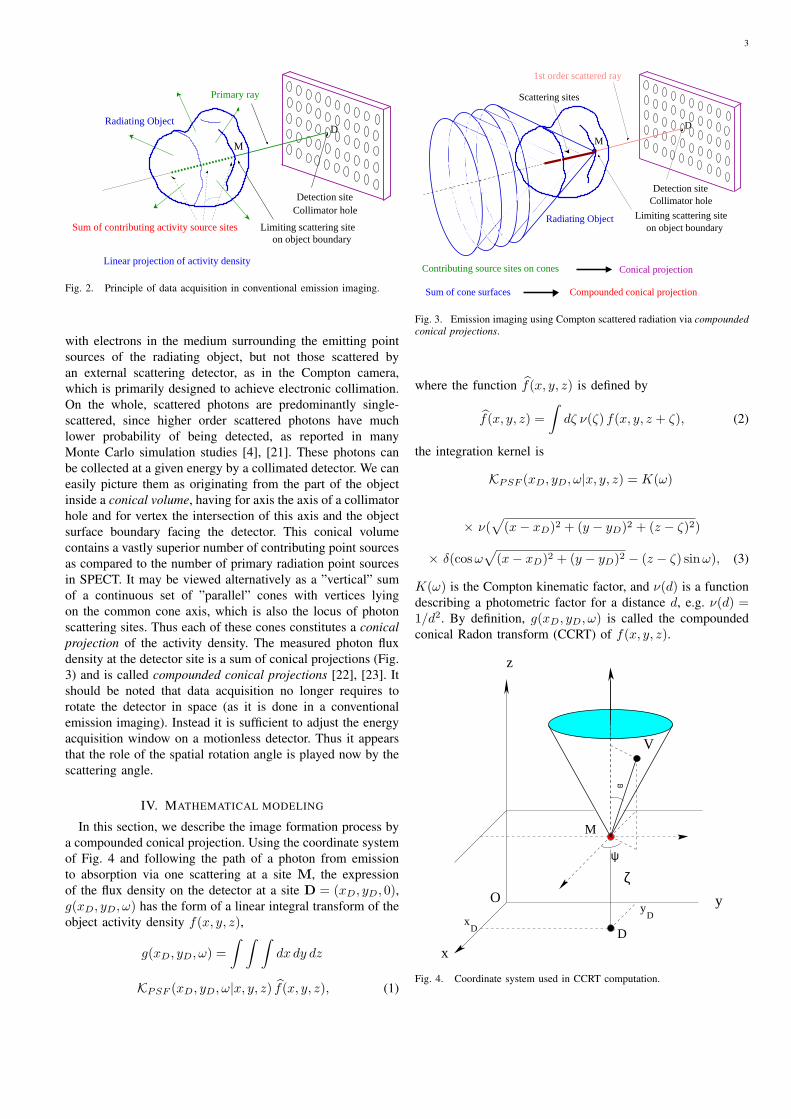
III. HOW ARE SCATTERED PHOTONS USED?In conventional SPECT, only primary photons (e.g. of
energy 140 keV for 99mTc) are collected by a collimatedgamma camera, registering the contribution of the sum ofpoint sources lying on a line as linear projection of the objectactivity density (see Fig. 2).
To get a set of complete data needed for three-dimensionalreconstruction, it is necessary to move the detector in spacearound the object, so as to get all the lines traversing the objectin all directions. Hence for a given single linear projection,only voxels lying on a straight line are concerned. Because ofphoton attenuation and the presence of a physical collimator,few from these primary photons (actually one out of 104)would actually reach the detector [19].
A natural alternative to imaging with primary photons isto use the more plentiful scattered photons, as advocated in[20]. Of particular interest are photons generated by scattering
3
M
D
Sum of contributing activity source sites
Collimator hole
Radiating Object
Primary ray
Linear projection of activity density
Limiting scattering site on object boundary
Detection site
Fig. 2. Principle of data acquisition in conventional emission imaging.
with electrons in the medium surrounding the emitting pointsources of the radiating object, but not those scattered byan external scattering detector, as in the Compton camera,which is primarily designed to achieve electronic collimation.On the whole, scattered photons are predominantly single-scattered, since higher order scattered photons have muchlower probability of being detected, as reported in manyMonte Carlo simulation studies [4], [21]. These photons canbe collected at a given energy by a collimated detector. We caneasily picture them as originating from the part of the objectinside a conical volume, having for axis the axis of a collimatorhole and for vertex the intersection of this axis and the objectsurface boundary facing the detector. This conical volumecontains a vastly superior number of contributing point sourcesas compared to the number of primary radiation point sourcesin SPECT. It may be viewed alternatively as a ”vertical” sumof a continuous set of ”parallel” cones with vertices lyingon the common cone axis, which is also the locus of photonscattering sites. Thus each of these cones constitutes a conicalprojection of the activity density. The measured photon fluxdensity at the detector site is a sum of conical projections (Fig.3) and is called compounded conical projections [22], [23]. Itshould be noted that data acquisition no longer requires torotate the detector in space (as it is done in a conventionalemission imaging). Instead it is sufficient to adjust the energyacquisition window on a motionless detector. Thus it appearsthat the role of the spatial rotation angle is played now by thescattering angle.
IV. MATHEMATICAL MODELING
In this section, we describe the image formation process bya compounded conical projection. Using the coordinate systemof Fig. 4 and following the path of a photon from emissionto absorption via one scattering at a site M, the expressionof the flux density on the detector at a site D = (xD, yD, 0),g(xD, yD, ω) has the form of a linear integral transform of theobject activity density f(x, y, z),
g(xD, yD, ω) =∫ ∫ ∫
dx dy dz
KPSF (xD, yD, ω|x, y, z) f(x, y, z), (1)
M
D
Detection siteCollimator hole
1st order scattered ray
Contributing source sites on cones Conical projection
Scattering sites
Limiting scattering site on object boundary
Radiating Object
Sum of cone surfaces Compounded conical projection
Fig. 3. Emission imaging using Compton scattered radiation via compoundedconical projections.
where the function f(x, y, z) is defined by
f(x, y, z) =∫
dζ ν(ζ) f(x, y, z + ζ), (2)
the integration kernel is
KPSF (xD, yD, ω|x, y, z) = K(ω)
× ν(√
(x− xD)2 + (y − yD)2 + (z − ζ)2)
× δ(cos ω√
(x− xD)2 + (y − yD)2 − (z − ζ) sin ω), (3)
K(ω) is the Compton kinematic factor, and ν(d) is a functiondescribing a photometric factor for a distance d, e.g. ν(d) =1/d2. By definition, g(xD, yD, ω) is called the compoundedconical Radon transform (CCRT) of f(x, y, z).
V
z
ψ
ϖ
x
yx
D
yD
Oζ
D
M
Fig. 4. Coordinate system used in CCRT computation.

4
−5
0
5
−5
0
5
0
5
10
15
20
X axis
Scattering angle = 50°
Y axis
PS
F w
ith c
ollim
ator
(P
hoto
ns)
Fig. 5. Two-dimensional plot of the PSF at scattering angle of 50 degreeswith collimator.
We have not included photon attenuation factors to avoidunnecessary complications which would mask the main idea.
The inversion of the kernel KPSF (xD, yD, ω|x, y, z) is thenobtained via a kind of central slice theorem in Fourier spaceof the detector plane for the function f∗, to which one shouldadd a deconvolution to get the Fourier transform F (u, v, w)of f(x, y, z), see [22], [24]:
F (u, v, w) =∫
dσ exp[2iπσw][−|z|√u2 + v2]
J (w)∫ ∞
0
t dt J1(2π|z|t√
u2 + v2)
[Y (ω − π/2)
∂
∂t
G(u, v, t)K(t)
+ Y (π/2− ω)∂
∂t
G(u, v,−t)K(−t)
],
(4)where
- J (w) is the Fourier transform of ν(x),- J1(x) is the Bessel function of order 1,- Y (x) is the Heaviside unit step function,- t = tan ω,- G(u, v, t), the Fourier transform of g(xD, yD, t),- K(t) is the Compton kinematic factor as a function of t.Finally f(x, y, z) is recovered by three-dimensional Fourier
transform.
V. POINT SPREAD FUNCTION AND RESULTS OFSIMULATIONS
A way to get an idea of what the CCRT could be or do isto construct its Point Spread Function (PSF). To concentrateon the scattered imaging principle, we make some simplifyingassumptions to stress only the effects of scattering [22]:• absence of attenuation for the propagating radiation [19],• constant density of electrons ne in the scattering medium,
The PSF turns out to be explicitly computable as an analyticfunction. It has the shape of a Mexican hat as shown in Fig.5 [24].
The gamma detector operates now at a fixed position. Nocoincidence detection, as in Compton cameras, is required.
010
010
0
2
4
x 104
010
010
0
2
4
x 104
010
010
0
2
4
x 104
010
010
0
2
4
x 104
010
010
0
2
4
x 104
010
010
0
2
4
x 104
010
010
0
2
4
x 104
010
010
0
2
4
x 104
010
010
0
2
4
x 104
010
010
0
2
4
x 104
010
010
0
2
4
x 104
010
010
0
2
4
x 104
010
010
0
2
4
x 104
010
010
0
2
4
x 104
010
010
0
2
4
x 104
010
010
0
2
4
x 104
Fig. 6. Original object (cylinder) in a cube consisting of 16 transaxial planes.
Numerical simulations have demonstrated the feasibility ofthis new imaging principle [24]. Issues related to higherorder scattering contribution, nonuniform attenuation, Pois-son emission noise, detection sensitivity, collimator efficiencyare part of ongoing research with innovative extensions totransmission imaging, leading eventually to an efficient dual-modality imaging procedure [25].
To provide more convincing arguments regarding the vi-ability of this idea, we present numerical simulations whichillustrate the reconstruction of a simple cylindrical object usingthe analytic inversion formula with the following workingconditions:• the used γ-detector is a conventional SPECT camera. It
has discretized dimensions N length units × N lengthunits. We have chosen N = 16 to keep the calculationsrequired at a reasonable level.
• the scattering medium is represented by a cube of dimen-sions N × N × N ,
• the electron density in biological medium is ne = 3.5×10²³ electrons/cm3,
• the radionuclide employed is Tc-99 with an activityconcentration corresponding to 4.84 × 10¹⁰ counts perminutes per cm3,
• the acquisition time per projection is set to 0.1 sec,• the 3D original object (cylinder of height 6 arbitrary
units) is placed at the center of the scattering medium(cube),
• the distance from the camera to the upper face of thescattering medium cube is l = 200 arbitrary units.
Fig. 6 represents the original object. Fig. 7 shows theseries of images of the object at various scattering angles ω(50 < ω < 1750). In Fig. 8, the reconstructed object in theabsence of noise is illustrated with a relative mean square error(RMSE) = 1.2%, which is perfectly reasonable. We observe agood performance of the CCRT for modeling the new imagingprocess.
Concerning spatial resolution, the intrinsic resolution de-pends on the camera design (collimator, crystal, photomulti-

5
010
010
0
0.05
010
010
0
0.05
010
010
0
0.05
010
010
0
0.05
010
010
0
0.05
010
010
0
0.05
010
010
0
0.05
010
010
0
0.05
010
010
0
0.05
010
010
0
0.05
010
010
0
0.05
010
010
0
0.05
010
010
0
0.05
010
010
0
0.05
010
010
0
0.05
010
010
0
0.05
Fig. 7. Series of images parameterized by the angle of Compton scatteringω (50 < ω < 1750).
010
010
0
5
x 104
010
010
0
5
x 104
010
010
0
5
x 104
010
010
0
5
x 104
010
010
0
5
x 104
010
010
0
5
x 104
010
010
0
5
x 104
010
010
0
5
x 104
010
010
0
5
x 104
010
010
0
5
x 104
010
010
0
5
x 104
010
010
0
5
x 104
010
010
0
5
x 104
010
010
0
5
x 104
010
010
0
5
x 104
010
010
0
5
x 104
Fig. 8. Reconstructed object in the absence of noise (RMSE = 1.2%).
plier tubes and measurement electronics). The reconstructedsystem resolution is further determined by the reconstructionalgorithm used. The inclusion of scattered radiation increasesconsiderably the number of detected photons, which mightcontribute to improve the signal to noise ratio (SNR) andthe resolution of the imaging system. To evaluate accuratelythe spatial resolution, it is necessary to use real data and tocompare it with conventional methods which do not make useof scattered radiation. At the present time, it is too early touse our preliminary simulation results for this purpose. Thiswork is ongoing using realistic experimental conditions.
Since our main objective in this paper is to show how toexploit advantageously Compton scattered radiation to suggesta new imaging principle, we focus on results illustrating theimage formation process as well as image reconstruction fromscattered photons.
In real situations, of course, one must take into account otherfactors such as photon attenuation by the medium, Poissonnoise and the imperfections of the detector system includingthe collimator and electronics.
The case of uniform attenuation (often assumed in theliterature) was included in our recent work [24]. The ex-act treatment of inhomogeneous attenuation poses enormousmathematical difficulties. Concerning emission noise, severalapproaches have been suggested to deal with it such asMaximum Likelihood or wavelets method. They may be usedfor ”denoising” the measured data beforehand or jointly withthe inversion process.
As for the imperfections of the detector, the standard wayfor treating this problem is to make use of a response functionusually modeled as a Gaussian defined both in spatial andenergy coordinates. These issues are discussed in detail in [18].
VI. HOW TO INCREASE SENSITIVITY?As mentioned earlier, the presence of a mechanical collima-
tor restricts severely the sensitivity of the imaging process. Wehave recently advocated a new functional modality followingthe principle of emission imaging by scattered gamma-rayswithout mechanical collimation [25], [26]. Removing thecollimator from the detector allows gamma rays to reach adetecting pixel from all directions coming from the upper-half space of this site, therefore increasing the strength of thesignal (Fig. 9). An introductory study in two dimensions hasrecently been performed [27] to demonstrate conclusively thefeasibility of this idea and to motivate the present work.
collimator
detector
Object
Scatteringmedium
Fig. 9. Two imaging modalities using Compton scattered radiation with andwithout collimation.
The modeling of the image formation process is thendone by a generalized compounded conical Radon transform,whereby one sums over conical projections at one detectionpixel over all cone vertices in the upper half space. Fig. 10shows the position of one conical projection in the generalizedCCRT.
This transform is obtained by summing over all sites M fora given site D on the detector. The mathematical expressionof one arbitrary conical projection is quite involved and givenin [23]. The summation over all such objects can be stillexpressed as a linear integral transform of the activity densityf(x, y, z)
g(D, ω) =∫
dx dy dz KPSF∗(D, ω|x, y, z) f(x, y, z) (5)
Unfortunately, the explicit form of KPSF∗(D, ω|x, y, z) istoo complicated to yield a simple interpretation, and will not

6
D
M
Sϖ
n
φ
x
y
z
Fig. 10. A conical projection in the generalized CCRT.
be addressed here. However, this PSF, although no longer acomputable function is in fact an integral of the electronicdensity over the surface of a torus of revolution whose axisis the line connecting the point source to the detection point(Fig. 11).
γ
x
y
z
O
ω
S
D
M
r
ψ
d
Scattering medium
Detector
Cutoff
α
l
Fig. 11. Torus surface of scattering sites for the case without collimator.
The shape of the PSF is now completely different comparedto the collimated geometry (Fig. 12).
To compare with the collimated detector geometry, Fig. 13shows the computed PSFs for both cases at a scattering angleof 30 degrees. The PSF without collimator is about 10 timesstronger than that with collimator.
To demonstrate the viability of this idea, we have useddata generated for a simple object, and applied classicalalgebraic reconstruction methods. We have taken a simplesource immersed in a cubic scattering medium. The source
−5
0
5
−5
0
5
10
20
30
40
50
X axis
Scattering angle = 50°
Y axis
PS
F w
ithou
t col
limat
or (
Pho
tons
)
Fig. 12. Plots of the PSF at a scattering angle of 50 degrees withoutcollimator.
Fig. 13. Comparison of the PSF with (lower blue line) and without collimator(upper red line).
itself consists of two concentric cubes with different activityconcentrations (Fig. 14).
A 256 × 256 pixels detector is placed on the xy plane.The pixel size is 0.4× 0.4 mm2. The scattering medium is arectangular box of dimensions 30 cm by 30 cm by 15 cm,which is at a distance of 1 cm above the planar detector.The electronic density inside the scattering medium is ne =3.341023 electrons/cm3 since most biological tissues have anelectronic structure close to that of water. The radionuclideused in this simulation is 99mTc, which emits photons at anenergy of 140.1 keV. The scattering medium is discretizedwith 13 voxels in x and y axis directions and with 9 voxelsin z axis direction. The detector is reduced to 13× 13 pixels.We construct the weight matrix of the medium by calculatingfrom our previous models, for each point of the mesh, thePSF of the detector at the different scattering angles. Thereconstruction is carried out using the conjugated gradientmethod with positivity constraint, see Fig. 15.
These preliminary results are incentive to pursue our inves-tigation on this new imaging modality.

7
Fig. 14. Two representative slices of the original object illustrating twotransaxial slices corresponding to the 6th and 9th planes, respectively.
Fig. 15. Two representative slices of the reconstructed images correspondingto the object shown in Fig. 14.
VII. CONCLUSION AND PERSPECTIVES
In this work, we have presented a new approach to Comptonscatter imaging. This concept exploits the gamma radiationnaturally scattered by the bulk of the emitting object usinga stationary gamma camera. Two operating modalities areexamined. The first one uses a standard SPECT camera andits working principle is based on the inversion of a newintegral transform, referred to as the compounded conicalRadon transform (CCRT). Numerical simulations shows thatit is feasible, probably with new generation of solid-statedetectors having very high energy resolution. The secondone operates without mechanical collimation and coincidencecircuitry and is based on a generalized CCRT. Althoughno analytic inversion formula exists at present, preliminarynumerical calculations with data generated using a forwardmodel show promising results.
Future challenging topics to be discussed in this imagingprocess are:
- Stability and robustness against noise and perturbations,- Accounting for non-uniform attenuation in the scattering
medium,- Treatment of higher-order scattering events [28],
From a technical standpoint, a new camera conception witha built-in high spatial and energy resolutions without a heavyand cumbersome mechanical apparatus for spatial rotation isto be designed. It would be well-suited for operation underchallenging conditions. This should be highly attractive fora number of fields of application such as medical imaging,industrial non-destructive control, surveillance of radioactivehazardous material storage (or transport), localization of con-taminations in a nuclear reactor, ... etc.
VIII. ACKNOWLEDGMENTS
This work was supported by the Swiss National ScienceFoundation under grant No. 3152A0-102143 and by the FrenchMinistry of Research under grant ACI NIM TRC 2004.
REFERENCES
[1] H Tuy, An inversion formula for cone-beam reconstruction, SIAM J.Appl. Math., vol. 43, pp. 546-552, 1983.
[2] S Zhao, H Yu and G Wang, A unified framework for exact cone-beamreconstruction formulas, Med. Phys., vol. 32, pp. 1712-1721, 2005.
[3] Wernick M, Aarsvold J. Emission Tomography: The Fundamentals ofPET and SPECT. San Diego: Academic Press, 2004.
[4] Zaidi H and Koral K F Scatter modelling and compensation in emissiontomography. Eur. J. Nucl. Med. Mol. Imaging Vol. 31, pp. 761-782, 2004.
[5] P G Lale, The examination of internal organs using gamma ray scatterextension to megavoltage radiotherapy, Phys. Med. Biol., vol.4, pp. 159-167, 1959.
[6] M Lenti, A 3-D imaging device using Compton scattering off the body,Nucl. Instrum. Meth. Phys. Res. A, vol. 538, pp. 457-462, 2008.
[7] R Guzzardi and G Licitra, A critical review of Compton imaging, CRCCritical Reviews in Biomedical Engineering, vol.15, pp. 237-268, 1998.
[8] G Harding, Inelastic photon scattering: effects and applications in biomed-ical science and industry, Radiat. Phys. Chem., vol 50, pp. 91-111, 1997.
[9] S J Norton, Compton scattering Tomography, J. Appl. Phys. vol. 76, pp.2007-2015, 1994.
[10] E M A Hussein, Radiation scattering methods for nondestructive testingand imaging, Int. Adv. Nondestr. Test. vol. 14, 301-321, 1989.

8
[11] S R Gautam, F F Hopkins, R Klinksiek and I L Morgan, Comptoninteraction tomography I: feasibility studies for applications in earthquakeengineering, IEEE Trans. Nucl. Sci., vol. 30, No. 2, pp. 1680-1684, 1983.
[12] R Todd, J Nightingale and D Everett, ”A proposed gamma camera”,Nature, vol. 251, pp. 132-134, 1974.
[13] M. Singh, An electronically collimated gamma camera for single photonemission computed tomography. Part 1: Theoretical considerations anddesign criteria”, Med. Phys., vol. 10, pp. 421-427, 1983.
[14] M Singh and D Doria, Single photon imaging with electronic collima-tion, IEEE Trans. Nucl. Sci., vol. 32, No 1, pp. 843-847, 1985.
[15] J Gerl, Gamma-ray imaging exploiting the Compton effect, Nucl. Phys.A, vol. 752, pp. 688-695, 2005.
[16] A Braem, M Chamizo Llatas, E Chesi, et al. Feasibility of a noveldesign of high-resolution parallax-free Compton enhanced PET scannerdedicated to brain research. Phys. Med. Biol., Vol. 49, pp. 2547-2562,2004.
[17] R D Evans, The atomic nucleus. New York, McGraw-Hill, 1955.[18] M K Nguyen, C Faye, L Eglin and T T Truong, Apparent image
formation by Compton-scattered photons in gamma-ray imaging, IEEESignal Processing Letters, vol. 8, pp. 248-251, 2001.
[19] Zaidi H and Hasegawa B, Determination of the Attenuation Map inEmission Tomography, J. Nucl. Med. vol. 44, pp. 291-315, 2003.
[20] P C Johns, R J Leclair and M P Wismayer, Medical X-ray Imagingwith scattered photons, in Proc. Opto-Canada: SPIE Regional Meetingon Optoelectronics, Photonics and Imaging, SPIE TD01, pp. 355-357,2002.
[21] H. Zaidi, Relevance of accurate Monte Carlo modeling in nuclearmedical imaging, Med. Phys., vol. 26, pp. 574-608, 1999.
[22] M K Nguyen and T T Truong, On an integral transform and its inversein nuclear imaging, Inverse Problems, vol. 18, pp. 265-277, 2002.
[23] T. T. Truong, M. K. Nguyen and H. Zaidi, The mathematical founda-tions of 3D Compton scatter emission imaging, International Journal ofBiomedical Imaging, doi: 10. 1155/2007/92780, 2007.
[24] M K Nguyen M K, T T Truong, H D Bui and J L Delarbre, A novelinverse problem in γ-rays emission imaging, Inverse Problems in Scienceand in Engineering, vol. 12, pp. 225-246, 2004.
[25] M. K. Nguyen, T.T. Truong, J. L. Delarbre, C. Roux and H. Zaidi, Novelapproach to stationary transmission scanning using Compton scatteredradiation, Phys. Med. Biol., vol. 52, pp. 4615-4632, 2007.
[26] C. Driol, M. K. Nguyen and T. T. Truong, Modeling and simulationresults on high sensitivity scattered gamma-ray emission imaging, Simula-tion Modelling Practice and Theory, DOI: 10.1016/j.simpat.2008.05.008,2008.
[27] M. K. Nguyen, C. Driol, T. T. Truong and H. Zaidi, Towards anew concept for high sensitivity Compton scatter emission imaging,Journal of the European Optical Society: Rapid Publications, vol. 3, DOI10.2971/jeos.2008.08010, 2008.
[28] C E Floyd, R J Jaszczak and G C Harris, Energy and spatial distributionof multiple order Compton scatter in SPECT: a Monte carlo investigation,Phys. Med. Biol., vol. 29, pp. 1217-1230, 1984.