ON A MICROSCOPIC PHASE IN THE LIFE CYCLE OF Title...
12
Title ON A MICROSCOPIC PHASE IN THE LIFE CYCLE OF NEMALION PULVINATUM GRUNOW (NEMALIONACEAE, RHODOPHYTA) Author(s) Umezaki, Isamu Citation PUBLICATIONS OF THE SETO MARINE BIOLOGICAL LABORATORY (1967), 15(4): 311-318 Issue Date 1967-12-20 URL http://hdl.handle.net/2433/175472 Right Type Departmental Bulletin Paper Textversion publisher Kyoto University
Transcript of ON A MICROSCOPIC PHASE IN THE LIFE CYCLE OF Title...

TitleON A MICROSCOPIC PHASE IN THE LIFE CYCLE OFNEMALION PULVINATUM GRUNOW(NEMALIONACEAE, RHODOPHYTA)
Author(s) Umezaki, Isamu
Citation PUBLICATIONS OF THE SETO MARINE BIOLOGICALLABORATORY (1967), 15(4): 311-318
Issue Date 1967-12-20
URL http://hdl.handle.net/2433/175472
Right
Type Departmental Bulletin Paper
Textversion publisher
Kyoto University

ON A MICROSCOPIC PHASE IN THE LIFE CYCLE OF NEMALION
PUL VIN A TUM GRUNOW (NEMALIONACEAE, RHODOPHYTA) 1)
lsAMU UMEZAKI
Department of Fisheries, Faculty of Agriculture, Kyoto University, Maizuru
With Plates VIII-X and 4 Text-figures
Nemalion pulvinatum GRUNOW (Nemalionaceae, Rhodophyta) is a taxonomically problematical species. Recently, Miss B. T. CHiu ( 1961) reported that Nemalion pulvinatum should be placed as a species of Dermonema. I continue to use the name Nemalion pulvinatum, because its complete taxonomy is not yet treated. Previously, the general life history of Nemalionaceae had been thought to be haplobiontic since SvEDELIUS (1915) and KYLIN (1916) showed that the zygote nucleus in Scinaia and Nemalion respectively underwent meiosis. Recently, MAGNE (196la, 196lb; 1964a,
1964b) reported new facts concerning the nuclear cycle of the family, rectifying
earlier statements of SvEDELIUs, K YLIN and others. He found that in Lemanea,
Nemalion and Scinaia the fertilized carpogonium did not undergo meiotic division.
VoN SToscH (1965) in his cytological and culture studies of Liagorafarinosa demonstrated MAGNE's theory: that is, the species showed a heteromorphic life history and its carpospore germlings gave rise to presumed sporophytes represented by microscopic
phase. MARTIN (according to a copy of her manuscript) also observed that carpospores of Nemalion elminthoides in culture developed into creeping filaments bearing monospores and that exactly similar creeping filaments from the sea gave rise to Nemalion plants.
In order to investigate the life cycle of Nemalion pulvinatum I cultured the carpospores and observed that they gave rise on germination to Acrochaetium-like filaments bearing monosporangia and that the presumed sporophyte reproduced repeatedly by
monospores for a certain period. No tetrasporangia have been observed on these plants.
Materials and Methods
Fertile plants of Nemalion pulvinatum were collected on May 22, 1966 and on April 20, 1967 from Shirahama, Wakayama Prefecture, where they are common from
April to June. The plants collected were brought to the Department of Fisheries,
1) Contributions from the Seta Marine Biological Laboratory, No. 477.
Publ. Seto Mar. Biol. Lab., XV (4), 311-318, 1967. (Article 19)
312 I. UMEZAKI
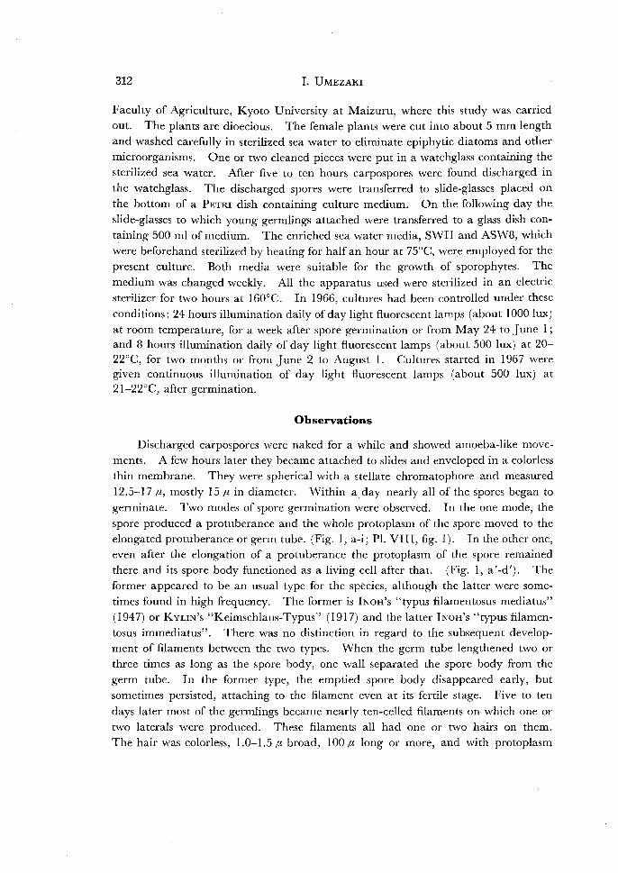
Faculty of Agriculture, Kyoto University at Maizuru, where this study was carried out. The plants are dioecious. The female plants were cut into about 5 mm length and washed carefully in sterilized sea water to eliminate epiphytic diatoms and other microorganisms. One or two cleaned pieces were put in a watchglass containing the
sterilized sea water. After five to ten hours carpospores were found discharged in the watchglass. The discharged spores were transferred to slide-glasses placed on the bottom of a PETRI dish containing culture medium. On the following day the
slide-glasses to which young germlings attached were transferred to a glass dish containing 500 ml of medium. The enriched sea water media, SWII and ASW8, which
were beforehand sterilized by heating for half an hour at 75°C, were employed for the present culture. Both media were suitable for the growth of sporophytes. The
medium was changed weekly. All the apparatus used were sterilized in an electric sterilizer for two hours at 160°C. In 1966, cultures had been controlled under these
conditions: 24 hours illumination daily of day light fluorescent lamps (about 1000 lux)
at room temperature, for a week after spore germination or from May 24 to June l ; and 8 hours illumination daily of day light fluorescent lamps (about 500 lux) at 20-220C, for two months or from June 2 to August l. Cultures started in 1967 were given continuous illumination of day light fluorescent lamps (about 500 lux) at 2l-22°C, after germination.
Observations
Discharged carpospores were naked for a while and showed amoeba-like movements. A few hours later they became attached to slides and enveloped in a colorless
thin membrane. They were spherical with a stellate chromatophore and measured
12.5-17 fl., mostly 15 fJ. in diameter. Within a day nearly all of the spores began to
germinate. Two modes of spore germination were observed. In the one mode, the
spore produced a protuberance and the whole protoplasm of the spore moved to the elongated protuberance or germ tube. (Fig. I, a-i; PI. VIII, fig. I). In the other one, even after the elongation of a protuberance the protoplasm of the spore remained there and its spore body functioned as a living cell after that. (Fig. l, a'-d'). The former appeared to be an usual type for the species, although the latter were some
times found in high frequency. The former is INoH's "typus filamentosus mediatus"
(1947) or KvuN's "Keimschlaus-Typus" (1917) and the latter INoH's "typus filamentosus immediatus". There was no distinction in regard to the subsequent development of filaments between the two types. When the germ tube lengthened two or three times as long as the spore body, one wall separated the spore body from the
germ tube. In the former type, the emptied spore body disappeared early, but sometimes persisted, attaching to the filament even at its fertile stage. Five to ten
days later most of the germlings became nearly ten-celled filaments on which one or
two laterals were produced. These filaments all had one or two hairs on them. The hair was colorless, 1.0-1.5 fJ. broad, I 00 fJ. long or more, and with protoplasm
MicroscoPic Phase of Nemalion Pulvinatum 313
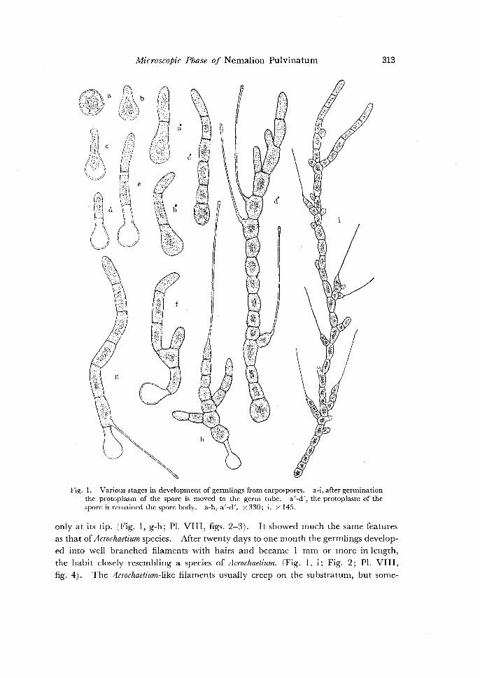
Fig. 1. Various stages in development of germlings from carpospores. a-i, after germination the protoplasm of the spore is moved to the germ tube. a'-d', the protoplasm of the spore is remained the spore body. a-h, a'-d'. x 330; i. X 145.
only at its tip. (Fig. I, g-h; Pl. VIII, figs. 2-3). It showed much the same features as that of Acrochaetium species. After twenty days to one month the germlings developed into well branched filaments with hairs and became I mm or more in length,
the habit closely resembling a species of Acrochaetium. (Fig. 1, i; Fig. 2; Pl. VIII, fig. 4). The Acrochaetium-like filaments usually creep on the substratum, but some-

314 I. UMEZAKI
times their laterals or terminals became upright and branched freely. They did not develop into a heterotrichous habit. When cultured in continuous light the Acrochaetium-like filaments reached a length of 3 mm to 5 mm two months after germination and were best developed under these conditions. In cultures of the year 1966 the filaments became fertile and produced monosporangia on July 1 or 30 to 37 days after germination. In 1967, monosporangia formation and release of fertile
monospores occurred on May 18 or 20 to 25 days after germination. Both the
cultures of 1966 and 1967 produced monosporangia successively, and released
monospores for about one month or a little more under the same conditions. Sessile monosporangia were born laterally on the nearly basal or middle cells of
the filament, or terminally or laterally on the short branches of one, two, or three
Fig. 2. Well developed filament from a carpospore. X 100.
cells. They are 10-15 p. X 12.5-17.5 f1. in size, hemispherical, short clavate or conical in side view. (Fig. 3; Pl. IX, fig. 1-4). The sporangium is enveloped in the wall composed of three layers: the outer layer is a membrane of the filament itself, being burst at its apex; and the middle and inner layers appear to be a membrane secreted from the sporangium itself. The monospores were liberated by rupture of the apex of the sporangium wall. The naked monospores showed amoe
boid movements like carpospores for a while after liberation. After a few hours the spores fixed to slides and were enveloped in a colorless membrane. They were
12.5-16 p. in diameter, the size being equal to that of carpospores. Immediately the spores began to germinate and developed into the Acrochaetium-like filaments.

MicroscoPic Phase of Nemalion Pulvinatum
Fig. 3. Part of a filament showing two well developed monosporangia and liberation of a monospore. X 600.
a
~b
f
Fig. 4. Monospore and its germlings showing vanous stages in development. X 300.
315

316 l. U MEZARI
(Fig. 4; Pl. X, figs. 1-2). The germination mode was much the same as that of
carpospores. After a monospore was released, sometimes a secondary new one was
found produced within the emptied sporangium wall. Monosporangia on the same
filament had been successively produced for a month or a little more. When these
monospore germlings had been cultured under the same condition, they again grew
fertile sporophytes bearing monosporangia in a month after germination. However,
under these conditions no tetrasporangia have been formed on the Acrochaetium-Iike
filaments, even in long continuous cultures.
Discussion
By SvEDELms's cytological investigation on Scinaia (1915) and KYLIN's on Nema
lion (1916) the Nemalionaceae has hitherto been believed as having haplobiontic life
cycle, although some exceptions were found. That is, meiosis occurred in the ferti
lized carpogonium so that the carposporophytes and the carpospores were thought
to be haploid. However, MAGNE's recent cytological investigations ( 1961 a, 1961 b;
1964a, 1964b) overthrew such previous opinions and showed that in some genera of
Nemalionaceae carpospores as well as carpospore germlings were diploid. More
over, Von SToscH's (1965) research confirmed :MAGNE's results: that is, Liagora
Jarinosa, a species ofNemalionaceae, alternates the haplobiontic gametophyte with the
diplobiontic sporophyte represented by a microscopic phase. MARTIN (according to
a copy of her manuscript) observed also that creeping sporophyte filaments of Nema
lion elminthoides in the field gave rise to gametophytes. In additon, sporophytes of
Liagora farinosa and Nemalion elminthoides reproduced asexually by monospores, accord
ing to VoN STOSCH (1. c.) and MARTIN (1. c.), respectively. Acrochaetium species
bearing monosporangia, which grew on Liagora had been collected by ABBOTT ( 1962).
VoN STOSCH ( 1. c.) collected also same Acrochaetium bearing monosporangia growing on Liagora. These collections suggest that Acrochaetium-like plants bearing monospores
in field may be derived from certain species ofNemalionaceae. And, the sporophytes
of Liagorafarinosa (VoN STOSCH, I. c.), Nemalion elminthoides (MARTIN, I. c.) and Nema
lion pulvinatum (present study) appear to reproduce by monospores for a certain season, although the stage may not be an essential part of the life cycle of these species like
tetrasporophytes.
Summary
I. Carpospores of Nemalion pulvinatum gave nse on germination to the Acro-
chaetium-like filaments. 2. The Acrochaetium-like filaments became sporophytes bearing monosporangia. 3. The sporophytes reproduced repeatedly by monospores for a certain period.
4. It has been known that Nemalion pulvinatum alternates the macroscopic

Microscopic Phase of Nemalion Pulvinatum 317
gametophyte with the dwarf Acrochaetium-like sprophyte, although no tetrasporophyte
has been observed.
Acknowledgtnents
I wish to express my sincere gratitude to Dr. I. A. ABBOTT of the Hopkins Marine Station of Stanford University, Pacific Grove, for her careful reading of the manuscript. I also wish to thank Dr. M. T. MARTIN of the University College of North Wales, Bangor, for sending me a copy of her manuscript.
REFERENCES
ABBOTT, I. A. 1962. Some Liagora-inhabiting species of Acrochaetium. Occ. Pap. B. P. BISHOP Mus., Honolulu, Hawaii, 23(6): 77-120.
Cmu, B. T. 1961. (Quoted from I. A. ABBOTT's Helminthora and Helminthocladia from California. Hydrobiol. 25: 88-98, 1965).
INOH, S. 1947. Germinations of seaweed species. 255 pp., Tokyo. (In japanese). KYLIN, H. 1916. tiber die Befruchtung und Reduktionsteilung bei Nemalion multifidum. Ber. deutsch.
bot. Ges. 34: 257-271. --- 1917. tiber die Keimung der Florideensporen. Arkiv f. Botanik. 1+: 1-25. MAGNE, F. 1961 a. Sur le cycle cytologic du Nemalion helminthoides (VELLEY) BATTERS. Com pt. R. A cad.
Sci. Paris, 252: 157-159. --- 1961b. Sur Ia caryologie de deux Rhodophycees considerees jusqu'ici comme a cycle cyto
logique entierement haplophasique. Compt. R. Acad. Sci. Paris, 252: 4023-4024. --- 1964a. Les Rhodophycees a cycle haplophasique existent-elles? Fourth Int. Seaweed Sym.
(1961). Proc. P. 112-116. --- 1964b. Recherches caryologie chez les Floridees (Rhodophycees). Cah. Bioi. Mar. 5: 461-
671. MARTIN, M. T. (In Press). Observations on the life-history of Nemalion helminthoides (VELL. in WITH.)
BATT. SvEDELIUs, N. 1915. Zytologisch-entwicklungsgeschichtliche Studien iiber Scinaiafurcellata, ein Beitrag
zur Frag der Reduktionsteilung der nicht tetrasporenbildenden Florideen. Nov. Acta Reg. Scient. Ups. ser. 4, 4: 1-55.
VoN SToscn, H. A. 1965. The sporophyte of Liagorafarirtosa LAMOUR. Br. Phycol. Bull. 2(6): 486-497.

318 I. UMEZAKI
EXPLANATION OF PLATES VIII-X
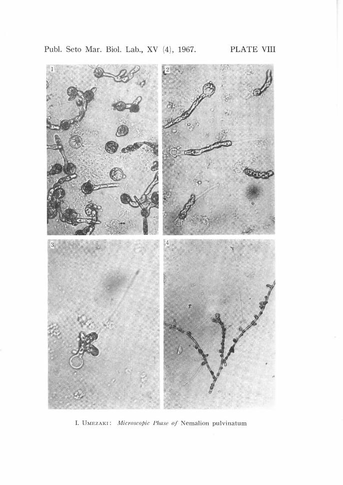
PLATE VIII. 1, carpospores and their germinations. X 310. 2, carpospore germlings
in three day culture. X 310. 3, carpospore germling bearing a hair. X 310. 4,
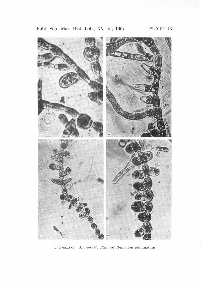
carpospore germling bearing monosporangia. X 35. PLATE IX. 1-2, part of a sporophyte bearing monosporangia and germling from a
monospore. 1 X 200; 2 X 310. 3, well matured monosporangium on a filament
and germling from a monospore. X 460. 4, emptied three monosporangia from
each of which monos pore liberated. X 270. PLATE X. 1, monospores and their germlings are found near sporophytes from which
monos pores liberated. X 100. 2, three germlings from monos pores. X 200.
Publ. Seto Mar. Bioi. Lab., XV (4), 1967. PLATE VIII
I. UMEZAKI: Microscopic Phase of Nemalion pulvinatum
Publ. Seto Mar. BioL Lab., XV (4), 1967 PLATE IX
I. UMEZAKI: Microscopic Phase of Nemalion pulvinatum
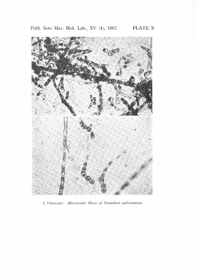
Publ. Seto Mar. Biol. Lab., XV (4), 1967. PLATE X
1 . . ~{ · ·. . ·~.~ ...
.. ... ~ t ~
· o
Q
~~ I
)I ..
I. UMEZAKI : Microscopic Pha se of Nemalion pulvinatum