OMT for Tinnitus: Evidence Based...
64
OMT for Tinnitus: Evidence Based Applications Joshua Alexander, DO, MPH Scripps Clinic San Diego, California Assistant Professor of Neurosciences University of California San Diego
Transcript of OMT for Tinnitus: Evidence Based...
OMT for Tinnitus: Evidence Based ApplicationsJoshua Alexander, DO, MPH
Scripps Clinic San Diego, CaliforniaAssistant Professor of Neurosciences
University of California San Diego

Objectives
• Recognize somatosensory tinnitus• Propose management • Perform OMT for somatosensory tinnitus

Roadmap
• Cases• Auditory Pathways• Introduction to tinnitus• Research• Treatment
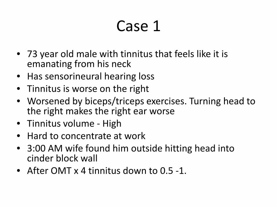
Case 1• 73 year old male with tinnitus that feels like it is
emanating from his neck• Has sensorineural hearing loss• Tinnitus is worse on the right • Worsened by biceps/triceps exercises. Turning head to
the right makes the right ear worse• Tinnitus volume - High• Hard to concentrate at work• 3:00 AM wife found him outside hitting head into
cinder block wall• After OMT x 4 tinnitus down to 0.5 -1.

Case 2
• 52 year old male in high speed MVC resulting in blunt trauma to the left side of the head
• Constant tinnitus – high intensity• Sensorineural hearing loss• Does not modulate• Did not respond to OMT

• Why does one patient respond and not the other?
• Let’s find out.

AUDITORY PATHWAYS

Auditory Pathway
• Tonotopically organized – higher frequency toward the outer spiral of the cochlea and therefore the outer part of the cochlear nerve (division of CN VIII)
• Spiral ganglion just outside the cochlea to cochlear nerve
• Dorsal and Ventral cochlear nuclei– Wrapped around lateral aspect of the inferior
cerebellar peduncle at the pontomedullary junction Blumenfeld H. Neuroanatomy through clinical cases. Chapter 12. Sinauer 2002.

Auditory Pathway
• Ascend bilaterally to the inferior colliculi– Mainly via lateral lemniscus– Multiple deccusations of fibers
• On to the medial geniculate nuclei of the thalamus and then to the primary auditory cortex at the Superior Temporal Gyrus(Heschl’s/ Brodmann 41)
• But there is more detail…
Auditory Pathway
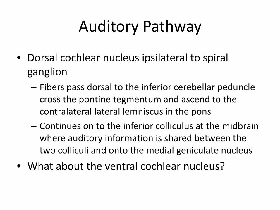
• Dorsal cochlear nucleus ipsilateral to spiral ganglion– Fibers pass dorsal to the inferior cerebellar peduncle
cross the pontine tegmentum and ascend to the contralateral lateral lemniscus in the pons
– Continues on to the inferior colliculus at the midbrain where auditory information is shared between the two colliculi and onto the medial geniculate nucleus
• What about the ventral cochlear nucleus?

Auditory Pathway• Many but not all fibers of the ventral cochlear nucleus
pass ventral to the inferior cerebellar peduncle to synapse bilaterally at the superior olivary nuclear complex in the rostral medulla and caudal pons– Localization?
• Fibers cross and form the trapezoid body– White matter
• Fibers ascend from the superior olivary nuclear complex bilaterally via the lateral lemniscus to the inferior colliculi of the dorsal midbrain seated under the thalamus
Blumenfeld H. Neuroanatomy through clinical cases. Chapter 12. Sinauer 2002.

Auditory Pathway
• Decussation at the inferior colliculi– Fibers pass dorsal and ventral to the cerebral aqueduct– Fibers ascend from the brachium of the inferior colliculi to
the medial geniculate nuclei• Brachium travels lateral to the superior colliculus to the posterior
thalamus
• Medial geniculate nucleus – Posterior inferior thalamus above the cerebral peduncle– Primary auditory cortex via auditory radiations
• Superior Temporal Gyrus/Heschl’s Gyrus behind the Sylvian fissure/lateral sulcus
Basic Auditory Modulation
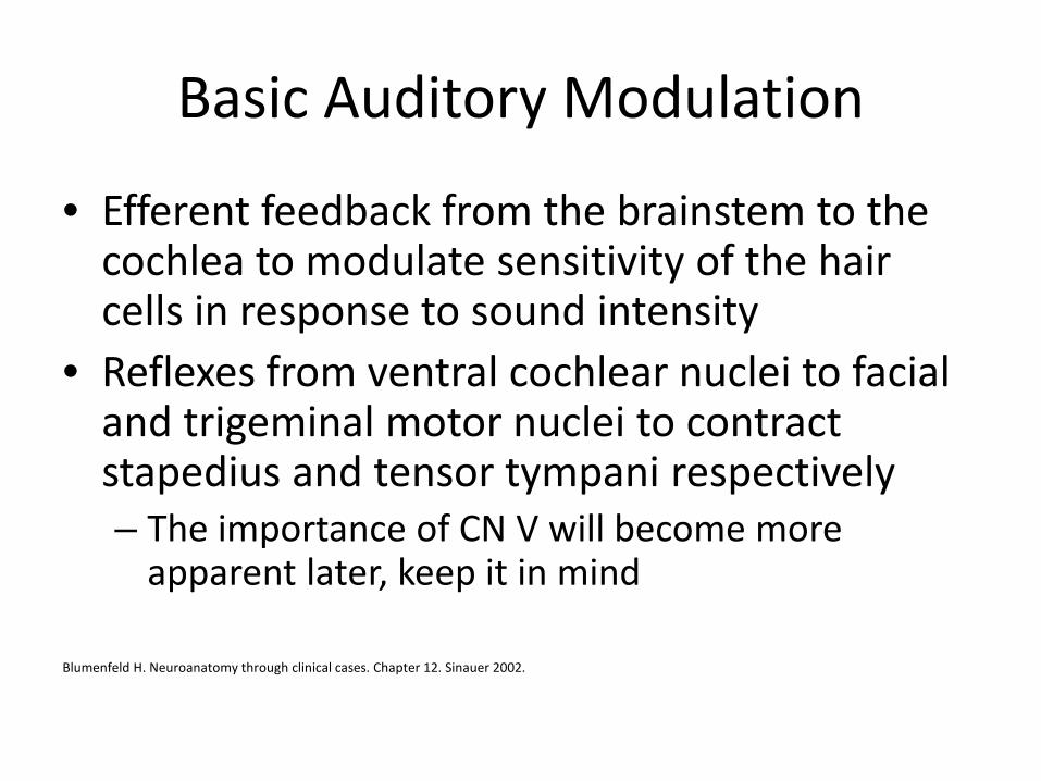
• Efferent feedback from the brainstem to the cochlea to modulate sensitivity of the hair cells in response to sound intensity
• Reflexes from ventral cochlear nuclei to facial and trigeminal motor nuclei to contract stapedius and tensor tympani respectively– The importance of CN V will become more
apparent later, keep it in mind
Blumenfeld H. Neuroanatomy through clinical cases. Chapter 12. Sinauer 2002.
Other Auditory Pathways
• Cochlear nuclei to the reticular formation to the thalamus to limbic system & hypothalamus & cortical structures
• Straightforward right, so why tinnitus then…
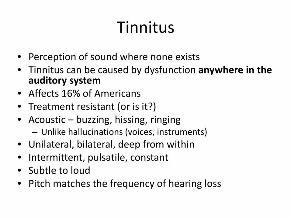
Tinnitus• Perception of sound where none exists• Tinnitus can be caused by dysfunction anywhere in the
auditory system• Affects 16% of Americans• Treatment resistant (or is it?)• Acoustic – buzzing, hissing, ringing
– Unlike hallucinations (voices, instruments)• Unilateral, bilateral, deep from within• Intermittent, pulsatile, constant• Subtle to loud• Pitch matches the frequency of hearing loss

Tinnitus
• Objective– Audible by the examiner– Inner body source of generator
• Tensor tympani/palatal myoclonus• Blood flow alteration
– Neurology & ENT referral!
• Subjective (more common & our focus here)– Patient hears

Tinnitus: to whom do you belong?
• Long thought to be a disease of the cochlea only– Damage to hair cells– Dead regions of cochlea seen on pathology– Managed by ENT
• Perhaps it is a neurological disease– Tinnitus persists after auditory nerve section– Treatments targeting the cochlea are of variable
efficacy– Auditory association cortex shows activity in tinnitus

Subjective Tinnitus• Sensorineural hearing loss• Somatosensory
– TMJ– Whiplash– Cervical spine disorders
• Vascular insufficiency• Bone disease• Infection• Metabolic Derangements• CNS pathology• Autoimmune Disease• Medications

Epidemiology
• Male• Age• Hearing impairment• Noise exposure

Associated Symptoms
• Irritable• Anxious• Frustrated• Depression• Insomnia• Hyperacusis• Difficulty hearing (distinct from hearing loss)• Poor concentrationLagguth et al. Tinnitus: causes and clinical management. Lancet Neurology. 2013; 12:920-30.

Pathophysiology
• Usually there is cochlear damage (hair cell) abnormal neural activity in the central auditory pathway perception of tinnitus
• Can also happen if the auditory nerve is injured• Not everyone with hearing loss gets tinnitus• Patients with tinnitus may have a normal
audiogram– These patients usually have cochlear dead regions
though– Is Tinnitus akin to phantom limb?

Neural Mechanisms• Multifactoral – auditory deprivation causes changes to neural
plasticity which alters neural activity and triggers compensation for absent input; abnormal somatosensory afferent input from the neck and face affect the central auditory pathways
• Cortex- reduced inhibition in the deafferented area, edge regions which receive input extend into the deafferented area – Is this compensation for sound loss or the generator of tinnitus –
unknown• Magnetoencephalography and EEG show gamma band activity in
the auditory cortex in patients with normal function and in those with tinnitus– Is this a problem of failure to inhibit?
Lagguth et al. Tinnitus: causes and clinical management. Lancet Neurology. 2013; 12:920-30.
Neural Mechanisms• Tinnitus changes are not restricted to the auditory
cortices• Anterior insula, anterior cingulate, thalamus, amygdala,
hippocampus– Abnormal amygdala – cingular cortex seen in patients with
chronic pain syndromes and somatiform disorders• Maybe nucleus accumbens and subgenual cingulate
cortex• Tinnitus changes the brain over time and covers a
number of neural networks – it becomes locked in– Maybe
Lagguth et al. Tinnitus: causes and clinical management. Lancet Neurology. 2013; 12:920-30.

Neurotinnitus?
• Dorsal cochlear nucleus (DCN) exhibits dysfunctional excitability in tinnitus– Treat this excitability and treat the tinnitus? – Giving NMDA receptor antagonist reduced
abnormal plasticity in the DCN in an animal model• Much more to come• Memantine/Namenda is an NMDA antagonist
– Keep the DCN in mind for later
Tagoe et al. Saturation of long-term potentiation in the dorsal cochlear nucleus and its pharmacological reversal in an experimental model of tinnitus. ExpNeurol. 2017 Feb 16; 292:1-10.

SOMATOSENSORY TINNITUS

Somatosensory Tinnitus
• Tinnitus modulated by movement of the head/neck– Pitch and volume changes
• Specific maneuvers– Cervical spine flexion, extension, side bending, rotation– Pressure to the forehead, occiput, vertex, mandible,
temple, and other sites with resistance– Clench teeth, open mouth, protrude jaw, slide jaw side to
side
Ralli et al. Somatosensory Tinnitus: correlation between cranio-cervico-mandibular disorder history and somatic modulation. Audiology and Neurology 2016;21: 372-382.

Somatosensory Tinnitus
• Risk of tinnitus is 8.37 times higher in patients with temporomandibular disorders
• 79.67% of patients with history of TMJ or cervical spine disorders have tinnitus modulation with specific musculoskeletal maneuvers– Tested maneuvers increased loudness in 94% – 75% have unilateral tinnitus
Buergers R, Kleinjung T, Behr M, Vielsmeier V: Is there a link between tinnitus and temporo- mandibular disorders? J Prosthet Dent 2014; 111:222–227. Ralli et al. Somatosensory Tinnitus: correlation between cranio-cervico-mandibular disorder history and somatic modulation. Audiology and Neurology 2016;21: 372-382.

Somatosensory Tinnitus
• Is there interaction between the musculoskeletal system and auditory system to explain this?
• Animal model shows the dorsal cochlear nucleus receives projections from the auditory nerve, trigeminal nerve, and the dorsal column ganglia.
Shore S et al. Mechanisms underlying somatic tinnitus. Progress in Brain Research. Vol 166, Chapter 10.

Back to Auditory Pathways
• Dorsal Cochlear Nucleus– Receive somatosensory input
• Does this inhibit sound?
– Hyperexcitability in tinnitus
– We don’t know what this means but the integration of somatic afferents with auditory afferents begs the question…

Somatosensory Tinnitus
• If there is a musculoskeletal connection, could this type of tinnitus be amenable to osteopathic manipulation?

RESEARCH

Research
• Alexander J et al. Response of Tinnitus to Osteopathic Manipulation. OMED October 2015.
• Oostendorp RA et al. Cervicogenic somatosensory tinnitus: an indication for manual therapy plus education? Manual Therapy. 2016 Jun; 23:106-13.
• Michaels S et al. Does multi-modal cervical physical therapy improve tinnitus in patients with cervicogenic somatic tinnitus. Musculoskeletal Science and Practice, 2016 26; 125-131.

Study 1: Who responds to OMT
• Retrospective of 20 patients at a tertiary center– 11 responded
• 80% had worsening tinnitus with musculoskeletal activity– Head turning, cervical flexion, cervical side bending, chewing– 100% have somatic dysfunction of the head and neck– Average reduction in perceived tinnitus volume 76.9%– All have sensorineural hearing loss superimposed– Responded after 3.25 treatments; treated average of 5.89 times
– 9 non-responders• None have change in tinnitus with musculoskeletal activity• 33% have somatic dysfunction• Etiology: auditory trauma, blunt cochlear trauma,
sensorineural hearing loss

Study 2: effect of manipulation on somatosensory tinnitus
• 122 Dutch participants with cervicogenic somatosensory tinnitus– Had to have cervical dysfunction by history and examination– Divided into groups receiving manipulation and manipulation +
counseling• Specific manipulation protocol • Primary outcome: reduction in Visual Analog Scale (VAS) for
Tinnitus• 38.5% female• Average duration with tinnitus 7.3 years• Average treatments ~10• No adverse effects• Study showed significant reduction in VAS score
– 5.9 for manipulation only group (+/- 17.7; p 0.01)– 12.3 for manipulation + counseling (+/- 20.3; p 0.00)

Study 3: effect of physical therapy on somatosensory tinnitus
• Delayed-start randomized control trial • Standardized cervical physical therapy for patients with
somatosensory tinnitus• 38 Belgian patients from tertiary tinnitus clinic
– Group 1: 6 weeks of treatment, 6 weeks of follow-up– Group 2: 6 week delay, 6 weeks treatment, 6 weeks follow-up
• Tinnitus Functional Index (TFI)– Measures tinnitus impact in 8 areas
• TFI 48 (severe tinnitus) at baseline• TFI decreased to 44 (SD 22 p0.04) immediately after
treatment• TFI increased to 47 (SD 22) 6 weeks after treatment

QUESTIONS? CRITICISM? COMMENTS?

TREATMENT

Treatment Time
• Pterygoids – 10 minutes total• Diaphragms – 20 minutes total• Auditory Pathways – 40 minutes +
– This one takes time and patience
• Clean up & questions- 20 minutes

Treatment
• Our focus today– Pterygoids – do this if nothing else!– Tentorium cerebelli– Auitory pathways
• You can do detailed anatomy review on your own of the pterygoids, mandible, temporal bone, diaphragms, etc…
• Also remember this is a component of a larger treatment model. Integrate this into the person, do not impose it on them.
• You may need to treat a number of other things first!

Treatment
• Medial Pterygoid– CN V via mandibular branch which also innervates
tensor tympani– Deep and superficial head
• Origin: lateral pterygoid plate and palatine bone• Insertion: ramus and angle of mandible
– Orientation• Downward, lateral, posterior
– Action• Mandible elevation, excursion, and protrusion

Treatment
• Lateral Pterygoid– Innervation: lateral pterygoid nerve from CN V– Origin: Superior head from greater wing of
sphenoid; Inferior head on the lateral pterygoidplate
– Insertion: condyloid process of mandible and TMJ– Action: Opens mandible, protrusion– Orientation: Superior head inferomedial
• Inferior head superomedial


Treatment• Pterygoids
– Inhibition using your pinkie • Patients can do this at home with their index finger
– and masseter massage
– Follow muscle fibers– Monitor PRM
• Treat the system– Place your other hand under the cervical spine and cradle
the neck. Monitor C2 and use fluid technique to treat the pterygoids and upper cervical muscles – usually splenius cervicis
– Integrate CN V if needed (it’s right behind your pinkie)– Lab

Treatment
• Treat the diaphragms – Tentorium cerebelli, (Sibson’s fascia), respiratory
diaphragm, pelvic diaphragm– You may find venous sinus technique more helpful
in treating the tent– Let’s talk about the tent a little– Lab (time dependent)

Treatment• Tentorium cerebelli
– Attached to the falx cerebri in the midline• Straight sinus• Superior position makes it seem tented
– Anterior portion attaches to the anterior clinoid process• Forms lateral cavernous sinus• Deep tentorial notch is U shaped
– Anterolateral attachment to superior petrous, extends laterally to the posterior clinoid process
• Superior petrosal sinus– Posterolateral margin separates to 2 layers of dura & attach at the
transverse sulci of the occipital bone. Anteriorly it attaches to the posterior-inferior angles of the parietal bone
– Posterior attachment to internal occipital protuberance • Transverse sinus


Treatment
• Sibson’s Fascia/suprapleural membrane– Attaches to the inner border of the first rib and
transverse process of C7
• You know the respiratory and pelvic diaphragms

THE AUDITORY PATHWAYSTinnitus treatment

Tinnitus Treatment
• Treat the auditory pathways from the cochlea to the auditory cortices– Be mindful of
bilateral pathways– Lab
Netter. CIBA Vol 1.


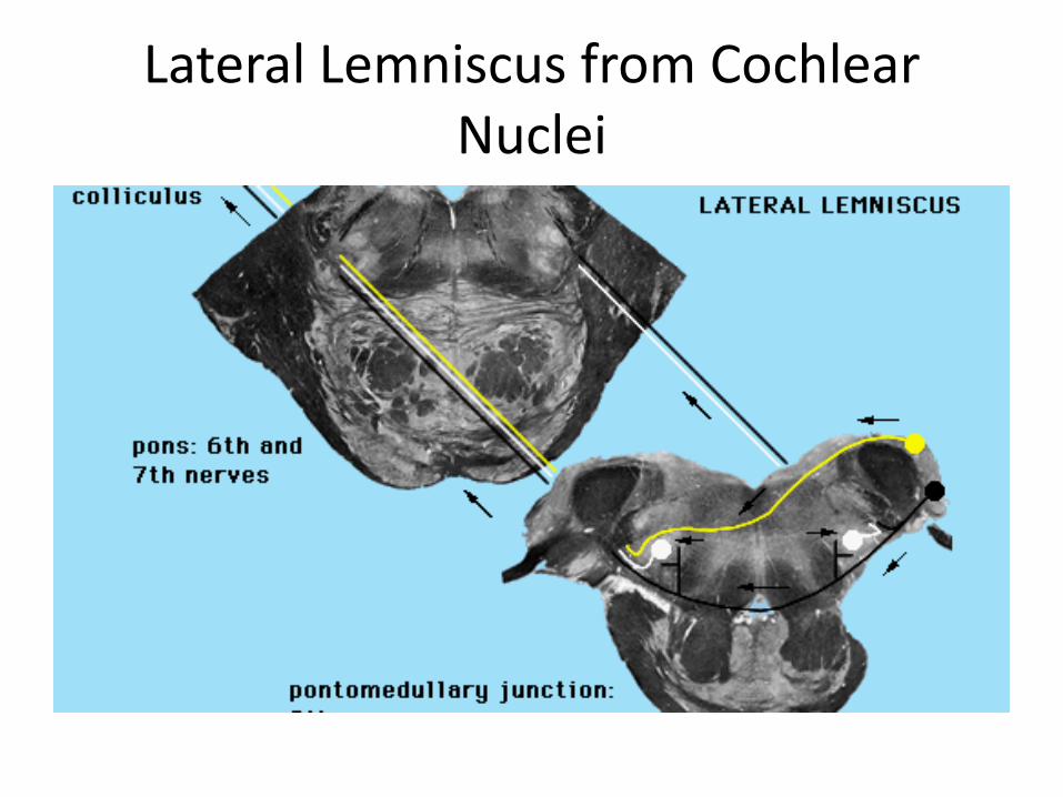
Lateral Lemniscus from Cochlear Nuclei

Rostral Pons Lateral Lemniscus extremely lateral

Midbrain Inferior Colliculus and Lateral Lemniscus


Inferior Colliculus to MGN in red

Inferior colluculi and Thalamus


Red = Heschl’s Gyrus

Treat Bilaterally
• Cochlea• Auditory nerve• Cochlear nuclei
– Start ventral and look at the superior olivary complex then come down and look at the dorsal cochlear nucleus then treat as one unit
• Inferior colliculi– Mind the decussaiton
• Heschl’s Gyrus• Auditory Association Cortex

Tinnitus Treatment: other thoughts
• Find where temporal motion is being restricted– Does patient have gliosis, fascial restriction?
• Treat petrous portion of the temporal bone• Fascial unwind of the ear and external
auditory canal• Treat the CSF/ventricles

Tinnitus Treatment: other thoughts
• Digastric• SCM – Occipitomastoid release and/or direct
inhibition• Align the jaw• Liberate the atlas• Treat the pelvis• Look at the fascia• Treat the arm if levator scapula is involved

END