OKAP REVIEW OF PEDIATRIC OPHTHALMOLOGY AND
16
1 Lisabeth S. Hall, MD Director Pediatric Ophthalmology and Strabismus OKAP REVIEW 2008 Pediatric Ophthalmology and Strabismus I. Development of the visual system II. Sensory anomalies OKAP REVIEW 2008 Pediatric Ophthalmology and Strabismus III. Amblyopia OKAP REVIEW 2008 Pediatric Ophthalmology and Strabismus IV. Anatomy of the EOM’s Summary, review and quiz OKAP REVIEW 2008 Pediatric Ophthalmology and Strabismus Sensory system COLOR Sensory system FORM Sensory system LOCATION visual space = Sensorimotor system ¾ visual sensations precipitate a chain of motor responses that move the eyes Sensory system + Motor system
Transcript of OKAP REVIEW OF PEDIATRIC OPHTHALMOLOGY AND

1
Lisabeth S. Hall, MDDirector
Pediatric Ophthalmology and Strabismus
OKAP REVIEW 2008Pediatric Ophthalmology and
Strabismus
I. Development of the visual systemII. Sensory anomalies
OKAP REVIEW 2008Pediatric Ophthalmology and
Strabismus
III. Amblyopia
OKAP REVIEW 2008Pediatric Ophthalmology and
StrabismusIV. Anatomy of the EOM’s
Summary, review and quiz
OKAP REVIEW 2008Pediatric Ophthalmology and
StrabismusSensory system
COLOR
Sensory systemFORM
Sensory system
LOCATION visual space = Sensorimotor system
visual sensations precipitate a chain of motor responses that move the eyes
Sensory system + Motor system

2
Visual development
Vision requires:
1) intact optical system
2) photo-pigment-mediated transformation of light into wave action potentials
Visual development
Vision requires:
3) synapses with feedback
4) precise binocular mapping of the environment onto the retina,
lateral geniculate body and occipital cortex
NeurophysiologyAnterior visual system
stimulus received by retinal photoreceptorsoptic nerveoptic tractoptic chiasm
NeurophysiologyLateral Geniculate Body
LGN or LGBthalamus
NeurophysiologyLateral Geniculate Body
receives afferent fibers from the anterior visual pathwayrelays information to primary visual cortexmechanism unknown
NeurophysiologyLateral Geniculate Body
organized in 6 layers• 6 - outermost
• 1 - innermost
• uncrossed, ipsilateral - 2, 3, 5
• crossed, contralateral - 1, 4 6
Lateral Geniculate Body
important clinicallymust know for BOARDS:
2 cell types:
• Magnacellular neurons• Parvocellular neurons
VisionNeurophysiology - LGN
M CELLS
Magno-large
parafoveal, peripheral
WHERE
Neurophysiology - LGN
P cells
color, two point discrimination
Parvo - small
WHAT

3
NeurophysiologyOccipital lobe
also called:striate cortexBrodman’s area 17
Development of normal binocular vision
must understand concepts of
• visual space
• visual direction
Visual space
Objective• objects in physical space outside of and
independent of our visual system
Subjective• conscious awareness of objects and
perception by our brain
Development of normal binocular vision
stimulation of any retinal area results in visual sensation from a subjective visual direction
Retinal correspondence
retinal areas in the two eyes share a common subjective visual direction
Visual direction
normally fovea = visual axis = straight ahead
Normal Retinal Correspondence
FRFL
Retinal correspondence
NRC
corresponding retinal points are located on the same meridian and at the same distance from the fovea in each eye
Normal Retinal CorrespondenceEmpirical Horopter
FRFLTRTL
NL NR
FRFL TR
TL NLNR
cyclopean eye
Normal Retinal CorrespondenceEmpirical Horopter

4
Empirical horopter
clinically defined area (3D space) where all points are seen singly
Vieth-Muller circle
model based upon assumption that eye is a perfect sphere
Empirical horopter
fusion exists
all points are seen singly
requires NRC by definition
NO stereopsis
Panum’s areaarea around horopter where non-corresponding retinal points are stimulated without diplopia
•based upon ability to fuse slightly disparate images•stereopsis exists•single binocular vision
Stereopsis
relative localization of visual objects in depthlimited by distance (<20 feet)remember:
monocular clues - important in interpretation of depth
StereopsisTitmus stereo test
allows stereopsis by presenting disparate images
fly = 4000 sec of arc
9/9 circles = 40 sec
Random-dot stereogramno monocular clues
Sensory adaptations to strabismus
suppressionARCdiplopiaconfusion

5
Diplopia Sensory adaptations to strabismus
confusion- rare- perception of 2 images superimposed
Confusion
Suppression Suppression
strabismus
confusing images originating from the retina
central inhibition to avoid diplopia
typically seen in children
clinically, can give clues about etiology and age of onset of strabismus
dense amblyopia
Sensory adaptations to strabismus
ARC fusion
- simultaneous perception of similar images - stimulate retinal points which normally do not correspond
ARC
FRFL NR
Sensory testingWhy bother?
can give insight into etiology of strabismus
can help with surgical plan
helps to prepare the patient post-op
• i.e. diplopia
Tests for retinal correspondence
Based upon principle that images that are less “alike” are harder to fuse
Tests that make images less alike (red filter) are more dissociating and therefore reveal suppression easier

11
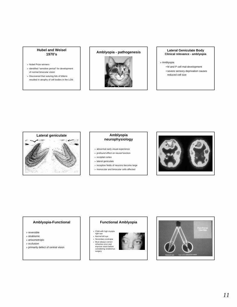
Hubel and Weisel1970’s
Nobel Prize winners
identified “sensitive period” for development of normal binocular vision
Discovered that suturing lids of kittens resulted in atrophy of cell bodies in the LGN
Amblyopia - pathogenesis Lateral Geniculate BodyClinical relevance - amblyopia
Amblyopia
• M and P cell mal-development
• severe sensory deprivation causes reduced cell size
Lateral geniculate Amblyopianeurophysiology
abnormal early visual experience
profound effect on neural function
occipital cortex
lateral geniculate
receptive fields of neurons become large
monocular and binocular cells affected
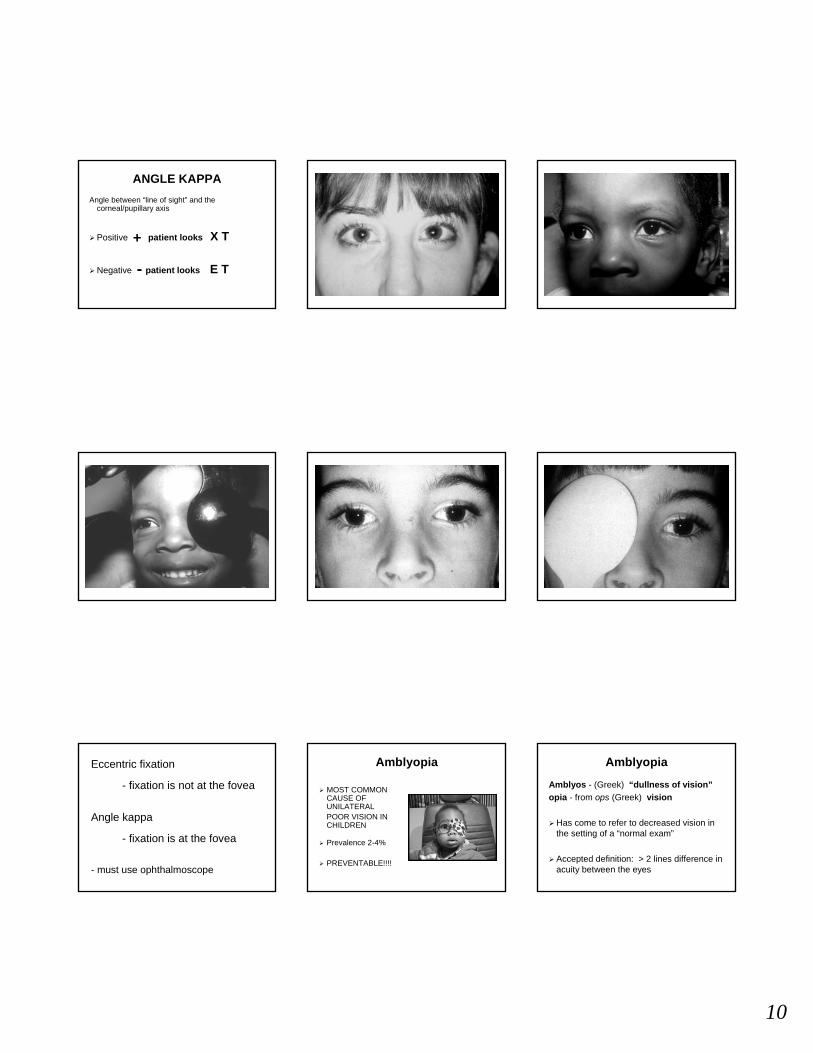
Amblyopia-Functional
reversiblestrabismicanisometropicocclusionprimarily defect of central vision
Functional Amblyopia
Child with high myopia right eyeNormal left eyeSecondary exotropiaMust always correct refractive error and improve vision before considering strabismus surgery

12
Amblyopia - Organic
typically refers to ocular anomalies preventing optimal acuityabnormality may be subtle or undetectable“irreversible”may be diagnosed after failure to respond to occlusion therapymust remember that organic amblyopia may have superimposed functional amblyopia
Occlusion AmblyopiaOrganic causeCan result from patching
Bilateral Amblyopia Amblyopia - diagnosis
Pre-verbalfixation preferencevertical prism test (8-10 PD)acuity
• OKN, FPL, Teller, VEP
VerbalAllen pictures, numbers, letters> 2 lines difference
Amblyopia - Strabismus
FixationIn classic “textbook”congenital esotropia infants alternate their fixation and do not become amblyopic

13
Amblyopiacrowding phenomenon
ACUITY
ISOLATED > LINEAR
in amblyopes
Amblyopia - Treatment
correct refractive errorstreat ocular problems - i.e. cataract, ptosisocclusionpenalization (i.e. atropine)follow closelyocclusion amblyopia - always check Va in “better” eye
Essentials of treatment
Patient and family understanding and involvementMotivation/rewardsRealistic goalsMake it fun and easyKnow when to stop
Must take treatment seriouslyOften make contract with older kidsCapitalize on their interestsVisual challenging is essential
Essentials of treatment
Pediatric Eye Disease Investigator Group
Amblyopia Treatment Study Group
Looking at measurement of visual acuityEfforts to standardize
Effects of treatment on child and family
Comparison ofDrops and patching
Shorter vs. longer occlusion
Archives of Ophthalmology 2003;121:603-611189 children<7 y w/ moderate amblyopia (20/40-20/80)Randomized to 2h/d vs.. 6h/d patchingBoth groups performed > 1 h per day of near visual activitiesCompliance consistent with other studies (poor)4 m follow upSimilar improvement in 2 groups
Amblyopia Study GroupPatching Regimens Anatomy of the EOM’s
Ocular alignment is determined by the extraocular muscles and their surrounding tissues
PRIMARY POSITION• the eye and head are directed straight
ahead• medial walls are parallel• lateral walls are 45 o from medial walls• in primary position: SO, IO - 51o
SR, IR - 23o
MR, LR - 90o

14
EOM’S OriginAnnulus of Zinn
oval fibrous ring at the orbital apexEOMs originate at annulus
EXCEPT:• inferior oblique• superior oblique• levator palpebrae
superioris
Origin of the EOMInferior Oblique
maxillary bone, adjacent to lacrimal fossa, posterior to orbital rim
Superior Obliqueorbital apex above annulus (functional origin at trochlea
LPSorbital apex above annulus
Insertions of the EOM’sSpiral of Tillaux
Rectus muscles insert into sclera anterior to the equator via tendonsTheir anatomic relationship at the insertions forms the Spiral of TillauxThinnest sclera -(.3mm) just posterior to the insertion
Insertions of the EOM’sOblique muscles
insert posterior to the equator
SO longest tendon, courses inferior to SR
IO shortest tendon, courses inferior to IR
Cardinal positions and yolk muscles
LLRRMR
LSRRIO
RSRLIO
RLRLMR
RIRLSO
LIRRSO
Cardinal positions and yolk muscles
15
EOM’S function
function dependent upon position of the globe1o,2o,3o action in the primary position:
• medial, lateral recti - adduct, abduct
• sup, inf oblique’s - elevate, depress, intort, extort, abduct
• sup, inf recti - elevate, depress, intort extort, adduct
EOM’S function
CLINICAL CORRELATION
A and V patterns due to oblique overaction
ET in thyroid patients with tight inferior recti
EOM - Innervation
CN III LPS, SR upperMR, IR, IO lower
CN IV SO
CN VI LR
Anatomy of the EOM’sVascular supply
Major: muscular branches of the
ophthalmic artery
Additional: Lacrimal artery to LR
Infraorbital to IO, IR
Vascular supply
MOST OFTEN
Recti muscles - contain 2 anterior ciliary arteries
Exception - Lateral rectus contains 1
Vascular supply
Recti muscles - perfuse anterior segment via anterior ciliary arteries
Oblique muscles - do not contribute to anterior segment circulation
Vascular supply
Clinical Relevance
• Surgery on multiple recti muscles contraindicated due to risk of anterior segment ischemia
SummaryNRC
Horopter• based upon NRC• points stimulate corresponding retinal elements• single vision and fusion exists
Panum’s area• slightly disparate points• allows stereopsis• outside - have physiologic diplopia
Normal Retinal CorrespondenceEmpirical horopter
FRFL TR
TL NLNR
cyclopean eye
16
SummaryRetinal correspondence
Retinal correspondence
ARC NRC
Binocular
SummaryAmblyopia
major preventable cause of visual loss
strabismus, anisometropia, high hyperopia, and myopia at risk
maximum FT occlusion = 1 week/year of life
“critical period” for development of binocular vision
possibly “at risk” until 10 years of age
SummaryAnatomy
Anterior segment circulation from 4 recti• all have 2 except LR has one ACA
All EOM orig. from annulus except IO, SO, LPS
Recti insert anterior to equator, obliques-posterior
Inferior-extorters, Superior-intort
Recti - adduct, Obliques - abduct
Obliques course inferior to recti
GOOD LUCK!!!!
QUIZ