Office Bearers of IAP Orissa State Branch 2010iaporissa.org/Downloads/OJP2010.pdfEmail:...
58
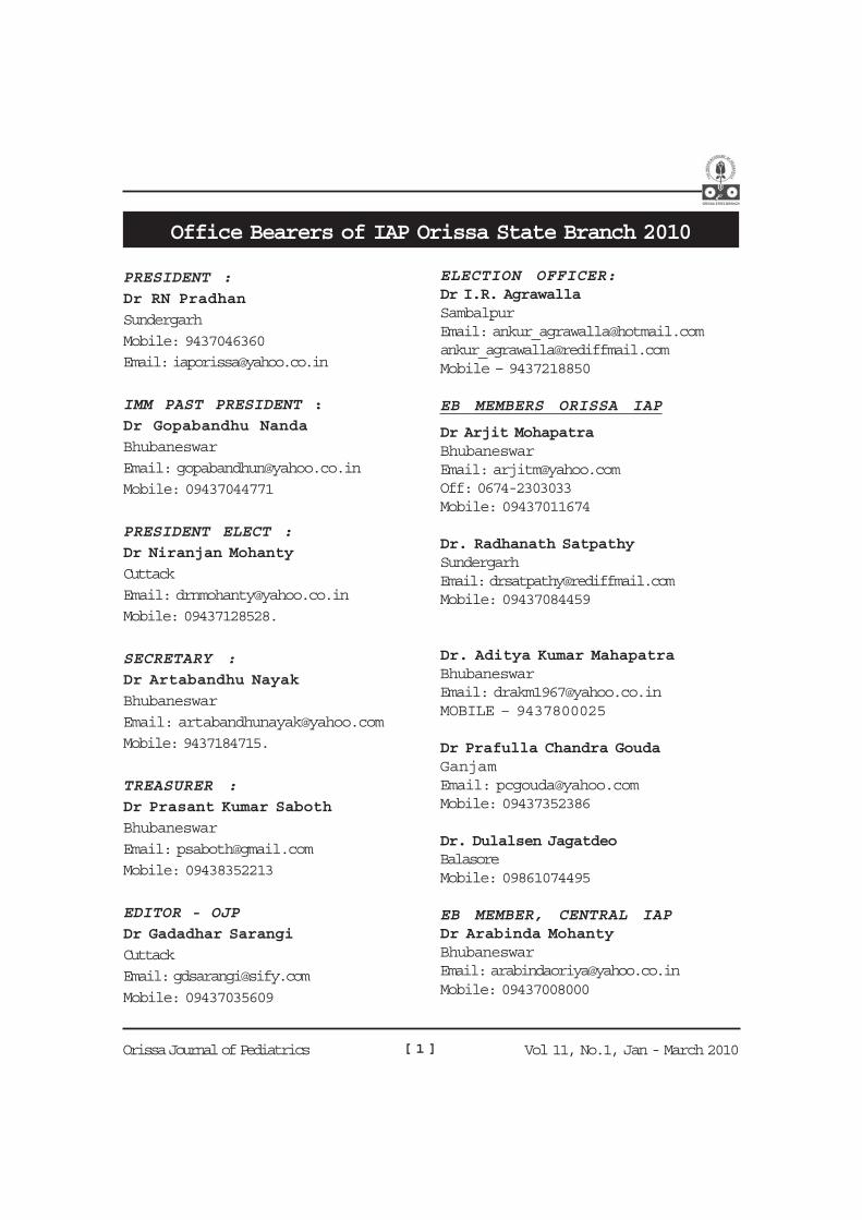
[ 1 ] Orissa Journal of Pediatrics Vol 11, No.1, Jan - March 2010 PRESIDENT : Dr RN Pradhan Sundergarh Mobile: 9437046360 Email: [email protected] IMM PAST PRESIDENT : Dr Gopabandhu Nanda Bhubaneswar Email: [email protected] Mobile: 09437044771 PRESIDENT ELECT : Dr Niranjan Mohanty Cuttack Email: [email protected] Mobile: 09437128528. SECRETARY : Dr Artabandhu Nayak Bhubaneswar Email: [email protected] Mobile: 9437184715. TREASURER : Dr Prasant Kumar Saboth Bhubaneswar Email: [email protected] Mobile: 09438352213 EDITOR - OJP Dr Gadadhar Sarangi Cuttack Email: [email protected] Mobile: 09437035609 ELECTION OFFICER: Dr I.R. Agrawalla Sambalpur Email: [email protected] [email protected] Mobile – 9437218850 EB MEMBERS ORISSA IAP Dr Arjit Mohapatra Bhubaneswar Email: [email protected] Off: 0674-2303033 Mobile: 09437011674 Dr. Radhanath Satpathy Sundergarh Email: [email protected] Mobile: 09437084459 Dr. Aditya Kumar Mahapatra Bhubaneswar Email: [email protected] MOBILE – 9437800025 Dr Prafulla Chandra Gouda Ganjam Email: [email protected] Mobile: 09437352386 Dr. Dulalsen Jagatdeo Balasore Mobile: 09861074495 EB MEMBER, CENTRAL IAP Dr Arabinda Mohanty Bhubaneswar Email: [email protected] Mobile: 09437008000 Office Bearers of IAP Orissa State Branch 2010
Transcript of Office Bearers of IAP Orissa State Branch 2010iaporissa.org/Downloads/OJP2010.pdfEmail:...
[ 1 ]Orissa Journal of Pediatrics Vol 11, No.1, Jan - March 2010
PRESIDENT :Dr RN Pradhan
Sundergarh
Mobile: 9437046360
Email: [email protected]
IMM PAST PRESIDENT :Dr Gopabandhu Nanda
Bhubaneswar
Email: [email protected]
Mobile: 09437044771
PRESIDENT ELECT :Dr Niranjan Mohanty
Cuttack
Email: [email protected]
Mobile: 09437128528.
SECRETARY :Dr Artabandhu Nayak
Bhubaneswar
Email: [email protected]
Mobile: 9437184715.
TREASURER :Dr Prasant Kumar Saboth
Bhubaneswar
Email: [email protected]
Mobile: 09438352213
EDITOR - OJPDr Gadadhar Sarangi
Cuttack
Email: [email protected]
Mobile: 09437035609
ELECTION OFFICER:Dr I.R. AgrawallaSambalpurEmail: [email protected][email protected] – 9437218850
EB MEMBERS ORISSA IAP
Dr Arjit MohapatraBhubaneswarEmail: [email protected]: 0674-2303033Mobile: 09437011674
Dr. Radhanath SatpathySundergarhEmail: [email protected]: 09437084459
Dr. Aditya Kumar MahapatraBhubaneswarEmail: [email protected] – 9437800025
Dr Prafulla Chandra GoudaGanjamEmail: [email protected]: 09437352386
Dr. Dulalsen JagatdeoBalasoreMobile: 09861074495
EB MEMBER, CENTRAL IAPDr Arabinda MohantyBhubaneswarEmail: [email protected]: 09437008000
Office Bearers of IAP Orissa State Branch 2010

[ 2 ]Orissa Journal of Pediatrics Vol 11, No.1, Jan - March 2010
JOURNAL COMMITTEE
Editor :
Gadadhar Sarangi
Associate Editors :
Jnanindra Nath Behera - Cuttack
Radhanath Satapathy - Rourkela
Executive Members :
Birakishore Rath
Aswini Kumar Mohanty
Radha Tripathy
Niranjan Mohanty
Duryodhan Samal
Sapan Kumar Murmu
Prakash Chandra Panda
Bimbadhar Rath
N. C. Mohanty
Advisory Board :
Rabi Kumar Satapathy
Govind Ch. Samal
Rudra Pradhan
B. K. Bhuyan
Imatram Agarwal
Chintamani Panda
K. J. Prusty
D. J. J. Swamy
Saroj Kumar Satapathy
Sneha Das
S. S. Mohapatra
Ambuja Pujari
Pitabas Routray
A. B. Nayak (Ex Officio)
CONTENTS
Office Bearers of IAP Orissa
State Branch 2010 1
Journal Committee 2
From The Editor’s Desk... 3
Annual Report IAP Orissa 2009
A.B. Nayak, Secretary 4
Auditors Report 7
Hair Changes In Malnutrition
Gadadhar Sarangi
B. K. Mohapatra
Swarnalata Mohapatra 8
Chikungunya Fever
Baldev S. Prajapati
Rajal B. Prajapati
Panna S. Patel 13
Rota Virus Gastroenteritis
Gadadhar Sarangi 21
Evaluation Of Score For Neonatal
Acute Physiology In Predicting
Neonatal Mortality
E.Jagadish Venkat Ragavan
J.N.Behera, Aswini.K.Mohanty
S.L.Das 34
The Enigma Of Pain Abdomen In
Children
Nimain C. Mohanty 44
Arthropathy As A Presenting
Symptom Of Wilson’s Disease -
A Case Report
Rachita Sarangi
D K Dash, B K Rath
S S Mohapatra 56
[ 3 ]Orissa Journal of Pediatrics Vol 11, No.1, Jan - March 2010
From The Editor’s Desk...Orissa Journal of Pediatrics is the official Journal of Indian Academy of Pediatrics,
Orissa State Branch. It has seen a lot of ups and downs with the growth and stagnation ofthe State Branch. The Infant mortality rate as well as the Neonatal mortality rate of the Stateis decreasing at pace with the national figures but still we are left with the second highestfigure next to Jharkhand. Infectious diseases are rampant in Pediatrics. The situation hasimproved much in the costal districts and industrial townships but the tribal areas havewitnessed the worst ravage from cholera last year. Health facilities getting adequate day byday with wide gap in distribution. Health in private sector is ready to provide facilities ofNational and International standards but watershed areas even do not have the approach tominimum health facilities. Added to the problems is the consumer protection. Law views lifewith at most respect and to keep it going anything and everything possible over the earth isdeemed to be done. In contrast a large segment of our community is unable to spend onedibles. Government is in no way ready to subsidies the total health expenditure of allindividuals. This keeps a health professional in tight hands. Knowledge to manage and savelife is not deficient but the resource to do the same is not available. The confusion of legal,moral and ethical concerns made our hands tight culminating in unnecessary referrals andover crowding of the referral canters with its ultimate negative out come. Compounding theterrible situation is the apex Govt. institutions where accountability only exits in pen andpapers. The culminating effect for a poor man is to land up in a no mans land for medicalhelp. Private areas are so costly that they remain unapproachable.
In this scenario the class of physicians affected most are the Pediatricians who carefor an unproductive segment of the society, for whom family budget is at the lowest minimumbut emotional overplay is maximum. With each loss of a newborn or child the county alsolooses maximum mean serving age for life expectancy is going high as on today. Multipletraining sessions were undertaken and being undertaken exposing Pediatricians, generalpractitioners and paramedicals in child survival but the results are not very encouraging.Some where something is not correct. May be we feel the negativity but no word to expressit. The very approach of training those who are already trained and repeatedly train them insubjects that they can not reproduce is becoming counter productive. What USA is doingtoday with its newborns with such an expansive Health network if we try to reproduce itover here may be it is bound to fail. In spite of chasing the care providers if we could havetracked the beneficiaries we might have been more successful. We provide them what wefeel to be their need but the ground reality may be something different and with the cumulativeexperience one can vow for the later.
Let the grass root Pediatrician take his decision as to what best can be done to protectchildren in his territory. Let him fix his goal. Let all agencies help him in achieving his goal.IAP with its mammoth membership should open the way and leap forward to unify publicwith Govt. initiatives and other organizations working for children.
Gadadhar Sarangi The Editor
[ 4 ]Orissa Journal of Pediatrics Vol 11, No.1, Jan - March 2010
Dear friends I thank all of you from the core of my heart for your cooperation and
timely advice. I am indebted to our President Dr. G.B.Nanda and Treasurer Dr. Prasant
Saboth for their timely guidance and help. I am also thankful to all the Executive members,
Dr. Gadadhar Sarangi, our ex. President, Dr. Niranjan Mohanty, our central executive body
member and the IAP Khurda team for their advice and cooperation throughout the year for
organizing all the academic activities. The events organized during the year – 2009 are-
1. State IAP NNF Conference:-
State IAP NNF Conference was held on 11th April 2009 at Hotel Suryansh,
Bhubaneswar. Mrs. Manorama Mohapatra, Editor, the Samaja, inaugurated the
Conference and urged the pediatricians to treat the children as own. Various topics
were discussed in the academic fiesta. My heartfelt thanks to Dr. Sailaja Nandan
Parida, President and Dr. Arikhit Swain, Secretary of NNF ,Orissa chapter as well Dr.
Arijit Mohapatra, Organising Chairperson and Dr. Biswajit Mishra, Organising
Secretary of the conference for successfully conducting the event.
2. Midterm CME on Basic Pediatrics: - The CME was conducted under the
stewardship of Dr. Gadadhar Sarangi, Prof. Pediatrics, Hi-Tech Medical College,
Bhubaneswar on 12th July 2009. 17 topics were discussed by in and out house resource
persons. The outhouse resource persons were Dr. Amar Verma, Dr. K.K Ghosh, Dr.
Sabitri Bhagat and Dr. Tapan Ghosh.
3. Workshops: - Ganjam District branch conducted a workshop on “Childhood Asthma”
in MKCG Medical College, Berhampur on 9th June 2009. It is the only branch to conduct
a workshop this year. I salute the branch.
4. ORS Day & Week Celebration: - All the district branches of IAP celebrated the
ORS Week from July 23rd to July 29th in schools and hospitals as well in medical
colleges.IAP Orissa State & Khurda branch jointly celebrated the ORS day in Shantipalli
slum, Shaheed Nagar, Bhubaneswar. The function was presided by Sj.Ananta Narayan
Jena,Mayor, Bhubaneswar Municipality. Dr. Arabinda Mohanty, Secretary, IAP Khurda
District Branch spoke on management of diarrhoea and importance of ORS. Dr. Reddys
Lab Pharma Company helped IAP, Orissa to celebrate the week. I sincerely thank the
organizing personnel of all branches for celebrating the week and Dr. Reddy’s lab for
its generosity for the cause of children.
ANNUAL REPORT IAP ORISSA 2009
Dr. A.B. Nayak, Secretary
[ 5 ]Orissa Journal of Pediatrics Vol 11, No.1, Jan - March 2010
5. World Breast Feeding Week: - All the district branches celebrated the week in their
respective places and stressed the beneficial effects of breast feeding. A joint celebration
of the week by IAP Orissa & IAP Khurda branch was organized near Bhagabanpur
High School, Patrapara on 1st August 09. Mrs. Anu Garg IAS, the commissioner cum
principal Secretary, Health and Family welfare, Govt. of Orissa, Director of Family
Welfare, Deputy Director. Nutrition and representatives of NRHM, UNICEF, CARE
INDIA and State Council for Child Welfare and State Social Welfare Board attended
the function. The Secretary spoke on Govt’s commitment to breast feeding. Dr. Arabinda
Mohanty, convenor, Orissa IMS act cell discussed on importance of Exclusive Breast
feeding and IMS act. Other dignitaries also spoke on the occasion. It was followed by
a quiz competition amongst young mothers. Later, local health workers and villagers
staged a play on breastfeeding.
6. PALS COURSE for doctors: - It was conducted at SVPIPGIP, Sishu Bhawan,
Cuttack on 28th & 29th August 2009 under the guidance of Dr. Niranjan Mohanty
HOD Pediatrics, SVPIPGIP & SCB Medical College. Dr. Bishwajit Mishra and Dr.
Arakhita Swain were the Zonal & local Co-ordinator respectively. Certificates were
distributed to successful candidates by the Organising team. I thank the organizers for
the event. Dr. G. B. Nanda President IAP Orissa State Branch & Prof. Dr. Chinara,
Principal, SCB Medical College, Cuttack inaugurated the PALS course.
7. PALS COURSE for Nurses- It was conducted for the first time in Orissa under the
co-ordinatorship of Dr. Bishwajit Mishra at Hi-Tech Medical College, Bhubaneswar.
40 Nurses attended it and the successful candidates were given certificates by the
Organising team. I congratulate Dr. Bishwajit Mishra for conducting the event for the
first time in Orissa & East Zone. President IAP Orissa Dr. G. B. Nanda formally stared
the course.
8. IAP QUIZES:-IAP Quiz 2009 for Undergraduate & Post Graduates, both college
and divisional round were conducted successfully along with NNF Neonatology PG
Quiz under the leadership of Prof. Dr. Niranjan Mohanty on 17th September 2009 and
10th October 2009 respectfully at SCB Medical College Cuttack. President Dr. G. B.
Nanda, Secretary Dr. A.B. Nyayk & Treasurer Dr. Prasant Saboth of IAP Orissa State
Branch, divisional coordinator Dr. JN Behera, Neonatalogy PG Quiz coordinator Dr.
Atul Samal, President NNF Orissa, Professors from SCB Medical College, SVPIPGIP,
Sishu Bhawan, Hi-tech Medical College, KIMS, IMS were present in the programme.
IAP thanks to Prof. Dr. Niranjan Mohanty for conducting it successfully.

[ 6 ]Orissa Journal of Pediatrics Vol 11, No.1, Jan - March 2010
9. Participation of State IAP in activities of Govt. and other organizations-
IAP President Participated in two State level meetings of Govt. of Orissa on
“Management of Swine Flu infection. The Meetings were presided by Hon ble
Chief Minister of Orissa.
Prof. Niranjan Mohanty and Dr. G. B. Nanda attended IAP zonal workshop on
“Advance Vaccinology “in Kolkata.
President IAP participated in State level meeting on introduction of MMR Vaccine
in National immunization schedule.
10. IAP Orissa State Branch Secretariat & Guest house:-A piece of land was
purchased at Patia Chhack, Bhubaneswar in February 2009 for IAP.
A memorandum of Understanding was signed with IMA, Bhubaneswar on 29th July
2009 to permanently occupy the 3rd Floor (2400sqft) of IMA house, near Capital
Hospital, Bhubaneswar, to use as IAP State branch Secretariat & IAP guest house.
The building is expected to complete by the end of 2009. IAP Orissa State branch will
have its own Secretariat & guest house.
11. Growth Development & Behavior Disorder (GDBD) Chapter: - I congratulate
Prof. Dr. S.N. Parida being elected as National President of Growth Development &
Behavior Disorders subspecialty chapter of Indian Academy of Pediatrics. It is pride
for IAP Orissa State Branch.
12. IAP Neocon-2009:- Indian Academy of Pediatrics, Orissa State Branch is hosting
the 2nd National Conference of IAP Neocon-2009 from 06.11.2009 to 08.11.2009
with the conference theme “Every Newborn – Our Responsibility”. I congratulate
Dr. Arjit Mohapatra and Dr. Bishwajit Mishra, the Organizing Chairman and Secretary
& their Organising team for successfully conducting it.
Long live IAP. Long live IAP, Orissa State Branch.

[ 7 ]Orissa Journal of Pediatrics Vol 11, No.1, Jan - March 2010
AUDITORS REPORT

[ 8 ]Orissa Journal of Pediatrics Vol 11, No.1, Jan - March 2010
Introduction:
Hair is a cutaneous appendage
typical of human skin. In the evolutional
process it has lost most of its protective
functions in human beings. The scalp hair andeye-lashes retain the property of protection
to some extent. Thus human race becomes
the least hairy mammal on earth. Tactile
perception is one of the important minor
functions it serves because of its rich nervenet-work.(1)
Hair is formed of hard keratin with
high sulphur content that is responsible for its
extraordinary tensile strength.(2) The Cortex
is made up of a low sulphur fibrillar
component tightly packed in a sulphur rich
matrix. The fibrillar component consist of
macrofibrills of 7 nm thick, arranged in a
HAIR CHANGES IN MALNUTRITIONGadadhar Sarangi*, B. K. Mohapatra **, Swarnalata Mohapatra ***
Abstract :
Protein Calorie Malnutrition is rampant in the developing world. The growth and form of hair is
affected as a part of the generalized growth retardation. Under scanning election microscope irregularity
of the hair tip, niche formation under the crests of wavy nodal lines, linear brakeage in the shaft and
herniation of the hair material through the lateral wall are observed.
Key Word :
Protein Calorie Malnutrition (PCM), Marasmic-Kwashiorkor, Scanning Electron Miscoscope.
longitudinal laminated form which on cross
section gives a thumb print appearance.(3) In
Malnutrition the amino acid contents in the
hair changes. The cysteine content was
reported to be significantly less and correlates
with the degree of malnutrition.(4)
Protein Calorie Malnutrition (PCM) is
a condition, which results from deprivation
of protein and calorie from the diet.
Marasmus and Kwashiorkor are two extreme
states of PCM, the former resulting from
predominant calorie deficiency while the later
from protein deficiency. Because of the non-
availability of protein or protein getting utilized
for calorie the growth and repair of body gets
affected and consequently the growth and
texture of hair also gets affected in these
conditions.
*Senior Consultant, THE CHILD, Cuttack Orissa**Senior Scientist, Regional Research Laboratory, Bhubaneswar, Orissa***Medical Officer – “THE CHILD”, Cuttack, OrissaAddress of Correspondence :Dr. Gadadhar Sarangi“THE CHILD”, B.K. Road,Ranihat, Cuttack – 753001, ORISSA

[ 9 ]Orissa Journal of Pediatrics Vol 11, No.1, Jan - March 2010
The slow growth rate of Marasmic
Child slows down the hair growth. The main
changes are of texture and a lack of pliability,
giving rise to rough, lusterless, straight hair
that can be easily pluckable and is brittle. (5)
In marasmus the hair is fine and dry, the
diameter of the hair bulb is reduced to a third
of normal and almost all follicles are in
telogen.(6) Johnson and his colleagues
reported significant difference in shaft
diameter, percentage of anogen and telogen
between well nourished and severely
Malnourished children. As it is not suitable to
asses different grades of PCM the method was
not recommended for PCM assessment is field
conditions.(7)
In Kwashiorkor the hair is sparse, thin,
brittle, wavy and silky or shows a mixed
picture with rough and standing hair as in
marasmus. The degree of hair loss varies from
mild to serve. Hairs are easily pluckable but
the hair growth rate is adequate. The colour
may be golden, blondy, rusty or light brown.
The colour changes may be generalized or
patchy. Black hair turns brown and brown
hair turns blonde. The hair grown during the
period of malnutrition is pale, therefore
alternate bands of dark and pale hair are seen
in a single strand (Flag sign)(5). The hair may
be prematurely gray or show a “pepper and
salt” appearance and become sparse, fine
and brittle. The hair shafts may show
constrictions which increase their vulnerability
to trauma. In kwashiorkor the hair follicles are
more in anogen although most are atrophic(8).
In moderate to severe PCM the
morphological charges in scalp hair include
reduction in diameter of the hair shaft, bulb
and medulla. The medulla may even be
absent.(9,10) In both the forms of PCM hair is
brittle and easily shed and partial or complete
alopecia may occur. The hair is lusterless and
if normally black, may assume a reddish tinge.(11,12)
Material & Methods :
To examine the hair in malnutrition a
case of marasmic kwashiorkor was selected.
There was partial alopecia. The hairs were
light brown, sparse, fine, brittle and easily
pluckable. The hairs were examined under
Scanning Electron Microscope (SEM) with
different magnifications. For this purpose a
JEOL model (JSM 35 cf), SEM was used.
Observations :
Under optical microscope no visible
difference in the hair shaft or the tip between
normal and malnourished hair is observed.
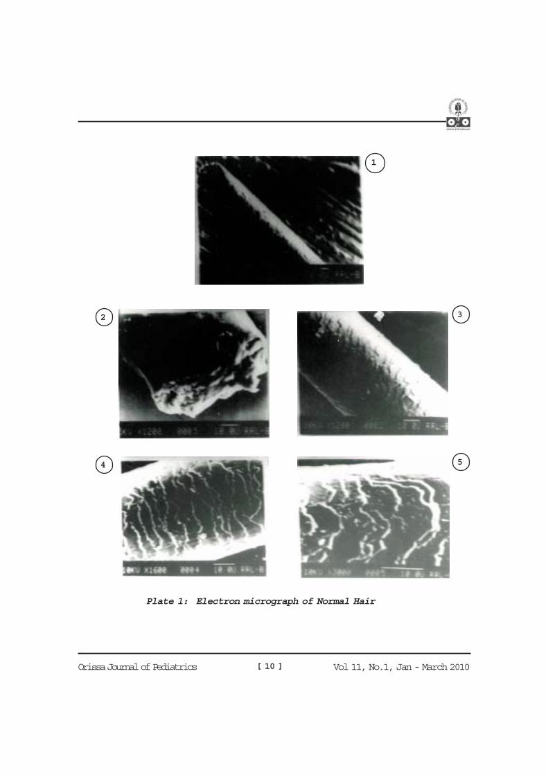
When viewed under SEM at low
magnification (X-540), the tips look more or
less pointed (Pl 1.1 and Pl 2.1). But in
magnification more than 1200 times, the tip
appears wavy and lamellar (Pl.1.2 ). In
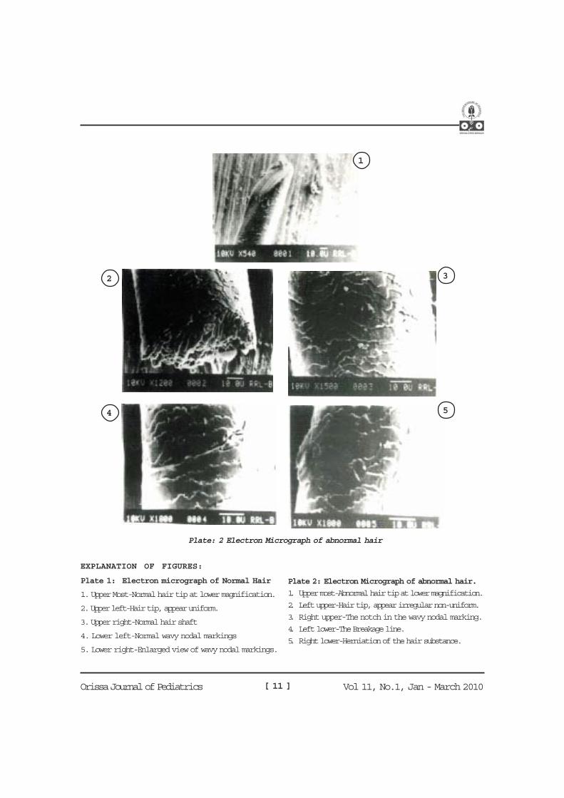
malnutrition the hair tip is irregular and the
projections are uneven (Pl.2.2). This finding
is observed in most of the hair tips.
[ 10 ]Orissa Journal of Pediatrics Vol 11, No.1, Jan - March 2010
1
Plate 1: Electron micrograph of Normal Hair
2 3
4 5
[ 11 ]Orissa Journal of Pediatrics Vol 11, No.1, Jan - March 2010
1
2 3
4 5
Plate: 2 Electron Micrograph of abnormal hair
EXPLANATION OF FIGURES:
Plate 1: Electron micrograph of Normal Hair
1. Upper Most-Normal hair tip at lower magnification.
2. Upper left-Hair tip, appear uniform.
3. Upper right-Normal hair shaft
4. Lower left-Normal wavy nodal markings
5. Lower right-Enlarged view of wavy nodal markings.
Plate 2: Electron Micrograph of abnormal hair.
1. Upper most-Abnormal hair tip at lower magnification.
2. Left upper-Hair tip, appear irregular non-uniform.
3. Right upper-The notch in the wavy nodal marking.
4. Left lower-The Breakage line.
5. Right lower-Herniation of the hair substance.

[ 12 ]Orissa Journal of Pediatrics Vol 11, No.1, Jan - March 2010
There are normal wavy nodalmarkings interspersed regularly in the hairshaft. They are in one plane. In normal hairthe pattern is evident in higher magnifications.The single plane disposition is obvious evenin 3000 times magnification (Pl.1.4 &5). Inhair of malnutrition the nodal wavy pattern ismaintained but the height of the wave is moreand they are more acute. There is a nichebelow the crest of the wave and there isshedding visible in the area indicating thatthey are in different planes. This observationis met frequently but not invariably (Pl.2.3)
Usually in the malnourished hairthere are breakage lines often extending fromone side to the other in the shaft. At times it ispartial or complete. This is better visible in1800 times magnification (Pl.2.4). To thenaked eye or in lower magnifications this lineis not very much appreciated.
One of the rare findings inmalnutrition is the protrusion of the hairsubstance in the lateral aspect of the hair shaft,delineated properly in 1800 magnification.This may be an out-growth or may beherniation of the hair substance out throughthe weak cortical tissue of the hair.
Summary & Conclusion:
The hair of a case of Marasmic-kwashiorkor was studied under scanningelectron microscope. In higher magnificationfew unusual findings like (a) Irregularity of thehair ends, (b) Increase in crest height alongwith niche formation, (c) Breakage line in thehair shaft, and (d) Herniation of the substancethrough lateral wall were observed. Thesefindings briefly describes the ultra structureof malnourished hair which has hither to notbeen reported in literature so far.
References :
1. Winkelmaan RK. The innervations of a hairfollicle.Ann NY Acad Sci. 1959; 83: 400- 407
2. U. Jakubovie HR., Ackerman AB. Structureand function of the skin. In : Moschella S,Hurley H, Edt. Dermatology 2nd ed.Philadelphia : W.B. Saunders, 1985:1-103
3. Breathnach AS. An atlas of ultra structureof human skin.London : Churchill Livingstone 1971
4. Menafee E and Friedman M. Estimation ofstructural components of abnormal humanhair from amino acid analyses. J. Pr. Chem1985, 5: 333-341
5 Desai SC, Seth RA, Udani PM. Nutrition andhair anomalies.In : Orfanos, Montanga, Stuffgen. Edt Hairresearch.Berlin : Springer 1981.
6. Bradfield RB, Cordario A, Graham GG. Hairroot adaptation to marasmus. Lancet 1969,ii : 1395 – 1396.
7. Johnsons AA, Latham MC, Roe DA. Anevaluation of the use of changes in hair rootmorphology in the assessment of Protein-Calorie Malnutrition.
8. Bradfield RB, Bailey MA. Hair root responseto under nutrition. In montanga W, DobsonRL Edt Hair growth. Oxford : Pregamon,1968 : 109 – 119
9. Visweswara Rao K, Bhatia RYP, Pralhad RaoN. Morphological changes in Scalp hair inmild and moderate forms of protein caloriemalnutrition. Indian J Med Res 1978; 68:631 – 640.
10. Visweswara Rao K. Bhatia RYP, Pralhad RaoN. Morphological changes in scalp hair ofchildren with severe forms of protein caloriemalnutrition. Indian J Med. Res. 1978,67:86-96.
11. EL- Hefnawi H, Shukri AS, Rashed A.Kwashiorkor in United Arab Republic Br. J.Dermatol 1965, 77, 137 – 150.
12. Sims RT. Hair growth in Kwashiorkor. Arch.Dis Child. 1967, 42 : 397 – 400.
[ 13 ]Orissa Journal of Pediatrics Vol 11, No.1, Jan - March 2010
Introduction :
The name “Chikungunya” is derived
from the word “Swahili” meaning ‘that which
bends up’ in reference to the stooped posture
developing as a result of the arthritic
symptoms of the disease. The name is derived
from Makonde, one of the African languages.
It is also spelt as Chiken Gunea. It is spread
by bite of ‘Aedes Aegypti’ mosquito. Abrupt
onset of fever with chills, flushing of skin and
joint pain are main characteristics of the
disease. It is almost always self limiting and
rarely fatal.
Chikungunya virus was first isolated
from the serum of a febrile person in Tanzania
in 1953. Since then, it has been isolated
repeatedly from several countries in Africa.
The virus was also identified in many parts
of Asia.
CHIKUNGUNYA FEVER
Baldev S. Prajapati*, Rajal B. Prajapati**, Panna S. Patel***
Chikungunya Virus :
Viruses in the families of Togaviridae,
Flaviviridae and Bunyaviridae are principally
arthropod-borne or spread as zoonoses.
(Table-1).
Chikungunya virus is a Group IV (+)
RNA virus belonging to Togaviridae with
genus Alphavirus and species Chikungunya.
Ross River, O’nyong-nyong are other viruses
belonging to the same family with genus
alphavirus. They have been associated with
similar syndrome.
Transmission :
Chikungunya virus is most commonly
transmitted to humans through the bite of an
infected mosquito, specifically mosquitoes of
the Aedes genus like Aedes Aegypti. They
usually bite during day time (Daylight) hours.
Culex and Mansonia mosquitoes can also
transmit the disease. Once the mosquitoes are
infected, they remain so throughout life.
* Associate Professor, Sheth L. G. General Hospital, Smt. N.H.L. MunicipalMedical College, Ahmedabad
Aakanksha Children Hospital and Neonatal Nursery
Nava Vadaj Road, Ahmedabad
** Associate Professor, Sheth V.S. General Hospital,
Smt. N.H.L. Municipal Medical College, Ahmedabad
***Pediatric Consultant

[ 14 ]Orissa Journal of Pediatrics Vol 11, No.1, Jan - March 2010
Family Genus Virus (examples) Clinical M anifestations
Togaviridae Alphavirus • Chikungunya
• Eastern equine encephalitis
Fever, Rash, Arthritis
Encephalitis
Rubivirus • Rubella Rash,Arthritis
Flaviviviridae Flavivirus • Yellow fever H epatitis, haemorrhagic fever.
• St. Louis encephalitis Febrile illness with headache, encephalitis
• Dengue Fever, Rash, H aemorrhagic fever
Hepatitis C like • Hepatitis C H epatitis
Pestivirus • Bovine viral diarrhea Diarrhoea
Bunyaviridae Bunyavirus • La Crosse Aseptic meningitis Encephalitis
Phlebovirus • Sandfly fever Fever
Nairovirus • Congo-Crimean Haemorrhagic fever
H aemorrhagic fever
Hantavirus • Hantaan,Seoul, Belgrade, Sin Nombre
Non-Cardiogenic pulmonary oedema.
Tospovirus • Tomato spotted wilt Spotted wilt in plants
Table-1
Arboviruses

[ 15 ]Orissa Journal of Pediatrics Vol 11, No.1, Jan - March 2010
Outbreaks of Dengue and
Chikungunya usually involve small towns
while outbreaks of o’nyong-nyong and West
Nile fever usually involve villages.
Vertical Maternal Fetal Transmission :
In March 2005, an epidemic of
chikungunya virus began in the southern
portion of Reunion Island. There was report
of acute chikungunya to 84 pregnant women.
74 mothers had infection quite earlier to the
delivery date and their newborns appeared
asymptomatic. Conversely, 10 newborns had
severe attacks after birth and required
prolonged hospitalization. Four babies
developed meningo-encephalitis, three
disseminated intravascular coagulation. Six
babies required NICU care with intubation
and assisted ventilation. No infant died. These
cases were confirmed by specific serology
testing or PCR or both for mothers and
newborns. It was noted that all severe cases
involved women with viremia and fever in the
intrapartum period.
Epidemiology :
Africa : Chikungunya virus is transmitted in
the forests of tropical Africa by aedes aegypti
mosquito. The vertebrate portion of the cycle
is provided by non-human primates such as
monkeys and baboons which amplify and
maintain virus circulation. It is thought that
endemic circulation and moving epidemics
in troops of primates are responsible for
survival of the virus and local spill over into
human population. In Africa villages or rural
areas these mosquitoes may then infect
humans and substantial viremia measure
suggest that humans, in appropriate setting
may contribute to mosquito infection, leading
to further virus amplification. This becomes
particularly important when domestic
breeding of Aedes Aegypti is present in large
number, a situation that may lead to village
and large urban epidemics in Africa. The
classical chikungunya epidemic which
occurred in Tanzania in 1953, resulted when
Aedes Aegypti borne disease moved through
multiple villages over an expense exceeding
5,000 Kms. In studies of individual dwellings,
there was a highly significant trend for multiple
cases to occur once a single case had
occurred. This of course, could be a reflection
of flight – range of Aedes Aegypti vectors and
human habits.
Asia : Transmission in Asia follows a different
pattern from that seen in Africa, being
primarily transmitted from human to human
by Aedes Aegypti. Although Asian monkeys
develop significant viremia after chikungunya
virus inoculation and have been found to
harbour antibodies to it, they have never been
shown to participate in any important way in
the maintenance or amplification of the virus
in the continent.

[ 16 ]Orissa Journal of Pediatrics Vol 11, No.1, Jan - March 2010
Chikungunya activity in Asia has been
documented since its isolation in Bangkok,
Thailand in 1958. Other South-East Asian
countries which have experienced
chikungunya outbreaks are Cambodia,
Vietnam, Burma, Srilanka and India. A series
of epidemics usually lasting a single year have
been reported from Sri Lanka and India.
India : First out-break of chikungunya in
India was in 1963 in Kolkatta. It was
responsible for extensive dengue like infection
with occasional haemorrhagic manifestations.
Chikungunya virus was isolated from cases
with severe disease like haemorrhagic
manifestations. Serious cases were more
common among infants, young children,
elderly and immunocompromised patients.
Severe manifestations were less common
among young adults. In 1964, there was an
epidemic of chikungunya in Vellore, Madras
and Pondichery. In 1973, a small localized
out-break was reported from Barsi, Sholapur
district in Maharashtra State. No out-break
was reported from India after 1973 till
2005.13,14
Again, there was increase in
incidence of fever cases in Maharashtra State
since December, 2005. 258 villages from 15
districts have reported 34,725 fever cases till
April, 2006. Similarly, 18,529 cases of fever
with arthritis or arthralgia have been reported
from seven districts of Karnataka State since
December, 2005. In Andhra Pradesh, 5,671
cases were reported from December, 2005 to
February, 2006. Orissa experienced the out-
break of the disease with almost 4,900 cases
in February and March,2006. It is estimated
that thousands of people suffered from
chikungunya during its recent out-break in
2007 in Gujarat.
Clinical Features :
The incubation period is usually 2-3
days with a range of 1-12 days. Chikungunya
is an acute viral infection of abrupt onset.
Fever, rash and joint involvement are
characteristic features of this disease. Fever
rises abruptly often reaching to 39 to 40oC
(102-104oF) and may be accompanied by
chills. This acute phase lasts for 2 to 3 days.
The temperature may remit for 1-2 days,
resulting in a “Saddle-back” fever curve.
The arthralgias are polyarticular,
migratory in type. Small joints of hands,
wrists, ankles and feet are commonly affected.
Larger joints are less commonly involved.
Pain on movement is worse in the morning,
improved by mild exercise and exacerbated
by strenuous exercise. Joint swelling may
occur but collection of fluid is uncommon.
Patients with milder articular manifestations
[ 17 ]Orissa Journal of Pediatrics Vol 11, No.1, Jan - March 2010
are usually symptom free within a few weeks,
while patients with severe joint involvement
require months to resolve completely.
Generalized myalgias as well as back and
shoulder pain is common. Joint involvement
is less common and if it is presents, it is of
mild degree in children compared to adults.
Young children may present with irritability
and excessive crying due to myalgia.
Cutaneous manifestations are typical
with many patients presenting with flush over
the face and trunk. This is usually followed
by maculopapular rashes all over the body.
The trunks and limbs are commonly involved
but face, palms and soles may also show
lesions.
Headache, photophobia, retro orbital
pain and conjunctival infection are other
common manifestations during acute illness.
Sore throat due to pharyngitis is also fairly
common.
Severe manifestations of the disease
like meningo-encephalitis are rare. They may
develop in newborns, infants, elderly and
immuno-compromised patients.
Lymphadenopathy is unusual feature of the
disease. Chikungunya outbreaks result in
several hundreds or thousands of cases but
deaths are rarely encountered. Death may
occur due to pre-existing medical disease.
Differential diagnosis of chikungunya
include Dengue, Dengue haemorrhagic fever,
Rose River and Onyong-nyong viral
infections.
Diagnosis :
Though definitive diagnosis can only
be made by Laboratory means, Chikungunya
should be suspected when epidemic disease
occurs with characteristic triad of fever, rash
and joint manifestations. The following
criterias are suggested for case definition.
(Table-2)
Table-2
Case definition of Chikungunya
Suspected case
An acute illness characterized by sudden
onset of fever with several of following
symptoms – joint pain, headache,
backache, photophobia, arthralgia, rash.
Probable case
As above and positive serology (when single
serum sample is obtained during acute
phase or during convalescence).
Confirmed cases
A probable case with any of the following :
1. Four fold HI antibody difference in
paired samples
2. Detection of IgM antibodies
3. Virus isolation in serum
4. Detection of CHIKV nucleic acid in sera
by RT-PCR
[ 18 ]Orissa Journal of Pediatrics Vol 11, No.1, Jan - March 2010
Suspected Case :
Acute illness characterized by sudden
onset of high grade fever maybe with chills,
associated with other symptoms such as joint
pain, skin rash, headache, photophobia, retro
orbital pain, backache, myalgia etc.
Probable Case :
As above and positive serology when
single serum sample is obtained during acute
phase or during the convalescence.
Confirmed Case :
A probable case with any of the
following :
Four fold haemagglutination inhibition
(HI) antibodies difference in paired serum
samples.
Detection of IgM antibodies.
Virus isolation from serum.
Detection of Chikungunya virus nucleic
acid in sera by RT-PCR.
Laboratory Tests :
The facility for laboratories working
on chikungunya available in our country is at
National Institute of Virology, Pune and
National Institute of Communicable Diseases,
Delhi.
Serological Diagnosis :
Virus specific IgM antibodies are readily
detected by Capture ELISA in patients
recovering from Chikungunya infection
and they persist for about 6 months.
Haemagglutination Inhibition (HI)
antibodies appear with ceasation of
Viremia. All patients will be positive by
day 5 to 7 of the illness.
Collection, Storage and Transportation of the
Sample : (Table-3)
Laboratory diagnosis depends on the
quality of sample, time of collection of sample,
its storage and necessary precautions taken
during transportation.
Table-3
Collecting storage andtransportation of sample
For serology :
Blood
Acute sample – within 5 day of onset ofillness
Convalescent or paired sample – 10-14days after first sample
Transport :
Transport to laboratory at 2-8oC as soonas possible. Do not freeze.
If more than 24 hours delays is expected,the serum should be separated and storedfrozen.
For isolation of virus and RT-PCR :
Blood – Collect within 5 days of illness(transport within 48 hour in cold, preferablyfrozen).
[ 19 ]Orissa Journal of Pediatrics Vol 11, No.1, Jan - March 2010
For Serology :
First sample should be collected 5days after the onset of illness. Convalescentor paired sample should be collected 10 to 14days after the first sample.For Isolation of the virus and RT-PCR :
Blood for isolation of virus and RT-PCR should be collected within first 5 days ofillness. These samples should be sentimmediately, within 48 hours to the referrallaboratory. It should preferably be frozen.Transportation :
Specimens should be transported tothe laboratory as soon as possible. Thetemperature should be maintained 2 to 8oCduring transport of the specimens. The wholeblood should not be frozen as hemolysis mayinterfere with results of tests.
If more than 24 hours delay fortransportation of specimens is expected, theserum should be separated from blood andserum should be stored frozen.Management :
There is no specific treatment forchikungunya. The illness is usually self limitingand will resolve in due course of time.
Supportive care with rest is indicatedduring the acute joint symptoms. Theantipyretics and analgesic drugs can be usedfor symptomatic relief. The commonly useddrugs are paracetamol 15 mg/kg/dose,Ibuprofen 8 to 10 mg/kg/dose and Diclofenacsodium 1 to 3 mg/kg/day. Aspirin, Nimesulideand other NSAIDs should be avoided toprevent gastric bleeding and other hazards.In unresolved arthritis refractory to NSAID,Chloroquine 250 mg/day is recommended.There is no role of antibiotics. Steroids shouldnot be used. It has been observed that at times
it may cause serious complications. Mildexercise tend to improve joint symptoms, butstrenuous exercise may exacerbate thesymptoms.Prevention And Control :
There is no specific medicine orvaccine available against chikungunyainfection. Vector control is thus very importantmeasure for prevention of the diseasetransmission. Elimination of mosquitobreeding sites or source reduction is aneffective method of control of the disease.Aedes Aegypti is typically a container habitatspecies and breeds primarily in artificialcontainer and receptacles. Aedes Aegyptimosquito does not fly more than 100 metersfrom its original shelter.Control of Mosquito Breeding :
All water tanks, barrels, cisterns, trashcontainers need to be covered tightly witha lid.Remove or empty water in old tyres, tincans, buckets, drums, bottles or from otherplaces where mosquitoes breed.Clogged gutters and flat roofs that mayhave poor drainage need to be checkedregularly.Water in bird baths and plant pots or diptrays should be changed at least twice aweek.Pets water bowls need to be emptied daily.In ornamental water tanks and garden,larvivorous fish (Guppy fish) need to beintroduced. They eat mosquito larvae.Weeds and tall grass should be cut short.Adult mosquitoes look for these shadyplaces to rest during hot day light hours.In case water containers can not beemptied regularly, temephos(1 ppm) should be applied.

[ 20 ]Orissa Journal of Pediatrics Vol 11, No.1, Jan - March 2010
Protection from Mosquito Bites :Use of insecticide (Permethrin) treated
mosquito nets or curtains has been found
effective. Especially children should sleep
under insecticide treated nets during day
time.
Insecticide spray kills the mosquitoes.
Fogging operations with 2% pyrethrum is
also recommended.
It is very essential that people should be
educated for all these measures and
scientific information regarding the
disease.
Chikungunya Virus Vaccine :This vaccine has passed through
various phases. It was conducted a Phase II,
randomized, double-blind, placebo-
controlled, safety and immunogenicity study
of a serially passaged, plaque-purified live
chikungunya (CHLK) vaccine in 73 healthy
adult volunteers. It was found promising and
highly immunogenic. Certain well-tolerable
side effects were noted. It may become
available for use in practice in the future.13
References :
1. CD Alert Vol.10 : No.2, Chikungunya Fever,
February, 2006; 1-8. Monthly Newsletter of
National Institute of Communicable Diseases,
Directorate General of Health Services,
Government of India.
2. Lam SK, Chug KB. Alpha viruses. WHO
collaborating centre for Arbovirus Reference
and Research, Department of Medical
Microbiology, Faculty of Medicine, University
of Malaya, Kuala Lumpar. Http://www.
vadscorner.com/alphaviruses.html
27.8.2006.
3. Long S.S., Pickering LK, Prober CG (Eds)
Principles and Practice of Pediatric Infectious
Diseases, 1st edn. New York; Churchill
Livingstone 1997.
4. Kliegman RM, Jenson HB, Behrman RE et
al. (Eds) Nelson – Textbook of Pediatrics. 18th
edn. New Delhi : Elsevier, 2008.
5. Passi GR. Global update. Disease Watch –
Chikungunya fever. Indian Pediatr 2006, 43
: 751.
6. Kapse A. Chikungunya – A Re-emerging
Disease. IAP Bulletin Academy Today,
September, 2006, 25-27.
7. Chikungunya (Internet Information) IMA
GSB, New Bulletin, September, 2006.
8. Robillard PY, Boumahni B, Gerardin P et
al. Vertical maternal fetal transmission of the
chikungunya virus. Ten cases among 84
pregnant women. Press Med 2006, 35 : 785-
788.
9. http://www.who.int/csr/don/2006_03_17/en/
index.html.
10. http://www.cbwinfo.com/Biological/
Pathogens/CHLK.html.
11. http://www.cdc.gov/travel/other2006/
chikungunya_india.htm.
12. Edelman R,Tacket Co., Wasserman SS, et
al. Phase II safety and immunogenicity study
of live chikungunya virus vaccine TSI-GSD-
218 American Journal of Tropical Medicine
and Hygiene, 2000, 62: 681-685.
13. Bhat S: Chikungunya Fever – A Re-emerging
viral disease. In Gupte S(Ed) Recent
Advances In Pediatrics-18 : Hot Topics 1st
edn. New Delhi. Jaypee Brothers Medical
Publishers (P) Ltd. 2009, 229-236.
14. Chhabra M, Mittal V, Bhattacharya D et al.
Chikungunya fever : a re-emerging viral
infection. Indian J Med Microbiol 2008,26 :
5-12.

[ 21 ]Orissa Journal of Pediatrics Vol 11, No.1, Jan - March 2010
Introduction :
Brener and Horne is 1959 describeda novel technique of electron microscopywhich ushered a new era in the field ofexperimental and diagnostic virology. Rotavirus in 1963 was first described as an agentof infantile murine diarrhoea. 10 years afterin1973 Bishop and his associates observedby electron microscopy, in the duodenalepithelium of children with diarrhea, a 70 nmvirus at Royal children’s hospital in Melbourneof Australia(1). Subsequently in 1974 Flewttand colleagues advanced the name“rotavirus” based on its “wheel-like”appearance (2).
ROTA VIRUS GASTROENTERITISGadadhar Sarangi*
Within 5 years of this discovery
rotavirus was recognized as the most common
cause of diarrhoea in infants and young
children world wide, accounting for
approximately one third of cases of severe
diarrhoea requiring hospitalization.
The Virus :
Rota Viruses are 70nm icosahedral,
none enveloped, double stranded RNA virus
belongs to the family Reoviridae. Rota Viruses
are classified as groups, subgroups and
serotypes. The groups do not have any
antigenic relations. Group A: Predominantly
affect humans and Group B in China and
Group C occasionally reported to be
pathogenic. Other 4 groups (D, E,F&G) are
not seen in humans.
Rota Viruses are subdivided to two
subgroups (I & II) depending upon the VP6
protein present in the inner capsid.
The serotypes are determined
depending upon the VP4 (Protease cleaved/
P Protein) and VP7 (Glycoprotein / G Protein)
* Professor Pediatrics, Hi-Tech Medical College, Bhubaneswar
Abstract :
Rotavirus is one of the commonest causes of severe gastroenteritis in infants and young children.The mortality and severity is high in malnourished children. Laboratory diagnosis is difficult in the fieldbut clinical features well correlates with the laboratory findings. Maintenance of fluid and electrolytes isthe cornerstone of therapy. Vaccine remains the answer for this widespread disease. Trials are on to
reduce the cost and to make it available to the developing world.
RotavirusReprinted from emerging infectious
DiseasesVolume 4 No. 4

[ 22 ]Orissa Journal of Pediatrics Vol 11, No.1, Jan - March 2010
The individual genomic RNA segment
can reassort independently producing a
Reassortant particle of mixed parentage which
could theoretically lead to the emergence of
110 different G and P combinations. G1, G4,
G9 with P4 and P8 serotypes with
combinations are predominant world wide
causing 90% infection in industrialized
countries and 68% infection in Asian
countries(4). GI P (8) is the globally
predominant strain followed by G3 P(8), G2
P(4) and G4 (8). G9 strains have emerged in
the early 2000s and have become
Distribution of Death due to Rotavirus gastroen-teritis World Over
Reprinted from Nat Med 1997; 3:10-11
predominant in some regions of the world
including Europe and parts of Eastern Asia.
Less usual strains like G10P(11) and G12
P(6) are also evolved in India.(5)
Both G & P proteins induce neutralizing
antibodies and may be involved in protective
immunity.
Epidemiology :
Rotaviruses are the leading cause of
severe diarrhoeal diseases and dehydration
in infants and children worldwide under the
age of 5years. It has been estimated that
rotavirus infection is responsible for 111
million episodes of diarrhoea requiring home
care and 2 million hospitalization from the
same cause with approximate 4, 40,000
deaths in under fives, 82% of which occurs
in the developing world (6) almost 100,000
deaths occurs each year in India alone and
double the number are lost in African
countries (7).
The Virus has three layered capsid. The
outer capsid is made up of VP4 (P Protein)
encoded by 4th gene and VP7 (G Protein)
encoded by 7, 8, 9 genes. They define the
serotype of the virus. The inner capsid is
made up of VP6 protein encoded in 6th gene
which is most abudant and immunogenic in
the virion. Anti VP6 antibodies inhibit virus
transcytosis through the intestinal epithelium
barrier(3) The internal shell surrounds the 11
segment double stranded RNA genome.
The Three layered capsid of the virusReprinted from “Fields virology” 3rd Edt. Vol.2

[ 23 ]Orissa Journal of Pediatrics Vol 11, No.1, Jan - March 2010
As rotavirus infects all the children below5years and the infectivity is very high, it isunlikely to reduce disease burden by improvingwater, food or sanitation. Vaccination remainsthe only alternative. Natural immunity issuggested because of infrequent occurrence ofmore than one episode of rotavirus diarrhoeain a child and decreased incidence of thedisease with increasing age (13). Furthermoreprotection increased with each infectionagainst moderate to severe disease, lessagainst, mild and least against asymptomaticinfection (14).
The global scenario recently gettingchanged with the advent of new oral rotavirusvaccines (15).
In a study from south India overall Rotavirus infection amongst children with acutediarrhoea was 22.55% with no genderspecificity. The study revealed 66.1% G2serotype followed by G4 (13.6%) G1 (9.3%)and G3 (1.7%). Dual infection was presentin 9.3% dominated by G1 - G2 (63.6%). Thedominantly present serotype was G2 (P4)(10).
In a multicentric analysis from 40published study from all over India out of13,000 pediatrics inpatients with diarrhoea,28% neonates and 18% children had rotavirusinfection. G1 and G2 were the commonserotypes in children where as G9 (P11) wasisolated more frequently from neonates. 50%of infection occurring within 6months and75% within 9months attracts the feasibilityfor neonatal vaccination (16).
The study by G. Kang et al from 18 citiesof India, among children attending hospitalfor diarrhoea the rotavirus infection waspresent in 23.4%. There was markedgeographical differences with G1 serotype beingcommon in western India. Group B viruses
Distribution of Death due to Rotavirus
gastroenteritis World Over
Virtually all the children are infected by
the time they reach two to three years of age.
Most symptomatic episodes occur between
3months and 2years with a peak incidence
between 7 and 15months (8). Neonatal
rotavirus infections are mostly asymptomatic
and caused by unusual strains as reported
from six hospitals from India (9). The disease
has definite winter seasonality in temperate
climate and year round exposure in tropical
countries. In south India a mean minimum
temperature of 24oC predisposed more to
rotavirus infection (10).
GIP(8), G3P(8), G4P(8) and G2P(4)
strains dominate world over (11). Unusual
strains are common is several developing
countries like those with serotype G9
accounted for 9.5% of all rotaviruses from a
multicenter collection in India (12).
Strain Distribution World Over
Reprinted from “journal Infect Dis 1996: 174: S30-36

[ 24 ]Orissa Journal of Pediatrics Vol 11, No.1, Jan - March 2010
were detected from Kolkata and Pune. G6 G8G9 and G9(P19) were reported from western,southern and eastern India, may be as a resultof zoonotic transmission (17).
Pathophysiology :
Rotavirus infection has a wide spectrumfrom asymptomatic to sever disease whichdepends upon both the Virus and Host. In theVirus some alleles of VP4 are not virulent; virusstrains got attenuated in the host and arespecies specific. In host, age and nutritionalstates are the two variables that modifiesresponse. Malnutrition delays recovery bymodifying intestinal inflammatory response.Increase of age elevates the neutralizingantibodies. Rate of epithelial cell replacementand fluid absorption are also age dependent.New born do not possess the age deponentprotease expression for cleavage of VP4.
Rotavirus infects the mature absorptivevillous epithelium of upper 2/3rds of the smallintestine and is confined to the mucosa.Though found in lamina propria andlymphatics, replication in these sites andsystemic spread does not occur inimmunocompetent host.
The Pathological lesions from Rota virusinfection varies from no visible lesions throughslight lesions like enterocyte vacuolization andloss to large lesions like villous blunting andcrypt hyperplasia but there is no absolutecorrelation between histological lesions anddisease symptoms (18). Diarrhoea does notfollow intestinal cellular damage ratherprecedes it, as diarrhoea appears on 5th daywhere as intestinal lesions on 6th day inexperiment with rabbits. Rotavirus affects theintestinal brush border membrane Na+glucose and Na+ leuicine cotransport system(19) to cause diarrhoea.
In the course of events rotavirus got
attached to the enterocytes. It gets internalized
to the cell loosing the outer capsid and
activates the virion associated transcriptase.
The viral proteins and RNA concentrate in
the cytoplasm is called viroplasms. From the
viroplasm replication takes place as well as
NSP4 (Non structural Protein 4) which has
an enterotoxin like activity, is released which
in turn releases ca++ from the endoplasmic
reticulum. Ca++ in turn disrupts the
microvillar cytoskeletal network resulting in
cell necrosis and malabsorptive diarrhoea,
lowers expression of disacharidages and
inhibits Na+ solute transport system resulting
in osmotic diarrhoea. A component of
secretory diarrhoea was suggested with
elevated PGE2 in the cell (18). Enteric nervous
system (ENS) block reduces rotavirus
diarrhoea up to 67% suggests a role of
activation of ENS resulting in increased
intestinal Motility by rotavirus infection (20).
Replication of Rota Virus

[ 25 ]Orissa Journal of Pediatrics Vol 11, No.1, Jan - March 2010
Clinical Manifestations :
The disease spreads among no immune
children through person to person contact,
feco oral route, air borne droplets or contact
with contaminated toys(8). Children from low
socioeconomic back ground and low birth
weight infants have an increased risk for
hospitalization (21). Both symptomatic and
asymptomatic patients shed rotavirus in their
stool for 7 to 10 days. but shedding can
happen to last for several weeks. The virus is
highly resistant in the environment and can
survive for months in stools at room
temperature (22).
The incubation period ranges from
1to3days but mostly within 48hours and there
is often exposure to other children with
diarrhoeal illness. It starts with anorexia and
vomiting with low or moderate grade pyrexia,
watery bloodless copious diarrhoea and
abdominal cramps. Dehydration often is the
presenting complaint. Diaper dermatitis,
disproportionate tachycardia, weight loss and
signs of dehydration are obvious physical
findings. Hyperactive bowel sounds are the
most common physical finding.
Rotavirus infection is usually localized
to the intestine but involvement of extra
intestinal sites, including respiratory, liver,
kidney, lymph nodes and the central nervous
system has been reported. Rota virus has been
isolated from blood within 3 days from 64.3%
at Rota positive cases from stool. When
rotavirus was only isolated from stool children
had high fever but no extra intestinal
manifestations (23).
Rota virus affected children develop
acidosis while passing an acid stool. The
presence of reducing substances in the stool
suggests of significant carbohydrate
malabsorption (24).
Rotavirus infection is frequently
associated with respiratory symptoms than
with diarrhoeas of other etiology. (25)
Depending upon the duration and
severity of diarrhoea, grade of dehydration
and mode of therapy, Puuska and Vesikari
had devised a point scoring system to classify
the severity of diarrhoea. The total score given
was 20. Up to 9 was taken as mild, 9 to 17 as
severe and beyond 17 was taken as fatal. (26)
However more than 10 watery stools a day
can be taken as severe gastroenteritis. Rota
virus diarrhoeas were more severe with a
score of 11.0 ± 3.7 to non Rota virus
diarrhoea with median score of 5.6 ± 3.2 (26).
Modification of Markov model
delineates the natural infection probabilities.
Those none immunized/not infected
previously, with first infection either remained
asymptomatic, develop mild or severe
infection. In the second challenge they remain
either asymptomatic or develop mild/severe
symptoms. With 3rd infection the clinical
scenario changes to no or mild manifestation.
The 4th infection gives results like 3rd infection
with more cases being asymptomatic. Further
infection fails to produce disease.

[ 26 ]Orissa Journal of Pediatrics Vol 11, No.1, Jan - March 2010
Mrkovs model

[ 27 ]Orissa Journal of Pediatrics Vol 11, No.1, Jan - March 2010
Diagnosis :
It is well known that administration of
antibiotics will not be of any help in case of
rotavirus diarrhoea or any other viral
diarrhoea. Therefore rapid diagnosis of Rota
virus infection is the need of the day. A
number of diagnostic assays have been
developed to detect the virus and/or to
demonstrate the serological response induced
by the virus in the host.
Electron microscopy, viral culture and
Reverse Transcriptase polymerase chain
reaction (RT-PCR) are highly sensitive but
needs costly equipments, reagents and trained
man power which is not possible in field
conditions.
Enzyme Immunoassays give 90%
specificity and sensitivity and can be
performed is the field conditions. ELISA and
latex Agglutination are found to be as sensitive
and specific as polyacrylamide Gel
Electrophoresis in the diagnosis of rotavious
diarrhoea(27). Rapid Immuno-
chromatographic tests available as
commercial test kits have the same sensitivity
and specificity as ELISA.
ELISA Test is taken as the golden
standard for diagnosis in the field conditions
even though at times it fails to detect viral
antigens in the stool with high titer of
corresponding antibody. In this test stool is
allowed to react with plastic beads washed
with guinea pig antirotavirus antibody for 3
hours at 45oC. After washing, the beads are
allowed to react with rabbit antirotavirus
antibody conjugated with horse-raddish
peroxidase for 1 hour at 45oC. After final
incubation the beads are kept in a buffer of
Hydrogen peroxide and 0 phenyle diamine.
The reaction is made to stop with 1N HCL
and the concentration of the oxidized product
is measured at 492 nm wavelength in a
quantum 2 Photo spectrometer(28).
Latex Agglutination test - In this test 1
drop of the buffered stool is added to I drop of
detection latex which contains rabbit
antirotavirus antibody. A control with inactivated
rotavirus is also provided. The mixture is rotated
at 90-110 rpm for 5 minutes. If agglutination in
the test exceeds than the control, the test in taken
as positive (29).
Polyacrilamide Gel Electrophoresis (PAGE)
- This procedure detects the RNA of the virus.
The stool is treated with sodium dudecyl
sulphate to disrupt the virus. Then treated with
phenolchloroform to deproteinise the nucleic
acid. Homogenized and heated to prevent
recoiling of RNA. Electrophoresis is done in
a polyacrylamide gel and stained with silver
nitrate (28).
Rapid Immunochromatographic Test -
The test strip contains a mobile monoclonal
mouse origin antirotavirus antibody
conjugated to colloidal gold particle. When
the rotavirus in the stool migrate to the
immobilized antibody area, a positive test
band becomes visible (29). It is easy to perform
and is available as single commercial test kits.
[ 28 ]Orissa Journal of Pediatrics Vol 11, No.1, Jan - March 2010
Laboratory Findings :
Blood reflects features of isotonic
dehydration with acidosis. The stools are free
from blood or leukocytes. Often reducing
sugars are detected with an acidic stool (30).
Treatment :
Avoidance and treatment of
dehydration are the main goals in therapy.
ORS for some dehydration and intravenous
fluids for severe dehydration are mandatory.
Usually cases recover with fluid and electrolyte
replacement. The second objective is
maintenance of adequate nutrition in the face
of vomiting more so when the child is
malnourished.
Antibiotics and antiviral drugs have no
role in rotavirus diarrhoea. Antiemetics and
antidiarrhoeals also do not offer added
benefits.
Zinc is involved in epithelial barrier
integrity, tissue repair and immune function.
Diarrhoea can be associated with increase
in fecal Zn loss by blocking enterohepatic
circulation. Efficiency of Zn treatment on
diarrhoea duration include an improved
absorption of water and electrolytes by the
intestine and quicker generation of gut
epithelium (31).
Zn works better with malnourished
children and children with Zn deficiency and
in intracellular pathogenic diarrhoea like
rotavirus and enterpathogenic Ecoli
diarrhoea.
The mean duration of acute diarrhoea is
significantly low in those receiving in. Children
receiving Zn are reported to have 18.8%
reduction in stool frequency, 15% shortening of
diarrhoeal duration, 17.9% probability of
reduction in acute diarrhoea cases most of
whom are due to rotavirus (32).
There is evidence of clinically significant
benefit of probiotics in the treatment of acute
infectious diarrhoea in infants and children,
particularly in rotavirus gastroenteritis.
Lactobacillus G G showed the most consistent
effect. (33)
The washing of the hand by tap water
alone and with soap reduces the viral titers
by 72.5% to 83.6%. Alcohol with savlon
reduced virus titer by 99%, where as the
reductions by proviodine, dettol and hibisol
ranged from 95 to 97%. However aqueous
solution of chlorhexidine is significantly less
effective in virus removal (34).
Single dose of human scrum
immunoglobulin given orally with 300mgs/kg
body weight significantly reduced the duration
of diarrhoea, hospitalization and excretion of
rotavirus in the stool.(35) This treatment is still
considered experimental (30).
Prevention :
Good hygiene reduces viral
transmission but the infectivity of the virus is
so high and efficient that it attacks all children
under 5years of age, be it developed or
underdeveloped country. Therefore some times
the virus has been described as a democratic

[ 29 ]Orissa Journal of Pediatrics Vol 11, No.1, Jan - March 2010
virus. Vaccine remained the mainstay in
prevention of rotavirus disease. In Asia,
universal rotavirus immunization would avert
about 1,10,000 deaths, 1.4 million
hospitalizations and 7.7 million out patients
visits. (36)
Natural infection protects partially
against reinfection. Reinfection boosts and
broadens natural immunity. Complete
protection against severe gastroenteritis is
acquired after the second infection. There fore
vaccination which mimics natural infection,
in early age may not prevent rotavirus disease
but prevents severe forms of the disease that
is responsible for morbidity and mortality (37).
Despite of the superficial nature of the
infection, rotaviruses induce both local and
systemic immune responses. Orally
administered rotavirus antibodies successfully
treated chronic rotavirus infection and
diarrhoea in immuno-compromised children.
Single oral dose of gamma globulin reduced
the duration of illness and viral shedding. (4,
5, 10, 16)
Animal studies suggested that presence
of rotavirus antibody in the intestinal lumen
was co-related with protection against disease(38).
These observations indicate that
intestinal immunity protects against rotavirus
diarrhoea and the success of rotavirus vaccine
depends upon its availability to induce
mucosal immune response.
The protection after primary infection
is with homotypic response but repeat
infection develops both homotypic as well as
heterotypic response against virus of different
serotypes. (39) Placentally transferred maternal
antibodies are speculated to protect an infant
below 3 months of age. How ever serum
neutralizing antibodies often poorly correlates
with the disease.
Various vaccines with different
combinations or with one serotype have been
introduced in the market with the hypothesis
of getting heterotypic response and prevention
of severe disease after two or subsequent
infection / immunization as delineated in
modified Marcov model.
A tetravalent rhesus-human reassortant
vaccine was introduced in USA market in
August 1998 and was subsequently
withdrawn due to unexpected adverse event
of intussusception. (40)
A human P8-G1 Rotavirus strain
(RIX4414) isolated from the stool of a sick 15
months old boy from USA was purified and
attenuated by passage in vero cells. The
vaccine with two oral doses showed 70% to
85% protective efficacy against severe disease
including those induced by non G1 serotypes.(41),
(42) It has been tested in 60 countries in a
large multicentric trial with infants from 6 to
14 weeks of age without increased risk of
inteussusception within 30days of vaccine
administration.(43)

[ 30 ]Orissa Journal of Pediatrics Vol 11, No.1, Jan - March 2010
A pentavalent human bovinereassortant vaccine by reassortment betweenthe naturally attenuated bovine rotavirus strain(WC3) and five different human rotavirusstrains with serotype G1, G2, G3, G4 andP(8) respectively have developed.Administered in 3 doses orally the vaccine didnot have any increased risk ofintussusception. It offered 74% protectionagainst G1-G4 rota gastroenteritis
(44).
The vaccine was shown not to interferewith immunogenesity of Hib, DPT, Hep B,conjugated pneumococcal and inactivatedpolio vaccine or with concomitantadministration of oral polio vaccine.
(45)
Trials are on in developing countriesfrom poor settings for immunogenesity,interference with oral polio vaccine and safetyin HIV positive infants. The results arepromising. WHO has recommended thevaccine since available evidence indicatesthat efficacy data can be extrapolated topopulations with similar mortality patternsregardless of geographical locations.
The vaccine conferred protection tochildren in the countries of their origin butfailed during field trials in some developingcountries. On the other hand a vaccine withhigh efficacy is yet to be developed.
(46)
As an Indian initiative, two rotavirusvaccines are developed by using nursery strain116E and human-bovine reassortant strainI321 which are under field trial.
(47), (48)
In china, a lamb derived monovalentP(12) G1live attenuated 3 dose oral vaccinewas used with 60% neutralizing antibodyresponse.
(49)
The benefits of rotavirus vaccine inUSA are observed with 50% reduction inincidence of infection during 2007-2008.During first 18weeks of 2008 only 6% of
samples tested positive compared to 51% in2006 and 54% in 2007 over the same period.(50)
The current price of the oral vaccinesmade them unaffordable in poor countriesand new rotavirus vaccine approaches gotinitiated with inactivated virus vaccine, DNAvaccine, VP6 subunit vaccine and vaccinewith virus like particles. The route ofadministration varies from parenteral,intranasal, intrarectal to oral and are foundto be immunogenic in mice and rabbits
(51)
The vaccine adverse reaction isobserved in less than 5% cases. Fussiness,irritability, cough, running nose, fever, loss ofappetite and vomiting are the reactionsobserved.
History of uncorrected congenitalmalformation of the gastrointestinal tract thatwould expose the infant to intussusception isthe lone contraindication in vaccineadministration.
Precautions have to be exercised withhistory of allergy to the components of vaccineincluding the latex rubber used foradministration. In acute gastroenteritis thevaccine administration has to be delayed.Safety and efficacy has not been evaluatedin chronic gastro intestinal disorders as wellas primary or secondary immunodeficiency.
Points To Remember :Rota Virus gastroenteritis is common ininfants and children.The episodes are more severe than thegastroenteritis of other etiology.Clinical diagnosis correlates well with labdiagnosis in severe cases.Antibiotics are unwarranted in Rotainfection.Effective vaccine is available and trailsare on for availability of cheap vaccine

[ 31 ]Orissa Journal of Pediatrics Vol 11, No.1, Jan - March 2010
References
1. Bishop RF, Davidson GP, Holmes IH, RuckBJ. Virus particles in the epithelial cell ofduodenal mucosa from children with acutenon bacterial gastroenteritis. Lancet 1973;1: 128-33.
2. Bass ES, Pappano DA, Humiston SG.Rotavirus. Pediatr Review 2007; 28:183-191.
3. Schwartzcornil I, Benureau Y, Greenburg H,Hendrickson BA, CohenJ. Heterologousprotection induced by the inner capsidProteins of rotavirus requires transcytors ofmucosal immunoglobulins. J. virol 2002; 76: 8110-17.
4. Santos N, Hoshino Y. Global sistribution ofrotavirus sorotypes / Genotypes and itsinplication for the development andimplimantation of an effective Rotavirusvaccine Rev. Med. Virol 2005; 15 : 29-56.
5. Stoele AD, Ivanoff B. Rotavirus strainscirculating in Africa during 1996-1999:Emergence of G9 strain and P(6) strains.Vaccine 2003, 21 : 361-67.
6. Parashar UD, Hummelman EG, Bresee JS,Miller MA, Glass RI. Global illness anddeaths caused by rotavirus disease inchildren.Emerg Infect Dis.2003;9:565 – 72.
7. Molback K, Fischer TK, Mikkelsen CS. Theestimation of mortality due to Rota virusinfection in sub Saharan Africa. Vaccine2000; 19: 393-395.
8. Gleize S, Desselberger U, Tatochenko V,Rodrigo C, Salman N, Meznor Z, et al.Nosocomial rotavirus infection in Europeancountries: a review of the epidemiology,severety and economic burden of hospitalacquired rotavirus disease. Padiatr Infect. DisJ 2006: 25 : S 12-21.
9. Cicirello HG, Das BK, Gupta A, Bhan MK,Gentsch JR, Kumar R et al. High prevalenceof Rota Virus infection among neonates bornat hospitals in Delhi, India: Predispositionof new borns for infection with unusualrotavirus. Ped Infect Dis J 1994; 13: 720 -24.
10. Sarvanan P, Ananthan S, AnanSubramanian M, Rotavirus infection amonginfants and young children in Chennai, southIndia. Ind J of med microbiol. 2004; 22(4) :212-221.
11. Gentsch JR, Woods PA, Ramachandran M,Das BK, Leite JP, Alfieri A, et al. Review ofG and P typing results from a globalcollection of rotavirus strains: Implicationsfor Vaccine development. J Infect Dis 1996;174: S 30-6.
12. Ramchandra M, Das BK, Vij A, Kumar R,Bhambal SS, Kesari M. et al. Unusualdiversity of human rotavirus G and PGenotypes in India. J Clin Microbiol 1996;34: 436-9.
13. Bishop RF, Barnens Gl, Cipriani E, Lund JS.Clinical immunity after neonetal rota virusinfection : A prospective longitudinal studyin young children. N Eng J Med 1983; 309 :72-76.
14. Velazquez FR, Maston DO, Calva JJ,Guerrero ML, Morron AL, Carter campbellS, Rotavirus infection in infants as protectionagainst subsequent infections. N Eng J Med.1996; 335: 10 22 -28.
15. Parasar UD, Bresee JS, Glass RI. The globalburden of diarrhea disease in children. BullWorld Health Organ. 2003, 81:236.
16. Jain V, Parashar UD, Glass RI, Bhan MK.Epidemiology of rotavirus in India. Indian JPediatr 2001; 68(9) : 855-62.
17. Kang G, Kelkar SD, Chitamber SD, Ray P,Naik T. Epidemiological profile of rotaviralinfection in India: Challenges for the 21stcentury. J Infect Dis. 2005; 192 suppl, 1 :120-126.
18. Robert F. Ramig. Pathogenesis of Intestinaland systemic Rotavirus Infection Journal ofvirology. 2004, 78, (19):10213-10220.
19. Halaihel N, Lievin V, Alvarado F, VasseurM. Rotavirus infection impairs intestinalbrast border memberane Na (+) solutecotransport activities in young rabbits.
AM J Physiol Gastrointest Liver Physiol,2000, 279(3): 587-96
[ 32 ]Orissa Journal of Pediatrics Vol 11, No.1, Jan - March 2010
20. Lundgren O, A timar - Peregrin, K. Persson,S. Kordasti, I. Uhnoo and L. svensson. Roleof enteric rervous system in the fluid andelectrolyte secretion of rota virus diarrhoea.Science, 2000, 287: 491-495.
21. Dennehy PH, Cortese MM, Begue RE, JaegerJL, Roberts NE, Zhang R et al. A casecontrolled study to determine risk factors forhospitalision for rotavirus gastro enteritis inU.S. children. Pediatr Infect Dis J. 2006, 25;1123-31.
22. Fischor TK, Bresee JS, Glass RI. Rotavirusvaccines and prenvetion of hospital acquireddiarrhoea in children. Vaccine 2004; 22suppl 1: S49-59.
23. Chapping E, Azzari C, Moriondo M, Galli L,Martino M, Viraemia is a common finding,in immunocompetent children with rotavirusinfection.
J Med Virol. 2005; 76 : 265-267.
24. DA Sack, M Rhoads, A Molla, AM Molla,MA Wahed. Carbohydrate malabsorption ininfants with rotavirus diarrhoea. AM J ClinNutr. 1982 : 36; 1112-1118.
25. Gurwith M, Wenman W, Hinde D, FelthamS, Grenberg H.A prospective study ofrotavirus infection in Infants and youngchildren.
The J.Inf. Dis. 1981, 144(3). 218-224
26. Ruuska T and Vesikari T. Rotavirus diseasein Finnish childran : use of numorical scoresfor clinical soverity of diarrhoeal episides.Scand J Inf Dis 1990; 22:259-267.
27. Altindis M, Yavru S, Simsek A, Ozkul A, CeriA, Koc H, Rotavirus Infection in children withacute diarrhoea as detected by latexagglutition, ELISA and Polyacrylamide GelElectrophoresis Ind. Pediatr. 2004; 41:590-594.
28. B.selb, H.G.Balimeister, G.Maass,H.W.Doerr. Detection of Human Rotavirusby necleic acid analysis in comparision toEnzyme linked immunoassay and Electronmicroscopy. Eur. J. Clin. Microbiol 1985. 4(1): 41-45.
29. J. Buser, L. Risch, T. Rutz, S.manang andJ. Munzinger. Comparition of Rotavirusagglutination test with two rapid immiunochromatographic test devices for detectionof rotavirus in Human feces. EuropeanJournal of clinical microbiology & InfectiousDiseases. 2001, 20(4): 295-296
30. Dorsey M. Bass in Behrman E., KliegmanM., Jenson B Ed ted Nelson Text book ofpediatrics, 17th Edition.
Rotavirus and other agents of viralgastroenteritis. Page 1081-83.
31. Bettger WJ, O’Dell BL. A criticalphysiological role of Zinc in the structure andfunction of biomembranes. Life Sci. 1981;28: 1425 1438.
32. Lukacik M, Thomas RL, Aranda JV. A meta-analysis of the effects of oral zinc in thetreatment of Acute and Persistant Diarrhoea.Pediatrics. 2008; 121:326-336.
33. Szajewska H, Mrukowicz JZ. Probiolics in thetreatment and prevention of acute infectiusdiarrhoea in infants and children: asystematic review of published randomized,double blind, Placebo controlled trials. Jpediatric Gastroenterol Nutr. 2001; 33: (2S)17-25.
34. Ansari SA, Sattar SA, Spring Thorpe VS,Wells GA and Tostowaryk W. In vivo Protocolfor testing efficacy of hand-washing agentsagainst viruses and bacteria. Experimentswith rotavirus and Escherichia coli. ApplEnviron Microbial. 1989; 55 (12): 3113-3118.
35. Guarino A, Canni RB, Russo S, Albano F,Rubino A, Canani MB, Ruggeri FM, DonelliG. Oral Immunoglobulins for the treatmentof Acute Rotavirus Gastroenteritis.
Pediatrics, 1994; 93: 12-16.
36. WHO, Initiatives for vaccine research, WHO.Proceedings of the 7th global vaccineresearch forum. Bangkok; 2006.
[ 33 ]Orissa Journal of Pediatrics Vol 11, No.1, Jan - March 2010
37. Parez M. Rotavirus gastroentoritis : Why to
back up the development of new vaccines ?
Comp Immunol Microbiol Infect Dis 2008;
31: 253-69.
38. Ward R. Mechanism of protection against
rotavirus in humans and mice. J. Infect Dis
1996; 174:S 51-58.
39. Offit PA. Host factors associated with
Protection against rotavirus disease : the skies
are clearing J Infect Dis 1996; 174 : S 59-
64.
40. CDC with drawl of rotavirus vaccine
recommendation. MMWR, Mor Mortal wkly
Rep 1999; 48:1007.
41. Salinas B, Perez Schael I, Linhares AC, Ruiz
Palacios GM, Guerrero ML, Yarzabal JP, et
al. Evaluation of safety, immunogenicity and
efficacy of an attenuated rotavirus vacine,
RIX 4414 : A randomized, Placebo controlled
trial in Latin American infants. Pediatr Infect
Dis J 2005; 24:807-16
42. Linhares AC, Velazquez FR, Perez-Schael I,
Saez-Llorens X, Abate H, Espinoza F, et al.
Efficacy and safety of an oral live attenuated
human rotavirus vaccine against rotavirus
gestroenteritis during the first 2 years of life
in Latin American infants : a randomised,
druble blind, placebo controlled phase III
study. Lancet 2008; 371 : 1181-89.
43. Ruiz-Palacios GM, Perez-Schael I, Velazquez
FR, Abate H, Breuer T, Clemens SC et al.
Safety and efficacy of an attenuated vaccine
against severe rotavirus gastroenteritis. N Engl
J Med 2006; 354: 11-22.
44. Vesikari T, Matson DO, Dennehy P, Van
Damme P, Santosham M, Rodriguez Z,
et al. Safety and efficacy of a pentavalent
human - borine (WC3) reassortant rotavirus
vaccine. M Eng J med 2006; 354: 23-33.
45. Rodriguez ZM, Goveia MG, Stek JE, Dallas
MJ, Boslego JW, Dinubile MJ, et al.
Concomitant use of an oral live pentavalent
human-bovine reassortant rotavirus vaccine
with licensed parenteral pediatric vaccines
in the united states. Pediatr Infect Dis J 2007;
26 : 221 - 7.
46. T.N. Naik. Rapid diagnosis of rotavirus
infection: key to prevent unnecessary use of
antibiotics for treatment of childhood
diarrhoea. Indian J med res 2004, 119,
(PPV - vi)
47. Gentsch JR, Das BK, Jiany B, Bhan MK,
Glass RI. Similarity of the VP4 protein of
human rotavirus strain 116E to that of
bovine B 223 strain. Virology 1993, 194 :
424-430.
48. Das M, Dunn Sj, Woode GN, Greenberg HB,
Rao CD. Both surface proteins (VP4 and
VP7) of an asymptomatic neonatal rotavirus
strain (I 321) have high level of sequence
identity with the homologous proteins of a
serotype 10 bovine rotavirus. Virology 1993,
194: 374 - 379.
AMJ Physiol Gastrointest Liver Physiol, 2000,
279(3) : 587 - 96.
49. Parashar UD, Gibson CJ, Bresse JS, Glass
RI; Rotavirus and severe childhood diarrhoea.
Emerg Infect Dis. 2006; 12:304-306.
50. CDC, Delayed onset and diminished
magnitude of rotavirus activity-United States
November 2007- May 2008. MMWR Morb
Mortal wkly Rep 2008; 57:697-700.
51. World Health Organization, Diarrhoea
Diseases (Update February 2009)

[ 34 ]Orissa Journal of Pediatrics Vol 11, No.1, Jan - March 2010
ABSTRACT :
Objectives : To evaluate the validity of the Score for Neonatal Acute Physiology as a predictor
of neonatal mortality, duration of ICU stay. The study was also undertaken to evolve the best cut-off
SNAP scores for predicting neonatal mortality in different gestational ages and birth weights. Methods:
264 newborns admitted to NICU during 2 yrs were evaluated for SNAP. Results : SNAP correlated well
with mortality, the best cut-off SNAP value was 13 with 76.5% sensitivity 97.2% specificity, 86% positive
predictive value and 94% negative predictive value. Best cut-off value for ELBW infants was 6, VLBW
was found to be 9 with 87.5% sensitivity and 100% specificity and in LBW was observed to be 13 with
89% sensitivity and 100% specificity. In infants
≤
32 wks cut-off value was observed to be 9 with 89%
sensitivity and 100% specificity and above 33 wks a cut-off value of 11 was found to have 90% sensitivity
and 100% specificity. The median duration of ICU stay among survivors increased as the SNAP increased
with values ranging from 4 to 8 days median duration of ICU stay and in dead babies the median
duration of ICU stay was 5 days with a SNAP of 6-10 and 4 days above that score. Conclusion :
SNAP is a good measure of severity of illness irrespective of differences in the gestational age, birth
weight and various morbid conditions. SNAP is a mandatory evaluation of function of all organ system
in a sick baby as against the traditional practice on concentrating on the presenting features.
KEYWORDS : Illness severity scoring, Neonate Mortality, NICU performance, SNAP
EVALUATION OF SCORE FOR NEONATAL ACUTEPHYSIOLOGY IN PREDICTING NEONATAL MORTALITY
E.Jagadish Venkat Ragavan*, J.N.Behera**, Aswini.K.Mohanty***, S.L.Das***
Introduction :
There is a wide variation in the
neonatal mortality among the various NICU
of both the government and private sector in
India. Recent studies have demonstrated
substantial variations in mortality and
morbidity among neonatal intensive care
units (NICUs). If these differences reflect
variations in the effectivenes of current NICU
technology, it would suggest that major
improvement in neonatal mortality and
* PG Student** Associate Professor*** ProfessorDepartment of Pediatrics, S.C.B. Medical College & Hospital, Cuttack, Orissa
[ 35 ]Orissa Journal of Pediatrics Vol 11, No.1, Jan - March 2010
reductions in complications might be
achieved if all NICUs performed as well as
those with the best outcomes. A variety of
risk adjustment, scores have been derived and
advocated for use in assessing neonatal
mortality. Clinical risk index for babies (CRIB)
score was created to predict mortality for
infants born of less than 32 wks gestation at
birth. (1) The new score of CRIB-II (2), an
improved version of CRIB was intended to
improve predictions for smaller, very
premature infant and to exclude variables that
could be influenced by care given to the infant,
needs to be further validated. National
Therapeutic Intervention Scoring System
(NTISS) in based on the treatments received
by an infant rather than measuring
pathophysiological factors. As treatment
depends on policy and practice in units, it
can vary greatly and it is not possible to
compare units using this type of adjustment.(3)
Berlin score includes a number of subjective
factors which limit its roles as an means of
objective comparison between units.(4)
National Institute of Child Heath & Human
Development (NICHHD) score was created
using factors noted at admission of newborns
weighing 500 gm - 1500gm has not been used
extensively since development (5)
Illness severity scores are now well
accepted as essential tools when comparing
healthcare providers. SNAP score estimates
mortality risks in hospital even within narrow
birth weight categories & highly correlates
with several measures of medical resource
use including therapeutic intensity nursing
workload.(6) SNAP is a valid measure of illness
severity which is specific for intensive care.(6)
The present study was conducted to
evaluate the validity of the Score for Neonatal
Acute Physiology (SNAP) as a predictor of
neonatal mortality, duration of ICU stay in
our neonatal intensive care unit. The study
was also undertaken to evolve the best cut-
off SNAP scores for predicting neonatal
mortality in different gestational ages and
birth weights.
Subject & Methods :
The material for this prospective cohort
study consisted of all newborn infants
admitted to the intensive care unit of SVP
Postgraduate institute of Pediatrics, an
attached Instituition of SCB Medical College,
Hospital, Cuttack over a period of 2 years.
Inclusion criteria :
All newborn infants admitted to our
intensive care unit were prospectively
evaluated within 24 hours of admission for
various SNAP parameters (Table I). Thus 694
babies were subjected to SNAP scoring.

[ 36 ]Orissa Journal of Pediatrics Vol 11, No.1, Jan - March 2010
TABLE -I SCORE FOR NEONATAL ACUTE PHYSIOLOGY
PARAMETER RANGE 1- POINT RANGE 3 - POINT RANGE 5 - POINT RANGEBlood Pressure
High 66 - 80 81-100 > 100Low 30 - 55 20-29 < 20
Heart rateHigh 180 - 200 201 - 250 > 250Low 80 - 90 40 - 79 < 40
Respiratory rate 60 - 100 > 100 ....Temperature F 95 - 96 92 - 94.9 < 92PO2, mm Hg 50 - 65 30 - 50 < 30PO2 / FiO2 ratio 2.5 - 3.5 0.3 - 2.49 < 0.3PCO2, mm Hg 50 - 65 30 - 50 < 30Oxygenation Index 0.07 - 0.2 0.21 - 0.4 > 0.4Hematocrit %
High 66 - 70 > 70 ....Low 30 - 35 20 - 29 < 20
WBC count x 100 2 - 5 < 2 ....Immature : Total ratio > 0.21 .... ....ANC 500 - 999 < 500 ....Platelet count ( x 1000) 30 - 100 < 30 ....BUN (mg/dl) 40 - 80 > 80 ....Creatinine (mg/dl) 1.2 - 2.4 > 2.5 ....Urine output (ml/kg/hr) 0.5 - 0.9 0.1 - 0.49 ....Indirect bilirubin (mg/dl)
> 2 kg 15 - 20 > 20 ....< 2 kg 5 - 10 > 10 ....
Direct bilirubin > 2 .... ....Sodium (2mEq/L)
High 150 - 160 161 - 180 > 180Low 120 - 130 < 120 ....
Potassium (mEq/L)High 6.6 - 7.5 7.6 - 9 > 9Low 2 - 3.9 < 2 ....
Calcium (mg/dl)High > 12 .... ....Low 5 - 6.9 < 5 ....
Glucose (mg/dl)High 150 - 250 > 250 ....Low 30 - 40 < 30 ....
Serum bicabonateHigh > 33 .... ....Low 11 - 15 < 10 ....
Serum pH 7.2 - 7.3 7.1 - 7.19 < 7.1Seizure Single Multiple ....Apnea Responsive to Unresponsive to stimulation Complete apnea
stimulationStool guaiac test Positive .... ....
[ 37 ]Orissa Journal of Pediatrics Vol 11, No.1, Jan - March 2010
Exclusion criteria :
1. Death or discharge from the neonatalintensive care unit in the first 24 hours oflife.
2. Neonates admitted with multiplecongenital anomalies.
3. Those who were shifted to NICU forobservation only.
4. Where SNAP scoring could not becompleted for one reason or the other.
By applying the above rigid exclusioncriteria 430 newborns were excluded from thestudy and finally 264 newborn infantsremained in our study group. All the neonatesunder study underwent careful clinicalassessment and received the necessarytreatment while concurrently being evaluatedfor SNAP, which consisted of 26 parameters.All the measurements were done usingstandard techniques and methods. Theparameters used in SNAP along with thescores are given in. One point is assigned foreach parameter, when the physiologicdeviation was sufficiently abnormal to meritcareful monitoring. Three points wereassigned when deviation was such that mostclinicians would alter therapy to correct it.Five points were given to items that was lifethreatening. Scores range from 0-42, higherthe score higher is the risk of adverse outcome.
Statistical analysis was done using theMann Whitney Test, wherever appropriateand the p value was computed. Sensitivity,specificity, positive and negative predictivevalues of SNAP score was evaluated indifferent groups of neonates. ReceiverOperating Characteristic curves wasconstructed to derive the best cut-off SNAPvalues with emphasis on minimizing the false-positives, while maintaining an optimum levelof sensitivity (>0.5).
Results :
During the study years newbornsconstituted 17.5% of all admissions of which27.1% were admitted to ICU. 35.6% of thebabies were admitted in the age groupbetween 72 hours and 7 days followed by30.4% babies admitted in 24 to 72 hrs agewith equal contribution under 24 hrs of ageand of more than 7 days each approximately17%. Among them 64.9%, 34% and 1.1% ofthe babies constituted term. Preterm andpostterm births respectively. Among thepreterms 13.6% were very preterm and 1.1%were extremely premature infant. 55.7% ofwhich 11.4% were very low birth weightinfants and 3.4% were extremely low birthweight infants. 44.3% of infants had birthweight 2.5kg or more. In the present studysepsis was more prevalent (42%) followed byHIE, MAS and RDS in 40.8%, 13.6% and3.4% respectively.
SNAP & Mortality (Table- II)
With SNAP value of 21 and abovemortality reached 100%. Between the scoreof 11 and 20 mortality is between 55.6 to71.4% with mean SNAP in survivors was5.58 and mean SNAP in dead babies was16.18 with Z-value of difference of 6.332 andp-value <0.001. The mean SNAP scoreamong survivorsin various subgroups wasranging from 4.73 to 6.13 whereas the meanSNAP score among dead babies was rangingfrom 15.33 to 18. The nonparametric test(Mann Whitney U test) showed a highlysignificant (p-value<0.0001) associationbetween SNAP score and mortality with a Zvalue of difference ranging from 3.937 to11.009 among various sub-groups. Below thescore of 11 the mortality is very low. The bestscore with higher specificity and sensitivity
[ 38 ]Orissa Journal of Pediatrics Vol 11, No.1, Jan - March 2010
lies at a cut-off SNAP value of 13 with 76.5%sensitivity 97.2% specificity, 86% positivepredictive value and 94% negative predictivevalue (13 was considered as the cut-off valuebased on the ROC curve (Fig 1).
Snap & Birth weight (Table-III)
The best estimate of sensitivity and
specificity was observed at a cut-off value of
6, which showed 100% sensitivity, 100%
specificity, 100% positive predictive value,
and 100% negative predictive value in ELBW
category. The best estimate of sensitivity and
specificity was observed at a cut-off value of
9, which showed 87.5% sensitivity, 100%
specificity, 100% positive predictive value,
and 69% negative predictive value in VLBW
category. The best estimate of sensitivity and
specificity was observed at a cut-off value of
15, which showed 64.3% sensitivity, 97%
specificity, 73% positive predictive value, and
95% negative predictive value in babies
weighing more than 1500 gms.
TABLE -II SCORE FOR NEONATAL ACUTE PHYSIOLOGY
SNAP AND MORTALITY ( n = 264)
SNAP TOTALNO. OFCASES
SURVIVAL DEATHS
NUMBER PERCENTAGE NUMBER PERCENTAGE
0 - 5 150 150 100 0 0
6 - 10 54 45 80 9 20
11 - 15 27 12 44.4 15 55.6
16 - 20 21 6 28.6 15 71.4
21 - 25 9 0 0 9 100
26 - 30 3 0 0 3 100
TOTAL 264 213 80.7 51 19.3
Mean SNAP in suvivors is 5.58, Mean SNAP in dead babies is 16.18
Z- Value of difference of 6.332, p - value < 0.001
Fig -1SNAP AND MORTALITY
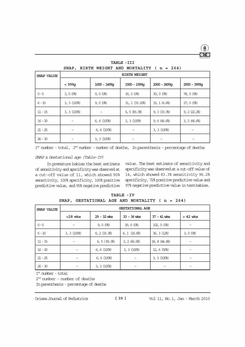
[ 39 ]Orissa Journal of Pediatrics Vol 11, No.1, Jan - March 2010
SNAP & Gestational age (Table-IV)
In premature babies the best estimateof sensitivity and specificity was observed ata cut-off value of 11, which showed 90%sensitivity, 100% specificity, 100% positivepredictive value, and 95% negative predictive
value. The best estimate of sensitivity andspecificity was observed at a cut-off value of14, which showed 83.3% sensitivity 96.2%specificity, 72% positive predictive value and97% negative predictive value in term babies.
TABLE -IIISNAP, BIRTH WEIGHT AND MORTALITY ( n = 264)
SNAP VALUE BIRTH WEIGHT
< 999g 1000 - 1499g 1500 - 1999g 2000 - 2499g 2500 - 3999g
0 - 5 3, 0 (0%) 9, 0 (0%) 30, 0 (0%) 30, 0 (0%) 78, 0 (0%)
6 - 10 3, 3 (100%) 9, 0 (0%) 31, 1 (33.32%) 15, 1 (6.6%) 27, 0 (0%)
11 - 15 3, 3 (100%) – 6, 5 (83.3%) 9, 3 (33.3%) 9, 2 (22.2%)
16 - 20 – 6, 6 (100%) 3, 3 (100%) 9, 6 (66.6%) 3, 2 (66.6%)
21 - 25 – 6, 6 (100%) – 3, 3 (100%) –
26 - 30 – 3, 3 (100%) – – –
1st number - total, 2nd number - number of deaths, In parenthesis - percentage of deaths
TABLE -IVSNAP, GESTATIONAL AGE AND MORTALITY ( n = 264)
SNAP VALUE GESTATIONAL AGE
<28 wks 29 - 32 wks 33 - 36 wks 37 - 41 wks > 42 wks
0 - 5 – 9, 0 (0%) 39, 0 (0%) 102, 0 (0%) –
6 - 10 3, 3 (100%) 6, 2 (33.3%) 6, 1 (16.6%) 36, 3 (12%) 3, 0 (0%)
11 - 15 – 6, 5 ( 83.3%) 3, 2 (66.6%) 18, 8 (44.4%) –
16 - 20 – 6, 6 (100%) 3, 3 (100%) 12, 6 (50%) –
21 - 25 – 6, 6 (100%) – 3, 3 (100%) –
26 - 30 – 3, 3 (100%) – – –
1st number - total2nd number - number of deathsIn parenthesis - percentage of deaths

[ 40 ]Orissa Journal of Pediatrics Vol 11, No.1, Jan - March 2010
SNAP & Morbid conditions (Table-V)
With SNAP values between 6 and 10
the mortality was highest in RDS group
(100%) followed by HIE III (50%) and
SEPSIS group with 25%. In the score range
of 11 to 15 there was no mortality in HIE I
SNAP & ICU stay (Table-VI)
The median length of stay in ICU of
surviving infants was least (4days) with score
below 5, and MLOS steadily increased as the
score increased, stay being 8 days above the
score of 16. The minimum duration of ICU
stay for any surviving infant was 3 days and
and HIE II but 2/3rd infants died in SEPSIS
and MAS whereas all died in HIE III group.
With SNAP values in 16 to 20 ranges 3/4th of
SEPSIS cases died and all infants in HIE III
and MAS died. Beyond 21 score there there
was 100% mortality in SEPSIS and HIE III
groups.
the maximum duration was 12 days. The
median length of stay in ICU of dying infants
was 5 days with score between 6 and 10,
and MLOS remained 4 days above the score
of 11. The minimum duration of ICU stay for
any dying infant was 3 days and the
maximum duration was 9 days in our study
group.
TABLE - VSNAP, MORBID CONDITIONS AND MORTALITY ( n = 264)
SNAPVALUE
MORBID CONDITIONS
RDS SEPSIS HIE-I HIE - II HIE - III MAS
0 - 5 3, 0 (O%) 72, 0 (0%) 12, 0 (0%) 30, 0 (0%) 15, 0 (0%) 18, 0 (0%)
6 - 10 3, 3 (100%) 12, 3 (25%) 3, 0 (0%) 18.0 (0%) 6, 3 (50%) 12, 0 (0%)
11 - 15 3, 3 (100%) 6, 4 (66.6%) 3, 0 (0%) 6, 0 (0%) 6, 6 (100%) 3, 2 (66.6%)
16 - 20 – 12, 9 (75%) – 3, 0 (0%) 3, 3 (100%) 3, 3 (100%)
21 - 25 – 6, 6 (100%) – – 3, 3 (100%) –
26 - 30 – 3, 3 (100%) – – – –
1st number - total2nd number - number of deathsIn parenthesis - percentage of deaths
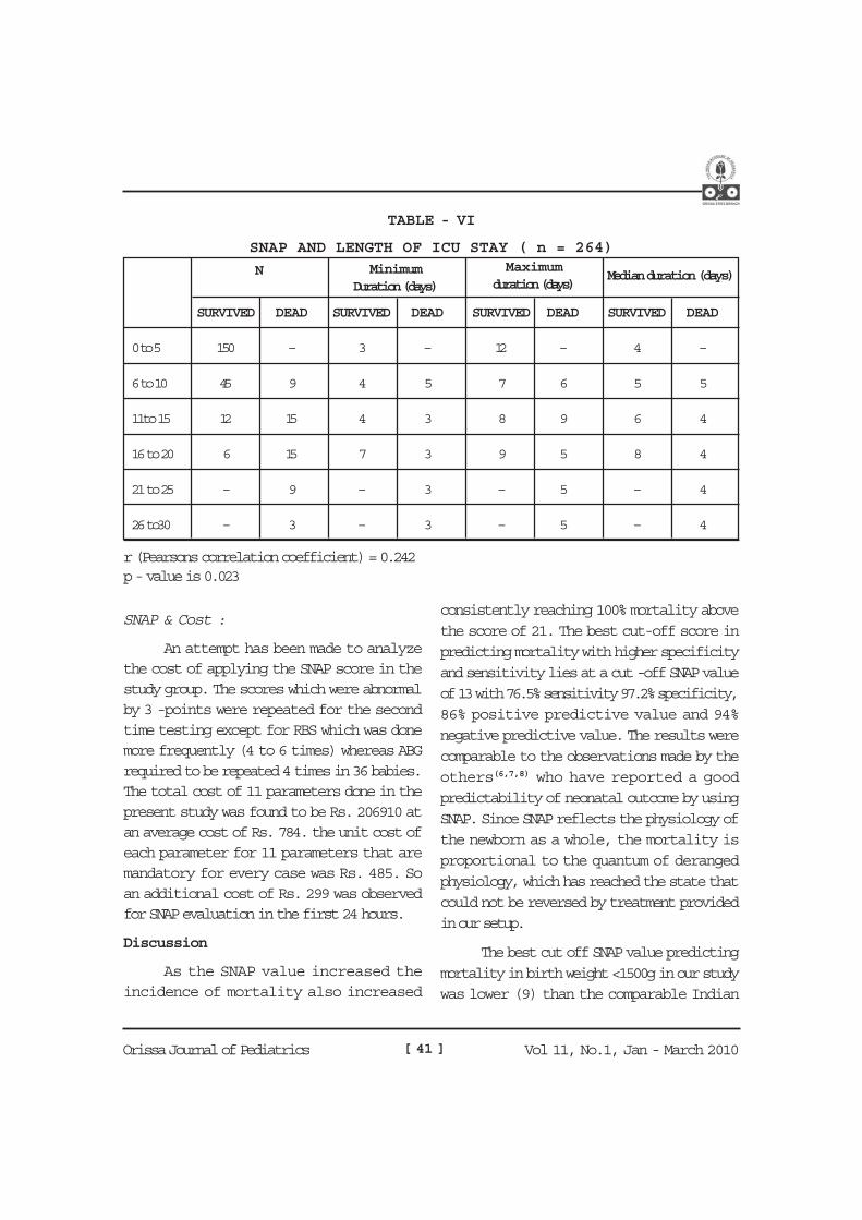
[ 41 ]Orissa Journal of Pediatrics Vol 11, No.1, Jan - March 2010
SNAP & Cost :
An attempt has been made to analyzethe cost of applying the SNAP score in thestudy group. The scores which were abnormalby 3 -points were repeated for the secondtime testing except for RBS which was donemore frequently (4 to 6 times) whereas ABGrequired to be repeated 4 times in 36 babies.The total cost of 11 parameters done in thepresent study was found to be Rs. 206910 atan average cost of Rs. 784. the unit cost ofeach parameter for 11 parameters that aremandatory for every case was Rs. 485. Soan additional cost of Rs. 299 was observedfor SNAP evaluation in the first 24 hours.
Discussion
As the SNAP value increased theincidence of mortality also increased
TABLE - VI
SNAP AND LENGTH OF ICU STAY ( n = 264)
N
SURVIVED DEAD SURVIVED DEAD SURVIVED DEAD SURVIVED DEAD
0 to 5 150 – 3 – 12 – 4 –
6 to 10 45 9 4 5 7 6 5 5
11to 15 12 15 4 3 8 9 6 4
16 to 20 6 15 7 3 9 5 8 4
21 to 25 – 9 – 3 – 5 – 4
26 to30 – 3 – 3 – 5 – 4
r (Pearsons correlation coefficient) = 0.242p - value is 0.023
MinimumDuration (days)
Maximumduration (days)
Median duration (days)
consistently reaching 100% mortality abovethe score of 21. The best cut-off score inpredicting mortality with higher specificityand sensitivity lies at a cut -off SNAP valueof 13 with 76.5% sensitivity 97.2% specificity,86% positive predictive value and 94%negative predictive value. The results werecomparable to the observations made by theothers(6,7,8) who have reported a goodpredictability of neonatal outcome by usingSNAP. Since SNAP reflects the physiology ofthe newborn as a whole, the mortality isproportional to the quantum of derangedphysiology, which has reached the state thatcould not be reversed by treatment providedin our setup.
The best cut off SNAP value predictingmortality in birth weight <1500g in our studywas lower (9) than the comparable Indian
[ 42 ]Orissa Journal of Pediatrics Vol 11, No.1, Jan - March 2010
studies (P. P. Maiya et al(7) had the best cut-
off score of 10) whereas above 1500 g the
best cut off score was 15 in both the studies.
This difference in SNAP value was due to
the absence of adequate nursing care, better
facilities to manage these VLBW babies. The
low birth weight by itself confers higher
mortality to these sick newborns as shown
by the low SNAP values with higher
mortality.(9)
The best cut off SNAP value predicting
mortality in preterm in our study was lower
(11) than other comparable Indian studies
(P.P.Maiya et al had the best cut-off score of
15) whereas in term babies the best cut off
score was 15 in the author’s studies and 14
in our study. This difference signifies the better
available facilities, efficient care of preterm
babies in those setting compared to our
NICU.
The outcome in our work with respect
to sepsis cases and HIE II was similar with
other comparable Indian studies(7) but in
other morbid condition, the mortality
increases with increase in SNAP values in
both the studies.
The duration of ICU stay of dead
babies in different SNAP ranges. With lower
SNAP values between 6-10 the MLOS was 5
days above the score of 11 with range of 3 to
9 days. There is no comparable literature for
length of ICU stay of dead babies. However
the shorter of ICU stay of dead babies shows
that our early intervention is probably not
adequate and requires more extensive
analysis to arrive at a conclusion.
Benefit accrued out of SNAP were
1. Alerting the doctors and nurses for more
aggressive management.
2. Higher rate of survival (213 cases) as
against mortality of 51 cases in a ratio of
4.2:1.
3. A high risk of mortality of 74% was
observed with a score above 13. In this
high-risk group it was possible to rescue
the remaining 26% of babies because of
increased alertness signaled by SNAP.
Similarly below a score of 13 there was
7% mortality and was due to
complication during the course of illness,
which could not be predicted by the initial
SNAP.
4. Because of the significant predictive value
observed in prognosticating mortality risk
and duration of ICU stay, the cost of
treatment required can be explained to
the parents with a more certainty.
5. SNAP can be used effectively for research
purposes and for comparison of efficiency
in management in different ICU stratifying
the newborn infants into various score
groups, randomizing the cases and
ultimately relating to the outcome.
6. The overall cost benefit analysis shows
that additional expenditure of Rs. 300 is
very insignificant in terms of the life saved,
minimizing the parental anxiety and
tension and a constant inspiration to
improve the ICU management at par with
better ICU’s in the country.

[ 43 ]Orissa Journal of Pediatrics Vol 11, No.1, Jan - March 2010
7. SNAP is a mandatory evaluation of
function of the organ system in a sick baby
as against the traditional practice on the
presenting features. This approach leads
to better homeostasis by effective
supportive management with better
outcome.
Conclusion :
SNAP score is a good measure of
severity of illness irrespective of differences
in the gestational age, birth weight and
various morbid conditions. When it is applied
on admissions or during the first 24 hours it
gives good information of the severity of
referral cases received in ICU. When SNAP
score alone is applied to measure the risk of
mortality it is found to have a linear
relationship i.e., with increasing score the
mortality also increases. A cut-off value of 13
has 76.2% sensitivity, 97.2% speciicity and
84.9% positive predictive value beyond which
the mortality increases exponentially. But
within the same range of birth weight of
gestational age the SNAP bears a linear
relation with mortality. SNAP is a mandatory
evalution of function of all the organ system
in a sick baby as against the traditional
practice on concentrating on the presenting
features. This approach leads to better
homeostasis by effective supportive
management with better outcome.
References :
1. Rautonen J, Makela A, Boyd H, etal; CRIB
& SNAP; assessing the risk of death for
preterm neonates, Lancet, 1994; 343: 1272
- 1273
2. Parry GT, TuckerJ, Tarnow- Mordi W; CRIB-
II an update of the Clinical risk index for babies
score, Lancet, 2003; 361: 1789 - 1791.
3. Gray JE, Richardson DK, Mc Cormick MC,
etal; Neonatal Therapeutic Intervention
Scoring System, a therapy based severity of
illness index; Pediatrics, 1992; 90 : 561- 567.
4. Maier RF, Casper- Karweck UE, Gravel EL
etal, A comparison of two mortality risk scores
for very low birth weight infants, clinical risk
index for babies & Berlin Score; Intens Care
Med 2002 ; 28: 1332- 1335.
5. Horbar JD, Onstad L, Wright E, etal;
Predicting mortality risk for infants weighing
501 to 1500 gms at birth; a National Institute
of Health Neonatal
Research Network Report, Crit Care Med
1993, 21 : 12-183.
6. Richardson DK, GrayJE, Mc Cormick MC,
etal, Scorefor Neonatal Physiology: a
physiological severity index for neonatal
intensive-care, Pediatrics 1993; 91 : 617-623.
7. P.P. Maiya etal; Role of Score for neonatal
acute physiology in predicting neonatal
mortality, Indian Journal of Pediatrics, Vol-
68 - Sep, 2001.
8. Eleni Petridox etal; Outcome prediction in
Greek Neontal Intensive Care units using a
score for neonatal acute physiology (SNAP),
Pediatrics, 1998 : Jun, 101(6) : 1070
9. Richardson DK, Phibbs CS, Gray JE, Mc
Cormick MC, Workman Daniels K, Goldmann
DA; Birthweight and illness severity,
independent predictors of neonatal mortality,
Pediatrics, 1993; 91 : 969 - 975.

[ 44 ]Orissa Journal of Pediatrics Vol 11, No.1, Jan - March 2010
Introduction :Very often than not, abdomen in
children has been an enigma to the attending
physician. The term by and large, is
descriptive in nature than a diagnostic
terminology. While all the presenting
symptoms do not always fit into a particular
organic entity as per book description, as
many as 13% are of functional in nature. On
the other hand, few of the kids do need life
saving emergency interventions. Hence it is
very important to recognize the red flag signs
in time. With drastic change in our social
milieu and rapid introduction of advanced
diagnostic technologies, the so called
functional group is shrinking further. Also
important is to realize the peculiar path
physiology of abdominal pain in case of
children, so different from adults! Hence this
review:
Seeking an immediate diagnosis for
abdominal pain, particularly in children, is like
exploring the so called Pandora’s Box. A
working diagnosis is easy if our approach is
rational but often vexed otherwise in our busy
office practice setting. It deserves its due, in
terms of time and patience on part of the care
giver as well as parents. Unlike adults and
grown-up children, abdominal and pelvic pain
presented differently in infants:
THE ENIGMA OF PAIN ABDOMEN IN CHILDRENNimain C. Mohanty
1. Screaming or persistent crying
2. Restlessness
3. Irritability
4. Squirming
5. Grunting respiration
6. Flexion of thighs over abdomen or,
7. Refusal to feed
Mostly we encounter children presenting
abdominal pain around umbilicus or in the
midline. It has got an embryogenic basis:
Most primitive viscera develop as mid-line
structures, having bilateral and symmetric
innervation.
Location of pain is perceived at a level
where afferent nerves enter spinal cord
segments.
Referred pain (Hyperalgesia) is felt over
cutaneous dermatomes supplied by same
neural segment e.g.- Pancreatic pain over
mid-back and inter-scapular (T5-9)
region; hepato-biliary origin pain being
referred to right sub-scapular region.
Classification of pain abdomen :
Classifying pain abdomen on the basis of its
location depends on the back-ground of
development, innervation and pain
perception.

[ 45 ]Orissa Journal of Pediatrics Vol 11, No.1, Jan - March 2010
Classifying pain abdomen on the basis of itsorigin
Visceral: Midline discomfort, poorly localised,accompay nausea, emesis, diphoresis.
Parietal: More intense, well localised,aggravated with movement.
Referred: To same dermatome represented byaffected viscera.
In combinations of any of the three above.
To further understand the very basis of theabove, we must realize the existence of myo-enteric nervous system (MNS) which of latehas generated much interest.
Visceral pain:
Nociceptors are present through-out wallsof abdominal viscera and supportingstructures.
Once generated, pain conducted throughsmall, un-myelinated slow conducting C-fibres; whose termination occurs 4-5spinal segments above. Therefore, theresulting pain is less characterised andpoorly localised
GI mucosa is insensitive to pricking,
cutting, crushing and therefore do not
generate impulses to such stimuli. Hence,
mucosal biopsy, polypectomy are
painless.
Parietal pain
Pain from superficial structures e.g. -
peritoneum is conducted by both small,
slow ‘C’ fibres and large, thinly myelinated,
rapidly conducting ‘A’ fibres as wel.
A and delta fibres in parietal & supporting
structures also respond to tactile, thermal,
chemical discriminative stimuli e.g.-
location and intensity.
Patient lies still as movement exaggerates
parietal pain whereas restless in visceral
pain which is vague and poorly localised.
Therefore, pain in appendicitis is initially
perceived high up, may be around umbilicus
initially but gets localised to right flank
(Macburny’s point) when local peritonitis sets
in.
PrimitiveStructures Segments Localisation
Foregut: Oesophagus, stomach, T5-T9 Xiphi-umbilicus
duodenum, liver, GB, pancreas (Midline)
Midgut: Small intestine, appendix, T8-L1Periumbilical
ascending & proximal 2/3 of
tansverse colon
Hindgut: Distal 3rd of trasverse colon, T11-L1 Umb-Symphisis
Descending, recto-sigmoid col pubis (Midline)
Nephrogenic cord: Kidneys, ureters, T10-L1 Lateralised
(Mesoderm) ovaries, tubes

[ 46 ]Orissa Journal of Pediatrics Vol 11, No.1, Jan - March 2010
Acute pain abdomen in children:
Deserves prompt evaluation to rule out any
surgical condition for immediate intervention.
Any of the following should arouse suspicion
(Red flag signs):
1. Sudden distension,
2. Bilious vomiting,
3. Exaggerated or absence of bowel sounds,
4. Guarding or rebound tenderness.
Classification basing upon etiology:
Etiology wise classification and detailed
discussion are exhaustive and beyond the
scope in this article. Following causes are
shown to complete the list.
I. Intra-abdominal Causes
A. GIT & Mesentery
1. Infantile Colic
2. APD & H. Pylori Infection
3. Zolinger Ellison Syndrome
4. Dietary indiscretion
5. Appendicitis
6. Intussusception
7. Malrotation
8. Volvulus
9. Intra-abdominal hernia
10. Meckel’s diverticulum
11. Mesenteric cyst
12. Duplication
13. Intestinal Polyp
14. Incarcenated Hernia
15. Intestinal obstruction
16. Constipation
17. Sigmoid Volvulus
18. Parasites
19. Bacterial enteritis
20. Aerophagia
21. Ulcerative colitis
22. Crohn’s disease
23. Food allergy, CMPA
24. Lactose intolerance
25. Cystic fibrosis
26. Hereditary angio-oedema
27. Mesenteritis
B. Urinary Tract
1. Obstruction
2. Systemic haemorrhagic diseases, Renalcalculus
3. Hyper-calciuria
4. A G N
5. PUJ obstruction
C. Liver & Gall Bladder
1. Viral hepatitis
2. Cholecystitis
3. Cholelithiasis
4. Passive congestion of liver
5. Sickle cell crisis
6. Choledochal cyst
7. Hydrops of Gall Bladder
8. Hepatic SOL
9. Budd-Chiari Syndrome
D. Spleen
1. Traumatic rupture
2. Splenomegaly
3. Congestive splenomegaly

[ 47 ]Orissa Journal of Pediatrics Vol 11, No.1, Jan - March 2010
F. Pancreas
1. Acute pancreatitis
2. Congenital fibrosis
3. Pancreaticpseudocyst
G. Ovaries & uterus
1. Torsion, cyst or tumor
2. Hematocolpus
3. Dysmenorrhea
4. Pelvic inflammatory disease
5. Endometriosis
6. Ectopic pregnancy
H. Lymph nodes
1. Mesenteric lymphadenitis
2. Iliac adenitis
3. Lymphoma
4. TB
I. Bacterial peritonitis
J. Mesenteric vein thrombosis
K. Pelvic osteomyelitis
L. Superior mesenteric artery syndrome
II. Extra-abdominal
A. Rt lower-lobe pneumonia
B. Cardiac origin
1. RHD
2. Pericarditis
3. Congenital endocardial fibroelastosis
C. CNS, spinal cord and spine
1. Abdominal epilepsy
2. SOL in brain
3. Herpes zoster
4. TB spondylitis
5. Spinal cord tumour in dorso-lumbarregion
6. Discitis, collapse of vertebra
D. Blood
1. Haemolytic anemias
2. Sickle cell crisis
3. Leukemia
4. HSP
5. Hemophilia
6. Acute infectious mononucleosis
E. Metabolic
1. Lead
2. Hyperparathyroidism
3. Addison’s disease
4. DKA
5. Hypoglycemia
6. Hyperlipoproteinemia
7. Acute porphyria
8. Hereditary angio-oedema
9. Familial paroxysmal polyserositis
F. Miscellaneous
1. Periarteritis nodosa
2. Arachnidism
3. Epidemic myalgia
4. Rheumatoid arthritis
5. Abdominal stitch
6. Mesenteric arteritis
III. Recurrent and Chronic Pain
Abdomen:
These terms are descriptive terms than
diagnostic; often used interchangeably.
Defined as pain occurring at least 3 episodes
over last 3 months (Apley,1958), severe
enough to affect daily activities. Based on his
criteria, out of 1000 school children studied,

[ 48 ]Orissa Journal of Pediatrics Vol 11, No.1, Jan - March 2010
13-15% school children complained on daily
to weekly basis, affecting their routine. Girls
reported more frequently than boys. All such
children do need evaluation although most do
not have underlying disease. No organic
pathology reported in 90% of such cases those
days with the available investigations. The
Rome III criteria of paediatric RAP includes
abdominal pain of more than 8 weeks
duration, as a modification over Rome II
criteria (Rasquin,2006; Hyman,2006).
History important.
Better be obtained directly focusing on:
Timing, frequency, location, quality, assoc
symptoms e.g. diaphoresis, nausea,
diziness, stool frequency, completeness of
evacuation.
Precipitating factors - food, viral g/e, stress,
anxiety, medication, menses, wt loss.
Growth failure, delayed puberty, fever,
rash, joint pain.
Family h/o IBD, APD, epilepsy, peer
relations. RAP among 1st degree relatives.
Red flag symptoms / signs indicatingsignificant pathology:
Involuntary weight loss, reduced growth
Pain awakening the child from sleep at
night
Significant vomiting, chronic / severe
diarrhea, unexplained fever
Clubbing, rash, arthritis, P/R findings /
bleeding PR or haematemesis
Localised pain away from umbilicus
Family h/o IBD, organomegaly (Liver,
spleen), mass, tenderness, ascites, bruits.
Investigations:
Stool, Urine, CBC, ESR, CRP, Urea, S/E,
LFT, screening for celiac, UTI
Predictive value of investigations;
(AAP Technical report, 2005)
There is insufficient evidence in support
of blood examination to look for organic
pathology, even in presence of red-flag
signals.
Ultrasonography, pH probe studies and
endoscopy have little additional
diagnostic yield in absence of red-flag
signals.
H. pylori infestation is as common inchildren with RAP as those without.
Management:
Most do not need medication.
Reassure that there is no evidence of
underlying pathology.
Those with persistent symptoms may need
multi-disciplinary approach, also
involving family.
Possible placebo effect may be borne in
mind.
Medications on selective basis.
Classification of basing upon organic or non-organic (Functional):
Organic: Discrete cause identified in 5 to 10%of cases; Yield improved to 33% applying
modern tools. Recognisation of conditionse.g.- IBD, COH MAS, APD, Celiac improved
the yield. Intestinal worm / parasite
infestations, particularly Ascariasis or
Giardiasis; constipation, GERD are commonin the Indian sub-continent.
[ 49 ]Orissa Journal of Pediatrics Vol 11, No.1, Jan - March 2010
Non-organic (Functional):
That can’t be explained on basis of structuralor biochemical abnormalities with presentlyavailable tools, but regarded as genuine. It isnot synonymous with psychogenic or‘Imaginary’ pain. ‘Functional’ must be labeledonly by exclusion. It is challenging and mustbe appreciated as an abnormality in ‘Brain-gut’ interaction and not merely as amanifestation of symptoms in somepsychogenic illness. The Abnormality couldbe any where at levels of gut, spinal affer-ents, central autonomic relay system, brain
Functional GI Disorders (FGID, ROMEII classification)
G2. Abdominal pain
G.2.a.1. Ulcer like dyspepsia
G.2.a.2. Dysmotility like dyspepsia
G.2.a.3. Unspecified dyspepsia
G.2.b. IBS
G.2.c. Functional abdominal painsyndrome
G.2.d. Abdominal migraine /epilepsy
G.2.e. Aerophagia
G.2a. Functional Dyspepsia:
Usual Presentation: Child complainsepigastric discomfort immediately after food,often awaking patient at night. Also hasnausea, fullness of abdomen; restricting foodintake. No h/o retro-sternal pain, vomiting,regurgitation, pain on swallowing. Is afraidto eat and misses school bus for morningsymptoms. No weight loss, growthmaintained. No nocturnal symptoms., no
vomiting, regurgitation or haematemesis.
Differential Diagnosis :
Unlike to be Acid related (Gastritis,
duodenitis, oesophagitis) since there is no
pain, nausea, heart burn related to eating,
night time awakening. Neither could be due
to H. Pylori (Also can be present without any
specific symptom), Eosinophilic oesophagitis,
Gastro-enteritis (Usually associated with
atopy, abdominal pain, dysphagia, diarrhoea,
eosinophilia), Crohn’s, Coeliac (Diarrhoea, wt
loss, pain, night awakening, other
constitutional symptoms; presence of all these
symptoms not a must also.)
Diagnosis:
Symptoms consistent with dyspepsia >12
wks as per Rome II criteria.
Not necessarily consecutive in preceding
12 months of pain / discomfort above
umbilicus (As per ROME III: H2a criteria,
at least once per week for 2 months)
Common in 50% of referred children and
5-10% among school children.
No organic disease to explain, even after
UGI endoscopy.
Not exclusively relieved by defecation or
associated with altered stool consistency.
Types:
1. Ulcer like, 2. Dysmotility like, 3. Overlap
symptoms.
Investigations:
UGI endoscopy: Can be delayed in
favour of a therapeutic trial of empric
therapy, except in cases of wt loss,
vomiting, hematemesis, odynophagia,
dysphagia.

[ 50 ]Orissa Journal of Pediatrics Vol 11, No.1, Jan - March 2010
UGI Ba series only for studying GIanatomy; not good for mucosalabnormality for which mucosal biopsyrecommended.Nuclear scintigraphy: (If gastroparesis issuspected).USG (Only if biliary / pancreatic disease)H. Pylori Screening: Sero testing difficultto interpret in children, may be false +ve.Can’t be used to monitor treatment. Stableisotope urea breath test not standardisedfor children, hence endoscopic biopsy isthe gold standard.
Management:Depends on predominant symptom ofdyspepsia:
Eliminate caffeine, fats, large meals,alcohol and smoking.If symptom response unequivocal,empiric treatment with H2 blockers,occasionally followed by proton pumpinhibitors. Failure to respond orrequirement of frequent drug therapywarrants endoscopy.Prokinetics (Metclopramide) indysmotility variant. Erythrocin, a motilinreceptor antagonist, can be tried.AntiemeticsLow dose tricyclic anti-depressants (10-25 mg HS) often helpful in patients notresponding to prokinetic agents.
G.2.b. Irritable Bowel Syndrome:Usual Presentation: Pain abdomen in a gown-up child, waxing and waning, may beassociated with intermittent diarrhoea. Painoccurs almost daily over lower quadrant;worse in morning some times sharp andcrampy, forcing patient to close thighs overabdomen. There may be nausea but no
vomiting. Pain may worsen with food intake.There is a feeling of urgency to defecate, paindecreasing after defecation, yet a sense ofincomplete evacuation. Usually there is h/opain of less intensity but less often for pastfew years. O/E: Normal anthropometry anddevelopment. Child may look pale withdiaphoresis. No apparent structural ormetabolic cause. Lab reports generally withinnormal limits. As per Rome-III (H.2.b),abdominal pain recurrent abdominal pain ordiscomfort for at least 3 days in a month overlast 3 months; associated with 2 or more offollowing:1. Discomfort or pain associated with 2 or
more of these in 25% of time: (a) Improvedafter defecation, (b) Onset associated withchange in stool frequency or (c) Form(Appearance)
2. No evidence of inflammatory, anatomic,metabolic or neoplastic process to explainsymptoms.
Differential Diagnosis : MAS, Carbohydrateintolerance (Explossive, watery, with gases,peri-anal redness), Giardiasis (Diarrhoea,steatorrhea, pain, gas, bloating, weight loss),UTI (Pain lower abdomen, dysuria,haematuria; suggestive of urine exam),Crohn’s, ulcerative colitis, eosinophilic gastro-enteritis etc.Patho-physiology:Patients with functional GI disorders haveidentifiable alterations in motility andsensitivity:
Delayed transit of contrast seen inconstipation predominant IBS andincreased in diarrhoea predominant IBS.Exaggerated motility response stimulisuch as- stress and meals seen.
[ 51 ]Orissa Journal of Pediatrics Vol 11, No.1, Jan - March 2010
Visceral hyper-sensitivity due to alterationsin neuro-physiology at gut level (Myentericand mucosal plexuses involved inepithelial transport, secretion, sensationand GI motility), at spinal cord or at highercentres.Within the enteric nervous system (ENS)in gut, reflexes generated by activation of‘Intrinsic Primary Afferent Neuron’(IPANS) which increase it’s excitabilitythrough repetitive stimulations, outlastingfor hours which leads to an enhancedEPANS activity and there on to CNS.Stimuli include infection, trauma andinflammation.Inflammation of gut mucosa leads tochanges in smooth muscle activity, entericnerves and composition ofneurotransmitters.Visceral hypersensitivity results in alteredprocessing of sensory inputs at CNS level(HPA, locus ceruleus, amygdala, para-vertebral & dorsal motor nuclei); Vagus(Parasympathetic) being the primarycommunication link.Hypothalamus also involved in panic andpost- traumatic stress disorders.Hence anxiety / depression can affectperception of pain signals arising out ofgut.
ManagementValidate symptoms to patient, Placeboresponse rate is around 40%.Cut fats, caffeine, alcohol; add fibers inconstipation predominant IBS.Lactose restriction only if H2 breath test+ve.Anticholinergic therapy for diarrhoeapredominant or variable forms.Dicyclamine / hyosciamine could be tried.
Bed time low dose Anti-depressants acts
via affect neuro-transmission at
muscarinic receptor, serotonin and nor-
epinephrine reuptake.
Alosetron (Antagonists of serotonin
receptors e.g. 5HT3, 5HT4) lead to delay
bowel movements and colonic transit
time.
Tagaserod, a partial antagonist of 5HT4
receptor, decreases small bowel and
colonic transit time. It also increases
phasic activation of colon; decrease pain,
bloating and constipation.
Add-on: Cognitive behavioral therapy.
Abdominal Migraine:
Usual Presentation: Long history of
intermittent pain abdomen of acute onset.
Usually comes early in the morning, almost
every month. Child appears pale, diaphoretic,
prefers dark room. Intense pain around
umbilicus lasting for 6-12 hours,
accompanied by nausea; often vomiting. Falls
asleep at end of each episode, fine on
awaking. No symptom between episodes.
Strong family h/o migraine (65% of 1st or 2nd
order). Normal growth. No hepato-biliary,
pancreatic, metabolic, renal disease or SOL.
Diagnosis:
Abdominal migraine is present among 2% of
children as per ROME-II classification:
3 or more paroxysmal acute episodes of
intense mid-line pain lasting > 2 hours to
several days, with symptom free intervals
lasting weeks to months.
[ 52 ]Orissa Journal of Pediatrics Vol 11, No.1, Jan - March 2010
No structural GI, CNS, metabolic,
biochemical abnormality; and any 2 of
following associated features:
1. Headache
2. Photophobia
3. Family history,
4. One sided headache
Aura: Visual (blurred / restricted), sensory
(Tingling, numbness), motor (Slurring,
aphasia or paralysis)
(As per Rome III H2c: 2 or more episodes
in preceding 12 months and above plus
Paroxysmal episodes Lasting for 1 hour
or more; 2 episodes of vomiting, anorexia,
nausea, headache, photophobia, palor;
No evidence of inflammatory, anatomic,
metabolic or neoplastic process to explain
patient’s symptoms.)
Differential Diagnosis :
Anatomic abnormalities (Malrotation, web,
strictures, duplications, intussusceptions),
Genito-urinary (UTI, hydronephrosis, uretero-
pelvic junction obstruction, nephrolithiasis
where associated dysuria, hematuria,
proteinuria are usually present), ovarian cyst,
pregnancy, allergic eosinophilic oesophagitis
/ Gastro-enteritis, infections (Giardia, familial
mediterranian fever, metabolic (Diabetes,
lead, aminoacidosis showing growth failure,
developmental delay and polydipsia etc)
Management:
Diagnostic :
CBC, ESR, biochy (S/E, LFT, Metabolic),
USG, Imaging of brain if indicated
(Raised ICT), Urine RE/ME
Therapeutic: (Focus on preventing episodes)
1. Propranolol (10-20 mg BD) acts by B-
blockade of cerebral arteries. 75% children
had excellent response.
2. Ciproheptadine acts as anti-serotonin oras Ca channel blocker. 33% had excellentresponse; addl 50% had less frequent andless intense episodes.
3. Misc: Pizotifen – Serotonin receptorantagonist, Carbamazepine, Tricyclineanti-depressants.
‘Functional Pain Abdomen’Usual Presentation:Complain of pain over 3 months, diffuse, mostof the day time (Never awakened at night).Initially may develop viral infection with fever,pain, diarrhoea and vomiting along with otherfamily members. While others recover andother symptoms subside, abdominal paincontinues. May have mild nausea but novomiting, haematemesis or constipation. Lossof school days. Feels fatigued, often getsheadache. Poor scholastic performance, nosense of belonging, felt teased by friends. O/E: WNL except diffuse abdominal tenderness.Assessment:Seems to have significant organic disease,possibly some psycho-social factorscontributing. Fits to ROME-II criteria of“Generic functional Abdominal Pain” i.e.- 12weeks of continuous abdominal pain in aschool age child or adolescent; having no oronly occasional relationship with psychogenicevent; some loss of daily function, no feigning,insufficient criteria for other functional GIdisorders to explain. Deserve detailed physicaland per-abdominal examination, includinggrowth.
[ 53 ]Orissa Journal of Pediatrics Vol 11, No.1, Jan - March 2010
Rome III:(H2d) Episodic or continousabdominal pain, insufficient criteria for otherfunctional GI disorders, No evidence ofinflammatory, anatomic, metabolic orneoplastic process to explain patient’ssymptoms.Investigations:CBC, ESR, Stool, Urine RE, ME, C/S,biochemistry profile, breath H2, USG,particularly when pancreatic or hepto-biliarydisease suspected.Differential Diagnosis : Acid related, unusualamong children: Gastritis, duodenitis,oesophagitis(Pain, nausea, heart burn, related to food,night time awakening).Infection; Parasites e.g.- Giardiasis(Diarrhoea, pain, bloating, weight loss).UTI (Pain), Allergic: Eosinophilic GE(Diarrhoea, pain, atopy,Eosinophilia- related to food); Inflammatory:Crohn’s, Ulcerative colitis,Crohn’s, Coeliac, TB, Pancreatitis(Constitutional symptoms e.g. fever, arthragia,diarrhoea with or without blood, pain, wt.loss, growth failure, night awaking).Management
Reassurance of child and parentsregarding nature of functional GI
disorders and their possible link with viralailment.Dietary modifications, althoughimprovement is minimal.Anticholinergic worth trying but data donot support efficacyTricyclic anti-depressants usedoccasionally.Attention to psycho-social aspects(Increased nociceptive signals from
Gut resulting from previous viral infection,in a child having mild depression or anxiety,can lead to continued GI discomfort)Functional Abdominal pain SyndromeMust include childhood functional abdominalpain at least 25% of the time and 1 or moreof the following (Rome III H2d1):1. Some loss of daily functioning2. Additional somatic symptoms e.g.-
headache, limb pain, or difficulty insleeping.
Aerophagia :Usual Presentation:Abdominal distension, pain, flatulence,intermittent distention, marked as dayprogresses, does’t want to eat. Normal bowelmovement. Passes large amount of gas perrectum, loud belching. O/E: Abdominaldistension but soft, no mass, bowel sounds-present. ROME-II described as: 24 wks historywhich need not be consecutive in preceding12 months with 2 or more of the followingsymptoms / signs (Mostly by observation)(Rome III H1c):1. Air swallowing, 2. Abdominal distensionby intra-lumen air,3. Repeated belching and / or 4. Increased
flatus; Otherwise healthyDifferential Diagnosis :Aerophagia, Coeliac, Intestinal pseudo-obstruction, Bacterial over load of smallbowel, Hirschprung’s disease, CHO and othermal-absorption disorders and Cystic fibrosis.Diagnosis :
Breath hydrogen test to t/o CHO MASUSG if difficult to palpate for massReassuranceIf associated with stressful events oranxiety, identify the factor responsibleand suitable behavior modification /psychotherapy
[ 54 ]Orissa Journal of Pediatrics Vol 11, No.1, Jan - March 2010
Avoid chewing gums, carbonated drinks
The ‘Infantile Colic’ has been described under
functional category. It is common among 10
to 15% of infants. GER, CMPA, hyper-
sensitivity to colonic distension, poor maternal
bonding etc are attributed. Children are said
to out-grow the problem by 5 to 6 months of
age. Parents need to be reassured accordingly
after excluding of all organic conditions.
Conclusion :
Functional pain abdomen can occur as a
well defined clinical entity or as
part of a poorly defined clinical syndrome.
Clinician is obliged to rule out organic
conditions in all cases. Appropriate
to take a bio-psycho-social approach.
Understand close interaction of gut and
brain; allowing child, family and physician
to address pain at multiple levels.
Further research needed to understand
exact biochemical process in ENS and
CNS; thereby expanding therapeutic
horizons.
ROME III CRITERIA FOR FUNCTIONAL
ABDOMINAL PAIN IN CHILDREN:
H2a. Functional dyspepsia:
Must include-
1. Persistent recurrent pain or discomfort
centered in upper abdomen (Above
umbilicus)
2. Not relieved by defecation or associated
with onset of a change in stool frequency
or stool form (As in IBS).
3. No evidence of an inflammatory,
anatomic, metabolic, or neoplastic
process that explains the subject’s
symptoms.
H1c. Aerophagia*Must include at least 2 of following-1. Air swallowing2. Abdominal distension due to intra-luminal
air3. Repetitive belching and /or increased
flatus.H2b. Irritable Bowel syndrome*Must include all of following-1. Abdominal discomfort or pain associated
with 2 or more of following at least 25%of time:(a) Improved with defecation(b) Onset associated with change in stool
frequency.(c) Onset associated with change in stool
form.2. No evidence of an inflammatory,
anatomic, metabolic, or neoplasticprocess that explains the subject’ssymptoms.
H2c. Abdominal migraine#;Must include all of following-1. Paroxysmal episodes of intense acute
peri-umbilical pain lasting for an 1 houror more.
2. Intervening periods of usual health lasingfor weeks and months
3. Pain interferes with normal activities4. Pain associated with 2 or more of
following: Anorexia, nausea, vomiting,headache, photophobia, pallor.
4. No evidence of an inflammatory,anatomic, metabolic, or neoplasticprocess that explains the subject’ssymptoms.
H2d. Childhood Functional AbdominalPain*:
[ 55 ]Orissa Journal of Pediatrics Vol 11, No.1, Jan - March 2010
Must include all of following-1. Episodic or continuous pain2. Insufficient criteria for other functional GI
disorders3. No evidence of an inflammatory,
anatomic, metabolic, or neoplasticprocess that explains the subject’ssymptoms.
H2d1. Childhood FunctionalAbdominal Pain syndrome*:Must include Childhood FunctionalAbdominal pain at least 25% of time and 1or more of following-1. Some loss of daily functioning2. Additional somatic symptoms such as
headache, limb pain or sleeping difficulty.* Symptoms at least once per week, forminimum 2 months before diagnosis;# On 2 or more occasions in preceding 12months.Points to Remember :1. We must take adequate time in history
taking.2. let’s keep in mind the peculiar patho-
physiology in terms of ENS in case ofchildren
3. The red-flag signs must always be bornein mind for early detection of any surgicalcondition and prompt intervention, whereever required.
4. we must minimize investigations to thebasics first e.g.- CBC, Stool, urine andUSG abdomen at the most, addition ofadvanced modalities can be made onspecific indication as such additions donot increase the diagnostic yieldimpressively when generalized.
5. Follow-up is very important.
References1. Apley J et al: Recurrent abdominal pain: A field
survey of 1000 school children. Arch dis Child1958; 33:165-170
2. Caplan A et al. Validation of pediatric Rome IIcriteria for FGID using questionnaire onpediatric GI symptoms. J PediatricGastroenterol and Nutrition, 2005; 100: 2079-2082.
3. Aziz Q et al. Brain-Gut axis in health anddisease. Gastroenterology; 1998: 114:559-578.
4. Donna K. et al: Recurrent abdominal pain inchildren: Pediatric GE and nutrition. PCNA;2006: 49;1:53-7
5. Lori Mahajan and Robert W: Chr Abd pain ofChildhood and Adolescence in Ped GE; 2005:03-13.
6. American Academy of Pediatrics sub-committeeon Chronic Pain Abdomen in children.Pediatrics, 2005; 115: 812-815
7. Huang RC et al. Prevalence and pattern ofchildhood abdominal pain in an Australian Genpractice. J Ped Child Health 2000;36:349-353.
8. NASPGHAN clinical report on chronicabdominal pain in children: Jl of ped gastroand nutrition; 40: 245248, march 2005.
9. Hyman PE et al. Childhood functional GIdisorders. Gastroenterol, 2006; 130:1519-1526.
10. Rasquin WA et al. Childhood functional GIdisorders.Gastroenterol, 2006; 130: 1527-1537.
11. Brenner AR, Sandhu BK. Recurrent painabdomen in childhood: The functional Element:Indian Pediatrics,2009; 46:375-379.
12. Devanarayana NM et al. Recurrent AbdominalPain in Children. Indian Pediatrics, 2009;46:389-399.
13. Yip WC et al. Value of Abdominal sonographyin the assessment of children with abdominalpain. J WC et al.Clin Ultrasound. 1998;26:397400.
14. Huertas – Ceballos A et al. Pharmacologicalinterventions for RAP in childhood. Cochrane
Database Syst Rev 2004; 1: CD 003017.
[ 56 ]Orissa Journal of Pediatrics Vol 11, No.1, Jan - March 2010
Introduction
Wilson’s disease is an inborn error of
metabolism characterized by toxic
accumulation of copper in liver, brain, cornea
and other tissue. It occurs world wide with
an estimated prevalence of 1 in 30-50.000
and is one of the leading causes of chronic
liver disease in Indian children. (1)As it can
have a varied clinical presentation a high
index of suspicion is necessary for diagnosis.
We had recently encountered a case where
the initial manifestation was arthropathy
simulating collagen vascular disease and was
treated as a case of SLE in another
institution.
Case Report
A 13 Year old female child presented
with history of joint swelling and restriction
of movement particularly involving the small
joints for six month. She had gradual
deterioration of scholastic performance for the
ARTHROPATHY AS A PRESENTING SYMPTOM OFWILSON’S DISEASE -A CASE REPORT
Rachita Sarangi *D K Dash*, B K Rath *, S S Mohapatra **
same duration followed by dysarthria ,
dystonia and bizzare behaviour for four
months. Fifteen days prior to hospitalisation
she had progressive abdominal distention,
swelling of whole body and breathlessness.
She was the 1st born female child of
nonconsanginous marriage and enjoyed a
sound health until six month prior to
hospitalization. Her growth and development
was at par. There was no history of jaundice,
blood transfusion and urinary symptoms.
On examination she was found to have
pallor, anasarca, genu valgum, double
malleolus, and rachitic rosary with knock knee
suggestive of rickets which was well supported
by radiological findings. Abdominal
examination revealed ascites, splenomegaly,
Prominent veins over the abdomen (features
of portal hypertension) with right sided pleural
effusion.
* Department of Paediatrics
** Department of Radiodiagnosis & imaging
IMS & Sum Hospital, Bhubaneswar

[ 57 ]Orissa Journal of Pediatrics Vol 11, No.1, Jan - March 2010
1. K F Ring
2. Widening of the wrists
3. Features of Ricket
Investigations revealed anemia (Hb=8.4gm
%), deranged liver function test in the form
of total billirubin 1.3mg/dl, direct billirubin 0.5
mg/dl, SGOT = 67 IU/L, SGPT = 60 IU/l,
Alkaline phosphatase=497 IU/L,serum
albumin = 1.7gm/dl, PT=35.3 Sec with INR
= 2.79 . Serum ceruloplasmin was low
(0.09gm/L) and 24 hr urinary copper
measured 131.2 microgram/L. She had
negative ANA, ASMA, dsDNA value and
antigen study against Hepatitis B also showed
negative result. Complete KF ring was found
in both eyes on slit lamp examination.
Discussion:
Wilson’s disease is an autosomal recessive
disorder characterized by degenerative
changes in brain, liver disease with Kayser
Fleischer ring in the cornea. The gene
responsible for this disorder is located in
chromosome 13 at q14-q21 (2). Most patients
with Wilson’s disease are diagnosed between
1st to 4th decades of life, (3) although the clinical
disease rarely presents before the age of five. (4)
According to Sternlieb et al(5) the initial
manifestations are hepatic 43%, Neurologic
34%, Psychiatric 10%, bony manifestation
12% and renal 1 %. A peculiar manifestation
of Wilsons disease in India is

[ 58 ]Orissa Journal of Pediatrics Vol 11, No.1, Jan - March 2010
“Osseomuscular” type with bony deformity
suggestive of resistant ricket (6) .The initial
presentation with arthropathy was 8% and
the mean age at diagnosis was 10.8 (±3.4yr)
with survival 90% . According to Walche et
al the most common radiological abnormality
was a generalized increase of radiolucency
i.e skeletal demineralization followed by
premature osteoarthrosis. Changes in the
spine were common and included
osteochondritis, reduction in interverterbral
joint space and osteoarthrosis. Other bony
changes include fluffy irregularity of femoral
trochanter, Osteochondritis dissecans of
knees, Osteophytic protrusion at bony ends
and Osteophytes at joint margin. (7)
There is no gold standard for diagnosis
of Wilson’s disease. It is based on clinical
and laboratory investigations like urinary
Cu>100 microgram /24hr, hepatic Cu >250
microgram/dl, serum cerruloplasmin<20
microgram/dl and presence of KF Ring.(8)
References:
1. Pandit AN, Bavdekar AR, Bhave SA, Wilson’s
disease. Indian J. Paediatrics 2002;69:785-
91.
2. HouwenR.H et al. H714Q Mutation in
Wilson’s disease is associated with late
neurological presentation. Journal Med Genet
1995;32:480-482
3. Schoen RE, Sterilieb I. Clinical aspects of
Wilson’s disease. American J. Gastroenterol
1990; 85 :1453-57
4. Sternlieb I. diagnosis pf wilson’s disease.
Gastroenterology 1978;74:787-789
5. Sternlieb I. Scheinberg IH : Wilson’s disease.
In Wright R and others, editor : liver billiary
disease : Pathophysiology, diagnosis&
management, LONDON, 1979, WB Saundar
S, 774.
6. Bavdekar AR . Therapeutic and management
challenges in Wilson’s disease. Journal of
Gastroenterology and hepatology, 2004 ;19:
391-395
7. J M Walshe, D N Golding et al. Arthropathy
of Wilson’s disease.study of clinical and
radiological features in 32 patients.Ann
Rheum dis.1977 April; 36(2):99-111
8. Gow PJ etal. Diagnosis of Wilson’s disease:An
experience over three decades.Gut
2000;46;415-419s