of very low density lipoproteins: immunologic ... · PDF fileIntrahepatic assembly of very low...
12
Intrahepatic assembly of very low density lipoproteins: immunologic characterization of apolipoprotein B in lipoproteins and hepatic membrane fractions and its intracellular distribution Roger A. Davis,',* Annamarie B. Prewett,* Daniel C. F. Chan,t James J. Thompson,** Roy A. Borchardt,tt and William R. Gallaher** Cell and Molecular Biology Unit,' Hepatobiliary Research Center, and Division of Medical Oncology,t University of Colorado Medical School, Denver, CO; Department of Microbiology, Immunology, and Parasito- logy, * Louisiana State University Medical School, New Orleans, LA; and Department of Biochemistry, 11 Duke University Medical School, Durham, NC Abstract Monoclonal antibodies, prepared against rat apoB, were used to examine apoB structure in serum lipoproteins and characterize the forms and localization of apoB in liver mem- brane fractions and cultured hepatocytes. Of the several anti- bodies obtained, four, having separate epitopes, were charac- terized. Western blot analysis showed that three (DB11, F4, and LB14) antibodies recognized both apoBL and apoBs. One anti- body (HB41) recognized only apoBL. This antibody showed unusual properties. Competition ELISA assays showed that the epitope recognized by HB41 was more effectively expressed on low density lipoproteins (LDL) compared to very low density lipoproteins (VLDL). In addition, treatment of lipoproteins with detergents and sulfhydryl reducing agents also increased the expression of the HB41 epitope. Since HB41 has been found to inhibit LDL binding to hepatocyte receptors, these data in- dicate that the HB41 epitope is located on the carboxy-terminal side of the apoBs junction (probably within the LDL receptor binding domain). Western blotting hepatic microsomal subfrac- tions showed that in the rough and smooth microsomes, HB41 recognized only apoBL, while in the Golgi it recognized both apoBL and a protein having a molecular weight slightly smaller. In contrast, Western blotting with a polyclonal antibody known to recognize both apoBL and apoBs showed that, in rough and smooth microsomes, proteins in addition to apoBL and apoBs having molecular weights between 120,000 and 30,000 were recognized. These proteins, likely to be proteolytic fragments of apoB, were barely detectable in the Golgi. Additional biosyn- thetic studies show that the [35S]methionine-labeled proteins smaller than apoB were immunoprecipitated from the rough microsome subfraction. Pulse-chase experiments show that these are produced with the same kinetics as full-size apoBL and apoBs, indicating that they are not incomplete nascent chains. Finally, immunofluorescence microscopy was used to determine the localization of monoclonal epitopes. ApoB monoclonal anti- bodies that recognized exclusively apoBL (HB41) and apoBL and apoBs (DB11) produced an immunofluorescence pattern characteristic of the endoplasmic reticulum, but not the Golgi. These data suggest that, in cultured rat hepatocytes, the majority of both molecular weight forms of apoB are local- ized in the endoplasmic reticulum, the initial site of VLDL assembly. The additional finding that proteolytic fragments of apoB are enriched in the microsomal fraction suggests that if the proteolysis occurs during subcellular fractionation, immature apoB is susceptible to proteolysis. Moreover, if proteolysis occurred in vivo, the data suggest that degradation of apoB in the endoplasmic reticulum may, in part, determine the portion of apoB entering the VLDL assembly/secretion pathway. -Davis, R. A., A. B. Prewett, D. C. F. Chan, J. J. Thomp- son, R. A. Borchardt, and W. R. Gallsiher. Intrahepatic assembly of very low density lipoproteins: immunologic characterization of apolipoprotein B in lipoproteins and hepatic membrane fractions and its intracellular distribution. J Lipid Res. 1989. 30: 1185-1196. Supplementary key words apoB monoclonal antibodies low density lipoproteins endoplasmic reticulum Apolipoprotein B (apoB) is an unusually large protein that is essential for the assembly and secretion of VLDL (1-4). In the rat there are two major molecular weight forms of apoB: one having a molecular weight of 550,000 (apoBL), the other form (apoBs) about 48% as large (1, 2). In both rats and humans, the large form is secreted by the liver, but not the intestine (1, 2). In humans, the small molecular weight form is secreted exclusively by the in- testine, whereas in rats the small form is secreted by' both the liver and intestine (1, 2). Recent evidence shows that Abbreviations: VLDL, very low density lipoproteins; LDL, low den- sity lipoproteins; HDL, high density lipoproteins; apoB, apolipoprotein B; PBS, phosphate-buffered saline; SDS, sodium dodecylsulfate; DTT, dithiothreitol; HAT, hypoxanthine-aminopterin-chymidine mixture; ELISA, enzyme-linked immunosorbent assay; PAGE, polyacrylamide gel electrophoresis. 'To whom correspondence should be addressed at: Box B-158, Univer- sity of Colorado Health Sciences Center, 4200 East 9th Avenue, Denver, CO 80262. Journal of Lipid Research Volume 30, 1989 1185 by guest, on May 6, 2018 www.jlr.org Downloaded from
Transcript of of very low density lipoproteins: immunologic ... · PDF fileIntrahepatic assembly of very low...

Intrahepatic assembly of very low density lipoproteins: immunologic characterization of apolipoprotein B in lipoproteins and hepatic membrane fractions and its intracellular distribution
Roger A. Davis,',* Annamarie B. Prewett,* Daniel C. F. Chan,t James J. Thompson,** Roy A. Borchardt,tt and William R. Gallaher**
Cell and Molecular Biology Unit,' Hepatobiliary Research Center, and Division of Medical Oncology,t University of Colorado Medical School, Denver, CO; Department of Microbiology, Immunology, and Parasito- logy, * Louisiana State University Medical School, New Orleans, LA; and Department of Biochemistry, 11 Duke University Medical School, Durham, NC
Abstract Monoclonal antibodies, prepared against rat apoB, were used to examine apoB structure in serum lipoproteins and characterize the forms and localization of apoB in liver mem- brane fractions and cultured hepatocytes. Of the several anti- bodies obtained, four, having separate epitopes, were charac- terized. Western blot analysis showed that three (DB11, F4, and LB14) antibodies recognized both apoBL and apoBs. One anti- body (HB41) recognized only apoBL. This antibody showed unusual properties. Competition ELISA assays showed that the epitope recognized by HB41 was more effectively expressed on low density lipoproteins (LDL) compared to very low density lipoproteins (VLDL). In addition, treatment of lipoproteins with detergents and sulfhydryl reducing agents also increased the expression of the HB41 epitope. Since HB41 has been found to inhibit LDL binding to hepatocyte receptors, these data in- dicate that the HB41 epitope is located on the carboxy-terminal side of the apoBs junction (probably within the LDL receptor binding domain). Western blotting hepatic microsomal subfrac- tions showed that in the rough and smooth microsomes, HB41 recognized only apoBL, while in the Golgi it recognized both apoBL and a protein having a molecular weight slightly smaller. In contrast, Western blotting with a polyclonal antibody known to recognize both apoBL and apoBs showed that, in rough and smooth microsomes, proteins in addition to apoBL and apoBs having molecular weights between 120,000 and 30,000 were recognized. These proteins, likely to be proteolytic fragments of apoB, were barely detectable in the Golgi. Additional biosyn- thetic studies show that the [35S]methionine-labeled proteins smaller than apoB were immunoprecipitated from the rough microsome subfraction. Pulse-chase experiments show that these are produced with the same kinetics as full-size apoBL and apoBs, indicating that they are not incomplete nascent chains. Finally, immunofluorescence microscopy was used to determine the localization of monoclonal epitopes. ApoB monoclonal anti- bodies that recognized exclusively apoBL (HB41) and apoBL and apoBs (DB11) produced an immunofluorescence pattern characteristic of the endoplasmic reticulum, but not the Golgi. These data suggest that, in cultured rat hepatocytes, the majority of both molecular weight forms of apoB are local- ized in the endoplasmic reticulum, the initial site of VLDL
assembly. The additional finding that proteolytic fragments of apoB are enriched in the microsomal fraction suggests that if the proteolysis occurs during subcellular fractionation, immature apoB is susceptible to proteolysis. Moreover, if proteolysis occurred in vivo, the data suggest that degradation of apoB in the endoplasmic reticulum may, in part, determine the portion of apoB entering the VLDL assembly/secretion pathway. -Davis, R. A., A. B. Prewett, D. C. F. Chan, J. J. Thomp- son, R. A. Borchardt, and W. R. Gallsiher. Intrahepatic assembly of very low density lipoproteins: immunologic characterization of apolipoprotein B in lipoproteins and hepatic membrane fractions and its intracellular distribution. J Lipid Res. 1989. 30: 1185-1196.
Supplementary key words apoB monoclonal antibodies low density lipoproteins endoplasmic reticulum
Apolipoprotein B (apoB) is an unusually large protein that is essential for the assembly and secretion of VLDL (1-4). In the rat there are two major molecular weight forms of apoB: one having a molecular weight of 550,000 (apoBL), the other form (apoBs) about 48% as large (1, 2). I n both rats and humans, the large form is secreted by the liver, but not the intestine (1, 2). In humans, the small molecular weight form is secreted exclusively by the in- testine, whereas in rats the small form is secreted by' both the liver and intestine (1, 2). Recent evidence shows that
Abbreviations: VLDL, very low density lipoproteins; LDL, low den- sity lipoproteins; HDL, high density lipoproteins; apoB, apolipoprotein B; PBS, phosphate-buffered saline; SDS, sodium dodecylsulfate; DTT, dithiothreitol; HAT, hypoxanthine-aminopterin-chymidine mixture; ELISA, enzyme-linked immunosorbent assay; PAGE, polyacrylamide gel electrophoresis.
'To whom correspondence should be addressed at: Box B-158, Univer- sity of Colorado Health Sciences Center, 4200 East 9th Avenue, Denver, C O 80262.
Journal of Lipid Research Volume 30, 1989 1185
by guest, on May 6, 2018
ww
w.jlr.org
Dow
nloaded from

in humans the two different molecular weight forms of apoB are derived from a single gene via a unique RNA modification that results in the formation of a stop codon and therefore a truncated translation form (apoB-48) (5-7). As predicted by the mRNA, apoB-48 is comprised of the amino terminal 48% of apoB-100 (6). Monoclonal antibody mapping studies show that essentially all anti- bodies that react with human apoB-48 also react with apoB-100, whereas some monoclonal antibodies recognize apoB-100, but not apoB-48 (8-11).
The tissue-specific expression of the two major mole- cular weight forms of apoB implies that functional differ- ences may exist for the amino terminal and carboxyl terminal halves. Since apoB-48 appears to be the sole pro- tein found in intestinal lipoproteins, the amino terminal 48% of apoB-100 is probably sufficient for lipoprotein assembly. The carboxyl terminal half of apoB-100 con- tains what is thought to be’the unique domain responsible for recognition by the LDL receptor (3, 4). Monoclonal antibodies specific for apoB-100 block the binding of LDL to the LDL receptor on human fibroblasts (12). The epi- topes recognized by these antibodies have been mapped to the LDL receptor binding domain which is located near the midpoint of the carboxyl terminal half of apoB-100 (12, 13). The additional finding that this epitope is ex- pressed on LDL but not on VLDL has led to the sugges- tion that loss of lipid via lipolysis exposes the cryptic antibody epitope (and presumably the LDL receptor binding domain) (13). Variable expression of the LDL receptor binding domain on apoB-100 may play an impor- tant physiologic role via directing plasma clearance of lipolyzed VLDL particles.
To gain an understanding of rat apoB structure, we em- barked on the goal of isolating monoclonal antibodies against rat apoB. These antibodies have proved useful in delineating some of the rather unique molecular features exhibited by rat apoB in serum lipoproteins, hepatic mi- crosomal membrane fractions, and cultured hepatocytes.
MATERIALS AND METHODS
Chemicals, culture supplies, and reagents were ob- tained from suppliers as described (14). Male Sprague- Dawley rats were obtained from Charles River Breeding Laboratories (Wilmington, MA) and were fed chow and water ad libitum. After the rats were anesthetized with ether, the livers were excised and homogenized. Pro- teolytic inhibitors were added and the liver homogenate was separated into rough, smooth, and Golgi microsomal membrane fractions as described in detail (14). Individual fractions were characterized using marker enzymes as previously described (14).
BALB/c female mice were obtained from Southern Animal Farms (Pratteville, AL) and fed chow and water ad libitum. They were injected intraperitoneally with 0.5
ml of Freund’s complete adjuvant containing 500 pg of apoBL which was purified by chromatography on Sepharose CL 6B following its solubilization delipidation as described (14). After 2 weeks, the mice were boosted with 500 pg of apoBL in incomplete Freund’s followed by a final injection of 500 pg of apoB in 0.9% saline. Three days later, the mice were killed by cervical dislocation and their spleens were removed, dissected, and minced using a garlic press.
Preparation of hybridomas
The BALB/c mouse myeloma line, P3x63 E.g.8.653, a nonproducing, nonsecreting recipient plasmacytoma, was obtained from J. Kearney of the University of Alabama, Birmingham. Suspension cultures were grown in 75-cm2 plastic flasks containing Dulbecco’s modified minimum essential medium (DMEM) and 10% (v/v) fetal calf serum (GIBCO). Cells were seeded at 1 x 10⁵/ml and passed when at 2-3 x 10⁶/ml. Before each fusion, the cells were checked for continued sensitivity to hypoxan- thine-aminopterin-thymidine (HAT-modified DMEM) and found to be stably sensitive.
Normal peritoneal macrophages were used as feeder layer cultures after initial fusion and during subcloning of hybridomas. These were obtained by killing a BALB/c mouse, injecting 5 ml of PBS intraperitoneally with a 21- gauge needle, massaging the distended abdomen for 2 min, and then withdrawing the saline. The cells were then centrifuged at low speed, washed DMEM containing 10 % fetal calf serum, and plated at a density of lo3 cells per well in 96-well culture dishes.
Production of monoclonal antibodies
Erythrocyte-free splenocytes (lo8- lo9) obtained from the inoculated mice described above were mixed with 0.1 times the number of myeloma cells by gentle suspension of a mixed cell pellet for 5 min in 50 % polyethylene glycol 4000 (E. Merck, Darmstadt, Germany). The cells were gently resuspended in DMEM without serum, and then washed further by resuspension in DMEM containing 10% fetal calf serum. The cells were plated in 96-well tissue culture plates over a lawn of IO4 peritoneal macrophages/well, which had been plated 24 h previously. At 24 h after fusion, HAT medium was added (0.1 mM thymidine/O.l mM hypoxanthine/0.8 ph4 aminopterin) and the plates were incubated undisturbed for 2 weeks ex- cept for addition of two additional drops of HAT medium at 1 week.
Stocks of monoclonal antibody were readily obtained by harvesting the supernatant from hybridoma cultures after a minimum of 4 days in culture. In some cases, selected cultures in which antibody production appeared to be best late in the growth cycle were allowed to over- grow to up to lo7 cells/ml and then the cells were dis- carded after harvesting the medium. In other instances,
1186 Journal of Lipid Research Volume 30, 1989
by guest, on May 6, 2018
ww
w.jlr.org
Dow
nloaded from
particularly for immunoprecipitation from hepatocyte cell culture medium, ascites fluid was used as a source of anti- body. BALB/c mice were primed with Pristane (Aldrich) 1 week prior to intraperitoneal injection of lo7 freshly harvested hybridoma cells. When the mice became ab- dominally distended they were monitored daily, and the ascites fluid was collected by drainage of the peritoneal cavity with an 18-gauge needle.
Lipoprotein preparation Lipoproteins were prepared by ultracentrifugation of
serum containing 0.02% sodium azide, 0.02% EDTA, and 0.01% BHT, according to a modification of the method of Havel, Eder, and Bragdon (15)) using a Beck- man L5-50 centrifuge and SW4O rotor. The plasma or lipoproteins were adjusted to the appropriate density by addition of KBr. Fractions collected included d < 1.006 g/ml (VLDL), 1.006<d < 1.063 g/ml (LDL), and 1.063<d < 1.21 g/ml (HDL), as well as a total lipopro- tein fraction, d < 1.21 g/ml. The lipoprotein prepara- tions were dialyzed against 10 mM Dulbecco's phosphate- buffered saline, pH 7.4, containing 0.02% EDTA and 0.02 % sodium azide. Protein determinations were made by the methods of Lowry et al. (16) and Bradford (17), us- ing bovine serum albumin as standard.
ELISA and competitive ELISA
Lipoproteins (0.05 ml of a d < 1.21 g/ml fraction ob- tained from rat serum) containing 0.025 mg of total pro- tein were added to each well of a 96-well polystyrene microtiter plate (Immunlon 2, Dynatech, Alexandria, VA) in 0.2 M carbonate. buffer, pH 9.5, and the plate was incubated overnight at 4OC. After washing 3 times in PBS containing 0.05 % Tween 20 (PBS-T), the plates could be stored wet at 4OC for weeks before use without any change in immunoreactivity (data not shown). In a given assay, a dilution of antibody was added at 37OC for 45 min, followed by washing and addition of rabbit anti- mouse IgG antibody for 45 min, washing again, and adding substrate. The enzyme substrate was 100 PI of 2, 2'-azine-di(3-ethyl-benzthiazole sulfate) (ABTS) at 1 mg/ml in 0.015 % hydrogen peroxide-0.2 M PBS. Absor- bance of the reaction was read using a Bio-Rad ELISA reader. Details of each ELISA assay are described in figure and table legends.
Electrophoresis and immunoblotting Polyacrylamide gel electrophoresis (PAGE) in linear
3.5-10 '$6 gradients employed the discontinuous buffer system of Laemmli (18). Electrophoresis and immuno- blotting were performed using the techniques of Towbin et al. (19). Molecular weights were estimated by the relative migration of standards (high molecular weight obtained from Sigma Chemical Co., St. Louis, MO) us- ing 2-20 % linear gradients of polyacrylamide.
Pulse-chase studies using cultured rat hepatocytes
Hepatocytes were subjected to pulse-chase studies us- ing [35S]methionine, as described in detail (14). Briefly, following a 10-min pulse with translation grade [35S]me- thionine, cells were chased with unlabeled methionine, harvested at the indicated times, and fractionated into rough, smooth, and Golgi microsomal membrane frac- tions. The individual fractions were subjected to im- munoprecipitation and, following separation on SDS/PAGE, the gels were autoradiographed.
Indirect immunofluorescent microscopy
To ensure specificity of immunologic reactivity, monoclonal antibody IgG-s were used (DB11 and HB41). Mouse IgGs were purified from hybridoma culture medium using a Bio-Rad MAPS IgG purification HPLC column. Rat hepatocytes, prepared as described in detail (14), were plated on glass cover-slips coated with poly-L- lysine. Cells were washed 3 times with PBS and then fixed in methanol ( - 2OOC) for 5 min, followed by a wash in acetone ( - 2OOC). The slides were washed again with PBS and then incubated with the indicated IgG for 1 h at 37OC in an incubator. The slides were washed again with PBS, and then incubated with a fluorescein-conju- gated goat anti-mouse IgG for 1 h at 37OC. After washing with PBS, the slides were treated with anti-fade (p-dia- minobenzene) and examined using a microscope equipped with a fluorescent light source and a camera as described (20).
RESULTS
Monoclonal antibodies to four epitopes expressed by apoB
A series of 21 hybridomas was obtained from a series of five fusions of P3x63 Af8.653 cells with splenocytes de- rived from mice immunized with delipidated rat apoB. Of these, 11 were confirmed as apoB-specific by immunoblot- ting. From the entire series of 21, 7 were consistently high producers of monoclonal antibody with comparable ELISA titers, and were subjected to ELISA co-titrations to determine the number of epitopes of apoB represented in the series. Evidence for five distinct epitopes was ob- tained, but only some were verifiable by immunoblotting as specific for apoB. These latter are shown in Table 1. Of the four monoclonal antibodies in this co-titration ex- periment, 5E12 and LB14 do not result in additive bind- ing, and thus appear to represent closely apposed or identical antigenic determinants. Antibodies DBll and F4 result in additive binding in combination with any anti- body other than themselves. This series of four monoclon- als therefore recognizes three separate epitopes on apoB.
An additional monoclonal antibody, HB41, behaved quite differently in ELISA such that it could not be co-
Davis ct al. Monoclonal antibodies to rat apoB 1187
by guest, on May 6, 2018
ww
w.jlr.org
Dow
nloaded from

TABLE 1. Co-titration of anti-rat apoB monoclonal antibodies
Second Antibody (Absorbance at 414 nm)
First Antibody 5E12 LB14 DBll F4
5E12 (0.722) 0.792 1.226 1.139 LB14 0.838 (0.771) 1.171 1.085 DBll 1.205 1.195 (0.577) 0.935 F4 1.169 1.555 1.002 (0.475)
Increasing amounts of the indicated monoclonal antibodies were added to 96-well culture dishes which were pre-coated with d < 1.21 g/ml total lipoprotein fraction obtained from rat serum. From this experiment, the amount of antibody needed to give maximum reaction by ELISA was determined. This amount was used to examine whether-the reaction of one monoclonal antibody interfered with the reaction of another to the d < 1.21 g/ml lipoprotein coat on the plate. This was determined by first adding one antibody to the pre-coated 96-well dish, incubating for 1 h at 37OC, and then washing. The second antibody was then added and incubated with the d < 1.21 g/ml lipoprotein-coated plate, previously incubated with the first antibody. A goat anti-mouse I@-horseradish peroxidase-conjugated antibody was then added and the absorbance was determined as described. Each value represents the mean of t h m in- dividual determinations.
titrated with the above series. However, its unique im- munoblotting (Fig. 1) supports the assignment of unique epitope, for a total of four distinct epitopes among the series of antibodies confirmable as specific for rat apoB.
The immunoglobulin isotopes of antibodies representa- tive of these four epitopes were determined by double diffusion Ouchterlony analysis, using standard anti- mouse immunoglobulin reagents. HB41 and F4 were found to be IgG1, kappa; while DBll and LB14 were found to be IgGBa, kappa.
Specificity of monoclonal antibodies for the individual molecular weight forms of apoB
To examine expression of the monoclonal antibody epi- topes by the different molecular weight forms of apoB, a d < 1.21 g/ml fraction of rat serum was electrophoresed and subjected to immunoblotting (Fig. 1). The results show that three of the epitopes were found on both mole- cular weight forms. The fourth, represented by mono- clonal HB41, reacted exclusively with the large molecular form. (A 100-fold increase in the concentration of HB41 used in Fig. 1 failed to show any reactivity with the small molecular weight form; data not shown.)
was developed. After reaction of the antibody with a dilu- tion series of VLDL or LDL in suspension, the mixture was allowed to react with the lipoproteins (d < 1.21 g/ml) coated on the plastic 96-well dish. The antibody that was free to bind to the coated dish was then quantitated by ELISA. It should be pointed out that for a specific anti- body, the same solution of antibody, lipoprotein competi- tor, and the same d < 1.21 g/ml coated 96-well plate were used. Thus, a direct comparison can be made in regard to the relative abilities of VLDL versus LDL to bind to the particular antibody. The results of the competitive ELISA are plotted as a fraction of control (absence of competing lipoprotein) versus apoB concentration of the competing lipoproteins (either VLDL or LDL) (Fig. 2). VLDL and LDL showed similar ability to compete with LBf4, suggesting that the epitope recognized by LB14 is equally expressed by both VLDL and LDL. DBll is less sensitive to competition, but is competed by both VLDL and LDL. F4 is competed with a much more shallow slope, suggesting that its epitope is not as efficiently ex- pressed by both VLDL and LDL. In contrast, HB41 is
&activity of monoclonal antibodies with lipoproteins
The recognition of VLDL and LDL particles by the four monoclonal antibodies was examined by ELISA. Since VLDL and LDL Coat the Plastic Plates with Fig. I. Western blot analysis of rat serum lipoproteins by monoclonal markedly different efficiency @e., VLDL showed a re- and polyclonal antibodies. A total lipoprotein fraction (d < 1.21 g/ml) auced ability to adhere to the plastic dish), a competitive was electrophoresed using a 2-20% linear gradient of polyacrylamide.
ELISA using a constant Coat of rat d < d m l Serum the indicated antibody, followed by the peroxidase-conjugated I@. The gels were electroblotted onto nitrocellulose paper and reacted with
fraction, and a constant amount of monoclonal antibody Lane a, HB41; b, LB14; C. F4; d, DB11; e, polyclonal rabbit antiserum.
1188 Journal of Lipid Research Volume 30, 1989
by guest, on May 6, 2018
ww
w.jlr.org
Dow
nloaded from

L E A '0
4 0 1 1
, I , , 0.31 1.0 3.1 10.0 0.31 1.0 3.1 10.0 31.0
MlCROg APO B/ML
Fig. 2. Ability of VLDL and LDL to compete in ELISA assay. Microtiter 96-well plastic dishes were coated with the same d < 1.21 g/ml lipoprotein fraction obtained from rat serum. Aliquots obtained from the same pool of serum VLDL and LDL containing the indicated concentration of apoB were added to the incubation medium containing the indicated monoclonal antibody, which was obtained as an aliquot from the same pool of I@. The amount of apoB (both apoBs and apoB,) was quantitated by determining the mass of total protein in each fraction, determining the relative portion of each apoprotein (by densitometry of Coomassie blue staining of SDS-PAGE) and calculating the mass (assuming equal staining with the dye). Following 2 h incuba- tion at 37OC, the antibody/lipoprotein solution was added to the 96-well plastic plate precoated with d < 1.21 g/ml lipoproteins. The amount of antibody retained by the plastic dishes was then determined using a horseradish peroxidase-conjugated goat anti-mouse IgG. Values are ex- pressed as a fraction of the control (no competitive lipoprotein added). Since the same solutions of antibodies, lipoprotein competitors, and d < 1.21 g/ml lipoprotein coat were used for each reading, for a given antibody, the results obtained with VLDL are directly comparable to the results obtained with LDL. Furthermore, it should be noted that the ratio of apoBL/apoBs was 1:l for VLDL and 3:l for LDL. Each value gives the mean of three individual determinations. Variation of each de- termination was less than 5%.
displaced with markedly different efficiency by VLDL and LDL: only very poorly by VLDL (65% inhibition of binding) versus competition by LDL comparable with other monoclonal antibodies (65 5% inhibition of binding). This experiment has been repeated three times (using different lipoprotein preparations) and the same results were obtained. Thus, there was about a tenfold difference between the ability of VLDL compared to LDL to be recognized by HB41. In some cases, addition of com- peting lipoprotein produced an enhancement of binding (Le., > 1.0) due to the precipitation of antigen-antibody complexes and their sticking to the plastic dish. Den- sitometric quantitation of the relative amount of apoBL and apoBs showed that, compared to VLDL, LDL con- tained threefold enrichment of apoBL. Thus, while some of the preferential reactivity of HB41 with LDL (com- pared to VLDL) is caused by an enrichment of apoBL in LDL, this threefold difference cannot account for the ten-
fold difference in binding. The marked difference in the ability of VLDL and LDL to displace binding of HB41 is consistent with its epitope being expressed on LDL, while being obscured on VLDL.
Sensitivity of the HB41 epitope to SDS and DTT Expression of the HB41 eiptope was sensitive to both
SDS and DTT. Addition of SDS (up to a final concentra- tion of 0.005 5%) increased the expression of the HB41 epi- tope by 80% (Fig. 3). In marked contrast, adding the same concentration of SDS had essentially no effect on the expression of the epitopes recognized by all the other monoclonal antibodies. Concentrations of SDS of 0.01 76 or higher decreased the ELISA reading for all the antibo- dies tested. These data show that at least some HB41 epi- topes are cryptic and can be exposed by treatment with SDS. Additional data show that the nonionic detergent Triton X-100 (0.1%) also increased the reactivity of VLDL toward HB41 to the same extent as that observed with SDS (data not shown).
Similar to its sensitivity toward detergents, the HB41 epitope was also uniquely sensitive to DTT (Fig. 4). Ad- dition of DTT increased the reactivity of lipoproteins toward HB41, whereas no effect was seen on the reactivity toward the other monoclonals (data for LB14 reactivity are shown in Fig. 4). These data suggest that the epitope recognized by HB41 requires disulfide bond reduction for expression.
I I 1
I I
0.001 0.01 0.1 PER CENT SDS
Fig. 3. Effect of SDS on reactivity of monoclonal antibodies. ELISA assays were performed as described in Fig. 2, except no competitive lipo- proteins were added. SDS was added at the indicated concentration prior to adding the indicated antibody. Results are presented as the frac- tion of the control value (antibody addition without SDS).
Davis et al. Monoclonal antibodies to rat apoB 1189
by guest, on May 6, 2018
ww
w.jlr.org
Dow
nloaded from
1 HB4l
+ DTT
,'- DTT I
1 1 1 1 1 1 1
1/2w 1/64 1/16 1/4
1 LB14
1/2w 1/84 1/18 1/4
ANTIBQDY DILUTION
Fig. 4. Effect of DTT on reactivity of monoclonal antibodies. ELISA assays were performed as described in Fig. 3, except 5 mM DTT was added instead of SDS. Results are expressed as a fraction of control (no DTT added).
Antibody recognition of intrahepatic forms of apoB
To examine the intracellular forms of apoB recognized by the antibodies, rat liver was fractionated into subcel- lular microsomal membrane fractions as described (14). The enrichment of specific marker enzymes is shown in Table 2. The marker enzyme data are consistent with the designation of each fraction. Two antibodies were used for these studies: a rabbit polyclonal antiserum that recog- nized both molecular weight forms of apoB having both native and SDS-denatured conformations, and the apoBL- specific monoclonal: HB41. In the rough and smooth microsomes the polyclonal antiserum recognized both apoBL and apoBs and several minor proteins having molecular weights between apoBL and apoBs, as well as several additional proteins having molecular weights be- tween 120,000 and 30,000 (Fig. 5 ) . One particular 60,000 dalton protein was barely detectable in the rough micro- somes, greatly enriched in the smooth microsomes, and
essentially absent from the Golgi. In the Golgi fraction, the polyclonal antiserum recognized mainly apoBL and apoBs and a protein slightly smaller than apoBL. In ex- periments directed toward examining the topography of apoB in the endoplasmic reticulum, we performed West- ern blot analyses of 60 different preparations of liver subcellular fractions. While the relative intensities of the bands were quite variable, in essentially all experiments bands corresponding in molecular weights of 40,000 to 60,000 were clearly present. Thus, while the relative abundance and molecular weights of the small peptides were somewhat variable from preparation to preparation, they were always found.
In the rough and smooth membrane fractions, the HB41 monoclonal antibody reacted essentially only with apo BL (Fig. 5). In the Golgi fraction, HB41 also reacted with a protein slightly smaller than apoBL (which is thought to be PII) (21). Control experiments using unin- jected rabbit serum, monoclonal antibody (against chick- en ovalbumin) that did not react with rat liver proteins, and the second antibodies only (horseradish peroxidase- linked goat anti-rabbit and mouse IgG) showed no detec- table reactivity to apoBL, apoBs, or the 60,000 dalton protein (present mainly in the smooth microsomal frac- tion; data not shown).
The protein slightly smaller than apoBL, which was recognized by both the polyclonal antiserum and HB41 (Fig. 5), is likely to be PII, described by Reuben et al. (21) as a proteolytic cleavage product of apoBL produced dis- tal to the Golgi in the secretion pathway. This protein (PII) contains the HB41 epitope, is a major form found in the d < 1.21 g/ml total lipoprotein fraction obtained from rat serum, but is not found in rough or smooth mi- crosomal fractions (Fig. 5). The presence of PI1 in the Golgi fraction, but not the rough or smooth microsomes, suggests that the Golgi fraction is contaminated with en- dosomes while the rough and smooth microsomes are not. The existence of forms of apoB having molecular weights smaller than mature secreted apoB and sharing antigenic epitopes recognized by the polyclonal antibody is pro-
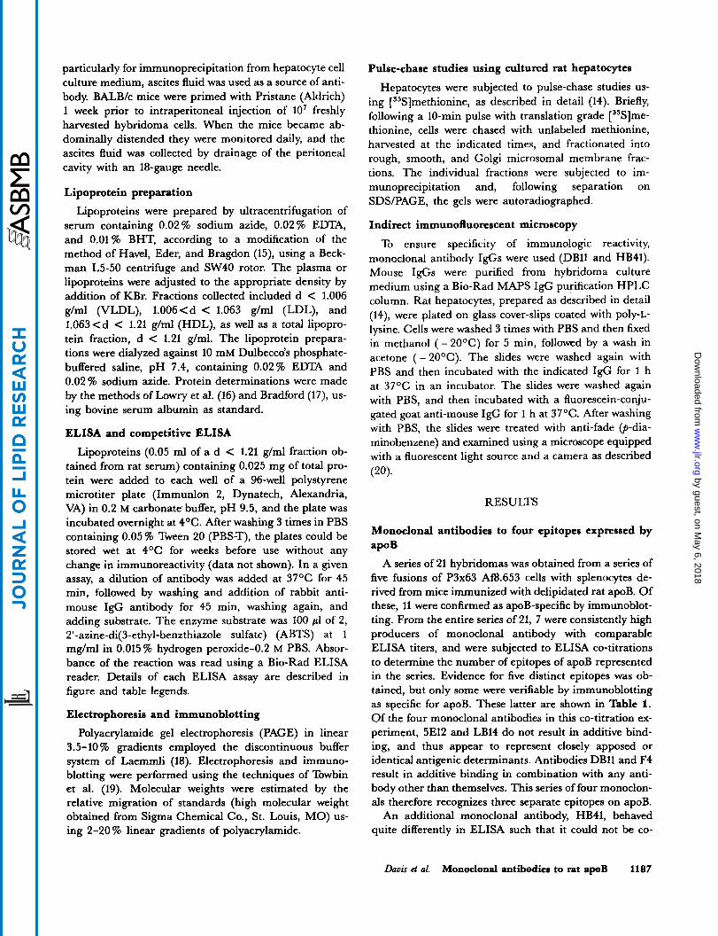
TABLE 2. Characterization of microsomal membrane fractions
Fold-Enrichment'
% Distribution % Distribution UDP-Galactosyl Glucose-6- Acid Fraction of Protein of RNA Transferase Phosphatase Phosphatase 5'-Nudeotidase
Rough 54 77 1.5 4 .1 2.3 1 . 7 Smooth 40 22 2.5 3.5 1.2 6.7 Golgi 6 1 56.3 1.9 3.1 1 1 . 1
Livers were homogenized and separated into membrane fractions by density gradient ultracentrifugation. Each fraction was subjected to protein, RNA, and enzyme activity analysis as described (14). Values represent mean of three individual determinations.
"Fold-enrichment was calculated as the specific activity of the designated fraction x the specific activity of the total homogenate.
1190 Journal of Lipid Research Volume 30, 1989
by guest, on May 6, 2018
ww
w.jlr.org
Dow
nloaded from

Fig. 5. Western blot analysis of hepatic microsomal fractions with apoB-specific antibodies. Liven from rats fed a b libitum. taken between 9 and 11 AM. wcrc homogenized in the presence of protcolytir inhibi- ton, and microsomal membrane fractions (rough. smooth, and Golgi) were obtained by sucrose density ultracentrifugation. An aliquot of each fraction (rough and smooth. 0.4 mg; Golgi. 0.1 mg) which represented 3, 3.6, and 20% of the total fraction, respectively) was applied. Antibo- dies used wcrc rabbit polyclonal antiserum (odd-numbcred lanes) and apoBI:specific HR41 (even-numbered lanes). Detection was obtained as described in Fig. I . A d < 1.21 glml total lipoprotein fraction obtained from rat serum was used as a control (lanes 1 and 2). The migration of apoBL and apoBs is given. The arrow indicates the migration of a 67,000 dalton protein standard.
Co-localization of apoB polyclonal and monoclonal epitopes in the endoplasmic reticulum of cultured rat hepatocytes
Previous studies using subcellular fractionation and immunoprecipitation show that in cultured rat hepato- cytes most of the newly synthesized apoBr. and apoBs is located in the rough and smooth microsomal fractions (14). We examined the localization of apoB1- and apoBs in cultured rat hepatocytes using immunofluorescence. Two monoclonals were used: one that recognized both apoB1- and apoBs (DB11) and one that only recognized apoBL (HB41).
Cultured rat hepatocytes were fixed with organic sol- vents (to remove neutral lipids) and then reacted with a purified IgGs obtained from the culture medium of the designated hybridoma cell. Preliminary studies (ELISA
bably the result of proteolysis. The presence of these pep- tides in the rough and smooth microsomes suggests that they might be formed from apoB in the secretory pathway. We, therefore, examined whether these peptides would be biosynthetically labeled with [35S]methionine.
Immunoprecipitation of [35SS]methionine-labeled apoB
Hepatocytes were pulsed for 10 min with [35Sjmethion- ine and then chased with unlabeled methionine. Cells were harvested at 0, 10, 30, 45, 90, and 180 min, separated into membrane fractions, and immunoprecipi- tated with polyclonal antiserum. The rate constants and quantitation of the relative incorporation of [35S]methion- ine into apoB and albumin have been reported (14). The autoradiographs of the immunoprecipitates showed that, in addition to apoBL and apoBs, two distinct proteins were labeled: one having an apparent molecular weight of 120,000 and another having an apparent molecular weight of 60,000 (Fig. 6). There were other proteins la- beled to a lesser degree. Moreover, during the chase period, the 120,000 dalton band disappeared at a rate comparable to that displayed by both molecular weight forms of apoB (Fig. 6) . In contrast, the band having a molecular weight of approximately 60,000 showed little, if any, reduction in intensity during the chase period (Fig. 6) . These data show that neither the 120,000 dalton or 60,000 dalton proteins are incomplete nascent chains of either apoB1- or apoBs.
6 8 K D
1 2 3 4 5 6 Fig. 6. Immunoprecipitation of pulsekhase ["SJmethionine-labcled rough microsomes. Cultured rat hepatocytes were pulsed for 10 min with 1 0 0 pCilml ["SJmcthionine. The cells were washed and then chased with unlabeled culture medium containing a 1000-fold excess of unlabeled methionine. Cells were harvested and then frarrionatcd into rough. smooth. and Golgi subfractions. The individual fractions were added to burners containinx 0.1 % SDS. boiled. rooled to rnom tempera- ture. and immunoprecipitated with rabbit polyclonal antiserum specific for apoR (see Western blot in Fig. 5). Following separation on a 2-20% linear gradient of polyacrylamide. the gels were drird and subjected to autoradiography for 2 weeks. The designation of standards of apoRl, and apoR, are shown. An estimation of 68.000 daltons (determined from the migration of protein standards) is also shown. The following samples are shown: lane 1. 0-min time point; lane 2, 10-min chase; lane 3, 20-min chase; lane 4, 45-min chase; lane 5,90-min chase; lane 6. 180- min rhasr.
Douis et al. Monoclonal antibodies to rat apoB 1191
by guest, on May 6, 2018
ww
w.jlr.org
Dow
nloaded from

assay) show that this treatment increases the expression of the monoclonal antibody epitopes. The cells were then treated with fluorescein-conjugated second antibody. Fluorescence microscopy afforded a distribution of apoB epitopes in cultured rat hepatocytes. Most of the immu- nofluorescence pattern produced with DBl1, which recog- nizes both apoRI- and apoRs, was localized throughout the cytoplasm (Fig. 7A), characteristic of the fluorescence produced by antibodies to the endoplasmic reticulum
(22). In addition, the immunofluorescence pattern pro- duced by apoRL-specific monoclonal I g G HB41 (Fig. 7B) was the same as that produced by DBll. To show that our immunofluorescence microscopy technique and reagents could discriminate Golgi, hepatocytes from the same preparation were reacted with a monoclonal antibody found to be specific for Golgi (characterized by D. C. F. Chan). The immunofluorescence pattern of Golgi-specific antibody was clearly different from that produced with
Fig. 7. Immunofluorescence microscopy of using monoclonal antibodies. Immunofluorescence studies were car- rird out as described in Methods. Cultured rat hepatocytes were reacted with the following monoclonal I$s: DBll (frame A), HB41 (frame R). and Golgi specific monoclonal 6A3 (frame C ) . For further comparison, the Golgi- specific monoclonal antibody 6A3 was also reacted with human skin fibroblasts (frame D).
1192 Journal of Lipid Research Volume 30, 1989
by guest, on May 6, 2018
ww
w.jlr.org
Dow
nloaded from

either apoB antibody (Fig. 7C). Finally, the G,olgi-specific antibody was reacted with human fibroblasts (Fig. 7D), again producing a pattern characteristic of Golgi (22). Thus, the Golgi-specific antibody recognizes a protein that is expressed by both fibroblasts and liver. Moreover, the data show that in cultured rat hepatocytes, epitopes expressed by both apoBL and apoBs are localized to the endoplasmic reticulum.
DISCUSSION
The major goal of this research was to produce mono- clonal antibody reagents with which to probe the struc- ture and localization of apoB in lipoproteins, hepatic membranes, and in liver cells. Consistent with previous studies using monoclonal antibodies directed against hu- man apoB (8-ll), all antibodies that recognize small molecular weight apoB also recognize large molecular weight apoB (i.e., LB14, F4, and DBll). In contrast, a monoclonal antibody (HB41) was obtained that recog- nized large molecular weight apoB, but did not react with small molecular weight apoB. These data are consistent with sequence analysis which shows that the small mole- cular weight form of apoB encompasses the amino- terminal domain which extends approximately to the midpoint of apoBL (6). Additional data show that the apoBL-specific antibody (HB41) recognizes a cryptic do- main in VLDL, which upon treatment with detergents (SDS and Triton X-100) becomes accessible. Furthermore, the HB41 epitope show the unusual characteristic of being sensitive to MT treatment of lipoproteins causes a substantial increase in the expression of the HB41 epi- tope. Studies of the LDL receptor binding domain of apoB using monoclonal antibodies that recognize epitopes in this portion of apoB-100 show that some of the epitopes are poorly expressed on VLDL and are expressed more efficiently on LDL (13). Detailed analysis of the amino acid sequence of human apoB-100 suggests that the LDL receptor binding domain is likely to lie on the carboxyl terminal third of the molecule encompassing a thrombin cleavage site (the T2/T3 junction) (3, 4, 12, 13). Monoclo- nal antibody epitopes lying within this area of human apoB-100 show a dependency on lipid for expression (12) and are thought to be encompassed by an intramolecular disulfide bond (4). However, studies on pig apoB indicate that the putative disulfide bridge is not required for recog- nition by the LDL receptor (23). Studies characterizing the lipoprotein receptors on cultured rat hepatocytes show that HB41 blocks the specific binding of '251-labeled LDL (L. Junker and R. Davis, unpublished data). While we have not yet determined the amino acid sequence of the HB41 epitope, the combined data suggest that it lies proximal to or within the LDL receptor binding domain
on apoBL. Furthermore, in regard to apoB structure/ function relationships, the differential expression of the HB41 epitope on VLDL and LDL, probably in response to lipid (Figs. 2 and 3) mimics the expression of the LDL receptor binding domain epitopes reported for human apoB-100 (13).
The specificity of the monoclonal antibody (HB41) allowed us to examine the expression by intracellular membranes of the apoBL epitope. Consistent with previous results obtained using cultured rat hepatocytes, apoB was found in the rough, smooth, and Golgi micro- somal fractions (Fig, 5). However, there were consistent differences in the molecular weight of the peptides that reacted with different antibodies. In the rough and smooth microsomal fractions there were two major pro- teins that reacted with the polyclonal antibody: apoBL and apoBs. Moreover, the smooth microsomes were found to be enriched (relative to the Golgi fraction) with several peptides smaller than apoB which react with the apoB-specific antiserum. Since no PII, a major plasma form produced from apoBL prior to secretion in the Golgi (21), was detected in either the rough or smooth microsomal fractions, it is unlikely that the rough and smooth microsomal fractions are contaminated with en- dosomes. In marked contrast, the Golgi fractions con- tained a different spectrum of immunoreactive proteins: proteins the size of apoBL, PII, and apoSs were detected by the polyclonal antibody, whereas the 60,000 dalton protein was not (Fig. 5). Reuben et al. (21) failed to detect PI1 in the Golgi fraction, and concluded that the pro- teolytic cleavage producing PI1 may have occurred be- yond the Golgi but prior to secretion. Our finding of an immunoreactive protein similar in size to PI1 in the Golgi membrane fraction might be due to our different method of isolation. Also, it is possible that the different Golgi membrane fractions may have contained different amounts of multivesicular bodies (24), and that some of the PI1 might represent the endocytotic pathway rather than the secretion pathway.
To determine the origin of the truncated forms of apoB, fluorograms of apoB immunoprecipitated from pulse- chase experiments (Fig. 6 of ref. 14) were analyzed. In ad- dition to apoBL and apoBs, two major and several minor labeled proteins were present. One having an apparent molecular weight of 120,000 disappeared with kinetics similar to that for apoB. Another having a molecular weight close to 60,000, displayed a much slower rate of decrease during the chase period. These data indicate that neither of the small peptides immunoprecipitated by the polyclonal antiserum are likely to be precursors to mature apoB. It is more likely that the small peptides are either small translation products of the apoB mRNA, produced by modification of the initial transcription product, or that they are proteolytic products. [35S]Methionine- labeled peptides smaller than apoB that are specifically
Davis et al. Monoclonal antibodies to rat apoB 1193
by guest, on May 6, 2018
ww
w.jlr.org
Dow
nloaded from

immunoprecipitated by apoB-specific antibodies appear in the autoradiograms reported by Reuben et al. (Fig. 5 of ref. 21), suggesting that these peptides are not unique to our experimental systems.
Since the 60,000 dalton protein found in the rough and smooth microsomal fraction reacted only with the poly- clonal antiserum (not the apoBL-specific HB41 monoclo- nal antibody), this peptide is a truncated form of apoB which does not contain the HB41 epitope. Since it is not possible to completely rule out proteolysis (even when several proteolytic inhibitors have been added to the buffers), we cannot determine whether the truncated forms of apoB are produced in vivo or during the mem- brane isolation step. However, the presence in the rough and smooth, but not the Golgi, microsomal fractions of the major truncated form of apoB (having a molecular weight of 60,000) suggests that apoB in the early portion of the secretory pathway @e., rough and smooth en- doplasmic reticulum) is susceptible to proteolysis, whereas in the distal portion (i.e., Golgi) it is not. Since in the same membrane fractions there are no detectable immunoreactive forms of albumin, apoE, or apoA-IV, which are smaller than their mature secretory forms (data not shown), the proteolysis of apoB appears selective.
In previous studies, we have found that only a portion of de novo synthesized apoB is secreted; the remainder appears to be degraded intracellularly (14). We have pro- posed that intracellular degradation is one mechanism responsible for determining apoB secretion rates (14). While the finding of a selective presence of proteolytic fragments of apoB in the rough and smooth, but not the Golgi, microsomes is consistent with our hypothesis that the site of apoB degradation is in the endoplasmic reticu- lum, more data are needed to prove this point.
Previous pulse-chase studies showed that, in cultured rat hepatocytes, the rate of movement out of the en- doplasmic reticulum is rate-limiting for secretion (14). Furthermore, quantitation of [ 35S]methionine steady- state labeled apoB in subcellular fractions obtained by density gradient ultracentrifugation showed that most of the label was in the rough and smooth microsomal frac- tion (14). In contrast, other hepatocyte culture models (i.e., HepG2 (25) and estrogen-stimulated chicken cells (26)) display kinetics consistent with the proposal that movement out of the Golgi is rate-limiting for secretion. The controversy regarding the rate-limiting step for secre- tion includes both apoB and albumin. The kinetics dis- played by cultured rat hepatocytes showing that move- ment out of the endoplasmic reticulum is the slowest step is consistent with the kinetics displayed by most secretory proteins (27) and several other culture models including rat hepatoma cells (28) and human HepG2 cells (29). Since two different results have been obtained using the same line of hepatoma cells (Le., HepG2 (25, 29)), the
differences cannot be ascribed to different culture models. A major goal of this study was use of the specific nature
of monoclonal antibodies to probe the intrahepatic locali- zation of apoB. To rule out the possibility that membrane fraction cross-contamination (an inherent short-coming of cell fractionation techniques) may have been responsi- ble for the intrahepatic distribution of de novo synthe- sized apoB showing that the majority was in the endo- plasmic reticulum of cultured rat hepatocytes, we used immunofluorescence microscopy. The immunofluores- cence data obtained using monoclonal antibodies that recognize both apoBL and apoBs (DB11) and the apoBL- specific monoclonal HB41 showed that the majority of apoB epitopes produce a fluorescence distribution consis- tent with the endoplasmic reticulum but not the Golgi (Fig. 7A, B). With the proviso that apoB epitopes are uni- formly accessible to the antibody reagents,, our findings are consistent with the proposal that most of the apoB in cultured rat hepatocytes resides in the endoplasmic reticu- lum. Furthermore, using the same methods but a Golgi- specific monoclonal antibody, we were able to obtain a fluorescence pattern characteristic of Golgi (Fig. 7C). Thus, our methods can discriminate the immuno- fluorescence pattern of a Golgi-specific epitope.
Earlier studies by Alexander, Hamilton, and Have1 (30) show that immunoreactive apoB was detected in the rough endoplasmic reticulum unassociated with VLDL particles. Moreover, apoB was found associated with VLDL in junctional complex between the rough and smooth endoplasmic reticulum (30). Their data showed, for the first time, that VLDL is assembled with the en- doplasmic reticulum. The molecular processes through which apoB orchestrates the assembly of a VLDL in the endoplasmic reticulum remain poorly defined. Our find- ings that in cultured rat hepatocytes movement out of the endoplasmic reticulum is the slowest step in VLDL secre- tion (14) and that most of the apoB is located in the en- doplasmic reticulum are consistent with hypothesis that assembly of the VLDL particle (in the endoplasmic retic- ulum) is rate-limiting for secretion. The additional find- ing of this study showing the selective presence of pro- teolytically cleaved apoB fragments in the rough and smooth, but not the Golgi, microsomal fractions suggests that a form of apoB residing in the endoplasmic reticulum is accessible to proteolysis. Attempts to inhibit this pro- teolysis with proteolytic inhibitors have been unsuccessful (data not shown). If the proteolysis takes place during the membrane isolation, the susceptibility of this form of apoB to proteolysis implies a unique molecular feature of the immature form of apoB present in the endoplasmic reticulum. If, on the other hand, the proteolysis takes place in vivo, degradation of this form of apoB may play a fundamental role in regulating VLDL assemblyhe- cretion.
1194 Journal of Lipid Research Volume 30, 1989
by guest, on May 6, 2018
ww
w.jlr.org
Dow
nloaded from

The authors thank Monica Malone-McNeal, Leonita Liu, Tom Sand, and Jane Archambault-Schexnayder for expert technical assistance. This research was supported by grants HL-25596,
Health.
12. Marcel, Y. T., T. L. Innerarity, C. Spilman, R. W. Mahley, A. Protter, and R. W. Milne. 1987. Mapping of human apolipoprotein B antigenic determinants. Arteriosclmsis. 7:
13. Krul, E. S., Y. Kleinman, M. Kinoshita, B. Pfleger, K. Oida, A. Law, J. Scott, R. Pease, and G. Schonfeld. 1988. Regional mecificities of monoclonal anti-human aDoliDo-
HL-41624, and DK-34914 from the National Institutes of 166-175.
Manum$ received 5 January 1989 and in revired form 13 Manh 1989.
1.
2.
3.
4.
5.
6.
7.
8.
9.
10.
11.
REFERENCES
Kane, J. P., D. A. Hardman, and H. E. Paulus. 1980. Heterogeneity of apolipoprotein B: isolation of a new spe- cies from human chylomicrons. h c . Natl. h a d . Sci. USA.
Krishnaiah, K. V., L. E Walker, J. Borensztajn, G. Schonfeld, and G. S. Getz. 1980. Apolipoprotein B variant derived from rat intestine. Pmc. Natl. Acad. Sci. USA. 77:
Yang, C. Y., S. H. Chan, S. H. Gianturco, W. A. Bradley, J. T. Sparrow, M. Tanimura, W. H. Li, D. A. Sparrow, H. DeLoof, M. Rosseneu, F. S. Lee, Z. W. Gu, A. M. Gotto, Jr., and L. Chan. 1986. Sequence, structure, receptor- binding domains and internal repeats of human apolipo- protein B-100. Nature. 323: 738-742. Knott, T. J., R. J. Pease, L. M. Powell, S. C. Wallis, S. C. Rall, Jr., T. L. Innerarity, B. Blackhart, W. H. Taylor, Y. Marcel, R. Milne, D. Johnson, M. Fuller, A. J. Lusis, B. J. McCarthy, R. W. Mahley, B. Levy-Wilson, and J. Scott. 1986. Complete protein sequence and identification of structural domains of human apolipoprotein B. Nature. 323:
Powell, L. M., S. C. Wallis, R. J. Pease, Y. H. Edwards, T. J. Knott, and J. Scott. 1987. A novel form of tissue- specific RNA processing produces apolipoprotein B-48 in intestine. Cell. 50: 831-840. Chen, S. H., G. Habib, C. Y. Yang, Z. W. Gu, B. R. Lee, S. A. Weng, S. R. Silberman, S. J. Cai, J. P. Deslypere, M. Rosseneu, A. M. Gotto, Jr., W. H. Li, and L. Chan. 1987. Apolipoprotein B-48 is the product of a messenger RNA with an organ-specific in-frame stop codon. Science. 238:
Hospattankar, A. V., K. Higuchi, S. W. Law, W. Meglin, and H. B. Brewer. 1987. Identification of a novel in-frame translational stop codon in human intestine apoB mRNA. Biochem. Bi@hys. Res. Commun. 148: 279-283. Tikkanen, M. J., R. Dargar, B. Pfleger, B. Goneu, J. M. Davie, and G. Schonfeld. 1982. Antigenic mapping of hu- man low density lipoproteins with monoclonal antibodies. J Lipid Res. 23: 1032-1038. Marcel, Y. L., M. Hogue, R. Theolis, Jr., and R. W. Milne. 1982. Mapping of antigenic determinants of human apolipoprotein B using monoclonal antibodies against low density lipoproteins. J. Biol. Chem. 257: 13165-13168. Curtiss, L. K., and T. S. Edgington. 1982. Im- munochemical heterogeneity of human plasma apolipopro- tein B. Apolipoprotein B binding of mouse hybridoma anti- bodies. J. Biol. Chem. 257: 15213-15221. Mao, S. J. T., R. E. Kazman, J. C. Silverfield, M. C. Alley, K. Kluge, and C. G. Fathman. 1982. Immunochemical pro- perties of human low density lipoproteins as explored by monoclonal antibodies: binding characteristics distinct from those of conventional serum antibodies. Biochim. Biophys. Acta. 713: 365-374.
77: 2465-2469.
3806-3810.
734-738.
363-366.
14.
15.
16.
17.
18.
19.
20.
21.
22.
23.
24.
25.
26.
27.
28.
1 1
pro;ein B antibodies. J. Lipid Res. 29: 937-947. Borchardt, R. A., and R. A. Davis. 1987. Intrahepatic assembly of very low density lipoproteins: rate of transport out of the endoplasmic reticulum determines rate of secre- tion. J. Biol. Chem. 262: 16394-16402. Havel, R. J., H. Eder, and J. Bragdon. 1955. The distribu- tion and chemical composition of ultracentrifugally separated lipoproteins in human serum. J. Clin. Invest. 34:
Lowry, 0. H., N. J. Rosebrough, A. L. Farr, andR. J. Ran- dall. 1951. Protein measurement with the Folin phenol rea- gent.J. Biol. Chem. 193: 265-275. Bradford, M. M. 1976. A rapid and sensitive method for the quantitation of microgram quantities of protein utiliz- ing the principle of protein-dye binding. Anal. Biochem. 72: 240-254. Laemmli, U. K. 1970. Cleavage of structural proteins dur- ing the assembly of the head of bacteriophage T4. Nature.
Towbin, H., T. Staehelin, and J. Gordon. 1979. Elec- trophoretic transfer of proteins from polyacrylamide gels to nitrocellulose sheets: procedure and some applications. Pmc. Natl. Acad. Sci. USA. 76: 4350-4354. Chan, D. C. E, and T. T. Puck. 1989. Involvement of vimentin in the reverse transformation reaction. Pmc. Natl. Ad. Sci. USA. 86: 2747-2751. Reuben, M. A., K. L. Svenson, M. H. Doolittle, D. E Johnson, A. J. Lusis, and J. Elovson. 1988. Biosynthetic relationships between three rat apolipoprotein B peptides. J. Lipid Res. 29: 1337-1347. Willingham, M. C., and I. Pastan. 1983. An Atlas of Immunofluorescence in Cultured Cells. Academic Press, New York. Ebert, D. L., N. Maeda, S. W. Lowe, J. Hasler-Rapacz, J. Rapacz, and A. D. Attie. 1988. Primary structure compari- son of the proposed low density lipoprotein (LDL) receptor binding domain of human and pig apolipoprotein B: im- plications for LDL-receptor interactions. J. Lipid &s. 29:
Hornick, C. A., R. L. Hamilton, E. Spaziani, G. H. En- ders, and R. J. Havei. 1985. Isolation and characterization of multi-vesicular bodies from rat hepatocytes: an organelle distinct from secretory vesicles of the Golgi apparatus. J. Cell. Biol. 100: 1558-1569. Bostrom, K., M. Wettesten, J. Boxen, G. Bondjers, 0. Wiklund, and S. 0. Olofsson. 1986. Pulse-chase studies of the synthesis and intracellular transport of apolipoprotein B-100 in Hep G2 cells. J. Biol. Chem. 261: 13800-13806. Bamberger, M. J., and M. D. Lane. 1988. Assembly of very low density lipoprotein in the hepatocyte. Differential transport of apoproteins through the secretory pathway. J. Biol. Chem. 263: 11868-11878. Lodish, H. E 1988. Transport of secretory and membrane glycoproteins from the rough endoplasmic reticulum to the Go1gi.J. Biol. Chem. 263: 2107-2110. Strous, G. J. A. M., and Lodish, H. F. 1980. Intracellular
1345-1353.
227: 680-685.
1501 - 1509.
Davis et al. Monoclonal antibodies to rat apoB 1195
by guest, on May 6, 2018
ww
w.jlr.org
Dow
nloaded from

transport of secretory and membrane proteins in hepatoma
709-717. 29. Lodish, H. E, N. Kong, M. Snider, andG. J. A. M. Strous.
1983. Hepatoma secretory proteins migrate from rough en-
doplasmic reticulum to Golgi at characteristic rates. Nutun.
30. Alexander, C. A., R. L. Hamilton, and R. J. Havel. 1976. Subcellular localization of B apoprotein of plasma lipopro- teins in rat livers. J. Cell Biol. 69 241-263.
cells infected by vesicular stomatitis virus. Cell. 22: 304: 80-83.
1196 Journal of Lipid Rerearch Volume 30, 1989
by guest, on May 6, 2018
ww
w.jlr.org
Dow
nloaded from