OF PERIOP&ONTICS RESTORATIVE DENTISTRY(Int J Periodontics Restorative Dent . 2012;32:521-531.)...
12
Specia l rep ri nt from THE INTERNATIONAL JOURNAL OF PERIOP&ONTICS RESTORATIVE DENTISTRY Vo l. 32 o Copyright © 2014 by Quint ess ence Publ Co, Inc 5 12
Transcript of OF PERIOP&ONTICS RESTORATIVE DENTISTRY(Int J Periodontics Restorative Dent . 2012;32:521-531.)...

Specia l rep ri nt from
THE INTERNATIONAL JOURNAL OF
PERIOPampONTICS RESTORATIVE
DENTISTRY Vo l 32
o Copyright copy 20 14 by Quintessence Publ Co Inc
5 12
The International Journal of Periodontics amp Restorative Dentistry
521
A Novel Approach to Root Coverage The Pinhole Surgical Technique
ltUmiddot ~_ f j - ~~ ~
-~
John C Chao 005
Free connective tissue graft techniques are currently considered th e most
predictable surgical m ethod for root coverage However morbidity associated
with secondary gra ft sites has generated interest in other meth ods The purpose
of th is study was to in vestigate the feas ibility of a novel surgical approach to
root coverage the pinhole surgical technique (PST) Th is retrospective study
examined the results of PST used for 43 consecutive patients on 12 1 rece ssion
sites of which 85 were Class l or I and 36 were Class II Mean initia l recession for
all sites was 34 plusmn 1 0 mm The mean assessment period was 18 plusmn 67 months
No secondary surgical site was necessary and on ly bioresorbable membrane
or acellular dermal matrix was use d as graft material PST require d no releasing
incision sharp dissection or suturing (wh en a bioresorbable m embrane was used) Only one incision of 2 to 3 mm (for entry) was necessary for th e entire procedure
Predictability of PST fo r Class I and I sites m e asured as freq uency of complete
root coverage was 8 12 Effectiveness of PST fo r Class I and I sites measured
as mean percent defect reduction was 940 plusmn 148 Wh en data from Class I
II and III sites were combined predictab ility and effective ness we re 694 and
884 plusmn 198 respectively Th e mean duration per procedure was 223 plusmn 10 1
minutes Th e mean level of patient subjective esthetic satis fa ction was 95 1 and
was rea lized within a mean 734 plusmn 135 days Postoperative com plications were
minimal These results indicate that PST holds p romise as a minimally invasive
predictable effective and time- and cost-effe ctive method for obtaining optimal
patient-based outcomes (Int J Pe riodont ics Restorat ive Dent 201232521-531)
Private Pract ice Alham bra California
Corresponden ce to Dr John C Ch ao 100 S First Street A lhambra CA 9180 1
fax 626-284-8584 email johnchaoddssbcgloba lnet
The reestabl ishment of a st able
periodont ium concomitant with an
optima l patient-centered outcome
is said to be the objective of perishy
odontal reconstructive surgery1-3
To meet t his object ive various reshy
constructive su rg ica l techn iques
for root coverage have been deshy
veloped and reported over the
yea rs4-12 Current ly free connect ive
tissue graft (FCTG) techniques are
considered the gold standard and
the most predi ctab le approach for
complete root coverage However
FCTG techniq ues are associated
with donor site complications such
as postoperative pain bleedi ng
and swe lling 13 An in-depth analysis
of FCTG and other current techshy
niques led to t he observation t hat
all of t hese methods require a coroshy
nal approach for th e entry incision
re leasing incisions flap e levation
or graft placement4-13 In contrast
th is article reports on a nove l vesshy
tibu lar surgica l techn ique t he pinshy
hole surgica l technique (PST) The
pu rposes of this study were t o exshy
am ine the predictability and effecshy
ti veness of PST and to assess its
effect on patient-based outcomes
Volume 32 Number 5 2012
522
Fig 1 Trans-Mucosal Papilla Elevators
Method and materials
In a practice-based retrospective
study covering a period of 33
months 43 patients (16 men 27
women) between the ages of 31 and
84 years (mean 57 plusmn 142 years)
with gingival recessions on 121
teeth (71 maxillary 50 mandibular)
were treated consecutively and exshy
clusively with PST in the random
order they presented themselves
The mean follow-up assessment
period was 18 plusmn 67 months (range
5 to 33 months) All cases were acshy
counted for with no patient lost to
follow-up Miller Class I and II sites
numbered 85 there were 36 Class
III sites Of the total 121 teeth treatshy
ed 98 presented baseline recesshy
sion measurements 2 30 mm For
each patient a mean of 281 recesshy
sion sites were treated per surg ica I
appointment It was the routine and
preferred practice of the implemenshy
tation of PST to treat not one but
multiple sites when present all at
one time (range 3 to 10 sites)
Inclusion criteria for this retshy
rospective study were as follows
American Society of Anesthesioloshy
gists Physical Status I or 1114 and no
Fig 2 Full-thickness flap elevation
contra indications for periodonta l
surgery presence of maxillary or
mandibular single or multiple bucshy
cal recessions classified as Class I
II III or a combination thereof abshy
sence or presence of restorations or
crowns and an identifiable cementoshy
enamel junction (CEJ) absen ce of
previous periodontal surgical treatshy
ment at the involved sites history
of compliance with oral hygiene
instructions and periodontal recall
absence of plaque and bleeding on
probing at the surgical sites and
no history of smoking in the 5 years
prior to treatment
Clinical parameters
Preoperatively at the surgical apshy
pointment at the third month
and every 3 or 6 months thereafter
depending on patients needs for
periodic checkups the following
four parameters were measured
(1) recession depth (RD) the disshy
tance from the marginal ging iva at
the midbuccal aspect of the root
to the CEJ or coronal margin of
the restoration (2) probing su lcus
depth (PD) (3) clinical attachment
Fig 3 Elevation of the papillae on each side of the affected tooth
level (CAL) the sum of RD and PD
and (4) keratinized gingiva (KG) the
height of the keratinized gingiva or
distance from the marginal gingiva
to the mucogingival junction Adshy
d itionally the quality of color and
t issue match was assessed by the
cli nician at all follow-up appointshy
ments Clinica l data regarding color
and tissue match and photographs
t aken at each follow-up session
were compared to those obtained
preoperatively for the purpose of
assessing tissue changes and rate
of healing Initial and follow-up RD
as observed on study casts were
measured independent of clinically
procured RD data to verify accurashy
cy of clinical measurements
Surgical method
All surgeries were performed by the
author Following injection of loshy
cal anesthetic caries restorations
surface irregularities and convexishy
ties on the root were removed and
planed using rotary burs ultrasonic
instruments and hand curettes Usshy
ing a no 12 scalpel (Bard-Parker)
a minimal horizontal incision of 2
The International Journal of Periodontics amp Restorative Dentistry
523
to 3 mm was made in the alveolar
mucosa near the base of the vestishy
bule apical to the recipient site(s)
In cases with mandibu lar premolar
involvement the incision was made
near the base of the vestibule suffishy
cientl y mesial to the root of the first
premolar such that in the judgment
of the clinician the incision posed
no risk of injury to the mental nerve
Specia lly designed instruments
(Trans-Mucosal Papillae Elevators
[TMPEsL H amp H) were inserted
through the entry incision to elevate
a full-thickness flap (Figs 1 and 2)
Elevation of the flap was guided by
visualization of the shape and moveshy
ment of the instruments through the
mucosa and gingiva l t issue The flap
was then extended coronal ly and
horizontally to allow for elevation of
the two adjacent papillae on each
side of the denuded root(s) (Fig 3)
The inclusion of at least four papilshy
lae is a unique feature of PST This
interproximal extension of t he f lap
resulted in a freely movable fla p
which was then positioned coroshy
nally to extend beyond the CEJ
For stabilization of the flap a malshy
leable bioresorbable membrane
(BM Bio-Gide Geistlich) was used
Fig 4 (left) PST graft pliers
Fig 5 (right) Placement of the 8M graft material
fo r 100 root defects whi le acell ushy
lar dermal mat rix (ADM Alloderm
BioHorizons) was used for the other
21 Two to fou r 2 X 12-mm strips of
BM presoaked in sterile water were
threaded one by one through the
entry incision using PST graft p liers
(H amp H) and tucked into the subg inshy
gival spaces under the papil lae and
marginal soft t issue (Figs 4 and 5)
The actual number of strips used
depended on the amount of mateshy
rial needed to secure the flap in th e
desired position
Tissue tension created by d isshy
tention or pouching of t he fl ap
was suffi cient in all cases to hold
the graft strips in place without sushy
tures surg ical d ressing o r tissue
adhesive Gentle digital pressure
was applied to t he flap fo r apshy
proximately 5 minutes The ent ry
incision was left to hea l by first inshy
tention without suturing
ADM was used in 21 sites The
slippery nature of ADM requ ired a
novel sling sutu ring techn ique A
2 X 5-mm stri p of ADM was t ied
at each end with a separate 4-0
24-mm 38c bioresorbable sutu re
(Vicryl Ethicon) Each need le was
threaded through the entry incision
to emerge from under the fac ial marshy
gina l g ingiva of the reci p ient root
One need le was t hen threaded unshy
der t he mesial contact and the other
under the distal The ends of th e
graft were al lowed to sl ip t hrough
the entry incision by t ugg ing on one
end and then the other from the
oral apsect Tugg ing both sutures
simu ltaneously advanced the entire
g raft strip along with the overlying
fl ap coronally enough to cover the
CEJ Threading each sutu re under
the opposite contacts al lowed the
sutures to be tightened and knotted
from the facia l aspect This manner
of suturing stabilized the flap Loose
ends of the b ioresorbable sutu res
were cut and removed when they
appeared during follow-up appointshy
ments (Figs 6a t o 6f)
Postoperative inst ructions inshy
cluded use of a ch lorhexdine glucoshy
nate 012 oral ri nse (Peridex 3M
ESPE) and avoidance of b rushing at
th e surgical site for 6 weeks Ea ch
patient was assessed for expected
clinical signs of early heal ing the
next business day and the followi ng
week Patients were further checked
at 3 and 6 weeks Light debrideshy
ment was done at each fol low-up
Volume 32 Number 5 2012
524
Fig 6a Needle is threaded through the entry incision to e merge under the facial marginal g ingiva of th e recipient root
Fig 6b Needle is threaded under th e mesial contact
Fig 6d The d ista l needle is passed under the mesia l contact to appear at the facial aspect
appointment as necessary At the
sixth week patients were instructed
on the roll brushing technique usshy
ing an extra-soft toothbrush Thereshy
after patients were re-assessed at
every periodontal maintenance apshy
pointment which was generally evshy
ery 3 months
Fig 6e Tugging b oth sutures from the fa cial aspect simultaneously advances the entire graft strip coronally Sutures are tied and th e knot is tugged under th e fl ap
Questionnaire and information
collection
Using a questionnaire a staff memshy
ber interviewed each patient reshy
garding the following patient-based
outcome variables The first vat-i able
was esthetics described by Zucchelshy
Ii and De Sanctis as a completely
satisfying result for the pati ent1 5
Each patient was asked to rate his o r
her degree of esthetic satisfact ion
on the basis of any set of criteria
Fig 6c A ne edle at the other end of the graft has been passed under the flap and unde r th e distal contact to appear at the oral aspect Tuggin g on on e end and th en th e oth er from the facial aspect allowed the ends of the g raft to slip th rou gh the entry incision
Fig 6f The suturing technique from th e facial perspe ctive
personal to the patient expressed
as a percentage (0 [total dissatshy
isfaction] to 100 [complete satshy
isfaction]) To add a ti me-to-event
measurement the patient was furshy
t her asked to state the time (day)
t he esthetic improvement (or lack o~
was first not iced Each patient also
was asked to rate complications reshy
lated to pain bleeding and swel lshy
ing on a scale from 0 to 1013 A
complication whether it was pain
b leeding or swelling was rated
The Inte rnational j ournal of Periodontics amp Resto rative Dentistry
525
as none to mild if the score was
o to 3 moderate if the score was
4 to 6 and severe if t he score was
7 to 10 13 Dentinal sensitivity was
rated by the patient on a scale of 0
to 10 according to the effect of hot
cold food and drink air toothbrushshy
ing and sweet and sour food on the
teeth16 Each patient also was asked
to rate overall satisfaction with t he
root coverage procedure as a pershy
centage (0 [totally unsatisfied] to
100 [complete satisfaction])
Statistical analysis
Quantitative data were recorded
as means plusmn standard deviations
Data were analyzed using the Stushy
dent t test for paired observations
to assess changes obtained with in
and between groups Kurtosis and
skewness curves were used to vershy
ify the normality of the data Th e
significance level for rejection of
the null hypotheses for all tests was
set at (X = 05
Results
Predictability was measured as the
percentage of the time duration
either complete root coverage or
near complete (gt 90 ) root covshy
erage was achieved 14 Of the 121
sites 85 were Miller Class I and II
and 36 were Miller Class III When
Class III sites were incl uded with
data from Class I and II sites comshy
plete root coverage was achieved
in 694 of sites and 90 defect
coverage was obtained in 777 of
Clinical parameters (mean plusmn SO)
Parameters All sites Class I and II Class III
No of sites 121 85 36
Assessment period (mo) 18 plusmn 67 20 plusmn 67 15 plusmn 52
Baseline recession (mm) 34plusmn 10 33plusmn10 36 plusmn 11
Follow-up recession (mm) 04 plusmn 08 02 plusmn 05 09 plusmn 10
Defect coverage (mm) 30plusmn11 31 plusmn 11 27 plusmn09
defect coverage 884 plusmn 198 940 plusmn148 755 plusmn 240
No of sites with complete 84 69 15 root coverage
complete root coverage 694 812 417
root coverage 90 777 906 472
Baseline PO (mm) 26 plusmn 07 26 plusmn 06 24 plusmn 09
Follow-up PO (mm) 12 plusmn 04 12 plusmn 04 13 plusmn 05
PO reduction (mml 14 plusmn 08 15 plusmn 07 11 plusmn 09
Baseline CAL (mm) 60 plusmn 13 59 plusmn 12 60 plusmn 16
Follow-up CAL (mm) 16 plusmn 09 14 plusmn 06 22 plusmn 11
CALgain (mm) 44 plusmn 14 46 plusmn 13 38 plusmn 15
Baseline KT (mm) 08 plusmn 16 11plusmn 18 00 plusmn 00
Follow-up KT (mm) 30 plusmn 11 33 plusmn 08 25 plusmn 13
KTgain (mm) 13plusmn 19 12 plusmn 20 15 plusmn 17
so =standard deviation PD =probing depth CAL =clinical attachment level KG = keratinized gingiva
sites When only the 85 Class I and age and mean defect reduction
sites were computed complete were 884 and 30 plusmn 11 mm
defect coverage was attained in respectively When only Class
812 of sites and near complete and sites were included in the
defect coverage was observed in calculation mean percent defect
90 6 of sites coverage and mean defect reducshy
Effecti veness was measured tion were 94 and 31 plusmn 11 mm
as th e mean percent defect covshy respectively (Table 1) The mean
era ge and mean defect reducshy postoperative measu rements ofthe
t ion 14 Mean baseline recession for other relevant parameters for al l
all si t es was 34 plusmn 10 mm When 121 sites were positive PD reducshy
all 121 sites were computed tion 14 mm CAL gain 44 mm
the mean percent defect cover- and KT gain 13 mm (Table 1)
Volume 32 Number 5 201 2
I
526
Figs 7a and 7b Single surgery on multiple sites with ADM (a) Presurgical photograph (b) follow~up 3 years later
Patient-based outcomes
Pain
Intensity (degree plusmn SD) 08 plusmn 08
No pain () 6 (140)
Mild pain () 32 (744)
Moderate pain () 3 (70)
Severe pain () 2 (46)
DUration (day plusmn 5D) 26 plusmn 15
Bleeding
Intensity (degree plusmn SD) 07 plusmn 05
No bleeding () 14 (326)
Mild bleeding() 29 (6741
Moderate bleeding () 0(00)
Severe bleeding () 0(00)
Duration (day plusmn SD) 12 plusmn 11
Swelling
Intensity (degree plusmn SD) 08 plusmn 05
No swelling () 11 (256)
Mild swelling () 30 (698)
Moderate swelling () 2 (46)
Severe swelling () 0(00)
Duration (day plusmn SD) 20 plusmn 18
SD =standard deviation
The mean number of recession sites treated per
procedure was 28 The mean follow-up assessment
period was 18 plusmn 67 months (range 5 to 33 months)
(Table 1) In a subset of 10 patients with 20 root recesshy
sion sites the mean duration of the PST procedure per
recession site was 223 plusmn 101 minutes
Regard ing patient-based outcomes the results of
the patient questionnaire showed that the mean pashy
tient esthetic sat isfact ion was 949 plusmn 10 Examples
of preoperative and follow-up photographs are shown
in Figs 7a and 7b Furthermore this esthetic result was
observed by patients within a mean of 74 plusmn 135 days
The mean overal l pat ient satisfaction over the course of
the study was 95 1 plusmn 12
Table 2 further summarizes the levels and durashy
tions of symptoms of pain swelling and bleeding
Twenty-five patients reported root sensitivity prior
to surgery O f t hose 25 patients 12 (48) reported senshy
sit ivity after surgery No other adverse events or complishy
cations in addition to t hese symptoms were observed
Clinical notes and photographs showed healing
to be uneventful in all cases Complete healing for all
cases was observed to have taken place at the 6-week
follow-up vis it Furthermore clinical data and followshy
up photographs indicated no observable differences
in color and tissue match between pre- and postopshy
erative gingival tissue in all cases at the first 3-month
fo llow-up visit and all other follow-up visits thereafter
(Fig 7b)
Mean percent defect coverage derived from meashy
suring initia l and follow-up recession on study casts
The Internationa l Journa l of Periodontics amp Restorative Dentistry
527
(865) was compared to that obshy
tained from intraoral measurements
(879) Since there was no signifishy
cant difference between the two
the clinical data with respect to reshy
cession were further confirmed
While predictability is meashy
sured by frequency of defect covershy
age effectiveness is measured by
mean percent defect coverage 14
The criterion for successful mean
defect coverage is 80 to 10012
USing PST mean percent defect
coverage for Class I and II sites was
951 through the course of the asshy
sessment period of 18 plusmn 67 months
(range 5 to 33 months) Most noshy
tably this result was first observed
by patients within a mean of 74
days Although clinical data and
photographic records indicated the
presence of at least some mild deshy
II
1) 01
l
528
Intragroup comparisons
Baseline Follow-up recession recession
No of teeth (mmplusmnSD) (mm plusmn SD)
Maxilla 71 34 plusmn 10 02 plusmn 05
Mandible 50 33 plusmn 10 07 plusmn 10
Cariousrestored 45 35 plusmn 09 06 plusmn 09
Intact roots 76 33 plusmn 11 03 plusmn 07
Age lt 575 y 62 34plusmn 11 03 plusmn 06
Agegt 575 y 59 33 plusmn 10 05 plusmn 09
Early group 53 33plusmn11 01 plusmn 02
Later group 68 35 plusmn 10 06 plusmn 09
ADM 21 36 plusmn 11 03 plusmn 08
BM 100 34 plusmn 10 04 plusmn 08
ADM = acellular dermal matrix BM = bioresorbable membrane
600 mg) Bleeding and swelling for
PST pat ients were mild and of short
duration (see Table 2) The relatively
rapid d iminishment of symptoms
in PST patients is coincidental with
the quickness of hea ling observed
cli nically and in postoperative
photographs
Twenty-five patients in this study
reported sensitivity prior to surgery
Of those 12 (48) reported sensishy
t ivity after surgery In a study by Pini
Prato et ai 4 of 10 (40) patients
with preoperative dentinal sensitivity
continued to experience sensitivity
postoperatively31
Table 3 compares PST intrashy
g roup d ifferences A slight but signifshy
icant statistical difference was noted
between maxillary and mandibular
teeth in terms of follow-up recesshy
sion (02 plusmn 05 and 07plusmn 10 mm
respectively) Significant statistical
differences in FCTG resu lts between
mandibular and maxi llary teeth
were also found by Chambrone and
Chambrone28 In the latter study
an FCTG procedure involving mulshy
t iple sites was performed for 28 pashy
tients half of whom were treated
for mandibular recessions while the
other half were treated for mu ltiple
maxillary recessions A ll sites were
either Class I or II Mean fin al reshy
cession depths for mandibular and
maxillary groups were 021 and 007
mm respectively a threefold difshy
ference Interestingly results with
PST also showed an approximate
threefold difference between the
mandibular and maxillary proceshy
dures (07 and 02 mm respec-
Defect coverage defect (mmplusmnSD) coverage
32plusmn11 936
27 plusmn 109 794
29 plusmn 10 832
30 plusmn 11 915
31plusmn12 901
28 plusmn 09 854
32plusmn11 960
29 plusmn 10 831
30 plusmn 13 914
29 plusmn 10 869
tively) The greater final RD in the
PST study as compared to t hat of
Chambrone and Chambrone28 may
be due to the inclusion of Class III
sites in the PST study PST Class III
defects accounted for 16 of 50 manshy
dibular sites and 20 of 71 maxillary
sites Chambrone and Chambrone
cited depth of the vestibular forshy
nix flap tension flap thickness and
mucogingival phenotype as posshy
sible proximal links to explain their
findings28 This difference between
mandibular and maxillary groups
may also be a result of the possibilshy
ity that functional mechanical forces
act much more heavily on wound
margins in the mandible than in the
maxilla as suggested by Amarante
et al 32 It should also be noted that
even though Class III cases were in-
The International Journal of Periodontics amp Restorative Dentist ry
529
cluded 794 defect coverage for
all mandibular PST procedures sti ll
measured favorably against the crishy
terion for successfu l mean defect
coverage suggested by Greenwel l
et ai which was 80 to 10012 Furshy
ther investigations focusing on the
effects of PST or FCTGs in mandibushy
lar sites are recommended
With respect to nonintact roots
results with PST were concordant
with those of a previous study by
Goldstein et al 33 which concluded
that coverage of previously carious
or restored roots is just as predictshy
able as coverage of intact roots
In PST cases no significant dif shy
ferences in treatment results were
evident between younger and older
age groups
With regard to the surgeon s
learning curve as a possible factor
for bias1 8 comparing the results
of an earlier group with those of
a later group categorized accordshy
ing to the time of surgery yielded
percent defect coverage resul ts
of 960 and 831 respectively
Since defect coverage for the early
group was slightly higher though
not statistica lly significantly better
than that of t he later group effect
of the surgeons lea rn ing curve o r
progressive improvement as a posshy
sible avenue of bias was not apparshy
ent (see Table 3) In add ition with
respect to compa ring resu lts beshy
tween BM and ADM no sign ificant
differences emerged (see Table 3)
Aside from the intragroup reshy
sults reported in Table 3 th is study
also addressed the issue of se lecshy
tion bias18 of the treated sites
During the observation period
al l patients needing root covershy
age surgery were offered PST
along with FCTG procedures but
al l patients preferred the PST and
were treated as they wished Thus
patients being treated consecutiveshy
ly with the same procedure (PST) in
the random order they presented
t hemselves addressed the issue of
selection bias to the extent posshy
sibl e in this retrospective study
Results indicate that with PST
mu lt iple sites (see Fig 7a) may be
treated simultaneously in signifishy
cantly less time and therefore may
incur lesser costs Recession sites
treated (procedures) per appointshy
ment for this study and the study
by Griffin et al13 were 28 and 145
respectively
According to Griffin et al 13 the
most significant risk indicator for
postoperative pain was time durashy
tion of the procedure particularly
for those who received autogenous
grafts The d ifference in mean dushy
ration of surgery per recess ion si te
(procedure) between t his study and
the study by Griffin et al13 was subshy
stantial and sig nificant 22 3 plusmn 101
(range 18 to 40) and 45 1 plusmn 191
minutes respectively
Thus it is reasonable to conshy
cl ude that w ith in the limits of th is
study PST may be deemed a preshy
dictable effect ive min imally invashy
sive and t ime- and cost-effective
alternative to FCTG techn iques fo r
obtaining optima l patient-based
outcomes In light of the potenti al
impact of PST on patient benefits
furth er invest igation through ran shy
domized controlled t rials to prove
its p lausibil ity is warranted
Disclosure
Dr Chao has a patent (no 8007278) for
TMPE instruments and a trademark regisshy
tered for Pinho le and PST
References
1 M iller PD Jr Regenerative and reconstrucshytive periodontal plastic surgery Mucoginshygival surgery Dent Clin North Am 1988 32287-306
2 Rocuzzo M Bunino M Needleman I Sanz M Periodontal plastic surgery for treatment of localized recessions A sysshytematic review J Clin Periodontol 2002 29178-194
3 Oates TW Robinson M Gunsol ley Jc Surgical therapies for the treatment of gingival recession A systematic review Ann Periodontol 20038303-320
4 Consensus report Mucogingival therapy Ann Periodontol 1996 1702-706
5 Mi ll er PD Jr Root coverage using a free soft tissue autograft following citric acid appl ication II Treatment of the ca rious root Int J Periodontics Restorative Dent 19833(5) 39-51
6 Cairo F Pagl iaro U Nieri M Treatment of gingiva l recession with coronally adshyvanced f lap procedures A systematic reshy
view J Clin Periodontol 200835(suppl) 136-162
7 Tarnow DP Semilunar coronally reposishytioned flap J Clin Periodontol 1986 13 182-185
8 Harris RJ Connective tissue grafts comshyb ined with either double pedicle grafts o r coronal ly positioned ped icle grafts Resu Its of 266 consecutive ly treated deshyfects in 200 patients Int J Periodontics Restorative Dent 200222 463-471
9 Kimble KM Eber RM Soehren S Shyr Y Wang HL Treatment of gingival recesshysion using a coll agen membrane with or w ithout the use of demineralized freezeshydried bone allograft fo r spa ce mainteshynance J PeriodontoI200475210-220
10 Pil loni A Paolanton io M Camargo PM Root cove rage with a coronally positioned flap used in combination with enamel mashytrix derivative 18-month clinica l eva luashytion J Peridontol 200677 2031 -2039
11 Moses 0 Artzi Z Sculean A et al Com shyparative study of two root coverage procedures A 24-month fo llow-up mulshyticenter study J Periodonto l 200677 195- 202
Volume 32 Number 52012
530
12 Greenwell H Bissada NF Henderson RD Dodge JR The deceptive nature of root coverage results J Periodontol 20007 1 1327-1337
13 Griffin TJ Cheung WS Zavras A I Dashymoulis PD Postoperative compl ications foll owing gingival augmentation proceshydures J Periodontol 2006772070-2079
14 Ma loney WJ Weinberg MA Implemenshytation of the American Society of Anesshythesiologists Physical Status classification system in periodontal p ractice J PerishyodontoI2008791124-1126
15 Zucche ll i G De Sanctis M Treatment of mu ltiple recess ion-type defects in pa shytients with esthetic demands J Periodon shytol 200071 1506- 151 4
16 Pereira R Chava VK Efficacy of a 3 poshytassium nitrate desensitizing mouthwash in the treatment of dentinal hypersensishytivity J Peridontol 2001 721720-1725
17 Greenwel l H Fiorelli ni J Giannobi le W et al Oral reconstructive and corrective considerations in periodontal t herapy J Periodontol 20057 6 1588-1600
18 Clauser C Nieri M Franceschi D Pagliashyro U Pin i-Prato G Evidence-based mushycog ingival therapy Part 2 Ordinary and individual patient data meta-a nalyses of surgical treatment of recession using complete root coverage as the outcome va riable J PeriodontoI200374741-756
19 Rotundo R Nieri M Mori M Clauser C Prato GP Aesthet ic perception after root coverage procedure J Cli n Periodontol 200835705-712
20 Pagliaro U Nieri M Franceschi D Clauser C Pin i-Prato G Evidence-based mucoshygingival therapy Part I A critica l review of the literatu re on root coverage proceshydures J Periodonto l 200374709-740
21 Miller PD Jr Root coverage using the free soft t issue autograft citric acid appl icashytion III A successful and predictable proshycedure in areas of deep-wide recess ion Int J Periodontics Restorative Dent 1985 5(2)15-37
22 Holbrook T Ochsenbein C Complete coverage of the denuded root surface with a on e-stage g ing ival graft Int J Perishyodontics Restorat ive Dent 19833(3)8-27
23 Nelson S The subped icl e connective tisshysue graft A bi laminar reconstructive proshycedure for the coverage of denuded root su rfaces J Periodontol 1987 5895-102
24 Borghetti A Garde lla JP Th ick gingishyval au tograft for th e coverage of g ingishyval recess ion A cl inical evaluation Int J Peri odontics Restorative Dent 1990 1 0 216-229
25 Tolmie PN Ru bins RP Buck GS Vagianos V Lanz Jc The predictabil ity of root covshyerage by way of free ging ival autografts and citric acid application An evaluation by multiple clinicians Int J Periodontics Restorative Dent 1991 11 261-271
26 Harris RJ Root coverage with connective tissue grafts An eva luation of short- and long-term results J Periodontol 200273 1054- 1059
27 Rossberg M Eickholz P Raetzke P Ratshyka-Kruger P Long-term results of root coverage with connective t issue in the envelope technique A report of 20 cases Int J Periodontics Restorative Dent 2008 2819-27
28 Chambron e LA Chambrone L Sub-epithelial connect ive tissue grafts in the t reatment of multiple recession -type deshyfects J Periodontol 200677909- 916
29 Paolantonio M Dolci M Esposito P et al Subped icle acellu lar dermal matrix graft and autogenous connect ive tissue g raft in the treatment of gingival recessions A comparative 1-year cl inical study J Perishyodontol 200273 1299-1307
30 Wessel JR Tatakis DN Patient outcomes following subepithe lial connective tissue g raft and free gingiva l graft procedures J Peridontol 200879425-430
31 Pin i Prato G Pagl iaro U Ba ldi C et al Coronally adva nced flap procedure fo r root coverage Flap with tension versus flap without tension A randomized conshyt rolled clinica l study J Periodontol 2000 711 88-20 1
32 Amarante SA Leknes KN Skavland J Lie T Coronally positioned flap proceshydures with or without a b ioabsorbab le membrane in the treatment of human ginshygival recession J Periodontol 20007 1 989-998
33 Go ldstein M Nasatzky E Gou ltschin J Boyan BD Schwartz Z Coverage of preshyvious ly carious roots is as p redictable a procedure as coverage of intact roots J PeriodontoI2002731419-1 426
The International Journal of Periodontics amp Restorative Dentist ry

The International Journal of Periodontics amp Restorative Dentistry
521
A Novel Approach to Root Coverage The Pinhole Surgical Technique
ltUmiddot ~_ f j - ~~ ~
-~
John C Chao 005
Free connective tissue graft techniques are currently considered th e most
predictable surgical m ethod for root coverage However morbidity associated
with secondary gra ft sites has generated interest in other meth ods The purpose
of th is study was to in vestigate the feas ibility of a novel surgical approach to
root coverage the pinhole surgical technique (PST) Th is retrospective study
examined the results of PST used for 43 consecutive patients on 12 1 rece ssion
sites of which 85 were Class l or I and 36 were Class II Mean initia l recession for
all sites was 34 plusmn 1 0 mm The mean assessment period was 18 plusmn 67 months
No secondary surgical site was necessary and on ly bioresorbable membrane
or acellular dermal matrix was use d as graft material PST require d no releasing
incision sharp dissection or suturing (wh en a bioresorbable m embrane was used) Only one incision of 2 to 3 mm (for entry) was necessary for th e entire procedure
Predictability of PST fo r Class I and I sites m e asured as freq uency of complete
root coverage was 8 12 Effectiveness of PST fo r Class I and I sites measured
as mean percent defect reduction was 940 plusmn 148 Wh en data from Class I
II and III sites were combined predictab ility and effective ness we re 694 and
884 plusmn 198 respectively Th e mean duration per procedure was 223 plusmn 10 1
minutes Th e mean level of patient subjective esthetic satis fa ction was 95 1 and
was rea lized within a mean 734 plusmn 135 days Postoperative com plications were
minimal These results indicate that PST holds p romise as a minimally invasive
predictable effective and time- and cost-effe ctive method for obtaining optimal
patient-based outcomes (Int J Pe riodont ics Restorat ive Dent 201232521-531)
Private Pract ice Alham bra California
Corresponden ce to Dr John C Ch ao 100 S First Street A lhambra CA 9180 1
fax 626-284-8584 email johnchaoddssbcgloba lnet
The reestabl ishment of a st able
periodont ium concomitant with an
optima l patient-centered outcome
is said to be the objective of perishy
odontal reconstructive surgery1-3
To meet t his object ive various reshy
constructive su rg ica l techn iques
for root coverage have been deshy
veloped and reported over the
yea rs4-12 Current ly free connect ive
tissue graft (FCTG) techniques are
considered the gold standard and
the most predi ctab le approach for
complete root coverage However
FCTG techniq ues are associated
with donor site complications such
as postoperative pain bleedi ng
and swe lling 13 An in-depth analysis
of FCTG and other current techshy
niques led to t he observation t hat
all of t hese methods require a coroshy
nal approach for th e entry incision
re leasing incisions flap e levation
or graft placement4-13 In contrast
th is article reports on a nove l vesshy
tibu lar surgica l techn ique t he pinshy
hole surgica l technique (PST) The
pu rposes of this study were t o exshy
am ine the predictability and effecshy
ti veness of PST and to assess its
effect on patient-based outcomes
Volume 32 Number 5 2012
522
Fig 1 Trans-Mucosal Papilla Elevators
Method and materials
In a practice-based retrospective
study covering a period of 33
months 43 patients (16 men 27
women) between the ages of 31 and
84 years (mean 57 plusmn 142 years)
with gingival recessions on 121
teeth (71 maxillary 50 mandibular)
were treated consecutively and exshy
clusively with PST in the random
order they presented themselves
The mean follow-up assessment
period was 18 plusmn 67 months (range
5 to 33 months) All cases were acshy
counted for with no patient lost to
follow-up Miller Class I and II sites
numbered 85 there were 36 Class
III sites Of the total 121 teeth treatshy
ed 98 presented baseline recesshy
sion measurements 2 30 mm For
each patient a mean of 281 recesshy
sion sites were treated per surg ica I
appointment It was the routine and
preferred practice of the implemenshy
tation of PST to treat not one but
multiple sites when present all at
one time (range 3 to 10 sites)
Inclusion criteria for this retshy
rospective study were as follows
American Society of Anesthesioloshy
gists Physical Status I or 1114 and no
Fig 2 Full-thickness flap elevation
contra indications for periodonta l
surgery presence of maxillary or
mandibular single or multiple bucshy
cal recessions classified as Class I
II III or a combination thereof abshy
sence or presence of restorations or
crowns and an identifiable cementoshy
enamel junction (CEJ) absen ce of
previous periodontal surgical treatshy
ment at the involved sites history
of compliance with oral hygiene
instructions and periodontal recall
absence of plaque and bleeding on
probing at the surgical sites and
no history of smoking in the 5 years
prior to treatment
Clinical parameters
Preoperatively at the surgical apshy
pointment at the third month
and every 3 or 6 months thereafter
depending on patients needs for
periodic checkups the following
four parameters were measured
(1) recession depth (RD) the disshy
tance from the marginal ging iva at
the midbuccal aspect of the root
to the CEJ or coronal margin of
the restoration (2) probing su lcus
depth (PD) (3) clinical attachment
Fig 3 Elevation of the papillae on each side of the affected tooth
level (CAL) the sum of RD and PD
and (4) keratinized gingiva (KG) the
height of the keratinized gingiva or
distance from the marginal gingiva
to the mucogingival junction Adshy
d itionally the quality of color and
t issue match was assessed by the
cli nician at all follow-up appointshy
ments Clinica l data regarding color
and tissue match and photographs
t aken at each follow-up session
were compared to those obtained
preoperatively for the purpose of
assessing tissue changes and rate
of healing Initial and follow-up RD
as observed on study casts were
measured independent of clinically
procured RD data to verify accurashy
cy of clinical measurements
Surgical method
All surgeries were performed by the
author Following injection of loshy
cal anesthetic caries restorations
surface irregularities and convexishy
ties on the root were removed and
planed using rotary burs ultrasonic
instruments and hand curettes Usshy
ing a no 12 scalpel (Bard-Parker)
a minimal horizontal incision of 2
The International Journal of Periodontics amp Restorative Dentistry
523
to 3 mm was made in the alveolar
mucosa near the base of the vestishy
bule apical to the recipient site(s)
In cases with mandibu lar premolar
involvement the incision was made
near the base of the vestibule suffishy
cientl y mesial to the root of the first
premolar such that in the judgment
of the clinician the incision posed
no risk of injury to the mental nerve
Specia lly designed instruments
(Trans-Mucosal Papillae Elevators
[TMPEsL H amp H) were inserted
through the entry incision to elevate
a full-thickness flap (Figs 1 and 2)
Elevation of the flap was guided by
visualization of the shape and moveshy
ment of the instruments through the
mucosa and gingiva l t issue The flap
was then extended coronal ly and
horizontally to allow for elevation of
the two adjacent papillae on each
side of the denuded root(s) (Fig 3)
The inclusion of at least four papilshy
lae is a unique feature of PST This
interproximal extension of t he f lap
resulted in a freely movable fla p
which was then positioned coroshy
nally to extend beyond the CEJ
For stabilization of the flap a malshy
leable bioresorbable membrane
(BM Bio-Gide Geistlich) was used
Fig 4 (left) PST graft pliers
Fig 5 (right) Placement of the 8M graft material
fo r 100 root defects whi le acell ushy
lar dermal mat rix (ADM Alloderm
BioHorizons) was used for the other
21 Two to fou r 2 X 12-mm strips of
BM presoaked in sterile water were
threaded one by one through the
entry incision using PST graft p liers
(H amp H) and tucked into the subg inshy
gival spaces under the papil lae and
marginal soft t issue (Figs 4 and 5)
The actual number of strips used
depended on the amount of mateshy
rial needed to secure the flap in th e
desired position
Tissue tension created by d isshy
tention or pouching of t he fl ap
was suffi cient in all cases to hold
the graft strips in place without sushy
tures surg ical d ressing o r tissue
adhesive Gentle digital pressure
was applied to t he flap fo r apshy
proximately 5 minutes The ent ry
incision was left to hea l by first inshy
tention without suturing
ADM was used in 21 sites The
slippery nature of ADM requ ired a
novel sling sutu ring techn ique A
2 X 5-mm stri p of ADM was t ied
at each end with a separate 4-0
24-mm 38c bioresorbable sutu re
(Vicryl Ethicon) Each need le was
threaded through the entry incision
to emerge from under the fac ial marshy
gina l g ingiva of the reci p ient root
One need le was t hen threaded unshy
der t he mesial contact and the other
under the distal The ends of th e
graft were al lowed to sl ip t hrough
the entry incision by t ugg ing on one
end and then the other from the
oral apsect Tugg ing both sutures
simu ltaneously advanced the entire
g raft strip along with the overlying
fl ap coronally enough to cover the
CEJ Threading each sutu re under
the opposite contacts al lowed the
sutures to be tightened and knotted
from the facia l aspect This manner
of suturing stabilized the flap Loose
ends of the b ioresorbable sutu res
were cut and removed when they
appeared during follow-up appointshy
ments (Figs 6a t o 6f)
Postoperative inst ructions inshy
cluded use of a ch lorhexdine glucoshy
nate 012 oral ri nse (Peridex 3M
ESPE) and avoidance of b rushing at
th e surgical site for 6 weeks Ea ch
patient was assessed for expected
clinical signs of early heal ing the
next business day and the followi ng
week Patients were further checked
at 3 and 6 weeks Light debrideshy
ment was done at each fol low-up
Volume 32 Number 5 2012
524
Fig 6a Needle is threaded through the entry incision to e merge under the facial marginal g ingiva of th e recipient root
Fig 6b Needle is threaded under th e mesial contact
Fig 6d The d ista l needle is passed under the mesia l contact to appear at the facial aspect
appointment as necessary At the
sixth week patients were instructed
on the roll brushing technique usshy
ing an extra-soft toothbrush Thereshy
after patients were re-assessed at
every periodontal maintenance apshy
pointment which was generally evshy
ery 3 months
Fig 6e Tugging b oth sutures from the fa cial aspect simultaneously advances the entire graft strip coronally Sutures are tied and th e knot is tugged under th e fl ap
Questionnaire and information
collection
Using a questionnaire a staff memshy
ber interviewed each patient reshy
garding the following patient-based
outcome variables The first vat-i able
was esthetics described by Zucchelshy
Ii and De Sanctis as a completely
satisfying result for the pati ent1 5
Each patient was asked to rate his o r
her degree of esthetic satisfact ion
on the basis of any set of criteria
Fig 6c A ne edle at the other end of the graft has been passed under the flap and unde r th e distal contact to appear at the oral aspect Tuggin g on on e end and th en th e oth er from the facial aspect allowed the ends of the g raft to slip th rou gh the entry incision
Fig 6f The suturing technique from th e facial perspe ctive
personal to the patient expressed
as a percentage (0 [total dissatshy
isfaction] to 100 [complete satshy
isfaction]) To add a ti me-to-event
measurement the patient was furshy
t her asked to state the time (day)
t he esthetic improvement (or lack o~
was first not iced Each patient also
was asked to rate complications reshy
lated to pain bleeding and swel lshy
ing on a scale from 0 to 1013 A
complication whether it was pain
b leeding or swelling was rated
The Inte rnational j ournal of Periodontics amp Resto rative Dentistry
525
as none to mild if the score was
o to 3 moderate if the score was
4 to 6 and severe if t he score was
7 to 10 13 Dentinal sensitivity was
rated by the patient on a scale of 0
to 10 according to the effect of hot
cold food and drink air toothbrushshy
ing and sweet and sour food on the
teeth16 Each patient also was asked
to rate overall satisfaction with t he
root coverage procedure as a pershy
centage (0 [totally unsatisfied] to
100 [complete satisfaction])
Statistical analysis
Quantitative data were recorded
as means plusmn standard deviations
Data were analyzed using the Stushy
dent t test for paired observations
to assess changes obtained with in
and between groups Kurtosis and
skewness curves were used to vershy
ify the normality of the data Th e
significance level for rejection of
the null hypotheses for all tests was
set at (X = 05
Results
Predictability was measured as the
percentage of the time duration
either complete root coverage or
near complete (gt 90 ) root covshy
erage was achieved 14 Of the 121
sites 85 were Miller Class I and II
and 36 were Miller Class III When
Class III sites were incl uded with
data from Class I and II sites comshy
plete root coverage was achieved
in 694 of sites and 90 defect
coverage was obtained in 777 of
Clinical parameters (mean plusmn SO)
Parameters All sites Class I and II Class III
No of sites 121 85 36
Assessment period (mo) 18 plusmn 67 20 plusmn 67 15 plusmn 52
Baseline recession (mm) 34plusmn 10 33plusmn10 36 plusmn 11
Follow-up recession (mm) 04 plusmn 08 02 plusmn 05 09 plusmn 10
Defect coverage (mm) 30plusmn11 31 plusmn 11 27 plusmn09
defect coverage 884 plusmn 198 940 plusmn148 755 plusmn 240
No of sites with complete 84 69 15 root coverage
complete root coverage 694 812 417
root coverage 90 777 906 472
Baseline PO (mm) 26 plusmn 07 26 plusmn 06 24 plusmn 09
Follow-up PO (mm) 12 plusmn 04 12 plusmn 04 13 plusmn 05
PO reduction (mml 14 plusmn 08 15 plusmn 07 11 plusmn 09
Baseline CAL (mm) 60 plusmn 13 59 plusmn 12 60 plusmn 16
Follow-up CAL (mm) 16 plusmn 09 14 plusmn 06 22 plusmn 11
CALgain (mm) 44 plusmn 14 46 plusmn 13 38 plusmn 15
Baseline KT (mm) 08 plusmn 16 11plusmn 18 00 plusmn 00
Follow-up KT (mm) 30 plusmn 11 33 plusmn 08 25 plusmn 13
KTgain (mm) 13plusmn 19 12 plusmn 20 15 plusmn 17
so =standard deviation PD =probing depth CAL =clinical attachment level KG = keratinized gingiva
sites When only the 85 Class I and age and mean defect reduction
sites were computed complete were 884 and 30 plusmn 11 mm
defect coverage was attained in respectively When only Class
812 of sites and near complete and sites were included in the
defect coverage was observed in calculation mean percent defect
90 6 of sites coverage and mean defect reducshy
Effecti veness was measured tion were 94 and 31 plusmn 11 mm
as th e mean percent defect covshy respectively (Table 1) The mean
era ge and mean defect reducshy postoperative measu rements ofthe
t ion 14 Mean baseline recession for other relevant parameters for al l
all si t es was 34 plusmn 10 mm When 121 sites were positive PD reducshy
all 121 sites were computed tion 14 mm CAL gain 44 mm
the mean percent defect cover- and KT gain 13 mm (Table 1)
Volume 32 Number 5 201 2
I
526
Figs 7a and 7b Single surgery on multiple sites with ADM (a) Presurgical photograph (b) follow~up 3 years later
Patient-based outcomes
Pain
Intensity (degree plusmn SD) 08 plusmn 08
No pain () 6 (140)
Mild pain () 32 (744)
Moderate pain () 3 (70)
Severe pain () 2 (46)
DUration (day plusmn 5D) 26 plusmn 15
Bleeding
Intensity (degree plusmn SD) 07 plusmn 05
No bleeding () 14 (326)
Mild bleeding() 29 (6741
Moderate bleeding () 0(00)
Severe bleeding () 0(00)
Duration (day plusmn SD) 12 plusmn 11
Swelling
Intensity (degree plusmn SD) 08 plusmn 05
No swelling () 11 (256)
Mild swelling () 30 (698)
Moderate swelling () 2 (46)
Severe swelling () 0(00)
Duration (day plusmn SD) 20 plusmn 18
SD =standard deviation
The mean number of recession sites treated per
procedure was 28 The mean follow-up assessment
period was 18 plusmn 67 months (range 5 to 33 months)
(Table 1) In a subset of 10 patients with 20 root recesshy
sion sites the mean duration of the PST procedure per
recession site was 223 plusmn 101 minutes
Regard ing patient-based outcomes the results of
the patient questionnaire showed that the mean pashy
tient esthetic sat isfact ion was 949 plusmn 10 Examples
of preoperative and follow-up photographs are shown
in Figs 7a and 7b Furthermore this esthetic result was
observed by patients within a mean of 74 plusmn 135 days
The mean overal l pat ient satisfaction over the course of
the study was 95 1 plusmn 12
Table 2 further summarizes the levels and durashy
tions of symptoms of pain swelling and bleeding
Twenty-five patients reported root sensitivity prior
to surgery O f t hose 25 patients 12 (48) reported senshy
sit ivity after surgery No other adverse events or complishy
cations in addition to t hese symptoms were observed
Clinical notes and photographs showed healing
to be uneventful in all cases Complete healing for all
cases was observed to have taken place at the 6-week
follow-up vis it Furthermore clinical data and followshy
up photographs indicated no observable differences
in color and tissue match between pre- and postopshy
erative gingival tissue in all cases at the first 3-month
fo llow-up visit and all other follow-up visits thereafter
(Fig 7b)
Mean percent defect coverage derived from meashy
suring initia l and follow-up recession on study casts
The Internationa l Journa l of Periodontics amp Restorative Dentistry
527
(865) was compared to that obshy
tained from intraoral measurements
(879) Since there was no signifishy
cant difference between the two
the clinical data with respect to reshy
cession were further confirmed
While predictability is meashy
sured by frequency of defect covershy
age effectiveness is measured by
mean percent defect coverage 14
The criterion for successful mean
defect coverage is 80 to 10012
USing PST mean percent defect
coverage for Class I and II sites was
951 through the course of the asshy
sessment period of 18 plusmn 67 months
(range 5 to 33 months) Most noshy
tably this result was first observed
by patients within a mean of 74
days Although clinical data and
photographic records indicated the
presence of at least some mild deshy
II
1) 01
l
528
Intragroup comparisons
Baseline Follow-up recession recession
No of teeth (mmplusmnSD) (mm plusmn SD)
Maxilla 71 34 plusmn 10 02 plusmn 05
Mandible 50 33 plusmn 10 07 plusmn 10
Cariousrestored 45 35 plusmn 09 06 plusmn 09
Intact roots 76 33 plusmn 11 03 plusmn 07
Age lt 575 y 62 34plusmn 11 03 plusmn 06
Agegt 575 y 59 33 plusmn 10 05 plusmn 09
Early group 53 33plusmn11 01 plusmn 02
Later group 68 35 plusmn 10 06 plusmn 09
ADM 21 36 plusmn 11 03 plusmn 08
BM 100 34 plusmn 10 04 plusmn 08
ADM = acellular dermal matrix BM = bioresorbable membrane
600 mg) Bleeding and swelling for
PST pat ients were mild and of short
duration (see Table 2) The relatively
rapid d iminishment of symptoms
in PST patients is coincidental with
the quickness of hea ling observed
cli nically and in postoperative
photographs
Twenty-five patients in this study
reported sensitivity prior to surgery
Of those 12 (48) reported sensishy
t ivity after surgery In a study by Pini
Prato et ai 4 of 10 (40) patients
with preoperative dentinal sensitivity
continued to experience sensitivity
postoperatively31
Table 3 compares PST intrashy
g roup d ifferences A slight but signifshy
icant statistical difference was noted
between maxillary and mandibular
teeth in terms of follow-up recesshy
sion (02 plusmn 05 and 07plusmn 10 mm
respectively) Significant statistical
differences in FCTG resu lts between
mandibular and maxi llary teeth
were also found by Chambrone and
Chambrone28 In the latter study
an FCTG procedure involving mulshy
t iple sites was performed for 28 pashy
tients half of whom were treated
for mandibular recessions while the
other half were treated for mu ltiple
maxillary recessions A ll sites were
either Class I or II Mean fin al reshy
cession depths for mandibular and
maxillary groups were 021 and 007
mm respectively a threefold difshy
ference Interestingly results with
PST also showed an approximate
threefold difference between the
mandibular and maxillary proceshy
dures (07 and 02 mm respec-
Defect coverage defect (mmplusmnSD) coverage
32plusmn11 936
27 plusmn 109 794
29 plusmn 10 832
30 plusmn 11 915
31plusmn12 901
28 plusmn 09 854
32plusmn11 960
29 plusmn 10 831
30 plusmn 13 914
29 plusmn 10 869
tively) The greater final RD in the
PST study as compared to t hat of
Chambrone and Chambrone28 may
be due to the inclusion of Class III
sites in the PST study PST Class III
defects accounted for 16 of 50 manshy
dibular sites and 20 of 71 maxillary
sites Chambrone and Chambrone
cited depth of the vestibular forshy
nix flap tension flap thickness and
mucogingival phenotype as posshy
sible proximal links to explain their
findings28 This difference between
mandibular and maxillary groups
may also be a result of the possibilshy
ity that functional mechanical forces
act much more heavily on wound
margins in the mandible than in the
maxilla as suggested by Amarante
et al 32 It should also be noted that
even though Class III cases were in-
The International Journal of Periodontics amp Restorative Dentist ry
529
cluded 794 defect coverage for
all mandibular PST procedures sti ll
measured favorably against the crishy
terion for successfu l mean defect
coverage suggested by Greenwel l
et ai which was 80 to 10012 Furshy
ther investigations focusing on the
effects of PST or FCTGs in mandibushy
lar sites are recommended
With respect to nonintact roots
results with PST were concordant
with those of a previous study by
Goldstein et al 33 which concluded
that coverage of previously carious
or restored roots is just as predictshy
able as coverage of intact roots
In PST cases no significant dif shy
ferences in treatment results were
evident between younger and older
age groups
With regard to the surgeon s
learning curve as a possible factor
for bias1 8 comparing the results
of an earlier group with those of
a later group categorized accordshy
ing to the time of surgery yielded
percent defect coverage resul ts
of 960 and 831 respectively
Since defect coverage for the early
group was slightly higher though
not statistica lly significantly better
than that of t he later group effect
of the surgeons lea rn ing curve o r
progressive improvement as a posshy
sible avenue of bias was not apparshy
ent (see Table 3) In add ition with
respect to compa ring resu lts beshy
tween BM and ADM no sign ificant
differences emerged (see Table 3)
Aside from the intragroup reshy
sults reported in Table 3 th is study
also addressed the issue of se lecshy
tion bias18 of the treated sites
During the observation period
al l patients needing root covershy
age surgery were offered PST
along with FCTG procedures but
al l patients preferred the PST and
were treated as they wished Thus
patients being treated consecutiveshy
ly with the same procedure (PST) in
the random order they presented
t hemselves addressed the issue of
selection bias to the extent posshy
sibl e in this retrospective study
Results indicate that with PST
mu lt iple sites (see Fig 7a) may be
treated simultaneously in signifishy
cantly less time and therefore may
incur lesser costs Recession sites
treated (procedures) per appointshy
ment for this study and the study
by Griffin et al13 were 28 and 145
respectively
According to Griffin et al 13 the
most significant risk indicator for
postoperative pain was time durashy
tion of the procedure particularly
for those who received autogenous
grafts The d ifference in mean dushy
ration of surgery per recess ion si te
(procedure) between t his study and
the study by Griffin et al13 was subshy
stantial and sig nificant 22 3 plusmn 101
(range 18 to 40) and 45 1 plusmn 191
minutes respectively
Thus it is reasonable to conshy
cl ude that w ith in the limits of th is
study PST may be deemed a preshy
dictable effect ive min imally invashy
sive and t ime- and cost-effective
alternative to FCTG techn iques fo r
obtaining optima l patient-based
outcomes In light of the potenti al
impact of PST on patient benefits
furth er invest igation through ran shy
domized controlled t rials to prove
its p lausibil ity is warranted
Disclosure
Dr Chao has a patent (no 8007278) for
TMPE instruments and a trademark regisshy
tered for Pinho le and PST
References
1 M iller PD Jr Regenerative and reconstrucshytive periodontal plastic surgery Mucoginshygival surgery Dent Clin North Am 1988 32287-306
2 Rocuzzo M Bunino M Needleman I Sanz M Periodontal plastic surgery for treatment of localized recessions A sysshytematic review J Clin Periodontol 2002 29178-194
3 Oates TW Robinson M Gunsol ley Jc Surgical therapies for the treatment of gingival recession A systematic review Ann Periodontol 20038303-320
4 Consensus report Mucogingival therapy Ann Periodontol 1996 1702-706
5 Mi ll er PD Jr Root coverage using a free soft tissue autograft following citric acid appl ication II Treatment of the ca rious root Int J Periodontics Restorative Dent 19833(5) 39-51
6 Cairo F Pagl iaro U Nieri M Treatment of gingiva l recession with coronally adshyvanced f lap procedures A systematic reshy
view J Clin Periodontol 200835(suppl) 136-162
7 Tarnow DP Semilunar coronally reposishytioned flap J Clin Periodontol 1986 13 182-185
8 Harris RJ Connective tissue grafts comshyb ined with either double pedicle grafts o r coronal ly positioned ped icle grafts Resu Its of 266 consecutive ly treated deshyfects in 200 patients Int J Periodontics Restorative Dent 200222 463-471
9 Kimble KM Eber RM Soehren S Shyr Y Wang HL Treatment of gingival recesshysion using a coll agen membrane with or w ithout the use of demineralized freezeshydried bone allograft fo r spa ce mainteshynance J PeriodontoI200475210-220
10 Pil loni A Paolanton io M Camargo PM Root cove rage with a coronally positioned flap used in combination with enamel mashytrix derivative 18-month clinica l eva luashytion J Peridontol 200677 2031 -2039
11 Moses 0 Artzi Z Sculean A et al Com shyparative study of two root coverage procedures A 24-month fo llow-up mulshyticenter study J Periodonto l 200677 195- 202
Volume 32 Number 52012
530
12 Greenwell H Bissada NF Henderson RD Dodge JR The deceptive nature of root coverage results J Periodontol 20007 1 1327-1337
13 Griffin TJ Cheung WS Zavras A I Dashymoulis PD Postoperative compl ications foll owing gingival augmentation proceshydures J Periodontol 2006772070-2079
14 Ma loney WJ Weinberg MA Implemenshytation of the American Society of Anesshythesiologists Physical Status classification system in periodontal p ractice J PerishyodontoI2008791124-1126
15 Zucche ll i G De Sanctis M Treatment of mu ltiple recess ion-type defects in pa shytients with esthetic demands J Periodon shytol 200071 1506- 151 4
16 Pereira R Chava VK Efficacy of a 3 poshytassium nitrate desensitizing mouthwash in the treatment of dentinal hypersensishytivity J Peridontol 2001 721720-1725
17 Greenwel l H Fiorelli ni J Giannobi le W et al Oral reconstructive and corrective considerations in periodontal t herapy J Periodontol 20057 6 1588-1600
18 Clauser C Nieri M Franceschi D Pagliashyro U Pin i-Prato G Evidence-based mushycog ingival therapy Part 2 Ordinary and individual patient data meta-a nalyses of surgical treatment of recession using complete root coverage as the outcome va riable J PeriodontoI200374741-756
19 Rotundo R Nieri M Mori M Clauser C Prato GP Aesthet ic perception after root coverage procedure J Cli n Periodontol 200835705-712
20 Pagliaro U Nieri M Franceschi D Clauser C Pin i-Prato G Evidence-based mucoshygingival therapy Part I A critica l review of the literatu re on root coverage proceshydures J Periodonto l 200374709-740
21 Miller PD Jr Root coverage using the free soft t issue autograft citric acid appl icashytion III A successful and predictable proshycedure in areas of deep-wide recess ion Int J Periodontics Restorative Dent 1985 5(2)15-37
22 Holbrook T Ochsenbein C Complete coverage of the denuded root surface with a on e-stage g ing ival graft Int J Perishyodontics Restorat ive Dent 19833(3)8-27
23 Nelson S The subped icl e connective tisshysue graft A bi laminar reconstructive proshycedure for the coverage of denuded root su rfaces J Periodontol 1987 5895-102
24 Borghetti A Garde lla JP Th ick gingishyval au tograft for th e coverage of g ingishyval recess ion A cl inical evaluation Int J Peri odontics Restorative Dent 1990 1 0 216-229
25 Tolmie PN Ru bins RP Buck GS Vagianos V Lanz Jc The predictabil ity of root covshyerage by way of free ging ival autografts and citric acid application An evaluation by multiple clinicians Int J Periodontics Restorative Dent 1991 11 261-271
26 Harris RJ Root coverage with connective tissue grafts An eva luation of short- and long-term results J Periodontol 200273 1054- 1059
27 Rossberg M Eickholz P Raetzke P Ratshyka-Kruger P Long-term results of root coverage with connective t issue in the envelope technique A report of 20 cases Int J Periodontics Restorative Dent 2008 2819-27
28 Chambron e LA Chambrone L Sub-epithelial connect ive tissue grafts in the t reatment of multiple recession -type deshyfects J Periodontol 200677909- 916
29 Paolantonio M Dolci M Esposito P et al Subped icle acellu lar dermal matrix graft and autogenous connect ive tissue g raft in the treatment of gingival recessions A comparative 1-year cl inical study J Perishyodontol 200273 1299-1307
30 Wessel JR Tatakis DN Patient outcomes following subepithe lial connective tissue g raft and free gingiva l graft procedures J Peridontol 200879425-430
31 Pin i Prato G Pagl iaro U Ba ldi C et al Coronally adva nced flap procedure fo r root coverage Flap with tension versus flap without tension A randomized conshyt rolled clinica l study J Periodontol 2000 711 88-20 1
32 Amarante SA Leknes KN Skavland J Lie T Coronally positioned flap proceshydures with or without a b ioabsorbab le membrane in the treatment of human ginshygival recession J Periodontol 20007 1 989-998
33 Go ldstein M Nasatzky E Gou ltschin J Boyan BD Schwartz Z Coverage of preshyvious ly carious roots is as p redictable a procedure as coverage of intact roots J PeriodontoI2002731419-1 426
The International Journal of Periodontics amp Restorative Dentist ry
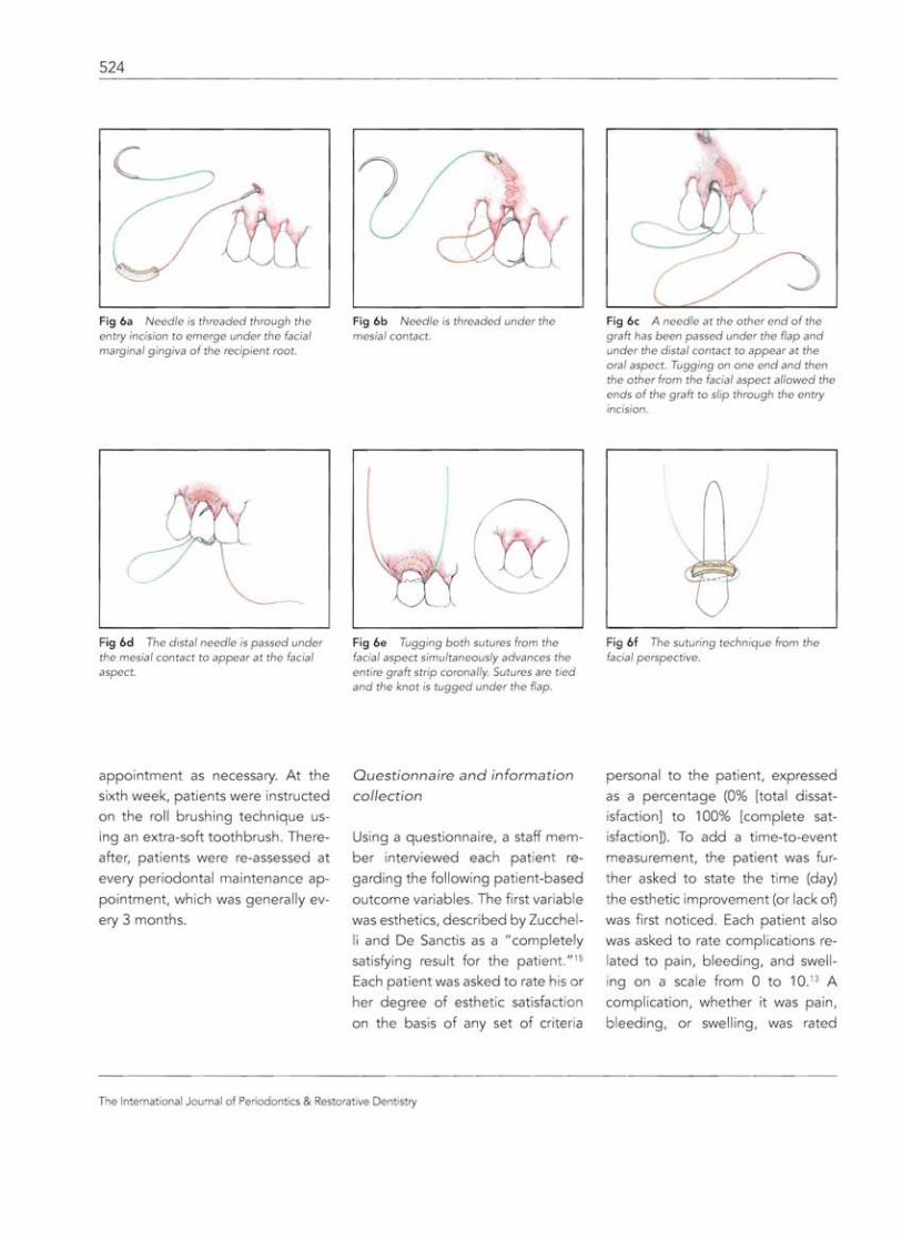
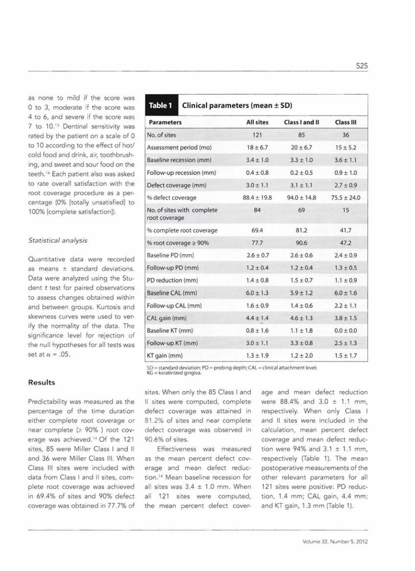