OF CHILDHOOD ASTHMA R. GAGLIARDO A. BONANNO1, A.M ...56... · in the lungs of sensitized...
14
PROOF PROOF INTERNATIONAL JOURNAL OF IMMUNOPATHOLOGY AND PHARMACOLOGY Vol. 26, no. 3, 0-0 (2013) 675 Key words: airway inflammation, airway epithelium, TGF-β1, childhood asthma, induced sputum Mailing address: Rosalia Gagliardo, PhD Istituto di Biomedicina e Immunologia Molecolare, Sezione di Immunopatologia e Farmacologia Clinica e Sperimentale dell’Apparato Respiratorio, CNR. Via Ugo La Malfa, 153 90146 Palermo, Italy Tel.: +39 091 6809150 Fax: +39. 091 6809122 e-mail: [email protected] TGF-β-targeting structural and inflammatory cells has been implicated in the mechanisms leading to the inflammatory and restructuring processes in asthma, suggesting an impact of TGF-β1 signaling on the development and persistency of this disease. We investigated the potential early involvement of TGF-β1 activity in the immunological and molecular mechanisms underlying progression of inflammation in childhood asthma. We evaluated the levels of TGF-β1 in induced sputum supernatants (ISSs) and the expression of small mother cell against decapentaplegic (Smad) 2 and Smad7 proteins in induced sputum cells (ISCs) from children with intermittent asthma (IA), moderate asthma (MA) and control subjects (C). Furthermore, we investigated the regulatory role of TGF-β1 activity on eosinophil and neutrophil adhesion to epithelial cells using adhesion assay, and on the granulocyte expression of adhesion molecule CD11b/CD18 Macrophage-1 antigen (MAC-1), by flow cytometry. We found that the levels of TGF-β1 are increased in ISSs of IA and MA in comparison to C, concomitantly to the activation of intracellular signaling TGFβ/Smads pathway in ISCs. In MA, TGF-β1 levels correlated with the number of sputum eosinophils and neutrophils. Furthermore, we showed the ability of sputum TGF-β1 to promote eosinophil and neutrophil adhesion to epithelial cells, and to increase the expression of MAC- 1 on the granulocyte surface. This study shows the activation of TGFβ/Smad signaling pathway in the airways of children with IA and, despite the regular ICS treatment, in children with MA, and provides evidence for the contribution of TGF-β1 in the regulation of granulocyte activation and trafficking. THE ROLE OF TRANSFORMING GROWTH FACTOR-β1 IN AIRWAY INFLAMMATION OF CHILDHOOD ASTHMA R. GAGLIARDO 1 , P. CHANEZ 2 , M. GJOMARKAJ 1 , S. LA GRUTTA 1 , A. BONANNO 1 , A.M. MONTALBANO 1 , C. DI SANO 1 , G. D. ALBANO 1 , D. GRAS 2 , G. ANZALONE 1 , L. RICCOBONO 1 and M. PROFITA 1 1 Institute of Biomedicine and Molecular Immunology, Unit of Immunopathology and Pharmacology of the Respiratory System, “A. Maurizio Vignola” Laboratories, Italian National Research Council, Palermo, Italy; 2 Department of Respiratory Diseases, AP-HM, INSERM U1067, UMR7733, Aix Marseille University, Marseille, France Received May, 3, 2013 – Accepted July 22, 2013 0394-6320 (2013) Copyright © by BIOLIFE, s.a.s. This publication and/or article is for individual use only and may not be further reproduced without written permission from the copyright holder. Unauthorized reproduction may result in financial and other penalties Transforming growth factor-β (TGF-β) has been implicated in the mechanisms leading to the deposition of extracellular matrix proteins in the airways of asthmatic patients and is a likely candidate in contributing to the inflammatory processes and structural changes (1-3). TGF-β1 is overexpressed in patients with asthma and is considered one of the major fibrogenic factors with potential immunomodulatory activities. TGF-β1 protein is expressed by structural cells, such as
Transcript of OF CHILDHOOD ASTHMA R. GAGLIARDO A. BONANNO1, A.M ...56... · in the lungs of sensitized...

PROOFPROOF
INTERNATIONAL JOURNAL OF IMMUNOPATHOLOGY AND PHARMACOLOGY Vol. 26, no. 3, 0-0 (2013)
675
Key words: airway inflammation, airway epithelium, TGF-β1, childhood asthma, induced sputum
Mailing address: Rosalia Gagliardo, PhDIstituto di Biomedicina e Immunologia Molecolare, Sezione di Immunopatologia e Farmacologia Clinica e Sperimentale dell’Apparato Respiratorio, CNR. Via Ugo La Malfa, 153 90146 Palermo, ItalyTel.: +39 091 6809150 Fax: +39. 091 6809122e-mail: [email protected]
TGF-β-targeting structural and inflammatory cells has been implicated in the mechanisms leading to the inflammatory and restructuring processes in asthma, suggesting an impact of TGF-β1 signaling on the development and persistency of this disease. We investigated the potential early involvement of TGF-β1 activity in the immunological and molecular mechanisms underlying progression of inflammation in childhood asthma. We evaluated the levels of TGF-β1 in induced sputum supernatants (ISSs) and the expression of small mother cell against decapentaplegic (Smad) 2 and Smad7 proteins in induced sputum cells (ISCs) from children with intermittent asthma (IA), moderate asthma (MA) and control subjects (C). Furthermore, we investigated the regulatory role of TGF-β1 activity on eosinophil and neutrophil adhesion to epithelial cells using adhesion assay, and on the granulocyte expression of adhesion molecule CD11b/CD18 Macrophage-1 antigen (MAC-1), by flow cytometry. We found that the levels of TGF-β1 are increased in ISSs of IA and MA in comparison to C, concomitantly to the activation of intracellular signaling TGFβ/Smads pathway in ISCs. In MA, TGF-β1 levels correlated with the number of sputum eosinophils and neutrophils. Furthermore, we showed the ability of sputum TGF-β1 to promote eosinophil and neutrophil adhesion to epithelial cells, and to increase the expression of MAC-1 on the granulocyte surface. This study shows the activation of TGFβ/Smad signaling pathway in the airways of children with IA and, despite the regular ICS treatment, in children with MA, and provides evidence for the contribution of TGF-β1 in the regulation of granulocyte activation and trafficking.
THE ROLE OF TRANSFORMING GROWTH FACTOR-β1 IN AIRWAY INFLAMMATION OF CHILDHOOD ASTHMA
R. GAGLIARDO1, P. CHANEZ2, M. GJOMARKAJ1, S. LA GRUTTA1,A. BONANNO1, A.M. MONTALBANO1, C. DI SANO1, G. D. ALBANO1, D. GRAS2,
G. ANZALONE1, L. RICCOBONO1 and M. PROFITA1
1Institute of Biomedicine and Molecular Immunology, Unit of Immunopathology and Pharmacology of the Respiratory System, “A. Maurizio Vignola” Laboratories, Italian National Research Council,
Palermo, Italy; 2Department of Respiratory Diseases, AP-HM, INSERM U1067, UMR7733, Aix Marseille University, Marseille, France
Received May, 3, 2013 – Accepted July 22, 2013
0394-6320 (2013)Copyright © by BIOLIFE, s.a.s.
This publication and/or article is for individual use only and may not be furtherreproduced without written permission from the copyright holder.
Unauthorized reproduction may result in financial and other penalties
Transforming growth factor-β (TGF-β) has been implicated in the mechanisms leading to the deposition of extracellular matrix proteins in the airways of asthmatic patients and is a likely candidate in contributing to the inflammatory
processes and structural changes (1-3). TGF-β1 is overexpressed in patients with asthma and is considered one of the major fibrogenic factors with potential immunomodulatory activities. TGF-β1 protein is expressed by structural cells, such as

PROOFPROOF
676
PROOF
R. GAGLIARDO ET AL.
epithelial cells, fibroblasts, and smooth muscle cells, and also by inflammatory cells, such as neutrophils and eosinophils. The signaling cascade following TGF-β binding to its receptor complex is via the Smad pathway (4). The activated TGF-β receptor induces phosphorylation of Smad2 and Smad3 which form heterooligomeric complexes with Smad 4. The complexes then translocate to the nucleus and regulate transcription of many inflammatory genes. Inhibitory Smads, including Smad6 and Smad7, have been identified as intracellular antagonists of TGF-β signaling by preventing TGF-β receptor activation (4). Upregulation of Smad proteins has been found in the lungs of sensitized ovalbumin-exposed mice, indicating that Smad-mediated signaling may be important in pathophysiology of allergic diseases (5).
TGF-β1 is a cytokine that affects many different cell processes in airways of asthmatic patients by targeting structural and inflammatory cells, suggesting an impact of TGF-β1 signaling on the development of this disease. The presence of chronic inflammation in airways is associated with bronchial obstruction and with remodeling of the airways (6). Airway epithelial cells are central to the pathogenesis of asthma and the function of the airway epithelium is further modified by local inflammatory/immune signals often promoting eosinophilic and neutrophilic inflammation in asthma (7). Granulocyte trafficking is regulated by surface molecules, such as CD11b/CD18 macrophage-1 antigen (MAC-1), that modulate the interaction with epithelial cells via intercellular adhesion molecules (ICAM-1), promoting granulocyte infiltration and survival in inflammatory sites (8).
It has been postulated that airway structural changes are the consequence of chronic inflammation, although there is evidence that suggests a dissociation between at least some of the inflammatory and remodeling airway events in childhood asthma (9, 10). In a previous study, we demonstrated that pro-remodeling process, although without defined effects, is initiated at the early steps of the disease, and is associated with airway inflammation, as demonstrated by the concomitant overexpression of IL-8, MMP-9 and TIMP-1 in induced sputum of asthmatic children (11). However, relatively few data are available on the definition of the immunological
and molecular mechanisms underlying progression of inflammation, and on the potential involvement of TGF-β1 activity in childhood asthma (11-14).
The aim of the present study was to investigate, both ex vivo and in vitro, the immunomodulatory role of TGF-β1 on the regulation of inflammatory and structural cells in childhood asthma, using non-invasive induced sputum method. We evaluated the levels of TGF-β1 in induced sputum supernatants and the expression of phosphorylated-Smad2 and Smad7 proteins in sputum cells from children with intermittent and moderate asthma. In addition, to further characterize the inflammatory and immunological events associated with the levels of TGF-β present in the airways of asthmatic children, we investigated, in vitro, the role of sputum TGF-β1, in the regulation of adhesion mechanisms of inflammatory cells to airway epithelium, and in the granulocyte activation and trafficking.
MATERIALS AND METHODS
SubjectsForty asthmatic children were recruited from the
outpatient clinic of the CNR. The diagnosis of asthma and the assessment of its severity and control were carried out at study entry according to Global Initiative for Asthma (15). Only 30 out of 40 asthmatic children were able to give suitable sputum samples and to be included in the study.
Ten children had intermittent asthma (IA), treated with short-acting ß2agonists on demand during the last 3 months; 20 children had moderate asthma (MA) treated with Fluticasone Propionate 250 µg (inhaler device Diskus [GlaxoSmithKline, UK]) plus Salmeterol 50 µg bid, for at least 3 months. This high dose of Fluticasone Propionate was used to control the disease, at least considering the clinical and functional parameters. The control group consisted of seven healthy children (C).
The study was approved by the Istitutional Ethics Committee of the Policlinic Hospital of Palermo University and complied with the Declaration of Helsinki. Written informed consent was obtained from the parents of the patients enrolled in the study.
Clinical assessment and compliance of patients Pulmonary function tests were performed as
recommended by the American Thoracic Society (16). Forced expiratory volume in one second (FEV1), forced vital capacity (FVC) were measured according to ATS guidelines and the best of 3 technically acceptable and

PROOFPROOF
677Int. J. Immunopathol. Pharmacol.
the manufacturer’s instructions. In order to measure biologically active TGF-β1, the samples were acidified using HCl for 1 h (22). After acidification the samples were neutralized to pH 7.0–7.4 with NaOH and measurements of TGF-β1 were performed immediately. The detection limit of the assay was 0.05 ng/ml. The TGF-β1 assay was validated in line with European Respiratory Society recommendations (23), assessing the effect of DTT on the recovery of exogenous recombinant TGF-β1 (500pg/ml) spiked in sputum samples from IA (n=3) and MA (n=3). The recovery of spiked TGF-β1 samples was 90±5.6%.
Immunocytochemistry analysisAfter thawing, immunocytochemical analyses for
phosphorylated Smad2 and Smad7 proteins, were evaluated in induced sputum cells (ISCs) using the labeled streptavidin-biotin method (Alkaline phosphatase Rabbit/Mouse/Goat; Universal LSAB + kit; Dako, Glostrup, Denmark), as previously described (11). Anti-phosphorylated Smad2 and anti-Smad7 antibodies (Cell Signalling Technology, Inc, MA, USA) were diluted 1:50.
Isolation of peripheral blood granulocytesPeripheral blood granulocytes (PBG) were isolated
from healthy child donors by means of dextrane sedimentation and centrifugation over Ficoll cushion, as previously described (24).
Eosinophil and neutrophil separationIn order to perform adhesion assay, neutrophils
and eosinophils were separated from the total PBG, by immunomagnetic cell sorting (25). Briefly, CD16+ neutrophils were magnetically labeled with CD16 microbeads (Miltenyi Biotec). Then the cell suspension was loaded onto a column placed in the magnetic field of a magnetic cell sorting separator (Miltenyi Biotec). The magnetically labeled CD16+ cells were retained on the column and the unlabeled eosinophils run through, and this cell fraction was depleted of CD16+ neutrophils. After removal of the column from the magnetic field, CD16+ positive neutrophils were eluted as the positive selected cell fraction.
Epithelial cell cultures16-HBE is a cell line that retains the differentiated
morphology and function of normal airway epithelial cells. The cells represent a clonal diploid (2n=6) cell line isolated from human lung previously used to study the functional properties of bronchial epithelial cells in inflammation and repair processes. 16-HBE cells were cultured as adherent monolayers in Eagle’s minimum essential medium (MEM) supplemented with 10% heat-inactivated (56°C, 30 min) fetal bovine serum (FBS),
reproducible manoeuvres was retained (16). Atopic status was assessed by skin prick tests (17) to aeroallergens commonly present in the Mediterranean area, and total serum IgE measurements. The compliance to treatment was assessed by checking the inhalation technique. We also measured basal plasma cortisol concentrations at 8:00a.m. by electrochemiluminescence. Results were expressed in nanomolar concentrations, and adherence to inhaled corticosteroids was considered satisfactory if cortisol was less than 100 nM (18, 19).
Sputum induction and processingEach subject underwent spirometry before the
beginning of the procedure. If FEV1 was over 75% of predicted at baseline and the child had not used a short-acting ß2-agonist within the previous 6 h, we performed the procedure without premedication. Patients were exposed to an aerosol of 4% hypertonic saline solution, monitoring the assessment of bronchial reactivity, as described elsewhere (11, 20).
Sputum processing was performed according to European Respiratory Society recommendation (21). Sputum was collected in previously weighed 50-ml sterile ampoules. The volume of the induced sputum was previously determined, and an equal volume of DTT (0.1% in saline; Sigma, St. Louis, MO) was added to the selected sputum. After homogenization, sputum samples were filtered (48-µm nylon cell strainer) and subsequently centrifuged at 800 g for 10 min to separate the supernatants from the cell pellet. The supernatants were then aspirated and frozen at -80°C for the subsequent biochemical analysis. The cells obtained from induced sputum were then cytocentrifuged (Cytospin 2, Shandon Instruments, Runcorn, UK) and stained with Diff-Quick (Merz-Dade, Dudingen, CH). Slides were read blindly by two independent investigators and differential cell counts were expressed as a percentage of 400 cells. The cytospins for immunocytochemistry were prepared on 3-aminopropyltriethoxysilane (APTEX)-coated slides by adding 100 µl of cell suspension (5x10⁵ cells/ml) into Shandon II cytocentrifuge cups and centrifuging at 180 g for 5 min. The air-dry slides were fixed in paraformaldehyde-lysine-periodate (PLP) for 30 min and in 15% sucrose in Dulbecco’s PBS for 30 min. The slides were stored at -80°C until use for immunocytochemical staining.
Measurement of TGF-β1
Determinations of TGF-β1 were assessed in induced sputum supernatants (ISSs) recovered from C, IA and MA children, using commercially available specific enzyme-linked immunoadsorbent assay kit (US Biological, and Amersham International plc, UK), according to

PROOFPROOF
678
PROOFconcentration used for the induced sputum processing. At the end of the incubation time, granulocytes were assessed as described below for macrophage-1 antigen (MAC-1) CD11b/CD18 expression.
Flow cytometry for MAC-1 expression Granulocytes were washed twice in PBS (containing
1% FCS and 0.02% Na-azide) and thereafter stained and subjected to flow cytometric analysis. The expression of MAC-1 on granulocytes was evaluated by flow-cytometry analyses by direct label immunofluorescence by a FACS Calibur™ flow cytometer (Becton Dickinson, CA, USA), as previously described, using (RPE)-conjugated mouse monoclonal antibody direct against an Anti-Human CD11b/CD18 (MAC-1) (DAKO A/S, Glostrup, Denmark). Granulocytes were gated by forward and side scatter and analysis was carried out on 50,000 acquired events for each sample. Results of flow cytometry were expressed as percentage of positive cells.
Statistical analysisStatistical comparisons in order to test differences
between the three groups (C, IA, MA) were made by use of the Kruskall-Wallis test followed by Bonferroni’s test correction for multiple comparisons. Data were expressed as median and inter quartile range. Correlations were calculated according to Spearman Rank test. ANOVA and unpaired t-test was used for the analysis of the data obtained from in vitro experimental conditions. Data were expressed as mean ± S.D.
RESULTS
Demographic and functional characteristics of patients
The demographic and functional characteristics of patients are reported in Table I. No statistically significant differences were found among the three study groups in terms of FEV1 values indicating that, on the basis of this parameter, the asthmatic patients were functionally under control. We observed a significant FEV1/FVC decline in MA in comparison to C, and in MA in comparison to IA.
Total and differential cell counts in induced sputum samples
The percentage of eosinophils in induced sputum samples was significantly higher in IA and MA than in C (Table II). The IA and MA also had a significantly increased percentage of sputum neutrophils and a significantly decreased percentage
1% MEM (non-essential aminoacids, Euroclone), 2 mM L-glutamine and gentamicin 250 µg/ml at 37°C in a humidified 5% CO2 atmosphere.
16-HBE stimulation for eosinophil and neutrophil adhesion
16-HBE (70,000 cells/well) were plated in standard 24-well culture plates in MEM 10% FCS and grown to confluence. After 24 h with MEM 1% FCS, medium was replaced and 16-HBE cells were stimulated with ISSs (10% in fresh MEM 1% FCS) from C, IA and MA for 24 h. To determine the contribution of TGF-β1 present in ISSs in regulating eosinophil and neutrophil adhesion capacity, ISSs from 5 C, 6 IA and 6 MA were pretreated with or without a monoclonal anti-TGF- β1 antibody (4 µg/ml) (R&D Systems Europe Ltd, Abingdon, UK) for 1 h at 4°C to neutralize sputum TGF- β1 activity, before co-incubation with 16-HBE.
Adhesion assayEosinophils and neutrophils were purified as described
above, resuspended in PBS (2 x 10⁶/ml) and labeled for 45 min at 37°C with an equal volume of the fluorochromic dye SFDA (50 µg/ml) (Molecular Probes). After labelling cells were washed, resuspended in PBS (0.4 x 10⁶/ml) and co-incubated with epithelial cells in 24-well culture plates. Medium containing ISSs, or DTT alone, was removed from the 16-HBE cultures, before the addition of labeled eosinophils or neutrophils.
The plates were incubated at 37°C for 25 min to allow eosinophils and neutrophils to contact and to adhere to the confluent 16-HBE, and total fluorescence was measured using an excitation wavelength of 485 nm and monitoring emission at 530 nm in a Wallac 1420 Victor multilabel counter (PerkinElmer Life and Analytical Sciences-Wallac OY, Turku, Finland). Later, non-adherent cells were removed by washing and fluorescence was measured to evaluate bound cells. Adhesion was expressed as percentage of the fluorescence ratio of bound cells on total cells. All test points were performed in triplicate. The baseline values represent the adhesion of eosinophils and neutrophils to unstimulated 16-HBE.
Peripheral blood granulocytes stimulation for MAC-1 expression
Granulocytes were resuspended and cultured at the concentration of 4x10⁵/ml in vials with fresh RPMI 1% FCS. ISSs from 5 C, 6 IA and 6 MA were pretreated with a monoclonal anti-TGF-β1 antibody (4 µg/ml) for 1 h at 4°C to neutralize sputum TGF-β1 activity. Thereafter, granulocytes were stimulated for 2 h with selected ISSs pretreated or not with anti-TGFβ neutralizing antibody, or incubated with medium containing DTT alone at the same
R. GAGLIARDO ET AL.

PROOFPROOF
679Int. J. Immunopathol. Pharmacol.
Table I. Demographic and functional characteristics of subjects.
Intermittent Moderate pValue Controls Asthma Asthma C/IA C/MA IA/MA
Number 7 10 20 Sex, M/F 5/2 6/4 12/8 Age 10(9-11) 8(7-10) 11(10-13) NS NS NS Total IgE(KU) 20(15-20) 522(308-857) 536(387-770) p<0.002 p<0.001 NS FEV1(%) 101(100-103) 96(94.5-104) 92(89-96) NS NS NS FEV1/FVC(%) 100(98.5-105) 93(88-109) 85(74-86) NS p<0.001 p<0.005 Asthma duration, yr NA 4(3-6) 6(4.5-7) NS Cortisol (nM)† 226(190-280) 194(170-225) 66(58-95) NS p<0.001 p<0.004 Results are expressed as median (25-75 percentiles). † Cortisol levels at study entry.
Table I. Demographic and functional characteristics of subjects.
Results are expressed as median (25-75 percentiles).† Cortisol levels at study entry.
Table II. Total and differential count of cells from induced sputum.
C IA MA C/IA C/MA IA/MA
Total cell counts (106cells/ml) 0.8(0.5-1.5) 1.5(0.8-1.9) 1.2 (0.6-1.8) NS NS NS
Squamous cells (%) 2.5(2-5) 2.5(1-4) 2(0-3) NS NS NS Macrophages (%) 90(89-95) 59.5(49.5-70.6) 58.5(25-63) ** ** NS Eosinophils (%) 0 5.5(3-8) 4.5(3-9) ** ** NS Neutrophils (%) 8(4-11) 28(6-33) 31.5(7-36) * ** NS Lymphocytes (%) 0(0-0.5) 3.5(1-3) 2.5(1.5-4.5) NS NS NS
Epithelial cells (%) 0 (0-2.6) 0.5 (0-2) 1(0-1.8) NS NS NS
Results are expressed as medians (25-75 percentiles) in respect to total cell counts. **, p<0.001; * , p<0.01; NS: not significant
Table II. Total and differential count of cells from induced sputum.
Results are expressed as medians (25-75 percentiles) in respect to total cell counts.**, p<0.001; * , p<0.01; NS: not significant
PROOFPROOF
680
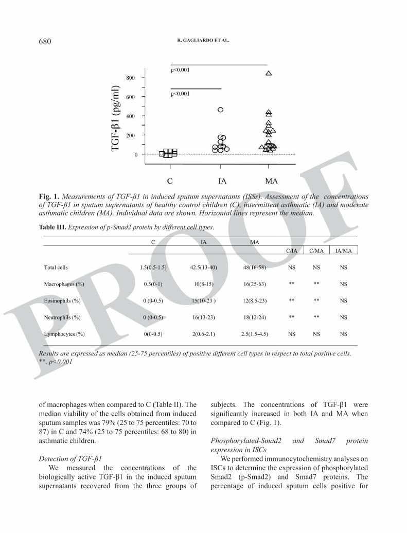
PROOFsubjects. The concentrations of TGF-β1 were significantly increased in both IA and MA when compared to C (Fig. 1).
Phosphorylated-Smad2 and Smad7 protein expression in ISCs
We performed immunocytochemistry analyses on ISCs to determine the expression of phosphorylated Smad2 (p-Smad2) and Smad7 proteins. The percentage of induced sputum cells positive for
of macrophages when compared to C (Table II). The median viability of the cells obtained from induced sputum samples was 79% (25 to 75 percentiles: 70 to 87) in C and 74% (25 to 75 percentiles: 68 to 80) in asthmatic children.
Detection of TGF-β1We measured the concentrations of the
biologically active TGF-β1 in the induced sputum supernatants recovered from the three groups of
Fig. 1. Measurements of TGF-β1 in induced sputum supernatants (ISSs). Assessment of the concentrations of TGF-β1 in sputum supernatants of healthy control children (C), intermittent asthmatic (IA) and moderate asthmatic children (MA). Individual data are shown. Horizontal lines represent the median.
Table III. Expression of p-Smad2 protein by different cell types.
C IA MA C/IA C/MA IA/MA
Total cells 1.5(0.5-1.5) 42.5(13-40) 48(16-58) NS NS NS
Macrophages (%) 0.5(0-1) 10(8-15) 16(25-63) ** ** NS Eosinophils (%) 0 (0-0.5) 15(10-23 ) 12(8.5-23) ** ** NS Neutrophils (%) 0 (0-0.5) 16(13-23) 18(12-24) ** ** NS Lymphocytes (%) 0(0-0.5) 2(0.6-2.1) 2.5(1.5-4.5) NS NS NS
Results are expressed as median (25-75 percentiles) of positive different cell types in respect to total posit**, p<0.001
Table IV. Expression of Smad7 protein by different cell types.
C IA MA C/IA C/MA
Total cells 20(16-23) 24(20-25) 23(15-30) NS NS
Macrophages (%) 10(2-8) 6.5(1-8) 6(3-7) NS NS Eosinophils (%) 4(3-8) 8(4-14) 7(5-9) NS NS Neutrophils (%) 4.5(2-9) 7(3-12) 8(7-11) NS NS Lymphocytes (%) 2(0-2) 2.5(1-4) 2(1.5-3) NS NS
Results are expressed as median (25-75 percentiles) of positive different cell types in respect to total posit**, p<0.001
Table III. Expression of p-Smad2 protein by different cell types.
Results are expressed as median (25-75 percentiles) of positive different cell types in respect to total positive cells.**, p<0.001
R. GAGLIARDO ET AL.

PROOFPROOF
681Int. J. Immunopathol. Pharmacol.
nucleus of IA and MA. No differences were detected between the two groups of asthmatic patients. The percentage of sputum macrophages, eosinophils and
p-Smad2 (Fig. 2A), were significantly higher in children with IA and MA than in C. We found the presence of p-Smad2 protein in both cytosol and
Table III. Expression of p-Smad2 protein by different cell types.
C IA MA C/IA C/MA IA/MA
Total cells 1.5(0.5-1.5) 42.5(13-40) 48(16-58) NS NS NS
Macrophages (%) 0.5(0-1) 10(8-15) 16(25-63) ** ** NS Eosinophils (%) 0 (0-0.5) 15(10-23 ) 12(8.5-23) ** ** NS Neutrophils (%) 0 (0-0.5) 16(13-23) 18(12-24) ** ** NS Lymphocytes (%) 0(0-0.5) 2(0.6-2.1) 2.5(1.5-4.5) NS NS NS
Results are expressed as median (25-75 percentiles) of positive different cell types in respect to total posit**, p<0.001
Table IV. Expression of Smad7 protein by different cell types.
C IA MA C/IA C/MA
Total cells 20(16-23) 24(20-25) 23(15-30) NS NS
Macrophages (%) 10(2-8) 6.5(1-8) 6(3-7) NS NS Eosinophils (%) 4(3-8) 8(4-14) 7(5-9) NS NS Neutrophils (%) 4.5(2-9) 7(3-12) 8(7-11) NS NS Lymphocytes (%) 2(0-2) 2.5(1-4) 2(1.5-3) NS NS
Results are expressed as median (25-75 percentiles) of positive different cell types in respect to total posit**, p<0.001
Table IV. Expression of Smad7 protein by different cell types.
Results are expressed as median (25-75 percentiles) of positive different cell types in respect to total positive cells.**, p<0.001
Fig. 2. Immunocytochemistry for phosphorylated-Smad2 (p-Smad2) (Panel A) and Smad7 (Panel C) in induced sputum cells from healthy C subjects (n=7), IA (n=10) and MA (n=14) children (magnification at x40). Panels B and D, individual data are shown (percentage of positive cells). Horizontal lines represent the median.

PROOFPROOF
682
PROOFTGF-β1 antibody significantly reduced the ability of eosinophils and neutrophils to adhere to epithelial cells (Fig. 3B and Fig. 4B).
Correlations In moderate asthmatic children, sputum TGF-β1
levels positively correlated with the number of sputum eosinophils and neutrophils (p=0.004, Rho=0.65; p=0.002, Rho=0.88, respectively) (Fig. 5A and 5B). In intermittent asthmatic children, no significant correlations were found between sputum TGF-β1 levels and the number of sputum eosinophils and neutrophils.
Effect of ISSs on the expression of MAC-1 in granulocytes
We showed that MAC-1 expression was
neutrophils positive for p-Smad2 was significantly higher in IA and MA than in C (Table III).
Similar levels of Smad7 protein expression were found in sputum cells from C, IA and MA, with no significant differences between the study groups (Fig. 2B and Table IV).
Effect of ISSs on eosinophil and neutrophil adhesion to 16-HBE
The stimulation of 16-HBE with ISSs obtained from intermittent and moderate asthmatic children, promoted higher levels of adherent eosinophils and neutrophils in comparison to 16-HBE stimulated with ISSs from control children or medium alone (Fig. 3A and Fig. 4A). DTT alone had no effects on eosinophil and neutrophil adhesion (data not shown). ISS neutralizing pre-treatment with anti-
Fig. 3. Adhesion of eosinophils to 16-HBE stimulated with ISSs. A) 16-HBE were stimulated with ISSs obtained from C (n=5), IA (n=6) and MA (n=6) for 24 h and then co-incubated with eosinophils, obtained from normal donors, for 25 min at 37°C before the eosinophil adhesion assay. Results are expressed as percentage of adhering eosinophils (% of fluorescence). B) 16-HBE were stimulated with ISSs obtained from C (n=5), IA (n=6) and MA (n=6), treated with or without a monoclonal neutralizing anti-TGF-β1 antibody for 1 h, before their addition to 16-HBE for 24 h. 16-HBE cells were then incubated with eosinophils obtained from normal donors for 25 min at 37°C before eosinophil adhesion assay. Results are expressed as percentage of inhibition of eosinophil adhesion vs untreated ISSs. The bars represent the means±SD of values. **p<0.001 vs C and baseline.
R. GAGLIARDO ET AL.

PROOFPROOF
683Int. J. Immunopathol. Pharmacol.
Fig. 4. Adhesion of neutrophils to 16-HBE stimulated with ISSs. A) 16-HBE were stimulated with ISSs obtained from C (n=5), IA (n=6) and MA (n=6) for 24 h and then co-incubated with neutrophils, obtained from normal donors, for 25 min at 37°C before the neutrophil adhesion assay. Results are expressed as percentage of adhering neutrophils (% of fluorescence). B) 16-HBE were stimulated with ISSs obtained from C (n=5), IA (n=6) and MA (n=6), treated with or without a monoclonal neutralizing anti-TGF-β1 antibody for 1 h, before their addition to 16-HBE for 24 h. 16-HBE cells were then incubated with neutrophils obtained from normal donors for 25 min at 37°C before neutrophil adhesion assay. Results are expressed as percentage of inhibition of neutrophil adhesion vs untreated ISSs. The bars represent the means±SD of values. *p<0.01 vs C and baseline; **p<0.001 vs C and baseline.
Fig. 5. Correlations between sputum TGF-β1 concentrations and sputum eosinophils (A), and between sputum TGF-β1 concentrations and sputum neutrophils (B), in moderate asthmatic children. Correlations were calculated according to Spearman test.

PROOFPROOF
684
PROOFregulation of adhesion mechanisms of inflammatory cells to airway epithelium, showing its potential role in the granulocyte activation and trafficking in the airways of asthmatic children.
TGF-β1 has a key role in orchestrating both inflammatory and remodeling processes in asthma (26). In bronchial biopsies of asthmatic children, the involvement was observed of TGF-β1 in changes of local tissue microenvironment promoting epithelial stress, structural pre-modeling and production of pro-inflammatory stimuli (9). Increased levels of TGF-β1 were observed in induced sputum of mild to moderate adult asthmatics (27) and in bronchoalveolar lavage fluid of children with severe asthma (28). The major activity of TGF-β1 is associated with eosinophils, although the action of TGF-β1 is also associated with other cell types, such as neutrophils, involved in the pathogenesis
increased in granulocytes stimulated with ISSs from IA and MA patients when compared to granulocytes stimulated with ISSs from C subjects. DTT alone had no effects on the granulocyte MAC-1 surface expression (data not shown). The pre-incubation of ISSs from IA and MA with anti-TGF-β1 neutralizing antibody reduced the expression of MAC-1 on granulocyte surfaces (Fig. 6A and 6B).
DISCUSSION
In the present study we show that the levels of TGF-β1 are increased in induced sputum of intermittent and moderate asthmatic children in comparison to controls, concomitantly to the activation of intracellular signaling TGFβ/Smads pathway in induced sputum cells. Furthermore, we demonstrate the contribution of TGF-β1 in the
Fig. 6. Flow cytometric analysis for MAC-1 protein surface expression in peripheral blood granulocytes (PBG) stimulated with ISSs from C, IA and MA. A) 16-HBE were stimulated for 24 h at 37°C with ISSs from C (n=5 ), IA (n=6) and MA (n=6), pretreated with or without neutralizing anti-TGFβ1 antibody for 1 h. The bars represent the means±SD of the percentage of positive cells. ##p<0.001 vs C; *p<0.01, ** p<0.001, before and after anti-TGFβ1 treatment. B) Representative flow cytometry of MAC-1 protein of each experimental condition (overlay of fluorescence intensity). The left peaks indicate representative flow cytometry of negative control (Dako, Denmark A/S).
R. GAGLIARDO ET AL.

PROOFPROOF
685Int. J. Immunopathol. Pharmacol.
had similar levels in IA, MA and C subjects. These results indicate that disturbed negative regulation of TGF-β/Smad signaling could exist in airways of IA and MA children, thus impairing the excess of Smad2-mediated activation. Moreover, the activation of TGFβ/Smad pathway in MA children, despite the ongoing ICS therapy, supports the concept that some patients display not completely inhibited molecular mechanisms of TGFβ/Smad pathway.
Airway epithelial cells respond to the cytokine milieu driving airway immune response and through the expression of adhesion molecules, may contribute to the recruitment of inflammatory cells, such as eosinophils and neutrophils, leading to the pathophysiological changes typical of the asthmatic airways (7, 9). Based on this knowledge, we evaluated the immunomodulatory role of TGF-β1 in childhood asthma, and its relationship with inflammatory and structural cells by investigating the interaction of sputum TGF-β1 with eosinophils and neutrophils, through the commitment of bronchial epithelium. We observed that the stimulation of human bronchial epithelial cells (16-HBE) with ISSs from IA and MA children promotes an increment of eosinophil and neutrophil adhesion to bronchial epithelial cells, in comparison to ISSs from C children. These findings suggest that the mechanisms of interactions between airway inflammatory microenvironment and epithelial cells facilitate the origin of signals promoting eosinophil and neutrophil recruitment. Furthermore, the depletion of TGF-β1, obtained with the pre-treatment of ISSs with anti-TGF-β1, reduced the percentage of adhesion of eosinophils and neutrophils to 16-HBE. Our results suggest that higher levels of bronchial TGF-β1, in childhood asthma, might be involved in the regulation of cellular adhesion mechanisms involving activated granulocytes and airway epithelium.
Corticosteroids are considered as first-line anti-inflammatory treatment, especially in chronic asthma, although the increase of TGF-β1 levels in vivo was shown to persist despite corticosteroid treatment in moderate to severe adult asthma (33). These observations suggested that anti-inflammatory therapy does not completely prevent inflammatory and remodeling processes associated with TGF-β1. We found that in moderate asthmatic children the depletion of TGF-β1 from ISSs showed a reduced
of asthma and persistence of inflammatory process (26). Eosinophils are recognized as major cellular mediators of airway inflammatory processes in asthma, although increased neutrophils are a feature of airway inflammation in a subgroup of adult and pediatric asthmatic patients with more severe disease (11, 29). In this scenario, eosinophilic and neutrophilic inflammation are not mutually exclusive in asthma, suggesting that the complex immunopathogenesis of inflammation is the result of immunological and molecular heterogeneity (11, 14, 30). Accordingly, our findings show increased levels of TGF-β1, in ISSs obtained from IA and MA children, compared with C subjects, together with the increased number of sputum eosinophils and neutrophils. These observations suggest that inflammation and pro-remodeling markers, such as TGF-β1, could be present simultaneously in the airways of asthmatic children. Although no significant differences in TGF-β1 levels were observed between IA and MA children, we identified the presence of a sub-population of MA with high sputum concentrations of TGF-β1, despite the regular inhaled corticosteroid (ICS) treatment. The precise initiation and progression of inflammatory events and the factors responsible for the variable efficacy of corticosteroid treatment are unknown in the airways of asthmatics. In accordance with our previous results (11, 14), these findings further suggest the presence of a biological heterogeneity in children with moderate asthma and the need to better characterize, especially in children, the immunologic and inflammatory mechanisms driving the asthma progression associated with TGF-β1.
The intracellular signaling TGFβ/Smads pathway and its relationship with airway inflammation have been extensively studied in human and in animal models (5, 31). Elevated phosphorylated Smad-2 levels have been demonstrated in bronchial biopsies obtained from adult asthmatic subjects, indicating that TGF-β1 signaling downstream from the receptor, is enhanced in asthma (31). Altered expression of Smad7 has been associated with inflammatory and fibrotic disorders with a regulator negative-feedback mechanism for TGF-β signaling (32). We observed that TGF- β/Smad2 pathway is active in airway sputum cells from children with IA and MA, while the expression of inhibitor Smad7

PROOFPROOF
686
PROOFACKNOWLEDGEMENTS
This study was supported by the Italian National Research Council (CNR), and Glaxo SmithKline.
P. Chanez has acted as a consultant for Actelion, AstraZeneca, Centocor, Chiesi, Glaxo SmithKline, MSD, Novartis and Nycomedand, and has received research support from Centocor and Schering-Plough. The rest of the authors declare that they have no conflict of interest.
REFERENCES
1. Bartram U, Speer CP. The role of transforming growth factor beta in lung development and disease. Chest 2004; 125:754-65.
2. Border WA, Noble NA. Transforming growth factor beta in tissue fibrosis. N Engl J Med 1994; 331:1286-92.
3. Vignola AM, Chanez P, Chiappara G, et al. Transforming growth factor-beta expression in mucosal biopsies in asthma and chronic bronchitis. Am J Respir Crit Care Med 1997; 156:591-9
4. Schmierer B, Hill CS. TGFbeta-SMAD signal transduction: molecular specificity and functional flexibility. Nat Rev Mol Cell Biol 2007; 8:970-82.
5. Rosendahl A, Checchin D, Fehniger TE, ten Dijke P, Heldin CH, Sideras P. Activation of the TGF-beta/activin-Smad2 pathway during allergic airway in-flammation. Am J Respir Cell Mol Biol 2001; 25:60-8.
6. Boxall C, Holgate ST, Davies DE. The contribution of transforming growth factor-beta and epidermal growth factor signalling to airway remodelling in chronic asthma. Eur Respir J 2006; 27:208-29.
7. Thompson AB, Robbins RA, Romberger DJ, Sisson JH, Spurzem JR, Teschler H, Rennard SI. Immunological functions of the pulmonary epithelium. Eur Respir J 1995; 8:127-49.
8. Yan SR, Sapru K, Issekutz AC. The CD11/CD18 (beta2) integrins modulate neutrophil caspase activation and survival following TNF-alpha or endotoxin induced transendothelial migration. Immunol Cell Biol 2004; 82:435-46.
9. Fedorov IA, Wilson SJ, Davies DE, Holgate ST. Epi-thelial stress and structural remodelling in childhood
granulocyte adhesion capacity to epithelium, supporting the concept that ICS used in the treatment of asthma does not completely down-regulate TGF-β1 levels and that the increased levels of TGF-β1 might be able to induce the persistence of inflammatory processes. Accordingly, we found that the percentage of eosinophils and neutrophils in induced sputum samples, positively correlate with sputum TGF-β1 levels in children with MA.
MAC-1 is a transmembrane protein containing α (CD11b) and β (CD18) chains, overexpressed on the surface of peripheral blood granulocytes of asthmatic children (34) and plays an important role in granulocyte activation and permeation into asthmatic airways (35). To clarify the relationship between TGF-β1 overexpression in the airways of asthmatic children and its role in the regulation of airway inflammation associated with granulocyte adhesion capacity, we tested the effects of TGF-β1 present in the induced sputum obtained from asthmatic children on MAC-1 expression. We showed that ISSs from IA and MA children increased the MAC-1 expression in comparison to ISSs from C in granulocytes isolated from healthy child donors and that the depletion of sputum TGF-β1 promotes the inhibition of MAC-1 in both asthmatic groups. These findings suggest that MAC-1 might be involved in TGF-β1 priming granulocytes with a potential role in promoting adhesion and tissue infiltration.
In conclusion, taken together our results suggest an impact of TGF-β1 signaling on cell-cell interaction, recruitment and modulation of inflammatory cells and describe for the first time a dynamic mechanism resulting from the interaction between TGF-β1 signaling activity and inflammatory cells through the commitment of bronchial epithelium, indicating a putative immunoregulatory role of TGF- β1 in the progression of airway inflammatory processes in asthmatic children.
Since airway inflammation is a heterogeneous process resulting from the release of several pro-inflammatory and fibrogenic factors, identifying biomarkers and immunomodulatory mechanisms in childhood asthma could contribute to better understand the disease pathogenesis and to direct more efficient therapies for these young asthmatic patients to control inflammation and airway structural change.
R. GAGLIARDO ET AL.

PROOFPROOF
687Int. J. Immunopathol. Pharmacol.
in children. Am J Respir Crit Care Med 2001; 164:1146-49.
21. Efthimiadis A, Spanevello A, Hamid Q, et al. Methods of sputum processing for cell counts, immunocytochemistry and in situ hybridisation. Eur Respir J Suppl 2002; 37:19s-23s.
22. Danielpour D. Improved sandwich enzyme-linked immunosorbent assays for transforming growth factor beta 1. J Immunol Methods 1993; 158:17-25.
23. Kelly MM, Keatings V, Leigh R, Peterson C, Shute J, Venge P, Djukanović R. Analysis of fluid-phase mediators. Eur Respir J Suppl 2002; 37:24s-39s.
24. Profita M, Sala A, Bonanno A, et al. Chronic obstructive pulmonary disease and neutrophil infiltration: role of cigarette smoke and cyclooxygenase products. Am J Physiol Lung Cell Mol Physiol 2010; 298:L261-9.
25. Profita M, Sala A, Bonanno A, et al. Increased prostaglandin E2 concentrations and cyclooxygenase-2 expression in asthmatic subjects with sputum eosinophilia. J Allergy Clin Immunol 2003; 112:709-16.
26. Moore B, Murphy RF, Agrawal DK. Interaction of TGF-beta with immune cells in airway disease. Curr Mol Med 2008; 8:427-36.
27. Profita M, Gagliardo R, Di Giorgi R, Bruno A, Riccobono L, Bonanno A, Bousquet J, Vignola AM. In vitro effects of flunisolide on MMP-9, TIMP-1, fibronectin, TGF-beta1 release and apoptosis in sputum cells freshly isolated from mild to moderate asthmatics. Allergy 2004; 59:927-32.
28. Brown SD, Baxter KM, Stephenson ST, Esper AM, Brown LA, Fitzpatrick AM Airway TGF-β1 and oxidant stress in children with severe asthma: association with airflow limitation. J Allergy Clin Immunol 2012; 129:388-96.
29. Wenzel SE, Schwartz LB, Langmack EL, Halliday JL, Trudeau JB, Gibbs RL, Chu HW. Evidence that severe asthma can be divided pathologically into two inflammatory subtypes with distinct physiologic and clinical characteristics. Am J Respir Crit Care Med 1999; 160:1001-8.
30. Bhakta NR, Woodruff PG. Human asthma phenotypes: from the clinic, to cytokines, and back again. Immunol Rev 2011; 242:220-32.
31. Sagara H, Okada T, Okumura K, Ogawa H, Ra C, Fukuda T, Nakao A. Activation of TGF-beta/Smad2
asthma. Thorax 2005; 60:389-94.10. Cokuğraş H, Akçakaya N, Seçkin, Camcioğlu Y,
Sarimurat N, Aksoy F. Ultrastructural examination of bronchial biopsy specimens from children with moderate asthma. Thorax 2001; 56:25-9.
11. Gagliardo R, La Grutta S, Chanez P, et al. Non-invasive markers of airway inflammation and remodeling in childhood asthma. Pediatr Allergy Immunol 2009; 20:780-90.
12. Pohunek P, Warner JO, Turzikova J, Kudrmann J, Roche WR. Markers of eosinophilic inflammation and tissue re-modelling in children before clinically diagnosed bronchial asthma. Pediatr Allergy Immunol 2005;16:43-51.
13. Marguet C, Dean TP, Basuyau JP, Warner JO. Eosinophil cationic protein and interleukin-8 levels in bronchial lavage fluid from children with asthma and infantile wheeze. Pediatr Allergy Immunol 2001; 12:27-33.
14. La Grutta S, Gagliardo R, Mirabella F, Pajno GB, Bonsignore G, Bousquet J, Bellia V, Vignola AM. Clinical and biological heterogeneity in children with moderate asthma. Am J Respir Crit Care Med 2003; 167:1490-5.
15. Global Initiative for Asthma (GINA). Workshop report, global strategy for asthma management and prevention. National Institutes of Health, National Heart, Lung and Blood Institute. Publication N. 02-3659.
16. Miller MR, Hankinson J, Brusasco V, et al; ATS/ERS Task Force. Standardisation of spirometry. Eur Respir J 2005; 26: 319-38.
17. Dreborg S. Skin testing. The safety of skin tests and the information obtained from using different methods and concentrations of allergen. Allergy 1993; 48: 473-5.
18. Eid N, Morton R, Olds B, Clark P, Sheikh S, Looney S. Decreased morning serum cortisol levels in children with asthma treated with inhaled fluticasone propionate. Pediatrics 2002; 109:217-21.
19. Hagg E, Asplund K, Lithner F. Value of basal plasma cortisol assays in the assessment of pituitary-adrenal insufficiency. Clin Endocrinol 1987; 26:221-6.
20. Jones PD, Hankin R, Simpson J, Gibson PG, Henry RL. The tolerability, safety, and success of sputum induction and combined hypertonic saline challenge

PROOFPROOF
688
PROOFIL-17, and type I and type III collagen expression. J Allergy Clin Immunol 2003; 111:1293-8.
34. Sale R, Sabatini F, Silvestri M, Serpero L, Petecchia L, Rossi GA. Concentration-dependent activity of mometasone furoate and dexamethasone on blood eosinophils isolated from atopic children: modula-tion of Mac-1 expression and chemotaxis. Int Immu-nopharmacol 2004; 4:1687-96.
35. Carlos TM, Harlan JM. Leukocyte-endothelial adhe-sion molecules. Blood 1994; 84:2068-101.
signaling is associated with airway remodeling in asthma. J Allergy Clin Immunol 2002; 110:249-54.
32. Asano Y, Ihn H, Yamane K, Kubo M, Tamaki K. Impaired Smad7-Smurf-mediated negative regulation of TGF-beta signaling in scleroderma fibroblasts. J Clin Invest 2004; 113:253-64.
33. Chakir J, Shannon J, Molet S, Fukakusa M, Elias J, Laviolette M, Boulet LP, Hamid Q. Airway remodeling-associated mediators in moderate to severe asthma: effect of steroids on TGF-beta, IL-11,
R. GAGLIARDO ET AL.