of Bluetongue Virus with Affinity for Single-Stranded RNA
7
Vol. 61, No. 11 In Vitro Phosphorylation and Purification of a Nonstructural Protein of Bluetongue Virus with Affinity for Single-Stranded RNA HENDRIK HUISMANS,* ALBERDINA A. VAN DIJK, AND ASNE R. BAUSKIN Department of Biochemistry, Veterinary Research Institute, Onderstepoort, South Africa 0110 Received 27 May 1987/Accepted 28 July 1987 A phosphorylated, nonstructural protein of bluetongue virus, NS2, is synthesized throughout the replication cycle in comparatively large amounts. The protein was detected in both the soluble and particulate fraction of the cytoplasm of infected cells. The particulate NS2 could be solubilized in 0.5 M NaCi. It was found that NS2 in the particulate fraction and immunoprecipitates of NS2 from the soluble protein fraction could be phosphorylated in vitro. It is not known whether the kinase involved is of cellular or viral origin, but after purification of NS2 by affinity chromatography on poly(U)-Sepharose it could still by phosphorylated in vitro Without the addition of exogenous protein kinase. The affinity of NS2 for nucleic acid was also investigated. The protein was found to bind to single-stranded RNA. In the presence of purified bluetongue virus mRNA, NS2 formend a complex with an estimated S value of about 22S. Bluetongue virus (BTV) is the prototype species of the genus Orbivirus and a member of the family Reoviridae (20). The genome consists of 10 segments of double-stranded RNA (dsRNA), each of which is transcribed into mRNA by a core-associated dsRNA-dependent RNA polymerase (12, 19). At least 10 translation products of the BTV mRNA species have been identified (14; A. A. van Dijk and H. Huismans, submitted for publication). These include the seven structural proteins, P1 to P7, and three distinct nonstructural proteins, NS1, NS2, and NS3. Two NS3 peptides, NS3 and NS3A, with molecular weights of 28,000 and 25,000, respectively, can be distinguished. They are both coded for by segment 10 and have almost identical peptide maps. Furthermore, a fourth possible nonstructural protein, designated NS4, has been identified. This protein has a molecular weight of about 16,000, and indications are that it is coded for by genome segment 9, which also codes for structural protein P6 (van Dijk and Huismans, submit- ted). We are interested in relationship between structure and function of the different BTV-specific nonstructural pro- teins. Very little is known about these proteins. The mor- phologically best-characterized protein is NS1, which is synthesized in very large amounts and accumulates in BTV- infected cells as tubular structures of various lengths and a side-to-side diameter of 68 nm (9). Nothing is known about the function of NS1, but it has been shown that it is coded for by one of the most highly conserved of the BTV genome segments (8a). The other major nonstructural protein, protein NS2, is also synthesized in large amounts. Preliminary investiga- tions have indicated a resemblance to aNS of reovirus in that NS2 has an affinity for single-stranded RNA (ssRNA) (8). Such ssRNA-binding nonstructural proteins with a molecu- lar weight of about 40,000 appear to be common to all members of the Reoviridae family and have also been demonstrated in the case of rotavirus (2). In the case of reovirus, uNS binds to reovirus mRNA (10) to form a 19S complex (5). The protein appears to play an * Corresponding author. early role in reovirus replication (13), and it is possible that it acts in the selection and condensation of the 10 mRNA species. The main distinction between BTV NS2 and the nonstructural protein equivalents in the other members of the Reoviridae family is that NS2 appears to be phosphoryl- ated (8). These preliminary results prompted a more detailed inves- tigation of the phosphorylation of NS2 and the ability of NS2 to hind to ssRNA. We have purified NS2 by affinity chro- matography and were able to show that purified NS2 can be phosphorylated in vitro without the addition of an exogenous protein kinase. The isolation of a 22S BTV mRNA-NS2 complex is also described. MATERIALS AND METHODS Cells and virus. The BHK-21 cells used in the investigation were grown as monolayers in Roux flasks, by using modified Eagle medium supplemented with 5% bovine serum. An attenuated strain of BTV serotype 10 (designated 10A) was used throughout the study. It was propagated in the BHK cells by using a plaque-purified, low-passage stock virus suspension (7). Preparation of 35S- and 32P-labeled S100 and P100 fractions from BTV-infected cells. Confluent monolayers of BHK-21 cells in Roux flasks were infected with BTV at a multiplicity of infection of 15 to 30 PFU per cell and incubated at 31°C until the start of the labeling period. After a rinse with methionine-free Eagle medium, the cells were incubated on a rocker platform for 2 h at 37°C in 5 ml of the same medium containing 15 ,uCi of [35S]methionine per ml. 32p labeling was done the same way, except that the cells were rinsed with phosphate-free Eagle medium and then labeled by using 10 ml of phosphate-free Eagle medium containing 10 ,uCi of 32p; per ml. The cells were harvested, concentrated by low-speed centrifugation, and then suspended at 5 x 10⁷ cells per ml in 0.01 STE-TX buffer (0.01 M NaCl, 10 mM Tris, 1 mM EDTA [pH 7.4], 0.5% Triton X-100). Nuclei were removed by centrifugation at 1,500 x g for 5 min and washed once with half the original volume of 0.01 STE-TX. The supernatants were combined (SlO fraction) and centrifuged for 2 h at 45,000 rpm in an SW50.1 rotor through a 2-ml layer of 40% 3589 JOURNAL OF VIROLOGY, Nov. 1987, p. 3589-3595 0022-538X/87/113589-07$02.00/0 Copyright © 1987, American Society for Microbiology Downloaded from https://journals.asm.org/journal/jvi on 23 November 2021 by 102.39.235.162.
Transcript of of Bluetongue Virus with Affinity for Single-Stranded RNA

Vol. 61, No. 11
In Vitro Phosphorylation and Purification of a Nonstructural Proteinof Bluetongue Virus with Affinity for Single-Stranded RNA
HENDRIK HUISMANS,* ALBERDINA A. VAN DIJK, AND ASNE R. BAUSKIN
Department of Biochemistry, Veterinary Research Institute, Onderstepoort, South Africa 0110
Received 27 May 1987/Accepted 28 July 1987
A phosphorylated, nonstructural protein of bluetongue virus, NS2, is synthesized throughout the replicationcycle in comparatively large amounts. The protein was detected in both the soluble and particulate fraction ofthe cytoplasm of infected cells. The particulate NS2 could be solubilized in 0.5 M NaCi. It was found that NS2in the particulate fraction and immunoprecipitates of NS2 from the soluble protein fraction could bephosphorylated in vitro. It is not known whether the kinase involved is of cellular or viral origin, but afterpurification of NS2 by affinity chromatography on poly(U)-Sepharose it could still by phosphorylated in vitroWithout the addition of exogenous protein kinase. The affinity of NS2 for nucleic acid was also investigated. Theprotein was found to bind to single-stranded RNA. In the presence of purified bluetongue virus mRNA, NS2formend a complex with an estimated S value of about 22S.
Bluetongue virus (BTV) is the prototype species of thegenus Orbivirus and a member of the family Reoviridae (20).The genome consists of 10 segments of double-strandedRNA (dsRNA), each of which is transcribed into mRNA bya core-associated dsRNA-dependent RNA polymerase (12,19). At least 10 translation products of the BTV mRNAspecies have been identified (14; A. A. van Dijk and H.Huismans, submitted for publication). These include theseven structural proteins, P1 to P7, and three distinctnonstructural proteins, NS1, NS2, and NS3. Two NS3peptides, NS3 and NS3A, with molecular weights of 28,000and 25,000, respectively, can be distinguished. They areboth coded for by segment 10 and have almost identicalpeptide maps. Furthermore, a fourth possible nonstructuralprotein, designated NS4, has been identified. This proteinhas a molecular weight of about 16,000, and indications arethat it is coded for by genome segment 9, which also codesfor structural protein P6 (van Dijk and Huismans, submit-ted).We are interested in relationship between structure and
function of the different BTV-specific nonstructural pro-teins. Very little is known about these proteins. The mor-phologically best-characterized protein is NS1, which issynthesized in very large amounts and accumulates in BTV-infected cells as tubular structures of various lengths and aside-to-side diameter of 68 nm (9). Nothing is known aboutthe function of NS1, but it has been shown that it is coded forby one of the most highly conserved of the BTV genomesegments (8a).The other major nonstructural protein, protein NS2, is
also synthesized in large amounts. Preliminary investiga-tions have indicated a resemblance to aNS of reovirus in thatNS2 has an affinity for single-stranded RNA (ssRNA) (8).Such ssRNA-binding nonstructural proteins with a molecu-lar weight of about 40,000 appear to be common to allmembers of the Reoviridae family and have also beendemonstrated in the case of rotavirus (2).
In the case of reovirus, uNS binds to reovirus mRNA (10)to form a 19S complex (5). The protein appears to play an
* Corresponding author.
early role in reovirus replication (13), and it is possible thatit acts in the selection and condensation of the 10 mRNAspecies. The main distinction between BTV NS2 and thenonstructural protein equivalents in the other members ofthe Reoviridae family is that NS2 appears to be phosphoryl-ated (8).These preliminary results prompted a more detailed inves-
tigation of the phosphorylation of NS2 and the ability of NS2to hind to ssRNA. We have purified NS2 by affinity chro-matography and were able to show that purified NS2 can bephosphorylated in vitro without the addition of an exogenousprotein kinase. The isolation of a 22S BTV mRNA-NS2complex is also described.
MATERIALS AND METHODSCells and virus. The BHK-21 cells used in the investigation
were grown as monolayers in Roux flasks, by using modifiedEagle medium supplemented with 5% bovine serum. Anattenuated strain of BTV serotype 10 (designated 10A) wasused throughout the study. It was propagated in the BHKcells by using a plaque-purified, low-passage stock virussuspension (7).
Preparation of 35S- and 32P-labeled S100 and P100 fractionsfrom BTV-infected cells. Confluent monolayers of BHK-21cells in Roux flasks were infected with BTV at a multiplicityof infection of 15 to 30 PFU per cell and incubated at 31°Cuntil the start of the labeling period. After a rinse withmethionine-free Eagle medium, the cells were incubated ona rocker platform for 2 h at 37°C in 5 ml of the same mediumcontaining 15 ,uCi of [35S]methionine per ml. 32p labeling wasdone the same way, except that the cells were rinsed withphosphate-free Eagle medium and then labeled by using 10ml of phosphate-free Eagle medium containing 10 ,uCi of 32p;per ml. The cells were harvested, concentrated by low-speedcentrifugation, and then suspended at 5 x 10⁷ cells per ml in0.01 STE-TX buffer (0.01 M NaCl, 10 mM Tris, 1 mM EDTA[pH 7.4], 0.5% Triton X-100). Nuclei were removed bycentrifugation at 1,500 x g for 5 min and washed once withhalf the original volume of 0.01 STE-TX. The supernatantswere combined (SlO fraction) and centrifuged for 2 h at45,000 rpm in an SW50.1 rotor through a 2-ml layer of 40%
3589
JOURNAL OF VIROLOGY, Nov. 1987, p. 3589-35950022-538X/87/113589-07$02.00/0Copyright © 1987, American Society for Microbiology
Dow
nloa
ded
from
http
s://j
ourn
als.
asm
.org
/jour
nal/j
vi o
n 23
Nov
embe
r 20
21 b
y 10
2.39
.235
.162
.

3590 HUISMANS ET AL.
a b c d e f q h i iA
B a b cd e f k1
FIG. 1. Autoradiogram of electrophoretically separated proteinsin BTV-infected BHK cells labeled with either [35S]methionine or
32P; at different intervals after infection. Monolayer cultures in Rouxflasks were infected with 30 PFU per cell and incubated at 31°C. Atthe following intervals p.i., the cells were labeled with 32p or 35Sprecursors as indicated under Materials and Methods: 2 to 4 h (lanea); 7 to 9 h (lane b); 11 to 13 h (lane c); 15 to 17 h (lane d); 20 to 22h (lane e); and 26 to 28 h (lane f). Control cells were labeled at 2 to4 h (lane g) and 20 to 22 h (lane h) after mock infection. The cellswere harvested after the labeling period. The cytoplasmic extractsof the [35S]methionine-labeled cells were analyzed by discontinuousSDS-PAGE (A). The results in panel B show immunoprecipitates ofthe S100 protein extracts from 32Pi-labeled cells after immunopre-cipitation of 200-pl portions of the different S100 fractions withrabbit serum that contained antibodies against the structural andnonstructural BTV proteins (7). An immunoprecipitation controlwith 35S-labeled soluble proteins is shown in lane k. Other controlsare 35S-labeled BTV proteins in purified virus (lane i) and in theparticulate fraction (lane j). 1 to 7, P1 to P7.
sucrose. The supernatant (S100) was divided in small por-tions and kept at -20°C. The pellet (P100) was suspended in0.01 STE (STE-TX without Triton X-100) (using 20% of theoriginal S10 volume) and stored at -20°C.
Preparation of SW fractions. For large-scale preparation, aP100 fraction was prepared from 109 BTV-infected BHKcells as described above and suspended in 4.5 ml of 0.01STE. NaCl was added to a final concentration of 0.5 M, andthe suspension was centrifuged for 1 h at 100,000 x g. Thesupernatant was divided in smaller portions and frozen at-70°C. For the large-scale purification of NS2, unlabeledsalt-wash (SW) fractions were mixed with a suitable amountof a 35S-labeled SW fraction prepared from BTV-infectedcells. SW fractions from uninfected cells were prepared thesame way as those from infected cells.
Preparation of 3H-labeled ssRNA and dsRNA. 3H-labeledBTV mRNA was prepared by in vitro transcription of
CsCI-purified BTV core particles in the presence of [3H]UTP(18). The 3H-labeled BTV dsRNA was obtained by phenol-sodium dodecyl sulfate (SDS) extraction of purified virus(12) that was labeled during the infection cycle with[3H]uridine. 3H-labeled cellular ssRNA was obtained byextraction of uninfected cells labeled during the cellulargrowth cycle with [3H]uridine. Both RNA preparations werepurified by two cycles of precipitation with 2 M LiCl. Theprecipitates were dissolved in water and stored in smallportions at -70°C.
Nitrocellulose-binding assay. A nitrocellulose-binding as-say was done as follows. Five-microgram quantities of eachof the different 3H-labeled nucleic acids were mixed withincreasing amounts of P100 SW fraction from infected oruninfected cells. The mixture was then diluted to a final saltconcentration of 0.2 M NaCl in 10 mM Tris hydrochloride,pH 7.4. After 30 min at 4°C, it was further diluted to 1.5 mlwith 0.1 STE and filtered through 0.45-,um-pore-size nitro-cellulose filter disks (Millipore Corp., Bedford, Mass.). Thefilters were washed at least four times with 5 ml of 0.1 STE,dried, and counted.Poly(U)-Sepharose affinity chromatography. A 4-cm
poly(U)-Sepharose 4B column in a Pasteur pipette wasequilibrated in 0.01 STE buffer containing 10 mM 2-mercaptoethanol. Before being loaded onto the column thesalt concentratiois of the S100 or SW fractions wereadjusted by dilution with 0.01 STE to 0.2 M NaCI. Sampleswere loaded on the column at a rate of 0.1 ml/min and washedwith equilibration buffer until, as measured by optical den-sity and radioactivity, the eluate was reduced to backgroundlevels. A linear gradient of 0.15 to 1.15 STE with 10 mM2-mercaptoethanol was then applied to the column. Peakfractions were pooled as indicated and analyzed by SDS-15% polyacrylamide gel electrophoresis (PAGE) by usingthe Laemmli buffer system (11). Proteins were concentratedby dialysis and lyophilization or alternatively by potassiumchloride-SDS precipitation (11).
In vitro phosphorylation assay. In vitro kinase activity wasassayed by using MTD buffer (0.01 M Tris hydrochloride[pH 8.0], 0.01 M MgCl2, 0.01 M dithiothreitol) containing 0.2,uCi of [y-32P]ATP (1 mCi/ml; 5,200 Ci/mol) as described byMoyer and Summers (15). The final assay mixture wasobtained by mixing equal amounts of sample and 2x con-centrated MTD buffer. The mixtures were incubated at 37°Cfor 30 min. 32p incorporation was detected by SDS-PAGEand autoradiography. Phosphoproteins that were labeledwith both 35S and 32p were characterized by a method whichmakes it possible to distinguish by autoradiography between32p and 35S-labeled components by using a 0.015-mm-thicksheet of aluminum foil to deflect 35S radiation (4).
RESULTS
Identification of NS2 in infected cells. The synthesis ofvirus-specific proteins in BTV-infected cells from 2 to 28 hpostinfection (p.i.) is shown in Fig. 1A. On discontinuousSDS-polyacrylamide gels in a Tris-glycine buffer system,protein NS2 of BTV migrates in a position between proteinsNS1 and P6 of BTV. This is different from the relativeelectrophoretic mobility of NS2 (formerly called P6A) onphosphate-buffered polyacrylamide gels in the presence ofurea. On these gels, NS2 has a higher electrophoreticmobility than P6 and migrates in a position that coincideswith that of a major labeled cellular protein (7). This haspreviously severely handicapped accurate assessment of thetime when NS2 is synthesized.
J. VIROL.
Dow
nloa
ded
from
http
s://j
ourn
als.
asm
.org
/jour
nal/j
vi o
n 23
Nov
embe
r 20
21 b
y 10
2.39
.235
.162
.

NONSTRUCTURAL PROTEIN OF BTV 3591
The 35S-methionine pulse-labeling experiment (Fig. 1A)illustrates that at a temperature of 31°C the majority ofvirus-specified proteins, including NS2, are synthesized be-tween 11 and 22 h p.i. Synthesis of cellular proteins declinesvery rapidly after 13 h p.i., and from 15 h p.i. almost allprotein synthesis is virus specified.A similar pulse-labeling experiment using 32Pi as labeled
precursor was also done. Proteins in the soluble (S10O)fraction of the cytoplasm were analyzed by immunoprecip-itation with a BTV-specific immune serum. The results areshown in Fig. 1B. Protein NS2 is the only 32P-labeled virusprotein. The intensity of the labeling at the different intervalsafter infection appears to coincide with that of the 35S-labeled NS2, indicating that NS2 is phosphorylated through-out the infection cycle.
Partial purification of NS2. 32P-labeled NS2 is also presentin the particulate fraction of infected cells (Fig. 2, lane a).This fraction, however, also contains a large number of32P-labeled cellular proteins as well as 32P-labeled dsRNA.The corresponding 35S-labeled proteins are shown in lane c(Fig. 2). The particulate NS2 was solubilized by treatmentwith 0.5 M NaCl. This resulted in a partial purification ofNS2 after removal of the particulate 35S-labeled viral pro-teins (Fig. 2, lane e). The majority of the small 32P-labeledcellular proteins and dsRNA was also removed (Fig. 2, laneb), but a number of 32P-labeled contaminants were stillpresent. Partially purified NS2-enriched protein extractsobtained by a high-salt wash of the particulate fraction arereferred to as SW fractions.
Binding of components in SW fraction to RNA. SW frac-tions were prepared from BTV-infected and uninfected cells
a b c d d c e
1-2-
5
6
7-
8
10
-1I- 2
.3-4
- 5NS1
NS2- 6
- 7
5
NS1
NS2
6
FIG. 2. Autoradiogram of electrophoretically separated BTVproteins solubilized by treatment of the particulate fraction of thecytoplasm of infected cells with 0.5 M NaCl. Cells were labeled witheither [35S]methionine or 32Pi at 15 to 17 h p.i. as indicated in thelegend to Fig. 1. The 32P- and 35S-labeled proteins in the particulatefraction are shown in lane a and both c lanes, respectively. Super-natants of the 32P- and 35S-labeled proteins after high-salt treatmentand centrifugation at 100,000 x g are shown in lanes b and e,respectively. Purified 35S-labeled BTV proteins are shown in lanesd. The positions of 32P-labeled BTV dsRNA genome segments inlanes a and b are indicated at the left of lane a. 1 to 7, P1 to P7.
100-
- 10
0)
n 60
zC 20
C100
60
20
10 20 30 40 50P100 salt-wash fraction (p1)
FIG. 3. Nitrocellulose binding assay of 3H-labeled nucleic acidto SW fractions from BTV-infected and uninfected cells. Thepreparation of [3H]RNA and the binding assay were done asdescribed in Materials and Methods. Quantities (5 ,ug) of BTVmRNA (A), cellular ssRNA (B), and BTV dsRNA (C) were mixedwith different amounts of P100 SW fractions either from BTV-infected cells (A) or from uninfected cells (o), filtered throughnitrocellulose filter disks, and the percent RNA bound was calcu-lated from the 3H counts bound to the filter relative to the totalcounts in each 5-,ug sample of RNA.
and then analyzed for their ability to bind BTV mRNA, BTVdsRNA, and cellular ssRNA, respectively (Fig. 3). It isevident that a component in the SW fraction of BTV-infectedcells binds both to the cellular ssRNA and to BTV mRNA.In the presence of an excess of SW fraction, most of theBTV mRNA and cellular RNA was bound to the nitrocellu-lose filters.To identify the binding component, fixed amounts of
35S-labeled SW fractions from BTV-infected cells weremixed with different quantities of 3H-labeled BTV mRNAand analyzed by zonal centrifugation in sucrose gradientsunder the conditions described in the legend to Fig. 4.The two amounts of 3H-labeled BTV mRNA used in the
experiment were chosen to represent conditions underwhich the mRNA was either in excess or limiting. Thesedimentation profiles indicate that in the absence of RNAall the 35S-labeled proteins sedimented at the top of thegradient. After the addition of a small amount of RNA, a
VOL. 61, 1987
~-I-0I
Dow
nloa
ded
from
http
s://j
ourn
als.
asm
.org
/jour
nal/j
vi o
n 23
Nov
embe
r 20
21 b
y 10
2.39
.235
.162
.
3592 HUISMANS ET AL.
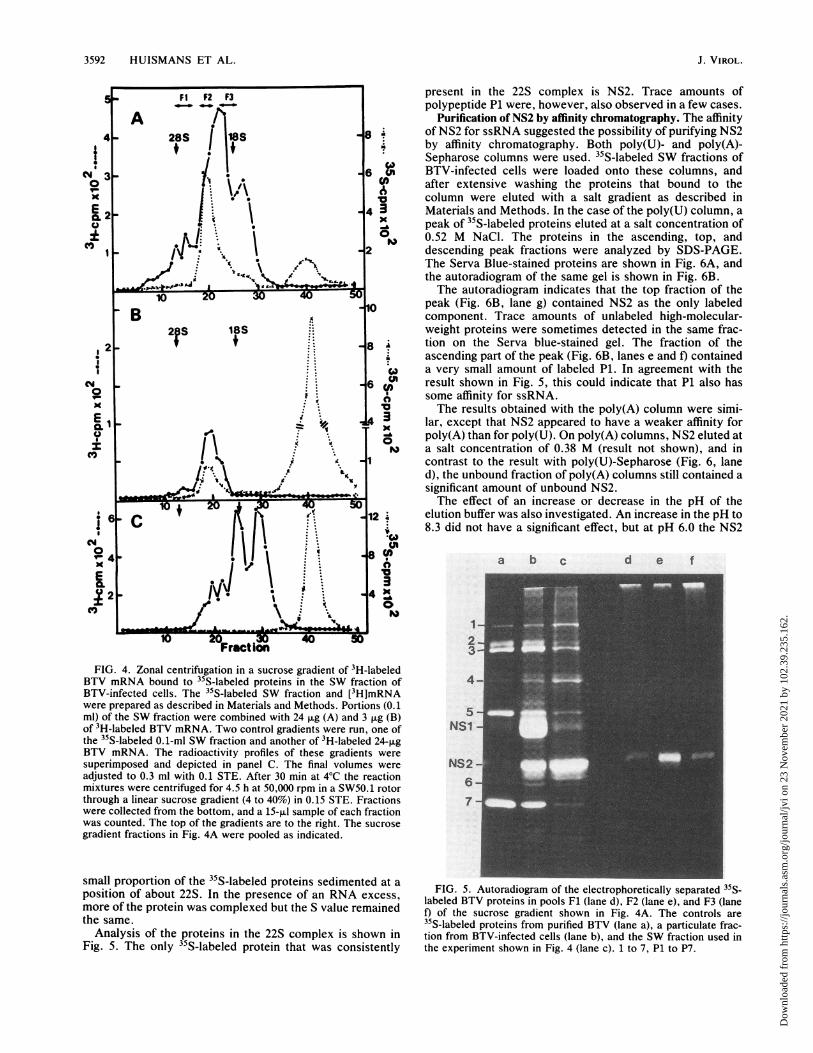
.-F1 P2 F3 present in the 22S complex is NS2. Trace amounts of_0F _-F2 F3
polypeptide P1 were, however, also observed in a few cases.A Purification of NS2 by affinity chromatography. The affinity
4 - 28S ~ 118S ~ 8 ~ of NS2 for ssRNA suggested the possibility of purifying NS2l+ l 1 + " by affinity chromatography. Both poly(U)- and poly(A)-
!1 6i| I . Sepharose columns were used. 35S-labeled SW fractions of
N < _ i . - 6 g BTV-infected cells were loaded onto these columns, ando
3\ \ <|> after extensive washing the proteins that bound to the
X I .. column were eluted with a salt gradient as described inE 1- f * 4 3 Materials and Methods. In the case of the poly(U) column, a0 t * . ^ peak of 35S-labeled proteins eluted at a salt concentration of
XI1t,1*\ | 0, 0.52 M NaCl. The proteins in the ascending, top, and1 _ /y . \ _2 descending peak fractions were analyzed by SDS-PAGE.
\ .~ 8 \ ,The Serva Blue-stained proteins are shown in Fig. 6A, and_/N..'wtr<.S w-the autoradiogram of the same gel is shown in Fig. 6B.- >,.x 2 i 4 aOIThe autoradiogram indicates that the top fraction of the
10 20 10 peak (Fig. 6B, lane g) contained NS2 as the only labeledB A component. Trace amounts of unlabeled high-molecular-
2 S 18S weight proteins were sometimes detected in the same frac-2 t .-. - 8 .^ tion on the Serva blue-stained gel. The fraction of theS..... ascending part of the peak (Fig. 6B, lanes e and f) contained
S .... *w a very small amount of labeled P1. In agreement with thesso 1- . -. 6 Q result shown in Fig. 5, this could indicate that Pl also has
some affinity for ssRNA.X . *. X The results obtained with the poly(A) column were simi-
E1X43i lar, except that NS2 appeared to have a weaker affinity forYoz - . O poly(A) than for poly(U). On poly(A) columns, NS2 eluted at
X | | \ .. *. | " a salt concentration of 0.38 M (result not shown), and in.'s.;i. 1 contrast to the result with poly(U)-Sepharose (Fig. 6, lane
d), the unbound fraction of poly(A) columns still contained asignificant amount of unbound NS2.The effect of an increase or decrease in the pH of the
612 elution buffer was also investigated. An increase in the pH to8.3 did not have a significant effect, but at pH 6.0 the NS2
cm~~~~~~~~~~~~~~~~U>°<4 IJ l18Ntn\a b c d e f
2 4. Z c i 4st
Fraction 3FIG. 4. Zonal centrifugation inaasucroseegradienttoff3H-labeledd
BTV mRNA bound to 35S-labeled proteins in the SW fraction of 4aBTV-infected cells. The 35S-labeled SW fraction and [3H]mRNAwere prepared as described in Materials and Methods. Portions (0.1 5ml) of the SW fraction were combined with 24 jig (A) and 3 jig (B)of 3H-labeled BTV mRNA. Two control gradients were run, one of NS1the 35S-labeled 0.1-ml SW fraction and another of 3H-labeled 24-jigBTV mRNA. The radioactivity profiles of these gradients weresuperimposed and depicted in panel C. The final volumes were NS2adjusted to 0.3 ml with 0.1 STE. After 30 min at 4°C the reaction 6mixtures were centrifuged for 4.5 h at 50,000 rpm in a SW50.1 rotorthrough a linear sucrose gradient (4 to 40%) in 0.15 STE. Fractions 7were collected from the bottom, and a 15-,lI sample of each fractionwas counted. The top of the gradients are to the right. The sucrosegradient fractions in Fig. 4A were pooled as indicated.
small proportion of the 35S-labeled proteins sedimented at aposition of about 22S. In the presence of an RNA excess, FIG. 5. Autoradio ram of the electrophoretically separated35S(more of the protein was complexed but the S value remained t) Of the sucrose gradient showl (laned)i F2 (lane e), and F3 (lanethe same. 35S-labeled proteins from purified BTV (lane a), a particulate frac-
Analysis of the proteins in the 22S complex is shown in tion from BTV-infected cells (lane b), and the SW fraction used inFig. 5. The only 35S-labeled protein that was consistently the experiment shown in Fig. 4 (lane c). 1 to 7, P1 to P7.
J. VIROL.
Dow
nloa
ded
from
http
s://j
ourn
als.
asm
.org
/jour
nal/j
vi o
n 23
Nov
embe
r 20
21 b
y 10
2.39
.235
.162
.

NONSTRUCTURAL PROTEIN OF BTV 3593
was contaminated with a large number of low-molecular-weight polypeptides. Therefore, a low pH for the elutionbuffer is not recommended.The highest yields of NS2 from affinity column purifica-
tions were obtained from SW fractions. However, themethod was found to be equally applicable to purifying NS2from soluble fractions of BTV-infected cells as well as toNS2 synthesized in vitro in a cell-free protein synthesissystem (results not shown). The method has also been usedto purify 32P-labeled NS2.
In vitro phosphorylation of NS2. To characterize the kinaseassociated with the phosphorylation of NS2, it was investi-gated whether NS2 could be phosphorylated in vitro as hasbeen described for the vesicular stomatitis virus phospho-protein NS (1). 35S-labeled particulate (P100) and soluble(S100) protein extracts were prepared from the cytoplasm ofuninfected and BTV-infected BHK cells. The soluble pro-
A
a b c d e f
-I1-2-3-4
- 5-NS1
- NS2-6
- 7
d e f g ha b c
3---.4- f.
"' JlW.
5-NS1-
NS2-6---
'r..
,m_sr
4'
7- _ -_.
W,-
B1- -
2 -
3--
4---
NS1 -
NS2- -
6-
7-
a b c
FIG. 7. Autoradiogram showing in vitro phosphorylation of invivo-synthesized BTV NS2 proteins. Unlabeled S100 and P100fractions were prepared from the cytoplasm of BTV-infected andmock-infected cells as described in Materials and Methods. Portionsof the S100 fractions, each representing approximately 106 cells,were immunoprecipitated with a BTV-specific serum as described inthe legend to Fig. 1. These immunoprecipitates, as well as equiva-lent amounts of the P100 fractions, were suspended in 30 iul of 2 mMTris, pH 8.0, and an equal volume of 2x MTD buffer containing[-y-32P]ATP was added. After 30 min of incubation, the samples wereanalyzed by means of SDS-PAGE and autoradiography. The resultsare shown as follows: immunoprecipitated S100 fraction of mock-
de f 9 h i infected cells (lane a); immunoprecipitated S100 fraction of BTV-infected cells (lane b); uninfected P100 fraction (lane c); andBTV-infected P100 fraction (lane d). Controls are I'S labeled: P100sample of BTV-infected cells (lane e) and purified BTV (lane f). 1 to7, P1 to P7.
FIG. 6. Electrophoretic separation of the proteins eluted from a
poly(U)-Sepharose column during the purification of NS2 from theSW fraction of BTV-infected cells. The 35S-labeled SW fraction wasprepared, loaded onto the column, and eluted with a linear 0.15 to1.15 STE gradient as described. A peak of labeled proteins elutedbetween fractions 66 and 75 of the gradient at a salt concentration of0.52 M NaCl. Proteins in the ascending part of the peak (fractions 66and 69) are analyzed in lanes e and f, the top fraction (fraction 71) isanalyzed in lane g, and the descending part of the peak (fractions 73and 75) is analyzed in lanes h and i. The Serva blue-stained gel isshown in panel A, and the autoradiogram from the same gel is shownin panel B. A fraction from the material that did not bind to thecolumn is shown in lane d. Other lanes: The controls are 35S-labeledproteins from purified BTV (lane a), a P100 fraction from BTV-infected cells (lane b), and the SW fraction loaded onto the column(lane c). 1 to 7, P1 to P7.
teins in the S100 extracts were concentrated by immunopre-cipitation with a BTV-specific serum. The different P100fractions and immunoprecipitates of the S100 fractions wereincubated in a phosphorylation mixture containing [y-32P]ATP and then analyzed as indicated in the legend to Fig.7. The result is shown in Fig. 7.The results indicate that a protein migrating in the position
of NS2 was phosphorylated in both the S100 immunopre-cipitate and the P100 fraction of BTV-infected cells (lanes band d). It is also evident from Fig. 7 that several nonviralproteins were phosphorylated. Since these proteins wereobserved in both the infected and the uninfected controls,the presence of a cellular protein kinase in the cellularextracts is implicated.To obtain more information about the kinase involved,
NS2 was purified from a 35S-labeled S100 fraction of BTV-infected cells by affinity chromatography as described in thelegend to Fig. 6. The 35S-labeled NS2 was concentrated fromthe high-salt column wash fractions by immunoprecipitation.The immunoprecipitate was assayed for in vitro kinaseactivity as indicated in the legend to Fig. 8.
Conditions for autoradiography were selected to discrim-inate between 35S- and 32P-labeled proteins. It is evident
VOL. 61, 1987
Dow
nloa
ded
from
http
s://j
ourn
als.
asm
.org
/jour
nal/j
vi o
n 23
Nov
embe
r 20
21 b
y 10
2.39
.235
.162
.

3594 HUISMANS ET AL.
a b c d
1234-
5-NS1
NS2
6
FIG. 8. Autoradiogram showing in vitro kinase activity in a
poly(U)-Sepharose-purified NS2 preparation. 35S-labeled NS2 was
purified from the S100 fraction of BTV-infected cells by poly(U)-Sepharose column chromatography as described in the legend toFig. 6. The viral proteins present in the NS2-containing peakfractions that eluted at 0.5 M NaCl were concentrated by immuno-precipitation and assayed for kinase activity as described in thelegend to Fig. 7. Lanes c and d were subjected to autoradiographythat distinguishes between 35S- and 32P-labeled components (4),whereas lanes a and b were subjected to normal autoradiography.Lanes: a, 35S-labeled P100 fraction of BTV-infected cells; b, 35S-labeled proteins in high-salt eluate from poly(U)-Sepharose column;c, phosphorylation of purified NS2 fraction at time zero, indicatingthat no 35S label is detected under these conditions; d, phosphory-lation of NS2 after 30 min of incubation. 1 to 7, P1 to P7.
from lane b that NS2 was the only 35S-labeled proteinrecovered from the column. After incubation for 30 min inthe phosphorylation mixture, NS2 was found to be labeledwith 32p. The column-purified NS2 preparation therefore stillcontained kinase activity. This kinase activity was thereforeeither associated with NS2 itself or was caused by thepresence of a kinase that copurified with NS2.
DISCUSSION
We have identified a nonstructural protein of BTV, proteinNS2, with affinity for ssRNA. The protein is synthesized incomparatively large amounts throughout the infection cycle.Synthesis can first be detected as early as 2 to 4 h p.i., withthe largest amounts being synthesized between 16 and 20 hp.i. The protein is characterized by anomalous electropho-retic migration. On phosphate-urea gels it has a higherelectrophoretic mobility than P6 (7), whereas on Tris-glycinegels it migrates slower than P6. It has also been observedthat on the latter gels NS2 very often separates into two very
closely spaced bands. This is suggestive of posttranslationalmodifications such as phosphorylation or glycosylation.There is no evidence for the glycosylation of NS2, but it hasbeen found that NS2 is the only virus-specified protein thatis phosphorylated. The presence of phosphorylated proteinshas been demonstrated in many viruses, and in a number ofcases the degree of phosphorylation seems to determine theextent of binding to viral nucleic acid (3, 16).
In the case of BTV, it was found that a component in theSW fraction of BTV-infected cells that contained NS2 as themajor viral protein could bind to ssRNA but not to dsRNA.
A complex with an estimated S value of 22S containing NS2as the sole protein component could be recovered frommixtures of BTV SW fractions and BTV mRNA. The S valueof the complex was found to be independent of themRNA/NS2 ratio. The narrow size distribution of the com-plex was rather surprising, in view of the heterogeneous sizedistribution of BTV mRNA. An equally heterogeneous sizedistribution of the BTV mRNA-NS2 complex would havebeen expected if the number of NS2 molecules that arebound to each mRNA were dependent solely on the length ofthe mRNA species. The possibility that only a limitednumber of NS2 molecules can bind to each mRNA, as hasbeen found in the case of reovirus (uNS (17), needs to beinvestigated. It is also possible that only certain of themRNA species bind NS2 effectively. Experiments withindividual mRNA species and highly purified NS2 of knownspecific activity are in progress to investigate this particularaspect in more detail.The affinity of NS2 for mRNA suggests that the NS2 in
infected cells is also bound to BTV mRNA or cellularssRNA. The S value of such complexes is small, and theyare therefore likely to be present in the soluble (S100)fraction of infected cells. The largest amount of NS2 is,however, not found in the soluble fraction but appears to beassociated with particulate material. Most of these NS2-associated particles sediment through a 40% sucrose cushionafter centrifugation at 100,000 x g for 1 h. Some of the NS2is also bound to low-density material that can be recoveredfrom the top of the 40% sucrose cushion. NS2 is therefore a"sticky" protein, which might also explain the nonspecificcoprecipitation of NS2 that is often observed during theimmunoprecipitation of BTV proteins (11).The particulate fraction of BTV-infected cells has been
used as starting material in the purification of NS2. It wasfound that, similar to particulate oNS in the case of reovirus(10), BTV NS2 can be solubilized from all particulate mate-rial by an SW with 0.5 M NaCI. If this treatment is done ata pH of 8.0 or more, the BTV capsid proteins are notsolubilized (11) and a significant purification of the NS2 fromother viral and cellular proteins is achieved. The subsequentpurification by poly(U)-Sepharose affinity chromatographyresulted in a fraction that contained NS2 as the only labeledcomponent. Serva blue-stained polyacrylamide gels, how-ever, often indicated the presence of very small traceamounts of contaminating cellular proteins.
This report presents the first evidence that NS2 can bephosphorylated in vitro. It was shown that immunoprecipi-tates of BTV-specific proteins from the S100 fraction ofinfected cells contained a protein kinase that was able tophosphorylate NS2. Similar results have been reported forthe NS protein of vesicular stomatitis virus (1). It was alsofound that NS2 in the P100 fraction of BTV-infected cellscan be phosphorylated in vitro. However, a number ofcellular proteins are also phosphorylated under these condi-tions. Results presented in this report also indicate that invivo phosphorylation of NS2 takes place throughout theinfection cycle. This implies that the in vitro 32p labeling ofalready phosphorylated NS2 is caused either by hyperphos-phorylation or by some form of phosphate exchange as theresult of a dynamic phosphorylation-dephosphorylation re-action. To investigate this aspect, we attempted to phos-phorylate NS2 synthesized in an in vitro translation reaction(results not shown). No phosphorylation could be demon-strated. These results are again in agreement with thoseobtained with vesicular stomatitis virus (6). An experimentwas also done in which in vivo-synthesized NS2 was purified
J. VIROL.
Dow
nloa
ded
from
http
s://j
ourn
als.
asm
.org
/jour
nal/j
vi o
n 23
Nov
embe
r 20
21 b
y 10
2.39
.235
.162
.

NONSTRUCTURAL PROTEIN OF BTV 3595
to separate it from the kinase activity. It was, however,found that poly(U)-Sepharose-purified NS2 preparations stillcontained kinase activity that phosphorylated NS2 in vitro.The most likely explanation is that the kinase is one of theminor cel!ular proteins that were often found to be associ-ated with purified NS2. Since a very small amount of P1 wasalso often seen to be present in purified NS2 preparations,the possibility of a viral protein kinase or even autophospho-rylation cannot be excluded. This aspect is under investiga-tion.
LITERATURE CITED1. Bell, J. C., E. G. Brown, D. Takayesu, and L. Prevec. 1984.
Protein kinase activity associated with immunoprecipitates ofvesicular stomatitis virus phosphoprotein NS. Virology 132:229-238.
2. Boyle, J. F., and K. V. Holmes. 1986. RNA-binding proteins ofbovine rotavirus. J. Virol. 58:561-568.
3. Clinton, G. M., B. W. Burge, and A. S. Huang. 1978. Effects ofphosphorylation and pH on the association of NS protein withvesicular stoinatitis virus cores. J. Virol. 27:340-346.
4. Cooper, P. C., and A. W. Burgess. 1982. Simultaneous detectionof 35S- and 32P-labeled proteins on electrophoretic gels. Anal.Biochem. 126:301-305.
5. Gomatos, P. J., 0. Prakash, and N. M. Stamatos. 1981. Smallreovirus particles composed solely of sigma NS with specificityfor binding different nucleic acids. J. Virol. 39:115-124.
6. Harmon, S. A., L. L. Marnel, and D. F. Sumlners. 1983. Themajor ribonucleoprotein-associated protein kinase of vesicularstomatitis virus is a host cell protein. J. Biol. Chem. 258:15283-15290.
7. Huismans, H. 1979. Protein synthesis in bluetongue virus in-fected cells. Virology 92:385-396.
8. Huismans, H., and H. M. Basson. 1983. The characterization ofa phosphorylated non-structural polypeptide of bluetongue vi-rus with affinity for RNA, p. 173-181. In R. W. Compans andD. H. L. Bishop (ed.), Double-stranded RNA viruses. ElsevierBiomedical Press, New York.
8a.Huismans, H., and M. Cloete. 1987. A comparison of differentcloned bluetongue virus genome segments as probes for the
detection of virus-specified RNA. Virology 158:373-380.9. Huismans, H., and H. J. Els. 1979. Characterization of the
tubules associated with the replication of three different orbivi-ruses. Virology 92:397-406.
10. Huismans, H., and W. K. Joklik. 1976. Reovirus codedpolypeptides in infected cells: isolation of two native mono-meric polypeptides with affinity for single-stranded and double-stranded RNA respectively. Virology 70:411-424.
11. Huismans, H., N. T. Van der Walt, M. Cloete, and B. J.Erasmus. 1987. Isolation of a capsid protein of bluetongue virusthat induces a protective immune response in sheep. Virology157:172-179.
12. Huismans, H., and D. W. Verwoerd. 1973. Control of transcrip-tion during expression of the bluetongue virus genome. Virology52:81-88.
13. Ito, Y., and W. K. Joklik. 1972. Temperature-sensitive mutantsof reovirus. III. Evidence that mutants of group D ("RNA-negative") are structural polypeptide mutants. Virology 50:282-286.
14. Mertens, P. P. C., F. Brown, and A. V. Sangar. 1984. Assign-ment of the genome segments of bluetongue virus type 1 to theproteins which they encode. Virology 135:207-217.
15. Moyer, S. A., and D. F. Summers. 1974. Phosphorylation ofvesicular stomatitis virus in vivo and in vitro. J. Virol. 13:455-465.
16. Scheidtmann, K.-H., M. Hardung, B. Echle, and G. Walter.1984. DNA-binding activity of simian virus 40 large T antigencorrelates with a distinct phosphorylation state. J. Virol. 50:1-12.
17. Stamatos, N. M., and P. J. Gomatos. 1982. Binding to selectedregions of reovirus mRNAs by a non-structural reovirus pro-tein. Proc. Natl. Acad. Sci. USA 79:3457-3461.
18. Van Dijk, A. A., and H. Huismans. 1980. The in vitro activationand further characterization of the bluetongue virus-associatedtranscriptase. Virology 104:347-356.
19. Verwoerd, D. W., H. J. Els, E.-M. De Villiers, and H. Huismans.1972. Structure of the bluetongue virus capsid. J. Virol. 50:783-794.
20. Verwoerd, D. W., H. Huismans, and B. J. Erasmus. 1979. Orbi-viruses, p. 285-345. In H. Fraenkel-Conrat and R. R. Wagner(ed.), Comprehensive virology 14. Plenum Publishing Corp.,New York.
VOL. 61, 1987
Dow
nloa
ded
from
http
s://j
ourn
als.
asm
.org
/jour
nal/j
vi o
n 23
Nov
embe
r 20
21 b
y 10
2.39
.235
.162
.