OET flashBAC Use Guide · Oxford Expression Technologies flashA™ User Guide 2015 5 | P a g e 5...
28
A guide to making recombinant baculoviruses using BacPAK6 or flashBAC™ User Guide 2015
Transcript of OET flashBAC Use Guide · Oxford Expression Technologies flashA™ User Guide 2015 5 | P a g e 5...
A guide to making recombinant
baculoviruses using BacPAK6 or flashBAC™
User Guide 2015
Oxford Expression Technologies flashBAC™ User Guide 2015
2 | P a g e
Contents
1 Limited Use License 3
2 flashBAC™ & BacBAK 6 Kit Contents 4
3 Essential Information and Technical Assistance 4
4 Safety Requirements 4
5 flashBAC™ & BacPAK6 kit and related products ordering information 5
6 Introduction to the Baculovirus Expression Vector System 7
Introduction to the BacPAK6 and flashBAC™ systems 9
7 Making recombinant baculoviruses: 13
Insertion of gene into, and choice of, transfer plasmid 13
Co-transfection of insect cells with flashBAC™ or BacPAK6 DNA and transfer plasmid
DNA to make recombinant virus 14
Plaque purification of recombinant BacPAK6 virus 17
Amplification of recombinant baculoviruses 20
Titration of recombinant baculoviruses 22
Making recombinant viruses in 24 well plate format 24
8 Analysis and optimisation of gene expression 25
9 Trouble Shooting and FAQ 26
10 References 28

Oxford Expression Technologies flashBAC™ User Guide 2015
3 | P a g e
1 Limited Use Licence for flashBAC™ virus DNA 1 In the License the following expressions shall have the following meanings:
DNA shall mean deoxyribonucleic acid;
Fee shall mean the fee invoiced for the Materials by the Licensor to the Licensee;
Licensee shall mean the purchaser of the Materials;
Licensor shall mean Oxford Expression Technologies Ltd;
Material shall mean the Licensor’s product known as flashBAC™ comprising either or both an agreed
quantity of DNA and the relevant User Guide;
Purpose shall mean the use by the Licensee of the Materials for the production of recombinant
proteins and/or viruses for Research purposes only;
Research shall mean the Licensor’s systematic search or investigation towards increasing the sum of
knowledge in the production of recombinant proteins and/or viruses;
User Guide shall mean the instructions provided with flashBAC™ to enable the Licensee to produce
recombinant proteins and/or viruses from the DNA.
2 The Licensor and the Licensee have agreed to enter into this Licence on the following terms and conditions.
3 The Licensee acknowledges and accepts that by opening and/or using the Materials it is agreeing to and
accepting these terms and condition. If the Licensee does not agree to these terms and conditions it must
immediately return all the Materials unused to the Licensor who shall issue a refund for the fee.
4 The Licensor has certain know0how and has developed a product that can be used to produce recombinant
proteins and/or viruses and has the right to exploit the product under, inter alia, patent applications
numbered EP1144666, WO0112829 and AU6460800.
5 This Licence shall commence on the date hereof and continue until the DNA has been used or destroyed.
6 The Licensor hereby grants to the Licensee and the Licensee hereby accepts a limited, non-exclusive, non-
transferable, licence to use the Materials for the Purpose and as otherwise set out in this licence.
7 The Licensee warrants to the Licensor that:
7.1 it shall only use the Materials for the purpose of Research; and
7.2 it shall not alter, reverse engineer, produce, manufacture or amplify the DNA; and
7.3 it shall not sell any protein and/or virus created pursuant to this Licence to any third party; and
7.4 it shall not provide any services to any third party using the Materials; and
7.5 if the Licensee desires to the Materials for any purpose other than the Purpose, it shall notify the Licensor
accordingly and procure a suitable licence prior to any such use.
8 The Licensee shall keep the DNA in accordance with the directions contained in the User Guide.
9 The Licensor shall raise an invoice to the Licensee for the Fee and the Licensee agrees to pay the same to the
Licensor within thirty (30) day of receipt of the invoice (unless otherwise agreed in writing).
10 The Materials are provided as is and neither the Licensor nor any staff acting on its behalf accepts any liability
whatsoever for any of the Materials or in connection with the Licensee’s possession, handling or use of the
Materials.
11 The Licensee’s remedy pursuant to this Licence shall be limited at the Licensor’s option to the replacement of
the Materials or a refund of the Fee paid by the Licensee.
12 Ownership of the Materials shall pass to the Licensee upon dispatch of the Materials by the Licensor to the
Licensee.
13 The Licensee shall indemnify the Licensor for any loss suffered by the Licensor as a result of the Licensee’s
breach of this licence and/or third party’s intellectual property rights.
14 This Licence is personal to the parties and shall not be assigned or otherwise transferred in whole or in part by
either party.
15 This Licence constitutes the entire agreement and understanding between the parties in respect of the
Materials and supersedes all previous agreements, understandings and undertakings in this respect and all
obligations implied by the law to the extent that they conflict with the express provisions of this Licence.
Oxford Expression Technologies flashBAC™ User Guide 2015
4 | P a g e
16 The invalidity, illegality or unenforceability of a provision of this Licence shall not affect or impair the
continuation in force of the remainder of this Licence.
17 The Licensor reserves the right to revoke this permission and may require the Licensee to return or destroy
any remaining DNA and/or the User Guide.
18 Clauses 1, 3, 7, 9, 10, 13, 16, 18-20 shall survive any termination or expiry of this Licence.
19 The interpretation construction and effect of this Licence shall be governed and construed in all respects in
accordance with the laws of England and the parties hereby submit to the non-exclusive jurisdiction of the
English courts.
20 The Contracts (Rights of Third Parties) Act 1999 shall have no application to this Licence whatsoever and the
parties do not intend hereunder to benefit any third party.
End of Limited Use Licence.
2 Kit Contents
All reagents and materials provided and referred to in this User Guide are for Research Purposes only.
1 flashBAC™ DNA (or BacPAK6 DNA). Store at 4⁰C.
2 Control transfer plasmid DNA (containing lacZ reporter gene). Store at -20⁰C. (flashBAC™ kits only)
3 Baculovirus Expression System User Guide. Download from www.oetltd.com.
4 Certificate of Analysis. Download from www.oetltd.com.
5 MSDS. Download from www.oetltd.com.
Note: Transfection reagent and insect cells are NOT supplied as part of this kit.
3 Essential Information and Technical Assistance
The information given in this User Guide is accurate to the best of our knowledge. It is a practical guide to allow
researchers to use the flashBAC™ (and BacPAK6) technology to produce recombinant baculoviruses. It is not intended
as a comprehensive guide to the baculovirus expression system or insect cell culture. Those experienced with the
baculovirus expression system may find that they are already familiar with much of the information provided.
Users are reminded that they may require other licences to use the baculovirus expression system or types of insect
cells and it is the responsibility of the user to ascertain ad act on this information.
For additional help or guidance please refer to the Trouble Shooting Section of this Guide and/or the Frequently Asked
Questions (FAQ) section of our website (www.oetltd.com). If these resources are unable to help, please contact us at
[email protected] and we will be pleased to help where possible. All technical assistance provided is given in good faith;
we cannot take any responsibility whatsoever for any results you obtain by relying on our assistance. We make no
warranties of any kind with respect to technical assistance or advice we provide.
4 Safety Requirements
These research products have not been approved for human or animal diagnostic or therapeutic use.
Procedures described within this User Guide should only be carried out by qualified persons trained in appropriate
laboratory safety procedures.
Always use good laboratory practice when handling this product.
Warning. Safety precautions may be necessary when handling some of the reagents described in this user guide.
Please refer to the material safety data sheets supplied by the appropriate manufacturer.
Oxford Expression Technologies flashBAC™ User Guide 2015
5 | P a g e
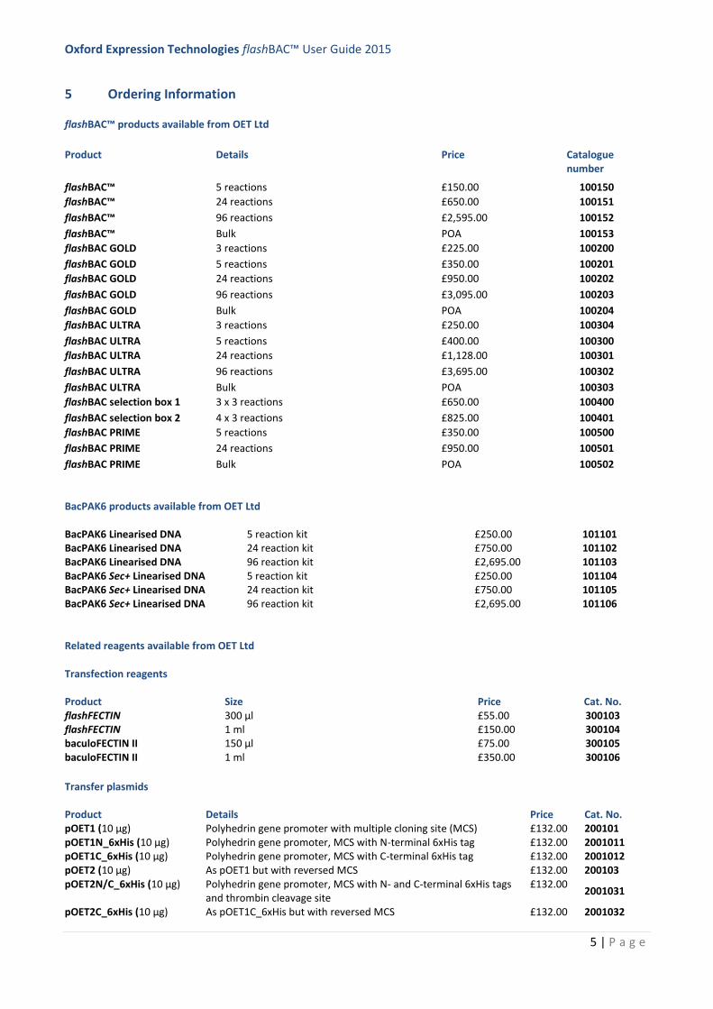
5 Ordering Information
flashBAC™ products available from OET Ltd
Product Details Price Catalogue number
flashBAC™ 5 reactions £150.00 100150
flashBAC™ 24 reactions £650.00 100151
flashBAC™ 96 reactions £2,595.00 100152
flashBAC™ Bulk POA 100153
flashBAC GOLD 3 reactions £225.00 100200
flashBAC GOLD 5 reactions £350.00 100201
flashBAC GOLD 24 reactions £950.00 100202
flashBAC GOLD 96 reactions £3,095.00 100203
flashBAC GOLD Bulk POA 100204
flashBAC ULTRA 3 reactions £250.00 100304
flashBAC ULTRA 5 reactions £400.00 100300
flashBAC ULTRA 24 reactions £1,128.00 100301
flashBAC ULTRA 96 reactions £3,695.00 100302
flashBAC ULTRA Bulk POA 100303
flashBAC selection box 1 3 x 3 reactions £650.00 100400
flashBAC selection box 2 4 x 3 reactions £825.00 100401
flashBAC PRIME 5 reactions £350.00 100500
flashBAC PRIME 24 reactions £950.00 100501
flashBAC PRIME Bulk POA 100502
BacPAK6 products available from OET Ltd
BacPAK6 Linearised DNA 5 reaction kit £250.00 101101 BacPAK6 Linearised DNA 24 reaction kit £750.00 101102 BacPAK6 Linearised DNA 96 reaction kit £2,695.00 101103 BacPAK6 Sec+ Linearised DNA 5 reaction kit £250.00 101104 BacPAK6 Sec+ Linearised DNA 24 reaction kit £750.00 101105 BacPAK6 Sec+ Linearised DNA 96 reaction kit £2,695.00 101106
Related reagents available from OET Ltd
Transfection reagents
Product Size Price Cat. No. flashFECTIN 300 μl £55.00 300103 flashFECTIN 1 ml £150.00 300104 baculoFECTIN II 150 µl £75.00 300105 baculoFECTIN II 1 ml £350.00 300106
Transfer plasmids
Product Details Price Cat. No. pOET1 (10 µg) Polyhedrin gene promoter with multiple cloning site (MCS) £132.00 200101 pOET1N_6xHis (10 µg) Polyhedrin gene promoter, MCS with N-terminal 6xHis tag £132.00 2001011 pOET1C_6xHis (10 µg) Polyhedrin gene promoter, MCS with C-terminal 6xHis tag £132.00 2001012 pOET2 (10 µg) As pOET1 but with reversed MCS £132.00 200103 pOET2N/C_6xHis (10 µg) Polyhedrin gene promoter, MCS with N- and C-terminal 6xHis tags
and thrombin cleavage site £132.00
2001031
pOET2C_6xHis (10 µg) As pOET1C_6xHis but with reversed MCS £132.00 2001032
Oxford Expression Technologies flashBAC™ User Guide 2015
6 | P a g e
pOET3 (10 µg) P6.9 gene promoter for late phase expression £132.00 200104 pOET4 (10 µg) As pOET3 but with reversed MCS £132.00 200105 pOET5 (10 µg) Dual expression with polyhedrin and p10 gene promoters £132.00 200106 pOET6 BacMAM (10 µg) CMV promoter for BacMam –mediated transduction of
mammalian cells £132.00
200107
pOET1 Gateway (10µg) pOET1 transfer plasmid with Gateway® technology £137.00 200108
baculoQUANT products available from OET Ltd
Product Details Price Catalogue number
baculoCOMPLETE protein expression kit + baculoQUANT all-in-one kit 5 + 100 reactions £895.00 400101 baculoQUANT All-in-one virus extraction & titration kit 100 reactions £365.00 100602 titrePLUS flashBAC all-in-one 5 + 100 reactions £463.50 100710 titrePLUS flashBAC all-in-one 24 + 100 reactions £913.50 100711 titrePLUS flashBAC GOLD all-in-one 5 + 100 reactions £643.50 100712
titrePLUS flashBAC GOLD all-in-one 24 + 100 reactions £1,183.50 100713 titrePLUS flashBAC ULTRA all-in-one 5 + 100 reactions £688.50 100714 titrePLUS flashBAC ULTRA all-in-one 24 + 100 reactions £1,344.00 100715 titrePLUS flashBAC PRIME all-in-one 5 + 100 reactions £643.50 100716 titrePLUS flashBAC PRIME all-in-one 24 + 100 reactions £1,183.50 100717
Cell line products available from OET Ltd
Product Frozen Live culture Medium Catalogue number
Sf9 cells ≥1 x 10⁷ cells per ampoule On request Serum-free 600100
Sf21 cells for plaque-assay ≥1 x 10⁷ cells per ampoule
On request TC100 with 10% serum
600105
Super Sf91 ≥1 x 10⁷ cells per ampoule On request Serum-free 600102
Super Sf92 ≥1 x 10⁷ cells per ampoule On request Serum-free 600103
Super Sf93
≥1 x 10⁷ cells per ampoule On request Serum-free 600104
Insect cell culture media products available from OET Ltd
Product Size Shipping conditions Medium Catalogue Number
BaculoGROW II 500 ml Room temperature Serum-free 500200
ESF 921 1 L Room temperature Serum-free Protein-free
500300
3 x 1 L 500301 5 x 1 L 500302 10 x 1 L 500303 Bulk > 10 L 500304
ES Transfection medium
20 ml 100 ml
Room temperature Serum-free, Animal free 500305 500306
ES production boost additive
100 ml Room temperature Serum-free, nutrient additive 500307
ESF 921 delta methionine
1 L Room temperature Serum-free, methionine free 500308
ESF 921 delta amino
acid deficient
1 L Room temperature Serum and amino acid free 500309
ES Custom media
production
Min. 20L Room temperature Custom to order 500310
Oxford Expression Technologies flashBAC™ User Guide 2015
7 | P a g e
6 Introduction to the Baculovirus Expression System and flashBAC™/BacPAK6 technology
6.1 Baculoviruses
Baculoviruses are insect viruses, predominantly infecting insect larvae of the order Lepidoptera (butterflies and
moths)1. A baculovirus expression vector is a recombinant baculovirus that has been genetically modified to contain a
foreign gene of interest, which can then be expressed in insect cells under control of a baculovirus gene promoter. The
most commonly used baculovirus for foreign gene expression is Autographa californica nucleopolyhedrovirus
(AcMNPV)2,3
. AcMNPV has a circular, double-stranded, super-coiled DNA genome (133894 bp; Accession NC_001623)4,
packaged in a rod-shaped nucleocapsid. The nucleocapsid can be extended lengthways and thus the DNA genome can
accommodate quite large insertions of DNA. The AcMNPV genome forms the basis of the flashBAC™ or BacPAK6 DNA
provided in this kit.
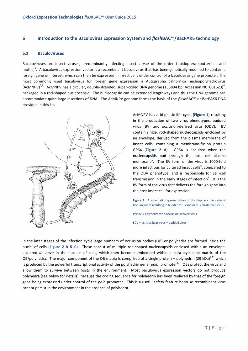
AcMNPV has a bi-phasic life cycle (Figure 1) resulting
in the production of two virus phenotypes: budded
virus (BV) and occlusion-derived virus (ODV). BV
contain single, rod-shaped nucleocapsids enclosed by
an envelope, derived from the plasma membrane of
insect cells, containing a membrane-fusion protein
GP64 (Figure 2 A). GP64 is acquired when the
nucleocapsids bud through the host cell plasma
membrane5. The BV form of the virus is 1000-fold
more infectious for cultured insect cells6, compared to
the ODV phenotype, and is responsible for cell-cell
transmission in the early stages of infection7. It is the
BV form of the virus that delivers the foreign gene into
the host insect cell for expression.
Figure 1. A schematic representation of the bi-phasic life cycle of
baculoviruses resulting in budded virus and occlusion-derived virus.
P/PDV = polyhedra with occlusion derived virus
ECV = extracellular virus = budded virus
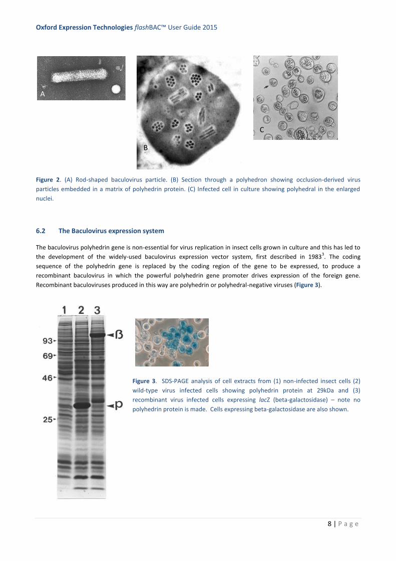
In the later stages of the infection cycle large numbers of occlusion bodies (OB) or polyhedra are formed inside the
nuclei of cells (Figure 2 B & C). These consist of multiple rod-shaped nucleocapsids enclosed within an envelope,
acquired de novo in the nucleus of cells, which then become embedded within a para-crystalline matrix of the
OB/polyhedra. The major component of the OB matrix is comprised of a single protein – polyhedrin (29 kDa)8,9
, which
is produced by the powerful transcriptional activity of the polyhedrin gene (polh) promoter13
. OBs protect the virus and
allow them to survive between hosts in the environment. Most baculovirus expression vectors do not produce
polyhedra (see below for details), because the coding sequence for polyhedrin has been replaced by that of the foreign
gene being expressed under control of the polh promoter. This is a useful safety feature because recombinant virus
cannot persist in the environment in the absence of polyhedra.
Oxford Expression Technologies flashBAC™ User Guide 2015
8 | P a g e
Figure 2. (A) Rod-shaped baculovirus particle. (B) Section through a polyhedron showing occlusion-derived virus
particles embedded in a matrix of polyhedrin protein. (C) Infected cell in culture showing polyhedral in the enlarged
nuclei.
6.2 The Baculovirus expression system
The baculovirus polyhedrin gene is non-essential for virus replication in insect cells grown in culture and this has led to
the development of the widely-used baculovirus expression vector system, first described in 19833. The coding
sequence of the polyhedrin gene is replaced by the coding region of the gene to be expressed, to produce a
recombinant baculovirus in which the powerful polyhedrin gene promoter drives expression of the foreign gene.
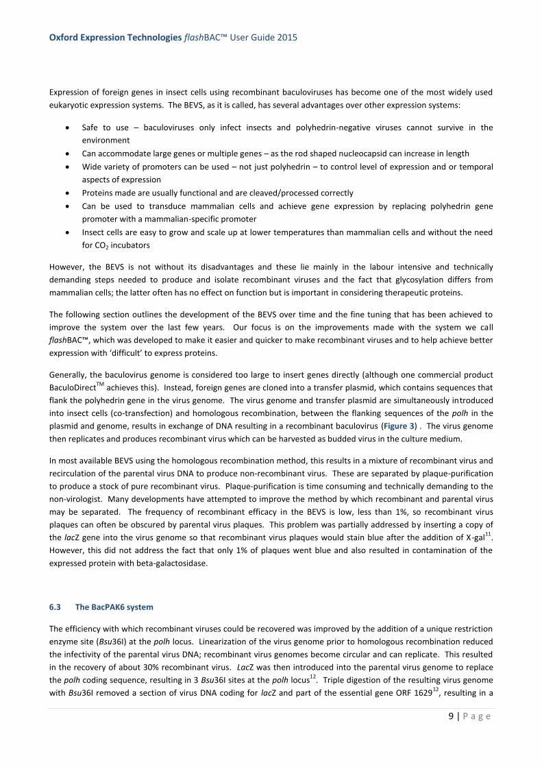
Recombinant baculoviruses produced in this way are polyhedrin or polyhedral-negative viruses (Figure 3).
Figure 3. SDS-PAGE analysis of cell extracts from (1) non-infected insect cells (2)
wild-type virus infected cells showing polyhedrin protein at 29kDa and (3)
recombinant virus infected cells expressing lacZ (beta-galactosidase) – note no
polyhedrin protein is made. Cells expressing beta-galactosidase are also shown.
A
C
B
B
B
Oxford Expression Technologies flashBAC™ User Guide 2015
9 | P a g e
Expression of foreign genes in insect cells using recombinant baculoviruses has become one of the most widely used
eukaryotic expression systems. The BEVS, as it is called, has several advantages over other expression systems:
Safe to use – baculoviruses only infect insects and polyhedrin-negative viruses cannot survive in the
environment
Can accommodate large genes or multiple genes – as the rod shaped nucleocapsid can increase in length
Wide variety of promoters can be used – not just polyhedrin – to control level of expression and or temporal
aspects of expression
Proteins made are usually functional and are cleaved/processed correctly
Can be used to transduce mammalian cells and achieve gene expression by replacing polyhedrin gene
promoter with a mammalian-specific promoter
Insect cells are easy to grow and scale up at lower temperatures than mammalian cells and without the need
for CO2 incubators
However, the BEVS is not without its disadvantages and these lie mainly in the labour intensive and technically
demanding steps needed to produce and isolate recombinant viruses and the fact that glycosylation differs from
mammalian cells; the latter often has no effect on function but is important in considering therapeutic proteins.
The following section outlines the development of the BEVS over time and the fine tuning that has been achieved to
improve the system over the last few years. Our focus is on the improvements made with the system we call
flashBAC™, which was developed to make it easier and quicker to make recombinant viruses and to help achieve better
expression with ‘difficult’ to express proteins.
Generally, the baculovirus genome is considered too large to insert genes directly (although one commercial product
BaculoDirectTM
achieves this). Instead, foreign genes are cloned into a transfer plasmid, which contains sequences that
flank the polyhedrin gene in the virus genome. The virus genome and transfer plasmid are simultaneously introduced
into insect cells (co-transfection) and homologous recombination, between the flanking sequences of the polh in the
plasmid and genome, results in exchange of DNA resulting in a recombinant baculovirus (Figure 3) . The virus genome
then replicates and produces recombinant virus which can be harvested as budded virus in the culture medium.
In most available BEVS using the homologous recombination method, this results in a mixture of recombinant virus and
recirculation of the parental virus DNA to produce non-recombinant virus. These are separated by plaque-purification
to produce a stock of pure recombinant virus. Plaque-purification is time consuming and technically demanding to the
non-virologist. Many developments have attempted to improve the method by which recombinant and parental virus
may be separated. The frequency of recombinant efficacy in the BEVS is low, less than 1%, so recombinant virus
plaques can often be obscured by parental virus plaques. This problem was partially addressed by inserting a copy of
the lacZ gene into the virus genome so that recombinant virus plaques would stain blue after the addition of X-gal11
.
However, this did not address the fact that only 1% of plaques went blue and also resulted in contamination of the
expressed protein with beta-galactosidase.
6.3 The BacPAK6 system
The efficiency with which recombinant viruses could be recovered was improved by the addition of a unique restriction
enzyme site (Bsu36I) at the polh locus. Linearization of the virus genome prior to homologous recombination reduced
the infectivity of the parental virus DNA; recombinant virus genomes become circular and can replicate. This resulted
in the recovery of about 30% recombinant virus. LacZ was then introduced into the parental virus genome to replace
the polh coding sequence, resulting in 3 Bsu36I sites at the polh locus12
. Triple digestion of the resulting virus genome
with Bsu36I removed a section of virus DNA coding for lacZ and part of the essential gene ORF 162912
, resulting in a
Oxford Expression Technologies flashBAC™ User Guide 2015
10 | P a g e
linear virus DNA (BacPAK6) that cannot replicate in insect cells. Co-transfection of insect cells with linearised BacPAK6
DNA and a transfer plasmid with foreign gene under control of polh, creates recombinant virus DNA in which ORF1629
is restored and the recircularised DNA can replicate to produce recombinant budded virus12
. This reduced even further
the chance of parental virus replicating and resulted in an increase in the recovery of recombinant virus to more than
90%*. It also introduced a useful blue-white selection system – with non-recombinant virus giving rise to blue plaques
and recombinant virus to white plaques. It was thus easier to achieve purified virus with a single round of plaque-
purification. *It is not 100% because it is impossible to ensure that every molecule of DNA is triple-digested and any
circular DNA remaining can replicate and produce non-recombinant virus.
The triple-cut linear BacPAK6 virus DNA is available from OET (see page 5). We are also pleased to offer BacPAK6-
Sec+, which has a deletion in the chitinase gene to aid expression of membrane targeted and secreted proteins.
Practical techniques to make recombinant BacPAK6 viruses are included in this User Guide.
Despite this fine tuning and optimisation of the system, a number of steps are still required to make recombinant
baculoviruses, thus making it more time consuming than bacterial expression systems and less amenable to scale up
and high throughput automation.
6.4 The flashBAC™ system
The flashBACTM
system13
is a new platform technology for the production and isolation of recombinant baculoviruses.
Importantly, flashBACTM
has been designed to remove the need for separation of recombinant virus from parental
virus, so no plaque-purification steps are needed. The production of recombinant virus has been simplified to a single
stage procedure that is fully amenable to high throughput manipulations – multiple recombinant viruses can be made
at one time using 24 well plates either manually or using simple robotic systems.
The flashBACTM
technology builds on the BacPAK6 technology. At the heart of the new system is an AcMNPV genome
that lacks part of the essential gene ORF 1629 and contains a bacterial artificial chromosome (BAC) at the polh locus,
replacing the polh coding sequence. The essential gene deletion prevents virus replication in insect cells and the BAC
allows the virus genome to be maintained in bacterial cells as a bacmid. Circular virus DNA is isolated from bacterial
cells and purified ready for use in flashBACTM
kits and co-transfections to make recombinant viruses.
A recombinant baculovirus is produced simply by co-transfecting insect cells with flashBACTM
DNA and a transfer
plasmid containing the gene to be expressed (Figure 4). Homologous recombination within the insect cells (1) restores
ORF 1629 allowing the recombinant virus to replicate (2) removes the BAC sequences and (3) inserts the foreign gene
under control of the polh promoter (or other promoter chosen that is in the transfer plasmid).
The recombinant virus budded virus is harvested from the co-transfection medium and becomes the seed stock (P0) of
recombinant virus. No selection systems are needed. However, the virus stock is not homogeneous in the way plaque-
purified virus is and for very large scale applications or for work that may be taken through regulatory processes, we
recommend a single round of plaque-purification. For most purposes, however, plaque-purification is not necessary
nor needed.
This single step procedure greatly facilitates the high throughput production of baculovirus expression vectors via
automated systems (Figure 5). However, it is just as useful for a research lab making one or two viruses in individual
dishes. It is very useful for the novice.
The flashBACTM
system is back-compatible with all transfer plasmids based on homologous recombination at the polh
locus. The OET website has details of most of these and they include single, dual, triple and quadruple expression
plasmids, those with purification tags at N and C termini, and other promoters including p10, p6.9, ie-1 and, CMV (for
mammalian cells). It is not compatible with pFASTBacTM
vectors and the Bac-toBac® system14
.
Oxford Expression Technologies flashBAC™ User Guide 2015
11 | P a g e
Since the launch of the original flashBACTM
DNA, we have made further modifications to help express difficult to express
proteins and the different flashBACTM
variants are now described:
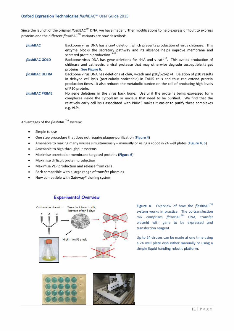
flashBAC Backbone virus DNA has a chiA deletion, which prevents production of virus chitinase. This enzyme blocks the secretory pathway and its absence helps improve membrane and secreted protein production
15-18.
flashBAC GOLD Backbone virus DNA has gene deletions for chiA and v-cath19
. This avoids production of chitinase and cathepsin, a viral protease that may otherwise degrade susceptible target proteins. See Figure 6.
flashBAC ULTRA Backbone virus DNA has deletions of chiA, v-cath and p10/p26/p74. Deletion of p10 results in delayed cell lysis (particularly noticeable) in TnHi5 cells and thus can extend protein production times. It also reduces the metabolic burden on the cell of producing high levels of P10 protein.
flashBAC PRIME No gene deletions in the virus back bone. Useful if the proteins being expressed form complexes inside the cytoplasm or nucleus that need to be purified. We find that the relatively early cell lysis associated with PRIME makes it easier to purify these complexes e.g. VLPs.
Advantages of the flashBACTM
system:
Simple to use
One step procedure that does not require plaque-purification (Figure 4)
Amenable to making many viruses simultaneously – manually or using a robot in 24 well plates (Figure 4, 5)
Amenable to high throughput systems
Maximise secreted or membrane targeted proteins (Figure 6)
Maximise difficult protein production
Maximise VLP production and release from cells
Back compatible with a large range of transfer plasmids
Now compatible with Gateway® cloning system
Figure 4. Overview of how the flashBACTM
system works in practice. The co-transfection
mix comprises flashBACTM
DNA, transfer
plasmid with gene to be expressed and
transfection reagent.
Up to 24 viruses can be made at one time using
a 24 well plate dish either manually or using a
simple liquid handing robotic platform.

Oxford Expression Technologies flashBAC™ User Guide 2015
12 | P a g e
Figure 5. Production and analysis
of a number of secreted
recombinant proteins using
flashBAC viruses (P1 stock) in Sf9
cells and probed with anti-His
antisera. Thanks to Dr Ray Owens
Oxford Protein Production Facility
for beta-testing flashBAC.
Figure 6. Expression of secreted proteins
1-6 using flashBAC (FB) or flashBAC GOLD
(FBG). Western blots probed with anti-His
antisera are shown as are densitometry
results to semi-quantify expression levels.
In most cases FBG improves secretion
levels of proteins.

Oxford Expression Technologies flashBAC™ User Guide 2015
13 | P a g e
7 A laboratory guide to making recombinant baculoviruses using either BacPAK6 or flashBAC™
7.1 Choice of transfer plasmid
Both the BacPAK6 and flashBAC systems use transfer plasmids to mediate transfer of the gene/s to be expressed into
the virus genome at the polh locus. A large number of transfer plasmids are available from OET Ltd and other suppliers.
Please see the OET website for more details or consult the following review articlesx,y,z
.
Transfer plasmids can be grouped as follows:
Polyhedrin gene promoter
Simple vector such as pOET1 or 2 that has a multiple cloning site (MCS) to insert your gene under control of the strong, very late polyhedrin gene promoter.
P10 gene promoter
Another strong, very late gene promoter.
Dual promoters For dual expression of genes, usually one under polyhedrin and one under P10 gene promoters, such as pOET5.
Multiple promoters
A mix of copes of polyhedrin and P10 promoters. Careful construct plans are needed to insert genes according to MCS and restriction sites available.
BacMAM plasmid promoters
These contain a mammalian promoter in place of polyhedrin so that the final recombinant virus can be used to effect gene expression in mammalian cells e.g. pOET6 (CMV promoter)
Late gene promoters
Use baculovirus gene promoters that are expressed earlier than polyhedrin and P10, in the late phase of gene expression. Useful for secreted or membrane targeted protein where polyhedrin/P10 has not worked, or for proteins that need processing before the virus shuts off host cell protein production. E.g. pOET3 and 4 use the p6.9 gene promoter.
Purification tags Several transfer plasmids will give the option for N- or C- terminal tags such as His, Strep, HA to aid protein purification. These may also have a cleavage site to release the final product from the tag.
Fusion vectors Some transfer plasmids allow the gene to be expressed as a fusion product with a fluorescent protein, for example, to allow visualisation by microscopy.
Signal peptides
Generally, the natural signal peptide of a protein will work in insect cells but if you want to add a signal peptide or use an insect virus one, then the signal peptides of either AcMNPV GP64 or chitinase work very well. Adding a signal peptide to a protein that is not normally secreted may not work.
Start codons Translation will start at the first ATG after the promoter so check constructs carefully to ensure there is no inadvertent additional ATG.
Codon optimisation
There is no general data to show that codon optimisation is needed, however, if you are getting your gene synthesised then it makes sense to optimise for insect cells
Membrane anchors
Many people try and secrete membrane protein domains by removing membrane anchor domains – this works sometimes but not always.
When cloning genes into transfer plasmids note:
Check the gene is in the correct orientation with respect to the promoter
Check that the first ATG after the promoter is the start codon you want to initiate translation in the mRNA
Oxford Expression Technologies flashBAC™ User Guide 2015
14 | P a g e
Check you have a stop codon
Check that any fusion or purification tags are in frame and after any signal peptide sequence (that will be
cleaved off)
Sequence any gene that has been cloned via PCR or gene synthesis. Check cloning junctions of genes cloned in
using RE digestion and ligation.
Ensure final plasmid is sterile as it will be used to transfect insect cells – you don’t want your cells getting
bugged.
Mini-prep type DNA works OK in transfections.
Contact us on [email protected] if you need advice or help with transfer plasmids.
7.2 Co-transfection of insect cells with BacPAK6 or flashBAC™ DNA and a transfer plasmid to make a
seed stock (P0) of recombinant baculovirus
This method uses cells prepared in individual 35mm cell culture dishes or 12 well plates. Protocol 7.6
provides an adaptation of this method for making multiple viruses using 24 well plates.
This method must be carried out using aseptic technique as the DNA complexes will be introduced in insect
cells in the absence of antibiotics.
Read through the whole protocol before starting to check you have all the reagents and equipment needed.
Check safety advice and MSDS data sheets where appropriate. We recommend wearing PPE such as lab
coats and gloves at all times.
Provided in the kit:
flashBAC™ DNA (any type) or BacPAK6 DNA (use 100 ng [5µl] DNA per co-transfection)
Positive control transfer plasmid DNA (expressing lacZ) (use 500 ng [5µl] per co-transfection) [flashBAC™ kit
only]
Also needed:
12 well plate or 35mm tissue culture dish seeded with a sub-confluent monolayer of Sf21 or Sf9 cells – one
dish/well for each co-transfection and one for a control
(See OET’s Cell Culture Manual for details on insect cell culture; it is vital for transfection success that cells used
are taken from a culture that is in log phase growth – virus can only replicate when cells are in log phase! A
sub-confluent monolayer is one in which there are spaces around each cell so there is room for each cell to
divide in the 24 hours after co-transfection)
Serum-free insect cell culture medium (we recommend using TC100 medium as a transfection medium but
most serum-free medium will also work)
Growth medium (serum-free or TC100 with 10% serum, as preferred)
Sterile transfer plasmid DNA containing gene to be expressed (see 8.1 for details) (500 ng per transfection)
Oxford Expression Technologies flashBAC™ User Guide 2015
15 | P a g e
Transfection reagent such as OET’s baculoFECTIN II or flashFECTIN (volume as indicated by the manufacturer)
Incubator set at 28⁰C
1% Virkon (Amtec) or other suitable disinfectant
Inverted phase contrast microscope
Plastic box to house dishes in the incubator
Sterile pipettes and bijoux or other polystyrene containers to make up the transfection mix; do not use micro-
centrifuge tubes made of polypropylene.
Method
1 For each co-transfection you require one 35mm dish, or one well of a 12 well plate, containing sub-confluent
Sf9 or Sf21 cells. If you are making a virus with the control vector provided in the flashBAC™ kit, add an extra
dish/well of cells. It is also good practice to have one dish/well for a mock-transfection in which no DNA is
added.
Do not use TniHi5 cells to make viruses as they are prone to mutations that affect gene expression.
Seed the dishes/wells with cells at least one hour before use to allow cells to attach and recover. Ensure cells
were taken from a log-phase culture of cells that were at least 90% viable. As a rough guide you need about
1.5 x 10⁶
Sf21 or Sf9 cells per 35mm dish to form a sub-confluent monolayer. For 12-well plates, add 0.4 x 10⁶
Sf9/Sf21 cells per well. The volume of medium should be 2 ml in 35mm dishes and 1 ml in 12-well plates.
Ensure cells are evenly distributed over the surface of the dish/well.
2 During the 1 hour incubation period above, prepare the co-transfection mix of DNA and transfection reagent.
For each co-transfection in either a 35mm dish or well of a 12-well plate, you need to mix in a polystyrene
tube, in the following order:
100 µl serum free medium (TC100 preferably or serum-free growth medium or ES Transfection Medium)
100 ng virus DNA from the kit (flashBAC™ or BacPAK6) [5µl]
500 ng transfer plasmid (5 µl lacZ transfer vector from flashBAC™ kit) or YOUR transfer vector
1.2 µl baculoFECTIN II
Mix (total volume = 111.2 µl) and leave at room temperature for 15 mins.
Set up a control transfection mix by omitting the DNAs, if wished.
Note. This protocol is optimised for using baculoFECTIN II. If using a different reagent, consult the protocol
supplied by the manufacturer.
3 If the plated cells were maintained in serum-containing medium, wash the monolayers twice with
TC100 without serum and then add 1 ml of TC100 without serum (or ES Transfection Medium) to
each 35 mm dish or well of a 12-well plate. If the cells were maintained in serum-free medium,
Oxford Expression Technologies flashBAC™ User Guide 2015
16 | P a g e
there is no need to wash at this step; simply remove and discard 1ml of medium from the 35mm
dishes.
4 All the 35mm dishes or wells of a 12-well plate should at this stage contain 1 ml of medium without
any serum.
Pipette the 111.2 µl transfection mix from stage 2 into each 35mm dish/well of a 12-well plate as
appropriate. Place in a sandwich box and incubate overnight at 28⁰C.
5 After overnight incubation, add one extra ml of growth medium to the 35mm dishes or replace the 1
ml medium in the 12-well plates with 1 ml growth medium. Continue the incubation for 4 more days
(5 day in total).
Growth medium may either be serum-free medium or TC100 with 10% serum.
Note. Cells in which virus has replicated appear different from mock-transfected cells so comparing
mock-transfected cells with experimental dishes can be a useful indicator that the transfection has
worked; infected cells appear more grainy with swollen nuclei.
6 Harvest the culture medium containing budded recombinant virus into a sterile container and store
in the dark at 4⁰C.
Note. If you prepared a control virus with the lacZ transfer plasmid in the flashBAC™ kit, you can
check for transfection success by staining the monolayer of cells left after harvesting the P0 virus;
add 1 ml of growth medium containing 15µl 2% v/v X-gal to the cell monolayer and leave for a few
hours to overnight for the blue colour to develop.
7 The next step depends on whether you have used BacPAK6 or flashBAC™ DNA.
flashBAC™ DNA: Your 1-2 ml stock of virus is your seed stock (P0), you now need to amplify this to
obtain a 50 ml P1 stock of virus for experimental work and freezing down (go to 8.4).
BacPAK6 DNA: You now need to plaque-purify your recombinant virus to obtain your seed stock (P0)
(go to 8.3)
Note: You can also plaque-purify virus produced using flashBAC™ DNA if required (go to 8.3)
Oxford Expression Technologies flashBAC™ User Guide 2015
17 | P a g e
7.3 Plaque-purification of recombinant BacPAK6 virus
The budded virus harvested after the co-transfection with BacPAK6 virus DNA will contain a mixture of parental virus
(about 10% blue) and recombinant virus (about 90% clear/colourless). These need to be separated by performing a
plaque-assay and picking individual plaques to amplify pure virus stocks. As long as well isolated plaques are picked,
only one round of plaque-purification is needed.
This is a multi-step method that enables you to isolate plaques and then amplify plaque-picked virus to produce a P0
seed stock of virus. Read through the whole method before starting to ensure you are aware of time scales and
reagents/equipment needed at each stage.
The OET Cell Culture Manual has details of insect cell culture.
Required:
Virus harvested from a co-transfection (see 7.2)
TC100 growth medium with serum (best; or serum free growth medium) - antibiotics (Penicillin, 10000
units/ml and Streptomycin, 10000 µg/ml in 0.85% saline; dilute 1:50 for use) may be added to plaque-assay
medium to help reduce the chance of contamination
Culture of Sf21 cells (preferred; or Sf9 cells) that are in log phase of growth and at least 90% viable
35 mm dishes and T25 flasks
Low temperature gelling (Sea-plaque) agarose for overlay (Sigma; 2% w/v solution in d.water). It is convenient
to make up small batches (15 ml) of agarose overlay by melting the agarose using a boiling water bath or
microwave oven (take care). Solidified agarose can be stored and re-melted prior to use. (Larger volumes may
also be prepared and melted multiple times). Cool to ‘hand hot’ before making up final overlay.
Sterile pipettes and bijoux or similar containers for making virus dilutions
Sterile Pasteur pipettes
Beaker with hand hot water as a temporary water bath
Plastic sandwich box
Incubator at 28⁰C
Phosphate-buffered saline (PBS)
Neutral Red stain (Sigma; 5 mg/ml in d.water, filter sterilize and store at room temperature). For use dilute 1
in 20 with PBS solution. Do not store diluted stain.
X-gal (2% v/v in DMF) to stain for blue plaques
1% Virkon (Amtec) or similar disinfectant
Oxford Expression Technologies flashBAC™ User Guide 2015
18 | P a g e
Inverted phase contrast light microscope
Lightbox to view plaques
Method
1 Seed 35mm cell culture dishes with a sub-confluent monolayer of healthy Sf21 cells (or Sf9 cells if Sf21 are not
available). See 8.2.1 for more details. Allow the cells to settle for at least one hour.
You need 12 dishes per virus.
Sf21 cells in TC100 with 10% serum give the largest easy-to-spot plaques because these cells have a well-
defined CPE (see Figure 7). Sf9 cells will also yield plaques but they are smaller, take longer to develop and are
not quite so easy to define. We have also noted that plaque assays conducted with Sf9 cells and serum free
medium produce plaques that quickly fade after staining with Neutral Red.
2 Make 1 in 10 dilutions of your transfection virus stock from 1 in 10 (10-1
) to 1 in 106 (10
-6). Use 50 µl virus and
450 µl growth medium as diluent at each step. Mix the virus and diluent between each step and change
tip/pipette each time to avoid carry-over.
3 Remove the medium from the dishes of cells using aseptic technique and add 100 µl of diluted virus drop wise
to the centre of each dish. Plate a range of dilutions and two plates per dilution – the aim is to get well
isolated plaques on at least one dilution. We normally plate the 1 in 100 (10-2
) to 10-6
dilutions in duplicate
dishes and use two dishes as mock infected controls (use medium only).
It is important that the cell monolayers do not dry out during this process of virus inoculation. Do not leave lids
off dishes for long periods. If working in a class 2 hood be aware the air flow can dry plates very quickly. If,
after staining, your monolayer appears with a shiny red patch devoid of cells, you have allowed the monolayer
to dry out.
4 Allow the virus to adsorb and be taken up into the cells at room temperature for 45 - 60 min. Rock the dishes
every few minutes to ensure even coverage of the inoculum. Do NOT put the cells in the incubator as they will
dry out.
5 During this time prepare the overlay. Dissolve agarose in water to 2% w/v by boiling (water bath or microwave
oven – take appropriate safety precautions). You need 1 ml per dish of cells. Cool the overlay to hand hot
(about 50-55⁰C) and add an equal volume of pre-warmed growth medium (28⁰C). Keep warm to prevent
setting (we use a temporary clean water bath comprising a beaker of hot tap water). You need 2 ml final
overlay per dish.
If the agarose in water sets, it is easy to melt again by boiling. If the agarose overlay with growth medium sets,
you cannot re-melt. You have to start again. We often prepare several small batches of agarose in water and
let them set and then melt each aliquot as we need it (15 ml is convenient).
6 At the end of the incubation period (4), remove the inoculum using a pipette and discard into Virkon or other
disinfectant. Working quickly, add 2 ml warm overlay to each dish allowing the agarose to flow down the side
of the dish and spread slowly over the monolayer of cells. Do NOT pipette into the centre of the dish.
Oxford Expression Technologies flashBAC™ User Guide 2015
19 | P a g e
Process one set of dishes per virus sample at a time. If working in a hood, keep the agarose overlay in a beaker
or sandwich box filled with warm water to delay solidification. If the agarose sets prematurely, you can leave
the dishes with virus inoculums for longer than 60 min without adverse effects. If you have removed the virus
and then find that your overlay medium has set, just add 0.1-0.2ml fresh medium to each plate to prevent
drying of cells. Prepare more agarose overlay medium and carry on, but don’t forget to remove the extra
medium you added to each dish!
7 Allow the agarose overlay to set at room temperature. Then add a 1 ml liquid overlay of growth medium to
feed the cells and prevent them from drying out.
8 Place the dishes in a plastic box and incubate at 28⁰C for 3-4 days. Three days for Sf21 and four days for Sf9
cells.
9 Add 1 ml growth medium containing 15 µl 2% v/v X-gal in DMF to each dish to stain for blue (parental)
plaques. Incubate at 28⁰C for 5-6 h.
Conveniently, this is done in the course of a normal working day. Blue plaques should start to develop during
this time.
10 Prepare the Neutral Red stain in water to 5 mg/ml d.water and filter sterilize or purchase ready made from
Sigma, for example. Dilute 1 in 20 with sterile PBS for use.
Different batches of Neutral Red may differ in their efficacy. Sometimes 1 in 40 dilutions give better results. Do
not store diluted stain, it will form a precipitate. The concentrated stock is stable at room temperature for
several months (if sterile).
11 Add 1 ml diluted neutral red stain to each dish. Do not remove the X-gal already added. Incubate at 28⁰C for
16 hours (overnight).
12 Decant all liquid and view plaques on a light box. Recombinant virus plaques will appear clear in a sea of red
healthy cells. Parental, non-recombinant plaques will stain blue with X-gal.
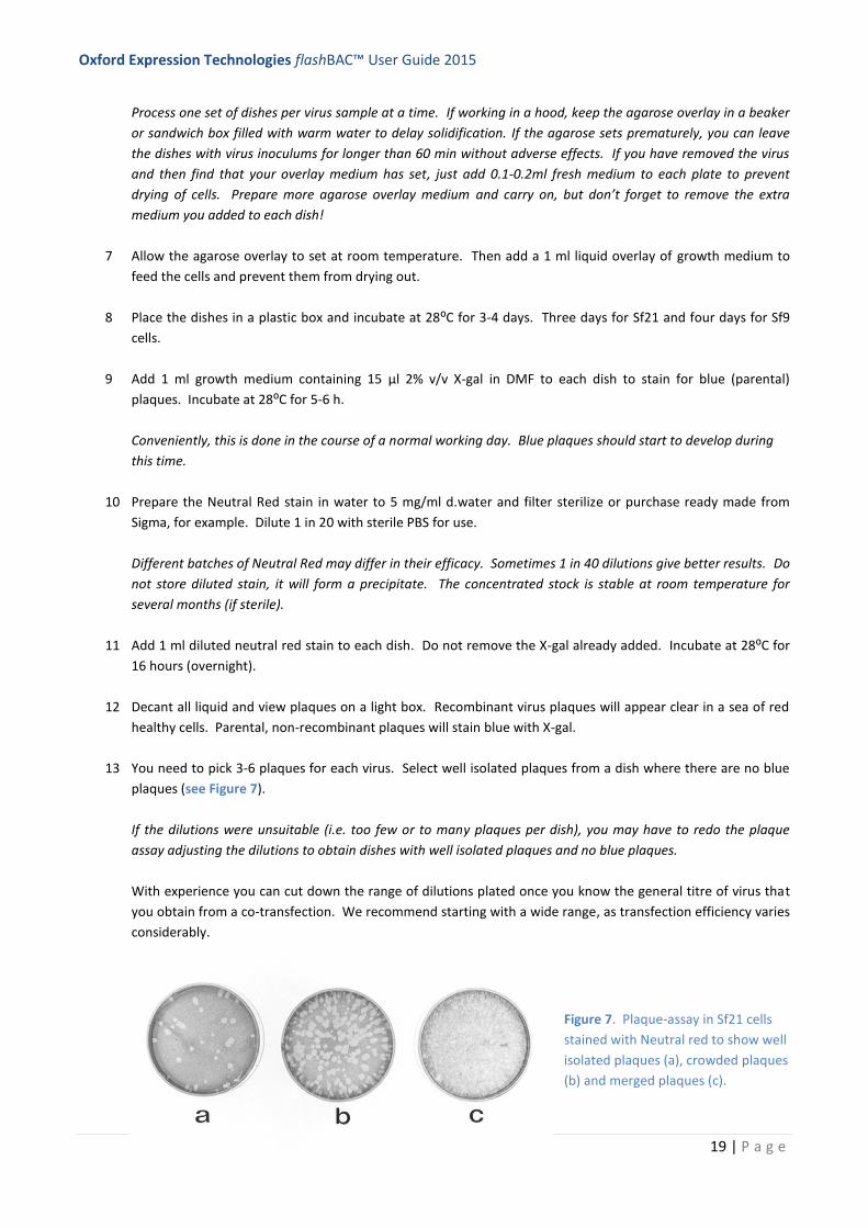
13 You need to pick 3-6 plaques for each virus. Select well isolated plaques from a dish where there are no blue
plaques (see Figure 7).
If the dilutions were unsuitable (i.e. too few or to many plaques per dish), you may have to redo the plaque
assay adjusting the dilutions to obtain dishes with well isolated plaques and no blue plaques.
With experience you can cut down the range of dilutions plated once you know the general titre of virus that
you obtain from a co-transfection. We recommend starting with a wide range, as transfection efficiency varies
considerably.
Figure 7. Plaque-assay in Sf21 cells
stained with Neutral red to show well
isolated plaques (a), crowded plaques
(b) and merged plaques (c).
Oxford Expression Technologies flashBAC™ User Guide 2015
20 | P a g e
14 To pick a plaque, you need to take up a plug of agarose from the centre of a plaque using a Pasteur pipette or
Gilson tip. Disperse the plug of agarose into 500 µl growth medium in a micro centrifuge tube and vortex to
release the virus from the agarose into the medium. Store in the dark at 4⁰C.
15 Amplify the plaque-picked virus by inoculating either a 35mm dish or a T25 flask of Sf21 or Sf9 cells using 100
µl (35 mm dish) or 250 µl (T25 flask) of your 500 µl as inoculum.
To do this, seed a 35 mm dish or T25 flask with cells to form a sub-confluent monolayer and after an hour or
so, remove the medium and replace with the inoculum for 45-60 mins. Then add 2 ml (dish) or 5 ml (T25 flask)
growth medium (no need to remove the inoculum) and incubate for 4-5 day at 28⁰C.
The cells should be well infected under the microscope at the end of the infection period.
16 Harvest the 2 ml or 5 ml of medium containing your P0 seed stock virus. Store at 4⁰C in the dark. Use this to
amplify a P1 working stock of recombinant virus to test gene expression (see 7.4).
17 The cell monolayers from the dish or flask used to amplify virus can be harvested and used to test for gene
expression or to isolate DNA to do a PCR to check that the gene has gone into the virus genome.
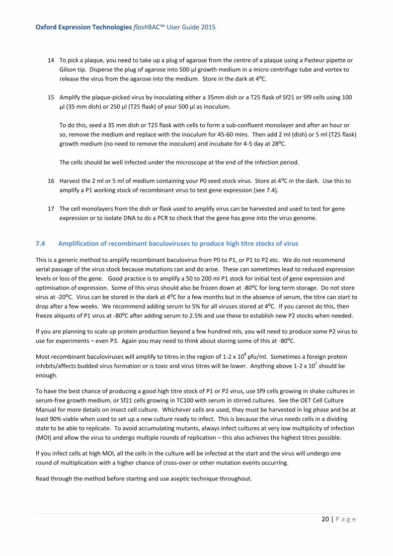
7.4 Amplification of recombinant baculoviruses to produce high titre stocks of virus
This is a generic method to amplify recombinant baculovirus from P0 to P1, or P1 to P2 etc. We do not recommend
serial passage of the virus stock because mutations can and do arise. These can sometimes lead to reduced expression
levels or loss of the gene. Good practice is to amplify a 50 to 200 ml P1 stock for initial test of gene expression and
optimisation of expression. Some of this virus should also be frozen down at -80⁰C for long term storage. Do not store
virus at -20⁰C. Virus can be stored in the dark at 4⁰C for a few months but in the absence of serum, the titre can start to
drop after a few weeks. We recommend adding serum to 5% for all viruses stored at 4⁰C. If you cannot do this, then
freeze aliquots of P1 virus at -80⁰C after adding serum to 2.5% and use these to establish new P2 stocks when needed.
If you are planning to scale up protein production beyond a few hundred mls, you will need to produce some P2 virus to
use for experiments – even P3. Again you may need to think about storing some of this at -80⁰C.
Most recombinant baculoviruses will amplify to titres in the region of 1-2 x 10⁸ pfu/ml. Sometimes a foreign protein
inhibits/affects budded virus formation or is toxic and virus titres will be lower. Anything above 1-2 x 10⁷ should be
enough.
To have the best chance of producing a good high titre stock of P1 or P2 virus, use Sf9 cells growing in shake cultures in
serum-free growth medium, or Sf21 cells growing in TC100 with serum in stirred cultures. See the OET Cell Culture
Manual for more details on insect cell culture. Whichever cells are used, they must be harvested in log phase and be at
least 90% viable when used to set up a new culture ready to infect. This is because the virus needs cells in a dividing
state to be able to replicate. To avoid accumulating mutants, always infect cultures at very low multiplicity of infection
(MOI) and allow the virus to undergo multiple rounds of replication – this also achieves the highest titres possible.
If you infect cells at high MOI, all the cells in the culture will be infected at the start and the virus will undergo one
round of multiplication with a higher chance of cross-over or other mutation events occurring.
Read through the method before starting and use aseptic technique throughout.
Oxford Expression Technologies flashBAC™ User Guide 2015
21 | P a g e
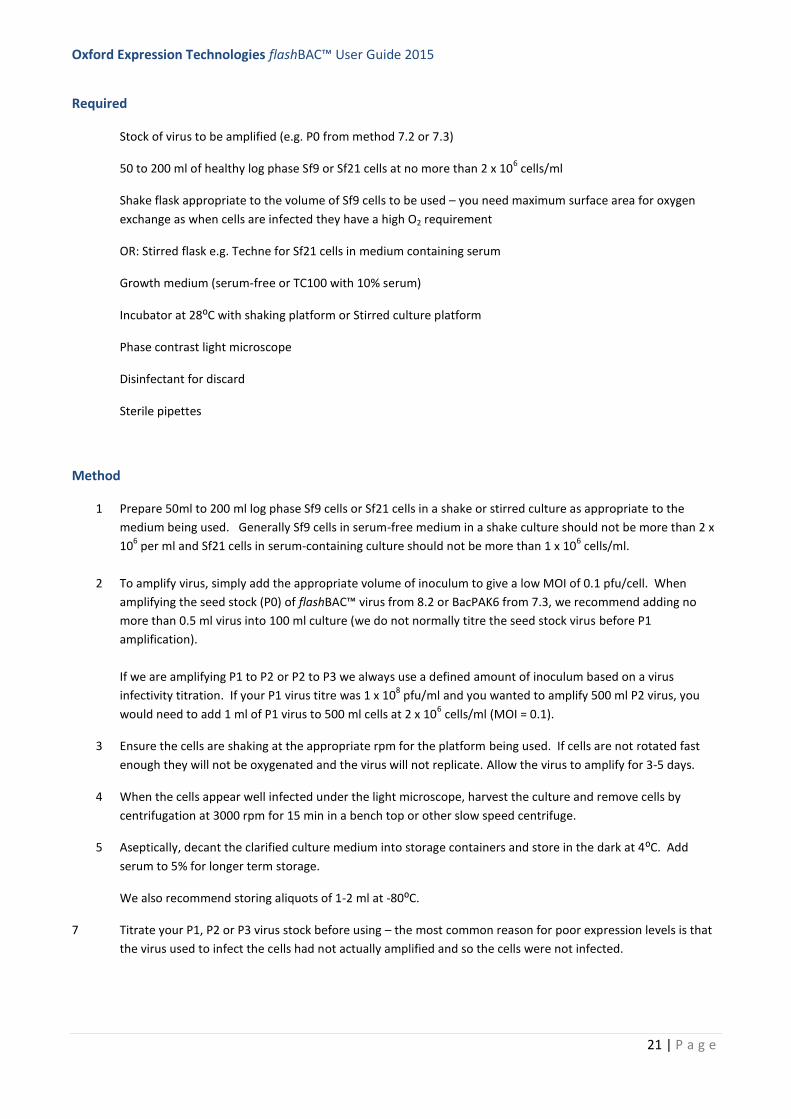
Required
Stock of virus to be amplified (e.g. P0 from method 7.2 or 7.3)
50 to 200 ml of healthy log phase Sf9 or Sf21 cells at no more than 2 x 10⁶ cells/ml
Shake flask appropriate to the volume of Sf9 cells to be used – you need maximum surface area for oxygen
exchange as when cells are infected they have a high O2 requirement
OR: Stirred flask e.g. Techne for Sf21 cells in medium containing serum
Growth medium (serum-free or TC100 with 10% serum)
Incubator at 28⁰C with shaking platform or Stirred culture platform
Phase contrast light microscope
Disinfectant for discard
Sterile pipettes
Method
1 Prepare 50ml to 200 ml log phase Sf9 cells or Sf21 cells in a shake or stirred culture as appropriate to the
medium being used. Generally Sf9 cells in serum-free medium in a shake culture should not be more than 2 x
106 per ml and Sf21 cells in serum-containing culture should not be more than 1 x 10
6 cells/ml.
2 To amplify virus, simply add the appropriate volume of inoculum to give a low MOI of 0.1 pfu/cell. When
amplifying the seed stock (P0) of flashBAC™ virus from 8.2 or BacPAK6 from 7.3, we recommend adding no
more than 0.5 ml virus into 100 ml culture (we do not normally titre the seed stock virus before P1
amplification).
If we are amplifying P1 to P2 or P2 to P3 we always use a defined amount of inoculum based on a virus
infectivity titration. If your P1 virus titre was 1 x 10⁸ pfu/ml and you wanted to amplify 500 ml P2 virus, you
would need to add 1 ml of P1 virus to 500 ml cells at 2 x 10⁶ cells/ml (MOI = 0.1).
3 Ensure the cells are shaking at the appropriate rpm for the platform being used. If cells are not rotated fast
enough they will not be oxygenated and the virus will not replicate. Allow the virus to amplify for 3-5 days.
4 When the cells appear well infected under the light microscope, harvest the culture and remove cells by
centrifugation at 3000 rpm for 15 min in a bench top or other slow speed centrifuge.
5 Aseptically, decant the clarified culture medium into storage containers and store in the dark at 4⁰C. Add
serum to 5% for longer term storage.
We also recommend storing aliquots of 1-2 ml at -80⁰C.
7 Titrate your P1, P2 or P3 virus stock before using – the most common reason for poor expression levels is that
the virus used to infect the cells had not actually amplified and so the cells were not infected.
Oxford Expression Technologies flashBAC™ User Guide 2015
22 | P a g e
You can titre your virus by plaque-assay – the Gold Standard (see 8.5) or by QPCR. OET has a convenient QPCR
titration kit (BaculoQuant) or OET provides a fast and cost-effective virus titration service (contact us at
[email protected]) for more details.
Note: Virus can also be amplified in monolayer cultures in T75 or T150 flasks. Simply seed the flasks to provide a sub-
confluent monolayer of cells. Remove the medium and add the inoculum to give the correct MOI (0.1 pfu/cell) (use
100 or 200 µl P0 virus from 8.2 or 8.3 diluted in medium to 500 µl (T75) or 1 ml (T150) per flask. After 45 mins
incubation, add 10-15 ml medium (T75) or 30 m medium (T150) and allow the virus to replicate for 3-5 days – until all
the cells are well infected. The titre of virus amplified in this way is not usually as high as that amplified in shake
cultures.
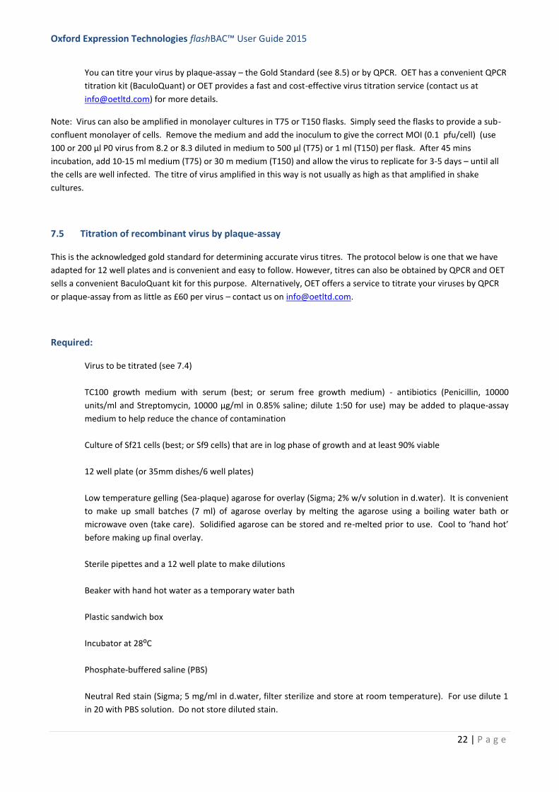
7.5 Titration of recombinant virus by plaque-assay
This is the acknowledged gold standard for determining accurate virus titres. The protocol below is one that we have
adapted for 12 well plates and is convenient and easy to follow. However, titres can also be obtained by QPCR and OET
sells a convenient BaculoQuant kit for this purpose. Alternatively, OET offers a service to titrate your viruses by QPCR
or plaque-assay from as little as £60 per virus – contact us on [email protected].
Required:
Virus to be titrated (see 7.4)
TC100 growth medium with serum (best; or serum free growth medium) - antibiotics (Penicillin, 10000
units/ml and Streptomycin, 10000 µg/ml in 0.85% saline; dilute 1:50 for use) may be added to plaque-assay
medium to help reduce the chance of contamination
Culture of Sf21 cells (best; or Sf9 cells) that are in log phase of growth and at least 90% viable
12 well plate (or 35mm dishes/6 well plates)
Low temperature gelling (Sea-plaque) agarose for overlay (Sigma; 2% w/v solution in d.water). It is convenient
to make up small batches (7 ml) of agarose overlay by melting the agarose using a boiling water bath or
microwave oven (take care). Solidified agarose can be stored and re-melted prior to use. Cool to ‘hand hot’
before making up final overlay.
Sterile pipettes and a 12 well plate to make dilutions
Beaker with hand hot water as a temporary water bath
Plastic sandwich box
Incubator at 28⁰C
Phosphate-buffered saline (PBS)
Neutral Red stain (Sigma; 5 mg/ml in d.water, filter sterilize and store at room temperature). For use dilute 1
in 20 with PBS solution. Do not store diluted stain.
Oxford Expression Technologies flashBAC™ User Guide 2015
23 | P a g e
1% Virkon (Amtec) or similar disinfectant
Inverted phase contrast light microscope
Lightbox to view plaques
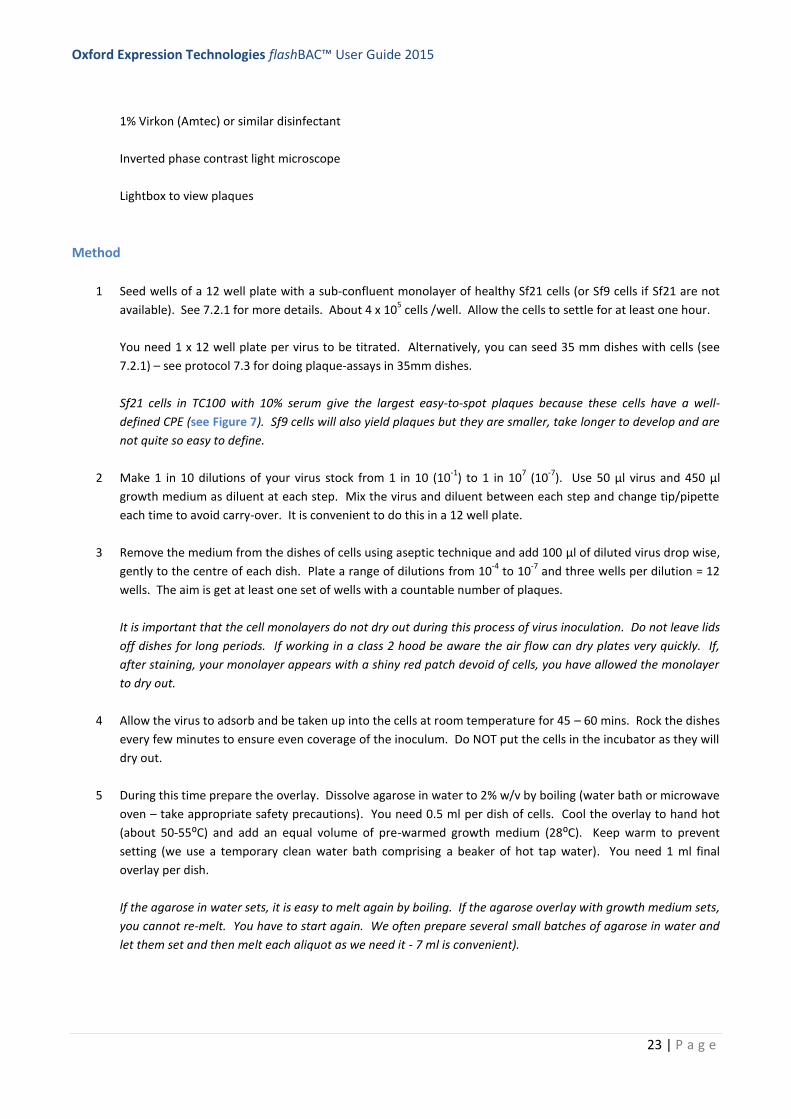
Method
1 Seed wells of a 12 well plate with a sub-confluent monolayer of healthy Sf21 cells (or Sf9 cells if Sf21 are not
available). See 7.2.1 for more details. About 4 x 10⁵ cells /well. Allow the cells to settle for at least one hour.
You need 1 x 12 well plate per virus to be titrated. Alternatively, you can seed 35 mm dishes with cells (see
7.2.1) – see protocol 7.3 for doing plaque-assays in 35mm dishes.
Sf21 cells in TC100 with 10% serum give the largest easy-to-spot plaques because these cells have a well-
defined CPE (see Figure 7). Sf9 cells will also yield plaques but they are smaller, take longer to develop and are
not quite so easy to define.
2 Make 1 in 10 dilutions of your virus stock from 1 in 10 (10-1
) to 1 in 107 (10
-7). Use 50 µl virus and 450 µl
growth medium as diluent at each step. Mix the virus and diluent between each step and change tip/pipette
each time to avoid carry-over. It is convenient to do this in a 12 well plate.
3 Remove the medium from the dishes of cells using aseptic technique and add 100 µl of diluted virus drop wise,
gently to the centre of each dish. Plate a range of dilutions from 10-4
to 10-7
and three wells per dilution = 12
wells. The aim is get at least one set of wells with a countable number of plaques.
It is important that the cell monolayers do not dry out during this process of virus inoculation. Do not leave lids
off dishes for long periods. If working in a class 2 hood be aware the air flow can dry plates very quickly. If,
after staining, your monolayer appears with a shiny red patch devoid of cells, you have allowed the monolayer
to dry out.
4 Allow the virus to adsorb and be taken up into the cells at room temperature for 45 – 60 mins. Rock the dishes
every few minutes to ensure even coverage of the inoculum. Do NOT put the cells in the incubator as they will
dry out.
5 During this time prepare the overlay. Dissolve agarose in water to 2% w/v by boiling (water bath or microwave
oven – take appropriate safety precautions). You need 0.5 ml per dish of cells. Cool the overlay to hand hot
(about 50-55⁰C) and add an equal volume of pre-warmed growth medium (28⁰C). Keep warm to prevent
setting (we use a temporary clean water bath comprising a beaker of hot tap water). You need 1 ml final
overlay per dish.
If the agarose in water sets, it is easy to melt again by boiling. If the agarose overlay with growth medium sets,
you cannot re-melt. You have to start again. We often prepare several small batches of agarose in water and
let them set and then melt each aliquot as we need it - 7 ml is convenient).

Oxford Expression Technologies flashBAC™ User Guide 2015
24 | P a g e
6 At the end of the incubation period (4), remove the inoculum using a pipette and discard into Virkon or other
disinfectant. Working quickly, add 1 ml warm overlay to each dish allowing the agarose to flow down the side
of the dish and spread slowly over the monolayer of cells. Do NOT pipette into the centre of the dish.
Process one set of dishes per virus sample at a time. If working in a hood, keep the agarose overlay in a beaker
or sandwich box filled with warm water to delay solidification. If the agarose sets prematurely, you can leave
the dishes with virus inoculums for longer than 60 min without adverse effects. If you have removed the virus
and then find that your overlay medium has set, just add 0.1-0.2ml fresh medium to each plate to prevent
drying of cells. Prepare more agarose overlay medium and carry on, but don’t forget to remove the extra
medium you added to each dish!
7 Allow the agarose overlay to set at room temperature. Then add a 0.5 ml liquid overlay of growth medium to
feed the cells and prevent them from drying out.
8 Place the dishes in a plastic box and incubate at 28⁰C for 3-4 days. Three days for Sf21 and 4 days for Sf9 cells.
9 Prepare the stain by dissolving Neutral Red in water to 5 mg/ml d.water and filter sterilize or purchase ready
made from Sigma, for example. Dilute 1 in 20 with sterile PBS for use.
Note some batches of Neutral Red may work better at 1 in 40 dilution – do not store diluted stain as it
precipitates.
10 Add 0.5 ml diluted neutral red stain to each dish and incubate for 3-4 hours.
11 Decant the remaining liquid and view plaques on a light box. Recombinant virus plaques will appear clear in a
sea of red healthy cells. It sometimes takes a few hours for plaques to be really visible.
12 Count the plaques from wells where there are a countable number of plaques (10-20). Average the plaque
count from the triplicate dishes and note the dilution that gave rise to these plaques.
13 Determine the virus titre as follows:
Average number plaques x dilution factor* x 10** = plaques/ml in the original virus stock.
*Inverse of dilution **because only 0.1 ml was added to dish
For example, if the average number of plaques was 15 taken from the 10-6
dilution wells, the virus titre would
be:
15 x 106 x 10 = 1.5 x 10
8 pfu/ml.
14 Note that virus titres will drop after storage at 4⁰C and so we recommend re-titrating virus before use if it has
been stored for more than 3-4 months.
8.6 A guide to using flashBAC in 24 well plate systems
The following is a guide to making recombinant flashBAC viruses in a 24 well plate format. This can be achieved
manually or the protocol can be adapted to use in a simple robotic system for liquid handling. In this way it is relatively
straightforward to make 24 recombinant viruses at one time.

Oxford Expression Technologies flashBAC™ User Guide 2015
25 | P a g e
Cells: Prepare a master mix of Sf9 cells in serum free medium at a density of 5 x 10⁵ cells/ml and dispense 0.4 ml (2 x
105 cells) per well. Allow cells to settle for one hour.
Transfection master mix: It is convenient to make this in the wells of a 96 well plate. Make up a master mix of 220 µl
TC100 medium w/o serum (or other serum-free medium) and 120 µl flashBAC DNA (5 µl per virus). Dispense 14 µl into
24 wells of a 96 well plate. Then add 5 µl of the correct transfer plasmid (500 ng DNA) and 1.2 µl baculoFECTIN II to
each of the 24 wells as appropriate, mix by pipetting up and down a few times and allow to stand for 15 min.
Add transfection mix to cells: Simply add the 20 µl transfection mix into each of the wells containing cells in the 24 well
plate. Seal to prevent evaporation and incubate at 28⁰C for 5 days.
Harvest recombinant virus by transferring the culture medium containing budded virus to the wells of a new 24 well-
plate, seal and store at 4⁰C in the dark.
To amplify virus, follow protocol 7.4 – as a guide use 250 µl to infect 50 – 100 ml of Sf9 cells.
8 Analysis and Optimisation of Gene Expression
This section provides a guide to the analysis of gene expression from recombinant virus made using either the BacPAK6
or flashBAC™ systems. It is not intended to be prescriptive simply a guide to help you get started.
8.1 Quick check for gene expression
After the co-transfection or after amplification of P0 virus to give P1, remaining cells in the monolayer can be harvested
and used to test for gene expression by SDS-PAGE and/or Western blotting. However, the expression levels are
variable at these stages so many people prefer to wait until they have a high titre stock of virus (P1 or P2). Some of the
expression after the co-transfection will also be transient expression from the transfer plasmid itself.
8.2 Test expression by infecting cells with high titre virus stocks
It is always best to test expression using a virus with a known titre. That way you can control the multiplicity of
infection. Normally the best levels of expression are obtained with high MOIs (5-10 pfu/cell) so that all the cells are
infected simultaneously and a synchronous infection is established. However, for a few proteins, best expression is
obtained at lower MOI. We therefore recommend that expression testing includes a range of MOI (0.5, 2.5 and 5 is a
good starting point).
It is convenient to monitor gene expression by setting up small-scale monolayer cultures in either 35mm dishes or the
wells of a 12 well plate. Set up monolayers in dishes/wells as described for co-transfections/plaque-assays (see 8.2/8.4)
and leave the cells to recover for an hour. Always take cells from log phase cultures to ensure that virus can infect the
cells and replicate – otherwise the polyhedrin gene promoter (or other virus promoter) will not be turned on and
expression levels will be very low.
Infect 35 mm dishes with 200 µl virus inoculum or 12 well plate wells with 100µl. Simply remove the medium, add the
inoculum drop wise and gently to the centre of the dish and leave to adsorb for 45-60 min, with occasional rocking of
the dishes. Then replace the growth medium (2 ml for 35 mm dishes and 1 ml for 12 well plates). Incubate at 28⁰C.
Always include a negative control (mock-infected cells) for comparison. If you have a known recombinant baculovirus,
you can add a positive control. If you purchased a flashBAC™ kit, you could make a recombinant virus with the control
lacZ transfer vector and use this to set control infections to look for beta-galactosidase production (see Figure 3).
Oxford Expression Technologies flashBAC™ User Guide 2015
26 | P a g e
We normally test expression by harvesting the cells and/or culture medium (as needed) at 72 hpi initially. If you want
to test the culture medium for secreted protein, harvest the medium, centrifuge to remove any floating cells and
decant into a fresh tube. If expression levels are expected to be on the low side, treat 1 ml of medium with Strataclean
resin®, which concentrates the protein ready for SDS-PAGE and/or Western blot analysis.
If the protein is intracellular, scrape the cells into the culture medium with a blue Gilson tip, pellet the cells in a
microcentrifuge tube. If liked, you can wash the dish with TE buffer to remove the last few cells, and add these to the
tube with the main bulk of cells. Wash the cell pellet with TE buffer and resuspend the cell pellet in SDS-PAGE loading
buffer and boil samples in the usual way.
We may later optimise expression by testing expression at multiple time points (see 9.3). It is well worth testing
expression in both Sf9 (Sf21) cells and TniHi5® cells. See the OET Cell Culture handbook for details of culturing Tni cells.
Sometimes there can be a large difference in the expression levels between these two cell lines. Whilst Tni cells should
not be used for virus amplification (due to accumulation of mutations), they can be an excellent cell line for protein
production and grow well in serum-free medium in shake cultures.
8.3 Optimisation of expression
Sometimes it is necessary to optimise expression levels. This is particularly important if you are going to scale up
production of protein – work here can save litres of medium and hard work later on. You can either set up multiple 35
mm dishes or 12 well plates (one dish/well per condition) or set up small (20 ml) shake cultures and take samples (2 ml)
at various time points. The latter is better if you are planning on scaling up in future. You may also need to do pilot
protein purification and small scale shake cultures can work well for this too.
Always do control mock-infected dishes or take samples prior to infecting shake cultures.
Parameters to optimise include:
Multiplicity of infection – start by comparing 1, 3 and 10 pfu/cell
Cell line (Sf9, Sf21, TniHi5®, SuperSf9 cells
Time to harvest 0 24, 38, 72, 96 hpi
flashBAC variant (see introduction)
8.4 Scaling up production
There are many ways to scale-up insect cell culture and hence virus or protein production. The simplest is to use large-
scale shake flasks. In this way up to 1.25 L cells can be infected at one time. The key to success is to ensure that flasks
are not overfilled (aim for maximum surface area) and that cells are shaken at a high rpm to ensure good aeration. GE
Healthcare’s wavebags® are also relatively easy to use but are expensive and require access to a Wave Bioreactor®.
The OET Cell Culture Handbook has more information on this topic.
9 Trouble shooting and FAQ
Q Why are my cells not growing well?
A The most likely problem with cells occurs when they have been allowed to reach stationary phase before
passaging. If this ‘stress’ happens to a culture 2 or 3 times, then the cells no longer grow properly. Always
check cells on a regular basis and do not let cultures overgrow. If this happens, go back to liquid nitrogen
stocks are set up a new culture. Far more important than passage number of the cell is the number of times
the culture has been stressed!
Oxford Expression Technologies flashBAC™ User Guide 2015
27 | P a g e
Cells that are not growing well should never be used to make recombinant viruses, amplify virus or test for
protein production because each of these techniques requires the virus to infect and replicate inside cells and
it can only do this is the cells are actively replicating – i.e. in log phase of growth.
Q My cells are not growing and have enlarged nuclei?
A See above, but they also may be contaminated with baculovirus. Start with a fresh cell culture. Never use
virus and stock cells in the same Class 2 hood. Always do cell culture work before virus work.
Q My co-transfection has not worked or become contaminated?
A See Q on cells above. Have you followed the protocol exactly? Try a different transfection reagent. The
plasmid DNA used in the co-transfection must be sterile – try precipitating with alcohol and re-suspending in
sterile TE. Check you medium is not contaminated. The flashBAC and BacPAK6 DNA is quality checked to
ensure it is sterile.
Q My virus has not amplified to high titre?
A See Q on cells above – this is the most likely problem. Did you infect cells with low MOI (0.1 pfu/cell)? High
MOI will lead to lower titres and very low MOI will work but you may need to leave the cells longer to achieve
high titres. Did he cells look infected (grainy and swollen nuclei under the microscope)? Could the foreign
gene product be affecting budded virus production?
Q Why don’t I see plaques in my plaque-assay?
Were the cells in good condition – see Q on cells above? Double check the cell density that was plated – too
high and cells cannot undergo enough rounds of replication to form a plaque (they will be like pin pricks and
hard to see) – too low and the cells do not close up to form a monolayer so edges of plaques can be ragged
and hard to spot. Are there any cells left at all – look under microscope – if the dishes dried out at any time
there will be no cells left and no plaques. Was the agarose overlay too hot – which may have killed the cells?
Did you remember the liquid overlay (with 10% for TC100)? Was the virus titre too low to be detected 0 try
again with lower dilutions or even neat virus plated out. Or was titre too high and you need to plate out
higher dilutions to see plaques – they may have merged together and be hard to see (did you change tips
between dilutions to avoid carry over!!). Was the Neutral red freshly diluted ready for use?
Q My plaque-assay overlay has cracks or fell out when inverting, or the plaques are smeared or only around the
edges of the dish?
A If the virus inoculum is not removed before adding the agarose overlay, it interferes with the gelling process
and can produce cracks. It may even cause the overlay to fall out when you tip off the stain. It may also allow
the virus to spread under the overlay and so the plaques appear smeared and diffuse. Always add inoculum to
the centre of the dish and rock the dish a few times during the incubation period to ensure even coverage of
the virus. Ensure the cells are also evenly distributed over the dish. Do not use a swirling motion at any time
as they simply sends cells and virus to the edges of the dish. Occasionally, multi-well plate wells do not have
perfectly flat surfaces – in our experience the worst culprits for this are 6 well plates and so we always use
individual 35 mm dishes.
Q I cannot detect any gene expression?
A Were the cells used for test expression in good condition – see answer above about cells. Did you use a virus
with a known titre (by plaque-assay or QPCR) – there may not have been any virus if you didn’t. Has the virus
been stored for some time before use - did you add serum to maintain the titre? If not re-titrate your virus
and try again. Only use QPCR on fresh virus. If you have a control virus, did that work? Very occasionally, the
Oxford Expression Technologies flashBAC™ User Guide 2015
28 | P a g e
gene is not stable – check that the gene is actually in the virus genome by PCR. Harvest cellular/virus DNA
from a 35 mm dish and use for PCR analysis. Is the gene properly under control of the polyhedrin gene
promoter – is the first ATG the ATG of your gene? If not, you need to address the construct and make a new
virus. If you are using tags to detect the gene, check they are in frame. Did you try optimising expression (see
above). In particular, sometimes T. ni cells yield protein when Sf9 do not. Finally, if you have exhausted all
avenues, there are a very, very few genes that for unknown reasons do not express. Most have been found to
be toxic to the cell. But check all of the above before thinking this!
10 References
1. Van Regenmortal, M. H. V. et al. (2000). Virus Taxonomy: Classification and Nomenclature of Viruses.
Seventh Report of the International Committee on Taxonomy of Viruses. San Diego: Academic Press.
2. Vail, P. V., Jay, D. L. & Hunter, D. K. In Proc. IVth Int. Colloq. Insect Pathology 297-304 (College Park, MD,
1971).
3. Smith, G. E., Summers, M. D. & Fraser, M. J. (1983). Molecular and Cellular Biology 3, 2156-2165.
4. Ayres, M. D., Howard, S. C., Kuzio, J., Lopez-Ferber, M. & Possee, R. D. (1994). Virology 202, 586-605.
5. Blissard, G. W. & Wenz, J. R. (1992). J. Virol. 66, 6829-6855.
6. Volkman, L. E. & Summers, M. D. (1977). J. Invertebr. Pathol. 30, 102-103.
7. Monsma, S. A., Oomens, A. G. P. & Blissard, G. W. (1996). J. Virol. 70, 4607-4616.
8. Summers, M. D. & Smith, G. E. (1978). Virology 83, 390-402.
9. Rohrmann, G. F. (1986). J. Gen. Virol. 67, 1499-1513.
10. Possee, R. D. & Howard, S. C. (1987). Nuc. Acids Res. 15, 10233-10248.
11. Kitts, P. A., Ayres, M. D. & Possee, R. D. (1990). Nuc. Acids Res. 11, 5667-5672.
12. Kitts, P. A. & Possee, R. D. (1993). Biotechniques 14, 810-817.
13. Patent applications EP1144666, WO0112829 & AU6460800.
14. Luckow, V. A., Lee, S. C. Barry, G. F. & Olins, P. O. (1993). J. Virol. 67, 4566-4579.
15. Hawtin, R. E. et al. (1995). Virology 212, 673-685.
16. Hawtin, R. E. et al. (1997). Virology 238, 243-253.
17. Thomas, C. A. et al. (1998). J. Virol. 72, 10207-10212.
18. Saville, G. P., Patmanidi, A. L., Possee, R. D. & King L. A. (2004). J. Gen. Virol. 85, 821-831.
19. Possee, R. D., Saville, G. P., Thomas, C. J., Patminidi, A. & King, L. A. (2001). In Prospects for the
development of insect factories. Proceedings of a Joint International Symposium of Insect COE Research
Program and Insect Factory Research Project. October 22-23. Tsukuba, Japan.
20. Vaughn, J. L., Goodwin, R. H., Tompkins, G. J. & McCawley, P. (1977). In Vitro 13, 213-217.
21. Hink, W. F. (1970). Nature 226, 466-467.
22. Hink, W. F. & Vail, P. V. (1973). J. Invertebr. Pathol. 22, 168-174.