![ocular emergencies-1.ppt [Read-Only] - ocw.usu.ac.idocw.usu.ac.id/.../emd166_slide_ocular_emergencies.pdf · Objectives of presentation Review ocular anatomy Understand basic ophthalmic](https://static.fdocuments.in/doc/165x107/5b5334587f8b9a575f8b6a7a/ocular-emergencies-1ppt-read-only-ocwusuacidocwusuacidemd166slideocular.jpg)
Ocular Urgencies & Emergencies
37
10/10/2016 1 Ocular Urgencies & Emergencies Richard B. Mangan, OD, FAAO Lexington, Kentucky [email protected] Subject Line: The Mangan Awards
Transcript of Ocular Urgencies & Emergencies

10/10/2016
1
Ocular Urgencies & EmergenciesRichard B. Mangan, OD, FAAO
Lexington, Kentucky
Subject Line: The Mangan Awards
10/10/2016
2
True Ocular Urgencies or Emergencies
Adnexa Lid Laceration
Orbit Orbital CellulitisBlow-Out FractureRetrobulbar Hemorrhage
Cornea Microbial KeratitisChemical Burn
Anterior ChamberAcute Angle Closure Glaucoma
Globe Ruptured GlobeIntraocular Foreign Body
Retina CRAO
Retinal Vein OcclusionsRetinal TearRetinal DetachmentEndophthalmitis
Neurological Giant Cell ArteritisCavernous Sinus ThrombosisHorner's SyndromeThird Nerve PalsyHomonymous Hemianopia
Triaging Ocular Urgencies & Emergencies
• Triage => is defined as the sorting of patientsaccording to the urgency of their need for care. Itcomes from the French verb trier, meaning to separate, sift,or select.
The goal of triage is to establish a level of urgency based on these common definitions:
• Immediate: Within 1-2 hours
• Urgent: Within 24 hours
• Soon: Within 1 week
• Elective: Within wks to months
• I-USE
• I => 1 hour
• U => 1 day
• S => 1 week
• E => 1 month
For Example
ASAP Within 1-2 hrs < 24 hours Within 1-2 Weeks
Chemical Burn Corneal Abrasion Recent Onset Diplopia Dry, Itchy, Watering
Acute Ocular Trauma Foreign Body Recent Onset PtosisFluctuating Vision X
weeks
Sudden Vision LossFlashes & Floaters+ Vision change
Distorted Vision for < 2 weeks Broken Glasses
Severe Pain Monocular Patients Photophobia Mild RednessBrow Pain with Nausea /
Vommitting Post-operative patients Mild Pain > 2-3 Days Lumps & bumps
Central Retinal Artery Occlusion
• Disruption of vascular perfusion in the central retinal artery leading to global retinal ischemia.
• Mainly due to Embolis or Thrombus at the lamina cribrosa.
Central Retinal Artery Occlusion
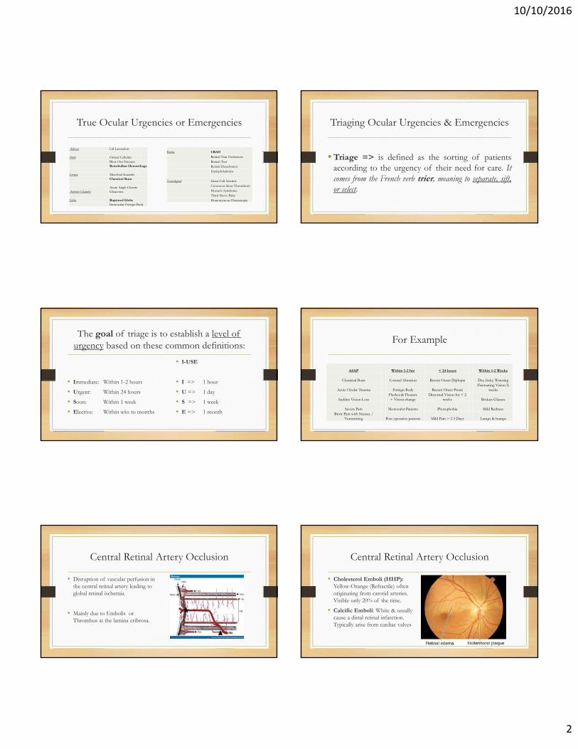
• Cholesterol Emboli (HHP): Yellow-Orange (Refractile) often originating from carotid arteries. Visible only 20% of the time.
• Calcific Emboli: White & usually cause a distal retinal infarction. Typically arise from cardiac valves
10/10/2016
3
Artery Occlusions
• Platelet Fibrin Emboli: Dull white and most commonly arise from a carotid thrombosus.
• Talc, Tumor, Septic, and Fat emboli are less common.
Central Retinal Artery Occlusion
• Symptoms: Unilateral, painless, acute vision loss
• 94% CF to LP vision
• +/- Amaurosis Fugax
• If VA is LP or worse, strongly consider Ophthalmic Artery occlusion
Central Retinal Artery Occlusion
• Signs: Superficial opacification or whitening of the retina in the posterior pole with a cherry-red spot in the center of the macula (which may be subtle)
• Absence of a cherry red spot likely indicated involvement of the ophthalmic artery.
Central Retinal Artery Occlusion
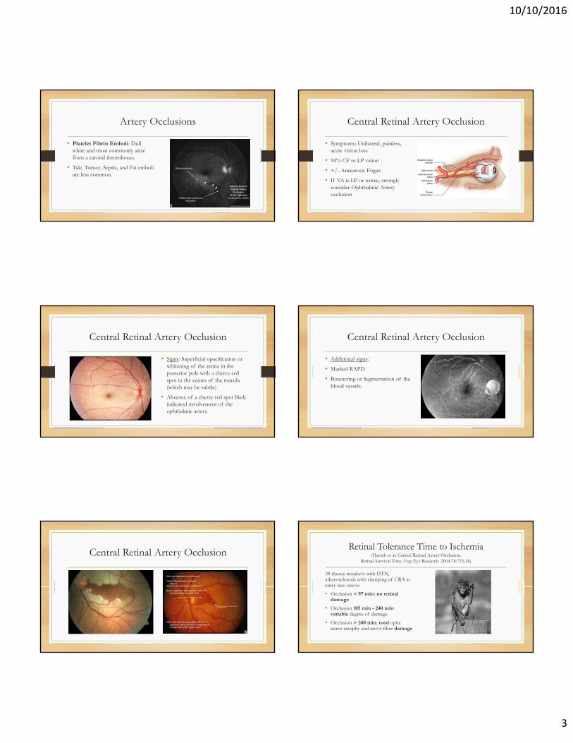
• Additional signs:
• Marked RAPD
• Boxcarring or Segmentation of the blood vessels.
Central Retinal Artery OcclusionRetinal Tolerance Time to Ischemia
(Hayreh et al. Central Retinal Artery Occlusion. Retinal Survival Time. Exp Eye Research. 2004.78:723-36)
38 rhesus monkeys with HTN, atherosclerosis with clamping of CRA at entry into nerve:
• Occlusion < 97 min: no retinal damage
• Occlusion 105 min - 240 min: variable degree of damage
• Occlusion > 240 min: total optic nerve atrophy and nerve fiber damage
10/10/2016
4
Timing is Critical
Initiation of Tx for CRAO Prognosis
Within 90 Minutes Fair (Complete)
Within 4 Hours Fair (Partial)
Within 6 Hours Guarded
After 6 Hours Poor
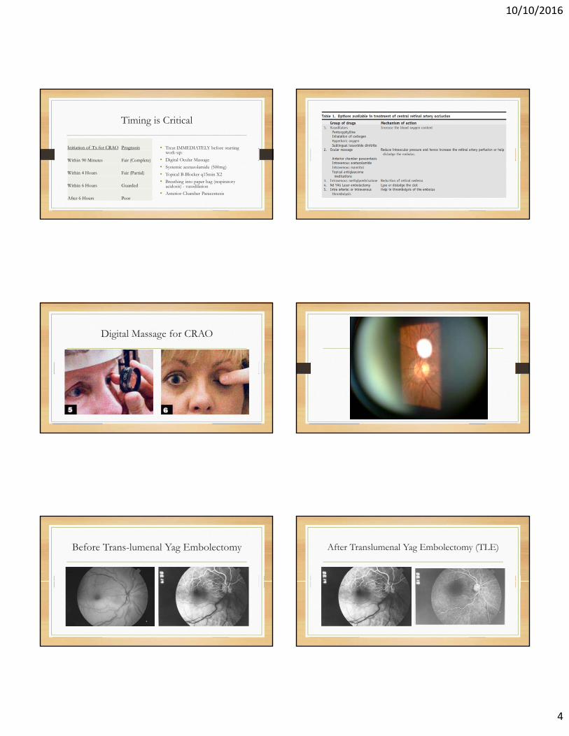
• Treat IMMEDIATELY before starting work-up:
• Digital Ocular Massage
• Systemic acetazolamide (500mg)
• Topical B-Blocker q15min X2
• Breathing into paper bag (respiratory acidosis) - vasodilation
• Anterior Chamber Paracentesis
Digital Massage for CRAO
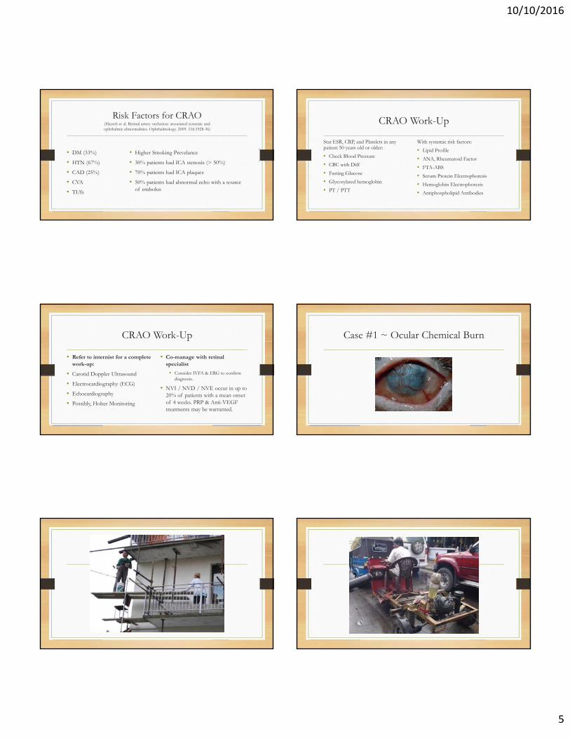
Before Trans-lumenal Yag Embolectomy After Translumenal Yag Embolectomy (TLE)
10/10/2016
5
Risk Factors for CRAO(Hayreh et al. Retinal artery occlusion: associated systemic andophthalmic abnormalities. Ophthalmology. 2009. 116:1928-36)
• DM (33%)
• HTN (67%)
• CAD (25%)
• CVA
• TIA’s
• Higher Smoking Prevelance
• 30% patients had ICA stenosis (> 50%)
• 70% patients had ICA plaques
• 50% patients had abnormal echo with a source of embolus
CRAO Work-Up
Stat ESR, CRP, and Platelets in any patient 50 years old or older:
• Check Blood Pressure
• CBC with Diff
• Fasting Glucose
• Glycosylated hemoglobin
• PT / PTT
With systemic risk factors:
• Lipid Profile
• ANA, Rheumatoid Factor
• FTA-ABS
• Serum Protein Electrophoresis
• Hemoglobin Electrophoresis
• Antiphospholipid Antibodies
CRAO Work-Up
• Refer to internist for a complete work-up:
• Carotid Doppler Ultrasound
• Electrocardiography (ECG)
• Echocardiography
• Possibly, Holter Monitoring
• Co-manage with retinal specialist
• Consider IVFA & ERG to confirm diagnosis.
• NVI / NVD / NVE occur in up to 20% of patients with a mean onset of 4 weeks. PRP & Anti-VEGF treatments may be warranted.
Case #1 ~ Ocular Chemical Burn
10/10/2016
6
Case #1
• You are escorting a patient to the front desk to check out. While there, you over hear your receptionist tell a patient to “bring him in immediately…we will work him right in!”
• When you inquire as to what the urgency is, your receptionist relays to you that your established patients husband accidently got splashed in the eye with a chemical he uses for fertilizing his crops. He is in severe pain and can barely open his eye(s).
Case #1
• Was this the right decision?
• Did your receptionist gather enough information?
• Are you prepared to handle such a problem?
• Where do we go from here?
Case #1 ~ Ocular Chemical Burn
• True Ocular Emergency!!!
• Whether by gas, liquid or solid, an acid or alkaline burn can cause irreversible damage to the eye and adnexa if urgent action is not taken.
10/10/2016
7
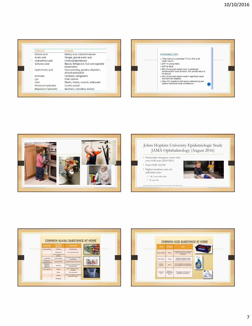
Johns Hopkins University Epidemiologic StudyJAMA Ophthalmology (August 2016)
• Nationwide emergency room visits over 4 full years (2010-2013)
• Total OCB: 144,149
• Highest incidence rates for individual years:
• 1 & 2 year olds; then
• 24 year old
Epidemiologic Trends of Chemical Ocular Burns in the United States - JAMA Ophthalmology

10/10/2016
8
Case #1 ~ Did the Receptionist Make The Right Decision?
Telephone Triage
• 1. Upon hearing of a chemical splash injury, make sure that the preliminary irrigation process begins on site before the patient seeks care.
• 2. If the chemical splash occurred outside of the workplace, remind the caller that the shower or an outdoor hose is an adequate option.
• 3. Attempt to determine time lapse between burn event and when irrigation started.
• 4. Attempt to determine the type of chemical that entered the eye(s).
• 5. Attempt to determine if the patient is wearing contact lenses. Irrigation should not stop in an effort to remove contact lenses.
Telephone Triage
• 6. Irrigation should take place for a minimum of 20 to 30 minutes before the patient is brought to the office or emergency room.
• 7. When the patient is ready to make the trip to the ER or office, remind them to bring the container that held the offending chemical. Important information may be obtained from the labeling.
• 8. If the injury occurred in the workplace, ask the patient to bring the MSDS (material safety data sheet) if available.
• 9. If the injury occurred where there is no or limited access to water for irrigation, refer them to the nearest emergency room or your office, whichever is closer.
• 10. Assist with dispatching emergency services as needed.
Special Situation
• If contamination with metallic lithium (i.e., lithium ion batteries), sodium, potassium, or magnesium has occurred, irrigation with water can result in a chemical reaction that causes burns to worsen. In these situations, the area should be covered with mineral oil and the metallic pieces should be removed with forceps and placed in mineral oil. If forceps are not available, soak the area with mineral oil and cover it with gauze soaked in mineral oil.
10/10/2016
9
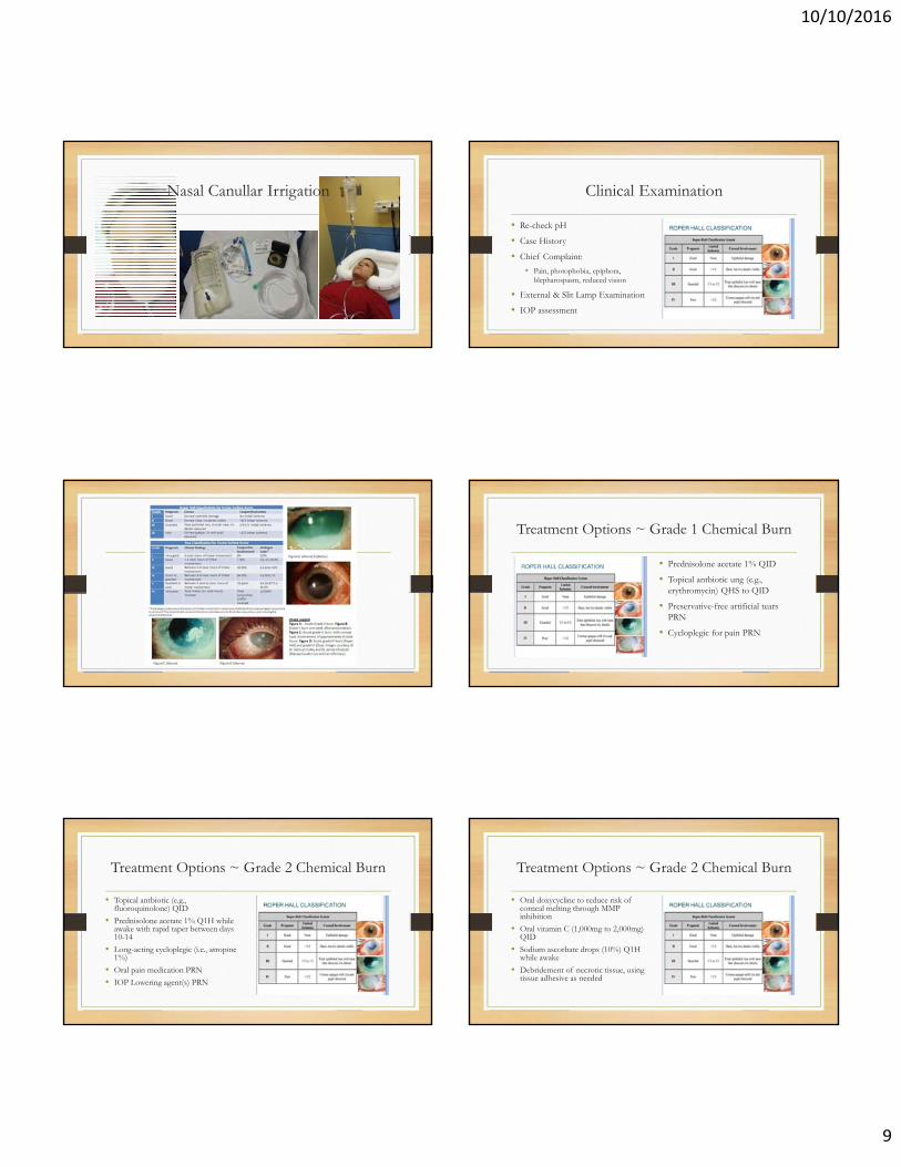
Nasal Canullar Irrigation Clinical Examination
• Re-check pH
• Case History
• Chief Complaint:
• Pain, photophobia, epiphora, blepharospasm, reduced vision
• External & Slit Lamp Examination
• IOP assessment
Treatment Options ~ Grade 1 Chemical Burn
• Prednisolone acetate 1% QID
• Topical antbiotic ung (e.g., erythromycin) QHS to QID
• Preservative-free artificial tears PRN
• Cycloplegic for pain PRN
Treatment Options ~ Grade 2 Chemical Burn
• Topical antbiotic (e.g., fluoroquinolone) QID
• Prednisolone acetate 1% Q1H while awake with rapid taper between days 10-14
• Long-acting cycloplegic (i.e., atropine 1%)
• Oral pain medication PRN
• IOP Lowering agent(s) PRN
Treatment Options ~ Grade 2 Chemical Burn
• Oral doxycycline to reduce risk of corneal melting through MMP inhibition
• Oral vitamin C (1,000mg to 2,000mg) QID
• Sodium ascorbate drops (10%) Q1H while awake
• Debridement of necrotic tissue, using tissue adhesive as needed
10/10/2016
10
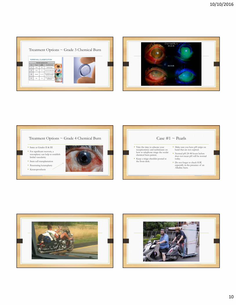
Treatment Options ~ Grade 3 Chemical Burn
Treatment Options ~ Grade 4 Chemical Burn
• Same as Grades II & III
• For significant necrosis, a tenonplasty can help re-establish limbal vascularity
• Stem cell transplantation
• Penetrating keratoplasty
• Keratoprosthesis
Case #1 ~ Pearls
• Take the time to educate your receptionist(s) and technicians on how to telephone triage the ocular chemical burn patient.
• Keep a triage checklist posted at the front desk.
• Make sure you have pH strips on hand that are not expired.
• Normal pH 24-48 hours before does not mean pH will be normal today.
• Do not forget to check IOP, especially in the presence of an Alkaline burn.

10/10/2016
11
Case #2
• An 84 year old woman develops a constant head and neck ache. She calls her HMO internist, who told her to take Motrin. Although she was a woman not given to complaining, she called the doctor again when the pain persisted. The internist told her that neck pain from arthritis is common at her age and not to worry, but the patient insisted on coming in to the doctor’s office.
Case #2
• The internists nurse practitioner took her blood pressure and gave her Relafin without drawing any tests. 2 weeks later she went blind in one eye. She was referred to the HMO ophthalmologist, who then referred her to a neuro-ophthalmologist. Unfortunately, 24 hours after the HMO ophthalmologist saw her, she went blind in the other eye.
Case #2
• It was later determined that she possessed many of the “classic” symptoms of GCA. The failure to recognize temporal arteritis had turned a functioning, independent woman into a blind person confined to home.
Giant Cell Arteritis (GCA)
10/10/2016
12
Temporal Arteritis (GCA)
• An occlusive inflammatory process causing ischemic disease. Affects medium & large-sized arteries. Etiology unknown (generic?, autoimmune?, infectious?)
• HA (New onset, localized, progressive) is the major feature in 65-90% of cases. Otherwise, patients may be vague with their symptoms.
• Age of Onset: >50 (incident increases w/ age)
• Incidence: 20 per 100,000; Prev: 200 / 100,000; Higher incidence in Caucasians and those of Scandinavian or Northern / Eastern European decent. Northern US.
• 4:1 female > male
Temporal Arteritis (GCA)
Unfortunately, if they are sitting in your chair, they most likely have already experienced:
Sudden, painless, non-progressive visual loss (count fingers) in at least one eye.
• Decreased VA => a “True” ophthalmic emergency.
Systemic History
• 50% of GCA patients begin with symptoms of anorexia, fever, malaise, myalgia, night sweats, and weight loss.
• Prodromal symptoms may occur for a few days or may even stretch out to weeks.
• Hallmark symptom of GCA is its new-onset localized headache.
• Localized to the temporal or occipital area, and may be occasionally may be diffuse or bilateral.
• 5-10% are ASYMPTOMATIC
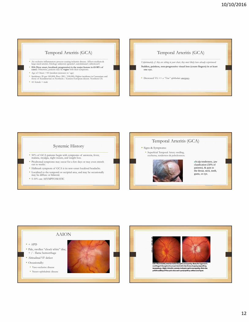
Temporal Arteritis (GCA)• Signs & Symptoms:
• Superficial Temporal Artery swelling, erythema, tenderness & pulselessness.
Scalp tenderness, jaw claudication (50% of patients), & pain in the throat, neck, teeth, gums, or eye.
r3
AAION
• + APD
• Pale, swollen “chock white” disc, + / - flame hemorrhage
• Altitudinal VF defect
• Occasionally:
• Vaso-occlusive disease
• Neuro-ophthalmic disease

Slide 77
r3 Do search to see about ausultation of temporal artery with stethoscope.rbm, 6/4/2008
10/10/2016
13
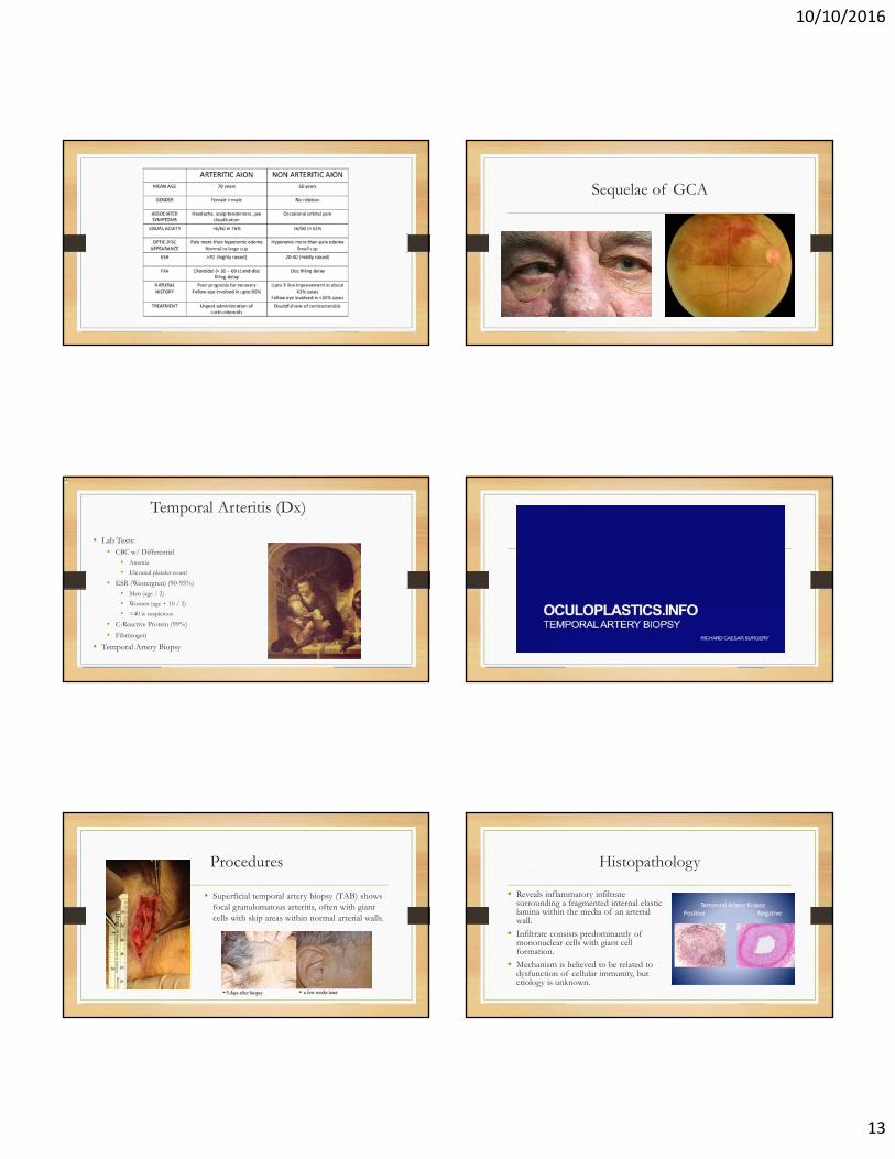
Sequelae of GCA
Temporal Arteritis (Dx)
• Lab Tests:• CBC w/ Differential
• Anemia
• Elevated platelet count
• ESR (Westergren) (90-95%)• Men (age / 2)
• Women (age + 10 / 2)
• >40 is suspicious
• C-Reactive Protein (99%)
• Fibrinogen
• Temporal Artery Biopsy
r4
Procedures
• Superficial temporal artery biopsy (TAB) shows focal granulomatous arteritis, often with giant cells with skip areas within normal arterial walls.
Histopathology
• Reveals inflammatory infiltrate surrounding a fragmented internal elastic lamina within the media of an arterial wall.
• Infiltrate consists predominantly of mononuclear cells with giant cell formation.
• Mechanism is believed to be related to dysfunction of cellular immunity, but etiology is unknown.

Slide 86
r4 What is the normal range for Fibrinogen?Look-up Color Duplex Ultrasonography of Temporal Arteries.rbm, 6/4/2008
10/10/2016
14
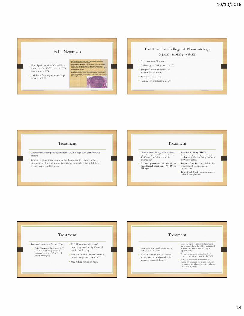
False Negatives
• Not all patients with GCA will have abnormal labs. 15-30% with + TAB have a normal ESR.
• TAB has a false negative rate (Skip lesions) of 5-9%.
The American College of Rheumatology5 point scoring system
• Age more than 50 years.
• A Westergren ESR greater than 50.
• Temporal artery tenderness or abnormality on exam.
• New onset headache.
• Positive temporal artery biopsy
Treatment
• The universally accepted treatment for GCA is high-dose corticosteroid therapy.
• Goals of treatment are to reverse the disease and to prevent further progression. This is of utmost importance especially in the ophthalmic arteries to prevent blindness.
Treatment
• First-line acute therapy without visual signs / symptoms => oral prednisone 40-60mg of prednisone ~or~ 1-2mg/kg/day.
• In the presences of visual orneurological symptoms => 80 to100mg/d
• Ranitidine 150mg BID PO (histamine type 2 receptor blocker) –or- Prevacid (Protein Pump Inhibitor) for GI protection.
• Fosamax Plus D – 10mg daily in the prevention of steroid induced osteoporosis
• Baby ASA (81mg) – decreases cranial ischemic complications.
Treatment
• Preferred treatment for AAION:
• Pulse Therapy: 3 day course of IV Solu-medrol (Methylpredisone) induction therapy of 15mg/kg/d (about 1000mg/d)
• 22 fold increased chance of improving visual acuity if started within the first day.
• Less Cumulative Dose of Steroids overall compared to oral Tx.
• May reduce remission rates.
Treatment
• Prognosis is poor if treatment is initiated > 48 hours.
• 30% of patients will continue to show a decline in vision despite aggressive steroid therapy.
• Once the signs of clinical inflammation are suppressed and the ESR is maintained at a low level, corticosteroids may be tapered slowly.
• No agreement exists as the length of treatment with corticosteroids for GCA.
• It may be reasonable to maintain the patient on treatment for 2 years to lessen the chances for relapses, although relapses have been reported.

10/10/2016
15
Case #3
• A 31 year old caucasion male presented for follow-up after being diagnosed with preseptal cellulitis OS. He claims compliance with his treatment regimen (Bactrim DS BID PO), but feels that his left eyelid has become more swollen and tender. He feels his vision has remained unchanged.
Case #3
• Entrance BCVA was 20/20 each eye. While EOM’s were full OU, the patient reported a pulling sensation OS when looking superior temporal. The left eye appeared slightly proptotic. Retropulsion assessment was inconclusive. Confrontational fields and pupil testing were unremarkable. Dilated funduscopic exam noted no apparent pathology of the posterior pole. Optic nerves were healthy in appearance with CD ratios of 0.3/0.3 OD, OS.
Case #3
• So despite being treated with oral antibiotics, his condition appears to be worsening. Could this be a case of antibiotic resistance? Is this pulling sensation suggestive of early stage orbital cellulitis? What is the next step in management given what we know?
10/10/2016
16
Periorbital and Orbital Cellulitis
Pathogenesis
• Sinusitis
• Extension of external ocular infection (ie hordeolum, dacryocystitis/dacroadenitis)
• Dental abscess
• Superficial break in the skin (ie infected bug bite, acne, eczema, periocular surgery or direct penetrating trauma)
• Hematogenous spread
Organisms
• Children:
• Haemophilus influenza type b (before Hib vaccine in 1985)
• Adults:
• Gram (+)
• Staphylococcus aureus (including MRSA)
• S. epidermidis
• Streptococcus pyogenes
• Enterococcus
• Gram (-)
• Pseudomonas (especially trauma)
• Klebsiella
• Fungal:
• Mucormycosis, Aspergillus species
Epidemiology
• Increased incidence during the winter due to the increased incidence of sinusitis
• No ethnic preferences
• Blindness occurs in up to 11% of cases
• In children, twice as common in males
• More common in children than adults: mean age 7-12 years old
10/10/2016
17
History
• Onset rapid or slow?
• Pain or tenderness to palpation?
• Recent fever, chills, rash or other systemic symptoms?
• Recent contact with other infected persons?
• Recent surgery (including eye or dental), hospitalization or trauma?
• Stiff neck or change in mental status?
• History of cancer, diabetes, HIV, organ transplantation, pulmonary or renal disease?
Clinical Signs and Symptoms
• Unilateral erythema of eyelid
• Swelling of eyelid
• Warmth of eyelid
• Tenderness of eyelid
• Blurred vision
• Ophthalmoplegia
• Proptosis
• Chemosis
Physical Exam
• Observe for degree of ocular swelling
• Assess extraocular movement
• Evaluate for foreign body
• Assess visual acuity
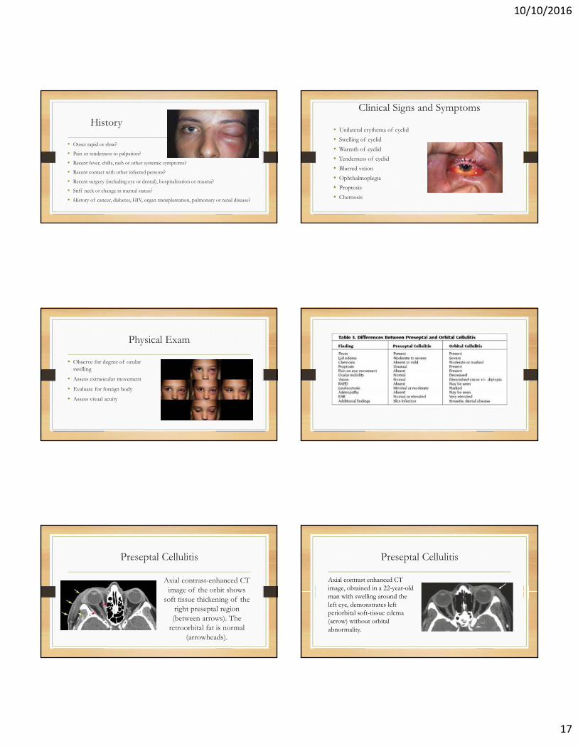
Preseptal Cellulitis
Axial contrast-enhanced CT image of the orbit shows
soft tissue thickening of the right preseptal region (between arrows). The
retroorbital fat is normal (arrowheads).
Preseptal Cellulitis
Axial contrast enhanced CT image, obtained in a 22-year-old man with swelling around the left eye, demonstrates left periorbital soft-tissue edema (arrow) without orbital abnormality.
10/10/2016
18
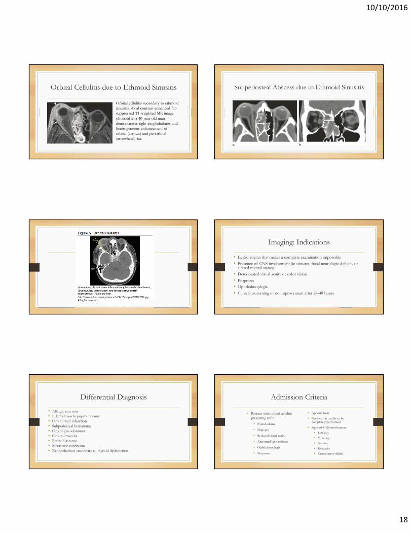
Orbital Cellulitis due to Ethmoid Sinusitis
Orbital cellulitis secondary to ethmoid sinusitis. Axial contrast-enhanced fat-suppressed T1-weighted MR image obtained in a 40-year-old man demonstrates right exophthalmos and heterogeneous enhancement of orbital (arrows) and periorbital (arrowhead) fat.
Subperiosteal Abscess due to Ethmoid Sinusitis
Imaging: Indications
• Eyelid edema that makes a complete examination impossible
• Presence of CNS involvement (ie seizures, focal neurologic deficits, or altered mental status)
• Deteriorated visual acuity or color vision
• Proptosis
• Ophthalmoplegia
• Clinical worsening or no improvement after 24-48 hours
Differential Diagnosis
• Allergic reaction• Edema from hypoproteinemia• Orbital wall infarction• Subperiosteal hematoma• Orbital pseudotumor• Orbital myositis• Retinoblastoma• Metastatic carcinoma• Exophthalmos secondary to thyroid dysfunction
Admission Criteria
• Patients with orbital cellulitis presenting with:
• Eyelid edema
• Diplopia
• Reduced visual acuity
• Abnormal light reflexes
• Ophthalmoplegia
• Proptosis
• Appears toxic
• Eye exam is unable to be completely performed
• Signs of CNS involvement:
• Lethargy
• Vomiting
• Seizures
• Headache
• Cranial nerve deficit
10/10/2016
19
Management
• Depends on the patient’s appearance, ability to take oral medications, compliance and clinical progression of the disease
• Empiric antibiotics should cover Staphylococcus and Streptococcus species, particularly MRSA
• Treat for 7-10 days for periorbital cellulitis
• Treat for 10-14 days for orbital cellulitis
• If no improvement in 24-48 hours consider consulting Infectious Disease, ENT and/or neurosurgery
Management
• Obtain blood culture in younger patients or those that appear systemically ill
• Culture ocular discharge
• Obtain orbital, epidural abscess or sinus fluid if patient requires surgery
Surgical Management
• Consider if:• > 9 years old
• Frontal sinusitis
• Non medial location of the subperiosteal abscess
• Large subperiosteal abscess
• Presence of gas in the abscess on CT suggesting an anaerobic etiology
• Recurrent episode of subperiostealabscess
• Nasal polyps which suggest chronic sinusitis
• Evidence of acute optic neuropathy
• Dental infection (likely an anaerobic infection)
Summary
• Orbital cellulitis is an emergency that requires prompt diagnosis and evaluation.
• Periorbital cellulitis and orbital cellulitis have distinct differences that can be elicited by careful history and physical examination
• If the physical exam cannot be fully completed for any reason, radiologic imaging is required
• Patients with systemic illness or evidence of orbital cellulitis or neurologic involvement require inpatient admission
• Improvement should occur within 24-48 hours with antibiotics

10/10/2016
20
Case #4:
Case #4
A 36-year-old white male presents to your office with a chiefcomplaint of intermittent pain on the right side of his head and orbitalarea along with redness and foreign body sensation in his right eye. Heworks in construction and is concerned that he may have gottensomething in his eye.
Case #4
External examination shows a mild ptosis (1.5mm) with minimal palpebral injection and no lid edema in the right eye. No foreign body of the cornea or bulbar conjunctiva is noted on a slit-lamp examination; nor is any evident on lid eversion. The cornea is clear, the anterior chamber is well formed and quiet. His unaided visual acuity is 20/20 each eye.
Case #4
• Pupil testing reveals anisocoria greatest under dim illumination and most noticeable during the first few seconds after the lights were turned down.
• The right pupil showed a delay in dilation consistent with “dilation lag” found in Horner’s syndrome.
• Anhidrosis is not evident and not reported by the patient.
Review of Anatomy
• Iris sphincter
• Iris dilator
• Parasympathetic pathway
• Sympathetic pathway
10/10/2016
21
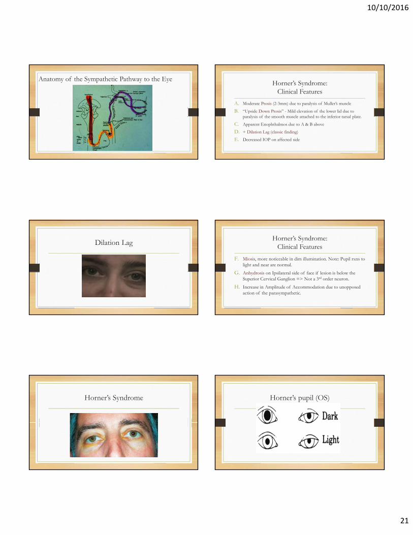
Anatomy of the Sympathetic Pathway to the EyeHorner’s Syndrome:
Clinical Features
A. Moderate Ptosis (2-3mm) due to paralysis of Muller’s muscle
B. “Upside Down Ptosis” - Mild elevation of the lower lid due to paralysis of the smooth muscle attached to the inferior tarsal plate.
C. Apparent Enophthalmos due to A & B above
D. + Dilation Lag (classic finding)
E. Decreased IOP on affected side
Dilation LagHorner’s Syndrome:
Clinical Features
F. Miosis, more noticeable in dim illumination. Note: Pupil rxns to light and near are normal.
G. Anhydrosis on Ipsilateral side of face if lesion is below the Superior Cervical Ganglion => Not a 3rd order neuron.
H. Increase in Amplitude of Accommodation due to unopposed action of the parasympathetic.
Horner’s Syndrome Horner’s pupil (OS)
10/10/2016
22
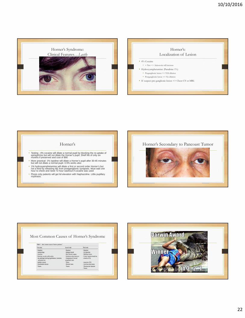
Horner’s Syndrome: Clinical Features…Lastly
Horner’s: Localization of Lesion
• 4% Cocaine
• + Test => Anisocoria will increase
• Hydroxyamphetamine (Paradrine 1%)
• Preganglionic lesion => YES dilation
• Postganglionic lesion => No dilation
• If suspect pre-ganglionic lesion => Chest CT or MRI.
Horner’s
• Testing ; 4% cocaine will dilate a normal pupil by blocking the re-uptake of epinephrine but will not dilate the Horner’s pupil. Shelf life of only six months if preserved and cost of $90
• More practical: 1% Iopidine will dilate a Horner’s pupil after 30-45 minutes but will not dilate a normal pupil. 0.5% works also
• 1% hydroxyamphetamine will dilate a first or second order Horner’s but not a third by releasing NE from postganglionic synapses. Must wait one hour to check and need 72 hour washout if cocaine was used
• Ptosis only patients will get lid elevation with Naphazoline. Little pupillary mydriasis.
Horner’s Secondary to Pancoast Tumor
Most Common Causes of Horner’s Syndrome

10/10/2016
23
Flashes & Floaters ~ How urgent is URGENT?
Case #5
• It’s Saturday night and you’re getting ready to sit down and watch a movie with your family. The answering service calls with a patient on the other line. They tell you that the patient is complaining of an increase in floaters after having a single bright flash of light. How many times have we all gotten this phone call, right?!? How many times have we gone into the office after hours or on weekends, only to dilate the patient to diagnose an uncomplicated PVD?
Case #5
• What do you tell the answering service? Do you simply agree to forego the movie and see the patient at your office in an hour? Do you ask them to call back if the floaters worsen before an early morning appointment on Monday? Do you ask them to be NPO after midnight and see them the next morning “just in case”? Or, do you need more information?
59 year Retrospective Meta-analysisHollands and Colleagues
• Found and reviewed 17 relevant studies looking at the association between flashes & floaters, PVD, and likelihood of retinal tear.
• If patients report flashes & floaters: 14% had a retinal break or tear.
• Flashes but no floaters: 13.7%
• Floaters but no flashes: 13.5%
• Add in “Subjective Visual Complaints” => Prevalence increases to 45%

10/10/2016
24
Case #5
• You have the answering service connect you directly with the patient. You determine that the patient is a 50 yo high myopewho had an onset of flashes & floaters 10 days earlier. She was seen by her optometrist same day and diagnosed with an uncomplicated PVD.
• She was educated on the warning signs of RD and instructed to call the office immediately should there be any worsening or change in her symptoms.
Case #5
• Hence her call. She reports an increase in floaters and feels that the lower part of her vision has become affected. Based on her symptoms, you ask her to meet you at your office this evening. Surprisingly, she asks if this could wait until tomorrow. She lives by herself in a rural town 45 – 50 minutes away. She does not feel comfortable driving herself and she is not sure she can manage a ride this evening. You explain to the patient that you are concerned that she may have a loose or torn retina and that the longer we wait in addressing it, the greater the risk of macular involvement and the potential for permanent vision loss. She said she would make some calls and call you back.
59 year Retrospective Meta-analysisHollands and Colleagues
• Patients previously diagnosed with an UNCOMPLICATED PVD have a 3.4% risk of developing a retinal tear within 6 weeks.
• Predictive symptoms for retinal tear in this subset of patients:
• Sudden increase in floaters (>10 floaters), and / or
• Subjective Vision Loss
• So do all retinal tears lead to detachment if undiagnosed?
Do all Retinal Tears lead to Detachment?
• NO
• Shea & Colleagues: 33-50% of all tears lead to detachment.
• 81 yo asymptomatic patient with self-sealing supero-temporal Horseshoe retinal tear, OS
Mechanism of RRD
• Size of retinal break
• Location of retinal break
• Residual vitreous traction
• Degree of vitreal syneresis (liquefied vitreous)
10/10/2016
25
Case #5
• Your patient calls you back and indicates she has found a ride and will meet you at your office in about an hour. Soon after she arrives, you determine that she has preserved central vision (BCVA 20/20- each eye), but has reduced confrontation fields inferiorly and slight asymmetry in her intra-ocular pressures (12mmHg OD, 16mmHg OS).
Case #5
• After she is dilated, slit-lamp exam of the vitreous increases your suspicion of retinal detachment after you notice some pigmentary “dusting”.
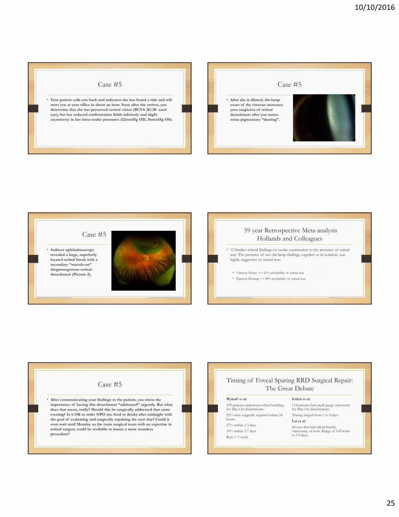
Case #5
• Indirect ophthalmoscopy revealed a large, superiorly located retinal break with a secondary “macula-on” rhegmatogenous retinal detachment (Picture 2).
59 year Retrospective Meta-analysisHollands and Colleagues
• 12 Studies related findings on ocular examination to the presence of retinal tear. The presence of two slit-lamp findings, together or in isolation, was highly suggestive of retinal tear:
• Vitreous Heme: => 62% probability of retinal tear
• Pigment Dusting => 88% probability of retinal tear.
Case #5
• After communicating your findings to the patient, you stress the importance of having this detachment “addressed” urgently. But what does that mean, really? Should this be surgically addressed that same evening? Is it OK to order NPO (no food or drink) after midnight with the goal of evaluating and surgically repairing the next day? Could it even wait until Monday so the main surgical team with an expertise in retinal surgery could be available to insure a more seamless procedure?
Timing of Foveal Sparing RRD Surgical Repair: The Great Debate
Wykoff et al:
199 patients underwent scleral buckling for Mac-On detachments:
52% were surgically repaired within 24 hours.
27% within 1-3 days
10% within 3-7 days
Rest: > 1 week
Erlich et al:
114 patients had small gauge vitrectomy for Mac-On detachments:
Timing ranged from 1 to 5 days
Lai et al:
66 eyes that had scleral buckle, vitrectomy, or both. Range of 0.8 hours to 5.9 days.

10/10/2016
26
Timing of Foveal Sparing RRD Surgical Repair: The Great Debate
• Prospective Analysis: Ho et al.
• 82 Mac-On RRD’s
• 87% showed no progression from presentation to surgery
• Remaining 13% showed an average rate of progression of 1.8 disc diameters per day!
R/B Ratio of delaying surgery
• Pro’s
• Optimal surgical conditions
• Most experienced staff
• Less cost for patient (surgery performed after hours costs 25% more)
• Cons
• Every eye is different. Some have progressed to Mac-off in as quickly as 4-8 hours.
• What if there is further delay from unforeseen events?
Case #6: Is HELP on the way?
• A 60-year-old African American male presented complaining of a red, irritated right eye with associated brow ache, and he felt his vision had decreased slightly in that eye.
Case #6: Is HELP on the way?
• He reported a history of glaucoma surgery (trabeculectomy) in both eyes approximately five years earlier and was not on any topical glaucoma medication currently.
• His uncorrected distance visual acuity was 20/50 OD and 20/25 OS, and pinhole acuity was 20/30 OD.
Case #6: Is HELP on the way?
• Slit-lamp examination revealed a superiorly located opalescent bleb surrounded by 3+ conjunctival injection.
• There appeared to have a mucopurulent infiltrate within the bleb in the right eye. The anterior chamber was deep, showing trace cell and flare, right eye.
Case #6: Is HELP on the way?
• Intraocular pressure (IOP) was 5mm Hg OD and 9mm Hg OS. NaFL strip application revealed a subtle leak from the infero-lateral edge of the bleb in the right eye. Dilated examination revealed advanced cupping in both eyes.
• The periphery was intact without choroidal effusions. Vitreous cells were not evident on slit-lamp exam or under binocular indirect ophthalmoscopy. B-scan ultrasound was acoustically normal in both eyes.
10/10/2016
27
Blebitis Acronyms to Remember
• HELP
• H = Hypotony
• E = Endophthalmitis
• L = Leak
• P = Pain
• RSVP
• R = Redness
• S = Sensitivity to Light
• V = Vision Loss
• P = Pain
Managing Late Bleb Leaks
• Conservative Approach
• Suppress Aqueous Production
• Antibiotic Prophylaxis
• Keep the eye lubricated
• Protection from trauma or mechanical rubbing
• Surgical Revision
• Needling the Bleb
• Autologous Blood Patching
• Compression Suturing
• Amniotic Membrane Grafting
Large Diameter BCL 4 Late Bleb Leaks
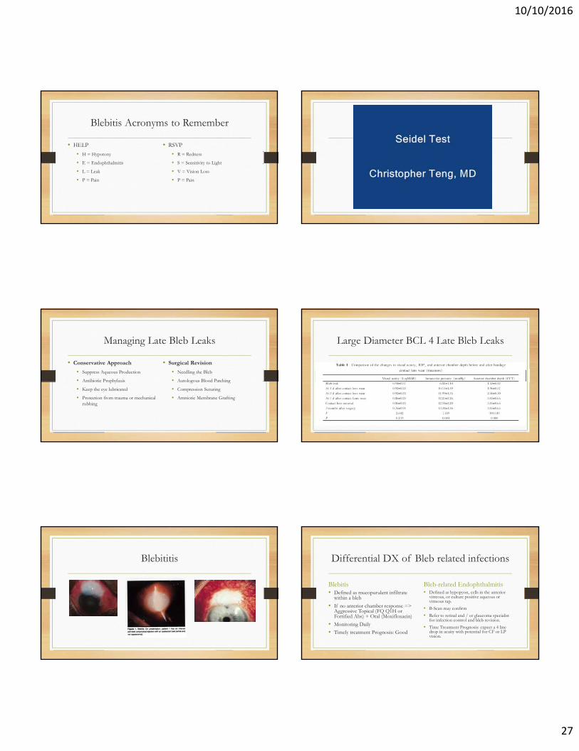
Blebititis Differential DX of Bleb related infections
Blebitis• Defined as mucopurulent infiltrate
within a bleb
• If no anterior chamber response => Aggressive Topical (FQ Q1H or Fortified Abs) + Oral (Moxifloxacin)
• Monitoring Daily
• Timely treatment Prognosis: Good
Bleb-related Endophthalmitis• Defined as hypopyon, cells in the anterior
vitreous, or culture positive aqueous or vitreous tap.
• B-Scan may confirm
• Refer to retinal and / or glaucoma specialist for infection control and bleb revision.
• Time Treatment Prognosis: expect a 4 line drop in acuity with potential for CF or LP vision.
10/10/2016
28
Topic #7: Third Nerve Palsy Parasympathetic Pathway
• Four neuron arc• Retina to the pretectal
nucleus in the midbrain (1)
• Pretectal nucleus to the EW nucleus (2)
• EW nucleus to the ciliary ganglion (3)
• Ciliary ganglion to the iris sphincter with short ciliary nerves (4)
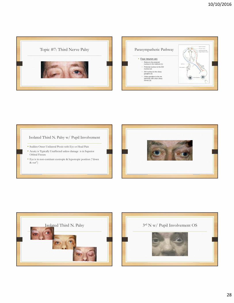
Isolated Third N. Palsy w/ Pupil Involvement
• Sudden Onset Unilateral Ptosis with Eye or Head Pain
• Acuity is Typically Unaffected unless damage is in Superior Orbital Fissure
• Eye is in non-comitant exotropic & hypotropic position (“down & out”)
Isolated Third N. Palsy 3rd N w/ Pupil Involvement OS

10/10/2016
29
Isolated Third N. Palsy w/ Pupil Involvement
• Posterior Communicating Artery Aneurysm (Most Common)
• Tumor or Trauma
• HZO
• Leukemia
• Uncal Herniation Syndrome
• Space Occupying Lesion
• Subdural Hematoma
• Pituitary Apoplexy
• Ischemic Vascular Disease (Rare)
Pupil sparing / Pupil involving
• Rule of thumb : Pupil sparing third nerve palsies tend to be ischemic while those involving the pupil tend to be due to aneurysms or tumors
• Not a firm rule
• Pupil sparing may become pupil involving so follow very closely
Pupil involving vs. pupil sparing Pupil involving vs. pupil sparing
Third Nerve Management
• Immediate Gad enhanced MRI / MRA if any question of aneurysmalinvolvement. Patient may complain of a severe headache and will often have other neurological signs
• If patient is diabetic or hypertensive and the pupil is not involved they can be followed closely without imaging studies
Third Nerve Management
• Patient education and reassurance a must
• Diplopia relief with patching
• Most ischemic palsies resolve over several months

10/10/2016
30
Case #8Patient History
• 68 year old white female.
• Ocular History:
CRAO, 2003.
• Medical history:
Diabetes
Renal Problems.
Recent Exam Findings
• July 2004
VA- 20/25, OU.
Cup-to-disc; .4/.4,OU.
• July 2005
VA- 20/25, OD, 20/60, OS.
Cup-to-disc; .6/.6, OD.
.9/.9, OS.
Present Exam Findings
• VA- OD- 20/25
OS- NLP
• PERRL + APD, OS.
• TA- 16, OD
67, OS
Observations, OS Neovascular Glaucoma
• Elevation of IOP.
• Painful red eye.
• Closure of anterior chamber angle from fibrovascular membrane formation.
10/10/2016
31
Causes
• Central retinal vein & artery occlusion.
40-60% of NVG cases.
• Diabetic retinopathy.
• Carotid artery occlusive disease.
• Chronic retinal detachments
• Usually occurs within 90 days of antecedent vascular occlusion.
Signs/Symptoms
• Acute onset of redness, pain, and blurred vision.
• Circumcilliary injection.
• Corneal edema.
• Deep anterior chamber with moderate flare.
• NVI/NVA.
Pathophysiology
• Stimulus= Lack of Oxygen.
• Hypoxic retinal tissue results in the release of vasoproliferative factors, i.e. VEGF.
• VEGF acts upon endothelial cells of viable capillaries to stimulate the formation of a new vessels.
• Once released, the angiogenic factors diffuse through the vitreous and posterior chamber into the aqueous and the anterior segment.
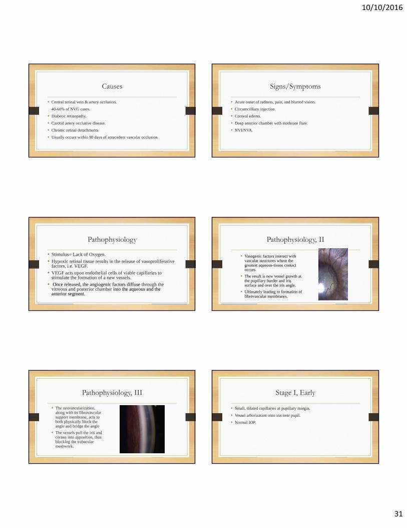
Pathophysiology, II
• Vasogenic factors interact with vascular structures where the greatest aqueous-tissue contact occurs.
• The result is new vessel growth at the pupillary border and iris surface and over the iris angle.
• Ultimately leading to formation of fibrovascular membranes.
Pathophysiology, III
• The neovascularization, along with its fibrovascularsupport membrane, acts to both physically block the angle and bridge the angle
• The vessels pull the iris and cornea into apposition, thus blocking the trabecular meshwork.
Stage I, Early
• Small, dilated capillaries at pupillary margin.
• Vessel arborization onto iris near pupil.
• Normal IOP.

10/10/2016
32
Stage II, Mid-Phase
• Involvement of anterior chamber angle.
• Radial vessel progression.
• Hyphema.
• Increase in IOP.
Stage III, Late
• Contraction of the fibrovascular membrane.
• 360o angle closure.
• Ectropion uvea.
• Significant anterior chamber reaction.
Management
• Medically treating neovascularglaucoma is challenging at best.
• Medical consult to rule out systemic disease.
• Duplex/Doppler scans to r/o carotid occlusive disease.
Medical Management
• If there is any degree of inflammation and ocular pain, prescribe a topical cycloplegic such as atropine 1% b.i.d. as well as a topical steroid such as Pred Forte.
IOP Control
• Medical therapy with topical ß-adrenergic antagonists, a-2 agonists, and topical or oral carbonic inhibitors lower IOP.
• Aqueous suppressants may be used in order to temporarily reduce IOP. However, chronic medical therapy is not indicated for neovascular glaucoma.
• Aqueous suppressants will temporize IOP and angle closure will continue.
Medical Management, II
• Ultimate management of NVG involves eradication of the vessels with PRP or cryo.
• Goal: destroy ischemic retina, minimize oxygen demand of the eye, and reduce the amount of VEGF being released.
• If a significant amount of the angle is in permanent synechial closure, filtering surgery must then be employed.

10/10/2016
33
However…
• What if the patient is, like ours, blind?
• The primary goal of treatment in this stage is pain control.
• For blind, painful eyes with uncontrollable IOP, options include continued medical therapy, cyclodestruction, retro bulbar alcohol injection, or enucleation.
But…
• Our patient was also not in pain.
• Plan of action:
Retinal consult.
Possible PRP to save cornea from decompensation.
Future Possibilities
• Anti-VEGF therapy.
• VEGF appears to produce its effect partly by being proinflammatory, leading to leukocyte adhesion and inflammation.
• VEGF can induce injury to the endothelium, cause fenestrations in endothelial cells, and cause breakdown of tight junctions.
Pointers…
• Retinal artery occlusions develop NVG in only 17 percent of cases and typically within four weeks post-occlusion.
• Miotics are contraindicated in any case where there is active inflammation. Prostaglandin analogs should likewise be avoided.

10/10/2016
34
Case #9: War Paint
• An 18-year-old male with a right eye injury presented with entrance acuity at the level of hand motion (HM) and an intraocular pressure (IOP) of 17mm Hg. An anterior segment examination revealed right upper lid edema and bruising.
Case #9: War Paint
• A slit-lamp examination confirmed corneal edema with mild folds centrally, a subconjunctival hemorrhage with chemosis and 3+ cell and flare with a 2mm hyphema in an adequately formed anterior chamber.
• The cornea and conjunctiva were seidel negative. The right pupil was 5mm with poor reactivity. The lens appeared centered, but it was difficult to asses due to poor clarity.
Case #9: War Paint
• Funduscopic exam was limited due to severe vitreous hemorrhage. A B-scan ultrasound did not suggest retinal detachment. The patient was referred to our staff retinal specialist the next day. A biometry was repeated and, once again, did not suggest retinal detachment.
Case #9: War Paint Case #9: War Paint
• Risk Factors for Open Globe:• Penetrating Lid Injury
• Hypotony + Hyphema
• Positive Seidel
• Peaked Pupil and / or APD
• Shallowed or absent anterior chamber
• 360 degree bullous sub-conj heme
• Lens dislocation
• Iridodialysis
10/10/2016
35
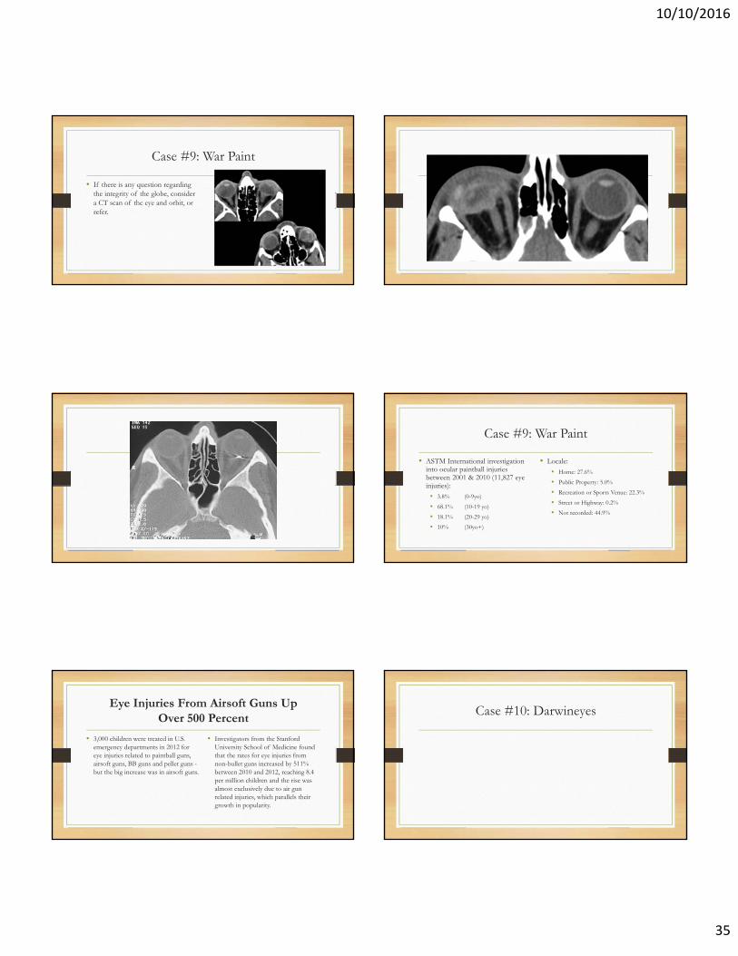
Case #9: War Paint
• If there is any question regarding the integrity of the globe, consider a CT scan of the eye and orbit, or refer.
Case #9: War Paint
• ASTM International investigation into ocular paintball injuries between 2001 & 2010 (11,827 eye injuries):
• 3.8% (0-9yo)
• 68.1% (10-19 yo)
• 18.1% (20-29 yo)
• 10% (30yo+)
• Locale:• Home: 27.6%
• Public Property: 5.0%
• Recreation or Sports Venue: 22.3%
• Street or Highway: 0.2%
• Not recorded: 44.9%
Eye Injuries From Airsoft Guns UpOver 500 Percent
• 3,000 children were treated in U.S. emergency departments in 2012 for eye injuries related to paintball guns, airsoft guns, BB guns and pellet guns -but the big increase was in airsoft guns.
• Investigators from the Stanford University School of Medicine found that the rates for eye injuries from non-bullet guns increased by 511% between 2010 and 2012, reaching 8.4 per million children and the rise was almost exclusively due to air gun related injuries, which parallels their growth in popularity.
Case #10: Darwineyes