OctOpus 600 See the new pulse in perimetry - Haag-Streit AG · OctOpus 600 See the new pulse in...
14
OCTOPUS 600 See the new pulse in perimetry Tradition and Innovation – Since 1858, visionary thinking and a fascination with technology have guided us to develop innovative products of outstanding reliability: Anticipating trends to improve the quality of life.
Transcript of OctOpus 600 See the new pulse in perimetry - Haag-Streit AG · OctOpus 600 See the new pulse in...
OctOpus 600See the new pulse in perimetry
tradition and Innovation – Since 1858, visionary thinking and a fascination with technology have guided us to develop innovative products of outstanding reliability: Anticipating trends to improve the quality of life.
02 | 03 pERIMEtRY YOu cAN tRust
OCTOPUS 600perimetry simplified
In 1972 Franz Fankhauser and others developed the principles and concepts of automated perimetry which resulted in the design of the first automated static perimeter, the Octopus 201, in 1974. Since then, Octopus has pioneered many significant innovations like the G-pattern, the direct projection system, fast strategies and outstanding software for visual field analysis.
Detecting visual field loss at the earliest possible stage, defining the opti-mum treatment and following up the patient to decide on the necessity of treatment changes or surgery are the main tasks of every glaucoma special-ist. Addressing this basic need, Haag-Streit introduced the Octopus 600 that combines early diagnosis and follow-up in a single compact-sized, stand-alone device.
In providing an instrument that seamlessly integrates into your practice en-vironment and increases patient throughput, Haag-Streit has focused on im-proving usability and ergonomics for both the operator and the patient.
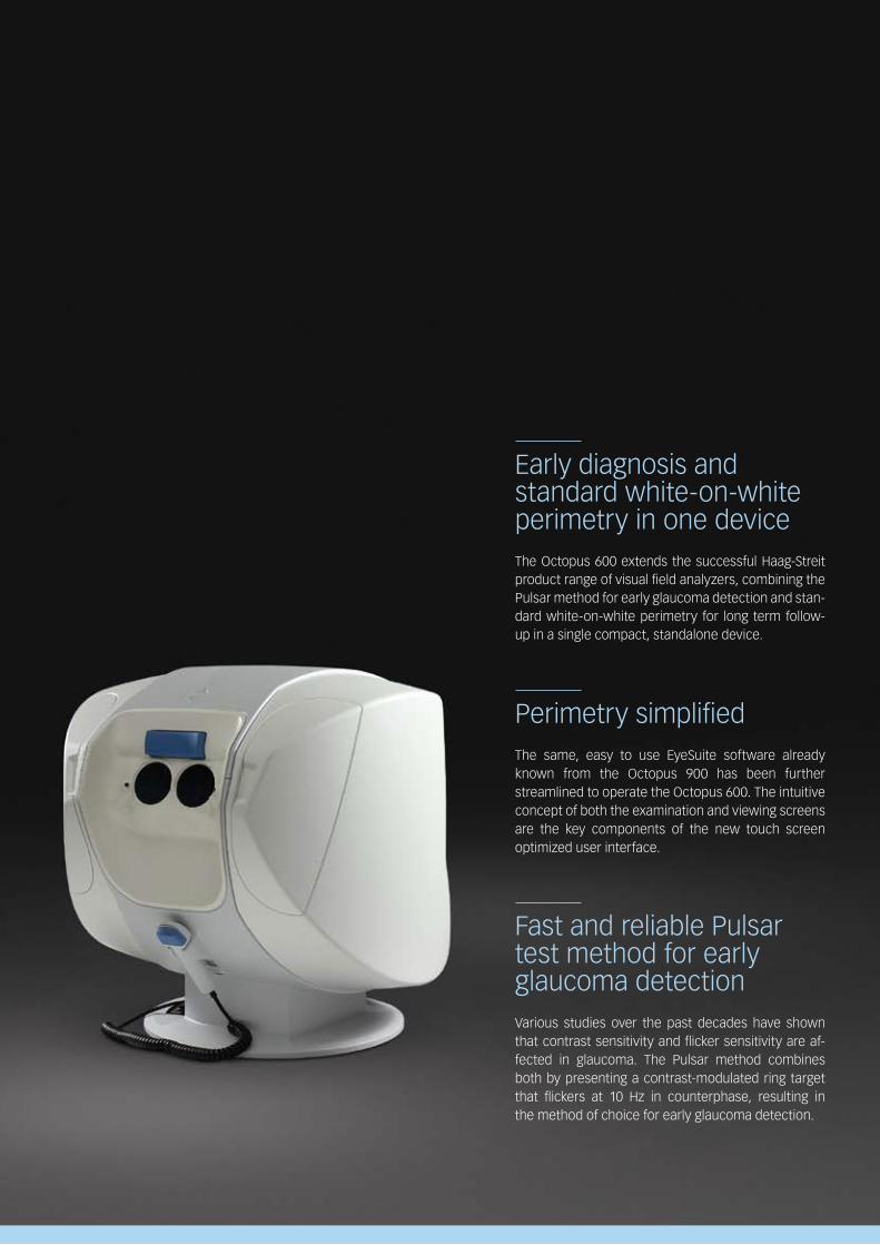
Early diagnosis and standard white-on-white perimetry in one deviceThe Octopus 600 extends the successful Haag-Streit product range of visual field analyzers, combining the Pulsar method for early glaucoma detection and stan-dard white-on-white perimetry for long term follow-up in a single compact, standalone device.
Perimetry simplifiedThe same, easy to use EyeSuite software already known from the Octopus 900 has been further streamlined to operate the Octopus 600. The intuitive concept of both the examination and viewing screens are the key components of the new touch screen optimized user interface.
Fast and reliable Pulsar test method for early glaucoma detectionVarious studies over the past decades have shown that contrast sensitivity and flicker sensitivity are af-fected in glaucoma. The Pulsar method combines both by presenting a contrast-modulated ring target that flickers at 10 Hz in counterphase, resulting in the method of choice for early glaucoma detection.
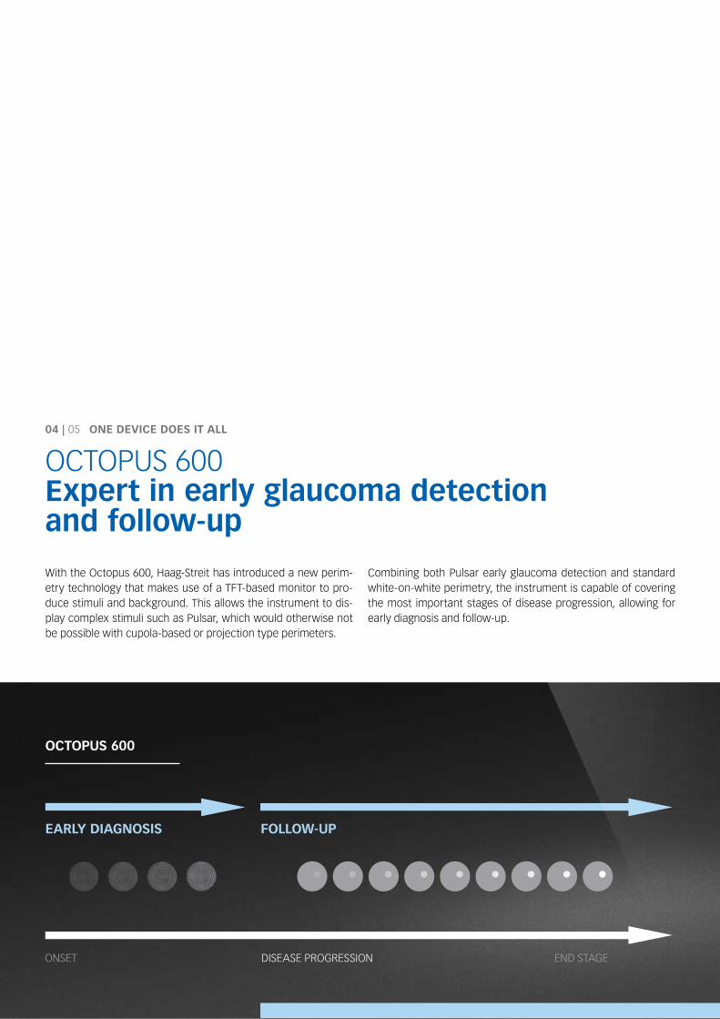
EARlY dIAgNOsIs FOllOw-up
04 | 05 ONE dEVIcE dOEs It All
OCTOPUS 600Expert in early glaucoma detection and follow-up
With the Octopus 600, Haag-Streit has introduced a new perim-etry technology that makes use of a TFT-based monitor to pro-duce stimuli and background. This allows the instrument to dis-play complex stimuli such as Pulsar, which would otherwise not be possible with cupola-based or projection type perimeters.
Combining both Pulsar early glaucoma detection and standard white-on-white perimetry, the instrument is capable of covering the most important stages of disease progression, allowing for early diagnosis and follow-up.
ONSET DISEASE PrOGrESSION END STAGE
OctOpus 600
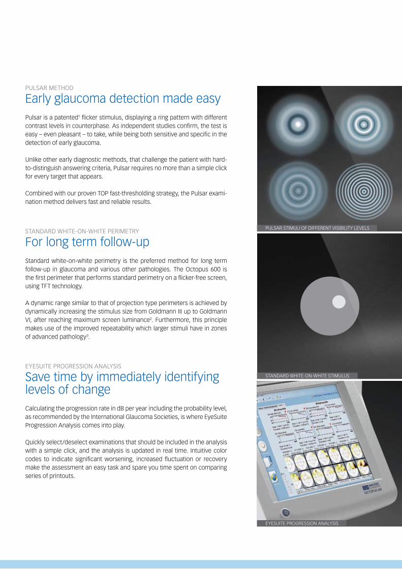
PUlSAr STImUlI OF DIFFErENT VISIbIlITy lEVElS
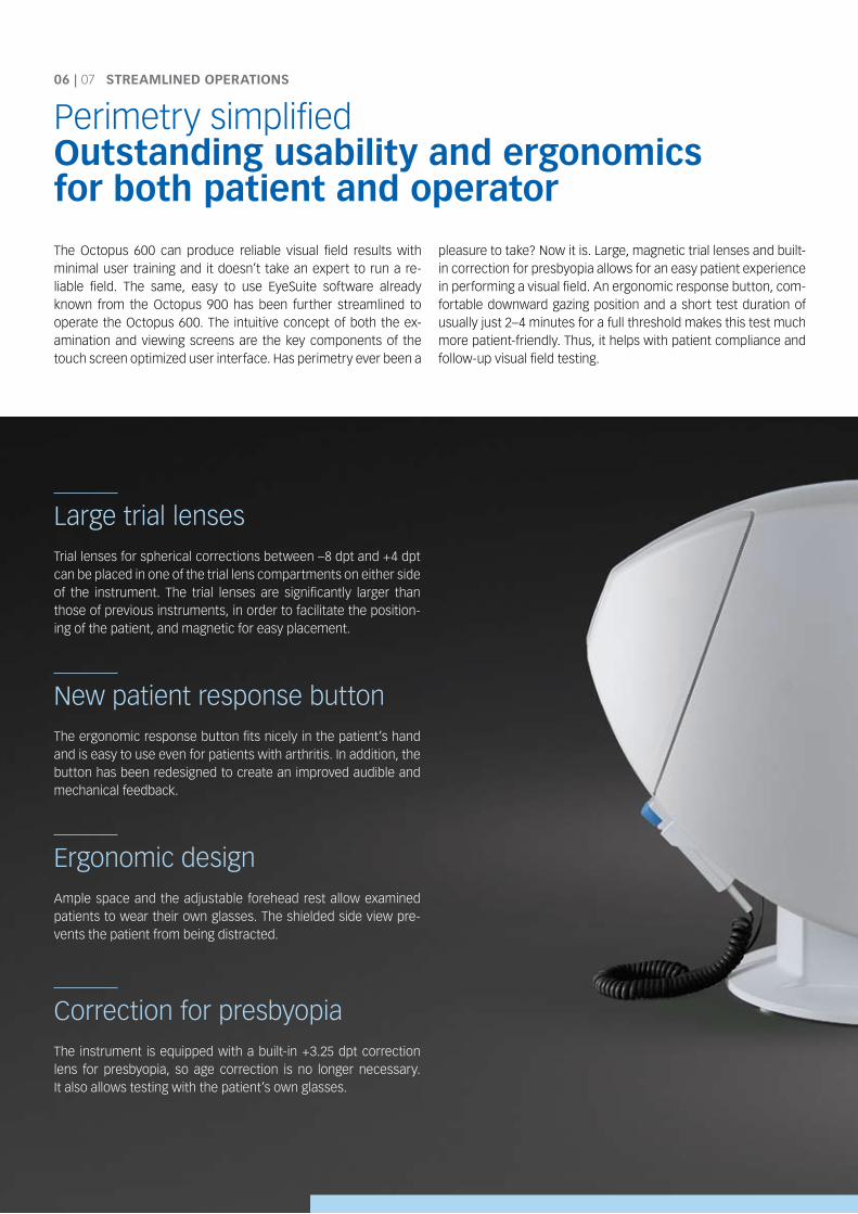
STANDArD WHITE-ON-WHITE STImUlUS
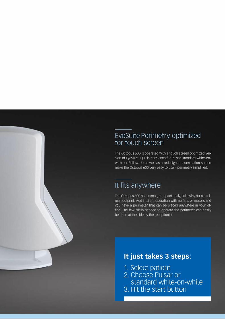
EyESUITE PrOGrESSION ANAlySIS
PUlSAr mETHOD
Early glaucoma detection made easyPulsar is a patented1 flicker stimulus, displaying a ring pattern with different contrast levels in counterphase. As independent studies confirm, the test is easy – even pleasant – to take, while being both sensitive and specific in the detection of early glaucoma.
Unlike other early diagnostic methods, that challenge the patient with hard-to-distinguish answering criteria, Pulsar requires no more than a simple click for every target that appears.
Combined with our proven TOP fast-thresholding strategy, the Pulsar exami-nation method delivers fast and reliable results.
STANDArD WHITE-ON-WHITE PErImETry
For long term follow-upStandard white-on-white perimetry is the preferred method for long term follow-up in glaucoma and various other pathologies. The Octopus 600 is the first perimeter that performs standard perimetry on a flicker-free screen, using TFT technology.
A dynamic range similar to that of projection type perimeters is achieved by dynamically increasing the stimulus size from Goldmann III up to Goldmann VI, after reaching maximum screen luminance2. Furthermore, this principle makes use of the improved repeatability which larger stimuli have in zones of advanced pathology3.
EyESUITE PrOGrESSION ANAlySIS
Save time by immediately identifyinglevels of changeCalculating the progression rate in db per year including the probability level, as recommended by the International Glaucoma Societies, is where EyeSuite Progression Analysis comes into play.
Quickly select/deselect examinations that should be included in the analysis with a simple click, and the analysis is updated in real time. Intuitive color codes to indicate significant worsening, increased fluctuation or recovery make the assessment an easy task and spare you time spent on comparing series of printouts.
The Octopus 600 can produce reliable visual field results with minimal user training and it doesn’t take an expert to run a re-liable field. The same, easy to use EyeSuite software already known from the Octopus 900 has been further streamlined to operate the Octopus 600. The intuitive concept of both the ex-amination and viewing screens are the key components of the touch screen optimized user interface. Has perimetry ever been a
pleasure to take? Now it is. large, magnetic trial lenses and built-in correction for presbyopia allows for an easy patient experience in performing a visual field. An ergonomic response button, com-fortable downward gazing position and a short test duration of usually just 2–4 minutes for a full threshold makes this test much more patient-friendly. Thus, it helps with patient compliance and follow-up visual field testing.
large trial lensesTrial lenses for spherical corrections between –8 dpt and +4 dpt can be placed in one of the trial lens compartments on either side of the instrument. The trial lenses are significantly larger than those of previous instruments, in order to facilitate the position-ing of the patient, and magnetic for easy placement.
New patient response buttonThe ergonomic response button fits nicely in the patient’s hand and is easy to use even for patients with arthritis. In addition, the button has been redesigned to create an improved audible and mechanical feedback.
Ergonomic designAmple space and the adjustable forehead rest allow examined patients to wear their own glasses. The shielded side view pre-vents the patient from being distracted.
Correction for presbyopiaThe instrument is equipped with a built-in +3.25 dpt correction lens for presbyopia, so age correction is no longer necessary. It also allows testing with the patient’s own glasses.
06 | 07 stREAMlINEd OpERAtIONs
Perimetry simplifiedOutstanding usability and ergonomics for both patient and operator
EyeSuite Perimetry optimizedfor touch screenThe Octopus 600 is operated with a touch screen optimized ver-sion of EyeSuite. Quick-start icons for Pulsar, standard white-on-white or Follow-Up as well as a redesigned examination screen make the Octopus 600 very easy to use – perimetry simplified.
It fits anywhereThe Octopus 600 has a small, compact design allowing for a mini-mal footprint. Add in silent operation with no fans or motors and you have a perimeter that can be placed anywhere in your of-fice. The few clicks needed to operate the perimeter can easily be done at the side by the receptionist.
It just takes 3 steps:
1. Select patient2. Choose Pulsar or standard white-on-white3. Hit the start button
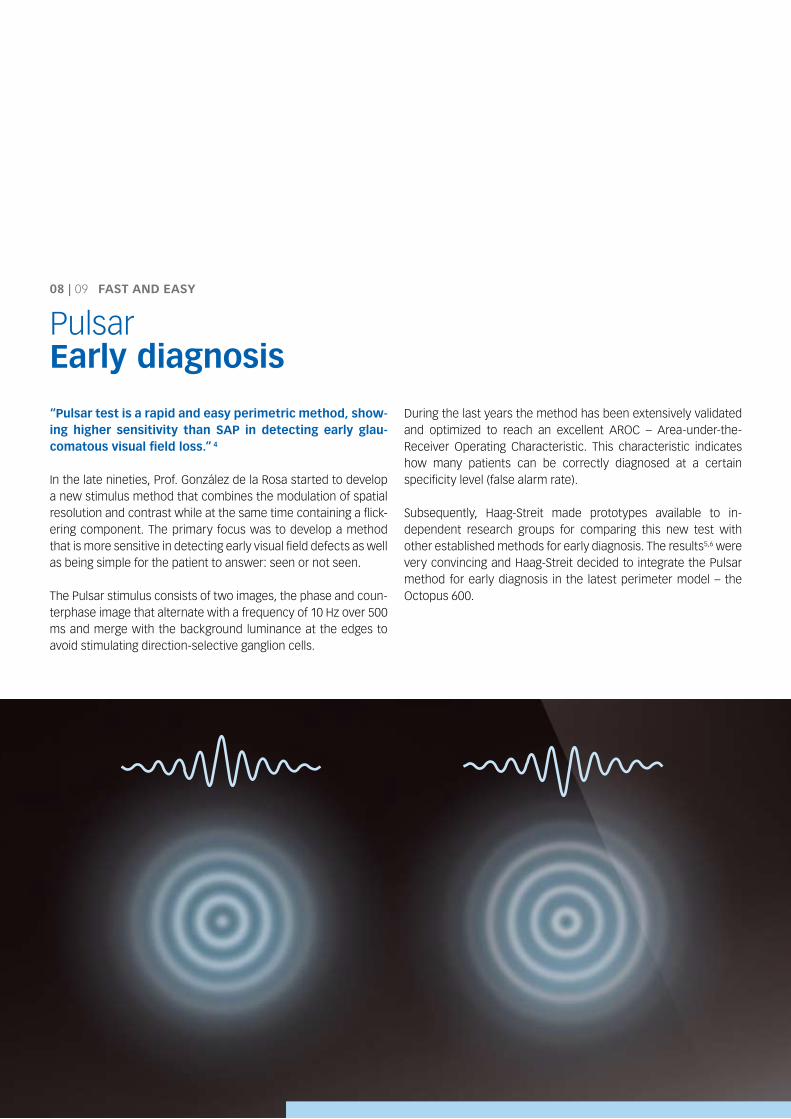
“pulsar test is a rapid and easy perimetric method, show-ing higher sensitivity than sAp in detecting early glau-comatous visual field loss.” 4
In the late nineties, Prof. González de la rosa started to develop a new stimulus method that combines the modulation of spatial resolution and contrast while at the same time containing a flick-ering component. The primary focus was to develop a method that is more sensitive in detecting early visual field defects as well as being simple for the patient to answer: seen or not seen.
The Pulsar stimulus consists of two images, the phase and coun-terphase image that alternate with a frequency of 10 Hz over 500 ms and merge with the background luminance at the edges to avoid stimulating direction-selective ganglion cells.
During the last years the method has been extensively validated and optimized to reach an excellent ArOC – Area-under-the- receiver Operating Characteristic. This characteristic indicates how many patients can be correctly diagnosed at a certain specificity level (false alarm rate).
Subsequently, Haag-Streit made prototypes available to in-dependent research groups for comparing this new test with other established methods for early diagnosis. The results5,6 were very convincing and Haag-Streit decided to integrate the Pulsar method for early diagnosis in the latest perimeter model – the Octopus 600.
08 | 09 FAst ANd EAsY
PulsarEarly diagnosis
1020
S
I
N T
A b
C D
E F
9
10
10 22125
5
5 5
819
19
18 12
7
7
7
614
1828
26
+
+
+
5.9
8.1
12.6
21.76.5
+
+
5.5
9
10
10 22125
5
5 5
819
19
18 12
7
7
7
614
1828
26
+
+
+
5.9
8.1
12.6
21.76.5
+
+
5.5
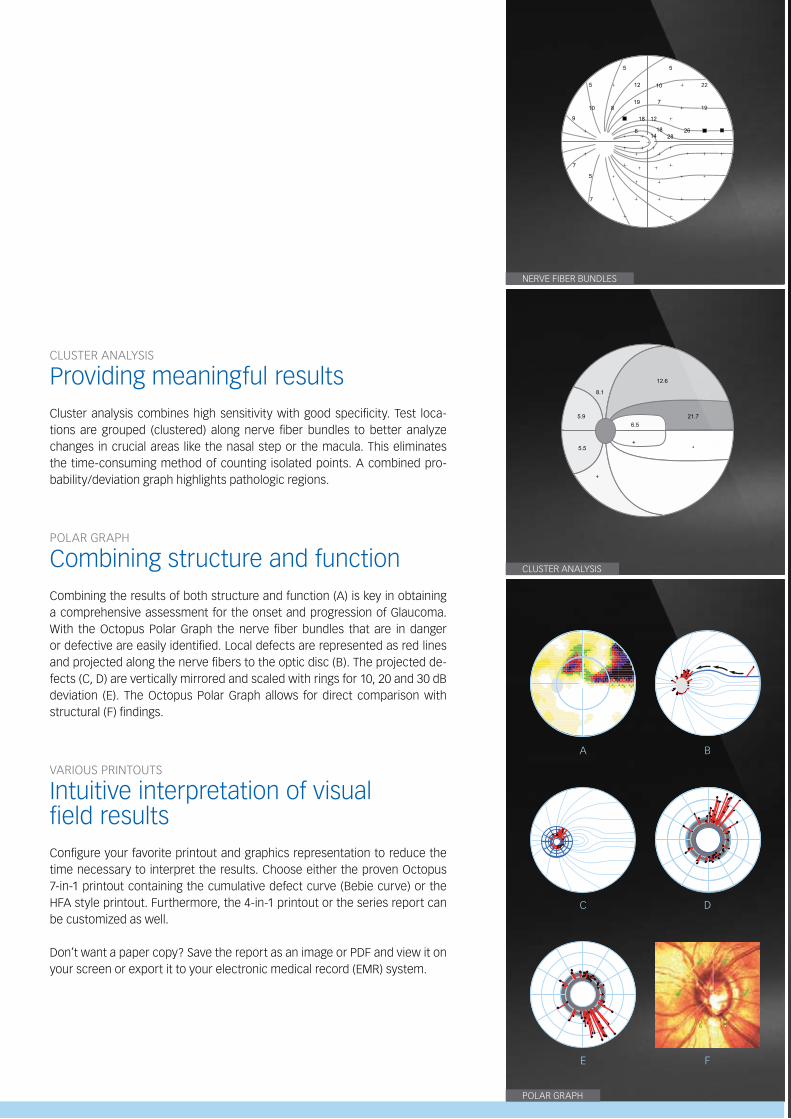
NErVE FIbEr bUNDlES
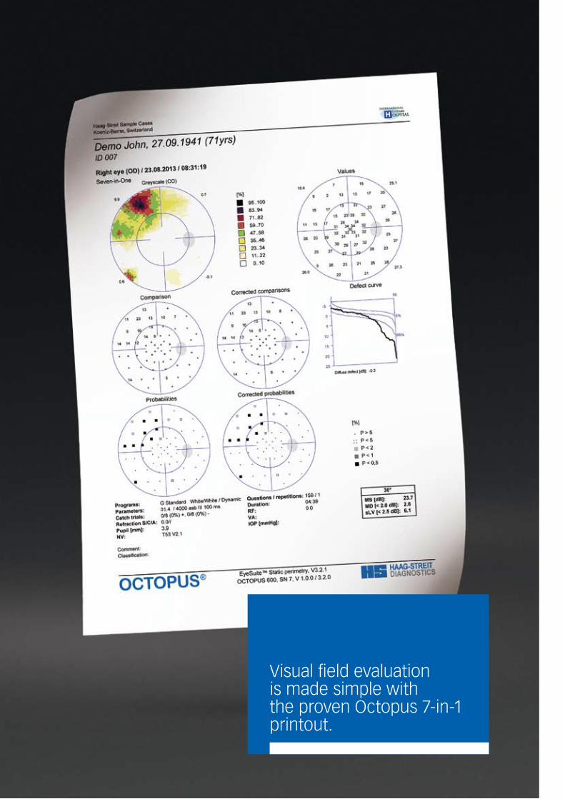
ClUSTEr ANAlySIS
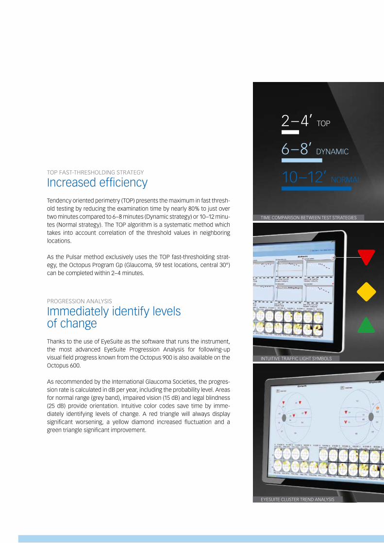
POlAr GrAPH
ClUSTEr ANAlySIS
Providing meaningful resultsCluster analysis combines high sensitivity with good specificity. Test loca-tions are grouped (clustered) along nerve fiber bundles to better analyze changes in crucial areas like the nasal step or the macula. This eliminates the time-consuming method of counting isolated points. A combined pro-bability/deviation graph highlights pathologic regions.
POlAr GrAPH
Combining structure and functionCombining the results of both structure and function (A) is key in obtaining a comprehensive assessment for the onset and progression of Glaucoma. With the Octopus Polar Graph the nerve fiber bundles that are in danger or defective are easily identified. local defects are represented as red lines and projected along the nerve fibers to the optic disc (b). The projected de-fects (C, D) are vertically mirrored and scaled with rings for 10, 20 and 30 db deviation (E). The Octopus Polar Graph allows for direct comparison with structural (F) findings.
VArIOUS PrINTOUTS
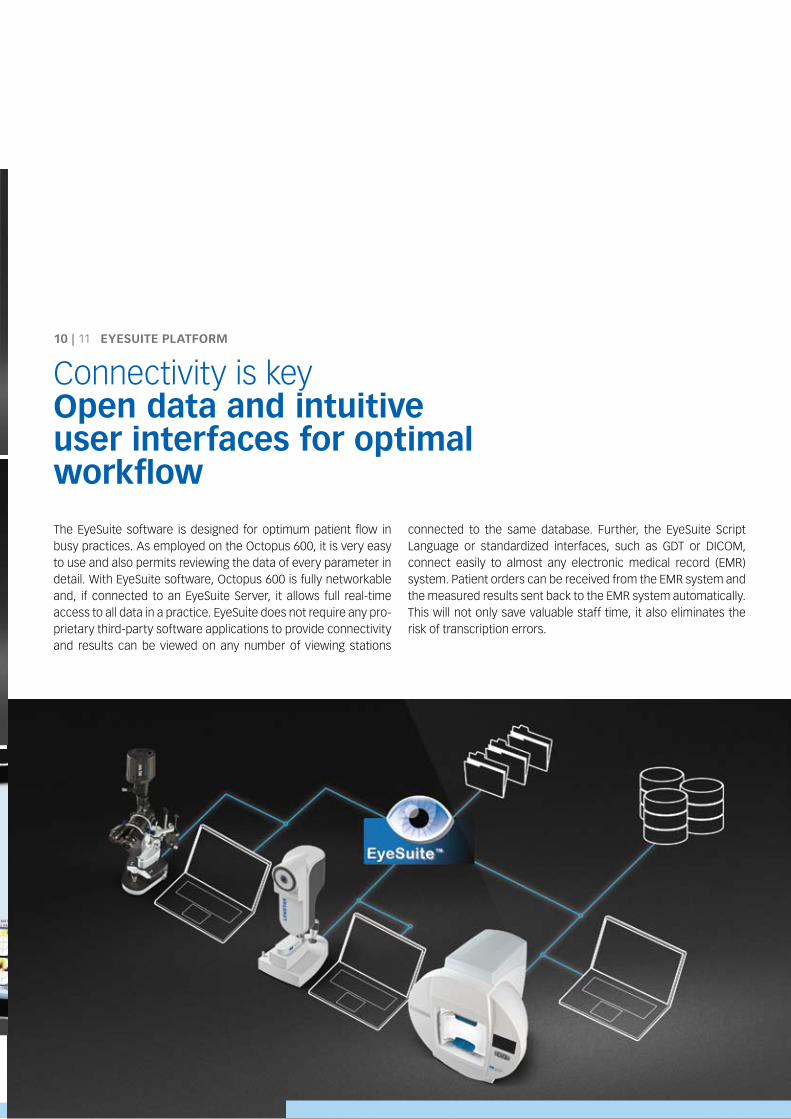
Intuitive interpretation of visual field resultsConfigure your favorite printout and graphics representation to reduce the time necessary to interpret the results. Choose either the proven Octopus 7-in-1 printout containing the cumulative defect curve (bebie curve) or the HFA style printout. Furthermore, the 4-in-1 printout or the series report can be customized as well.
Don’t want a paper copy? Save the report as an image or PDF and view it on your screen or export it to your electronic medical record (Emr) system.
1020
S
I
N T
Visual field evaluation is made simple with the proven Octopus 7-in-1 printout.
TOP FAST-THrESHOlDING STrATEGy
Increased efficiencyTendency oriented perimetry (TOP) presents the maximum in fast thresh-old testing by reducing the examination time by nearly 80% to just over two minutes compared to 6–8 minutes (Dynamic strategy) or 10–12 minu-tes (Normal strategy). The TOP algorithm is a systematic method which takes into account correlation of the threshold values in neighboring locations.
As the Pulsar method exclusively uses the TOP fast-thresholding strat-egy, the Octopus Program Gp (Glaucoma, 59 test locations, central 30°) can be completed within 2–4 minutes.
PrOGrESSION ANAlySIS
Immediately identify levels of changeThanks to the use of EyeSuite as the software that runs the instrument, the most advanced EyeSuite Progression Analysis for following-up visual field progress known from the Octopus 900 is also available on the Octopus 600.
As recommended by the International Glaucoma Societies, the progres-sion rate is calculated in db per year, including the probability level. Areas for normal range (grey band), impaired vision (15 db) and legal blindness (25 db) provide orientation. Intuitive color codes save time by imme-diately identifying levels of change. A red triangle will always display significant worsening, a yellow diamond increased fluctuation and a green triangle significant improvement.
EyESUITE ClUSTEr TrEND ANAlySIS
INTUITIVE TrAFFIC lIGHT SymbOlS
2 – 4’ TOP
6 – 8’ DyNAmIC
10 –12’ NOrmAl
TImE COmPArISON bETWEEN TEST STrATEGIES
The EyeSuite software is designed for optimum patient flow in busy practices. As employed on the Octopus 600, it is very easy to use and also permits reviewing the data of every parameter in detail. With EyeSuite software, Octopus 600 is fully networkable and, if connected to an EyeSuite Server, it allows full real-time access to all data in a practice. EyeSuite does not require any pro-prietary third-party software applications to provide connectivity and results can be viewed on any number of viewing stations
connected to the same database. Further, the EyeSuite Script language or standardized interfaces, such as GDT or DICOm, connect easily to almost any electronic medical record (Emr) system. Patient orders can be received from the Emr system and the measured results sent back to the Emr system automatically. This will not only save valuable staff time, it also eliminates the risk of transcription errors.
10 | 11 EYEsuItE plAtFORM
Connectivity is keyOpen data and intuitiveuser interfaces for optimalworkflow
EyESUITE ClUSTEr TrEND ANAlySIS
INTUITIVE TrAFFIC lIGHT SymbOlS
2 – 4’ TOP
6 – 8’ DyNAmIC
10 –12’ NOrmAl
TImE COmPArISON bETWEEN TEST STrATEGIES
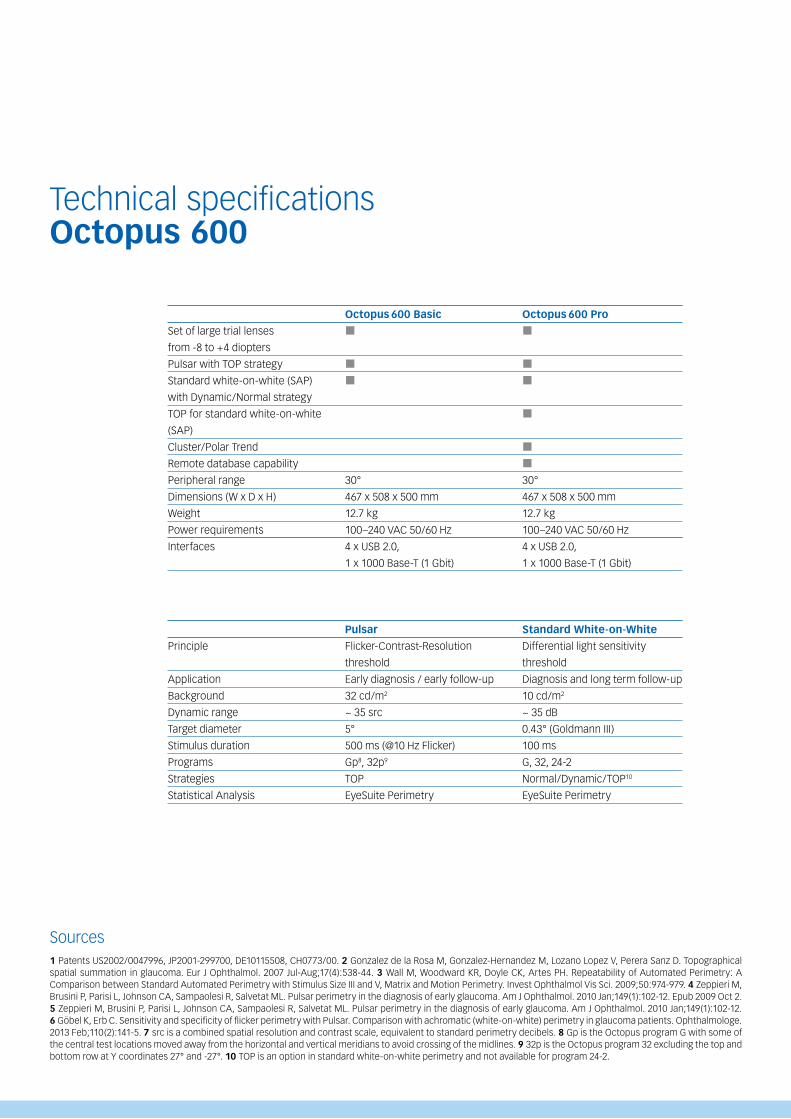
Set of large trial lenses
from -8 to +4 diopters
Pulsar with TOP strategy
Standard white-on-white (SAP)
with Dynamic/Normal strategy
TOP for standard white-on-white
(SAP)
Cluster/Polar Trend
remote database capability
Peripheral range
Dimensions (W x D x H)
Weight
Power requirements
Interfaces
Principle
Application
background
Dynamic range
Target diameter
Stimulus duration
Programs
Strategies
Statistical Analysis
Octopus 600 Basic
pulsar
Flicker-Contrast-resolution
threshold
Early diagnosis / early follow-up
32 cd/m2
~ 35 src
5°
500 ms (@10 Hz Flicker)
Gp8, 32p9
TOP
EyeSuite Perimetry
Octopus 600 pro
standard white-on-white
Differential light sensitivity
threshold
Diagnosis and long term follow-up
10 cd/m2
~ 35 db
0.43° (Goldmann III)
100 ms
G, 32, 24-2
Normal/Dynamic/TOP10
EyeSuite Perimetry
1 Patents US2002/0047996, JP2001-299700, DE10115508, CH0773/00. 2 Gonzalez de la rosa m, Gonzalez-Hernandez m, lozano lopez V, Perera Sanz D. Topographical spatial summation in glaucoma. Eur J Ophthalmol. 2007 Jul-Aug;17(4):538-44. 3 Wall m, Woodward Kr, Doyle CK, Artes PH. repeatability of Automated Perimetry: A Comparison between Standard Automated Perimetry with Stimulus Size III and V, matrix and motion Perimetry. Invest Ophthalmol Vis Sci. 2009;50:974-979. 4 Zeppieri m, brusini P, Parisi l, Johnson CA, Sampaolesi r, Salvetat ml. Pulsar perimetry in the diagnosis of early glaucoma. Am J Ophthalmol. 2010 Jan;149(1):102-12. Epub 2009 Oct 2. 5 Zeppieri m, brusini P, Parisi l, Johnson CA, Sampaolesi r, Salvetat ml. Pulsar perimetry in the diagnosis of early glaucoma. Am J Ophthalmol. 2010 Jan;149(1):102-12. 6 Göbel K, Erb C. Sensitivity and specificity of flicker perimetry with Pulsar. Comparison with achromatic (white-on-white) perimetry in glaucoma patients. Ophthalmologe. 2013 Feb;110(2):141-5. 7 src is a combined spatial resolution and contrast scale, equivalent to standard perimetry decibels. 8 Gp is the Octopus program G with some of the central test locations moved away from the horizontal and vertical meridians to avoid crossing of the midlines. 9 32p is the Octopus program 32 excluding the top and bottom row at y coordinates 27° and -27°. 10 TOP is an option in standard white-on-white perimetry and not available for program 24-2.
Sources
Technical specificationsOctopus 600
30°
467 x 508 x 500 mm
12.7 kg
100–240 VAC 50/60 Hz
4 x USb 2.0,
1 x 1000 base-T (1 Gbit)
30°
467 x 508 x 500 mm
12.7 kg
100–240 VAC 50/60 Hz
4 x USb 2.0,
1 x 1000 base-T (1 Gbit)
HAAG-STrEIT Holding AG
www.haag-streit-holding.com
HAAG-STrEIT AG
www.haag-streit.com
SPECTrOS AG
www.spectros.ch
HAAG-STrEIT medtech AG
www.haag-streit-medtech.com
HAAG-STrEIT France EUrl
www.haag-streit.fr
HAAG-STrEIT Far East
www.haag-streit-fareast.com
HAAG-STrEIT Surgical GmbH
www.haag-streit-surgical.com
möller-Wedel GmbH & Co KG
www.moeller-wedel.com
möller-Wedel Optical GmbH
www.moeller-wedel-optical.com
HAAG-STrEIT Deutschland GmbH
www.haag-streit.de
IPrO GmbH
www.ipro.com
ClEmENT ClArKE ltd.
www.clement-clarke.com
HAAG-STrEIT UK
www.haag-streit-uk.com
John Weiss ltd.
www.johnweiss.com
HAAG-STrEIT USA
www.haag-streit-usa.com
reliance medical Inc.
www.haag-streit-usa.com
Asetronics AG
www.asetronics.ch
Comlab AG
www.comlab.ch
Members of HAAg-stREIt group
©HAAG-STrEIT AG, 3098 Koeniz, Switzerland5. Edition/2013 -11
HS-Art.No. 1511.7220356.02040
HAAg-stREIt AgGartenstadtstrasse 103098 KoenizSwitzerlandPhone +41 31 978 01 11Fax +41 31 978 02 [email protected]