OCT changes of idiopathic epiretinal membrane after ...
5
Vallejo‑Garcia et al. Int J Retin Vitr (2020) 6:37 https://doi.org/10.1186/s40942‑020‑00239‑8 ORIGINAL ARTICLE OCT changes of idiopathic epiretinal membrane after cataract surgery Jose Luis Vallejo‑Garcia 1 , Mary Romano 1,2 , Luca Pagano 1,3* , Alessio Montericcio 1 , Alfredo Borgia 1 , Emanuela Morenghi 1 and Paolo Vinciguerra 1 Abstract Background: We reviewed our experience in the management of cataract and idiopatic epiretinal membrane sur‑ geries at the Humanitas Research Institute–Milan, Italy‑ over the past 3 years. Methods: We conducted a single center retrospective observational case series of patients that underwent sequen‑ tial cataract and idiopatic epiretinal membrane (ERM) surgeries from 2012–2015 in Humanitas Research Institute. Full data was obtained for 53 eyes of 57 patients. Patients with ERM secondary to uveitis or trauma or associated with simultaneous retinal detachment were excluded. Diabetic retinopathy, glaucoma, age‑related macular degeneration, and myopia of more than 6 diopters were exclusion criteria as well. Results: Cataract surgery was not associated with an ERM stage progression at one month follow up, but caused retinal inflammation that resulted in a significant increase in central macular thickness (CMT ), macular volume (MV), central macular edema (CME), IS/OS disruption (IS/OS) and neurosensory detachment (NSD). However, there was no significant change in Best corrected visual acuity (BCVA). Conclusion: We suggest that patients undergoing cataract surgery in the presence of epiretinal membranes need tight follow up to treat and control eventual macular inflammatory changes and eventual prompt vitrectomy if BCVA is threatened. Keywords: Epiretinal membrane, Cataract surgery, Retina, Peeling © The Author(s) 2020. This article is licensed under a Creative Commons Attribution 4.0 International License, which permits use, sharing, adaptation, distribution and reproduction in any medium or format, as long as you give appropriate credit to the original author(s) and the source, provide a link to the Creative Commons licence, and indicate if changes were made. The images or other third party material in this article are included in the article’s Creative Commons licence, unless indicated otherwise in a credit line to the material. If material is not included in the article’s Creative Commons licence and your intended use is not permitted by statutory regulation or exceeds the permitted use, you will need to obtain permission directly from the copyright holder. To view a copy of this licence, visit http://creativeco mmons.org/licenses/by/4.0/. The Creative Commons Public Domain Dedication waiver (http://creativecommons.org/publicdomain/ zero/1.0/) applies to the data made available in this article, unless otherwise stated in a credit line to the data. Introduction Epiretinal membrane (ERM), also commonly known as macular pucker, is an avascular fibrocellular membrane that forms at the vitreoretinal interface, causing visual disfunction and metamorphopsia [1]. In a recent review of the epidemiological studies available, prevalence was 9.1% of the total population [2]. e precise pathophysiology of this clinical entity is not yet known, a proliferation of hyalocytes in the setting of anomalous posterior vitreous detachment and vitreoschi- sis has been proposed as a possible mechanism in the early development of idiopathic ERMs [3]. e ERM can also be formed secondary to retinal vascular or inflam- matory diseases, trauma, tumours and intraocular sur- gery or inflammation [2]. e association between cataract surgery and ERM and the timing of the surgical procedures is to be determined, whether it is preferable a 2-step-sequential approach or a combined surgery [4–6]. Literature seems to have no clear answer yet, so it is important to analyse morpho- logically whether or not ERM progression is accelerated by phacoemulsification and if there is a consequence of sequential surgery in BCVA. e only specific study available on this topic dates back to 2008, were Hayashi found that foveal thickness and macular volume were not significantly influenced by cataract surgery and that the visual acuity was not Open Access International Journal of Retina and Vitreous *Correspondence: [email protected] 3 Department of Ophthalmology, Istituto Clinico Humanitas, Via Alessandro Manzoni 56, 20089 Rozzano, MI, Italy Full list of author information is available at the end of the article
Transcript of OCT changes of idiopathic epiretinal membrane after ...

Vallejo‑Garcia et al. Int J Retin Vitr (2020) 6:37 https://doi.org/10.1186/s40942‑020‑00239‑8
ORIGINAL ARTICLE
OCT changes of idiopathic epiretinal membrane after cataract surgeryJose Luis Vallejo‑Garcia1, Mary Romano1,2, Luca Pagano1,3* , Alessio Montericcio1, Alfredo Borgia1, Emanuela Morenghi1 and Paolo Vinciguerra1
Abstract
Background: We reviewed our experience in the management of cataract and idiopatic epiretinal membrane sur‑geries at the Humanitas Research Institute–Milan, Italy‑ over the past 3 years.
Methods: We conducted a single center retrospective observational case series of patients that underwent sequen‑tial cataract and idiopatic epiretinal membrane (ERM) surgeries from 2012–2015 in Humanitas Research Institute. Full data was obtained for 53 eyes of 57 patients. Patients with ERM secondary to uveitis or trauma or associated with simultaneous retinal detachment were excluded. Diabetic retinopathy, glaucoma, age‑related macular degeneration, and myopia of more than 6 diopters were exclusion criteria as well.
Results: Cataract surgery was not associated with an ERM stage progression at one month follow up, but caused retinal inflammation that resulted in a significant increase in central macular thickness (CMT), macular volume (MV), central macular edema (CME), IS/OS disruption (IS/OS) and neurosensory detachment (NSD). However, there was no significant change in Best corrected visual acuity (BCVA).
Conclusion: We suggest that patients undergoing cataract surgery in the presence of epiretinal membranes need tight follow up to treat and control eventual macular inflammatory changes and eventual prompt vitrectomy if BCVA is threatened.
Keywords: Epiretinal membrane, Cataract surgery, Retina, Peeling
© The Author(s) 2020. This article is licensed under a Creative Commons Attribution 4.0 International License, which permits use, sharing, adaptation, distribution and reproduction in any medium or format, as long as you give appropriate credit to the original author(s) and the source, provide a link to the Creative Commons licence, and indicate if changes were made. The images or other third party material in this article are included in the article’s Creative Commons licence, unless indicated otherwise in a credit line to the material. If material is not included in the article’s Creative Commons licence and your intended use is not permitted by statutory regulation or exceeds the permitted use, you will need to obtain permission directly from the copyright holder. To view a copy of this licence, visit http://creat iveco mmons .org/licen ses/by/4.0/. The Creative Commons Public Domain Dedication waiver (http://creat iveco mmons .org/publi cdoma in/zero/1.0/) applies to the data made available in this article, unless otherwise stated in a credit line to the data.
IntroductionEpiretinal membrane (ERM), also commonly known as macular pucker, is an avascular fibrocellular membrane that forms at the vitreoretinal interface, causing visual disfunction and metamorphopsia [1]. In a recent review of the epidemiological studies available, prevalence was 9.1% of the total population [2].
The precise pathophysiology of this clinical entity is not yet known, a proliferation of hyalocytes in the setting of anomalous posterior vitreous detachment and vitreoschi-sis has been proposed as a possible mechanism in the
early development of idiopathic ERMs [3]. The ERM can also be formed secondary to retinal vascular or inflam-matory diseases, trauma, tumours and intraocular sur-gery or inflammation [2].
The association between cataract surgery and ERM and the timing of the surgical procedures is to be determined, whether it is preferable a 2-step-sequential approach or a combined surgery [4–6]. Literature seems to have no clear answer yet, so it is important to analyse morpho-logically whether or not ERM progression is accelerated by phacoemulsification and if there is a consequence of sequential surgery in BCVA.
The only specific study available on this topic dates back to 2008, were Hayashi found that foveal thickness and macular volume were not significantly influenced by cataract surgery and that the visual acuity was not
Open Access
International Journalof Retina and Vitreous
*Correspondence: [email protected] Department of Ophthalmology, Istituto Clinico Humanitas, Via Alessandro Manzoni 56, 20089 Rozzano, MI, ItalyFull list of author information is available at the end of the article

Page 2 of 5Vallejo‑Garcia et al. Int J Retin Vitr (2020) 6:37
markedly impaired in the first year following surgery [7].
However, with the recent advances in image resolu-tion and acquisition speed, a newly introduced OCT-based classification was proposed by Govetto et al. [8], shifting the attention to the inner retina anatomy. They described continuous ectopic inner foveal layers (EIFL) as the essential element in their grading scheme, which also was correlated with visual impairment.
We have studied retrospectively the behaviour of the retinal configuration and BCVA in patients with ERM that underwent sequential cataract surgery and consec-utive vitrectomy after a month.
Materials and methodsThe aim of the study was to analyse the changes of the epiretinal membrane stage, macular status and BCVA before and after cataract surgery.
We conducted a single center retrospective observa-tional case series of patients that underwent sequential cataract and idiopathic epiretinal membrane surgeries from 2012–2015. Full data was obtained for 53 eyes of 57 patients.
Patients with ERM secondary to uveitis or trauma or associated with simultaneous RD were excluded. Diabetic retinopathy, glaucoma, age-related macular degeneration, and myopia of more than 6 diopters were exclusion criteria as well.
We classified our patients according to Govetto’s ERM classification [8] as follows.
Stage 1 was characterized by the presence of the foveal pit and well-conserved retinal layers.
In stage 2 there was the absence of the foveal pit, stage 3 characterised by the presence of ectopic inner foveal layers, whereas in stage 4 there was also a retinal layers disruption.
The presence of macular cystoid edema (CME), dis-ruption of the inner/outer segment (IS/OS) and neuro-sensory foveal detachment (NSD) was also recorded.
Patients were examined pre-cataract surgery and one-month post-surgery with full ophthalmologi-cal examination including BCVA, slit lamp and fun-dus examination and SD-OCT (Cirrus HD-OCT; Carl Zeiss, Dublin, California, USA) with protocols analy-sis of central macular thickness (CMT), macular vol-ume (MV). 11 patients had an extended follow up of 3 months due to delay of the vitrectomy. The following exclusion criteria were adopted: previous intraocular surgery or laser refractive procedure, any ocular or sys-temic morbidity significantly affecting VA (e.g. central corneal opacity, advanced glaucoma, retina vein occlu-sion or macular degeneration).
Statistical methodData were expressed as mean and standard deviation or number and percentage, as appropriated. The com-parison between preoperative and 1-month follow up was done with paired t student test or Wilcoxon test, as appropriated, for continuous variables, and with Mc Nemar test for categorical variables.
The association of the preoperative characteristics with BCVA changes was explored with linear regres-sion, while the association with diabetes was explored with logistic regression analysis.
The significance level was set at 0.05 for all test. Sta-tistical analysis was performed with Stata 15 software (StataCorp. 2017, College Station, TX: StataCorp LLC).
ResultsThe 57 eyes of 53 patients included 26 (49.1%) men, with a mean age of 71 ± 5 years, 7 (13.2%) patients had type 2 diabetes without retinopathy.
At baseline they presented with an epiretinal mem-brane stage 1 in 5 (8.9%) eyes, as stage 2 in 24 (42.9%) eyes, as stage 3 in in 22 (39.3%) eyes and as stage 4 in 5 (8.9%) eyes.
Pre-operative characteristics of the population are reported in Table 1.
Staging of the ERM remained stable after cataract surgery in all patients.
Table 2 reports the changing in visual parameters on month after surgery: there was no significant change in BCVA (p = 0.4605), while a statistically significant worsening of the following parameters was observed CMT, CME, disruption in the IS/OS and NSD, while MV showed a significant increasing.
Table 1 Preoperative population characteristics
N 57
Age (years) 71.6 ± 5.3
BCVA 0.46 ± 0.18
ERM staging 2.48 ± 0.79
1 5 (8.9%)
2 24 (42.9%)
3 22 (39.3%)
4 5 (8.9%)
CMT (µm) 443 ± 79
MV (mm3) 11.9 ± 1.4
CME 13 (22.8%)
IS/OS disruption 8 (14.0%)
NSD 3 (5.3%)
Page 3 of 5Vallejo‑Garcia et al. Int J Retin Vitr (2020) 6:37
The difference in 1-month with preoperative BCVA wasn’t associated with any of the preoperative character-istics (Table 3).
We also analyzed our diabetic patients’ group (7 patients in total), finding no statistically significant dif-ference when compared with the non-diabetic patients. The data are reported in Table 4. This analysis, however, is partially influenced by the low population number.
DiscussionCataract surgery is a known factor causing inflamma-tion of the eye. It has been described an upregulation of the inflammatory mediators in the aqueous and vitreous humors after surgical manipulation. The breakdown of the blood-aqueous and blood-retinal-barriers lead to an increased vascular permeability causing macular swelling with or without CME. Idiopathic epiretinal membranes are a result of a multifactorial activation of fibroblasts and the vitreous of these patients is known to have a higher concentration of inflammatory and profibrotic cytokines [9]. Chu et al. found that the relative risk of finding an increase in the relative risk of 5.60 in eyes with previously diagnosed ERM [10]. Our patient cohort dem-onstrated an incidence of postoperative CME at month 1 higher than what is reported in literature for patients with epiretinal membranes, which is around 15% [11]. Other studies [7] documented the presence or absence of ERM, not analyzing the stage of the condition, and we
suggested that the advanced stages of our ERM could be responsible of this increased risk. However, there was no statistically significant correlation between the stage of the ERM and the development of the CME (Fig. 1).
A second aspect to consider is the improvement in the OCT technology, which allowed us to be way more sensitive in CME detection. According to a recently pub-lished paper by Dong H. Yoon et al., we classified as CME even the microcystic macular edema that is in fact a mild form of CME [12]. This increased sensitivity is confirmed by the fact that even preoperatively, one-fourth of our patients already had CME.
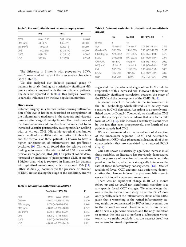
We also documented an increased rate of disruption of the inner/outer segment (IS/OS) and neuroretinal detachment (NSD) after phacoemulsification, all of them characteristics that are correlated to a reduced BCVA (Fig. 2).
Our data shows a statistically significant increase in all these variables. As literature has previously demostrated [7], the presence of an epiretinal membrane is an inde-pendent risk factor, which acts sinergically to increase the rate of these inflammatory consequences [13]. Detailed analysis of foveal OCT anatomy was performed demon-strating the changes induced by phacoemulsification in eyes with idiopathic advanced membranes.
There was no significant change in BCVA 1 month follow-up and we could not significantly correlate it to any specific foveal OCT changes. We acknowledge that one of the limitation of our study is that the BCVA does only partially reflect the inflammatory status of the ERM, given that a worsening of the retinal inflammatory sta-tus, might be compensated by BCVA improvement due to the cataract removal. However, most of our patient didn’t have a significant cataract, and the only indication to remove the lens was to perform a subsequent vitrec-tomy, so we might conclude that the cataract itself was not a cause for visual impairment.
Table 2 Pre and 1-Month post cataract surgery values
Pre Post P
BCVA 0.46 ± 0.18 0.45 ± 0.18 0.4605
CMT (µm) 443 ± 79 486 ± 99 < 0.0001
MV (mm3) 11.9 ± 1.4 12.4 ± 1.8 < 0.0001
CME 13 (22.8%) 32 (56.1%) < 0.0001
IS/OS 8 (14.0%) 16 (28.1%) 0.0047
NSD 3 (5.3%) 9 (15.8%) 0.0143
Table 3 Association with variation of BVCA
Coefficient (95% CI) P
Age −0.001 (−0.023–0.021) 0.906
Diabetes −0.070 (−0.399–0.259) 0.670
ERM staging 0.058 (−0.092–0.206) 0.448
CMT (µm) −0.000 (−0.002–0.001) 0.847
MV (mm3) 0.015 (−0.068–0.099) 0.713
CME 0.128 (−0.143–0.398) 0.349
IS/OS 0.247 (−0.075–0.570) 0.130
NSD −0.095 (−0.607–0.417) 0.711
Table 4 Different variables in diabetic and non-diabetic groups
DM No DM OR (95% CI) P
N eyes 8 49
Age 72.8 ± 8.2 71.4 ± 4.7 1.05 (0.91–1.21) 0.502
Gender (M) 6 (75.0%) 24 (49.0%) 3.13 (0.57–17.03) 0.188
ERM staging 2.29 ± 0.95 2.51 ± 0.77 0.68 (0.24–1.94) 0.478
BCVA 0.40 ± 0.18 0.47 ± 0.18 0.11 (0.00–6.07) 0.277
CMT (μm) 381 ± 72 452 ± 77 0.98 (0.97–1.00) 0.020
MV (mm3) 12.2 ± 1.8 11.8 ± 1.3 1.19 (0.70–2.01) 0.525
CME 2 (25.0%) 11 (22.5%) 1.15 (0.20–6.53) 0.873
IS/OS 1 (12.5%) 7 (14.3%) 0.86 (0.09–8.07) 0.893
NSD 2 (25.0%) 1 (2.0%) 16.0 (1.25–204) 0.033

Page 4 of 5Vallejo‑Garcia et al. Int J Retin Vitr (2020) 6:37
We do not know whether this intraocular inflam-mation prolonged for longer follow up periods, would cause a worsening in the BCVA or resolve without consequences. In our case the maximum follow up was 3 months, because all our patients already had surgical ERM.
Previous studies have shown that the clinical history of an ERM is not significantly influenced by cataract surgery. Our findings confirm that even with this new OCT-based classification focused in the inner retinal changes the ERM progression is not significantly wors-ened by cataract surgery at 1 month follow up, how-ever we are conscious that the short follow up is not enough to exclude the speed up of the natural history of ERM after phacoemulsification.
ConclusionStage of the ERM was not rapidly accelerated by cataract surgery. Patients with ERM are at higher risk for devel-oping inflammatory changes after cataract surgery such as cystoid macular edema, neurosensory detachment and alterations of the inner-outer segment layer. However, these are not associated with any worsening of the BCVA within the first month. We suggest that patients undergo-ing cataract surgery in the presence of epiretinal mem-branes need tight follow up to treat and control eventual macular inflammatory changes and eventual prompt vit-rectomy if BCVA is threatened.
Further prospective studies are needed to fully under-stand the consequences of cataract surgery in patients with advanced epiretinal membranes.
Fig. 1 OCT showing a stage 2 ERM pre (left) and post (right) phacoemulsification. It can be observed a slight increase in the CME
Fig. 2 OCT showing a stage 3 ERM pre (left) and post (right) phacoemulsification. It can be observed a significant increase in the CME and a small subfoveal NSD

Page 5 of 5Vallejo‑Garcia et al. Int J Retin Vitr (2020) 6:37
• fast, convenient online submission
•
thorough peer review by experienced researchers in your field
• rapid publication on acceptance
• support for research data, including large and complex data types
•
gold Open Access which fosters wider collaboration and increased citations
maximum visibility for your research: over 100M website views per year •
At BMC, research is always in progress.
Learn more biomedcentral.com/submissions
Ready to submit your research ? Choose BMC and benefit from:
Authors’ contributionsAll authors equally contributed to conception and design and to interpreta‑tion of data. All authors drafted the article and approved its final version. All authors read and approved the final manuscript.
Funding and AcknowledgementThis study received no specific grant from any funding agency in the public, commercial or not‑for‑profit sectors.
Availability of data and materialsThe data used to support the findings of this study are available from the cor‑responding author upon request.
Ethics approval and consent to participateThis article does not contain any studies with human participants or animals performed by any of the authors.
Consent for publicationAll authors have agreed on the publication of this paper.
Competing interestsAll authors declare that they have no relevant conflict of interest.
Author details1 Humanitas Clinical and Research Center, Humanitas University, Via Manzoni 56, 20089 Rozzano, Milan 20089, Italy. 2 Multidisciplinary Department of Medi‑cal, Surgical and Dental Sciences University of Campania Luigi Vanvitelli, Neaples, Italy. 3 Department of Ophthalmology, Istituto Clinico Humanitas, Via Alessandro Manzoni 56, 20089 Rozzano, MI, Italy.
Received: 5 December 2019 Accepted: 25 July 2020
References 1. Dikkaya F, Karaman Erdur S, Ozsutcu M, Aydin R, Kocabora MS, Aras C. The
significance of neutrophil‑to‑lymphocyte ratio in idiopathic epiretinal membrane. Int Ophthalmol. 2018;38(4):1393–7. https ://doi.org/10.1007/s1079 2‑017‑0597‑0.
2. Xiao W, Chen X, Yan W, Zhu Z, He M. Prevalence and risk factors of epireti‑nal membranes: a systematic review and meta‑analysis of population‑based studies. BMJ Open. 2017;7(9):e014644. https ://doi.org/10.1136/bmjop en‑2016‑01464 4.
3. Schumann RG, Gandorfer A, Ziada J, et al. Hyalocytes in idiopathic epiretinal membranes: a correlative light and electron microscopic study. Graefes Arch Clin Exp Ophthalmol. 2014;252(12):1887–94. https ://doi.org/10.1007/s0041 7‑014‑2841‑x.
4. Hamoudi H, Correll Christensen U, La Cour M. Epiretinal membrane surgery: an analysis of 2‑step sequential‑ or combined phacovitrectomy surgery on refraction and macular anatomy in a prospective trial. Acta Ophthalmol. 2017;96(3):243–50. https ://doi.org/10.1111/aos.13572 .
5. Dugas B, Ouled‑Moussa R, Lafontaine PO, et al. Idiopathic epiretinal mac‑ular membrane and cataract extraction: combined versus consecutive surgery. Am J Ophthalmol. 2010;149(2):302–6. https ://doi.org/10.1016/j.ajo.2009.09.011.
6. Alexandrakis G, Chaudhry NA, Flynn HWJ, Murray TG. Combined cataract surgery, intraocular lens insertion, and vitrectomy in eyes with idiopathic epiretinal membrane. Ophthalmic Surg Lasers. 1999;30(4):327–8. https ://doi.org/10.3928/1542‑8877‑19990 401‑21.
7. Hayashi K, Hayashi H. Influence of phacoemulsification surgery on pro‑gression of idiopathic epiretinal membrane. Eye. 2008;23(4):774–9. https ://doi.org/10.1038/eye.2008.161.
8. Govetto A, Lalane RA, Sarraf D, Figueroa MS, Hubschman JP. Insights into epiretinal membranes: presence of ectopic inner foveal layers and a new optical coherence tomography staging scheme. Am J Ophthalmol. 2017;175:99–113. https ://doi.org/10.1016/j.ajo.2016.12.006.
9. Zandi S, Tappeiner C, Pfister IB, Despont A, Rieben R, Garweg JG. Vitreal cytokine profile differences between eyes with epiretinal membranes or macular holes. Invest Ophthalmol Vis Sci. 2016;57(14):6320–6. https ://doi.org/10.1167/iovs.16‑20657 .
10. Chu CJ, Johnston RL, Buscombe C, Sallam AB, Mohamed Q, Yang YC. Risk factors and incidence of macular edema after cataract surgery: a database study of 81984 eyes. Ophthalmology. 2016;123(2):316–23. https ://doi.org/10.1016/j.ophth a.2015.10.001.
11. Schaub F, Adler W, Enders P, et al. Preexisting epiretinal membrane is associated with pseudophakic cystoid macular edema. Graefes Arch Clin Exp Ophthalmol. 2018. https ://doi.org/10.1007/s0041 7‑018‑3954‑4.
12. Yoon DH, Kang DJ, Kim MJ, Kim HK. New observation of microcystic macular edema as a mild form of cystoid macular lesions after stand‑ard phacoemulsification. Medicine. 2018;97(15):e0355. https ://doi.org/10.1097/MD.00000 00000 01035 5.
13. Hardin JS, Gauldin DW, Soliman MK, Chu CJ, Yang YC, Sallam AB. Cataract surgery outcomes in eyes with primary epiretinal membrane. JAMA Ophthalmol. 2018;136(2):148. https ://doi.org/10.1001/jamao phtha lmol.2017.5849.
Publisher’s NoteSpringer Nature remains neutral with regard to jurisdictional claims in pub‑lished maps and institutional affiliations.