Occipital Sulci of the Human Brain: Variability and ...2)_Iaria.pdf · On the lateral surface of...
17
Occipital Sulci of the Human Brain: Variability and Probability Maps GIUSEPPE IARIA 1,2 * AND MICHAEL PETRIDES 1 1 Montreal Neurological Institute, McGill University, Montreal, Quebec H3A 2B4, Canada 2 Dipartimento di Psicologia, Universita ` di Roma “La Sapienza,” CAP 00185 Rome, Italy ABSTRACT The morphological variation of the sulci of the occipital region of the human brain was examined in both the left and the right hemispheres in 40 normal adult human brains on magnetic resonance images. We identified the occipital sulci and marked their corresponding gray matter voxels on the magnetic resonance images, which had been transformed into the Montreal Neurological Institute standard proportional stereotaxic space in order to construct probability maps. In the medial occipital region, the calcarine sulcus was the longest and most constant sulcus. We identified, in the inferior part of the medial occipital lobe, the lingual sulcus and the posterior collateral sulcus, and, in the superior part, the inferior and superior sagittal sulci of the cuneus. On the lateral surface of the occipital lobe, the lateral occipital, the lunate, and the transverse and inferior occipital sulci were identified. The parieto-occipital fissure and the temporo-occipital incisure were also identified on the lateral and medial surfaces. Finally, the patterns of the occipital sulci and gyri were examined in 20 post-mortem human hemispheres fixed in formalin. Probability maps of the occipital sulci were constructed, which provide a quantitative description of the variability of the sulci in standard stereotaxic space and may be used to identify the location of voxels in other magnetic resonance images transformed into the same streotaxic space. These maps are a useful tool in the study of functional activations related to visual processing. J. Comp. Neurol. 501:243–259, 2007. © 2007 Wiley-Liss, Inc. Indexing terms: sulcus; visual cortex; morphology; lunate sulcus; calcarine sulcus; lateral occipital sulcus; transverse occipital sulcus The occipital region of the human brain is traditionally defined as extending from the occipital pole to the parieto- occipital fissure, dorsally, and to the temporo-occipital inci- sure, ventrally. In both the human and the nonhuman pri- mate brain, the occipital region is involved in visual information processing. By the beginning of the Twentieth Century, it was clearly established that the distinct cortex comprising the stripe of Gennari (i.e., the striate cortex), which lies on the banks of the calcarine sulcus, was involved in visual cortical processing (see, e.g., Bolton, 1900). The sulcal and gyral patterns of the occipital region of the human brain became the subject of investigation during the latter part of the Nineteenth Century and the first half of the Twentieth Century (see, e.g., Cunningham, 1892; Eber- staller, 1890; Elliot Smith, 1904a– c; Retzius, 1896). Much of this early work focused on the calcarine sulcus and its rela- tion to the striate cortex, producing a number of publications in which the detailed morphological relations of striate cor- tex, the calcarine sulcus, and the immediately surrounding sulci were outlined (see, e.g., Bolton, 1900; Elliot Smith, 1904a– c; Shellshear, 1926). Architectonic studies at the beginning of the Twentieth Century established that the striate cortex (area 17 in the Brodmann map, 1909; area OC in the Economo and Ko- skinas map, 1925) was surrounded by a ring of cortex with different architecture (area 18 in the Brodmann map; area OB in the Economo and Koskinas map) and then another larger region (area 19 in the Brodmann map; area OA in the Economo and Koskinas map). Thus, the architectonic studies had already implied the presence of more than one visual area in the occipital cortex. Beginning in the 1960s, Grant sponsor: Canadian Institutes of Health Research (CIHR); Grant number: MOR 14620. *Correspondence to: Giuseppe Iaria, PhD, Cognitive Neuroscience Unit, Montreal Neurological Institute, McGill University, 3801 University St., Montreal, Quebec, Canada H3A 2B4. E-mail: [email protected] Received 16 August 2006; Revised 12 October 2006; Accepted 31 October 2006 DOI 10.1002/cne.21254 Published online in Wiley InterScience (www.interscience.wiley.com). THE JOURNAL OF COMPARATIVE NEUROLOGY 501:243–259 (2007) © 2007 WILEY-LISS, INC.
Transcript of Occipital Sulci of the Human Brain: Variability and ...2)_Iaria.pdf · On the lateral surface of...
Occipital Sulci of the Human Brain:Variability and Probability Maps
GIUSEPPE IARIA1,2* AND MICHAEL PETRIDES1
1Montreal Neurological Institute, McGill University, Montreal, Quebec H3A 2B4, Canada2Dipartimento di Psicologia, Universita di Roma “La Sapienza,” CAP 00185 Rome, Italy
ABSTRACTThe morphological variation of the sulci of the occipital region of the human brain was
examined in both the left and the right hemispheres in 40 normal adult human brains onmagnetic resonance images. We identified the occipital sulci and marked their correspondinggray matter voxels on the magnetic resonance images, which had been transformed into theMontreal Neurological Institute standard proportional stereotaxic space in order to constructprobability maps. In the medial occipital region, the calcarine sulcus was the longest andmost constant sulcus. We identified, in the inferior part of the medial occipital lobe, thelingual sulcus and the posterior collateral sulcus, and, in the superior part, the inferior andsuperior sagittal sulci of the cuneus. On the lateral surface of the occipital lobe, the lateraloccipital, the lunate, and the transverse and inferior occipital sulci were identified. Theparieto-occipital fissure and the temporo-occipital incisure were also identified on the lateraland medial surfaces. Finally, the patterns of the occipital sulci and gyri were examined in 20post-mortem human hemispheres fixed in formalin. Probability maps of the occipital sulciwere constructed, which provide a quantitative description of the variability of the sulci instandard stereotaxic space and may be used to identify the location of voxels in othermagnetic resonance images transformed into the same streotaxic space. These maps are auseful tool in the study of functional activations related to visual processing. J. Comp. Neurol.501:243–259, 2007. © 2007 Wiley-Liss, Inc.
Indexing terms: sulcus; visual cortex; morphology; lunate sulcus; calcarine sulcus; lateral
occipital sulcus; transverse occipital sulcus
The occipital region of the human brain is traditionallydefined as extending from the occipital pole to the parieto-occipital fissure, dorsally, and to the temporo-occipital inci-sure, ventrally. In both the human and the nonhuman pri-mate brain, the occipital region is involved in visualinformation processing. By the beginning of the TwentiethCentury, it was clearly established that the distinct cortexcomprising the stripe of Gennari (i.e., the striate cortex),which lies on the banks of the calcarine sulcus, was involvedin visual cortical processing (see, e.g., Bolton, 1900). Thesulcal and gyral patterns of the occipital region of the humanbrain became the subject of investigation during the latterpart of the Nineteenth Century and the first half of theTwentieth Century (see, e.g., Cunningham, 1892; Eber-staller, 1890; Elliot Smith, 1904a–c; Retzius, 1896). Much ofthis early work focused on the calcarine sulcus and its rela-tion to the striate cortex, producing a number of publicationsin which the detailed morphological relations of striate cor-tex, the calcarine sulcus, and the immediately surroundingsulci were outlined (see, e.g., Bolton, 1900; Elliot Smith,1904a–c; Shellshear, 1926).
Architectonic studies at the beginning of the TwentiethCentury established that the striate cortex (area 17 in theBrodmann map, 1909; area OC in the Economo and Ko-skinas map, 1925) was surrounded by a ring of cortex withdifferent architecture (area 18 in the Brodmann map; areaOB in the Economo and Koskinas map) and then anotherlarger region (area 19 in the Brodmann map; area OA inthe Economo and Koskinas map). Thus, the architectonicstudies had already implied the presence of more than onevisual area in the occipital cortex. Beginning in the 1960s,
Grant sponsor: Canadian Institutes of Health Research (CIHR); Grantnumber: MOR 14620.
*Correspondence to: Giuseppe Iaria, PhD, Cognitive Neuroscience Unit,Montreal Neurological Institute, McGill University, 3801 University St.,Montreal, Quebec, Canada H3A 2B4.
E-mail: [email protected] 16 August 2006; Revised 12 October 2006; Accepted 31 October
2006DOI 10.1002/cne.21254Published online in Wiley InterScience (www.interscience.wiley.com).
THE JOURNAL OF COMPARATIVE NEUROLOGY 501:243–259 (2007)
© 2007 WILEY-LISS, INC.
neurophysiological investigations mapping the receptivefields of single neurons responding to visual stimuli in theoccipital region of nonhuman primates identified severalvisual areas that surround and extend anterior to thestriate cortex that now came to be referred to as area V1(see, e.g., Allman and Kaas, 1971; Zeki, 1969, 1974,1978a,b). In nonhuman primates, these additional visualareas hold an excellent relation to sulcal and gyral land-marks. For instance, in the dorsal occipital region, area V2occupies the posterior bank of the lunate sulcus, V3 andV3A are found on the annectant gyrus that is buried in thelunate sulcus, area V4 is located on the prelunate gyrus,area V5/MT is found on the posterior bank of the caudalsuperior temporal sulcus, and area V6 is found within theparieto-occipital fissure (for reviews see Felleman andVan Essen, 1991; Kaas, 2004; Sereno and Tootell, 2005).
In recent years, developments in functional neuroimag-ing in normal human subjects, such as functional mag-netic resonance imaging (fMRI), have permitted the map-ping of several visual areas of the human brain and havealready provided provisional identification of some of thevisual areas that were first described in nonhuman pri-mates (see Sereno and Tootell, 2005). These areas are V1,V2, V3, V3A, V4, V5/MT, and V6 (e.g., Anderson et al.,1996; Barton et al., 1996; Bense et al., 2006; de Jong et al.,1994; DeYoe et al., 1996; Dougherty et al., 2003; Dumoulinet al., 2000; Dupont et al., 1994; Hadjikhani et al., 1998;Hasnain et al., 1998; Itoh et al., 2005; Sack et al., 2006;Sereno et al., 1995; Shipp et al., 1995; Shulman et al.,1998; Stiers et al., 2006; Tootell et al., 1996, 1997; Tootelland Hadjikhani 2001; Vallines et al., 2006; Walters et al.,2003; Watson et al., 1993; Zeki et al., 1991). The firstvisual area outside the striate cortex that was identified inthe human brain was the motion area V5/MT, for which areasonably good relation with certain sulcal landmarkswas noted (Watson et al., 1993; Zeki et al., 1991). TheV5/MT motion area lay within a sulcus that had variouslybeen named the anterior occipital sulcus or an ascendingbranch of the inferior temporal sulcus (Cunnigham, 1892).Watson and colleagues (1993) noted the lack of standardterminology and adequate description of the sulci of theoccipital region of the human brain with the exception ofthe calcarine sulcus.
The lack of an adequate description of the sulcal pat-terns of the human occipital region, with the exception ofthe calcarine sulcus, which is evident from inspection ofany one of several modern standard textbooks of neuro-
anatomy (see, e.g., Carpenter, 1996; Nolte, 2002), makes itdifficult to establish clear relations between sulcal land-marks and identified visual areas with modern functionalneuroimaging. There have been no examinations of thesulcal patterns of the occipital region of the human brainin recent years, with the exception of the studies by Onoand colleagues (1990) on 25 human cadaver brains. Onoand colleagues (1990) examined the patterns formed bythe calcarine sulcus and its relation to the parieto-occipital fissure as well as the sagittal sulci of the cuneusand the lingual gyrus that immediately border the calcar-ine sulcus on the medial surface of the hemisphere. Thesulci of the lateral part of the occipital region, however,were not investigated by Ono and colleagues.
The aim of the present investigation was to examine thesulcal patterns on the medial and lateral surface of thehuman occipital region and to provide a quantitative de-scription of the variability of these sulci in standard ste-reotaxic space in the form of probability maps. Modernfunctional neuroimaging studies have relied on the Ta-lairach and Tournoux (1988) atlas to determine the loca-tion of functional activity in the human brain resultingfrom functional neuroimaging studies. Because the Ta-lairach and Tournoux atlas is based on one hemisphere ofa single brain, it does not provide any measure of anatom-ical variability. The proper use of a standard stereotaxicspace requires statistical statements of the variability ofthe location of a given brain structure (e.g., a sulcus)within that space in order to account for individual differ-ences. Probability maps have already been provided forthe cingulate and paracingulate sulci (Paus et al., 1996),the primary auditory region (Penhune et al., 1996), thepars opercularis of the inferior frontal gyrus (Tomaiuolo etal., 1999), the sulci of the orbital frontal cortex (Chiavaraset al., 2001), and the precentral sulci (Germann et al.,2005). The present study examines the anatomical vari-ability of the sulci of the human occipital region. Thefindings can be used to relate activation foci of visualprocessing obtained from functional neuroimaging studiesin a precise quantitative manner.
MATERIALS AND METHODS
Subjects
Magnetic resonance imaging (MRI) scans of both the leftand the right hemispheres of 40 human brains were ex-amined. The sample consisted of 17 females (mean age25.5 years, SD 5.3) and 23 males (mean age 25 years, SD5.3). All subjects were right-handed, and none had a pos-itive history of neurological and psychiatric disorders. Thesubjects were randomly selected from the InternationalConsortium for Brain Mapping project (Mazziotta et al.,1995a,b). All subjects gave informed consent. We also ex-amined 10 post-mortem human brains fixed in formalin(six females, four males; mean age 70.5 years, SD 9.2) toinvestigate the patterns of the occipital sulci and gyri.Photographs of the sulci patterns on the post-mortembrain were taken with a digital camera, and modificationsof the images (contrast and brightness) were made inAdobe Photoshop.
MRI
The MRI scans were performed on a Philips Gyroscan1.5-T superconducting magnet system. A fast-field echo
Abbreviations
acc accessory lateral occipital sulciACS anterior calcarine sulcusAOS anterior occipital sulcusBCS body of the calcarine sulcusIOS inferior occipital sulcusISGS inferior sagittal sulcusLiS lingual sulcusLOS lateral occipital sulcusLuS lunate sulcusPCS posterior collateral sulcus, i.e., the occipital extension of
the collateral sulcusPOF parieto-occipital fissureRCS retrocalcarine sulcusSSGS superior sagittal sulcusTO temporo-occipital incisureTOS transverse occipital sulcus
The Journal of Comparative Neurology. DOI 10.1002/cne
244 G. IARIA AND M. PETRIDES
3-D acquisition sequence was used to collect 160 contigu-ous 1-mm T1-weighted images (Tr � 18 msec, Te � 10msec, flip angle � 30°) in the sagittal plane. To normalizeand correct the images for interindividual differences ingross brain size, each MR volume was transformed intothe Montreal Neurological Institute (MNI) standardizedstereotaxic space (Evans et al., 1992; Mazziotta et al.,1995a,b), which is based on that of Talairach and Tour-noux (1988). For each MR volume, the transformation wasdetermined by an automated registration using 3D cross-correlation (Collins et al., 1994) to a target image that wasthe intensity average of 305 brain volumes previouslyaligned to the Talairach atlas (Evans et al., 1992). Theimage data were then resampled onto a standard gridwith cubical voxels 1 mm wide. The mediolateral (left–right) axis was defined by using the x-coordinate (posi-tive � right hemisphere), the rostrocaudal (anterior–posterior) axis by using the y-coordinate (positive �rostral to anterior commisure), and the dorsoventral(superior–inferior) axis by using the z-coordinate (posi-tive � superior to a horizontal line drawn through ante-rior and posterior commissures).
Localization of the occipital sulci
The occipital sulci were manually identified using DIS-PLAY, an interactive 3-D imaging software package (Mac-Donald, 1996). The software displays a 3-D view of thebrain surface as well as sections in the coronal, horizontal,and sagittal planes. The sections on the screen are auto-matically updated as the cursor is moved from voxel tovoxel in order to mark the location of the sulcus investi-gated. This automatic updating of the views on the screenallows the investigator to identify accurately the extentand direction of a particular sulcus. DISPLAY also gener-ates a histogram of the image intensity values, which isused to determine the upper intensity threshold for voxelsof the cerebrospinal fluid (CSF). All voxels were automat-ically classified according to tissue type (Kollokian, 1996).Initially, the voxels considered to contain CSF betweenthe sulcal banks were marked by the investigator. Thegray-matter voxels extending for 1 mm on either side ofthe banks of the sulcus, adjacent to those in the sulcalCSF, were automatically included in the set of voxelsconstituting the sulcus. The lateral and medial surfaces ofthe occipital lobe of the formalin-fixed post-mortem hu-man hemispheres were examined and photographed toidentify the typical sulcal and gyral patterns, as describedin the classical literature.
Probability mapping
After labeling of the voxels forming each of the sulci ofinterest, 3-D probability maps were constructed. For eachsulcus, the probability values are displayed by means of acolor scale. The minimum value of each scale is 0.1 (10% ofthe subjects included in this study). The highest probabil-ity value varied for different sulci. The maps were con-structed by dividing, at each particular 3-D stereotaxiclocation, the number of times that a voxel belonged to thesulcus of interest by the number of subjects examined.These probability values, which are displayed in color-coded maps, thus represent the likelihood that any voxelin MNI space will be classified as part of the sulcus. Forexample, if the value at the given x,y,z location is 0.6, thenthis location was occupied by a voxel that belong to thatsulcus in 60% of the subjects examined. The probability
maps are then superimposed on the intensity-averagedtarget image of 305 brains (Evans et al., 1992), and thestereotaxic x,y,z coordinate values are provided in theMNI standard proportional space. In other words, thesemaps quantify the spatial variability of the sulci in astandard stereotaxic space.
RESULTS
We identified 11 occipital sulci in 80 hemispheres andmarked the gray-matter voxels that constituted thesesulci in the MNI space. In addition, we identified andmarked the gray-matter voxels around the parieto-occipital fissure (POF) and the temporo-occipital incisure(TO). Figures 1–4 present photographs of the medial, lat-eral, basal, and dorsal surfaces, respectively, of the occip-ital lobe in post-mortem human hemispheres, illustratingpatterns of the occipital sulci. Figures 5–7 present coronal,horizontal, and sagittal sections, respectively, through theoccipital region of one MRI brain to identify the location ofthe sulci of interest. Examples of probability maps of thePOF and TO are provided in coronal section (POF, Fig. 8;TO, Fig. 9).
Calcarine sulcus
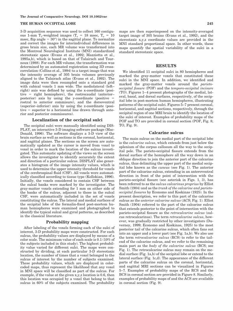
The main sulcus on the medial part of the occipital lobeis the calcarine sulcus, which extends from just below thesplenium of the corpus callosum all the way to the occip-ital pole. The parieto-occipital fissure extends from thedorsal surface of the hemisphere all the way down in anoblique direction to join the anterior part of the calcarinesulcus, thus delimiting the upper part of the medial occip-ital lobe known as the cuneus (see Fig. 1). The anteriorpart of the calcarine sulcus, extending in an anteroventraldirection in front of the point of intersection with theparieto-occipital fissure (see asterisk in Figs. 1, 3), hasbeen referred to as the sulcus calcarinus proprius by ElliotSmith (1904) and as the trunk of the calcarine and parieto-occipital fissures by Economo and Koskinas (1925). In thepresent description, we refer to this part of the calcarinesulcus as the anterior calcarine sulcus (ACS; Fig. 1). ElliotSmith (1904) referred to the part of the calcarine sulcusthat extends posterior to the point of intersection with theparieto-occipital fissure as the retrocalcarine sulcus (sul-cus retrocalcarinus). The term retrocalcarine sulcus, how-ever, was gradually restricted by other investigators (Du-vernoy, 1999; Economo and Koskinas, 1925) to the mostposterior tail of the calcarine sulcus, which often fans outinto an upper and a lower part (see Fig. 1a,b). We also usethe term retrocalcarine sulcus (RCS) to refer to the tail-end of the calcarine sulcus, and we refer to the remainingmain part as the body of the calcarine sulcus (BCS; seeFig. 1). The retrocalcarine sulcus may remain on the me-dial surface (Fig. 1a,b) of the occipital lobe or extend to thelateral surface (Fig. 1c,d). The appearance of the differentparts of the calcarine sulcus on the coronal, horizontal,and sagittal MRI sections can be visualized in Figures5–7. Examples of probability maps of the RCS and theBCS in coronal section are provided in Figure 8. Similarly,examples of probability maps of and the ACS are availablein coronal section (Fig. 9).
The Journal of Comparative Neurology. DOI 10.1002/cne
245THE HUMAN OCCIPITAL LOBE
Inferior and superior sagittal sulciof the cuneus
Just dorsal to the body of the calcarine sulcus, there isfrequently a sulcus running, more or less, parallel to it.This sulcus has been referred to as the inferior sagittalsulcus (ISGS) of the cuneus by Economo and Koskinas(1925) to distinguish it from another, somewhat less fre-quent superior sagittal sulcus of the cuneus (SSGS). Du-vernoy (1999) referred to the inferior sagittal sulcus of thecuneus as the paracalcarine sulcus, but this term is prob-ably best avoided, because the same name has been usedfor a sulcus that lies on the posterior bank of the parieto-occipital fissure (Elliot Smith, 1904a). In the presentstudy, we have adopted the terminology of Economo andKoskinas (1925) for the sulci of the cuneus. The appear-ance of the different patterns of these two sulci of thecuneus can be visualized in Figure 1 (photographs of post-mortem human brains) and in Figure 5–7 displaying thecoronal, horizontal, and sagittal MRI sections, respec-tively. Examples of probability maps of the ISGS andSSGS are provided in coronal section (ISGS, Fig. 9; SSGS,Fig. 10).
Posterior collateral and lingual sulci
In the inferior part of the occipital lobe (below the cal-carine sulcus), the posterior branch of the collateral sulcus(PCS) runs, more or less, parallel to the body of the cal-carine sulcus, thus delimiting the lingual gyrus (Figs. 3,7). Occasionally, within the lingual gyrus, there is a sulcusrunning, more or less, parallel to the body of the calcarinesulcus, dividing it into a superior and an inferior lingualgyrus. This sulcus has been referred to as the lingualsulcus (LiS) in the classical literature (e.g., Economo andKoskinas, 1925) and, more recently, as the intralingualsulcus by Ono and coauthors (1990). We have retained theclassical term lingual sulcus (Figs. 3, 7). The probabilitymaps of the PCS and the LiS are available in coronalsection (Fig. 10).
Lunate, lateral, inferior, and transverseoccipital sulci
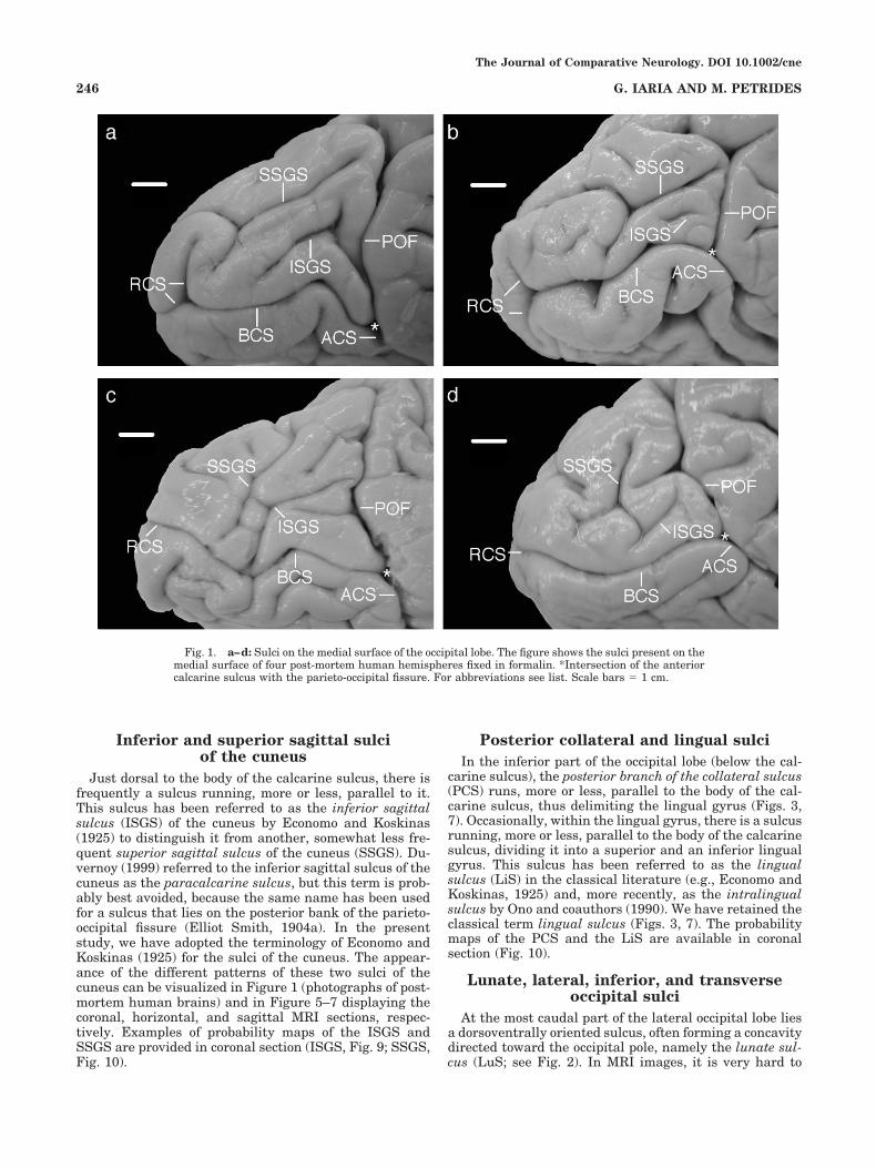
At the most caudal part of the lateral occipital lobe liesa dorsoventrally oriented sulcus, often forming a concavitydirected toward the occipital pole, namely the lunate sul-cus (LuS; see Fig. 2). In MRI images, it is very hard to
Fig. 1. a–d: Sulci on the medial surface of the occipital lobe. The figure shows the sulci present on themedial surface of four post-mortem human hemispheres fixed in formalin. *Intersection of the anteriorcalcarine sulcus with the parieto-occipital fissure. For abbreviations see list. Scale bars � 1 cm.
The Journal of Comparative Neurology. DOI 10.1002/cne
246 G. IARIA AND M. PETRIDES
identify this sulcus because of both its shape and its loca-tion on the lateral to medial curvature of the occipital pole(see Figs. 5–7). Immediately anterior to the lunate sulcus,the horizontally oriented lateral occipital sulcus (LOS) canbe identified (see Fig. 2; Duvernoy, 1999; Eberstaller,1890; Economo and Koskinas, 1925; Ono et al., 1990).Elliot Smith (1904a) referred to this sulcus as the prelu-nate sulcus (sulcus praelunatus). This sulcus usuallyblends, more or less, with the middle part of the lunatesulcus and divides the lateral surface of the occipital lobeinto a superior and an inferior part. In the inferior part ofthe occipital region, close to the base of the hemisphere, isthe inferior occipital sulcus (IOS), the caudal end of whichruns close to the most ventral part of the lunate sulcus(see Fig. 2). In the superior part of the occipital region, thetransverse occipital sulcus (TOS) can be identified (see Fig.2; Duvernoy, 1999; Elliot Smith, 1904a). This sulcus,which is more or less dorsoventrally oriented, lies caudalto the parieto-occipital fissure and joins the occipital ex-tension of the intraparietal sulcus. Economo and Koskinas(1925) referred to this sulcus as the sulcus occipitalisprimus. The appearance of these sulci on coronal, horizon-tal, and sagittal MRI sections can be appreciated in Fig-ures 5–7, respectively. Examples of probability maps of
the LuS and LOS are displayed in coronal section (LuS,Fig. 10; LOS, Fig. 8). Similarly, examples of probabilitymaps of the IOS and TOS are available in coronal section(IOS, Fig. 8; TOS, Fig. 10).
Gray-matter volumes of the occipital sulci
We analyzed the volumes of the intrasulcal gray matterobtained from each of the sulci identified in the occipitallobe. Two-way repeated-meausures analyses of variance(ANOVA) were performed to evaluate the effect of gender(female, male) and side (left and right hemisphere; with ccvolume as repeated measures) on each of the occipitalsulci. The main effect of the gender was statistically sig-nificant for the IOS (F1,38 � 6.4, P � 0.016) and LiS(F1,38 � 5.02, P � 0.031). On the other hand, the maineffect of side was statistically significant for the RCS(F1,38 � 6.25, P � 0.168), SSGS (F1,38 � 4.61, P � 0.038),TOS (F1,38 � 10.33, P � 0.003), and PCS (F1,38 � 6.73, P �0.013). Post hoc comparison (Duncan’s test) of these ef-fects showed that the larger gray-matter volumes werefound in males (vs. females) and in the left hemisphere(vs. right). No other significant main or interaction effectswere found for the other sulci. In Table 1 we report thevolumes of the occipital sulci in individual sulci and the
Fig. 2. a–d: Sulci on the lateral surface of the occipital lobe. The figure shows the sulci present on thelateral surface of four post-mortem human hemispheres fixed in formalin. For abbreviations see list.Scale bars � 1 cm.
The Journal of Comparative Neurology. DOI 10.1002/cne
247THE HUMAN OCCIPITAL LOBE

mean values according to gender (females, males) andhemisphere (left, right).
DISCUSSION
Although classic and modern descriptions of the mor-phology of the medial surface of the occipital region of thehuman brain are reasonably consistent with each other,descriptions of its lateral surface have generated consid-erable confusion. The same sulcus is frequently identifiedwith different names, and two obviously different sulci arereferred to by the same name (see below). Indeed, mostmodern neuroanatomy textbooks avoid any description ofthe sulci of the lateral occipital region of the human brain,providing only a general definition of the lateral occipitallobe as being that part of the posterior hemisphere that isbounded by the parieto-occipital fissure dorsally and bythe temporo-occipital incisure ventrally (see, e.g., Carpen-ter, 1996; Nolte, 2002). The present examination of themorphology of the occipital region in the MRIs of 80 cere-bral hemispheres (40 left and 40 right), as well as 20hemispheres of post-mortem brains, showed that, despitethe existence of considerable morphological variation, a
basic pattern governs the variability and can be used toidentify most of the sulci consistently. We provide below adescription of the basic pattern of the sulci of the lateraloccipital region, which is clearly illustrated in Figure 2a,with comments on the variability that this pattern exhib-its (Fig. 2b–d), and then we describe the less controversialmedial occipital region (Fig. 1).
Just anterior to the dorsoventrally directed lunate sul-cus, which is situated close to the occipital pole, extends amore or less horizontally arranged sulcus, the lateral oc-cipital sulcus. The lateral occipital sulcus (Fig. 2), whichhas also been called the praelunate sulcus by Elliot Smith(1904a–c), is a very important sulcus for the definition ofthe overall morphology of the lateral surface of the occip-ital region of the human brain because it divides it into asuperolateral and an inferolateral portion. The lunate sul-cus is often difficult to identify in MRIs because of itsposition on the curvature of the occipital pole, but thelateral occipital sulcus can be reliably identified in coronalMRI sections and can, therefore, be used to define thesuperolateral and inferolateral portions of the lateral oc-cipital lobe. Ventral or dorsal to the main lateral occipitalsulcus, there may be accessory lateral occipital sulci that
Fig. 3. a–d: Sulci identified on the basal surface of the occipital lobe. The figure shows the sulcipresent on the basal surface of four post-mortem human hemispheres fixed in formalin. Asterisksindicate intersection of the anterior calcarine sulcus with the parieto-occipital fissure. For abbreviationssee list. Scale bars � 1 cm.
The Journal of Comparative Neurology. DOI 10.1002/cne
248 G. IARIA AND M. PETRIDES
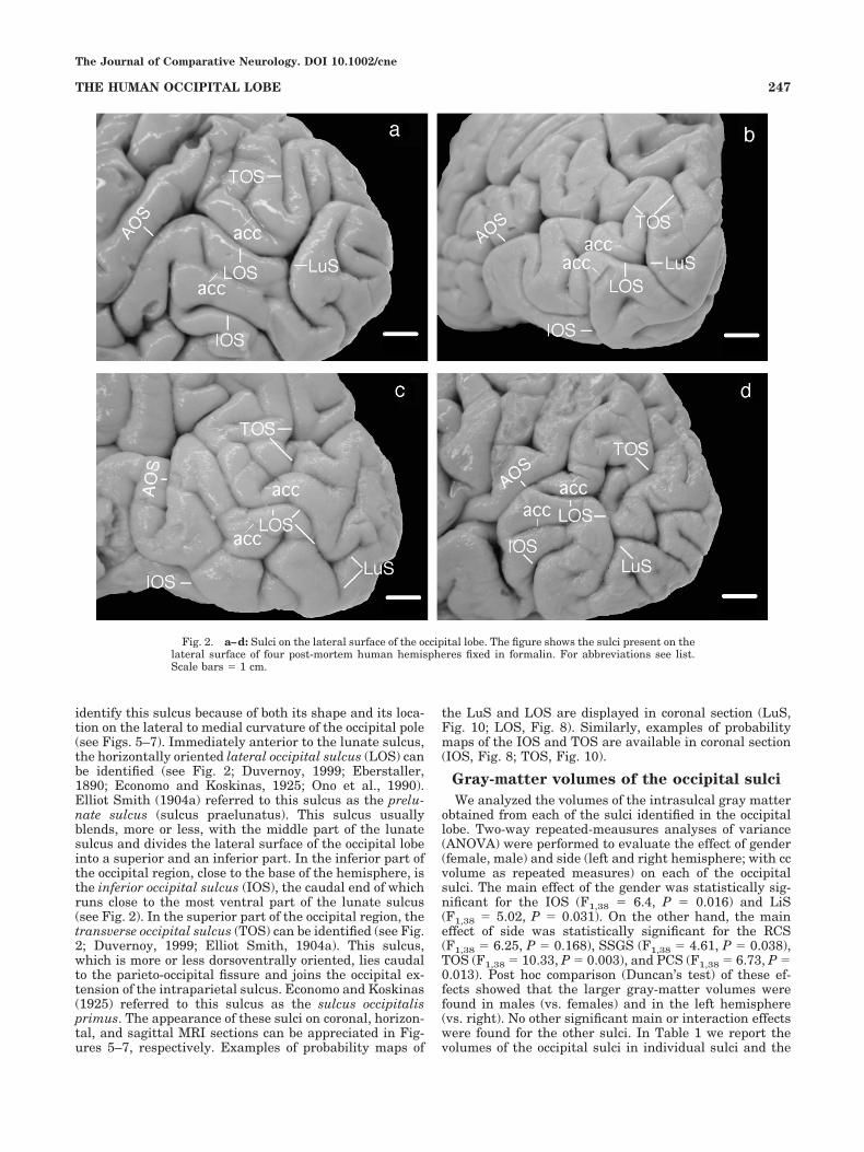
are shallower, shorter, and inconsistent (Fig. 2). By con-trast, it has never proved difficult to identify the mainlateral occipital sulcus, which has a rostral end that ap-proaches the anterior occipital sulcus and often blendswith it (superficially) and a caudal part that approachesand often blends, approximately, with the midpoint of thelunate sulcus (Fig. 2). The lunate sulcus is sometimesdivided into an upper and a lower branch by a submergedor an exposed narrow gyrus (the translunate gyrus), and,when this happens, either the dorsal or the ventral branchof the lunate sulcus may blend with the lateral occipitalsulcus. Occasionally, the lunate sulcus blends with thelateral occipital sulcus in such a manner that it appears asthe caudal tail of the lateral occipital sulcus (Fig. 2c).
The sulcus that marks the morphology of the superolat-eral portion of the human occipital region is the trans-verse occipital sulcus that runs in a dorsoventral directionabove the lateral occipital sulcus (Fig. 2a). The transverseoccipital sulcus emerges from the occipital extension of theintraparietal sulcus just caudal to the parieto-occipitalfissure and often forms a branch running in a dorsomedialdirection (i.e., above the intraparietal sulcus) and abranch running in ventrolateral direction below the in-traparietal sulcus. These dorsomedial and ventrolateral
branches may blend (Fig. 2a,d) or remain separate (Fig.2b,c) on the surface of the brain. The ventrolateral branchof the transverse occipital sulcus may extend ventrally tojust above the lateral occipital sulcus and anterior to theupper part of the lunate sulcus (Fig. 2), but in some brainsit may, superficially, appear to join the upper part of thelunate sulcus because it extends beneath the posteriorbank of the lunate sulcus. Various names have been usedto refer to the transverse occipital sulcus: Economo andKoskinas (1925) refer to the dorsomedial branch of thetransverse occipital sulcus as the sulcus parietalis trans-versus and its ventrolateral branch as the sulcus occipi-talis primus. Elliot Smith (1904a) treats the transverseoccipital sulcus as the tail of what he calls the paroccipitalsulcus (i.e., the occipital extension of the intraparietalsulcus). Eberstaller (1892) has referred to the transverseoccipital sulcus as the fissura occipitalis anterior, but thisname has been used for a completely different sulcus thatlies anterior to it by Wernicke (1876) and Elliot Smith(1904a), and this terminology has been adopted by mostmodern investigators (see, e.g., Malikovic et al., 2006; Onoet al., 1990) and in the present study (see Fig. 2).
Since the completion of our study, an article on thelunate sulcus of the human brain has been published by
Fig. 4. a–d: Sulci on the dorsal surface of the occipital lobe. The figure shows the sulci present on thedorsal surface of four post-mortem human hemispheres fixed in formalin. For abbreviations see list. Scalebars � 1 cm.
The Journal of Comparative Neurology. DOI 10.1002/cne
249THE HUMAN OCCIPITAL LOBE
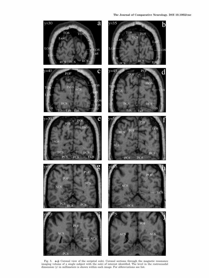
Fig. 5. a–j: Coronal view of the occipital sulci. Coronal sections through the magnetic resonanceimaging volume of a single subject with the sulci of interest identified. The level in the rostrocaudaldimension (y) in millimeters is shown within each image. For abbreviations see list.
The Journal of Comparative Neurology. DOI 10.1002/cne

Fig. 6. a–n: Horizontal view of the occipital sulci. Horizontal sections through the magnetic resonanceimaging volume of a single subject with the sulci of interest identified. The level in the dorsoventraldimension (z) in millimeters is shown within each image. For abbreviations see list.
The Journal of Comparative Neurology. DOI 10.1002/cne

Fig. 7. Sagittal view of the occipital sulci. Sagittal sections of the left (a–h) and right (i–n) hemi-sphere in the magnetic resonance imaging volume of a single subject with the sulci of interest identified.The level in the mediolateral dimension (x) in millimeters is shown within each image. For abbreviationssee list.
The Journal of Comparative Neurology. DOI 10.1002/cne
252 G. IARIA AND M. PETRIDES

Fig. 8. Probability maps on coronal sections of the parieto-occipitalfissure (POF), the retrocalcarine sulcus (RCS), and the lateral occip-ital sulcus (LOS) are displayed in Aa–f. Probability maps of the bodyof the calcarine sulcus (BCS) and the inferior occipital sulcus (IOS)
are displayed in Ba–f. The probability maps of the sulci are superim-posed on the average brain of the MNI (Evans et al., 1992), and thecoordinates provided are within the MNI standard proportional ste-reotaxic space.
The Journal of Comparative Neurology. DOI 10.1002/cne
253THE HUMAN OCCIPITAL LOBE
Allen and colleagues (2006). These investigators note thehigh variability in the shape of the lunate sulcus andreport that, when present, it is most often not a singlesulcus but rather a composite of two or more sulcal seg-ments. They defined the lunate as a continuous sulcusthat traverses a substantial portion of the lateral surfaceof the posterior occipital lobe. This definition would in-clude not only the lunate sulcus as defined by Elliot Smith(1904a–c) and by ourselves but also the transverse occip-ital sulcus and even the caudal part of the lateral occipitalsulcus. Indeed, Allen and colleagues (2006, p 871) pointout that, if a typical pattern can be identified in the com-posite lunate sulci they identified, “it is one in which thesuperior portion is formed by the extension of intrapari-etal sulcus (i.e., the transverse occipital sulcus), whichthen extends downward to form a junction with anotheroccipital sulcus (e.g., the lateral occipital sulcus or occipi-topolar sulcus, sensu Duvernoy, 1999).” If one were toadopt such a definition, the lunate sulcus of the humanbrain would clearly not be homologous to the lunate sulcusin nonhuman primate brains. We have defined the lunatesulcus as a short sulcus on the occipital pole that may
blend, superficially, with the ventralmost part of thetransverse occipital sulcus or the caudalmost part of thelateral occipital sulcus, but it is clearly distinct from theseother two sulci. Because in nonhuman primate brains thelunate sulcus lies close to the lateral border of the striatecortex, its homologue in the human brain can be consid-ered to be a sulcus only at the very posterior part of thelateral occipital lobe, where a small fraction of the striatecortex extends. We agree with Allen and colleagues (2006)that there has been considerable development of the oc-cipital region of the human brain. We believe that thetransverse occipital and lateral occipital sulci found on thehuman occipital lobe are new sulci and are related toprestriate cortical areas (see below), which, in the ma-caque monkey brain, lie on the annectant gyrus that ishidden within the nonhuman lunate sulcus and the pre-lunate gyrus that extends anterior to it.
Allen and colleagues (2006) report that the compositelunate sulcus, as they defined it for the human brain, wasobserved in 32.7% of the left hemispheres and 26.4% of theright hemispheres. These percentages are much lowerthan those reported by Ono et al. (1990) for the lunate
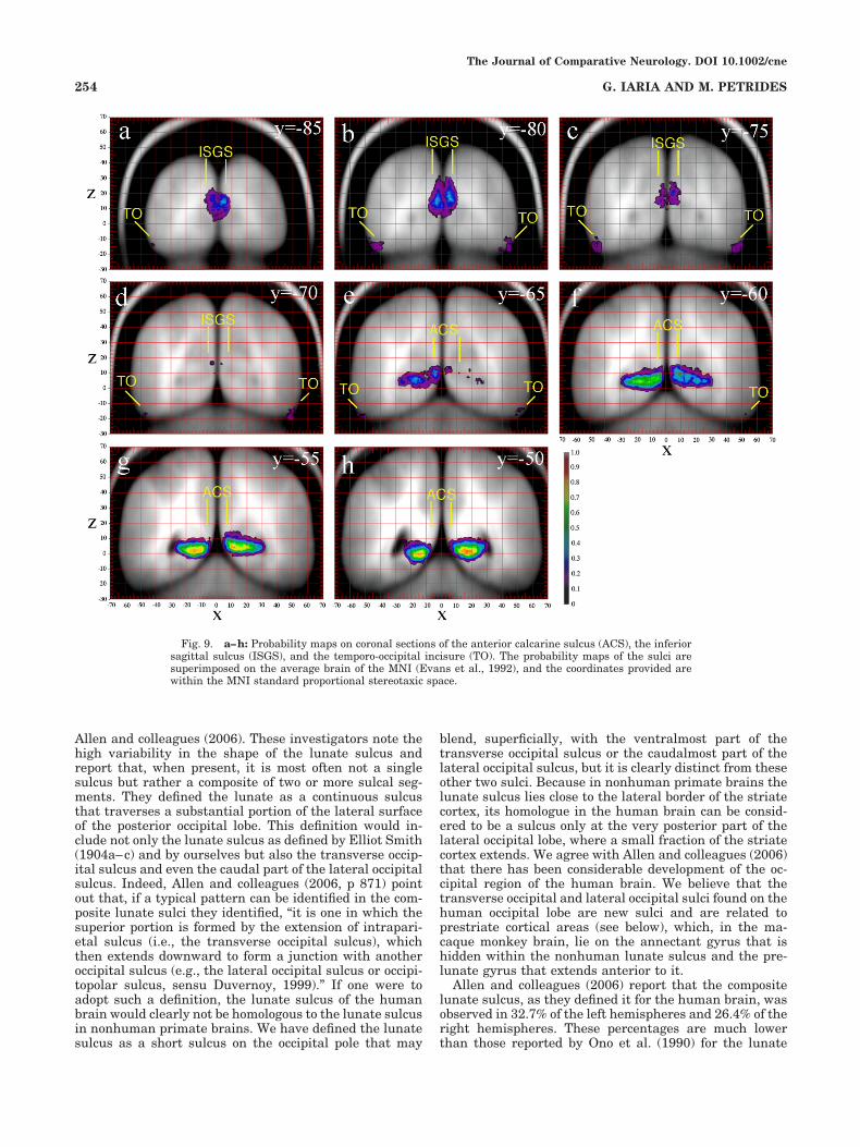
Fig. 9. a–h: Probability maps on coronal sections of the anterior calcarine sulcus (ACS), the inferiorsagittal sulcus (ISGS), and the temporo-occipital incisure (TO). The probability maps of the sulci aresuperimposed on the average brain of the MNI (Evans et al., 1992), and the coordinates provided arewithin the MNI standard proportional stereotaxic space.
The Journal of Comparative Neurology. DOI 10.1002/cne
254 G. IARIA AND M. PETRIDES

Fig. 10. Probability maps on coronal sections of the superior sag-ittal sulcus (SSGS) and the posterior collateral sulcus (PCS) aredisplayed in Aa–c. Ba–f displays the probability maps of the lingualsulcus (LiS) and the lunate sulcus (LuS). Finally, Ca–c displays the
probability maps of the transverse occipital sulcus (TOS). The prob-ability maps of the sulci are superimposed on the average brain of theMNI (Evans et al., 1992), and the coordinates provided are within theMNI standard proportional stereotaxic space.
The Journal of Comparative Neurology. DOI 10.1002/cne
255THE HUMAN OCCIPITAL LOBE

sulcus in cadaver brains: 64% in the left hemisphere and60% in the right hemisphere. In the MRIs that we exam-ined, the lunate sulcus could be unambiguously defined in50% of the left hemispheres and 45% of the right hemi-spheres. We must point out, however, that these percent-ages are underestimates of the true incidence of the lunatesulcus because of the difficulty in identifying this sulcus,which lies on the curvature of the occipital pole, in MRIvolumes.
The morphology of the medial surface of the occipitalregion is dominated by the calcarine sulcus (Fig. 1). Thedorsal part of the medial occipital region that lies abovethe calcarine sulcus and posterior to the parieto-occipitalfissure (POF) is known as the cuneus. The ventral part ofthe medial occipital region that lies below the calcarinesulcus as far as the occipital extension of the collateralfissure is the lingual gyrus. Within the cuneus, a numberof more or less horizontally running sulci, the inferior andsuperior sagittal sulci can be identified (Ono et al., 1990).Immediately above the calcarine sulcus, the ISGS of thecuneus can be identified and, farther dorsally, the SSGS of
the cuneus. Duvernoy (1999) has referred to these sulci asthe paracalcarine sulci, but this term should be avoidedbecause the term has been used to refer to sulci within oradjacent to the parieto-occipital fissure (see, e.g., ElliotSmith, 1904a). Within the lingual gyrus that lies betweenthe calcarine sulcus and the occipital extension of thecollateral sulcus, one can sometimes identify a sulcus thatis more or less well developed, the lingual (intralingual)sulcus. This sulcus can be clearly seen in Figure 1c andfaintly in Figure 1a,b,d.
Neurophysiological studies in nonhuman primates be-ginning in the 1960s have identified many cortical visualareas in the occipital lobe, i.e., many separate represen-tations of the visual field (Kaas, 2004; Zeki, 1978), andvarious schemes for naming these visual areas have beenproposed (Kaas, 2004; Zeki, 1978). In all these schemes,the striate cortex (Brodmann’s area 17) is referred to asthe first cortical visual area, i.e., V1. Although it wasknown since the beginning of the Twentieth Century thatthe striate cortex (i.e., Brodmann’s area 17 or V1), with itsdistinct and easily identified architectonic feature, the
TABLE 1. Volume (cc) of the Occipital Sulci for Individual Subjects and Mean Values According to Gender and Hemisphere1
POF TO BCS ACS RCS ISGS SSGS TOS LOS IOS PCS LiS LuS
L R L R L R L R L R L R L R L R L R L R L R L R L R
S1 (female) 27.5 18.4 2.5 3.4 14.1 9.8 5.6 4.5 4.1 1.2 0.4 0.7 1.3 1.0 7.0 3.2 1.5 2.9 1.1 0.3 4.4 2.9 1.4 1.8 0.0 0.0S2 (female) 15.4 18.7 2.2 1.3 5.3 8.5 3.6 5.0 0.0 1.5 2.9 1.3 0.6 1.8 7.2 5.0 1.6 1.6 1.5 0.2 0.5 1.3 1.8 1.6 1.0 0.0S3 (female) 25.3 29.0 1.3 0.6 10.0 11.0 4.1 6.6 2.2 1.7 3.0 3.0 1.6 1.2 12.9 12.6 7.1 17.0 2.1 7.6 7.7 5.3 2.1 2.6 0.0 0.0S4 (female) 26.6 30.5 2.2 1.0 11.2 10.7 7.0 7.9 6.7 4.4 1.5 2.2 0.8 2.1 19.6 15.7 4.8 19.8 4.8 1.0 12.5 8.6 1.9 0.8 2.0 0.0S5 (female) 16.6 16.7 0.9 0.8 6.4 12.9 8.0 7.7 0.7 1.1 5.3 1.5 1.3 4.1 3.8 2.8 2.4 1.2 0.9 0.9 3.1 1.9 2.3 1.8 1.0 1.3S6 (female) 28.8 27.2 1.0 0.7 15.8 13.4 9.8 9.4 7.3 4.0 1.5 0.8 0.4 2.0 5.3 6.8 4.1 5.9 1.0 0.4 1.2 1.4 0.9 1.1 5.6 2.6S7 (female) 19.2 18.0 0.8 0.8 10.9 7.0 4.1 6.2 5.6 0.0 0.4 2.4 1.0 0.8 8.3 6.8 8.2 5.2 1.8 2.1 9.3 3.9 1.8 0.6 3.6 0.7S8 (female) 22.0 18.6 0.6 0.7 8.9 8.1 9.9 6.5 1.5 2.6 0.7 2.1 6.4 0.9 12.5 12.4 5.4 3.8 0.3 0.3 6.6 2.4 0.7 0.8 0.0 0.0S9 (female) 24.6 27.5 1.3 0.8 10.1 8.1 8.0 7.8 2.6 2.4 0.7 1.0 1.7 1.2 6.3 10.4 4.6 3.6 1.1 1.1 3.9 1.4 0.6 2.3 0.0 0.0S10 (female) 42.9 38.9 1.1 0.8 14.5 11.2 12.1 10.6 2.1 2.5 1.9 2.5 1.6 0.6 19.5 5.0 8.1 1.8 1.1 0.5 4.5 4.8 4.9 1.3 2.5 0.0S11 (female) 11.8 13.3 0.9 0.7 7.1 2.8 3.3 3.0 0.8 2.6 0.8 0.4 1.3 0.3 3.3 4.3 1.8 1.5 0.7 0.9 1.3 1.6 0.8 0.4 1.4 1.0S12 (female) 38.3 39.4 1.0 1.9 17.3 17.8 6.1 5.4 10.2 4.0 1.8 1.8 1.8 2.7 5.6 6.4 10.2 6.3 1.2 1.0 5.1 5.1 3.0 1.0 0.0 0.0S13 (female) 20.1 18.7 1.0 0.9 11.8 10.1 6.9 5.4 1.4 0.3 0.6 1.5 2.5 1.2 4.4 3.4 2.0 0.5 0.2 0.5 2.0 1.1 0.8 0.4 0.0 0.0S14 (female) 37.4 20.0 2.1 0.8 18.6 12.4 9.5 3.5 4.6 4.0 4.5 2.6 1.8 1.2 11.4 5.3 11.4 9.8 2.2 1.9 1.6 6.9 0.2 1.0 3.9 4.0S15 (female) 19.2 22.0 1.2 1.2 7.6 9.5 3.1 5.4 1.7 1.2 0.7 1.6 1.0 1.0 6.6 3.8 2.0 3.3 1.4 0.2 0.4 0.9 1.1 0.6 0.0 0.0S16 (female) 38.7 35.1 0.8 0.8 0.0 41.0 6.9 12.1 3.5 2.9 1.9 4.3 4.1 1.2 10.6 10.6 10.8 10.1 1.0 1.5 1.7 1.3 0.5 0.3 0.0 0.0S17 (female) 13.3 8.7 0.5 1.0 8.3 11.3 4.0 3.7 2.2 1.3 1.1 1.8 0.4 1.6 6.3 4.2 0.6 1.1 0.5 0.2 0.2 0.4 0.3 0.1 0.0 0.0S18 (female) 24.3 25.8 1.6 1.4 22.2 15.0 6.5 4.4 1.4 1.0 8.3 1.8 1.6 2.2 4.8 5.0 36.3 12.1 9.3 1.7 7.5 5.5 2.7 3.6 1.2 0.5S19 (female) 22.5 19.9 0.7 0.4 10.0 12.3 10.5 7.5 3.2 0.6 0.7 5.4 1.5 2.8 4.8 2.6 1.1 0.8 6.6 2.9 5.9 5.1 3.2 7.1 2.9 0.8S20 (female) 27.0 30.2 0.8 0.8 8.8 7.3 7.0 8.3 2.2 2.4 0.9 0.8 3.5 1.4 2.6 0.8 9.5 2.7 2.9 4.1 2.4 1.5 1.4 0.3 0.0 0.0S21 (male) 11.2 17.9 1.6 4.7 7.2 9.0 3.3 4.0 2.4 2.0 1.0 1.3 1.1 1.4 7.3 8.0 8.0 9.6 3.1 3.8 10.2 4.6 0.6 1.7 4.4 0.0S22 (male) 19.8 13.3 1.0 1.6 8.9 12.1 5.5 9.0 8.0 3.8 3.5 1.9 2.5 0.4 10.4 12.0 13.1 6.3 4.5 3.8 3.8 3.0 1.9 0.6 0.6 1.7S23 (male) 14.9 18.1 2.3 2.7 10.1 8.2 3.7 4.1 2.9 0.0 3.8 1.5 2.4 1.2 5.3 6.3 3.6 6.4 4.2 2.4 3.9 6.3 2.5 3.4 0.0 0.0S24 (male) 27.2 16.6 0.0 0.8 8.9 10.6 4.8 4.8 2.2 2.0 0.6 1.1 0.3 2.1 2.0 2.6 4.3 7.9 1.1 1.9 4.3 2.4 3.6 2.5 0.0 1.7S25 (male) 25.6 20.8 0.9 1.3 11.3 5.4 7.3 4.5 2.7 9.6 1.1 0.8 0.5 0.7 12.5 7.0 14.2 10.9 4.1 2.6 7.7 1.0 1.6 3.4 0.0 0.0S26 (male) 29.2 21.6 0.8 2.4 8.0 8.9 7.3 5.6 2.7 0.7 1.3 0.9 6.2 0.4 3.2 4.9 6.6 5.6 1.7 0.7 5.2 5.8 5.6 3.1 0.8 5.0S27 (male) 22.2 8.7 0.5 0.6 10.8 10.9 5.8 5.5 1.6 1.9 1.5 0.8 6.2 1.3 1.7 1.9 1.1 3.8 0.8 1.2 0.9 1.6 2.5 3.5 0.0 0.0S28 (male) 23.3 27.2 0.5 0.7 4.8 4.7 15.1 0.0 1.6 0.7 0.0 2.0 3.6 0.4 14.3 8.7 6.9 1.4 4.2 2.0 4.6 4.0 0.7 0.9 1.4 0.0S29 (male) 14.8 24.7 0.6 0.7 11.7 10.5 6.6 6.4 3.4 1.0 2.8 2.6 2.7 1.7 11.4 6.8 6.8 5.2 9.3 1.7 2.0 5.5 2.0 2.5 3.7 3.8S30 (male) 13.0 20.9 1.3 0.8 8.3 11.7 18.3 0.0 1.3 0.0 2.8 1.3 0.6 1.5 11.6 7.0 5.5 4.0 0.6 0.7 3.1 3.5 1.7 0.3 0.6 0.2S31 (male) 39.5 31.1 0.9 0.9 15.4 13.3 10.4 10.3 1.4 5.0 0.8 2.1 1.5 1.4 3.9 7.3 6.6 7.9 3.1 1.0 10.6 5.7 1.6 0.6 13.0 7.6S32 (male) 32.8 37.0 1.2 1.3 16.3 13.2 8.6 12.7 5.1 4.5 2.3 2.4 1.1 0.6 12.2 8.6 6.4 2.6 3.8 3.3 4.6 1.2 1.9 1.2 1.0 2.1S33 (male) 24.7 17.3 1.2 0.7 12.2 6.5 9.2 6.7 2.8 1.2 0.4 0.3 3.9 3.0 9.1 7.9 3.2 1.6 0.8 0.8 7.2 1.6 5.2 3.0 0.0 0.0S34 (male) 41.2 41.2 2.0 0.7 19.1 17.1 7.6 7.0 3.1 4.4 0.8 2.5 2.6 2.4 21.4 9.1 10.3 0.7 1.7 0.9 5.6 5.8 3.6 0.7 5.5 0.9S35 (male) 25.8 15.5 1.1 0.7 14.1 13.5 11.3 12.7 2.8 2.6 4.0 2.2 1.5 0.9 6.4 2.2 6.3 6.0 1.4 1.8 1.4 1.3 1.0 0.8 0.0 1.7S36 (male) 20.4 14.1 0.6 1.3 15.1 9.0 8.8 8.3 2.0 1.2 3.1 1.6 2.1 1.8 5.0 5.2 4.3 3.0 1.0 0.4 2.0 1.2 0.7 0.4 0.0 0.9S37 (male) 30.9 25.5 1.7 0.5 8.7 11.5 10.1 14.6 4.4 3.1 2.9 2.1 4.9 3.1 7.0 6.7 7.0 4.2 0.9 2.7 2.7 3.7 1.3 1.1 0.0 0.0S38 (male) 40.5 42.5 1.2 1.0 19.4 20.7 18.3 10.9 2.0 1.6 3.2 1.2 1.1 0.7 8.4 7.3 7.6 1.0 1.3 0.9 7.0 3.2 0.9 1.0 0.0 0.0S39 (male) 14.3 40.4 1.0 0.3 4.3 7.9 5.9 9.0 0.9 0.6 0.2 0.9 0.9 0.6 5.8 2.4 6.4 2.4 0.7 2.4 3.2 3.7 3.9 0.6 3.4 0.6S40 (male) 9.3 11.4 0.3 0.9 6.7 3.5 6.8 6.1 2.8 1.3 3.0 2.0 5.4 2.1 5.4 2.7 0.7 0.6 0.2 0.5 1.1 0.7 1.7 0.8 0.0 0.0F Mean 25.2 23.6 1.2 1.1 11.1 12.1 6.6 6.5 3.6 2.3 1.7 1.8 1.7 1.5 8.8 7.0 5.1 5.6 1.4 1.2 3.9 3.0 1.5 1.1 2.6 1.9F SD 9.5 8.7 0.5 0.3 4.8 8.1 2.7 2.5 2.7 1.3 2.1 0.9 1.5 0.9 5.0 3.8 8.3 5.8 2.2 1.7 3.6 2.4 1.2 0.9 1.6 1.3M Mean 24.1 23.5 1.1 1.2 11.4 10.5 8.6 7.7 2.7 2.4 2.2 1.7 2.5 1.5 7.7 5.8 7.6 4.6 2.9 1.9 4.6 3.4 2.3 1.9 3.2 2.0M SD 9.0 9.7 0.5 1.0 4.7 4.0 4.0 3.1 1.5 2.1 1.8 1.0 1.8 0.9 4.7 2.9 7.1 3.3 2.6 1.1 2.7 1.9 1.4 1.6 3.5 2.1Mean 24.5 23.6 1.2 1.1 11.3 11.2 7.8 7.2 3.1 2.4 2.0 1.8 2.2 1.5 8.2 6.3 6.6 5.0 2.3 1.6 4.3 3.2 1.9 1.5 3.0 1.9SD 9.1 9.2 0.6 0.8 4.4 6.0 3.6 2.9 2.1 1.8 1.7 1.0 1.7 0.9 4.8 3.3 5.9 4.4 2.2 1.5 3.0 2.1 1.3 1.4 2.9 1.9MEAN (SD) 24.0 (9.1) 1.1 (0.7) 11.2 (5.4) 7.5 (3.4) 2.7 (2.0) 1.9 (1.4) 1.8 (1.4) 7.2 (4.2) 5.8 (5.3) 1.9 (1.9) 3.8 (2.6) 1.7 (1.4) 2.5 (2.1)
1F mean, mean for female subjects; F SD, standard deviation for female subjects; M mean, mean for male subjects; M SD, standard deviation for male subjects; mean, mean M �F by hemisphere; SD, standard deviation M � F by hemisphere; MEAN (SD), mean and standard deviation for total volumes; L, left hemisphere; R, right hemisphere; POF,parieto-occipital fissure; TO, temporo-occipital incisure; BCS, body of the calcarine sulcus; ACS, anterior calcarine sulcus; RCS, retrocalcarine sulcus; ISGS, inferior sagittal sulcus;SSGS, superior sagittal sulcus; TOS, transverse occipital sulcus; LOS, lateral occipital sulcus; IOS, inferior occipital sulcus; PCS, posterior collateral sulcus; LiS, lingual sulcus;LuS, lunate sulcus.
The Journal of Comparative Neurology. DOI 10.1002/cne
256 G. IARIA AND M. PETRIDES
stripe of Gennari, lies along both banks of the calcarinesulcus (see, e.g., Bolton, 1900; Elliot Smith, 1904a), thelocation of the many other functional visual areas thathave been identified in the monkey during the last 40years has only now began to be tentatively identified inthe human brain thanks to modern functional neuroimag-ing (see e.g., Tootell and Hadjikhani, 2001; Tootell et al.,1993, 1996; Walters et al., 2003; Watson et al., 1993; Zekiet al., 1991). The current identifications of homologues inthe human occipital lobe of the visual areas established inthe monkey should be treated with caution and should beregarded as tentative suggestions, because the methodol-ogies used in studies with monkeys and humans are verydifferent. In the monkey, the definition of a cortical visualarea is based on mapping of the visual field representationat the single-neuron level with microelectrode recording(Kaas, 2004; Zeki, 1978). In the human brain, attempts tomap these same areas are usually based on global signalchanges in fMRI, i.e., indirect measures of functional ac-tivity based on blood flow, and the precise location of anarea is likely to be affected by the details of blood vesseldistribution. Despite these limitations, several interestingstudies have already been carried out that have provideda tentative identification of many visual cortical areas inthe human occipital cortex that may be homologous toareas previously identified in monkeys. We shall commenthere on the possible relation of the sulci of the occipitalregion and these functionally defined visual cortical areas,although much work remains to be carried out before wecan be reasonably certain that these identifications arecorrect.
In the classic anatomical studies conducted at the be-ginning of the Twentieth Century, the relation of thecalcarine sulcus to the striate cortex was the subject ofintense investigation (see, e.g., Bolton, 1900; Elliot Smith,1904a). These studies showed that the anterior calcarinesulcus (Fig. 1), i.e., the extension of the calcarine sulcusanterior to the point of intersection with the parieto-occipital fissure, is the border between limbic cortex lyingon the isthmus and the striate cortex, which is found onlyon the ventral bank of the calcarine sulcus at this point.Caudal to the point of convergence of the parieto-occipitalfissure with the calcarine sulcus, i.e., on the body of thecalcarine sulcus (Fig. 1), the striate cortex extends on bothbanks of the sulcus (see, e.g., Antoni, 1914; Bolton, 1900;Brodmann, 1909; Economo and Koskinas, 1925; ElliotSmith, 1904a). The striate cortex extends outside the cal-carine sulcus farther caudally, i.e., close to the occipitalpole. According to Elliot Smith (1904a,b), in about 70% ofthe brains, the striate cortex extends around the occipitalpole to reach the lateral surface of the occipital region, andit is limited, more or less, by the lunate sulcus. Note that,unlike the brain of the macaque monkey, in which thelunate sulcus is always the border of area V1 with areaV2, the striate cortex in the lateral part of the humanoccipital pole may be close to the lunate sulcus (as in themacaque monkey), or it may stay behind it (see, e.g., ElliotSmith, 1904a). In other words, area V2, which in themonkey lies always within the posterior bank of the lu-nate sulcus, may spread in the human brain outside theposterior bank of the lunate sulcus onto the occipital pole.
The relation of the striate cortex (Brodmann’s area 17)to the calcarine sulcus was recently reexamined byAmunts and coauthors (2000). These investigators exam-ined 10 brains and in all cases area 17 (i.e., V1) was
located mainly in the depth of the calcarine sulcus, ex-tending onto the free surface in the most caudal sections.Area 18 (V2) surrounded area 17 dorsally and ventrally.Clarke and Miklossy (1990) studied the location of callosalconnections in the human occipital cortex because theyare known to provide an anatomical indication of thevertical meridian (i.e., the boundary) between V1 and V2.Area 17 (V1) occupied both banks of the calcarine sulcusand posteriorly (toward the occpital pole) extended ontoboth lips of the calcarine sulcus. The boundary of area 17(V1) and area 18 (V2) extended in an anteroposteriordirection close to the superior saggital sulcus, dorsally,and the lingual sulcus, ventrally. Below the lingual sulcusand still on the lingual gyrus, the ventral part of area V3(also known as VP) could be established on the basis ofcallosal connections (Clarke and Miklossy, 1990).
Functional neuroimaging studies have also identifiedthe primary visual cortical area along the calcarine sulcus(V1) of the human brain (see, e.g., DeYoe et al., 1996;Hadjikhani et al., 1998; Sereno et al., 1995; Tootell et al.,1997). These studies have shown that, proceeding dorsallyin the cuneus, strips of cortex running in an anteroposte-rior direction along the calcarine sulcus can be identifiedas the dorsal part of V2, V3, and V3A (accessory V3; DeYoeet al., 1996; Hadjikhani et al., 1998; Sereno et al., 1995;Tootell et al., 1997). All these areas extend beyond thecuneus onto the superior-lateral surface of the occipitallobe. The most dorsal area, V3A, runs along the transverseoccipital sulcus (Fig. 2) on the superior-lateral surface ofthe occipital lobe (Tootell et al., 1997). Recently, Pitzalisand coauthors (2006) have shown that, in the dorsalmostpart of the parieto-occipital sulcus of the human brain, thecontralateral visual hemifield could be mapped anteriorand medial to areas V2, V3, and V3A. This newly mappedarea is thought to be the human homologue of macaquearea V6 (Galletti et al., 1996, 1999).
Below the calcarine sulcus, on the lingual gyrus, again aseries of anteroposterior strips of cortex has been linkedwith the ventral part of V2 and the ventral part of V3 (alsoknown as VP; DeYoe et al., 1996; Hadjikhani et al., 1998;Sereno et al., 1995; Tootell et al., 1997). More ventrally,close to the collateral sulcus and extending lateral to it onthe fusiform gyrus, a visual area related to the processingof color has been identified via functional neuroimaging(Lueck et al., 1989; Zeki et al., 1991). This color-relatedarea was originally interpreted as the homologue of ma-caque monkey area V4 (Zeki et al., 1991), but others haveargued that it is not area V4 but a separate area, whichwas named V8 (Hadjikhani et al., 1998). Some neuroim-aging studies have located the ventral part of area V4(V4v) immediately after ventral area V3 and medial to thecolor area, which was originally found on the fusiformgyrus (DeYoe et al., 1996; Hadjikhani et al., 1998; Serenoet al., 1995; Tootell et al., 1997). These studies suggestthat the most ventral part of the lingual gyrus may in factcontain the ventral part of area V4 (i.e., V4v), which isseparate from the color area found on the fusiform gyrus.
According to the coordinates provided by Tootell andHadjikhani (2001), dorsal area V4 (V4d) would lie at therostral part of the lateral occipital sulcus, extending dor-sally behind the anterior occipital sulcus. Recall that areaV3A (i.e., area V3 accessory), which in the monkey lies inthe dorsal prestriate region between areas V3 and V4(Van Essen and Zeki, 1978; Zeki, 1978b), has been iden-tified in the human brain in the dorsal prestriate region
The Journal of Comparative Neurology. DOI 10.1002/cne
257THE HUMAN OCCIPITAL LOBE
along the transverse occipital sulcus (Tootell et al., 1997),i.e., just posterior to the location of dorsal area V4 on thedorsal part of the lateral occipital sulcus (Tootell andHadjikhani, 2001). If these interpretations are correct,then the superior lateral occipital cortex that lies abovethe lateral occipital sulcus includes the dorsal parts ofareas V2, V3, and V4 and the complete contralateral rep-resentation of area V3A. This would make this region ofthe human occipital cortex comparable to the cortex thatis hidden in the lunate sulcus and extending onto theprelunate gyrus in the macaque monkey, where areas V2,V3, V3A, and V4 can be found. The ventral parts of areasV2, V3 (VP), and V4 would then occupy the lingual gyrus.
Anterior to the transverse occipital sulcus lies the an-terior occipital sulcus (Fig. 2). The anterior occipital sul-cus has also been referred to as the posterior ascendingbranch of the second temporal sulcus (Eberstaller, 1892;Economo and Koskinas, 1925) or the posterior ascendingbranch of the inferior temporal sulcus (Cunningham, 1982;Watson et al., 1993). The cortical region close to the pointof intersection of the anterior occipital sulcus and thelateral occipital sulcus has been shown, via functionalneuroimaging methods (Dumoulin et al., 2000; Watson etal., 1993; Zeki et al., 1991), to be the locus of the humanhomologue of the visual cortical motion area demon-strated in the rhesus monkey by Zeki (1974) and namedV5 and in the owl monkey by Allman and Kaas (1971) andnamed MT. In a recent study, Malikovic and coauthors(2006) have shown a distinct architectonic area in thisregion of the cortex that is located most often in the depthof the sulci, i.e., the posterior bank of the anterior occipitalsulcus and the ventral bank of the lateral occipital sulcus.These investigators consider this distinct architectonicarea as the putative human visual motion area, i.e., thehomologue of monkey V5/MT.
Although many details remain to be established regard-ing the number of visual areas in the human occipital lobeand their correspondence to comparable areas in the mon-key, as well their relation to the morphology of the humanoccipital region, the available evidence has produced apreliminary scheme that is in line with what is knownfrom the monkey. The qualitative and quantitative de-scription of the sulci of the occipital lobe of the humanbrain that is provided here may help in evaluating anyrelations that might exist between functional areas andthe sulcal and gyral morphology of the human brain.
ACKNOWLEDGMENTS
We thank Steve Robbins and Mallar Chakravarty forhelp with the construction of the probability maps andfigures and Emily Rubin-Ferreira for technical assistance.
LITERATURE CITED
Allen JS, Bruss J, Damasio H. 2006. Looking for the lunate sulcus: amagnetic resonance imaging study in modern humans. Anat Rec A288:867–876.
Allman JM, Kaas JH. 1971. A representation of the visual field in thecaudal third of the middle temporal gyrus of the owl monkey (Aotustrivirgatus). Brain Res 31:85–105.
Amunts K, Malikovic A, Mohlberg H, Schormann T, Zilles K. 2000. Brod-mann’s areas 17 and 18 brought into stereotaxic space—where and howvariable? Neuroimage 11:66–84.
Anderson SJ, Holliday IE, Singh KD, Harding GF. 1996. Localization and
functional analysis of human cortical area V5 using magnetoencepha-lography. Proc Biol Sci 263:423–431.
Antoni NRE. 1914. Ausbreitung und Flachenbeziehungen der Area striataim menschlichen Gehirn. Fol Neurobiol 8:265–279.
Barton JJ, Simpson T, Kiriakopoulos E, Stewart C, Crawley A, Guthrie B,Wood M, Mikulis D. 1996. Functional MRI of lateral occipitotemporalcortex during pursuit and motion perception. Ann Neurol 40:387–398.
Bense S, Janusch B, Schlindwein P, Bauermann T, Vucurevic G, Brandt T,Stoeter P, Dieterich M. 2006. Direction-dependent visual cortex acti-vation during horizontal optokinetic stimulation (fMRI study). HumBrain Map 27:296–305.
Bolton JS. 1900. The exact histological localization of the visual area of thehuman cerebral cortex. Philos Trans R Soc Lond B Biol Sci 193:165–222.
Brodmann K. 1909. Vergleichende Lokalizationslehre der Grosshirnrindein ihren Prinzipien dargestellt auf Grund des Zellenbaues. Leipzig:Barth.
Carpenter MB. 1996. Core text of neuroanatomy. New York: Williams andWilkins.
Chiavaras MM, LeGoualher G, Evans A, Petrides M. 2001. Three-dimensional probabilistic atlas of the human orbitofrontal sulci instandardized stereotaxic space. Neuroimage 13:479–496.
Clarke S, Miklossy J. 1990. Occipital cortex in man: organization of callosalconnection, related myelo- and cytoarchitecture, and putative bound-aries of functional visual areas. J Comp Neurol 298:188–214.
Collins DL, Neelin P, Peters TM, Evans AC. 1994. Automatic 3D intersub-ject registration of MR volumetric data in standardized Talairachspace. J Comp Assist Tomogr 18:192–205.
Cunningham DJ. 1892. Contribution to the surface anatomy of the cerebralhemispheres. Dublin: Academy House.
de Jong BM, Shipp S, Skidmore B, Frackowiak RS, Zeki S. 1994. Thecerebral activity related to the visual perception of forward motion indepth. Brain 117:1039–1054.
DeYoe EA, Carman GJ, Bandettini P, Glickman S, Wieser J, Cox R, MillerD, Neitz J. 1996. Mapping striate and extrastriate visual areas inhuman cerebral cortex. Proc Natl Acad Sci U S A 9:2382–2386.
Dougherty RF, Koch VM, Brewer AA, Fischer B, Modersitzki J, WandellBA. 2003. Visual field representations and locations of visual areasV1/2/3 in human visual cortex. J Vis 3:586–598.
Dumoulin SO, Bittar RG, Kabani NJ, Baker CL Jr, Le Goualher G, PikeGB, Evans AC. 2000. A new anatomical landmark for reliable identi-fication of human area V5/MT: a quantitative analysis of sulcal pat-terning. Cereb Cortex 10:454–463.
Dupont P, Orban GA, De Bruyn B, Verbruggen A, Mortelmans L. 1994.Many areas in the human brain respond to visual motion. J Neuro-physiol 72:1420–1424.
Duvernoy H. 1999. The human brain: surface, three-dimensional sectionalanatomy and MRI, 2nd ed. Wien: Springer-Verlag.
Eberstaller O. 1890. Das Stirnhirn. Ein Beitrag zur Anatomie der Ober-flache des Grosshirns. Wien: Urban and Schwarzenberg.
Economo C, Koskinas GN. 1925. Die Cytoarchitektonik der Hirnrinde deserwachsenen Menschen. Berlin: Springer.
Elliot Smith G. 1904a. Studies in the morphology of the human brain withspecial reference to that of the Egyptians. No. I: the occipital region.Rec Egypt Gov School Med 2:123–173.
Elliot Smith G. 1904b. The morphology of the occipital region of thecerebral hemisphere in man and the apes. Anat Anz 24:436–451.
Elliot Smith G. 1904c. The morphology of the retrocalcarine region of thecortex cerebri. Proc R Soc Lond B Biol Sci 73:59–65.
Evans AC, Collins DL, Milner B. 1992. An MRI-based stereotactic brainatlas from 300 young normal subjects. Proceedings of the 22nd AnnualSymposium of the Society for Neuroscience, Anaheim, CA, p 408.
Felleman DJ, Van Essen DC. 1991. Distributed hierarchical processing inthe primate cerebral cortex. Cereb Cortex 1:1–47.
Galletti C, Fattori P, Battaglini PP, Shipp S, Zeki S. 1996. Functionaldemarcation of a border between areas V6 and V6A in the superiorparietal gyrus of the macaque monkey. Eur J Neurosci 8:30–52.
Galletti C, Fattori P, Gamberini M, Kutz DF. 1999. The cortical visual areaV6: Brain location and visual topography. Eur J Neurosci 11:575–582.
Germann J, Robbins S, Halsband U, Petrides M. 2005. Precentral sulcalcomplex of the human brain: morphology and statistical probabilitymaps. J Comp Neurol 493:334–356.
Hadjikhani N, Liu AK, Dale AM, Cavanagh P, Tootell RBH. 1998. Retino-
The Journal of Comparative Neurology. DOI 10.1002/cne
258 G. IARIA AND M. PETRIDES

topy and color sensitivity in human visual cortical area V8. Nat Neu-rosci 1:235–241.
Hasnain MK, Fox PT, Woldorff MG. 1998. Intersubject variability of func-tional areas in the human visual cortex. Hum Brain Map 6:301–315.
Itoh K, Fujii Y, Kwee IL, Nakada T. 2005. MT�/V5 activation withoutconscious motion perception: a high-field fMRI study. Magn Reson MedSci 4:69–74.
Kaas JH. 2004. Early visual areas: V1, V2, V3, DM, DL, and MT. In: KaasJH, Collins CE, editors. The primate visual system. New York: CRCPress. p 139–159.
Kollokian V. 1996. Performance analysis of automatic techniques for tissueclassification in magnetic resonance images of the human brain. MScThesis, Computer Science, Concordia University, Montreal, Canada.
Lueck CJ, Zeki S, Friston KJ, Deiber MP, Cope P, Cunningham VJ,Lammertsma AA, Kennard C, Frackowiack RS. 1989. The colour centrein the cerebral cortex of man. Nature 340:386–389.
MacDonald D. 1996. Program for display and segmentation of surfaces andvolumes. McConnell Brain Imaging Center, Montreal NeurologicalInstitute, Montreal, Quebec. Software available from http://www.bic.mni.mcgill.ca.
Malikovic A, Amunts K, Schleicher A, Mohlberg H, Eickhoff SB, Wilms M,Palomero-Gallagher N, Armstrong E, Zilles K. 2006. Cytoarchitectonicanalysis of the human extrastriate cortex in the region of V5/MT�: aprobabilistic, stereotaxic map of area hOc5. Cereb Cortex (April 7E-pub ahead of print).
Mazziotta JC, Toga AW, Evans A, Fox PT, Lancaster J. 1995a. A probabi-listic atlas of the human brain: theory and rationale for its develop-ment. Neuroimage 2:89–101.
Mazziotta JC, Toga AW, Evans A, Fox PT, Lancaster JL. 1995b. Digitalbrain atlases. Trend Neurosci 18:210–211.
Ono M, Kubik S, Abernathey CD. 1990. Atlas of the cerebral sulci. Stutt-gart: Thieme.
Nolte J. 2002. The human brain: an introduction to its functional anatomy.New York: Mosby.
Paus T, Tomaiuolo F, Otaky N, MacDonald D, Petrides M, Atlas J, MorrisR., Evans A. 1996. Human cingulate and paracingulate sulci: pattern,variability, asymmetry, and probabilistic map. Cereb Cortex 6:207–214.
Penhune VB, Zatorre RJ, MacDonald JD, Evans AC. 1996. Interhemi-spheric anatomical differences in human primary auditory cortex:probabilistic mapping and volume measurement from magnetic reso-nance imaging. Cereb Cortex 6:661–672.
Pitzalis S, Galletti C, Huang RS, Patria F, Committeri G, Galati G, FattoriP, Sereno MI. 2006. Wide-field retinotopy defines human cortical visualarea V6. J Neurosci 26:7962–7973.
Retzius G. 1896. Das Menschenhirn. Stockholm: G. Norstedt and Soener.Sack AT, Kohler A, Linden DE, Goebel R, Muckli L. 2006. The temporal
characteristics of motion processing in hMT/V5�: combining fMRI andneuronavigated TMS. Neuroimage 29:1326–1335.
Sereno MI, Tootell RBH. 2005. From monkeys to humans: what do we nowknow about brain homologies? Curr Opin Neurobiol 15:135–144.
Sereno MI, Dale AM, Reppas JB, Kwong KK, Belliveau JW, Brady TJ,Rosen BR, Tootell RB. 1995. Borders of multiple visual areas in hu-
mans revealed by functional magnetic resonance imaging. Science 268:889–893.
Shellshear JL. 1926. The occipital lobe in the brain of the Chinese withspecial reference to the sulcus lunatus. J Anat 61:1–13.
Shipp S, Watson JDG, Frackowiak RSV, Zeki S. 1995. Retinotopic maps inhuman prestriate visual cortex: the demarcation of areas V2 and V3.Neuroimage 2:125–132.
Shulman GL, Schwarz J, Miezin FM, Petersen SE. 1998. Effect of motioncontrast on human cortical responses to moving stimuli. J Neuro-physiol 79:2794–2803.
Stiers P, Peeters R, Lagae L, Van Hecke P, Sunaert S. 2006. Mappingmultiple visual areas in the human brain with a short fMRI sequence.Neuroimage 29:74–89.
Talairach J, Tournoux P. 1988. Co-planar stereotaxic atlas of the humanbrain. New York: Thieme.
Tomaiuolo F, MacDonald JD, Caramanos Z, Posner G, Chiavaras M, EvansAC, Petrides M. 1999. Morphology, morphometry and probability map-ping of the pars opercularis of the inferior frontal gyrus: an in vivo MRIanalysis. Eur J Neurosci 11:3033–3046.
Tootell RBH, Hadjikhani N. 2001. Where is “dorsal V4” in human visualcortex? Retinotopic topographic and functional evidence. Cerebral Cor-tex 11:298–311.
Tootell RBH, Dale AM, Mendola JD, Reppas JB, Sereno MI. 1996. fMRIanalysis of human visual cortical area V3A. Neuroimage 3:S358.
Tootell RBH, Mendola JD, Hadjikhani NK, Ledden PJ, Liu AK, Reppas JB,Sereno MI, Dale AM. 1997. Functional analysis of V3A and relatedareas in human visual cortex. J Neurosci 17:7060–7078.
Vallines I, Greenlee MW. 2006. Saccadic suppression of retinotopicallylocalized blood oxygen level-dependent responses in human primaryvisual area V1. J Neurosci 26:5965–5969.
Van Essen DC, Zeki SM. 1978. The topographic organization of rhesusmonkey prestriate cortex. J Physiol 277:193–226.
Walters NB, Egan GF, Kril JJ, Kean M, Waley P, Jenkinson M, WatsonJD. 2003. In vivo identification of human cortical areas using high-resolution MRI: an approach to cerebral structure–functin correlation.Prot Natl Acad Sci U S A 100:2981–2986.
Watson JD, Myers R, Frackowiak RS, Hajnal JV, Woods RP, Mazziotta JC,Shipp S, Zeki S. 1993. Area V5 of the human brain: evidence from acombined study using positron emission tomography and magneticresonance imaging. Cereb Cortex 3:79–94.
Wernicke C. 1876. Das Urwindungsystem des menschlichen Gehirns. ArchPychiatr 6:298–326.
Zeki SM. 1969. Representation of central visual fields in prestriate cortexof the monkey. Brain Res 34:19–35.
Zeki S. 1974. Functional organization of a visual area in the posterior bankof the superior temporal sulcus of the rhesus monkey. J Physiol 236:549–573.
Zeki SM. 1978a. Functional specialization of the visual cortex of the rhesusmonkey. Nature 274:423–428.
Zeki SM. 1978b. The third visual complex of rhesus monkey prestriatecortex. J Physiol 277:245–272.
Zeki S, Watson JDG, Lueck CJ, Friston KJ, Kennard C, Frackowiak RSJ.1991. A direct demonstration of functional specialization in humanvisual cortex. J Neurosci 11:641–649.
The Journal of Comparative Neurology. DOI 10.1002/cne
259THE HUMAN OCCIPITAL LOBE