Obstructive Sleep Apnea Treatment - UHCprovider.com Home · Obstructive Sleep Apnea Treatment Page...
25
Obstructive Sleep Apnea Treatment Page 1 of 25 UnitedHealthcare West Medical Management Guideline Effective 04/01/2019 Proprietary Information of UnitedHealthcare. Copyright 2019 United HealthCare Services, Inc. OBSTRUCTIVE SLEEP APNEA TREATMENT Guideline Number: MMG091.S Effective Date: April 1, 2019 Table of Contents Page COVERAGE RATIONALE ............................................. 1 DEFINITIONS .......................................................... 2 APPLICABLE CODES ................................................. 2 DESCRIPTION OF SERVICES ...................................... 3 CLINICAL EVIDENCE ................................................. 4 U.S. FOOD AND DRUG ADMINISTRATION ................... 19 REFERENCES .......................................................... 20 GUIDELINE HISTORY/REVISION INFORMATION .......... 24 INSTRUCTIONS FOR USE ......................................... 24 COVERAGE RATIONALE Nonsurgical Treatment Removable oral appliances are proven and medically necessary for treating Obstructive Sleep Apnea (OSA) as documented by a sleep study (e.g., polysomnography or Home Sleep Apnea Testing). Refer to the Medical Management Guideline (MMG) titled Attended Polysomnography for Evaluation of Sleep Disorders for further information. For many members, oral appliance therapy (OAT) may be an effective alternative to failed continuous positive airway pressure (CPAP) therapy. Documentation of the following is required: A member presenting with symptoms of OSA be seen in a face-to-face evaluation with a qualified physician (MD or DO) trained in sleep medicine prior to beginning treatment for OAT (AASM and AADSM, December 2012). A treating physician (MD or DO) must diagnose OSA and recommend course of treatment. If the member refuses CPAP therapy, documentation of the refusal from the patient’s treating physician (MD or DO) must be supplied. For medical necessity clinical coverage criteria, see MCG™ Care Guidelines, 23 rd edition, 2019, Oral Appliances (Mandibular Advancement Devices), A-0341 (ACG). The following are unproven and not medically necessary due to insufficient evidence of efficacy: Removable oral appliances for treating central sleep Apnea. Nasal dilator devices for treating Obstructive Sleep Apnea (OSA) Surgical Treatment The following surgical procedures are proven and medically necessary for treating Obstructive Sleep Apnea as documented by polysomnography. For medical necessity clinical coverage criteria, see MCG™ Care Guidelines, 23 rd edition, 2019. Uvulopalatopharyngoplasty (UPPP) - Uvulopalatopharyngoplasty (UPPP), A-0245 (ACG). Maxillomandibular Advancement Surgery (MMA) - Maxillomandibular Osteotomy and Advancement, A-0248 (ACG). Also see the Medical Management Guideline titled Orthognathic (Jaw) Surgery. Multilevel Procedures Whether Done in a Single Surgery or Phased Multiple Surgeries - Mandibular Osteotomy, A- 0247 (ACG). The following surgical procedures are unproven and not medically necessary for treating Obstructive Sleep Apnea due to insufficient evidence of efficacy: Laser-assisted uvulopalatoplasty (LAUP) Palatal implants Lingual suspension-also referred to as tongue stabilization, tongue stitch or tongue fixation Transoral robotic surgery (TORS) Related Medical Management Guidelines Attended Polysomnography for Evaluation of Sleep Disorders Orthognathic (Jaw) Surgery UnitedHealthcare ® West Medical Management Guideline UnitedHealthcare of California (HMO) UnitedHealthcare Benefits Plan of California (EPO/POS) UnitedHealthcare of Oklahoma, Inc. UnitedHealthcare of Oregon, Inc. UnitedHealthcare Benefits of Texas, Inc. UnitedHealthcare of Washington, Inc. Instructions for Use
-
Upload
truongtruc -
Category
Documents
-
view
214 -
download
0
Transcript of Obstructive Sleep Apnea Treatment - UHCprovider.com Home · Obstructive Sleep Apnea Treatment Page...
Obstructive Sleep Apnea Treatment Page 1 of 25 UnitedHealthcare West Medical Management Guideline Effective 04/01/2019
Proprietary Information of UnitedHealthcare. Copyright 2019 United HealthCare Services, Inc.
OBSTRUCTIVE SLEEP APNEA TREATMENT Guideline Number: MMG091.S Effective Date: April 1, 2019
Table of Contents Page COVERAGE RATIONALE ............................................. 1 DEFINITIONS .......................................................... 2 APPLICABLE CODES ................................................. 2 DESCRIPTION OF SERVICES ...................................... 3 CLINICAL EVIDENCE ................................................. 4 U.S. FOOD AND DRUG ADMINISTRATION ................... 19 REFERENCES .......................................................... 20 GUIDELINE HISTORY/REVISION INFORMATION .......... 24 INSTRUCTIONS FOR USE ......................................... 24 COVERAGE RATIONALE
Nonsurgical Treatment
Removable oral appliances are proven and medically necessary for treating Obstructive Sleep Apnea (OSA) as documented by a sleep study (e.g., polysomnography or Home Sleep Apnea Testing). Refer to the Medical Management Guideline (MMG) titled Attended Polysomnography for Evaluation of Sleep Disorders for further information. For many members, oral appliance therapy (OAT) may be an effective alternative to failed continuous positive airway pressure (CPAP) therapy. Documentation of the following is required:
A member presenting with symptoms of OSA be seen in a face-to-face evaluation with a qualified physician (MD or DO) trained in sleep medicine prior to beginning treatment for OAT (AASM and AADSM, December 2012).
A treating physician (MD or DO) must diagnose OSA and recommend course of treatment. If the member refuses CPAP therapy, documentation of the refusal from the patient’s treating physician (MD or
DO) must be supplied. For medical necessity clinical coverage criteria, see MCG™ Care Guidelines, 23rd edition, 2019, Oral Appliances
(Mandibular Advancement Devices), A-0341 (ACG). The following are unproven and not medically necessary due to insufficient evidence of efficacy: Removable oral appliances for treating central sleep Apnea. Nasal dilator devices for treating Obstructive Sleep Apnea (OSA) Surgical Treatment
The following surgical procedures are proven and medically necessary for treating Obstructive Sleep
Apnea as documented by polysomnography. For medical necessity clinical coverage criteria, see MCG™ Care Guidelines, 23rd edition, 2019. Uvulopalatopharyngoplasty (UPPP) - Uvulopalatopharyngoplasty (UPPP), A-0245 (ACG). Maxillomandibular Advancement Surgery (MMA) - Maxillomandibular Osteotomy and Advancement, A-0248 (ACG).
Also see the Medical Management Guideline titled Orthognathic (Jaw) Surgery. Multilevel Procedures Whether Done in a Single Surgery or Phased Multiple Surgeries - Mandibular Osteotomy, A-
0247 (ACG).
The following surgical procedures are unproven and not medically necessary for treating Obstructive Sleep Apnea due to insufficient evidence of efficacy: Laser-assisted uvulopalatoplasty (LAUP) Palatal implants Lingual suspension-also referred to as tongue stabilization, tongue stitch or tongue fixation Transoral robotic surgery (TORS)
Related Medical Management Guidelines
Attended Polysomnography for Evaluation of Sleep Disorders
Orthognathic (Jaw) Surgery
UnitedHealthcare® West Medical Management Guideline
UnitedHealthcare of California (HMO) UnitedHealthcare Benefits Plan of California (EPO/POS)
UnitedHealthcare of Oklahoma, Inc. UnitedHealthcare of Oregon, Inc.
UnitedHealthcare Benefits of Texas, Inc. UnitedHealthcare of Washington, Inc.
Instructions for Use
Obstructive Sleep Apnea Treatment Page 2 of 25 UnitedHealthcare West Medical Management Guideline Effective 04/01/2019
Proprietary Information of UnitedHealthcare. Copyright 2019 United HealthCare Services, Inc.
Implantable hypoglossal nerve stimulation Radiofrequency ablation of the soft palate and/or tongue base DEFINITIONS
Apnea: The cessation of airflow (≥90% decrease in airflow compared to baseline) lasting at least 10 seconds. Apneas are classified as obstructive, mixed, or central based on the pattern of respiratory effort. An obstructive Apnea is associated with continued or increased inspiratory effort throughout the entire period of absent airflow. A central Apnea is associated with absent inspiratory effort throughout the entire period of absent airflow. Mixed Apneas are associated with absent inspiratory effort in the initial portion of the event, followed by resumption of inspiratory effort in the second portion of the event (AASM Scoring Manual, 2017).
Apnea Hypopnea Index (AHI): The number of Apneas plus the number of Hypopneas, times 60, divided by total sleep time (AASM Scoring Manual, 2017). Home Sleep Apnea Testing: The use of unattended diagnostic studies to assess for OSA without the determination of sleep stage. The term specifies the condition being assessed (i.e., sleep Apnea) by current technology without
implying that “sleep” quality, staging or time are determined. Not all such studies are performed at home; however, that is where the vast majority of patients undergo these tests (AASM Style Guide, 2015). Also referred to as out-of-
center sleep testing or portable monitoring. Hypopnea: An abnormal respiratory event lasting at least 10 seconds associated with at least a 30% reduction in airflow and with at least a 3% decrease in oxygen desaturation from pre-event baseline or the event is associated with an arousal (AASM Scoring Manual, 2017).
Obstructive Sleep Apnea: The American Academy of Sleep Medicine (AASM) defines Obstructive Sleep Apnea as a sleep related breathing disorder that involves a decrease or complete halt in airflow despite an ongoing effort to breathe. OSA severity is defined as: mild for AHI or RDI ≥ 5 and < 15
moderate for AHI or RDI ≥ 15 and ≤ 30 severe for AHI or RDI > 30/hr Physician - any Doctor of Medicine or Doctor of Osteopathy who is properly licensed and qualified by law.
Respiratory Disturbance Index (RDI): The number of Apneas plus the number of Hypopneas plus the number of
Respiratory Effort-Related Arousals, times 60, divided by total sleep time (AASM Scoring Manual, 2017). Respiratory Effort-Related Arousal (RERA): A sequence of breaths characterized by increasing respiratory effort, inspiratory flattening in the nasal pressure or PAP device flow channel or an increase in end-tidal PCO2 (children) leading to an arousal from sleep. Respiratory effort-related arousals do not meet criteria for Hypopnea and have a minimum duration of at least 10 seconds in adults or the duration of at least two breaths in children (AASM Scoring Manual, 2017).
Respiratory Event Index (REI): Total number of respiratory events scored, times 60, divided by monitoring time. The REI is used for Home Sleep Apnea Testing (AASM Scoring Manual, 2017). APPLICABLE CODES
The following list(s) of procedure and/or diagnosis codes is provided for reference purposes only and may not be all
inclusive. Listing of a code in this guideline does not imply that the service described by the code is a covered or non-covered health service. Benefit coverage for health services is determined by the member specific benefit plan document and applicable laws that may require coverage for a specific service. The inclusion of a code does not imply any right to reimbursement or guarantee claim payment. Other Policies and Guidelines may apply.
CPT Code Description
0466T Insertion of chest wall respiratory sensor electrode or electrode array, including
connection to pulse generator (List separately in addition to code for primary procedure)
0467T Revision or replacement of chest wall respiratory sensor electrode or electrode array, including connection to existing pulse generator
Obstructive Sleep Apnea Treatment Page 3 of 25 UnitedHealthcare West Medical Management Guideline Effective 04/01/2019
Proprietary Information of UnitedHealthcare. Copyright 2019 United HealthCare Services, Inc.
CPT Code Description
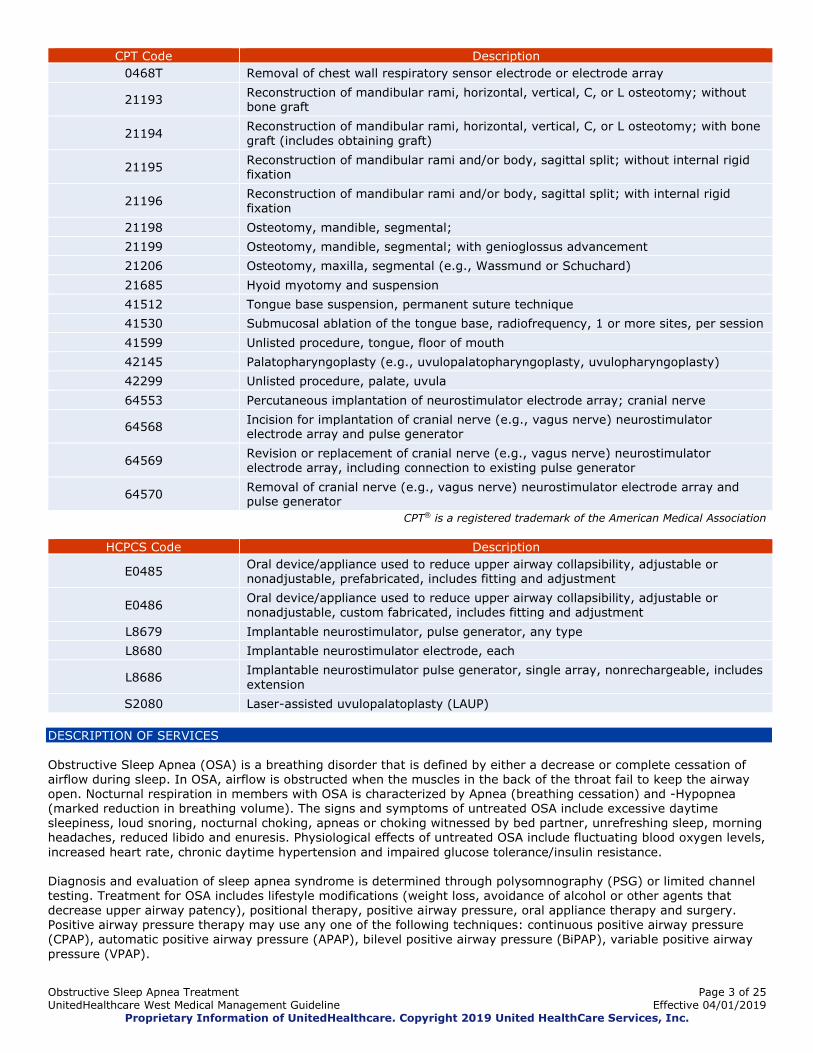
0468T Removal of chest wall respiratory sensor electrode or electrode array
21193 Reconstruction of mandibular rami, horizontal, vertical, C, or L osteotomy; without bone graft
21194 Reconstruction of mandibular rami, horizontal, vertical, C, or L osteotomy; with bone graft (includes obtaining graft)
21195 Reconstruction of mandibular rami and/or body, sagittal split; without internal rigid fixation
21196 Reconstruction of mandibular rami and/or body, sagittal split; with internal rigid fixation
21198 Osteotomy, mandible, segmental;
21199 Osteotomy, mandible, segmental; with genioglossus advancement
21206 Osteotomy, maxilla, segmental (e.g., Wassmund or Schuchard)
21685 Hyoid myotomy and suspension
41512 Tongue base suspension, permanent suture technique
41530 Submucosal ablation of the tongue base, radiofrequency, 1 or more sites, per session
41599 Unlisted procedure, tongue, floor of mouth
42145 Palatopharyngoplasty (e.g., uvulopalatopharyngoplasty, uvulopharyngoplasty)
42299 Unlisted procedure, palate, uvula
64553 Percutaneous implantation of neurostimulator electrode array; cranial nerve
64568 Incision for implantation of cranial nerve (e.g., vagus nerve) neurostimulator electrode array and pulse generator
64569 Revision or replacement of cranial nerve (e.g., vagus nerve) neurostimulator electrode array, including connection to existing pulse generator
64570 Removal of cranial nerve (e.g., vagus nerve) neurostimulator electrode array and pulse generator
CPT® is a registered trademark of the American Medical Association
HCPCS Code Description
E0485 Oral device/appliance used to reduce upper airway collapsibility, adjustable or nonadjustable, prefabricated, includes fitting and adjustment
E0486 Oral device/appliance used to reduce upper airway collapsibility, adjustable or nonadjustable, custom fabricated, includes fitting and adjustment
L8679 Implantable neurostimulator, pulse generator, any type
L8680 Implantable neurostimulator electrode, each
L8686 Implantable neurostimulator pulse generator, single array, nonrechargeable, includes extension
S2080 Laser-assisted uvulopalatoplasty (LAUP)
DESCRIPTION OF SERVICES
Obstructive Sleep Apnea (OSA) is a breathing disorder that is defined by either a decrease or complete cessation of
airflow during sleep. In OSA, airflow is obstructed when the muscles in the back of the throat fail to keep the airway open. Nocturnal respiration in members with OSA is characterized by Apnea (breathing cessation) and -Hypopnea (marked reduction in breathing volume). The signs and symptoms of untreated OSA include excessive daytime sleepiness, loud snoring, nocturnal choking, apneas or choking witnessed by bed partner, unrefreshing sleep, morning headaches, reduced libido and enuresis. Physiological effects of untreated OSA include fluctuating blood oxygen levels,
increased heart rate, chronic daytime hypertension and impaired glucose tolerance/insulin resistance. Diagnosis and evaluation of sleep apnea syndrome is determined through polysomnography (PSG) or limited channel testing. Treatment for OSA includes lifestyle modifications (weight loss, avoidance of alcohol or other agents that decrease upper airway patency), positional therapy, positive airway pressure, oral appliance therapy and surgery. Positive airway pressure therapy may use any one of the following techniques: continuous positive airway pressure (CPAP), automatic positive airway pressure (APAP), bilevel positive airway pressure (BiPAP), variable positive airway
pressure (VPAP).
Obstructive Sleep Apnea Treatment Page 4 of 25 UnitedHealthcare West Medical Management Guideline Effective 04/01/2019
Proprietary Information of UnitedHealthcare. Copyright 2019 United HealthCare Services, Inc.
Non-surgical oral appliances, worn during sleep, are an effective treatment option for snoring and OSA. These devices work by keeping the airway open in one of three ways: by pushing the lower jaw forward (a mandibular advancement device or MAD), by preventing the tongue from falling back over the airway (a tongue-retaining device) or by combining both mechanisms.
It is the position of the American Academy of Sleep Medicine (AASM) that dentists and physicians work collaboratively managing sleep-related breathing disorders with oral appliance therapy by conducting follow-up sleep testing to improve or confirm treatment efficacy along with periodic follow up visits (Ramar, 2015). A nasal dilator is a removable appliance that is placed just inside the nostril and is secured in place with hypoallergenic adhesive. Using small valves, the device increases pressure inside the nose by creating resistance
during exhalation to maintain an open airway during sleep (Theravent website). There are a variety of surgical options used to treat OSA. The intention of surgery is to create a more open airway so obstructions are less likely to occur. Implantable hypoglossal nerve stimulation systems are being evaluated as a way to relieve upper airway obstruction.
There are two hypoglossal nerve stimulation systems that are being evaluated: the Inspire® Upper Airway Stimulation device (Inspire Medical) and the aura6000™ Sleep Therapy System (ImThera Medical). A third device, the HGNS
System (Apnex Medical), has been discontinued and the manufacturer is no longer in business. The Inspire device is intended to treat moderate to severe OSA and is designed for use in member who are unable or unwilling to use a CPAP device. Inspire’s construction and implantation are comparable to those of a pacemaker: a surgeon implants the device containing a neurostimulator subcutaneously in the member’s chest with one lead attached to the member’s hypoglossal nerve (cranial nerve XII) at the base of the tongue and one lead implanted in the member’s chest. The
lead in the chest consists of a pressure sensor that detects breathing. Information about respiration rate is relayed to the device, which stimulates the hypoglossal nerve in the tongue. When stimulated, the tongue moves forward, thus opening the airway. The member can operate the device by remote control, which the member activates before going to sleep. The device turns on after 20 minutes to minimize disrupting the member’s sleep onset; the device turns off via remote when the member wakes. CLINICAL EVIDENCE
Nonsurgical Treatment
Oral Appliances
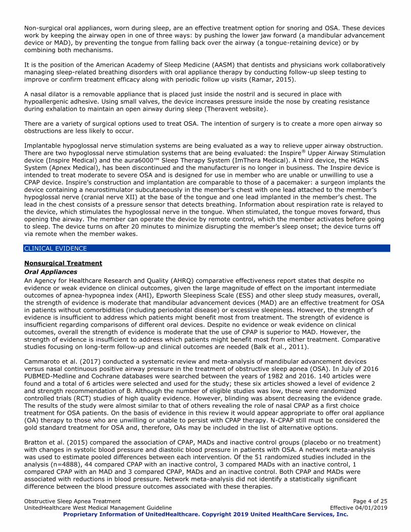
An Agency for Healthcare Research and Quality (AHRQ) comparative effectiveness report states that despite no
evidence or weak evidence on clinical outcomes, given the large magnitude of effect on the important intermediate outcomes of apnea-hypopnea index (AHI), Epworth Sleepiness Scale (ESS) and other sleep study measures, overall, the strength of evidence is moderate that mandibular advancement devices (MAD) are an effective treatment for OSA in patients without comorbidities (including periodontal disease) or excessive sleepiness. However, the strength of evidence is insufficient to address which patients might benefit most from treatment. The strength of evidence is insufficient regarding comparisons of different oral devices. Despite no evidence or weak evidence on clinical outcomes, overall the strength of evidence is moderate that the use of CPAP is superior to MAD. However, the
strength of evidence is insufficient to address which patients might benefit most from either treatment. Comparative studies focusing on long-term follow-up and clinical outcomes are needed (Balk et al., 2011). Cammaroto et al. (2017) conducted a systematic review and meta-analysis of mandibular advancement devices versus nasal continuous positive airway pressure in the treatment of obstructive sleep apnea (OSA). In July of 2016 PUBMED-Medline and Cochrane databases were searched between the years of 1982 and 2016. 140 articles were
found and a total of 6 articles were selected and used for the study; these six articles showed a level of evidence 2 and strength recommendation of B. Although the number of eligible studies was low, these were randomized
controlled trials (RCT) studies of high quality evidence. However, blinding was absent decreasing the evidence grade. The results of the study were almost similar to that of others revealing the role of nasal CPAP as a first choice treatment for OSA patients. On the basis of evidence in this review it would appear appropriate to offer oral appliance (OA) therapy to those who are unwilling or unable to persist with CPAP therapy. N-CPAP still must be considered the gold standard treatment for OSA and, therefore, OAs may be included in the list of alternative options.
Bratton et al. (2015) compared the association of CPAP, MADs and inactive control groups (placebo or no treatment) with changes in systolic blood pressure and diastolic blood pressure in patients with OSA. A network meta-analysis was used to estimate pooled differences between each intervention. Of the 51 randomized studies included in the analysis (n=4888), 44 compared CPAP with an inactive control, 3 compared MADs with an inactive control, 1 compared CPAP with an MAD and 3 compared CPAP, MADs and an inactive control. Both CPAP and MADs were associated with reductions in blood pressure. Network meta-analysis did not identify a statistically significant
difference between the blood pressure outcomes associated with these therapies.
Obstructive Sleep Apnea Treatment Page 5 of 25 UnitedHealthcare West Medical Management Guideline Effective 04/01/2019
Proprietary Information of UnitedHealthcare. Copyright 2019 United HealthCare Services, Inc.
In a randomized crossover trial, Phillips et al. (2013) compared the effects of continuous positive airway pressure (CPAP) and mandibular advancement device (MAD) therapy on cardiovascular and neurobehavioral outcomes in patients with obstructive sleep apnea (OSA). A total of 126 patients with moderate to severe OSA were randomly
assigned to a treatment order, and 108 completed the trial with both devices. Health outcomes were similar after 1 month of treatment. CPAP was more efficacious than MAD in reducing AHI but compliance was higher with MAD. The 24-hour mean arterial pressure was not inferior on treatment with MAD compared with CPAP; however, overall, neither treatment improved blood pressure. Sleepiness, driving simulator performance and disease-specific quality of life improved on both treatments by similar amounts, although MAD was superior to CPAP for improving four general quality-of-life domains.
Holley et al. (2011) conducted a retrospective analysis evaluating the efficacy of an adjustable oral appliance (aOA) in comparison with continuous positive airway pressure (CPAP) for treating obstructive sleep apnea (OSA). A total of 497 patients were given an aOA. The aOA reduced the mean apnea-hypopnea index (AHI) to 8.4 ± 11.4, and 70.3%, 47.6% and 41.4% of patients with mild, moderate and severe disease achieved an AHI < 5, respectively. Patients using an aOA decreased their mean Epworth Sleepiness Score by 2.71 at follow-up. CPAP improved the AHI by -3.43 when compared with an aOA, but when adjusted for severity of disease, this difference only reached significance for
patients with severe disease (-5.88). However, 70.1% of all patients achieved an AHI < 5 using CPAP compared with 51.6% for the aOA. Baseline AHI was a significant predictor of achieving an AHI < 5, and age showed a trend toward
significance. In comparison with past reports, more patients in this study achieved an AHI < 5 using an aOA. The authors concluded that aOAs are comparable to CPAP for patients with mild disease; however, CPAP is superior for patients with moderate to severe disease. In a multicenter, randomized controlled trial (n=101), Lam et al. (2007) compared the effectiveness of three
commonly used non-surgical treatment modalities in patients with mild to moderate OSA. Treatment groups consisted of conservative measures (sleep hygiene) only, continuous positive airways pressure (CPAP) in addition to conservative measures or an oral appliance in addition to conservative measures. The severity of sleep-disordered breathing was decreased in the CPAP and oral appliance groups compared with the conservative measures group and the CPAP group was significantly better than the oral appliance group. Overall, CPAP produced the best improvement in terms of physiological, symptomatic and quality of life measures, while the oral appliance was slightly less effective.
A Cochrane review concluded that while CPAP appears to be more effective in improving sleep disordered breathing, there is increasing evidence suggesting that oral appliances (OA) improve subjective sleepiness and sleep disordered breathing compared with a control. Until there is more definitive evidence on the effectiveness of OA in relation to CPAP, with regard to symptoms and long-term complications, it would appear to be appropriate to recommend OA
therapy to patients with mild symptomatic OSA, and those patients who are unwilling or unable to tolerate CPAP therapy. OA should not be considered as first choice therapy for OSA where symptoms and sleep disruption are severe
(Lim et al., 2006; updated 2008). Ferguson et al. (2006) conducted an evidence-based systematic review regarding the use of oral appliances for treating OSA and concluded that overall, patients with mild to severe OSA have a 52% chance of being able to control their sleep apnea using an appliance. Success rates ranged between 14 and 61% among patients with severe OSA (AHI defined as greater than 30 in some studies and great than 40 in others). Better success rates were seen in patients with lower AHI. OAs are on the whole less effective than CPAP but may be better accepted by patients than
nasal CPAP in studies where subjects used both treatments. OAs are not recommended as a first line treatment in patients with severe OSA. However, these patients might consider an OA if they have failed CPAP or upper airway surgery, recognizing that the results of OA therapy in severe OSA are unpredictable. The literature now provides better evidence for the efficacy of OAs and indications for use. Tegelberg et al. (2003) compared two different degrees of mandibular advancement with an intraoral appliance in 74
male patients with mild to moderate OSA. Thirty-eight patients received a dental appliance with 50% advancement
and 36 patients received a dental appliance with 75% mandibular advancement. Somnography was performed pre-treatment and after one year of treatment. Fifty-five patients completed follow-up after one year of treatment. In the group of 50% advancement, normalization (an apnea index of <5 and apnea/hypopnea index <10) was observed in 79% of the group. In the group of 75% advancement, normalization was observed in 73% of the group. Less than 5% of the patients reported symptoms from the stomatognathic system; one-third of the patients reported headaches more than once a week. Headaches significantly decreased after one year of treatment.
Thirty-five patients diagnosed with OSA unable to tolerate or non-compliant with CPAP were studied by Prathibha et al. (2003). These patients underwent sleep studies, used intraoral appliances for three months and had a repeat sleep study performed while using the appliance. Thirty-one patients completed the study. Patients with a pre-study AHI <20 benefited from the appliance, while the authors concluded that those patients with a pre-study AHI >20 did not.
Obstructive Sleep Apnea Treatment Page 6 of 25 UnitedHealthcare West Medical Management Guideline Effective 04/01/2019
Proprietary Information of UnitedHealthcare. Copyright 2019 United HealthCare Services, Inc.
Walker-Engstrom et al. (2002) randomized 95 patients with confirmed OSA to treatment with a dental appliance or uvulopalatopharyngoplasty. Patients underwent sleep studies before treatment and 1 year and 4 years after treatment. Thirty-two patients in the dental appliance group and 40 patients in the UPPP group completed the 4-year follow up. Success was defined as a reduction in the apnea index of at least 50%. The dental appliance group had a success rate
of 81%; the UPPP group had a success rate of 53%. An apnea index of <5 or an apnea/hypopnea index <10 was observed in 63% of the dental-appliance group and 33% of the UPPP group. The compliance rate of the dental appliance group was 62%. Seventy-five percent of the UPPP group were satisfied with their results and required no further complementary treatment. Gotsopoulos et al. (2002) evaluated the effect of a mandibular advancement splint (MAS) on daytime sleepiness and a range of other symptoms in 73 patients (59 men, 14 women) with mild to severe OSA. OSA severity subgroups
revealed a predominance of moderate and severe OSA, with 41 patients (56%) and 21 patients (29%) in each subgroup, respectively. Using a randomized crossover design, patients received 4 weeks of treatment with MAS and a control device (inactive oral appliance). At the end of each treatment period, patients were reassessed by questionnaire, polysomnography, and multiple sleep latency tests. Participants experienced significantly improved mean sleep latency on the multiple sleep latency test and Epworth sleepiness scale score with the MAS compared with the control device. The proportion of patients with normal subjective sleepiness was significantly higher with the MAS
than with the control device (82 versus 62%), but this was not so for objective sleepiness (48 versus 34%). Other OSA symptoms were controlled in significantly more patients with the MAS than with the control device.
In a randomized, controlled crossover study, Mehta et al. (2001) evaluated the efficacy of a mandibular advancement splint (MAS) in 28 patients with mild to severe OSA. Patients underwent three polysomnographs with either a control oral plate, which did not advance the mandible, or a MAS. Complete response (CR) was defined as a resolution of symptoms and a reduction in apnea/hypopnea index (AHI) to <5/hour, and partial response (PR) as a ≥ 50%
reduction in AHI, but remaining ≥ 5/hour. Twenty-four patients (19 men, 5 women) completed the protocol. Treatment outcome was similar across all categories of OSA severity, with complete response being achieved in some subjects with moderate and severe OSA. Subjective improvements with the MAS were reported by the majority of patients (96%). There were significant improvements in AHI, oxygen saturation and arousal index with MAS, compared with the control. The control plate had no significant effect on AHI and oxygen saturation. CR (n = 9) or PR (n = 6) was achieved in 62.5% of patients. The MAS is an effective treatment in some patients with OSA, including those patients with moderate or severe OSA.
Nasal Dilators
In a systematic review and meta-analysis, Camacho et al. (2016) evaluated internal (NoZovent) and external (Breathe Right Strips) nasal dilators as treatment for obstructive sleep apnea (OSA). Five studies were found for
internal dilators and nine studies for external. Twelve of the fourteen studies showed no significant change in the apnea-hypopnea index (AHI) with the use of the nasal dilators. The essential limitation of this study is the lower quality of published studies evaluating nasal dilators. Most studies were individual case-control or prospective case series studies with often smaller sample sizes lacking randomization and other significant drawbacks. Although nasal
dilators have demonstrated improved nasal breathing, they have not shown improvement in OSA outcomes, except with mild improvement in apnea when internal nasal dilators were used. In a randomized, partially blinded, placebo-controlled trial Rossi et al. (2013) evaluated the efficacy of the Provent nasal device for preventing the recurrence of obstructive sleep apnea (OSA) following continuous positive airway pressure (CPAP) withdrawal in patients with moderate-to-severe OSA. The goal of the study was to determine if OSA patients could occasionally substitute the Provent device for their CPAP. Sixty-seven patients with OSA receiving CPAP
were randomized to one of three groups for 2 weeks: continuing CPAP (n=23), active Provent (n=22) or placebo Provent (n=22). The three groups were similar at baseline and their mean apnea-hypopnea index (AHI) before CPAP treatment was 38 events per hour. Primary outcomes included for the active Provent versus the placebo Provent were OSA severity (oxygen desaturation index (ODI)), AHI and Epworth Sleepiness Scale (ESS) score. Secondary outcomes
for the active Provent versus the placebo Provent included ODI from ambulatory pulse oximetry and blood pressure (BP). For CPAP versus the active Provent or CPAP versus the placebo Provent, secondary outcomes included ODI/AHI, ESS and BP. OSA recurred in the active Provent and placebo Provent groups, and there was no significant difference in
ODI, AHI and ESS between active Provent and placebo Provent at 2 weeks. ODI from ambulatory pulse-oximetry and BP at 2 weeks were not different in the active Provent versus the placebo Provent groups. ODI, AHI and BP, but not ESS, were significantly higher in the active Provent and placebo Provent groups compared with CPAP. The authors concluded that Provent cannot be recommended as an alternative short-term therapy for patients with moderate to severe OSA already on CPAP.
Berry et al. (2011) conducted a multicenter randomized controlled trial investigating the efficacy of a nasal expiratory positive airway pressure (EPAP) device for treating OSA. Two hundred and fifty patients with mild to severe OSA were randomized to treatment with EPAP (n=127) or a similar sham device (n=123) for 3 months. A total of 229 completed week 1 sleep studies (119 EPAP, 110 sham). This group was the intention to treat (ITT) group. Of these, 173 had an
Obstructive Sleep Apnea Treatment Page 7 of 25 UnitedHealthcare West Medical Management Guideline Effective 04/01/2019
Proprietary Information of UnitedHealthcare. Copyright 2019 United HealthCare Services, Inc.
AHI > 5/hour on the device-off night and comprised the modified intention to treat (mITT) group (92 EPAP, 81 sham). One hundred ninety five patients in the ITT group (100 EPAP, 95 sham) and 144 patients in the mITT group (77 EPAP, 67 sham) completed the 3 month study. All patients underwent a baseline clinic evaluation that included the Epworth Sleepiness Scale (ESS). Polysomnography (PSG) was performed on 2 non-consecutive nights (random order: device-
on, device-off) at week 1 and after 3 months of treatment. At week 1, the EPAP device significantly decreased the AHI compared to device-off nights and the difference was significantly greater than with the sham device (52.7% versus 7.3%, ITT analysis). At 3 months, 51% of the EPAP device users had a 50% or greater reduction in the AHI on device-on compared to device-off nights. The authors concluded that nasal EPAP significantly reduced the AHI and improved subjective daytime sleepiness compared to the sham treatment in patients with mild to severe OSA with excellent adherence. This study is limited by short follow-up, patient-reported adherence, a large number of exclusion criteria and a modified intention to treat group. A potential for bias exists due to manufacturer sponsorship of the study.
Kryger et al. (2011) conducted a 13 center extension study of the 3-month Berry trial. This study was designed to evaluate the long-term effectiveness of EPAP. Forty-one patients from the EPAP arm who met adherence and efficacy criteria were continued on therapy and returned for polysomnography (PSG) after 12 months of treatment. From the analyzable subject cohort (n=34), results from the 12 month PSGs were compared against their baseline results. Median AHI was reduced from 15.7 to 4.7 events/h (week 1 device-off versus month 12 device-on). The decrease in
the AHI (median) was 71.3%. The Epworth Sleepiness Scale decreased from 11.1 ± 4.2 to 6.0 ± 3.2. The median percentage of reported nights used (entire night) was 89.3%. The authors reported that long-term adherence to EPAP
was excellent in those who had a positive clinical response at month 3 of the Berry trial. As with the original trial, this study is limited by patient-reported adherence, a large number of exclusion criteria and a modified intention to treat group. A potential for bias exists due to manufacturer sponsorship of the study. Patel et al. (2011) studied a one way nasal device using EPAP to identify appropriate patients for the therapy and
provide pilot data as to its potential mechanisms of action. Twenty patients with OSA underwent three nocturnal polysomnograms (NPSG) including diagnostic, therapeutic (with a Provent® nasal valve device) and CPAP. Nineteen of the 20 patients tolerated the device. The authors reported that the nasal valve device produced improvement in sleep disordered breathing in 75% of patients with OSA of varying severity, with 50% of patients reaching a clinically significant reduction in RDI. Although the study was not able to establish predictors of success or a definitive mechanism of action, the authors feel it helps define a restricted list of candidates for further investigation. A potential for bias exists due to manufacturer sponsorship of the study.
Walsh et al. (2011) evaluated tolerability, short-term efficacy and adherence of an EPAP nasal device in 59 OSA patients who refused CPAP or used CPAP less than 3 hours per night. After demonstrating tolerability to the EPAP device during approximately 1 week of home use, 47 patients (80%) underwent a baseline polysomnogram (PSG1).
Forty-three patients met AHI entry criteria and underwent PSG2 within 10 days of PSG1. Twenty four patients (56%) met pre-specified efficacy criteria and underwent PSG3 after 5 weeks of EPAP treatment. Compared to PSG1, mean
AHI was significantly lower at both PSG2 and PSG3. For most patients AHI at PSG3 was similar to AHI at PSG2. Device use was reported an average of 92% of all sleep hours. The authors concluded that improvements in AHI and Epworth Sleepiness Scale (ESS) scores, combined with the high degree of treatment adherence observed, suggest that the EPAP device tested may become a useful therapeutic option for OSA. Limitations of the study include lack of randomization and control, small sample size and short term follow-up. A potential for bias exists due to manufacturer sponsorship of the study.
In a multicenter, prospective study, Rosenthal et al. (2009) evaluated the efficacy of a novel device placed in the nares that imposes an expiratory resistance for the treatment of OSA and evaluated adherence to the device over a 30-day in-home trial period. Participants (n=34) with a baseline apnea-hypopnea index (AHI) ≥ 5 were evaluated. Treatment was well tolerated and accepted by the participants. The authors documented an overall reduction in AHI; however, therapeutic response was variable (and at times inconsistent) among the participants. Further research is required to identify the ideal candidates for this new therapeutic option in the management of OSA. A potential for
bias exists due to manufacturer sponsorship of the study.
Colrain et al. (2008) conducted a pilot study to test the hypothesis that the application of expiratory resistance via a nasal valve device would improve breathing during sleep in subjects with OSA and in primary snorers. Thirty men and women were recruited for the study. Twenty-four had at least mild OSA (AHI >5), and 6 were primary snorers. Subjects underwent 2 nights of polysomnographic evaluation, one with and one without a new nasal resistance device with the order of nights counterbalanced across participants. The device consisted of a small valve inserted into each
nostril calibrated to provide negligible inspiratory resistance, but increased expiratory resistance. Standard polysomnography was conducted to compare participants' sleep both with and without the device, with the scoring conducted blind to treatment condition. The apnea-hypopnea (AHI) and oxygen desaturation (O2DI) indices both significantly decreased, and the percentage of the night spent above 90% saturation significantly increased with device use. The results of this pilot study are suggestive of a therapeutic effect of expiratory nasal resistance for some
Obstructive Sleep Apnea Treatment Page 8 of 25 UnitedHealthcare West Medical Management Guideline Effective 04/01/2019
Proprietary Information of UnitedHealthcare. Copyright 2019 United HealthCare Services, Inc.
OSA patients and indicate that this technique is worthy of further clinical study. A potential for bias exists due to manufacturer sponsorship of the study. Professional Societies
American Academy of Sleep Medicine (AASM)
AASM makes the following recommendations regarding oral appliance therapy (Ramar et al., 2015): When oral appliance therapy is prescribed by a sleep physician for an adult patient with OSA, the guidelines
suggest that a qualified dentist use a custom, titratable appliance over non-custom oral devices. Strength of
recommendation: Guideline. Quality of evidence: Low. Benefits clearly outweigh harms. Sleep physicians should consider prescription of oral appliances, rather than no treatment, for adult patients with
OSA who are intolerant of CPAP therapy or prefer alternate therapy. Strength of recommendation: Standard. Quality of evidence: Moderate. Benefits clearly outweigh harms.
Qualified dentists should provide oversight, rather than no follow-up, of oral appliance therapy in adult patients with OSA to survey for dental-related side effects or occlusal changes and reduce their incidence. Strength of recommendation: Guideline. Quality of evidence: Low. Benefits clearly outweigh harms.
Sleep physicians should conduct follow-up sleep testing to improve or confirm treatment efficacy, rather than conduct follow-up without sleep testing, for patients fitted with oral appliances. Strength of recommendation: Guideline. Quality of evidence: Low. Benefits clearly outweigh harms.
Sleep physicians and qualified dentists should instruct adult patients treated with oral appliances for OSA to return
for periodic office visits, as opposed to no follow-up, with a qualified dentist and a sleep physician. Strength of recommendation: Guideline. Quality of evidence: Low. Benefits clearly outweigh harms.
AASM practice parameters on the treatment of central sleep apnea do not list oral appliances as a treatment option (Aurora et al., 2012). American College of Physicians (ACP)
The ACP developed a clinical practice guideline on the management of obstructive sleep apnea (OSA) in adults based on an AHRQ systematic review (Balk, et al., 2011). The guideline makes the following recommendations: All overweight and obese patients diagnosed with OSA should be encouraged to lose weight. (Grade: strong
recommendation; low-quality evidence) Continuous positive airway pressure treatment is recommended as the initial therapy for patients diagnosed with
OSA. (Grade: strong recommendation; moderate-quality evidence) Mandibular advancement devices as an alternative therapy to continuous positive airway pressure treatment is
recommended for patients diagnosed with OSA who prefer mandibular advancement devices or for those with
adverse effects associated with continuous positive airway pressure treatment. (Grade: weak recommendation; low-quality evidence) (Qaseem et al., 2013).
European Respiratory Society (ERS)
An ERS report on non-CPAP therapies for OSA concluded that evidence supports the use of mandibular advancement
devices in mild to moderate OSA. Nasal dilators cannot be recommended as effective treatments for OSA (Randerath et al., 2011). Surgical Treatment
An Agency for Healthcare Research and Quality (AHRQ) comparative effectiveness review concluded that CPAP remains the most effective treatment for OSA. The studies for surgical interventions are limited, and current evidence is insufficient to determine their relative effectiveness when compared to each other, to sham or no treatment or to other OSA interventions (Balk et al., 2011).
Caples et al. (2010) conducted a systematic review and meta-analysis of literature reporting outcomes following
various upper airway surgeries for the treatment of OSA in adults, including maxillomandibular advancement (MMA), pharyngeal surgeries such as uvulopalatopharyngoplasty (UPPP), laser assisted uvulopalatoplasty (LAUP) and radiofrequency ablation (RFA), as well as multi-level and multi-phased procedures. The authors found that the published literature is comprised primarily of case series, with few controlled trials and varying approaches to pre-operative evaluation and postoperative follow-up. Surgical morbidity and adverse events were reported but not systematically analyzed. The change in the apnea-hypopnea index (AHI) was the primary measure of efficacy.
Substantial and consistent reductions in the AHI were observed following MMA; adverse events were uncommonly reported. Outcomes following pharyngeal surgeries were less consistent; adverse events were reported more commonly. Papers describing positive outcomes associated with newer pharyngeal techniques and multi-level procedures performed in small samples of patients appear promising. Further research is needed to better clarify patient selection, as well as efficacy and safety of upper airway surgery in those with OSA.
Obstructive Sleep Apnea Treatment Page 9 of 25 UnitedHealthcare West Medical Management Guideline Effective 04/01/2019
Proprietary Information of UnitedHealthcare. Copyright 2019 United HealthCare Services, Inc.
In a Cochrane review, Sundaram and Lasserson (2005; reviewed 2008) evaluated surgical treatment for obstructive sleep apnea. Ten studies (602 participants) of mixed quality met the inclusion criteria. Data from eight studies were eligible for assessment in the review. No data could be pooled. The authors concluded that there are now a small number of trials assessing different surgical techniques with inactive and active control treatments. The studies
assembled in the review do not provide evidence to support the use of surgery in sleep apnea/hypopnea syndrome, as overall significant benefit has not been demonstrated. The participants recruited to the studies had mixed levels of AHI, but tended to suffer from moderate daytime sleepiness where this was measured. Short-term outcomes are unlikely to consistently identify suitable candidates for surgery. Long-term follow-up of patients who undergo surgical correction of upper airway obstruction is required. This would help to determine whether surgery is a curative intervention, or whether there is a tendency for the signs and symptoms of sleep apnea to re-assert themselves, prompting patients to seek further treatment for sleep apnea.
Uvulopalatopharyngoplasty (UPPP)
Using conventional surgical instruments, UPPP removes excess tissue from the soft palate and pharynx. The tonsils are also removed if present (ASAA, 2015). One RCT evaluated UPPP versus lateral pharyngoplasty for OSA hypopnea syndrome (OSAHS). This study found that lateral pharyngoplasty provided statistically significant improvements in daytime sleepiness and apnea-hypopnea index compared with UPPP; however, it was small (n=27) and involved a mean of only 8 months of follow-up (Cahali,
2004). Wilhelmsson et al. (1999) conducted the largest study (n=95), with follow-up data provided in three other articles (Walker-Engstrom 2000; 2002, Ringqvist 2003). This RCT, which evaluated UPPP versus nonsurgical treatment with a mandibular advancement device, provides limited evidence that the mandibular advancement device is more effective than UPPP. Patients randomized to the device had significant improvements in apnea index, apnea-hypopnea index, and blood oxygen saturation, relative to patients randomized to UPPP. However, 38% of patients in the device
treatment group were lost to follow-up or withdrew from the study due to noncompliance before 4 years of follow-up were completed. Another RCT of UPPP was conducted by Lojander et al. (1996), who performed two parallel RCTs in which patients were assigned to CPAP (n=44) or UPPP (n=32) by a team of medical experts and then randomized to treatment or no treatment. Although the results of this study suggest that UPPP and CPAP reduced symptoms of sleep apnea, the design of this study prevents direct comparison of results obtained with UPPP versus CPAP. Considering only the UPPP
arm of the trial, this procedure was found to provide statistically significant improvements in daytime sleepiness and snoring but not in decreases in blood oxygen saturation levels during sleep.
In a nonrandomized comparative study, Walker et al. (1997) investigated the efficacy and safety of UPPP (n=41) compared with LAUPP (n=38). The response rate, defined as a > 50% reduction in the postoperative respiratory disturbance index, was 51% of UPPP-treated patients and 47% of LAUPP-treated patients. Patients in the UPPP group
had higher respiratory disturbance indexes prior to surgery (52.1) compared with those who underwent LAUPP (30.3), which may have had an impact on outcome. Maxillomandibular Advancement Surgery (MMA) / Multilevel Surgery (MLS)
Maxillomandibular Advancement (MMA) is a procedure in which both the upper (maxillary) and lower (mandible) jaws are cut and reconfigured. Genioglossal advancement with hyoid myotomy (GAHM) is a procedure in which the tongue is advanced by cutting the chin bone and moving the chin forward. Then the hyoid bone is fixed to the mandible or thyroid cartilage. These procedures are intended to expand the airway and reduce OSA.
Most of the published literature addressing MMA surgery for treatment of obstructive sleep apnea (OSA) is of case series design. The variety of surgical techniques used, combinations of treatment, and patient selection criteria
presents some difficulty in comparison of results. Additionally, variation in what was termed as outcome success inhibits comparison of results. In a meta-analysis and systematic review of the clinical efficacy and safety of MMA in treating OSA, Holty et al. (2010) found that the mean apnea-hypopnea index (AHI) decreased from 63.9/h to 9.5/h following surgery. The pooled
surgical success and cure (AHI <5) rates were 86.0% and 43.2%, respectively. Younger age, lower preoperative weight, lower AHI and greater degree of maxillary advancement were predictive of increased surgical success. Most patients reported satisfaction after MMA with improvements in quality of life measures and most OSA symptoms. The authors concluded that MMA is a safe and highly effective treatment for OSA. Zaghi et al. (2016) conducted a meta-analysis on maxillomandibular advancement (MMA) as an invasive yet effective surgical option for obstructive sleep apnea (OSA). Inclusion criteria consisted of three things: 1) adult patients who
underwent MMA as treatment for OSA; 2) report of preoperative and postoperative quantitative outcomes for the
Obstructive Sleep Apnea Treatment Page 10 of 25 UnitedHealthcare West Medical Management Guideline Effective 04/01/2019
Proprietary Information of UnitedHealthcare. Copyright 2019 United HealthCare Services, Inc.
apnea-hypopnea index (AHI) and/or respiratory disturbance index (RDI); and 3) report of individual patient data. Patients who underwent adjunctive procedures at the time of MMA (including tonsillectomy, uvulopalatopharyngoplasty, and partial glossectomy) were excluded. Forty-five studies with individual data from 518 unique patients/interventions were included. Among patients for whom data were available, 197 of 268 (73.5%) had
undergone prior surgery for OSA. Mean (SD) postoperative changes in the AHI and RDI after MMA were -47.8 (25.0) and -44.4 (33.0), respectively; mean (SE) reductions of AHI and RDI outcomes were 80.1% (1.8%) and 64.6% (4.0%), respectively; and 512 of 518 patients (98.8%) showed improvement. The authors concluded that maxillomandibular advancement is highly effective treatment for OSA. Tan et al. (2017) collected evidence from published systematic reviews that evaluated pharyngeal airway changes related to mandibular advancement with or without maxillary procedures. The electronic databases of PubMed,
EMBASE, Web of Science, and Cochrane Library were searched in April of 2017. After screening 1642 articles, eleven systematic reviews were found to match the inclusion and exclusion criteria and thus included. MMA increases linear, cross-sectional plane and volumetric measurements of pharyngeal airways significantly, while reducing the apnea-hypopnea index (AHI) and the respiratory disturbance index (RDI) significantly. Most primary studies of the included systematic reviews were of moderate quality with only a few of high quality which might have affected the quality of those systematic reviews. MMA increases pharyngeal airway dimensions and is beneficial to patients suffering from
OSA, but more conclusive long-term results are needed.
Lin et al. (2008) conducted a systematic review and meta-analysis on outcomes in patients with sleep apnea/hypopnea syndrome (OSAHS) treated with multilevel surgery of the upper airway. After applying specific inclusion criteria, 49 multilevel surgery articles (58 groups) were identified including 1,978 patients. The mean minimal follow-up time was 7.3 months. Success was defined as a reduction in the apnea/ hypopnea index (AHI) of 50% or more and an AHI of less than 20. The success rate was 66.4%, and the overall complication rate was 14.6%.
The authors noted that while multilevel surgery for OSAHS is associated with improved outcomes, this clinical advantage is supported largely by level 4 evidence (case series without an internal control group). Future research should focus on prospective and controlled studies. In a prospective, nonrandomized comparative study, Dattilo and Drooger (2004) assigned 57 patients with OSA to MMA surgery (n=15) or palatal surgery combined with genioglossus advancement and hyoid suspension (n=42). Daytime sleepiness scores decreased 72% after MMA versus 43% after the palatal and other procedures. Parallel
improvements were seen in respiratory disturbance index, which decreased 83% after MMA versus 59% after the other procedures. Although these results suggest that MMA surgery is more effective than palatal surgery with genioglossus advancement and hyoid suspension, the statistical significance of differences between the treatment groups at baseline and after treatment was not reported.
Vilaseca et al. (2002) treated 20 patients with UPPP plus mandibular osteotomy with GAHM and concluded that
patients with mild and moderate OSA and multilevel obstruction in the upper airway may benefit from UPPP plus GAHM. Mean AHI was reduced from 60.5 to 44.6. CT90 (percentage of time with oxyhemoglobin saturation below 90%) decreased from 39.5% to 25.1%. The overall surgical success rate was 35% but increased to 57% in patients with moderate OSAS and to 100% in mild OSA. In the group of severe OSA, the success rate was only 9%.
Maxillomandibular advancement (MMA) provides a solution for OSA patients that have difficulty accepting lifelong
treatments with continuous positive airway pressure or mandibular advancement devices. De Ruiter et al. (2017) studied the success and failures of OSA treatment on sixty-two patients that underwent MMA for moderate and severe
OSA. The AHI was what was used to determine the success or failure of OSA treatment after MMA. A 71% success
rate was observed with a mean AHI reduction of 69%. A statistically significant larger neck circumference was measured in patients with failed OSA treatments following MMA, and older patients had failed OSA treatments with MMA: 58 vs. 53 years respectively. There was no difference in maxillary and mandibular advancements between success and failed MMA-treated OSA patients. The complications most frequently reported following MMA were
sensory disturbances in the inferior alveolar nerve and malocclusion. The results suggest that age and neck girth may be important factors that could predict susceptibility to OSA treatment failures by MMA. Neruntarat (2003a) studied the short term results of genioglossus advancement and hyoid myotomy with suspension in 31 patients with OSA. Six to 8 months post-surgery, the mean RDI decreased from 48.2 (+/- 10.8) to 14.5 (+/- 5.8). The lowest oxygen saturation increased from 81.8% (+/- 3.8) to 88.8% (+/- 2.9). Responders were defined as
patients who had a reduction in RDI of at least 50% and an RDI of less than 20 after surgery. Using these criteria, 70% of the patients responded to the surgery. Neruntarat (2003b) reported the long term results of genioglossus advancement and hyoid myotomy with suspension in 46 patients with OSA. The mean pre-operative RDI was 47.9 (+/-8.4). The follow-up time ranged from 37 to 46 months. The mean RDI at follow-up was 18.6 (+/- 4.1).
Obstructive Sleep Apnea Treatment Page 11 of 25 UnitedHealthcare West Medical Management Guideline Effective 04/01/2019
Proprietary Information of UnitedHealthcare. Copyright 2019 United HealthCare Services, Inc.
Lee et al. (1999) published results of a prospective study of 48 patients with OSA. Patients with nasal obstruction underwent nasoseptoplasty or treatment with nasal corticosteroid; then had UPPP and anterior mandibular osteotomy (AMO) or inferior sagittal osteotomy (ISO). Patients then were evaluated by PSG at 4-6 months, and those who were non-responders were given MMA (n=3). "Responder" was defined as an exhibitor of an RDI < 20 and with an O2
saturation > 95%. Thirteen patients did not complete the trial. Sixty-four percent (n=24/35) of patients were responders to UPPP and AMO or ISO. All three patients receiving MMA, responded to the treatment. The authors concluded that, in a properly selected patient population, staged reconstruction of the airway is efficacious. A study by Prinsell et al. (1999), reviewed the cases of 50 patients with OSA by PSG (RDI > 15, O2 saturation < 90%, and EDS) and with orohypopharyngeal narrowing caused by macroglossia with retropositioned tongue base, who underwent MMA. Success was defined by the authors as: RDI < 15, O2 saturation > 80%, and apnea index (AI) < 5,
OR a reduction in RDI and AI > 60% and an AI < 10. Findings were that all patients reported elimination of EDS, and that there was significant improvement in RDI, AI, O2 saturation, number of desaturations, blood pressure, BMI and sleep parameters. The authors concluded that surgery produced results comparable to use of CPAP. Radiofrequency Ablation of the Soft Palate and/or Tongue
Radiofrequency tissue volume reduction (RFTVR) involves the use of low-intensity radiofrequency energy to shrink the size of the uvula, soft palate and/or tongue. Somnoplasty™ and Coblation® are two trade names using this technology. The procedure may be performed in conjunction with other therapies.
Baba et al. (2015) conducted a systematic review and meta-analysis to determine the efficacy of temperature controlled radiofrequency tissue ablation (TCRFTA) to alleviate symptoms of OSA. A total of 20 studies were included in the meta-analysis. Effectiveness of TCRFTA was measured separately at the base of tongue and soft palate, and for multilevel intervention using the respiratory disturbance index (RDI), lowest oxygen saturation (LSAT), Epworth sleepiness scale (ESS) and bed partner's rating of snoring using a visual analogue scale (VAS snoring). The authors concluded that, in the short term, TCRFTA is clinically effective in reducing respiratory disturbance index (RDI) levels
and symptoms of sleepiness in patients with OSA syndrome when directed at the base of tongue or as a multilevel procedure but had limited efficacy on the soft palate. Author noted limitations include heterogeneity between studies, short term follow-up and inclusion of lower quality studies. Amali et al. (2017) conducted a randomized clinical trial which compared the efficacy of modified radiofrequency tissue ablation (MRFTA) with that of uvulopalatopharyngoplasty (UPPP) in patients with mild to moderate obstructive sleep apnea (OSA). Forty patients with mild to moderate OSA were randomly divided into two groups; one for UPPP
and the other for MRFTA. Evaluation was made immediately before surgery based on the apnea hypopnea index (AHI), Sleep Apnea Quality of Life Index (SAQLI) and Epworth Sleepiness Scale (ESS), and again 6 months
postoperatively. The results demonstrated the postoperative AHI scores were improved significantly in both groups, although the postoperative AHI in the UPPP group was significantly lower than in the MRFTA group (P = .02). Comparing postoperative ESS scores in the 2 groups showed no significant difference (P = .24) and the SAQLI total score were significantly higher in the MRFTA group. The authors concluded MRFTA as well as UPPP can greatly
improve daytime sleepiness and AHI, especially in patients with mild OSA. MRFTA proved to be more effective than UPPP to enhance quality of life of patients with OSA. Further studies with longer follow-up are required to evaluate long-term safety and efficacy of these procedures. A meta-analysis by Farrar et al. (2008) looked at sixteen studies using radiofrequency ablation (RFA) to treat OSA. The study found a 31% reduction in short-term Epworth Sleepiness Scale (ESS), which was maintained beyond 12 months. RFA resulted in a 31% reduction in short term and a 45% reduction in long-term respiratory disturbance
index (RDI) levels. Short-term results of the lowest O2 saturations failed to demonstrate improvement. The authors concluded that RFA seems to be a clinically effective tool that reduces ESS scores and RDI levels in patients with OSA syndrome. However, the design of included studies was unclear. The majority of studies appeared to be observational and lacked a control group. The review methodology was poorly reported and no assessment of the methodological
quality of included studies was reported. Included studies had small sample sizes and mainly short-term follow-up. Results of a randomized placebo-controlled trial comparing RFTVR and sham RFTVR of the tongue base, or tongue
base and palate, with nasal CPAP suggested that CPAP provided somewhat better results, since AI and AHI scores were lower when CPAP was used. However, these benefits were obtained only if patients complied adequately with CPAP treatment. Data obtained with the FOSQ and the ESS suggested that CPAP and RFTVR provided comparable improvements in OSA (Woodson 2003). Although upper airway RFTVR and CPAP were also found to provide comparable benefits in a small retrospective case-matched comparative trial and a prospective nonrandomized comparative study, none of the studies evaluating RFTVR versus CPAP involved any follow-up after the post-treatment
assessment (Woodson, 2001; Steward, 2004). Therefore, it is not known if RFTVR provided durable benefits. Franklin et al. (2009) conducted a systematic review evaluating the efficacy and adverse effects of surgery for OSA. The authors reported that only a small number of randomized controlled trials with a limited number of patients
Obstructive Sleep Apnea Treatment Page 12 of 25 UnitedHealthcare West Medical Management Guideline Effective 04/01/2019
Proprietary Information of UnitedHealthcare. Copyright 2019 United HealthCare Services, Inc.
assessing some surgical modalities for sleep apnea are available. For RFA, the studies reviewed did not support any benefit on daytime sleepiness, apnea reduction or quality of life. Two reviewed studies compared RFTVR of the palate and uvula and LAUPP in a randomized design. Although results of
one study suggested that these two procedures provided similar benefits, the statistical significance of differences between the RFTVR and LAUPP groups was not reported (Atef, 2005). In addition, the second study was small (n=17) and indicated that both RFTVR (palate) and LAUPP reduced snoring but did not significantly reduce other symptoms of mild sleep-disordered breathing (Terris, 2002a). In one randomized study, RFTVR of palate and uvula was compared to radiofrequency channeling (Bassiouny, 2007). Both methods were equally effective at 4 months post-treatment, the date of the final follow-up. Both methods significantly improved snoring and OAS. However, there was a nonsignificant trend that RFTVR may achieve improvements faster and may have a higher success and cure rates for
OAS (50% and 45%, respectively) than the channeling method (40% and 25%, respectively). It is not known whether the treatment effect can be maintained beyond the 4 months follow-up. Hofmann et al. (2006) compared temperature controlled RFTVR to conventional surgery using a non-randomized comparative design. Both UPPP and RFTVR reduced snoring, but UPPP led to improvement in AHI and HI, while RFTVR did not. While postoperative pain was shorter in duration for RFTVR, the number of treatments was higher, leading to
a comparable length of postoperative pain. Laser-Assisted Uvulopalatoplasty (LAUP)
Camacho et al. (2017) performed a systematic review and meta-analysis to evaluate the use of laser-assisted uvulopalatoplasty (LAUP) alone as a treatment for obstructive sleep apnea (OSA) in adults. Twenty-three adult studies including 717 patients were selected for review. Individual patient data analyses demonstrate a 23% success rate (≥50% reduction in apnea-hypopnea index (AHI) and <20 events/hr) and an 8% cure rate. Additionally, 44% of patients had worsening of their AHI after LAUP. In this meta-analysis, LAUP reduced AHI by 32% among all patients; while the LSAT only changed minimally. Individual data demonstrated a success rate of 23%, cure rate of 8%, and
worsening of the AHI among 44% of patients. There are three important points to note in this review: First, laser-assisted uvulopalatoplasty (LAUP) can potentially worsen obstructive sleep apnea. Second, primary snoring patients who no longer snore after LAUP should be tested for OSA post-operatively if they develop signs and symptoms of OSA. Third, given that reflexogenic dilation of the pharyngeal airway is at least partially mediated by pharyngeal mucosa afferent nerve fibers, it is possible that by destroying the surface of the soft palate with a laser, that there may be blunting of the reflexogenic dilation of the pharyngeal airway. The authors conclude that LAUP be performed with caution or not performed at all given the unfavorable results of currently published studies. Limitations in this review
are that most studies were case series studies, and only two were randomized controlled trials.
Two of the reviewed studies were randomized trials that evaluated LAUPP. Ferguson et al. (2003) conducted a small RCT (n=45) with 8 months of follow-up to evaluate LAUPP versus no treatment for mild OSA. Although patients who underwent an average of 2.4 LAUPP procedures had statistically significant improvements in snoring and apnea-hypopnea index relative to the control group, improvements in daytime sleepiness and sleep apnea QOL scores were not statistically significant. Moreover, the benefits were limited, corresponding to a 44% decrease in mean snoring intensity and 35% decrease in apnea-hypopnea index.
Terris et al. (2002a) also conducted a randomized trial of LAUPP but used a randomized crossover design in which patients were randomly assigned to LAUPP or RFA of the palate and then allowed to undergo the nonassigned treatment if their assigned treatment did not provide adequate improvement. Although this study was small (n=17) and involved only 16 weeks of follow-up, the results suggest that multiple LAUPP and RFA treatments of the palate reduce snoring but do not significantly reduce the other symptoms of sleep-disordered breathing such as daytime sleepiness or upper airway collapse.
An RCT conducted by Larrosa et al. (2004) focused primarily on LAUPP for treatment of snoring; however, it included
some patients with mild OSA and evaluated outcomes other than snoring intensity. Patients were randomized to LAUPP or a placebo surgery control group. This study was small (n=25) and did not involve any follow-up after the post treatment assessment at 3 months; however, it found that there were no statistically significant differences between the control group and LAUPP treatment group in snoring, daytime sleepiness, apnea-hypopnea index, or QOL measures. A shortcoming of the trial is that patients underwent only one LAUPP treatment rather than the multiple
treatments provided by Terris and Ferguson. Lysdahl et al. (2002) compared the outcomes of 121 patients treated for rhonchopathy, the majority of whom also reported apneas. Sixty-one were treated with uvulopalatopharyngoplasty and 60 with laser-assisted uvulopalatoplasty. The patients were requested to assess the frequency of symptoms associated with OSA prior to surgery, at 3-month follow up and 5 to 8 years postoperatively. Both groups reported significant improvements; however UPPP was superior to LAUPP in terms of all clinical effect parameters. However, the surgeries are not directly comparable as
more tissue is removed in UPPP, and the OSA was self-reported.
Obstructive Sleep Apnea Treatment Page 13 of 25 UnitedHealthcare West Medical Management Guideline Effective 04/01/2019
Proprietary Information of UnitedHealthcare. Copyright 2019 United HealthCare Services, Inc.
Lin et al. (2006) conducted a prospective, controlled trial in which they evaluated LAUPP as treatment for moderately severe or severe OSA in 25 subjects. After LAUPP, impedance in non-responders remained elevated, but impedance in responders returned to levels comparable to those in the 15 healthy controls.
Palatal Implants
Palatal implants consist of three small woven polyester inserts that are placed in the soft palate to stiffen the palate and thereby reduce the number of episodes of partial or complete blockage of breathing during sleep. Pillar® is a trade name using this technology. The woven consistency of the polyester inserts is designed to facilitate an inflammatory response that results in the formation of a fibrous capsule surrounding each insert (Pillar website). Choi et al. (2013) performed a meta-analysis of studies evaluating the efficacy of the Pillar implant for treating mild to moderate obstructive sleep apnea (OSA). Seven studies were included: 5 case series (n=287) and 2 controlled trials
(n=76). Mean follow-up duration ranged from 3 to 29 months. The Pillar implant significantly reduced the Epworth Sleepiness Scale and the apnea-hypopnea index (AHI) compared to pre-procedure values. The authors concluded that the Pillar implant has a moderate effect on mild to moderate OSA, but acknowledged that most of the relevant studies were case series and not placebo-controlled. Most studies were also limited by short-term follow-up. In a randomized, double-blind, placebo-controlled trial (n=22), Maurer et al. (2012) assessed the effects of palatal
implants in patients with mild to moderate sleep apnea due to palatal obstruction. Respiratory parameters and sleep efficiency (evaluated by polysomnography), snoring (evaluated by the bed partner) and daytime sleepiness (evaluated by ESS) were assessed before and 90 days after surgery. The apnea-hypopnea index (AHI), hypopnea index (HI) and lowest oxygen saturation (LSAT) showed statistically significant improvement in the treatment group. Snoring as rated by bed partners also showed statistically significant improvement within the treatment group. There was no statistical difference when comparing the means of the treatment group with the placebo group. There were no peri- or postoperative complications and no extrusions during the follow-up period. The study supports the idea that palatal
implants lead to a reduction in respiratory events in patients with mild to moderate OSA, although a statistically significant superiority of palatal implants over placebo could not be demonstrated in this trial. In addition, the significance of this study is limited by extremely small sample size. A National Institute for Health and Care Excellence (NICE) guideline states that current evidence on soft-palate implants for obstructive sleep apnea (OSA) raises no major safety concerns, but there is inadequate evidence that the procedure is efficacious in the treatment of this potentially serious condition for which other treatments exist.
Therefore, soft-palate implants should not be used in the treatment of OSA (NICE, 2007).
Friedman et al. (2008) performed a double-blinded, placebo-controlled RCT that enrolled 62 patients with mild-to-moderate OSA who underwent palatal implantation (Treatment Group, n=31) or mock implantation (Control Group, n=31). In the patients who completed 3 months of follow-up, mean AHI scores had decreased from 24 to 16 points for the Treatment Group versus an increase from 20 to 21 (1 4) points for the Control Group. Although improvements
were statistically significant, they were relatively small. In a multi-institution, double-blind, placebo-controlled study, Steward et al. (2008) randomly assigned one hundred patients with mild to moderate OSA and suspected retropalatal obstruction to treatment with three palatal implants or sham placebo. Palate implants demonstrated efficacy over placebo for several important outcomes measures with minimal morbidity, but overall effectiveness remains limited. The investigators concluded that further study is needed.
In a retrospective, nonrandomized, controlled study, Friedman et al. (2006a) evaluated the Pillar implant system alone and in combination with other procedures for treatment of mild-to-moderate OSA/hypopnea syndrome (OSAHS). A total of 125 patients (mean age 42 11 years) who had mild-to-moderate OSAHS were assigned to palatal implantation alone (Palatal Group, n=29), or in combination with other procedures. Most of the procedures other than
palatal implantation were not defined clearly. After a mean follow-up of 8 1 months, mean AHI for the Palatal Group had decreased from 13 8 to 12 13; however, this difference was not statistically significant compared with baseline. Using the criteria of AHI < 20 and > 50% reduction of AHI as "cured," Friedman reported that 7 (24%) Palatal Group
patients and 43 (34%) of all patients were "cured." A serious shortcoming of this conclusion is that many patients had an AHI < 20 at baseline, particularly in the Palatal Group, which had a baseline AHI of 13 8. Walker et al. (2006) studied the Pillar implant system in 53 patients in a 90 day multicenter noncomparative study. Inclusion criteria were OSA caused by palatal obstruction, an AHI score of 10 to 30, a BMI less than or equal to 32 kg/m2, age greater than or equal to 18 years, and a soft palate of sufficient length for the implants. Mean AHI score
decreased from 25 14 at baseline to 22 15 at 90 days follow-up. Although this decrease was small, it was statistically significant (P=0.05). The AHI score was reduced to below 10 in 12 (23%) patients; however, 18 (34%) patients experienced an increase in their AHI score.
Obstructive Sleep Apnea Treatment Page 14 of 25 UnitedHealthcare West Medical Management Guideline Effective 04/01/2019
Proprietary Information of UnitedHealthcare. Copyright 2019 United HealthCare Services, Inc.
Three other small, uncontrolled studies have been performed to evaluate the Pillar Palatal Implant System for mild-to moderate OSA. These studies enrolled 16 to 26 patients who had an AHI score of 5 to 30. These studies reported that, compared with baseline, patients obtained small-to-moderate but statistically significant improvements in outcomes such as AHI and Epworth Sleepiness Scale (ESS) scores at up to 1 year of follow-up; however, these studies do not
provide reliable evidence of efficacy since they did not involve any control or comparison groups (Friedman, 2006b; Goessler, 2007; Nordgard, 2007). Lingual Suspension/Tongue Fixation
Lingual suspension is intended to keep the tongue from falling back over the airway during sleep. This procedure involves inserting a bone screw into the lower jaw. A cable is then threaded through the base of the tongue and anchored to the bone screw. It is usually performed in conjunction with other procedures. No studies on the long-term success of this procedure are available, and there is little clinical data to demonstrate its efficacy.
Handler et al. (2014) performed a systematic review of suture-based tongue suspension procedures as a stand-alone therapy for hypopharyngeal obstruction in obstructive sleep apnea (OSA). The review also compared outcomes of tongue suspension as part of various multilevel approaches to OSA surgery. Studies published after 1997 were included and involved four cohorts: tongue suspension alone, tongue suspension with uvulopalatopharyngoplasty (UPPP), tongue suspension with genioglossus advancement (GA) plus UPPP and tongue suspension with genioglossus advancement with hyoid suspension (GAHM) plus UPPP. Twenty-seven studies were included. Six studies qualified for
the tongue suspension-alone group with a surgical success rate of 36.6%. Eight studies qualified for the cohort of tongue suspension with UPPP with a surgical success rate of 62.3%. Eighteen studies qualified for the remaining two cohorts: GA plus UPPP and GAHM plus UPPP. The surgical success rates for both were 61.1%. Surgical outcomes were similar among the various combined procedures. Author noted limitations include the inability to measure statistical significance due to lack of patient demographic data for the individual studies. Secondly, of the studies used to create the surgical cohorts, three were level 2 evidence, while the remaining 24 were considered level 4 evidence. Lastly, some studies used pre- and postoperative respiratory distress index (RDI), while others used the apnea-hypopnea
index (AHI), making comparisons difficult. In a multicenter, prospective case series, Woodson et al. (2010) assessed the safety and effectiveness of an adjustable lingual suspension device (Advance System) for treating OSA. Forty two surgically naive patients with moderate to severe OSA and tongue base obstruction underwent surgical insertion of a midline tissue anchor into the posterior tongue and connected to an adjustable mandibular bone anchor with a flexible tether. Outcomes included changes in AHI, sleepiness, sleep-related quality-of-life, snoring, swallowing, speech and pain. After six months, all
patients noted improvement for AHI, sleepiness and sleep-related quality of life. Post implant pain scores were mild to moderate at day one and resolved by day five. Device related adverse events included wound infection (7%) and
edema or seroma (5%), which resolved. However, in 31 percent of patients, asymptomatic tissue anchor barb fractures were observed radiographically. The tissue anchor failure rate of the tested device precludes its clinical use. Further investigation is warranted.
Kuhnel et al. (2005) conducted a prospective nonrandomized study (n=28) to demonstrate the efficacy of tongue base suspension with the Repose System in the treatment of OSA. PSG was performed before as well as three and 12 months after surgery. Lateral cephalometric radiography and videoendoscopy of the pharynx were performed preoperatively and postoperatively to identify morphological changes in the posterior airway space. A suspension suture anchored intraorally at the mandible was passed submucosally in the body of the tongue, with suture tightness adjusted individually. The posterior airway space was widened by at least 2 mm in 60% of cases. Daytime sleepiness improved subjectively in 67% of patients, and the RDI improved postoperatively in 55% of patients. The correlation
between posterior airway space widening and the improvements in daytime sleepiness and respiratory disturbance index was not significant. The authors concluded that surgical intervention in obstructive sleep apnea syndrome with the Repose System does not result in permanent anatomical change in the posterior airway space.
Miller et al. (2002) conducted a retrospective analysis of the Repose System for the treatment of OSA to describe preliminary experience using the system in conjunction with UPPP in the multilevel surgical approach. The authors evaluated 19 consecutive patients undergoing UPPP and the Repose System tongue base suspension for the
management of OSA during a one-year period (1998 through 1999). Fifteen patients had complete preoperative and postoperative PSG data. A 46% reduction in RDI was demonstrated at a mean of 3.8 months after surgery. The apnea index demonstrated a 39% reduction. The authors concluded that the Repose System in conjunction with UPPP has been shown to produce significant reductions in the RDI and apnea index, as well as a significant increase in oxygen saturation. Despite the improvement in these objective parameters, the overall surgical cure rate was only 20% (three of 15 patients) in this retrospective series. Further research is warranted to define the role of the Repose
System in the management of obstructive sleep apnea patients. Woodson et al. (2000) conducted a prospective multicenter uncontrolled study to evaluate the feasibility and short-term subjective effectiveness of a new tongue suspension technique using the Repose System in 39 patients with
Obstructive Sleep Apnea Treatment Page 15 of 25 UnitedHealthcare West Medical Management Guideline Effective 04/01/2019
Proprietary Information of UnitedHealthcare. Copyright 2019 United HealthCare Services, Inc.
snoring and OSA. Twenty- three patients completed 1 month and 19 completed 2 months of follow-up. In OSA patients, activity level, energy/fatigue, and sleepiness improved. Two-month outcomes were less (activity level, energy/fatigue, and sleepiness). Fewer changes were observed in snorers than in OSA patients. There were 6 complications (18%), including sialadenitis (4), gastrointestinal bleeding (1), and dehydration (1) after the procedure.
Authors concluded that further evaluation is required to demonstrate effectiveness. DeRowe et al. (2000) performed minimally invasive technique for tongue-base suspension with the Repose system in 16 patients with sleep-disordered breathing. Fourteen patients reported an improvement in daytime sleepiness, and their bed partners reported an improvement in snoring. The mean respiratory distress index before surgery was 35. Two months after surgery, the mean respiratory distress index was 17, an improvement of 51.4%. These preliminary results show the initial efficacy and safety of this new surgical procedure.
Transoral Robotic Surgery (TORS)
Based on studies using TORS to treat head and neck cancers, researchers are investigating the use of this technology for patients with obstructive sleep apnea. Miller et al. (2017) conducted systematic review and meta-analysis on the effect of transoral robotic surgery (TORS) base of tongue (BOT) reduction sleep-related outcomes in patients with obstructive sleep apnea (OSA). Studies on TORS BOT reduction as part of OSA treatment in adult patients with pre- and postoperative apnea-hypopnea index
(AHI) scores were included. Studies on TORS as treatment for diseases other than OSA were excluded. A total of six articles were reviewed and 353 patients met inclusion criteria. Pooled analyses (baseline vs. postsurgery) showed significant improvement in the following: AHI (44.3 ± 22.4 to 17.8 ± 16.5, P < .01), Epworth Sleepiness Scale (12.9 ± 5.4 to 5.8 ± 3.7, P < .01), lowest oxygen saturation (79.0 ± 9.5 to 84.1 ± 6.5, P < .01), and snoring visual analog scale (9.3 ± 0.8 to 2.4 ± 2.43, P < .01). Surgical success rate was 68.4%. Cure rate was 23.8%. The authors concluded TORS BOT is considered successful in the majority of adult patients with OSA, however further studies must be performed to optimize patient selection criteria to achieve higher rates of success.
Justin et al. (2016) conducted a systematic review of the literature evaluating the effectiveness, complications and safety of TORS for the treatment of OSA. Sixteen studies were included. TORS was almost always combined with other sleep surgery procedures. The summary estimate of the decrease in AHI using TORS as part of a multilevel surgical approach was 24.0. The summary estimate of a decrease in ESS score was 7.2 and of the overall surgical "success" (defined as AHI <20 and 50% reduction) was 48.2%. Three large studies reported complication rates with an average of 22.3%. The authors concluded that initial results for the use of TORS as part of a multilevel surgical
approach for OSA are promising for select patients. However, the morbidity may be greater than with other techniques, offsetting its advantages in visualization and precision. More prospective studies are needed to determine
the optimal role of this tool. In a prospective, nonrandomized trial using historical controls, Lee et al. (2012) assessed the use of transoral robot-assisted lingual tonsillectomy and uvulopalatopharyngoplasty for the surgical management of tongue base obstruction
in patients with obstructive sleep apnea. Twenty patients have completed the study to date. The rate of surgical success was 45%, and the rate of surgical response was 65%. The mean preoperative apnea-hypopnea index of 55.6 decreased by 56.7%, to a mean postoperative value of 24.1, and the minimum arterial oxygen saturation increased from the mean preoperative value of 75.8% to the mean postoperative value of 81.7%. The mean Epworth Sleepiness Scale score improved from 13.4 to 5.9. One patient had postoperative bleeding that required cauterization, resulting in a major complication rate of 4.2%. This study is limited by lack of randomization and small sample size.
Friedman et al. (2012) assessed the feasibility and efficacy of robotically assisted partial glossectomy without tracheotomy by comparing obstructive sleep apnea-hypopnea syndrome (OSAHS) outcomes with those of established techniques. Using a historical cohort study, 40 consecutive patients underwent transoral robotic surgery (TORS) for OSAHS and were followed up with regard to complications, morbidity and subjective and objective outcomes. Data
from 27 of these patients who underwent concomitant z-palatoplasty with 6-month follow-up were compared with those of 2 matched cohorts of patients who underwent either radiofrequency or Coblation reduction of the tongue base and z-palatoplasty. No major bleeding or airway complications were observed. Postoperative pain and length of
admission were similar between groups. All groups saw Epworth score and snore score improvement. Patients undergoing robot-assisted surgery took longer than their radiofrequency counterparts to tolerate normal diet and resume normal activity. Apnea hypopnea index (AHI) reduction averaged 60.5% ± 24.9% for TORS versus 37.0% ± 51.6% and 32.0% ± 43.3% for Coblation and radiofrequency, respectively. Only the robotic group achieved statistically significant improvement in minimum oxygen saturation. Surgical cure rate for TORS (66.7%) was significant compared with radiofrequency (20.8%) but not compared with Coblation (45.5%). The authors concluded
that it is feasible to perform robotically assisted partial glossectomy without the need for tracheotomy. This technique resulted in greater AHI reduction but increased morbidity compared with the other techniques studied. This study is limited by a retrospective design and small sample size.
Obstructive Sleep Apnea Treatment Page 16 of 25 UnitedHealthcare West Medical Management Guideline Effective 04/01/2019
Proprietary Information of UnitedHealthcare. Copyright 2019 United HealthCare Services, Inc.
Vicini et al. (2010) evaluated the feasibility, tolerability and efficacy of tongue base management using transoral robotic surgery (TORS) in patients with obstructive sleep apnea-hypopnea syndrome (OSAHS). Seventeen patients with OSAHS, principally related to tongue base hypertrophy, underwent TORS (Intuitive da Vinci®). Patients with a minimum follow-up of 3 months were evaluated. Ten patients [mean preoperative apnea-hypopnea index (AHI): 38.3
+/-23.5 SD] were included in the study. The postoperative polysomnographic results were fairly good (mean postoperative AHI: 20.6 +/- 17.3 SD), and the functional results (pain, swallowing and quality of life) were encouraging. Complications were rare and of minor importance. Transoral robotic tongue base management in patients with OSAHS primarily related to tongue base hypertrophy is feasible and well tolerable. The authors found these preliminary results encouraging and worthy of further evaluation. Hypoglossal Nerve Stimulation
A Hayes report concluded that the overall quality of the evidence evaluating hypoglossal nerve stimulation for treating OSA is very low. Stimulation of the hypoglossal nerve may provide a treatment option for patients with moderate-to-
severe OSA for whom CPAP has failed to provide relief, but the procedure may carry risks for complications and postimplantation surgical procedures. The evidence is still unclear as to whether improvements translate to better sleep quality and improved quality of life. Additional good-quality comparative studies with larger sample sizes are needed to define the patient population that is most likely to respond to this therapy option (Hayes, 2016. Updated 2018).
A National Institute for Health and Care Excellence (NICE) guideline states the current evidence on the safety and efficacy of hypoglossal nerve stimulation for moderate to severe OSA is limited in quantity and quality therefore the use of this procedure should only be used with special arrangements for clinical management, consent and research (NICE, 2017). Certal et al. (2015) conducted a systematic review and meta-analysis evaluating the safety and efficacy of hypoglossal nerve stimulation in the treatment of OSA. Six prospective studies (n=200) were included in the review.
At 12 months, the pooled data demonstrated statistically significant reductions in AHI, ODI and ESS. Similar reductions were observed at 3 and 6 months. Overall, the AHI was reduced between 50% and 57%, and the ODI was reduced between 48% and 52%. Despite using different hypoglossal nerve stimulators in each subgroup analysis, no significant heterogeneity was found in any of the comparisons, suggesting equivalent efficacy regardless of the system in use. The authors concluded that hypoglossal nerve stimulation therapy may be considered in selected patients with OSA who fail medical treatment; however, further studies comparing this therapy with conventional therapies are needed to definitively evaluate outcomes.
The Stimulation Therapy for Apnea Reduction (STAR) trial (Strollo et al. 2014) evaluated the clinical safety and
effectiveness of upper airway stimulation at 12 months for the treatment of moderate to severe obstructive sleep apnea. Using a multicenter, prospective, single-group, cohort design, an upper airway stimulation device was surgically implanted in patients with obstructive sleep apnea who had difficulty either accepting or adhering to CPAP therapy. The primary outcome measures were the apnea-hypopnea index (AHI; the number of apnea or hypopnea
events per hour, with a score of ≥15 indicating moderate-to-severe apnea) and the oxygen desaturation index (ODI; the number of times per hour of sleep that the blood oxygen level drops by ≥4 percentage points from baseline). Secondary outcome measures were the Epworth Sleepiness Scale, the Functional Outcomes of Sleep Questionnaire (FOSQ), and the percentage of sleep time with the oxygen saturation less than 90%. The study included 126 participants; 83% were men. The mean age was 54.5 years, and the mean body-mass index (the weight in kilograms divided by the square of the height in meters) was 28.4. The median AHI score at 12 months decreased 68%, from 29.3 events per hour to 9.0 events per hour; the ODI score decreased 70%, from 25.4 events per hour to 7.4 events
per hour. Secondary outcome measures showed a reduction in the effects of sleep apnea and improved quality of life. In the randomized phase, the mean AHI score did not differ significantly from the 12-month score in the nonrandomized phase among the 23 participants in the therapy-maintenance group (8.9 and 7.2 events per hour, respectively); the AHI score was significantly higher (indicating more severe apnea) among the 23 participants in the
therapy-withdrawal group (25.8 vs. 7.6 events per hour). The ODI results followed a similar pattern. The rate of procedure-related serious adverse events was less than 2%. The authors concluded that upper airway stimulation led to significant improvements in objective and subjective measurements of the severity of obstructive sleep apnea. The
lack of a control group limits the validity of the results of this study. This study was funded by Inspire Medical Systems. Follow-up studies of the same patient population at 18 and 36 months, indicate that the treatment effects are maintained over time. Limitations are the same as the original study (Strollo et al., 2015; Woodson et al., 2016).
In a subgroup analysis of the STAR trial, Woodson et al. (2014) assessed the efficacy and durability of upper airway stimulation via the hypoglossal nerve on obstructive sleep apnea (OSA) severity including objective and subjective clinical outcome measures. The study included a consecutive cohort of 46 responders at 12 months from a prospective phase III trial of 126 implanted participants. Participants were randomized to either therapy maintenance ("ON")
Obstructive Sleep Apnea Treatment Page 17 of 25 UnitedHealthcare West Medical Management Guideline Effective 04/01/2019
Proprietary Information of UnitedHealthcare. Copyright 2019 United HealthCare Services, Inc.
group or therapy withdrawal ("OFF") group for a minimum of 1 week. Short-term withdrawal effect as well as durability at 18 months of primary (apnea hypopnea index and oxygen desaturation index) and secondary outcomes (arousal index, oxygen desaturation metrics, Epworth Sleepiness Scale, Functional Outcomes of Sleep Questionnaire, snoring, and blood pressure) were assessed. Both the therapy withdrawal group and the maintenance group
demonstrated significant improvements in outcomes at 12 months compared to study baseline. In the randomized assessment, therapy withdrawal group returned to baseline, and therapy maintenance group demonstrated no change. At 18 months with therapy on in both groups, all objective respiratory and subjective outcome measures showed sustained improvement similar to those observed at 12 months. The authors concluded that withdrawal of therapeutic upper airway stimulation results in worsening of both objective and subjective measures of sleep and breathing, which when resumed results in sustained effect at 18 months. The authors state that reduction of obstructive sleep apnea severity and improvement of quality of life were attributed directly to the effects of the electrical stimulation of the
hypoglossal nerve. The author-reported limitations of this study include the selection bias of only including responders to upper airway stimulation device therapy and the lack of subject or investigator blinding. This study was funded by Inspire Medical Systems. In a prospective uncontrolled study, Van de Heyning et al. (2012) examined the safety and preliminary effectiveness of a second generation device, the Upper Airway Stimulation (UAS) system, and identified baseline predictors for
therapy success. UAS systems were implanted in patients with moderate to severe OSA who failed or were intolerant of continuous positive airway pressure (CPAP). The study was conducted in 2 parts. In part 1, patients were enrolled
with broad selection criteria. Apnea hypopnea index (AHI) was collected using laboratory-based polysomnography at preimplant and postimplant visits. Epworth Sleepiness Scale (ESS) and Functional Outcomes of Sleep Questionnaire (FOSQ) were also collected. In part 2, patients were enrolled using selection criteria derived from the experience in part 1. In part 1, 20 of 22 enrolled patients (two exited the study) were examined for factors predictive of therapy response. Responders had both a body mass index ≤32 and AHI ≤50 and did not have complete concentric palatal
collapse. Part 2 patients (n = 8) were selected using responder criteria and showed an improvement on AHI from baseline, from 38.9 ± 9.8 to 10.0 ± 11.0 at 6 months postimplant. Both ESS and FOSQ improved significantly in part 1 and 2 subjects. According to the authors, this study demonstrates that therapy with upper airway stimulation is safe and efficacious in a select group of patients with moderate to severe OSA who cannot or will not use CPAP as primary treatment. Limitations of this study included lack of control group and small sample size. The investigators acknowledged that the different implantation techniques and eligibility criteria used in the 2 parts of the study hampered interpretation of the study results. The study was funded by Inspire Medical Systems.
In a small prospective uncontrolled study, Vanderveken et al. (2013) evaluated the possible predictive value of drug-induced sleep endoscopy (DISE) in assessing therapeutic response to implanted upper airway stimulation (UAS) for obstructive sleep apnea (OSA). The authors reported on the correlation between DISE results and therapy response in
21 OSA patients (apnea-hypopnea index [AHI] 38.5 ± 11.8/h; body mass index [BMI] 28 ± 2 kg/m(2), age 55 ± 11 y, 20 male/1 female) who underwent DISE before implantation of a UAS system. Statistical analysis revealed a
significantly better outcome with UAS in patients (n = 16) without palatal complete concentric collapse (CCC), reducing AHI from 37.6 ± 11.4/h at baseline to 11.1 ± 12.0/h with UAS. No statistical difference was noted in AHI or BMI at baseline between the patients with and without palatal CCC. In addition, no predictive value was found for the other DISE collapse patterns documented. The authors concluded that the absence of palatal CCC during DISE may predict therapeutic success with implanted UAS therapy but additional studies are needed. The study was funded by Medical Systems.
Mwenge et al. (2012) studied targeted hypoglossal neurostimulation (THN) therapy with the aura6000™ System. The primary objective was to improve the polysomnographically determined apnea/hypopnea index (AHI) at 3 months, and maintain the improvement after 12 months of treatment. Thirteen out of 14 operated patients were successfully implanted. At 12 months, the AHI decreased from 45±18 to 21±17, a 53% reduction. The 4% oxygen desaturation index fell from 29±20 to 15±16 and the arousal index from 37±13 to 25±14. The Epworth sleepiness scale decreased from 11±7 to 8±4. THN was neither painful nor awakened patients, who all complied with therapy. There were two
transient tongue paresis. The authors concluded that THN is safe and effective to treat OSA in patients not compliant
with CPAP. The small sample size and lack of a control group compromises the validity of the results of this study. Schwartz et al. (2012) hypothesized that graded increases in hypoglossal nerve stimulation (HGNS) relieve pharyngeal obstruction progressively during sleep. Responses were examined in 30 patients with sleep apnea who were implanted with an HGNS system. Current (milliampere) was increased stepwise during non-REM sleep. Frequency and pulse width were fixed. At each current level, stimulation was applied on alternating breaths, and
responses in maximal inspiratory airflow (V(I)max) and inspiratory airflow limitation (IFL) were assessed. The authors found that HGNS produced marked dose-related increases in airflow without arousing patients from sleep. Increases in airflow were of sufficient magnitude to eliminate IFL in most patients and IFL and non-IFL subgroups achieved normal or near-normal levels of flow, suggesting potential HGNS efficacy across a broad range of sleep apnea severity. According to the authors, further studies in additional patients, sleep stages, and body positions are required to determine the clinical and physiologic predictors of this response.
Obstructive Sleep Apnea Treatment Page 18 of 25 UnitedHealthcare West Medical Management Guideline Effective 04/01/2019
Proprietary Information of UnitedHealthcare. Copyright 2019 United HealthCare Services, Inc.
Professional Societies
American Academy of Otolaryngology – Head and Neck Surgery (AAO-HNS)
No evidence-based clinical practice guidelines addressing the treatment of OSA were identified. The organization has
published several position statements related to OSA treatment options; however, these documents are based on an informal process of expert or committee consensus (AAO-HNS website). American Academy of Sleep Medicine (AASM)
The AASM recommends surgery as a treatment option for OSA when noninvasive treatments such as CPAP or oral appliances have been unsuccessful. The use of hypoglossal nerve stimulation is not addressed as a therapeutic option (Arora, 2010a).
A 2010 AASM practice parameter (Aurora, 2010a; Aurora, 2010b; Caples, 2010) on surgical options for OSA makes the following recommendations: Uvulopalatopharyngoplasty (UPPP): UPPP as a single surgical procedure, with or without tonsillectomy, does not
reliably normalize the AHI when treating moderate to severe OSA. Therefore, patients with severe OSA should initially be offered positive airway pressure (PAP) therapy, while those with moderate OSA should initially be
offered either PAP therapy or oral appliances. The clinical evidence for UPPP is very low quality (Option recommendation-either inconclusive or conflicting evidence or conflicting expert opinion). This recommendation is a change from the previous practice parameter.
Maxillomandibular Advancement (MMA) Surgery: MMA is indicated for surgical treatment of severe OSA in patients who cannot tolerate or who are unwilling to adhere to PAP therapy, or in whom oral appliances, which are more often appropriate in mild and moderate OSA patients, have been considered and found ineffective or undesirable. Although the clinical evidence is very low quality, studies tend to demonstrate consistent effectiveness in severe OSA. MMA is not well described in mild and moderate OSA making recommendations in less severe OSA unclear (Option recommendation-either inconclusive or conflicting evidence or conflicting expert opinion).
Multi-Level or Stepwise Surgery (MLS): Multi-level surgery, as a combined procedure or as stepwise multiple operations, is acceptable in patients with narrowing of multiple sites in the upper airway, particularly when UPPP
as a sole treatment has failed (Option recommendation-either inconclusive or conflicting evidence or conflicting expert opinion).
Radiofrequency Ablation (RFA): RFA can be considered as a treatment in patients with mild to moderate OSA who
cannot tolerate or who are unwilling to adhere to positive airway pressure therapy, or in whom oral appliances have been considered and found ineffective or undesirable. The clinical evidence for RFA is very low quality (Option recommendation-either inconclusive or conflicting evidence or conflicting expert opinion).
Laser-Assisted Uvulopalatoplasty (LAUP): LAUP is not routinely recommended as a treatment for OSA syndrome. LAUP does not generally normalize the AHI and the literature does not demonstrate significant improvement in secondary outcomes. Some studies actually saw worsening of the overall AHI. The clinical evidence for LAUP is low quality. (Standard recommendation – generally accepted patient-care strategy).
Palatal Implants: Palatal implants may be effective in some patients with mild obstructive sleep apnea who cannot tolerate or who are unwilling to adhere to positive airway pressure therapy, or in whom oral appliances have been considered and found ineffective or undesirable. There is limited research that adequately assesses the efficacy of palatal implants for the treatment of OSA. Available studies suggest marginal efficacy (Option recommendation-either inconclusive or conflicting evidence or conflicting expert opinion).
American Sleep Apnea Association (ASAA)
While positive airway pressure therapy is the first line of treatment for moderate to severe sleep apnea, patient
compliance represents a problem. For the noncompliant patient, surgery may be a feasible alternative. The surgeon must first determine what part of the upper airway is causing the obstruction to airflow. The sites of obstruction could
be anywhere in the upper respiratory tract including the nose, tongue and throat. There are many surgical options for the treatment of sleep apnea for patients who cannot tolerate CPAP therapy. Because the airway pattern and the severity of obstruction vary greatly between individuals, the surgical regimen must be catered to a particular individual. Often it takes a combination of procedures to achieve success. A logical
step-wise approach must be taken when a patient seeks surgery, and it is a requisite that the patient find a surgeon who understands both the pathophysiology of sleep apnea and the anatomy of the upper respiratory tract to ensure the best chance of success (ASAA, 2015). European Respiratory Society (ERS)
An ERS report on non-CPAP therapies for OSA concluded that maxillomandibular osteotomy seemed to be as efficient as CPAP in patients who refused conservative treatment. Radiofrequency tonsil reduction, tongue base surgery, uvulopalatal flap, laser midline glossectomy, tongue suspension and genioglossus advancement cannot be
Obstructive Sleep Apnea Treatment Page 19 of 25 UnitedHealthcare West Medical Management Guideline Effective 04/01/2019
Proprietary Information of UnitedHealthcare. Copyright 2019 United HealthCare Services, Inc.
recommended as single interventions. Uvulopalatopharyngoplasty, palatal implants and hyoid suspension should only be considered in selected patients and potential benefits should be weighed against the risk of long-term side-effects. Multilevel surgery is only a salvage procedure for OSA patients (Randerath et al., 2011).
U.S. FOOD AND DRUG ADMINISTRATION (FDA) Oral appliances for OSA are regulated by the FDA, but products are too numerous to list. See the following web site for more information (use product codes LRK or LQZ); Available at: http://www.accessdata.fda.gov/scripts/cdrh/cfdocs/cfPMN/pmn.cfm (Accessed September 19, 2018) The PROVENT® Professional Sleep Apnea Therapy (Ventus Medical, Inc.) received FDA approval (K090398) on April 3,
2009. The device is placed inside the nostrils and is intended for the treatment of obstructive sleep apnea. See the following website for more information: http://www.accessdata.fda.gov/cdrh_docs/pdf9/K090398.pdf. (Accessed September 19, 2018) Radiofrequency ablation (RFA) systems for surgery are regulated by the FDA as Class II devices, and a large number of these RFA systems have been approved via the 510(k) process. The following devices are among the RFA devices
specifically approved for coagulation of tissues in the head and neck. The Somnoplasty™ System, manufactured by Olympus (formerly Gyrus ENT), received 510(k) approval (K982717)
from the FDA on November 2, 1998. Intended for the reduction of the incidence of airway obstructions in patients suffering from upper airway resistance syndrome (URAS) or obstructive sleep apnea syndrome (OSAS), the system generates heat for creating finely controlled lesions at precise locations within the upper airway. As the tissue heals, it reduces tissue volume, opening the airway. Available at: http://www.accessdata.fda.gov/cdrh_docs/pdf/K982717.pdf. (Accessed September19, 2018)
Coblation® technology, manufactured by ArthroCare ENT, received 510(k) approval (K030108) from the FDA on February 3, 2003. The system is a bipolar, high frequency electrosurgical system indicated for ablation, resection and coagulation of soft tissue and hemostasis of blood vessels in otorhinolaryngology (ENT) surgery . Using low temperatures, the technology destroys tissue using radiofrequency energy to excite electrolytes in a conductive medium, such as saline. Available at: http://www.accessdata.fda.gov/cdrh_docs/pdf3/K030108.pdf. (Accessed September 19, 2018)
The AIRvance™ Tongue Suspension system (formerly Repose™), manufactured by Medtronic ENT, received 510(k) approval (K981677) from the FDA on August 27, 1999. The system is intended for anterior tongue base suspension by fixation of the soft tissue of the tongue base to the mandible bone using a bone screw with pre-threaded suture. It is also suitable for the performance of a hyoid procedure. It is indicated for the treatment of OSA and/or snoring.
Available at: http://www.accessdata.fda.gov/cdrh_docs/pdf/K981677.pdf. (Accessed September 19, 2018)
The Pillar® System for treating obstructive sleep apnea, manufactured by Medtronic ENT, received 510(k) approval (K040417) from the FDA on July 28, 2004. The system of palatal implants is intended to stiffen the soft palate tissue, which may reduce the incidence of upper airway obstruction in patients suffering from mild to moderate OSA. Available at: http://www.accessdata.fda.gov/cdrh_docs/pdf4/K040417.pdf. (Accessed September 19, 2018) The FDA granted premarket approval (PMA) on April 30, 2014, to the Inspire Upper Airway Stimulation (UAS) system (Inspire Medical Systems Inc.) (P130008), which includes the Model 3024 Implantable Pulse Generator, the Model
4063 Stimulation Lead, the Model 4323 Sensing Lead, the Model 2740 Physician Programmer, and the Model 3032 Patient Programmer for treatment of patients with an AHI ≥ 20 and ≤ 65. Inspire UAS is used in adult patients ≥ 22 years of age who have been confirmed to fail or cannot tolerate PAP treatments such as CPAP or BPAP machines, and who do not have a complete concentric collapse at the soft palate level. The device is also referred to as the Inspire II (search MNQ in the Product Code field at: 510(k) Premarket Notification Database). The FDA mandated 2 post-approval studies: a prospective, single-arm cohort study to evaluate the long-term safety of the device in 124
subjects over 5 years, and a multicenter, prospective, single-arm cohort study to evaluate long-term safety and
effectiveness of the device and effectiveness of the physician training program in 127 subjects over 5 years. Available at: https://www.accessdata.fda.gov/scripts/cdrh/cfdocs/cfpma/pma.cfm?id=P130008. (Accessed September 19, 2018) Additional Product Information
Advance System (Aspire Medical) is an adjustable tongue base suspension system that is not yet FDA approved for marketing in the U.S.
Aura6000 (ImThera Medical) is an implantable hypoglossal nerve stimulation system that is not yet FDA approved for marketing in the U.S.

Obstructive Sleep Apnea Treatment Page 20 of 25 UnitedHealthcare West Medical Management Guideline Effective 04/01/2019
Proprietary Information of UnitedHealthcare. Copyright 2019 United HealthCare Services, Inc.
REFERENCES
Amali A, Motiee-Langroudi M, Saedi B, Rahavi-Ezabadi S, et al. A comparison of uvulopalatopharyngoplasty and modified radiofrequency tissue ablation in mild to moderate obstructive sleep apnea: a randomized clinical trial. J Clin
Sleep Med. 2017 Sep 15;13(9):1089-1096.
American Academy of Otolaryngology - Head and Neck Surgery (AAO-HNS) website. Available at: http://www.entnet.org/sites/default/files/Guidance-Documents.pdf. Accessed September 19, 2018.
American Academy of Sleep Medicine (AASM). AASM Manual for the Scoring of Sleep and Associated Events: Rules, terminology and technical specifications. V2.5. April 2018.
American Academy of Sleep Medicine (AASM) and American Academy of Dental Sleep Medicine (AADSM). Policy
Statement on the Diagnosis and Treatment of Obstructive Sleep Apnea. December 7, 2012. Available at: https://aadsm.org/docs/jointpolicy.pdf. Accessed September 19, 2018.
American Sleep Apnea Association (ASAA). Varieties of OSA surgery. 2015. Available at: http://sleepapnea.org/treat/treatment-options/surgery.html. Accessed September 19, 2018.
Atef A, Mosleh M, Hesham M, et al. Radiofrequency vs. laser in the management of mild to moderate obstructive sleep apnea: does the number of treatment sessions matter? J Laryngol Otol. 2005;119(11):888-893.
Aurora RN and Morgenthaler TI. On the goodness of recommendations: the changing face of practice parameters.
Sleep. 2010b;33(10):1273-6.
Aurora RN, Casey KR, Kristo D, et al. Practice parameters for the surgical modifications of the upper airway for obstructive sleep apnea in adults. Sleep. 2010a;33(10):1408-1413.
Aurora RN, Chowdhuri S, Ramar K, et al. The treatment of central sleep apnea syndromes in adults: practice parameters with an evidence-based literature review and meta-analyses. Sleep. 2012 Jan 1;35(1):17-40.
Baba RY, Mohan A, Metta VV, et al. Temperature controlled radiofrequency ablation at different sites for treatment of obstructive sleep apnea syndrome: a systematic review and meta-analysis. Sleep Breath. 2015 Sep;19(3):891-910.
Balk EM, Moorthy D, Obadan NO, et al. Diagnosis and treatment of obstructive sleep apnea in adults. Comparative Effectiveness Review No. 32. (Prepared by Tufts Evidence-based Practice Center under Contract No. 290-2007-10055-1). AHRQ Publication No. 11-EHC052-EF. Rockville, MD: Agency for Healthcare Research and Quality. July 2011.
Bassiouny A, El Salamawy A, Abd El-Tawab M, Atef A. Bipolar radiofrequency treatment for snoring with mild to
moderate sleep apnea: a comparative study between the radiofrequency assisted uvulopalatoplasty technique and the channeling technique. Eur Arch Otorhinolaryngol. 2007 Jun;264(6):659-67.
Berry et al. A novel nasal expiratory positive airway pressure (EPAP) device for the treatment for obstructive sleep apnea: a randomized controlled trial. Sleep. 2011 Apr 1; 34(4): 479-485.
Bratton DJ, Gaisl T, Wons AM, et al. CPAP vs mandibular advancement devices and blood pressure in patients with obstructive sleep apnea: a systematic review and meta-analysis. JAMA. 2015 Dec 1;314(21):2280-93.
Cahali MB, Formigoni GG, Gebrim EM, et al. Lateral pharyngoplasty versus uvulopalatopharyngoplasty: a clinical, polysomnographic and computed tomography measurement comparison. Sleep. 2004;27(5):942-950.
Camacho M, Nesbitt NB, Lambert E, et al. Laser-assisted uvulopalatoplasty for obstructive sleep apnea: a systematic review and meta-analysis. Sleep. 2017 Mar 1;40(3).
Camacho M, Malu OO, Kram YA, et al. Nasal dilators (Breathe Right Strips and NoZovent) for snoring and OSA: a systematic review and meta-analysis. Pulm Med. 2016;2016:4841310.
Cammaroto G, Galletti C, Galletti F, et al. Mandibular advancement devices vs nasal-continuous positive airway
pressure in the treatment of obstructive sleep apnoea. Systematic review and meta-analysis. Med Oral Patol Oral Cir Bucal. 2017 Jul 1;22(4):e417-e424.
Caples SM, Rowley A, Prinsell JR, et al. Surgical modifications of the upper airway for obstructive sleep apnea in adults: a systematic review and meta-analysis. Sleep. 2010;33(10):1396-1407.
Certal VF, Zaghi S, Riaz M, et al. Hypoglossal nerve stimulation in the treatment of obstructive sleep apnea: A systematic review and meta-analysis. Laryngoscope. 2015 May;125(5):1254-64.
Choi JH, Kim SN, Cho JH. Efficacy of the Pillar implant in the treatment of snoring and mild-to-moderate obstructive sleep apnea: a meta-analysis. Laryngoscope. 2013 Jan;123(1):269-76.
Coblation website. Available at http://www.smith-nephew.com/. Accessed September 19, 2018.
Colrain IM, Brooks S, Black J. A pilot evaluation of a nasal expiratory resistance device for the treatment of obstructive sleep apnea. J Clin Sleep Med. 2008 Oct 15;4(5):426-33.
Obstructive Sleep Apnea Treatment Page 21 of 25 UnitedHealthcare West Medical Management Guideline Effective 04/01/2019
Proprietary Information of UnitedHealthcare. Copyright 2019 United HealthCare Services, Inc.
Dattilo DJ, Drooger SA. Outcome assessment of patients undergoing maxillofacial procedures for the treatment of sleep apnea: comparison of subjective and objective results. J Oral Maxillofac Surg. 2004;62(2):164-168.
DeRowe A, Gunther E, Fibbi A, et al. Tongue-base suspension with a soft tissue-to-bone anchor for obstructive sleep apnea: preliminary clinical results of a new minimally invasive technique. Otolaryngol Head Neck Surg. 2000 Jan;122(1):100-3.
De Ruiter MHT, Apperloo RC, Milstein DMJ, et al. Assessment of obstructive sleep apnoea treatment success or failure after maxillomandibular advancement. Int J Oral Maxillofac Surg. 2017 Nov;46(11):1357-1362.
ECRI Institute. Product Brief. Inspire II upper airway stimulation therapy (Inspire Medical Systems, Inc.) for treating obstructive sleep apnea. January 2014. Updated July 2016.
Epstein LJ, Kristo D, Strollo, PJ Jr. et al. Clinical guideline for the evaluation, management and long-term care of obstructive sleep apnea in adults. J Clin Sleep Med 2009;5(3):263-276.
Farrar J, Ryan J, Oliver E, et al. Radiofrequency ablation for the treatment of obstructive sleep apnea: a meta-analysis. Laryngoscope. 2008 Oct;118(10):1878-83.
Ferguson KA, Cartwright R, Rogers R, et al. Oral appliances for snoring and obstructive sleep apnea: a review. Sleep. 2006 Feb 1;29(2):244-62.
Ferguson KA, Heighway K, Ruby RR. A randomized trial of laser-assisted uvulopalatoplasty in the treatment of mild obstructive sleep apnea. Am J Respir Crit Care Med. 2003;167(1):15-19.
Franklin KA, Anttila H, Axelsson S, et al. Effects and side-effects of surgery for snoring and obstructive sleep apnea--a systematic review. Sleep. 2009 Jan;32(1):27-36.
Friedman M, Hamilton C, Samuelson CG, et al. Transoral robotic glossectomy for the treatment of obstructive sleep apnea-hypopnea syndrome. Otolaryngol Head Neck Surg. 2012 May;146(5):854-62.
Friedman M, Ibrahim H, Bass L. Clinical staging for sleep-disordered breathing. Otolaryngol Head Neck Surg. 2002 Jul;127(1):13-21.
Friedman M, Schalch P, Joseph NJ. Palatal stiffening after failed uvulopalatopharyngoplasty with the Pillar Implant System. Laryngoscope. 2006b;116(11):1956-1961.
Friedman M, Schalch P, Lin HC, et al. Palatal implants for the treatment of snoring and obstructive sleep apnea/hypopnea syndrome. Otolaryngol Head Neck Surg. 2008;138(2):209-216.
Friedman M, Vidyasagar R, Bliznikas D, et al. Patient selection and efficacy of pillar implant technique for treatment of snoring and obstructive sleep apnea/hypopnea syndrome. Otolaryngol Head Neck Surg. 2006a;134(2):187-196.
Goessler UR, Hein G, Verse T, et al. Soft palate implants as a minimally invasive treatment for mild to moderate obstructive sleep apnea. Acta Otolaryngol. 2007;127(5):527-531.
Gotsopoulos H, Chen C, Qian J, Cistulli PA. Oral appliance therapy improves symptoms in obstructive sleep apnea: a randomized, controlled trial. Am J Respir Crit Care Med. 2002 Sep 1;166(5):743-8.
Handler E, Hamans E, Goldberg AN, Mickelson S. Tongue suspension: an evidence-based review and comparison to hypopharyngeal surgery for OSA. Laryngoscope. 2014 Jan;124(1):329-36.
Hayes, Inc. Hayes Medical Technology Directory. Hypoglossal nerve stimulation for the treatment of obstructive sleep apnea. Landsdale, PA: Hayes Inc., March 2016. Updated February 2018.
Hofmann T, Schwantzer G, Reckenzaun E, et al. Radiofrequency tissue volume reduction of the soft palate and UPPP in the treatment of snoring. Eur Arch Otorhinolaryngol. 2006;263(2):164-170.
Holley AB, Lettieri CJ, Shah AA. Efficacy of an adjustable oral appliance and comparison with continuous positive airway pressure for the treatment of obstructive sleep apnea syndrome. Chest. 2011 Dec;140(6):1511-6.
Holty JE, Guilleminault C. Maxillomandibular advancement for the treatment of obstructive sleep apnea: a systematic review and meta-analysis. Sleep Med Rev. 2010 Oct;14(5):287-97.
Justin GA, Chang ET, Camacho M, et al. Transoral robotic surgery for obstructive sleep apnea: a systematic review and meta-analysis. Otolaryngol Head Neck Surg. 2016 May;154(5):835-46.
Kryger MH, Berry RB, Massie CA. Long-term use of a nasal expiratory positive airway pressure (EPAP) device as a treatment for obstructive sleep apnea (OSA). J Clin Sleep Med. 2011 Oct 15;7(5):449-53B.
Kuhnel T; Schurr C; Wagner B, et al. Morphological changes of the posterior airway space after tongue base suspension. Laryngoscope. 2005 Mar;115(3):475-80.
Kushida CA, Morgenthaler TI, Littner MR, et al. Practice parameters for the treatment of snoring and obstructive sleep apnea with oral appliances: an update for 2005. Sleep. 2006 Feb 1;29(2):240-3.
Obstructive Sleep Apnea Treatment Page 22 of 25 UnitedHealthcare West Medical Management Guideline Effective 04/01/2019
Proprietary Information of UnitedHealthcare. Copyright 2019 United HealthCare Services, Inc.
Lam B, Sam K, Mok WY, et al. Randomised study of three non-surgical treatments in mild to moderate obstructive sleep apnoea. Thorax. 2007 Apr;62(4):354-9.
Larrosa F, Hernandez L, Morello A, et al. Laser-assisted uvulopalatoplasty for snoring: does it meet the expectations? Eur Respir J. 2004;24(1):66-70.
Lee JM, Weinstein GS, O'Malley BW Jr, Thaler ER. Transoral robot-assisted lingual tonsillectomy and uvulopalatopharyngoplasty for obstructive sleep apnea. Ann Otol Rhinol Laryngol. 2012 Oct;121(10):635-9.
Lee, N., Givens, C. Jr., and Wilson, J., et al. Staged surgical treatment of obstructive sleep apnea syndrome: a review of 35 patients. J Oral Maxillofac Surg. 1999;57:382-385.
Lim J, Lasserson TJ, Fleetham J, Wright J. Oral appliances for obstructive sleep apnoea. Cochrane Database Syst Rev. 2006 Jan 25;(1):CD004435. Updated in 2008 with no change to conclusions.
Lin CC, Lee KS, Chang KC, et al. Effect of laser-assisted uvulopalatoplasty on oral airway resistance during wakefulness in obstructive sleep apnea syndrome. Eur Arch Otorhinolaryngol. 2006;263(3):241-7.
Lin HC, Friedman M, Chang HW, Gurpinar B. The efficacy of multilevel surgery of the upper airway in adults with obstructive sleep apnea/hypopnea syndrome. Laryngoscope. 2008 May;118(5):902-8.
Lojander J, Maasilta P, Partinen M, et al. Nasal-CPAP, surgery, and conservative management for treatment of obstructive sleep apnea syndrome: a randomized study. Chest. 1996;110(1):114-119.
Lysdahl M, Haraldsson PO. Uvulopalatopharyngoplasty versus laser uvulopalatoplasty: prospective long-term follow-up of self-reported symptoms. Acta Otolaryngol. 2002 October 1;122(7):752-7.
Maurer JT, Sommer JU, Hein G, et al. Palatal implants in the treatment of obstructive sleep apnea: a randomised, placebo-controlled single-centre trial. Eur Arch Otorhinolaryngol. 2012 Jul;269(7):1851-6.
Medtronic AIRvance website. Available at: http://www.medtronic.com/us-en/healthcare-professionals/products/ear-nose-throat/sleep-disordered-breathing/airvance-system-obstructive-sleep-apnea.html. Accessed May 2, 2018.
Mehta A, Qian J, Petocz P, et al. A randomized, controlled study of a mandibular advancement splint for obstructive sleep apnea. Am J Respir Crit Care Med. 2001 May;163(6):1457-61.
Miller SC, Nguyen SA, Ong AA, Gillespie MB. Transoral robotic base of tongue reduction for obstructive sleep apnea: A systematic review and meta-analysis. Laryngoscope. 2017 Jan;127(1):258-265.
Miller FR, Watson D, Malis D. Miller FR, Watson D, Malis D. Role of the tongue base suspension suture with The Repose System bone screw in the multilevel surgical management of obstructive sleep apnea Otolaryngol Head Neck Surg. 2002 Apr;126(4):392-8.
Mwenge GB, Rombaux P, Dury M, et al. Targeted hypoglossal neurostimulation for obstructive sleep apnoea: a 1-year pilot study. Eur Respir J. 2013 Feb;41(2):360-7.
National Institute for Health and Care Excellence (NICE). IPG241. Soft-palate implants for obstructive sleep apnoea. November 2007.
National Institute for Health and Care Excellence (NICE) IPG598. Hypoglossal nerve stimulation for moderate to severe obstructive sleep apnoea. November 2017.
Neruntarat C. Genioglossus advancement and hyoid myotomy under local anesthesia. Otolaryngol Head Neck Surg. 2003a Jul;129(1):85-91.
Neruntarat C. Genioglossus advancement and hyoid myotomy: short-term and long-term results. J Laryngol Otol. 2003b Jun;117(6):482-6.
Nordgard S, Hein G, Stene BK, et al. One-year results: palatal implants for the treatment of obstructive sleep apnea. Otolaryngol Head Neck Surg. 2007;136(5):818-822.
Patel AV, Hwang D, Masdeu MJ, et al. Predictors of response to a nasal expiratory resistor device and its potential mechanisms of action for treatment of obstructive sleep apnea. J Clin Sleep Med. 2011 Feb 15;7(1):13-22.
Phillips CL, Grunstein RR, Darendeliler MA, et al. Health outcomes of continuous positive airway pressure versus oral
appliance treatment for obstructive sleep apnea: a randomized controlled trial. Am J Respir Crit Care Med. 2013 Apr 15;187(8):879-87.
Pillar website. Available at: http://www.pillarprocedure.com. Accessed September 19, 2018.
Prathibha BN, Jagger RG, Saunders M, Smith AP. Use of a mandibular advancement device in obstructive sleep apnea. Journal of Oral Rehabilitation. 2003 May;30(5):507-9.
Prinsell, J. Maxillomandibular advancement surgery in a site-specific treatment approach for obstructive sleep apnea in 50 consecutive patients. Chest. 1999;116:1519-1529.
Obstructive Sleep Apnea Treatment Page 23 of 25 UnitedHealthcare West Medical Management Guideline Effective 04/01/2019
Proprietary Information of UnitedHealthcare. Copyright 2019 United HealthCare Services, Inc.
Qaseem A, Holty JE, Owens DK, et al. Management of obstructive sleep apnea in adults: a clinical practice guideline from the American College of Physicians. Ann Intern Med. 2013;159:471-483.
Quan SF, Schmidt-Nowara W. The role of dentists in the diagnosis and treatment of obstructive sleep apnea: consensus and controversy. J Clin Sleep Med. 2017 Oct 15;13(10):1117-1119.
Ramar K, Dort LC, Katz SG, et al. Clinical practice guideline for the treatment of obstructive sleep apnea and snoring with oral appliance therapy: an update for 2015. J Clin Sleep Med. 2015 Jul 15;11(7):773-827.
Randerath WJ, Verbraecken J, Andreas S, et al.; European Respiratory Society task force on non-CPAP therapies in sleep apnoea. Non-CPAP therapies in obstructive sleep apnoea. Eur Respir J. 2011 May;37(5):1000-28.
Riley, R., Powell, N., and Guilleminault, C. Maxillary, mandibular, and hyoid advancement for treatment of obstructive sleep apnea: a review of 40 patients. J Oral Maxillofac Surg. 1990a;48:20-26.
Riley, R., Powell, N., and Guilleminault, C. Maxillofacial surgery and obstructive sleep apnea: a review of 80 patients. Otolaryngol Head Neck Surg. 1989;101:353-361.
Riley, R., Powell, N., and Guilleminault, C. Maxillofacial surgery and nasal CPAP. A comparison of treatment for obstructive sleep apnea syndrome. Chest. 1990b;98:1421-1425.
Riley, R., Powell, N., and Guilleminault, C. Obstructive sleep apnea syndrome: a review of 306 consecutively treated surgical patients. Otolaryngol Head Neck Surg. 1993;108:117-125.
Riley, R., Powell, N., and Li, K., et al. Surgery and obstructive sleep apnea: long-term clinical outcomes. Otolaryngol Head Neck Surg. 2000;122:415-421.
Ringqvist M, Walker-Engstrom ML, Tegelberg A, Ringqvist I. Dental and skeletal changes after 4 years of obstructive sleep apnea treatment with a mandibular advancement device: a prospective, randomized study. Am J Orthod Dentofacial Orthop. 2003;124(1):53-60.
Rosenthal L, Massie CA, Dolan DC, et al. A multicenter, prospective study of a novel nasal EPAP device in the treatment of obstructive sleep apnea: efficacy and 30-day adherence. J Clin Sleep Med. 2009 Dec 15;5(6):532-7.
Rossi VA, Winter B, Rahman NM, et al. The effects of Provent on moderate to severe obstructive sleep apnoea during continuous positive airway pressure therapy withdrawal: a randomised controlled trial. Thorax. 2013 Sep;68(9):854-9.
Schwartz AR, Barnes M, Hillman D, et al. Acute upper airway responses to hypoglossal nerve stimulation during sleep in obstructive sleep apnea. Am J Respir Crit Care Med. 2012 Feb 15;185(4):420-6.
Somnoplasty website. Available at: http://medical.olympusamerica.com/products/somnoplasty. Accessed September 19, 2018.
Steward DL, Huntley TC, Woodson BT, et al. Palate implants for obstructive sleep apnea: multi-institution, randomized, placebo-controlled study. Otolaryngol Head Neck Surg. 2008 Oct;139(4):506-10.
Steward DL, Weaver EM, Woodson BT. A comparison of radiofrequency treatment schemes for obstructive sleep apnea syndrome. Otolaryngol Head Neck Surg. 2004;130(5):579-585.
Steward DL. Effectiveness of multilevel (tongue and palate) radiofrequency tissue ablation for patients with obstructive sleep apnea syndrome. Laryngoscope. 2004;114(12):2073-2084.
Strollo PJ Jr, Gillespie MB, Soose RJ, et al.; Stimulation Therapy for Apnea Reduction (STAR) Trial Group. Upper airway stimulation for obstructive sleep apnea: durability of the treatment effect at 18 months. Sleep. 2015 Oct 1;38(10):1593-8.
Strollo PJ Jr, Soose RJ, Maurer JT, et al.; STAR Trial Group. Upper-airway stimulation for obstructive sleep apnea. N Engl J Med. 2014 Jan 9;370(2):139-49.
Sundaram S, Lim J, Lasserson TJ. Surgery for obstructive sleep apnea. Cochrane Database of Systematic Reviews
2005, Issue 4. Art. No.:CD001004. DOI: 10.1002/14651858. CD001004.pub2. Content reviewed and assessed as up-to-date July 2008. Edited with no change to conclusion 2013.
Tan SK, Leung WK, Tang ATH, et al. How does mandibular advancement with or without maxillary procedures affect pharyngeal airways? An overview of systematic reviews. PLoS One. 2017 Jul 27;12(7):e0181146.
Tegelberg A, Walker-Engstrom ML, Vestling O, et al. Two different degrees of mandibular advancement with a dental
appliance in treatment of patients with mild to moderate obstructive sleep apnea. Acta Odontologica Scandinavica. 2003 Dec;61(6):356-62.
Terris DJ, Coker JF, Thomas AJ, et al. Preliminary findings from a prospective, randomized trial of two palatal operations for sleep-disordered breathing. Otolaryngol Head Neck Surg. 2002a;127(4):315-323.
Terris DJ, Kunda LD, Gonella MC. Minimally invasive tongue base surgery for obstructive sleep apnea. J Laryngol Otol. 2002b;116(9):716-721.
Obstructive Sleep Apnea Treatment Page 24 of 25 UnitedHealthcare West Medical Management Guideline Effective 04/01/2019
Proprietary Information of UnitedHealthcare. Copyright 2019 United HealthCare Services, Inc.
Theravent, Inc. website. http://www.proventtherapy.com/. Accessed September 19, 2018.
Van de Heyning PH, Badr MS, Baskin JZ, et al. Implanted upper airway stimulation device for obstructive sleep apnea. Laryngoscope. 2012 Jul;122(7):1626-33.
Vanderveken OM, Maurer JT, Hohenhorst W, et al. Evaluation of drug-induced sleep endoscopy as a patient selection tool for implanted upper airway stimulation for obstructive sleep apnea. J Clin Sleep Med. 2013 May 15;9(5):433-8.
Vicini C, Dallan I, Canzi P, et al. Transoral robotic tongue base resection in obstructive sleep apnoea-hypopnoea syndrome: a preliminary report. ORL J Otorhinolaryngol Relat Spec. 2010;72(1):22-7.
Vilaseca I, Morello A, Montserrat J et al. Usefulness of uvulopalatopharyngoplasty with genioglossus and hyoid advancement in the treatment of obstructive sleep apnea. Arch Otolaryngol Head Neck Surg. 2002;128(4):435-40.
Walker RP, Grigg-Damberger MM, Gopalsami C. Uvulopalatopharyngoplasty versus laser-assisted uvulopalatoplasty for the treatment of obstructive sleep apnea. Laryngoscope. 1997;107(1):76-82.
Walker RP, Levine HL, Hopp ML, et al. Palatal implants: a new approach for the treatment of obstructive sleep apnea. Otolaryngol Head Neck Surg. 2006;135(4):549-554.
Walker-Engstrom ML, Tegelberg A, Wilhelmsson B, et al. 4-Year follow-up of treatment with dental appliance or uvulopalatopharyngoplasty in patients with obstructive sleep apnea. Chest. 2002;121:739-46.
Walker-Engstrom ML, Wilhelmsson B, Tegelberg A, et al. Quality of life assessment of treatment with dental appliance
or UPPP in patients with mild to moderate obstructive sleep apnea. A prospective randomized 1-year follow-up study. J Sleep Res. 2000;9(3):303-308.
Walsh JK, Griffin KS, Forst EH, et al. A convenient expiratory positive airway pressure nasal device for the treatment of sleep apnea in patients non-adherent with continuous positive airway pressure. Sleep Med. 2011 Feb;12(2):147-52.
Wilhelmsson B, Tegelberg A, Walker-Engstrom ML, et al. A prospective randomized study of a dental appliance compared with uvulopalatopharyngoplasty in the treatment of obstructive sleep apnea. Acta Otolaryngol. 1999;119(4):503-509.
Woodson BT, Derowe A, Hawke M, et al. Pharyngeal suspension suture with repose bone screw for obstructive sleep apnea. Otolaryngol Head Neck Surg. 2000;122(3):395-401.
Woodson BT, Gillespie MB, Soose RJ, et al.; STAR Trial Investigators. Randomized controlled withdrawal study of upper airway stimulation on OSA: short- and long-term effect. Otolaryngol Head Neck Surg. 2014 Nov;151(5):880-7.
Woodson BT, Nelson L, Mickelson S, et al. A multi-institutional study of radiofrequency volumetric tissue reduction for OSAS. Otolaryngol Head Neck Surg. 2001;125(4):303-311.
Woodson BT, Soose RJ, Gillespie MB, et al.; STAR Trial Investigators. Three-year outcomes of cranial nerve stimulation for obstructive sleep apnea: the STAR trial. Otolaryngol Head Neck Surg. 2016 Jan;154(1):181-8.
Woodson BT, Steward DL, Mickelson S, et al. Multicenter study of a novel adjustable tongue-advancement device for obstructive sleep apnea. Otolaryngol Head Neck Surg. 2010 Oct;143(4):585-90.
Woodson BT, Steward DL, Weaver EM, Javaheri S. A randomized trial of temperature-controlled radiofrequency, continuous positive airway pressure, and placebo for obstructive sleep apnea syndrome. Otolaryngol Head Neck Surg. 2003;128(6):848-861.
Zaghi S, Holty JE, Certal V, Abdullatif J, Guilleminault C, Powell NB, Riley, RW, Camacho M. Maxillomandibular Advancement for Treatment of Obstructive Sleep Apnea: A Meta-analysis. JAMA Otolaryngol Head Neck Surg. 2016 Jan;142(1):58-66.
GUIDELINE HISTORY/REVISION INFORMATION
Date Action/Description
04/01/2019
Revised and reformatted coverage rationale:
o Simplified content o Replaced references to:
“Individual(s)” with “member(s)” “MCG™ Care Guidelines, 22nd edition, 2018” with “MCG™ Care
Guidelines, 23rd edition, 2019” Archived previous policy version MMG091.R
INSTRUCTIONS FOR USE This Medical Management Guideline provides assistance in interpreting UnitedHealthcare standard benefit plans. When deciding coverage, the member specific benefit plan document must be referenced as the terms of the member
Obstructive Sleep Apnea Treatment Page 25 of 25 UnitedHealthcare West Medical Management Guideline Effective 04/01/2019
Proprietary Information of UnitedHealthcare. Copyright 2019 United HealthCare Services, Inc.
specific benefit plan may differ from the standard benefit plan. In the event of a conflict, the member specific benefit plan document governs. Before using this guideline, please check the member specific benefit plan document and any applicable federal or state mandates. UnitedHealthcare reserves the right to modify its Policies and Guidelines as necessary. This Medical Management Guideline is provided for informational purposes. It does not constitute medical
advice. UnitedHealthcare may also use tools developed by third parties, such as the MCG™ Care Guidelines, to assist us in administering health benefits. UnitedHealthcare West Medical Management Guidelines are intended to be used in connection with the independent professional medical judgment of a qualified health care provider and do not constitute the practice of medicine or medical advice.
Member benefit coverage and limitations may vary based on the member’s benefit plan Health Plan coverage provided by or through UnitedHealthcare of California, UnitedHealthcare Benefits Plan of California, UnitedHealthcare of Oklahoma, Inc., UnitedHealthcare of Oregon, Inc., UnitedHealthcare Benefits of Texas, Inc., or UnitedHealthcare of Washington, Inc.