Obstetrics Manual for Merrygold Hospitals · working with more than 10,000 Tarang partners (ASHAs,...
111
1 Obstetrics Manual for Merrygold Hospitals Participant’s Manual 2008 Uttar Pradesh Social Franchising Project A project supported by USAID & SIFPSA. Implemented by HLFPPT
Transcript of Obstetrics Manual for Merrygold Hospitals · working with more than 10,000 Tarang partners (ASHAs,...

1
Obstetrics Manual for Merrygold Hospitals
Participant’s Manual 2008
Uttar Pradesh Social Franchising Project
A project supported by USAID & SIFPSA. Implemented by HLFPPT

2
Preface HLFPPT is an organization committed to work with various partners pioneering innovations for bettering health outcomes for the poor. Merrygold Health Network is one of such innovations in the field of Social Franchising. Merrygold Health Network, aims towards achieving an objective of improving Maternal and Child Health through increased access to low cost – high quality healthcare services, for rural and urban working poor in Uttar Pradesh. In U.P. Social Franchising Project (supported by USAID and SIFPSA), HLFPPT as an implementing agency, will be establishing 70 fully franchised Merrygold Hospitals at district level, 700 partially franchised Merrysilver Clinics at block level and will be working with more than 10,000 Tarang partners (ASHAs, Chemists, Fare price shop owners, Tarang health committee members, Opinion leaders, Anganwadi workers, Depot holders) and AYUSH practitioners at the village level by 2010. Two model hospitals are already established in Kanpur and Agra focusing on maternal and child health care. In our endeavour to make this a successful model, it was felt that training for doctors, nurses and other team members will be a key component to improve the quality of service delivery and equip the staff with appropriate knowledge and skills. “Obstetrics Manual for Merrygold Hospitals – 2008”, was designed under the guidance and expertise of Prof. Alokendu Chatterjee (Vice President, National Board of Examination, Past President FOGSI), Dr. Joydev Mukherjee (Prof., Department of Gynecology and Obstetrics, R.G. Kar Medical College, Kolkata) and Dr. Partho Mukherjee (Associate Professor, IPGME & R & SSKM Hospital, Kolkata) to meet the above objectives. It has been pre-tested with Merrygold L0 hospital staff at Kanpur and Agra. The inputs and feedbacks from the hospital staff and comments of review committee members from SIFPSA and ITAP, has given this manual the present shape. I am sure that this manual, when used by hospitals and clinics in the Social Franchising Project will as an enabling tool towards excellent service delivery. HLFPPT

3
Acknowledgement Manual on Obstetric conditions can prove as a guideline for managing cases during antenatal, intra-natal and post-partum period or when obstetric emergencies occur during pregnancy. I present “Obstetrics Manual for Merrygold Hospitals – 2008”, for better and more harmonized obstetrical and medical care. This manual is the result of sincere intent, aspirations and hard work of all those who are an integral part of the network.
I am grateful to Mr. G. Manoj, (CEO, HLFPPT) who has shown faith in my entire team to undertake the task of preparing this manual. My sincere thanks to Mr. Rajeev Kapoor I.A.S. (Executive Director - SIFPSA & Mission Director - NRHM), Mr. S. Krishnaswamy (General Manager Private Sector - SIFPSA), Dr. M. K. Sinha (General Manager Public Sector – SIFPSA), Ms. Savita Chauhan (Dy. General Manager Private Sector - SIFPSA), Dr. Lovleen Johari (Senior Reproductive Health Advisor, USAID) and Ms. Shuvi Sharma (Manager - Social Marketing & Franchising, ITAP) for their support and encouragement for developing this manual. I thank Dr. Brinda Frey, Dr. Vandana Naidu, Dr. Amrita Kansal and Dr. Vibha Bansal from HLFPPT for developing and designing this manual. I also thank Ms. Divya Babbar for providing secretarial assistance. My sincere thanks to Prof. Alokendu Chatterjee, Dr. Joydev Mukherjee, Dr. Patho Mukherjee, for their constant support and guidance in the development of this manual. I express deep appreciation and thanks to Dr. Vinita Das, Dr. Shikha Srivastava, Dr. Rinku Srivastava, Dr. Jyoti Vajpayee for reviewing this manual and providing their valuable comments. This manual has been pre tested by UPSF training team at both the L0 hospitals at Kanpur and Agra. Efforts made by Mr. Alok Tabelabux, Mr. B. K. Mishra from HLFPPT, in organizing trainings and active involvement of entire Merrygold hospital staff in training was commendable. Special mention needs to be made of Mr. Sharad Agarwal, Dr. Sanjeev Yadav, Dr. Brinda Frey, Mr. Rajeev Shukla, Mr. Gajendra Verma,, Ms. Preeti Dwivedi and entire U.P. Social Franchising team for their efforts, valuable time and support for arranging and organizing training program based on this manual. Dr. Vasanthi Krishnan Head, Technical Services Division HLFPPT

4
Design and Development Team – S.N. Names Designation & Organization 1. Dr. Vasanthi Krishnan Head, TSD, HLFPPT 2. Dr. Brinda Frey Head, Clinical services - Merrygold Health
Network, HLFPPT 3. Dr. Vandana Naidu Program Manager, HLFPPT 4. Dr. Vibha Bansal Program Manager – Training and Quality
Assurance, HLFPPT Review Team – S.N. Names Designation & Organization 1. Prof. Alokendu Chatterjee Vice President, National Board of
Examination, Past President FOGSI 2. Dr. Joydev Mukherjee Prof., Department of Gynecology and
Obstetrics, R.G. Kar Medical College, Kolkata
3. Dr. Patho Mukherjee Associate Professor, IPGME & R & SSKM Hospital, Kolkata
4. Dr. Vinita Das HOD, Queen Mary’s Hospital, KGMU, Lucknow
5. Dr. Shikha Srivastava Asst. Director health, UPHSDP, Lucknow 6. Dr. Rinku Srivastava APC-PS, SIFPSA, Lucknow 7. Dr. Jyoti Vajpayee Country Director, EngenderHealth

5
Abbreviations
AIDS Acquired Immuno Deficiency Syndrome ANC Ante Natal Care ANM Auxiliary Nurse Midwife APH Ante Partum Hemorrhage ASHA Accredited Social Health Worker AWW Angan Wadi Worker BP Blood Pressure CPD Cephalo - pelvic Disproportion CS Cesarean Section CVS Cerebro-Vascular Accidents EDD Expected Date of Delivery EFM Electronic Fetal Monitoring FHR Foetal Heart Rate FHS Foetal Heart Sound HIV Human Immunodeficiency Virus HLFPPT Hindustan Latex Family Planning Promotion Trust IFA Iron and Folic Acid IUD Intra Uterine Death LAM Lactional Amenorrhoea Method LMP Last Menstrual period MTP Medical Termination of Pregnancy PHC Primary Health Center PIH Pregnancy Induced Hypertension PPH Post Partum Hemorrhage PPNDT Preconception Pre-Natal Diagnostic Techniques P/V Per vaginum RR Respiratory rate PROM Premature Rupture of Membranes TT Tetanus Toxoid VBAC Vaginal Birth after Caesarean
6
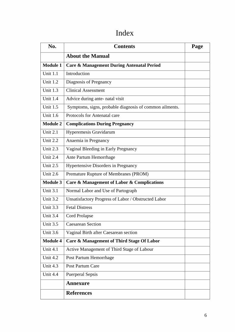
Index
No. Contents Page About the Manual
Module 1 Care & Management During Antenatal Period
Unit 1.1 Introduction
Unit 1.2 Diagnosis of Pregnancy
Unit 1.3 Clinical Assessment
Unit 1.4 Advice during ante- natal visit
Unit 1.5 Symptoms, signs, probable diagnosis of common ailments.
Unit 1.6 Protocols for Antenatal care
Module 2 Complications During Pregnancy
Unit 2.1 Hyperemesis Gravidarum
Unit 2.2 Anaemia in Pregnancy
Unit 2.3 Vaginal Bleeding in Early Pregnancy
Unit 2.4 Ante Partum Hemorrhage
Unit 2.5 Hypertensive Disorders in Pregnancy
Unit 2.6 Premature Rupture of Membranes (PROM)
Module 3 Care & Management of Labor & Complications
Unit 3.1 Normal Labor and Use of Partograph
Unit 3.2 Unsatisfactory Progress of Labor / Obstructed Labor
Unit 3.3 Fetal Distress
Unit 3.4 Cord Prolapse
Unit 3.5 Caesarean Section
Unit 3.6 Vaginal Birth after Caesarean section
Module 4 Care & Management of Third Stage Of Labor
Unit 4.1 Active Management of Third Stage of Labour
Unit 4.2 Post Partum Hemorrhage
Unit 4.3 Post Partum Care
Unit 4.4 Puerperal Sepsis
Annexure
References
7
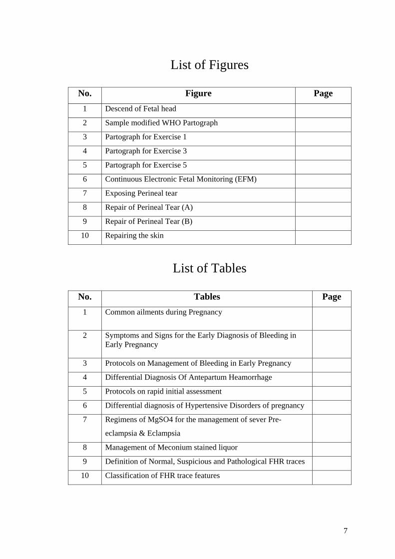
List of Figures
No. Figure Page 1 Descend of Fetal head
2 Sample modified WHO Partograph
3 Partograph for Exercise 1
4 Partograph for Exercise 3
5 Partograph for Exercise 5
6 Continuous Electronic Fetal Monitoring (EFM)
7 Exposing Perineal tear
8 Repair of Perineal Tear (A)
9 Repair of Perineal Tear (B)
10 Repairing the skin
List of Tables
No. Tables Page
1 Common ailments during Pregnancy
2 Symptoms and Signs for the Early Diagnosis of Bleeding in Early Pregnancy
3 Protocols on Management of Bleeding in Early Pregnancy
4 Differential Diagnosis Of Antepartum Heamorrhage
5 Protocols on rapid initial assessment
6 Differential diagnosis of Hypertensive Disorders of pregnancy
7 Regimens of MgSO4 for the management of sever Pre-
eclampsia & Eclampsia
8 Management of Meconium stained liquor
9 Definition of Normal, Suspicious and Pathological FHR traces
10 Classification of FHR trace features

8
About the Manual This Manual consists of four modules pertaining to essential and emergency obstetrics and newborn care. This manual has been created with an objective of making obstetricians aware regarding most recent developments. The differential diagnosis of important obstetrical complications like Ante partum hemorrhage has been put into tabular form, which makes it simpler and easier to follow. Similarly case studies on different complications will be helpful in adopting practical approach in management of cases.
9
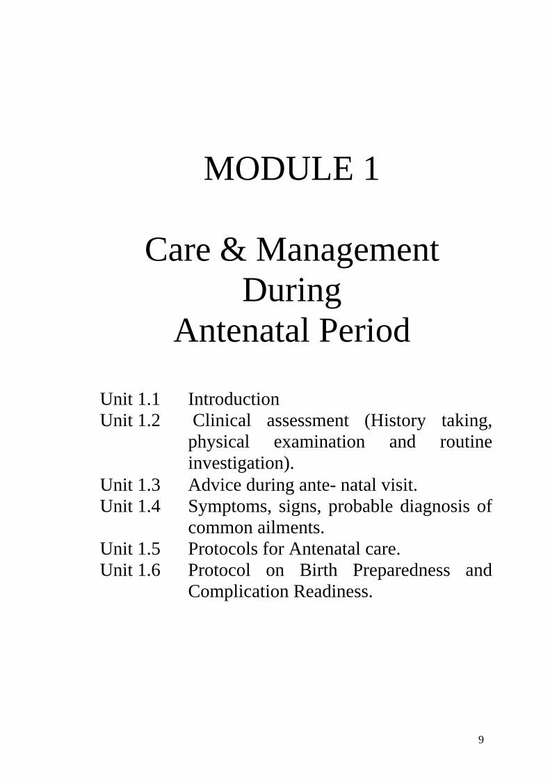
MODULE 1
Care & Management During
Antenatal Period Unit 1.1 Introduction Unit 1.2 Clinical assessment (History taking,
physical examination and routine investigation).
Unit 1.3 Advice during ante- natal visit. Unit 1.4 Symptoms, signs, probable diagnosis of
common ailments. Unit 1.5 Protocols for Antenatal care. Unit 1.6 Protocol on Birth Preparedness and
Complication Readiness.

10
About this Module In this module we will discuss about the care and management of a women during her antenatal period. It will also cover defined protocols related to antenatal registration, care, investigations and management, advice, and danger signs during antenatal period. Unit 1.1 Introduction Learning Objectives
• To diagnose pregnancy • To know about the appropriate timing, number and importance of ANC.
Pregnancy is not a disease but every pregnancy is at risk. Ensure that Ante-Natal care is used as an opportunity to detect and treat existing problems. Effective Ante-Natal care can provide a healthy mother and a healthy baby as an outcome. However, you must realize that even with the most effective screening tools available, one can not predict which woman will develop complications related to pregnancy and also when.
• Recognize that “ Every pregnancy is at risk” • Ensure that Antenatal care is used as an opportunity to detect and treat
existing problems. • Make sure that services are available to manage obstetric emergencies when
they occur. • Prepare pregnant women and their families for the eventuality of an
emergency. 1.1.1 Diagnosis of Pregnancy Symptoms: The woman may come to you with the following complaints:
• Cessation of Menstruation • Nausea with or without vomiting • Increased frequency of urine • Fatigue • Perception of foetal movements (after four months)
Signs: on examination you may find
• Breast enlargement • Changes in the skin colour of areola • Enlargement of abdomen • Discoloration of vaginal mucosa • Softening of cervix and uterus • Uterine enlargement • Internal and external ballottement • Ability to discern fetal parts

11
You can confirm the pregnancy by • Detection of HCG in urine within 3 months of amenorrhea • Detection of FHS at 20 weeks by auscultation • Perception of foetal movements on abdominal examination
* In case of doubt of congenital anomalies, an USG may be considered –refer clinical protocol 1. To ensure a normal pregnancy with the delivery of a healthy baby from a healthy mother and for screening the complications during pregnancy there are few points to be kept in mind, which are:
Early registration: All pregnant women should be registered early, at least before 12 weeks.
Antenatal card: Antenatal card should be duly filled in by nurse and doctor
for every woman registered, and handed over to the pregnant woman.
Number and timing of visits:. Ensure that every woman makes at least 3 Antenatal visits apart from registration. In cases of pregnancy without complications, these visits should be sufficient. Ideally, the registration should be done as soon as the pregnancy is suspected and first visit should be scheduled between 4th and 6th month (around 26 weeks). The second visit should be planned in the 8th month (32 weeks) and the third visit in the 9th month (36 weeks).
Source : (MOH&FW manual “Guidelines for Antenatal Care and skilled attendance at birth by ANMs and LHVs”)

12
Unit 1.2 Clinical assessment (History taking, physical examination and routine investigation) Learning Objectives
• Know the important points to be asked while collecting history to a pregnant woman.
• Understand and be able to perform physical examination on a pregnant woman.
• Understand the routine investigations to be done during antenatal period. 1.2.1 History Taking
Date of Last Menstrual Period: • Ask for date of 1st day of the last menstrual period (LMP) • Calculate the Expected Date of Delivery (EDD) = LMP + 9months and 7
days. This calculation is based on the assumption that the menstrual cycle was regular and it was a 28-30 days’ cycle. If the cycle varies, EDD will vary accordingly
• Calculate Period of Gestation in weeks: Period of gestation is to be expressed in terms of completed weeks. A fraction of more than 3 days is to be considered as completed week.
Age of the woman Duration of marriage Order of the pregnancy Gravida (GPAL- Gravida, Para, Abortion, Living children) Last child birth/ Last abortion History of present pregnancy Complaints during present pregnancy (to be asked during each visit):
• Excessive vomiting • fever • Palpitations, easy fatigability and breathlessness at rest • Puffiness of the face • Headache or blurring of vision • Passing smaller amounts of urine • Vaginal bleeding or Leaking of watery fluid per vaginam(P/V) • Pain abdomen at any stage of pregnancy • Decreased or absent foetal movements
History of problems during the previous pregnancy/ delivery (Obstetric
History)- • Abortion(s) or Premature birth(s), • Twins or multiple pregnancies • Stillbirths(s) or neonatal loss • Hypertensive disorder of pregnancies(if don’t know, ask for a history of
convulsion in previous pregnancies) • Duration of labor

13
• Malpresentation, such as breech delivery • Ante partum hemorrhage • Postpartum hemorrhage • Assisted delivery (vacuum extraction) • Delivery by caesarean section • Birth weight of previous baby • Any surgery on the reproductive tract • Iso-immunization (Rh-ve) in the previous pregnancy • History of drug intake or allergies. • Any treatment taken or taken drugs for infertility.
History of any systemic/ Medical illness (es)-
Relevant history of past medical illnesses e.g. Hypertension, Diabetes, Heart disease, Tuberculosis, Renal disease, Epilepsy, Asthma, Malaria, Thyroid or any disease for which she has been advised to take treatment, is to be elicited.
Family History: • systemic illness • Twins • Congenitally malformed baby • Thalassaemia
Personal History: • Contraceptive practices prior to pregnancy • Smoking, alcohol, tobacco or any other substance abuse • Bowel, bladder, appetite
1.2.2 Physical Examination
General Examination • Height • Weight • Blood Pressure • Pallor • Respiratory rate (RR) • Pedal edema • Generalized edema or puffiness of the face • Breast Examination to detect inverted nipples, crusting or soreness of
nipples and Lumps. Systemic Examination: to rule out heart disease, respiratory disease, neck glands
etc. Abdominal Examination: Abdominal examination is done to monitor the progress
of pregnancy and foetal growth and to check the foetal lie and presentation and any scar.
Arrangement for HIV counseling HIV counseling to precede HIV testing
1.2.3 Routine investigations
• Haemoglobin (Hb) Estimation

14
• ABO grouping & Rh typing • Urine testing for Albumin, Sugar and pus cells • Blood sugar screening should be done • Estimation of fasting and post prandial ( 2 hours after meals) blood glucose (PPBG), glucose tolerance test when indicated • VDRL (husband and wife) • HIV screening should be done only after counseling • Hepatitis B and C antigen
Repetition of investigations:
• Haemoglobin (Hb) Estimation. • Urine for Albumin and Sugar and pus cells

15
Unit 1.3 Advice during Ante- Natal Visit Learning Objectives
• Become aware of the advices to be given to a pregnant woman.
1.3.1 Important advises
• To bring ANC card at each visit. • Iron & folic acid (IFA) supplementation. • Injection Tetanus Toxoid (Inj. TT) administration ( three doses –first at
16weeks, second at 20 weeks ,third dose sixth month after the second dose) • Pregnant woman may continue her household chores throughout pregnancy if
not tired. • Hard and strenuous work should be avoided, especially in Ist and III trimester. • Pregnant woman should sleep for about 8 hours at night and take 2 hours of
rest during the daytime. • Should take daily bath but be careful against slipping in the bathing area due
to imbalance. • Clean, loose comfortable, preferably cotton clothes to be worn. • Retracted nipples to be corrected manually as soon as it is detected to avoid
problems during breast-feeding and apply some cream to make them soft. • Avoid supine position especially during late pregnancy. • Coitus to be avoided during the first trimester and during the last 6 weeks. • Travel by vehicles having jerks to be avoided. • Pregnant women and her attendant to be told about the symptoms and signs of
complications like pain in abdomen, watery discharge per vaginum, bleeding per vaginum, severe and persistent headache with blurring of vision, vomiting, swelling all over the body or feet, high grade fever, diminished foetal movements, undue breathlessness, palpitations, decreased urine output. Tell them to bring the pregnant woman to the hospital whenever any of these occur.
Dietary advice during pregnancy
The woman should be advised to eat more than her normal diet throughout her pregnancy. Remember, a pregnant woman needs about 300 extra kcal per day compared to her usual diet. The woman's food intake should be especially rich in proteins, iron, vitamin A and other essential micronutrients.

16
Unit 1.4 Symptoms, Signs, Probable Diagnosis of Common Ailments Learning Objectives
• To know about the symptoms, signs and management of common ailments during pregnancy.
1.4.1 Common Ailments during pregnancy The table given below gives a clear picture about the common ailments during pregnancy, their signs and symptoms and recommended actions to be taken.
Table 1: Common ailments during Pregnancy Symptoms Signs/investigations Most probable diagnosis Action(s) to be taken Vomiting during the first trimester
May be physiological Advise the woman to eat small frequent meals; chew before swallowing; avoid greasy food; eat lots of green vegetables and drink plenty of fluids. If vomiting is excessive in the morning, ask her to eat dry foods such as biscuits or toast after waking up in the morning.
Excessive vomiting, especially after the first trimester
The woman may be dehydrated
Hyperemesis gravidarum Refer to protocol on Hyperemesis Gravidarum.
Palpitations, easy fatigability, breathlessness at rest
Conjunctival and/or palmer pallor present Hb level <7 g/dl
Severe anaemia Refer to protocol on Anemia during pregnancy
Swelling of one or both feet or tightening of rings on fingers and toes. BP normal.
Rest with feet up
Puffiness of the face, generalized body oedema
- BP >140/90 mmHg. Proteinuria absent
Hypertensive disorder of pregnancy
Manage as per clinical protocol given for PIH

17
- BP >140/90 mm Hg Proteinuria present
- Pre-Eclampsia - Advise her on the danger signs of imminent eclampsia and eclampsia
Heartburn and nausea
Reflux - Advise the woman to avoid spicy and rich foods. - Ask her to take cold milk during attacks. - If severe, antacids may be prescribed.
Increased frequency of urination up to 10 - 12 weeks of pregnancy. No burning during micturition.
May be physiological due to pressure of the gravid uterus on the urinary bladder
Reassure her that it will be relieved on its own.
- Increased frequency of urination after 12 weeks, or persistent symptoms, or burning on urination
- Tenderness may be present at the sides of the abdomen and back - Body temperature may be raised
Urinary tract infection (UTI)
Refer to protocol on Urinary tract infection
Constipation Physiological Advise the woman to take more fluids, leafy vegetables and a fiber-rich Diet. If not relieved, give her Isabgol, 2 tablespoonfuls to be taken at bedtime, with water or with milk. Do NOT prescribe strong laxatives as they may start uterine contractions
Check the pulse and BP to assess for shock
Threatened abortion / spontaneous abortion / hydatidiform mole / ectopic pregnancy
Refer to protocol on Bleeding in Early pregnancy
Bleeding P/V, before 20 weeks of gestation
- Ask for history of violence
-Spontaneous abortion due to violence
Put her in touch with local support groups

18
Bleeding P/V, after 20 weeks of gestation
Check the pulse and BP to assess for shock
Ante partum haemorrhage
Do NOT carry out a vaginal examination under any circumstances. Refer to the next level/specialist
Blood peripheral smear is positive for malaria parasite.
Malaria Manage according to the NAMP guidelines for malaria in pregnancy.
Fever
Body temperature is raised
Site of infection somewhere, including possible sepsis
Refer to next level
FHS heard, and within the normal range of 120-160/ minute
Baby is normal
Fetal kick chart for 6 hours. If less than 4, refer to higher center where CTG available. if >4 , call the woman next day for reassessment. If the count is >4,then Reassure the woman
FHS heard, but the rate is<120/minute or >160/ minute
Foetal distress
Repeat FHS after 15 Minutes. If the FHS is still out of the normal range, refer.
Decreased or absent foetal movements (NOTE: foetal movements are felt only after about 4 months of gestation.
FHS not heard Intrauterine foetal death Inform the woman and her family that the baby might not be well. Refer to the next level
Vaginal discharge, with or without abdominal pain (not blood stained or bleeding)
RTI/STI Advise the woman regarding vaginal hygiene, i.e. cleaning the external genitalia with soap and Water.
Leaking of watery fluids P/V
Wet pads/cloths Premature rupture of Membrane.
Look if the woman is in labor / refer

19
Unit1.5 Protocols for Antenatal care Learning Objectives
• Understand and appreciate the protocols for Antenatal care in L0 and L1 hospitals of Merrygold health Network.
1.5.1 Registration at Reception
• Name, age, address, order of pregnancy (GPAL - Gravida, Para, Abortion, Live birth), LMP (Last Menstrual Period). Classification criteria (annex - 1) form to be filled up
• Patient goes to the nursing station. The Nurse checks height, BP, weight, urine sugar/ protein. Advises client for Hb %, Blood Group, PPBG/ 50 gm glucose challenge test.
1.5.2 Patient goes to Medical Officer's Room
• The Medical officer assesses the Patient's status and makes a decision between: Minimum four visits, if the classification criteria card does not include any ‘Yes’ answer. Even with 1 ‘YES’ answer, WHO focussed Antenatal care of four visits will not apply.
• Information about routine USG to exclude congenial abnormalities should be given to all women. It should be done between 18 weeks to 20 weeks gestational age. Any woman refusing to have an USG is responsible for the consequences.
• If low lying placenta is seen at 18-20 weeks, repeat USG at 34-36 weeks. Transvaginal sonography is recommended in early pregnancy. In case of low lying placenta, Trans abdominal scans are preferred
1.5.3 Management 1.5.3.1 During First Trimester of Pregnancy:
• Diagnosing pregnancy by urine pregnancy test • If test is positive, note the uterine size (PV examination) • Folic Acid (5 mg) supplementation only • If she wants MTP then refer to MTP Clinic
Management as per 'Basic Component of the new WHO Antenatal Care model (Annex - 1) General Antenatal Advice
• Nutrition support (Extra 300 kcal per day compared to her usual diet, especially rich in proteins, iron, vitamin A and other essential micronutrient is recommended).
• Iron supplementation • Folic Acid supplementation

20
• Advice to complete all 2 doses of TT course • Routine investigations
- Haemoglobin (Hb) Estimation - ABO grouping & Rh typing - Urine testing for Albumin, Sugar and pus cells - Blood sugar screening should be done - Estimation of fasting and post prandial ( 2 hours after meals) blood
glucose - (PPBG), glucose tolerance test when indicated - VDRL (husband and wife) - HIV screening should be done only after counseling - Hepatitis B and C antigen
• Advice about sexual intercourse, work and exercise • Birth preparedness and complication readiness * • Inform date of next visit • Counselling against abuse of alcohol and tobacco • Use of / continue use of condoms to prevent STI. • Restricting the use of other medicines without Doctor's advice • Danger signs that are to be noted by the woman and appropriately reported
to the care centre immediately: a. Any bleeding per vaginum any time b. Any discharge of water per vaginum c. Severe continuous headache d. Disturbance of vision e. Convulsions f. High fever and prolonged Malaise g. Unusual abdominal pain h. Difficulty in breathing
• In case of any deviation from normalcy then refer to other protocols as appropriate
21
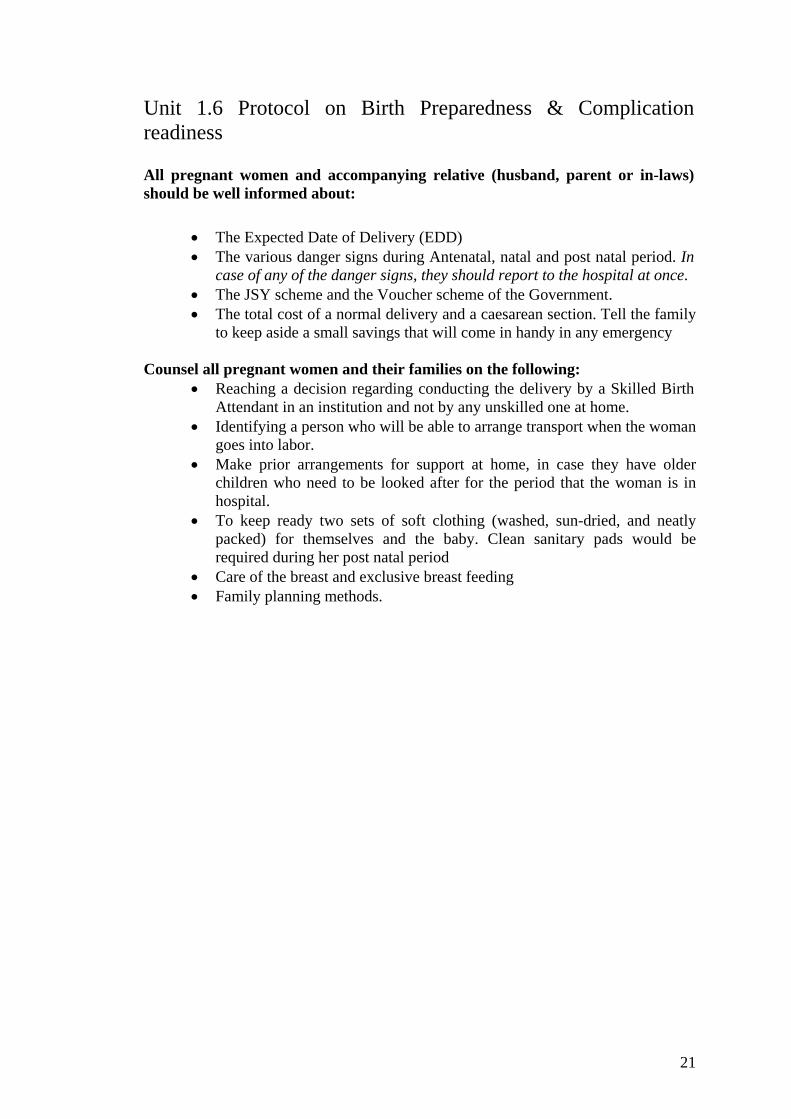
Unit 1.6 Protocol on Birth Preparedness & Complication readiness All pregnant women and accompanying relative (husband, parent or in-laws) should be well informed about:
• The Expected Date of Delivery (EDD) • The various danger signs during Antenatal, natal and post natal period. In
case of any of the danger signs, they should report to the hospital at once. • The JSY scheme and the Voucher scheme of the Government. • The total cost of a normal delivery and a caesarean section. Tell the family
to keep aside a small savings that will come in handy in any emergency Counsel all pregnant women and their families on the following:
• Reaching a decision regarding conducting the delivery by a Skilled Birth Attendant in an institution and not by any unskilled one at home.
• Identifying a person who will be able to arrange transport when the woman goes into labor.
• Make prior arrangements for support at home, in case they have older children who need to be looked after for the period that the woman is in hospital.
• To keep ready two sets of soft clothing (washed, sun-dried, and neatly packed) for themselves and the baby. Clean sanitary pads would be required during her post natal period
• Care of the breast and exclusive breast feeding • Family planning methods.

22
MODULE 2
Complications During Pregnancy
Unit 2.1 Hyperemesis Gravidarum Unit 2.2 Anaemia in Pregnancy Unit 2.3 Vaginal Bleeding in Early Pregnancy
Unit 2.4 Ante Partum Hemorrhage Unit 2.5 Rapid Initial Assessment and Management of
Shock Unit 2.6 Hypertensive Disorders in Pregnancy Unit 2.7 Premature Pre - Labor Rupture of
Membranes (PPROM) Unit 2.8 Preterm Labor Unit 2.9 Management of Breech Presentation
23
About this Module In this module, we will be discussing the different complications that can occur during the period of pregnancy. The participants will have an opportunity to work in groups on case studies, detail out the line of management as well as understand standard protocols that are laid down for each of the conditions. Unit 2.1 Hyperemesis Gravidarum Learning Objectives
• Discuss the condition of hyperemesis gravidarum during first trimester of pregnancy and its implications.
• Discuss the management of antenatal women with hyperemesis gravidarum. 2.1.1 Introduction It is a severe type of vomiting of pregnancy which has got deleterious effect on the health of mother and/or incapacitates her in day to day activities. More commonly seen in primigravida, twins/ multiple pregnancy and hydatiform mole. Symptoms:
- Can take nothing by mouth due to excessive vomiting - Severe retching and nausea is present - Diminished urine output
Signs:
- Progressive emaciation with loss of weight - dehydrated. Loss of skin elasticity - Tongue- dry, brown, coated, red or raw - Tachycardia - Ketonuria may be present - Rarely icterus (when it appears it is a grave sign)
2.1.2 Management Protocol
• Hospitalization • Reassure the woman and family. Counsel them regarding harmless nature of the
condition. • Start IV fluids slowly, either R/L or Dextrose Saline • Repeat urine examination every 4 hours till it becomes negative for ketone bodies.

24
• Anti – emetic Drugs-oral doxylamine succinate and B6; Promethazine( Phenargan) 25mg or Prochlorperazine (Stemetil) 5 mg. In patients not responding to above treatment, give Inj Ondensetreon 8mg I/V 12 hrly.
• Advise the woman to take small, frequent, carbohydrate rich meals. • Sympathetic but the firm handling of the patient is essential. • Once the vomiting stops and the dehydration is corrected, discharge after 24
hours. Case Study – Hyperemesis Gravidarum A 35 year old married shop assistant was referred as an emergency for antenatal assessment because of intractable vomiting. Marked nausea and sickness had complicated her first pregnancy, a breech delivery, and she was not therefore surprised when this persisted. Her second pregnancy had however been entirely normal. The vomiting initially occurred in the morning, but as the pregnancy progressed it occurred throughout the day and became progressively more severe. During the 48 hours prior to her admission, she had been unable to retain even clear liquids and had sought the help of her family doctor. He had been concerned at her general condition and the fact that her uterus appeared to be at least 4 weeks larger than the duration of her pregnancy, calculating from the date of her last pill withdrawal bleed. She had not experienced any other vaginal bleed or any pain since this time. There had been no change in her bowel habits and she had no urinary symptoms. Apart from an appendectomy at the age of 21 years her past history was unremarkable. There was no relevant family history. On examining she was clearly dehydrated and distressed by her constant nausea and persistent vomiting. Her temperature was 36.8 C, pulse rate 104 beats / minute and blood pressure 100/60 mm Hg, she did not appear anaemic and there was no abnormality on general examination. Her breasts were normal. Abdominal palpitation revealed a 16-week uterus, which was regular in outcome. There was no other suggestion of abdominal distension and her abdomen was non tender with normal bowel sounds. Vaginal examination was normal and bimanual palpation confirmed the uterus size. A pregnancy test was strongly positive. Questions 1. What is the differential diagnosis? 2. What investigation would you perform? 3. What are the complications of this condition? 4. Name a well-known authoress who died of this condition.
25
Key to Case Study: 1. The patient had hyperemesis gravidarum and uncertain dates.Her uterus is about 4
weeks larger than expected. Multiple pregnancy and hydatidform mole, both of which predisposes to hyperemesis in pregnancy must therefore be excluded. This can be done most easily by ultrasound scanning. The number of fetuses present can be counted and the gestation assessed by measurement of the biparietal diameter. With appropriate ultrasound intensity (low grain), an absence of fetal parts and a “classical snow storm” will confirm a hydatidform mole.
Gastroenteritis, hepatitis, cholecystitis, hiatus hernia, peptic ulcers and intestinal obstruction may all be mimicked by hyperemesis but can be excluded on clinical grounds in this case.
2. Blood should be sent for plasma electrolytes, blood urea estimation and Liver
function tests. A full blood count should also be obtained. The patient should be nursed in a quiet single room with no oral intake allowed, apart from occasional sips of water. Re hydration should be achieved by giving intravenous fluids and nausea quelled using antiemetics.
While awaiting the urea and electrolyte results, an intravenous infusion of dextrose saline BP should be commenced. Potassium supplements should be added to the infusion fluid when the plasma electrolyte concentrations are known. A careful fluid balance record must be maintained and the patient should be weighed daily. Suitable antiemetics include promethazine, 25-30 mg, or chlorpromazine, 25–50 mg, by intramuscular injection every 4-6 hours as required. These drugs cause drowsiness and may produce a dry mouth. Other drugs that are used include promazine and perphenazine. All these drugs, especially the last, may cause extrapyramidal neurological effects such as oculogyric crises. Vitamin C, vitamin B complex and vitamin B6 (pyridoxine) may be added to the infusion fluid if there has been prolonged starvation. Although there is no proven cause for hyperemesis, many of these women have emotional problems. Psychiatric consultation may be necessary and isolation from domestic stress and relatives may be beneficial. When the patient has stopped vomiting for 24-48 hours and has a normal urine output and normal plasma electrolytes, oral fluid intake should be increased to 50 ml hourly. If this is tolerated she should be given easily digested high energy foods (e.g. milk fruit juices etc) frequently in small amounts. If these are tolerated, the diet should progress to semi solids (e.g. boiled eggs, cereals etc). When the patient has returned to her normal diet and activity, she should be discharged home on oral antiemetic therapy. She should be advised not to drive or operate machinery while taking antiemetic drugs. Considerable anxiety has been expressed about the possible tetratogenic effects of antiemetic. Extensive studies in Britain and the United States of America have exonerated these preparations and demonstrated their safety in pregnancy.

26
3. Rare complications of persistent vomiting include rupture of the oesophagus, aspirated pneumonitis, hemorrhagic retiniitis and dental erosion.
4. Maternal death from hypermesis is recorded (Charlotte Bronte succumbed to the
disorder), but it is now rare and once treated there is no apparent effect on the pregnancy.
The ultrasound scan in this patient showed a normal 12-week pregnancy and her hyperemesis resolved rapidly with the therapy described. NOTE : In all cases of hyperemesis , exclude medical/surgical illness and go for ultrasonography .

27
Unit 2.2 Anaemia in Pregnancy Learning Objectives
• Discuss the existing situation of anemia in pregnancy and its implications • Implement the protocols for management of antenatal women with mild /
moderate / severe anemia. 2.2.1 Definition Anaemia in pregnancy is defined as an Hb level of <11 g/dl during pregnancy and in the immediate postpartum period. A pregnant woman with an Hb level of <7 g/dl is said to have severe anaemia. 2.2.2 Diagnosis Examine and investigate the woman for the following:
• Conjuctival pallor • Severe palmar pallor • Pallor of the tongue, palate and oral mucosa • RR (count for 1 minute) • Level of Hb
2.2.3 Protocols for Prevention and Management of Anaemia in
Pregnancy • Diagnosis - Hb% less than 11 gms% during pregnancy • Prophylaxis and treatment
Prophylaxis
From 16 weeks onwards: (If Hb% > 11 gms %): 60mg elemental iron + 1 mg Folic acid, OD till six weeks post partum Albendazole 400 mg HS one dose If Hb% between 7 gms% to 10.9 gms% (mild anemia) Change to therapeutic dosage, as under:
• 100 mg elemental Fe with upto 2 mg Folic acid once a day till 12 weeks postpartum
• Albendazole 400 mg HS one dose. • Check PCV, peripheral smear. Exclude other causes of anaemia if any.
Perform stool test (ova, worms), urine test (routine & microscopy). Perform Dental check up

28
Note: If recurrent vomiting after Fe supplementation: Change sulfate to fumarate and then to gluconate If repeated non-compliance and intolerance ascertained, then parenteral Fe supplementation may be considered
If Hb% between 5 to 6.9 gm%
In Early pregnancy:
• Admit patient and investigate extensively to exclude serious causes like malaria, bone marrow abnormalities, thalassemia, chronic bleeding disorders, marrow abnormalities, leukemias, etc.
• If Iron (Fe) deficiency confirmed and gestation age is : i. Below 32 weeks - give oral Fe as in 100 mg elemental Fe with
upto 2 mg folic acid upto 12 weeks postpartum. ii. Between 32 to 36 weeks - parenteral Fe should be given
iii. Over 36 weeks - whole blood transfusion If Hb% less than 5 gm%
1. Packed cell transfusion with furesemide IV administered 30 mins after initiating transfusion.
2. If Congestive Cardiac Failure (CCF) - Packed cell transfusion. Urgent involvement of a physician, which means the physician should be brought in and provide necessary treatment
Indications of Blood Transfusion for Anemia in Pregnancy
If Pregnancy less than 36 weeks:
a. Haemoglobin 5.0 g/dl or below, even without clinical signs of cardiac failure or hypoxia
b. Haemoglobin between 5 and 7.0 g/dl and in the presence of the following conditions:
• Established or incipient cardiac failure or clinical evidence of hypoxia
• Pneumonia or any other serious bacterial infection • Malaria • Pre-existing heart disease, not causally related to the anaemia.
If Pregnancy 36 weeks or more:
a Haemoglobin 6.0 g/dl or below with of without any other signs and symptoms
b Haemoglobin between 6.0 g/dl and 8.0 g/dl and in the presence of the following conditions:
• Established or incipient cardiac failure or clinical evidence of

29
hypoxia • Pneumonia or any other serious bacterial infection • Malaria • Pre-existing heart disease, not causally related to the anaemia
For Elective CS with anaemia in cases with H/O APH, PPH and Past CS, if:
a Hb% 8.0 to 10.0 gm%: then keep serum ready for cross matching (Blood
Group must be known) b Hb% < 8.0 gm%: then 2 units of Blood 'X' matched and made available Note-IV iron Sucrose compound, available for moderate anaemia, can be given if blood is not available

30
Unit 2.3 Vaginal Bleeding in Early Pregnancy Learning Objectives At the end of the session the participants will be able to
1. Discuss various causes of vaginal bleeding during early pregnancy. 2. Understand the clinical protocol for management of vaginal bleeding in early
pregnancy. 2.3.1 Introduction Vaginal bleeding during early pregnancy (up to 20 weeks of gestation) can be due to various types of abortions, ectopic pregnancy or the presence of a hydatidiform mole (molar pregnancy).
Table 2: Symptoms and Signs for the Early Diagnosis of Bleeding in Early Pregnancy
Symptoms and signs typically present
Symptoms and signs sometimes present
Probable diagnose
• Light bleeding • Closed cervix • The size of the uterus
corresponds to the gestational period
• Cramping / lower abdominal pain
• Uterus softer than normal
Threatened abortion
• Heavy bleeding • Dilated cervix • The size of the uterus
corresponds to the gestational period
• Cramping / lower abdominal pain
• No expulsion of the products of conception
• The uterus is tender
Inevitable abortion
• Heavy Bleeding • Dilated cervix • The size of the uterus is
smaller than that expected for the gestational period
• Cramping / lower abdominal pain
• History of partial expulsion of the products of conception
Incomplete abortion
• Light bleeding • Or blood stained
discharge • Closed cervix • The size of the uterus is
smaller than that expected for the gestational period
• Uterus softer than normal
• Light cramping / abdominal pain
• History of expulsion of the products of conception
Complete abortion
31
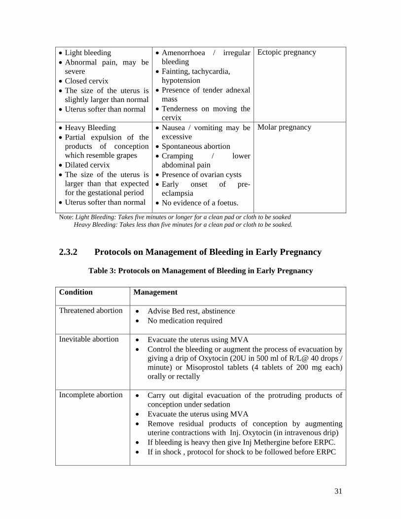
• Light bleeding • Abnormal pain, may be
severe • Closed cervix • The size of the uterus is
slightly larger than normal • Uterus softer than normal
• Amenorrhoea / irregular bleeding
• Fainting, tachycardia, hypotension
• Presence of tender adnexal mass
• Tenderness on moving the cervix
Ectopic pregnancy
• Heavy Bleeding • Partial expulsion of the
products of conception which resemble grapes
• Dilated cervix • The size of the uterus is
larger than that expected for the gestational period
• Uterus softer than normal
• Nausea / vomiting may be excessive
• Spontaneous abortion • Cramping / lower
abdominal pain • Presence of ovarian cysts • Early onset of pre-
eclampsia • No evidence of a foetus.
Molar pregnancy
Note: Light Bleeding: Takes five minutes or longer for a clean pad or cloth to be soaked Heavy Bleeding: Takes less than five minutes for a clean pad or cloth to be soaked. 2.3.2 Protocols on Management of Bleeding in Early Pregnancy
Table 3: Protocols on Management of Bleeding in Early Pregnancy Condition Management
Threatened abortion • Advise Bed rest, abstinence
• No medication required
Inevitable abortion • Evacuate the uterus using MVA • Control the bleeding or augment the process of evacuation by
giving a drip of Oxytocin (20U in 500 ml of R/L@ 40 drops / minute) or Misoprostol tablets (4 tablets of 200 mg each) orally or rectally
Incomplete abortion • Carry out digital evacuation of the protruding products of
conception under sedation • Evacuate the uterus using MVA • Remove residual products of conception by augmenting
uterine contractions with Inj. Oxytocin (in intravenous drip) • If bleeding is heavy then give Inj Methergine before ERPC. • If in shock , protocol for shock to be followed before ERPC

32
Complete abortion • Check for any retained products of conception by USG and / or bleeding
• No further management is required if the condition of the woman is stable.
Septic abortion • Give Paracetamol (1 tablet of 500 mg) to control fever
(temperature > 38oC) • Examine for the presence of any foreign body in the vagina • Thoroughly irrigate the vagina to remove any herbs, local
medications or caustic substances • Give the following antibiotics:
Inj. Ampicillin 2 g IV, every 6 hours, PLUS Inj. Gentamicin 5 mg/kg body weight, IV, every 24 hr
PLUS Inj. Metronidazole 400 mg in 100 ml infusion bottle to be
given IV every 8 hrs, until the woman is afebrile for 48 hrs
(To avoid phlebitis, change the infusion site every three days or at the first sign of inflammation)
• If the bleeding is minimal, evacuate the uterus after 48 hrs of antibiotic coverage, preferably use MVA

33
Unit 2.4 Ante Partum Hemorrhage Learning Objectives: At the end of the session we will be able to:
• Understand the differential diagnosis of ante-partum hemorrhage. • Discuss the protocol to be followed for APH cases.
Session Duration: 120 minutes 2.4.1 Definitions Vaginal bleeding occurring after 20 weeks of pregnancy or during labor (but before delivery of the baby) is known as antepartum haemorrhage (APH).
Table 4: Differential Diagnosis of Antepartum Heamorrhage
Criteria Placenta Praevia
Abruptio Placentae Uterine Rupture
Nature of the bleeding • Painless,
causeless and recurrent
• The bleeding is always revealed
• Painful, pain is often localized to start with and later becomes generalized, attributed to pre- eclampsia or trauma and is continuous.
• The bleeding is revealed, concealed, or usually mixed
• The bleeding often occurs after the woman has been in labor for a long time
• The bleeding may be concealed or mixed
General condition and anaemia
Proportional to the amount of blood loss.
Out of proportion to the visible blood loss in the concealed variety
Out of proportion to the visible blood loss
Feature of the uterus Not relevant Present in one-third of cases Not relevant
Height of the uterus • Proportional to the gestational age
• May be disproportionately enlarged in the concealed type
Feel of the uterus Soft and relaxed May be tense, tender and rigid
Uterine contour nor felt, occasionally, the uterus is felt separately on to one side
Malpresentation • Common: the head is high and floating
• Unrelated: head may be engaged
• Feotal parts felt superficially: malpresentation may be present
Localization of placenta Placenta is in the
lower segment of Placenta is in the upper segment
The placenta may be attached to the uterus or
A woman with APH, if not managed in time, can bleed to death within 12 hours of the start of bleeding. It is an important cause of maternal mortality.

34
the uterus may be lying free in the peritoneal cavity
Vaginal Placenta felt in the lower segment
The placenta is not felt in the lower segment
The presenting part is high up not felt: the contracted uterus may be felt on one side.
A careful P/S examination may be performed to rule out other causes of bleeding such as cervicits, trauma, cervical polyps or cervical malignancy. The presence of these, however, does not rule out placenta praevia. 2.4.2 Protocol on Prevention and Management of Ante partum Hemorrhage (APH) 2.4.2.1 Management of Placenta Praevia
• Assess the bleeding Up to Moderate Bleeding:
• Investigate for Hb%, Blood group and cross matching • Blood transfusion if required • Check the coagulation factors • USG to identify location of placenta as soon as possible • If placenta in lower segment: Expectant management in hospital if:
i. pregnancy < 37 weeks ii. baby alive
iii. woman's life not at risk
More than moderate bleeding Evidence by tachycardia and Hypotension • Transfuse blood liberally • Usual treatment of shock, if any • USG done to identify location of placenta • Terminate pregnancy
• Definitive treatment:
• CS for major degrees of placenta praevia • Vaginal delivery in selected cases of minor degrees of placenta praevia • Blood should be kept ready at time of delivery
2.4.2.2 Management of Abruptio Placenta Mild case: Manage expectantly (watch and wait, follow up after one week) but may go home after USG
Severe case:
• Restore blood volume through liberal IV fluids and Blood Transfusion. • Monitor coagulation profile and urine volume • Plan for early delivery.
35
2.4.2.3 Methods of delivery are as follows
First choice: Aim for vaginal delivery by Artificial Rupture of membranes and augmentation with oxytocin.
If: a. Response to induction & augmentation is poor b. Foetus in distress FHS present,
THEN GO FOR CAESAREAN SECTION Note: Exclude coagulation defects before CS 2.4.3 Case Study – Ante partum Hemorrhage 1. Antepartum haemorrhage
Kalavati 30 year old female resident of Etmadpur was admitted to the Merrygold hospital at Agra as a para 1+0 with a previous normal delivery conducted at a Merrysilver hospital 3 years back. She has been referred to the hospital at Agra from the same hospital as a term pregnancy with fresh vaginal bleeding and abdominal pain. On examination she is distressed with pain, is pale, pulse is 100 bpm, BP-110/80 mm/Hg, uterus is tender and the uterine contractions recorded were 3/10 minutes. Blood stains were noticed on her feet and between her toes.
• What is the most likely diagnosis • What are the risks to the mother and the fetus • How should you assess and manage this situation
Diagnosis: The most likely diagnosis is placental abruption. It is the premature separation of the normally situated placenta from the uterine wall resulting in hemorrhage prior to delivery of the fetus. Risk factors includes sudden uterine decompression, external trauma, uterine anomaly, increased maternal age, smoking and an unexplained elevation of maternal serum alpha protein in the second trimester. However most commonly no cause is found.. Bleeding can be in whole or in part concealed. It is important to differentiate placental abruption from placenta praevia {pp}.in pp bleeding is due to separation of a placenta situated in the lower uterine segment as a result of lower segment forming or the cervix dilating. It usually presents with small painless bleed in the early part of the third trimester, but severe bleeding can occur in associating with labor. Usually the bleeding settles spontaneously. Major degrees of pp are incompatible with vaginal delivery and CS is required. Because the placenta is in the lower uterine segment, mal-presentation and unstable lie are common. Ultrasound is usually used to delineate the placental site before conservative management is employed in minor degrees, a conservative approach is satisfactory.

36
Risks to the mother and fetus:
I. Maternal risks-hypovolaemic shock, acute renal failure , DIC , PPH{because of atonic uterus and DIC]
II. Fetal risks-premature delivery {spontaneous and iatrogenic}, fetal anaemia, fetal distress, IUGR, neurological defects in the surviving infants.
Survival rate depends on severity of abruption, the gestation, birth weight, amount of concealed haemorrhage. Assessment and Management: Assessment • Blood loss should be assessed [concealed +revealed]: -monitor pulse, blood
pressure, urinary output [urinary catheter is required] ,often central venous pressure.
• Observe for evidence of clinical shock • Uterine activity, uterine size [may be increasing due to concealed hmg.] • Coagulation screen , full blood count and cross-matching of a minimum of units
of blood ;urea and electrolytes may also be helpful • CTG and USG-to confirm viability of the fetus, to exclude pp [pp may be a
coincidental finding in 10% abruptions]. A large retro placental clot may be identified on USG though it is not a reliable technique.
Management • Good venous access through 2 large bore intravenous canula for resuscitation
with blood or plasma expanders. [Avoid use of dextran for fear of anaphylactoid reactions]
• Involve senior obstetricians, hematologists, and anesthetist in the management of the case.
One hour later the maternal condition was essentially unchanged from admission .she still had a borderline tachycardia, blood pressure is satisfactory and the uterus continues to contract at 3/10 min. and remains tender. CTG shows appropriate beat-to-beat variability with no decelerations in response to contractions. Coagulation screen shows a reduced haemoglobin-8.4gm/dl, reduced fibrinogen and increased fibrin degradation products, borderline prolongation of prothrombin time and partial thromboplastin time. Platelet count was still normal. Further monitoring is required, as serious haemostatic problems could result with the worsening of the coagulation screen. The best way to deal with this problem is to effect delivery to prevent further blood loss and coagulation failure. Blood product therapy,

37
such as infusion of fresh frozen plasma, may be required. Platelet concentrate is required only if the platelet count falls to<50x10
9/l , associated with the need for operative
delivery or spontaneous bleeding. As the patient is continuing to contract and therefore appears to be laboring with a significant abruption, she should be taken to theatre for examination, usually without anaesthesia. The theatre should beset for CS, as this might be required if the alternative or coexisting diagnosis of pp is found or if severe fetal distress occurs. On examination the cervix is found to be 5 cm dilated and fully effaced with no placenta palpable. The fetal head is at the level of ischial spines and is in the left occipito-anterior position. Amniotomy is usually performed to enhance labor and to assess for fresh meconium. If fetal distress occurs CS should be performed immediately as there is a strong and close association between diagnosis to delivery time and the perinatal mortality rate. However in the absence of fetal distress, labor which is usually extremely rapid, should be allowed to progress. An oxytocic to augment labor can be used .Risk of PPH should be kept in mind. In case of invtero death, vaginal delivery should be anticipated. This patient delivered a live and healthy male infant in less than an hour after amniotomy Patient should be explained about the risk of recurrence of abruption that may be as high as 1:8 to 1:12. This patient also received counseling about family planning since her family was complete.

38
Unit 2.5 Rapid Initial Assessment & Management of Shock Learning Objectives:
• How to assess and determine the degree of illness of a woman who presents with problems.
• Symptoms, signs and management of shock 2.5.1 Protocol on Rapid Initial Assessment When a woman of childbearing age presents with a problem, rapidly assess her condition to determine her degree of illness.
Table 5: Protocols on rapid initial assessment
Assess Danger Signs
Consider
Airway and breathing LOOK FOR: • Cyanosis (blueness) • Respiratory distress
EXAMINE: • Skin: pallor • Lungs: wheezing or rales
• Severe anaemia • Heart failure • Pneumonia • Asthma
Circulation (signs of shock)
EXAMINE: • skin: cool and clammy • pulse: fast (110 or more)
and weak • blood pressure: low
(systolic less than 90 mm Hg)
• Shock
Vaginal bleeding (early or late pregnancy or after childbirth)
ASK IF: • pregnant, length of
gestation • recently given birth • placenta delivered
EXAMINE: • vulva: amount of bleeding,
placenta retained, obvious tears
• uterus: atony • bladder: full
• abortion • ectopic pregnancy • molar pregnancy • Abruptio placentae • Ruptures uterus • Placenta praevia • Atonic uterus • Tears of cervix and
vagina • Retained placenta • Inverted uterus

39
DO NOT DO A VAGINAL EXAM AT THIS STAGE
Unconscious or convulsing
ASK IF: • Pregnant, length of
gestation
EXAMINE: • Blood pressure: high
(diastolic 90 mm Hg or more)
• Temperature: 38oC or more
• Eclampsia • Malaria • Epilepsy • Tetanus
Dangerous fever ASK IF: • weak, lethargic • frequent, painful urination
EXAMINE: • temperature: 38oC • unconscious • neck: stiffness • lungs: shallow breathing,
consolidation • abdomen: severe
tenderness • vulva: purulent discharge • breasts: tender
• urinary tract infection • malaria
• metritis • pelvic abscess • peritonitis • breast infection
• complications of
abortion
• pneumonia
Abdominal Pain ASK IF: • pregnant, length of
gestation • blood pressure: low
(systolic less than 90 mm Hg)
• pulse: fast (110 or more) • temperature: 38oC or
more • uterus: state of pregnancy
• ovarian cyst • appendicitis • ectopic pregnancy
• possible term or
preterm labor • amnionitis • abruption placentae • ruptured uterus
This list does not include all the possible problems that a woman may face in a pregnancy or the puerperal period. It is meant to identify those problems that put the woman at greater risk of maternal morbidity and mortality. The woman also needs prompt attention if she has any of the following signs:
40
• blood-stained mucus discharge (show) with palpable contractions • ruptured membranes • pallor • weakness • fainting • severe headaches • blurred vision • vomiting • fever • respiratory distress
The woman should be sent to the front of the queue and promptly treated. Implementing a Rapid Initial Assessment Scheme Rapid initiation of treatment requires immediate recognition of the specific problem and quick action. This can be done by:
• Training all staff—including clerks, guards, door-keepers or switchboard operators—to react in an agreed upon fashion (“sound the alarm”, call for help) when a woman arrives at the facility with an obstetric emergency or pregnancy complication or when the facility is notified that a woman is being referred;
• Clinical or emergency drills with staff to ensure their readiness at all levels; • Ensuring that access is not blocked (keys are available) and equipment is in
working order (daily checks) and staff are properly trained to use it; • Having norms and protocols (and knowing how to use them) to recognize a
genuine emergency and know how to react immediately; • Clearly identifying which women in the waiting room—even those waiting for
routine consultations—warrant prompt or immediate attention from the health worker and should therefore pass to the front of the queue
** agreeing on schemes by which women with emergencies can be exempted from payment, at least temporarily (local insurance schemes, health committee emergency funds). 2.5.2 Protocol on Management of Shock Shock is characterized by failure of the circulatory system to maintain adequate perfusion of the vital organs. Shock is a life-threatening condition that requires immediate and intensive treatment. Suspect or anticipate shock if at least one of the following is present:
• bleeding in early pregnancy (e.g. abortion, ectopic or molar pregnancy); • bleeding in late pregnancy or labor (e.g. placenta praevia, abruptio placentae,
ruptured uterus);

41
• bleeding after childbirth (e.g. ruptured uterus, uterine atony, tears of genital tract, retained placenta or placental fragments);
• infection (e.g. unsafe or septic abortion, amnionitis, metritis, pyelonephritis); • Trauma (e.g. injury to uterus or bowel during abortion, ruptured uterus, tears of
genital tract). Symptoms & Signs: Diagnose shock if the following symptoms and signs are present:
• fast, weak pulse (110 per minute or more); • Low blood pressure (systolic less than 90 mm Hg).
Other symptoms and signs of shock include:
• pallor (especially of inner eyelid, palms or around mouth); • sweatiness or cold clammy skin; • rapid breathing (rate of 30 breaths per minute or more); • anxiousness, confusion or unconsciousness; • Scanty urine output (less than 30 mL per hour).
Management of Shock Immediate Management
• SHOUT FOR HELP. Urgently mobilize all available personnel. • Monitor vital signs (pulse, blood pressure, respiration, temperature). • Turn the woman onto her side to minimize the risk of aspiration if she vomits and
to ensure that an airway is open. • Keep the woman warm but do not overheat her as this will increase peripheral
circulation and reduce blood supply to the vital centres. • Elevate the legs to increase return of blood to the heart (if possible, raise the foot
end of the bed). Specific Management
• Start an IV infusion (two if possible) using a large-bore (16-gauge or largest available) canula or needle. Collect blood for estimation of hemoglobin, immediate cross-match and bedside clotting (see below), just before infusion of fluids:
- Rapidly infuse IV fluids (normal saline or Ringer’s lactate) initially at the rate of 1 L in 15–20 minutes;
Note: Avoid using plasma substitutes (e.g. dextran). There is no evidence that plasma substitutes are superior to normal saline in the resuscitation of a shocked woman and dextran can be harmful in large doses.
- Give at least 2 L of these fluids in the first hour. This is over and above fluid replacement for ongoing losses.
42
Note: A more rapid rate of infusion is required in the management of shock resulting from bleeding. Aim to replace 2–3 times the estimated fluid loss.
• If a peripheral vein cannot be cannulated, perform a venous cut down • Continue to monitor vital signs (every 15 minutes) and blood loss. • Catheterize the bladder and monitor fluid intake and urine output. • Give oxygen at 6–8 L per minute by mask or nasal prongs.
Determining and Managing the Cause of Shock Determine the cause of shock after the woman is stabilized.
A. If heavy bleeding is suspected as the cause of shock: - Take steps simultaneously to stop bleeding (e.g. Oxytocics, uterine massage,
bimanual compression, aortic compression, preparations for surgical intervention);
- Transfuse as soon as possible to replace blood loss ; - Determine the cause of bleeding and manage: - If bleeding occurs during first 22 weeks of pregnancy, - suspect abortion, ectopic or molar pregnancy ; - If bleeding occurs after 22 weeks or during labor but before delivery,
suspect placenta praevia, abruption placentae or ruptured uterus ; - If bleeding occurs after childbirth, suspect ruptured uterus, uterine atony,
tears of genital tract, retained placenta or placental fragments. - Reassess the woman’s condition for signs of improvement
B. If infection is suspected as the cause of shock:
- Collect appropriate samples (blood, urine, pus) for microbial culture before starting antibiotics, if facilities are available;
- Give the woman a combination of antibiotics to cover aerobic and anaerobic infections and continue until she is fever-free for 48 hours :
- penicillin G 2 million units OR ampicillin 2 g IV every 6 hours; - PLUS Gentamicin 5 mg/kg body weight IV every 24 hours; - PLUS Metronidazole 500 mg IV every 8 hours.
Do not give antibiotics by mouth to a woman in shock.
C. If trauma is suspected as the cause of shock, prepare for surgical intervention. Reassessment
• Reassess the woman’s response to fluids within 30 minutes to determine if her condition is improving. Signs of improvement include:
- stabilizing pulse (rate of 90 per minute or less); - increasing blood pressure (systolic 100 mm Hg or more); - improving mental status (less confusion or anxiety); - increasing urine output (30 mL per hour or more).

43
• If the woman’s condition improves: - Adjust the rate of infusion of IV fluids to 1 L in 6 hours; - Continue management for the underlying cause of shock.
• If the woman’s condition fails to improve or stabilize, she requires further management.
Further Management
• Continue to infuse IV fluids, adjusting the rate of infusion to 1 L in 6 hours and maintain oxygen at 6–8 L per minute.
• Closely monitor the woman’s condition. • Perform laboratory tests including haematocrit, blood grouping and Rh typing and
cross-match. If facilities are available, check serum electrolytes, serum creatinine and blood pH.

44
Unit 2.6 Hypertensive Disorders in Pregnancy Learning Objectives: At the end of the session the participants will be able to: Discuss hypertensive disorders of pregnancy and its protocol, contribution of this condition to poor maternal and neonatal outcomes. 2.6.1 Hypertensive disorders in pregnancy Hypertensive disorders in pregnancy include the following conditions:
1. Pregnancy (has) induced hypertension after 20 weeks of pregnancy ( but no proteinuria) 2. Pre- eclampsia : Similar condition with proteinuria 3. Eclampsia (pre-eclampsia with superadded convulsions) 4. Chronic hypertension (hypertensive women becomes pregnant and hypertension
continues or may worsen) 5. Condition 4 above with superadded pre-eclampsia or eclampsia
2.6.2 Definitions Pre-eclampsia This is a condition specific to pregnancy, arising after the 20th week of gestation, characterized by hypertension and proteinuria. Oedema may also be present. Hypertension Hypertension is defined as: BP of 140/90 mmHg or more recorded on two occasions six hours apart Proteinuria Proteinuria is defined as a protein concentration of 0.3 g/L or more in at least two random urine samples collected 6 or more hours apart. A woman developing pre-eclampsia rarely has proteinuria before there is a rise in her BP. When proteinuria is present with a normal BP, it usually does not indicate pre-eclampsia but could indicate urinary tract infection (UTI), kidney disease or contamination of the sample, and is also found after prolonged standing. Oedema Oedema, especially pedal oedema, is commonly seen in normal pregnancy and is, therefore, not a reliable sign of pre-eclampsia except when oedema of the hands and/or face starts suddenly. Sometimes oedema is not obvious on examination but manifests itself only by excessive weight gain (this is called occult oedema or hidden oedema). An excessive weight gain of 1 kg or more in a week (or 3 kg in a month) is indicative of pre-eclampsia (the normal weight gain is about 0.5 kg per week, or 2 kg in a month). Oedema in a case of pre-eclampsia may occur at the following sites:

45
- The front of the legs (pre-tibial)/dorsum of the foot and over the ankles - Hands/fingers - Face, eyelid - Abdominal wall - Sacral area - Vulva
2.6.3 Differential diagnosis of Hypertensive Disorders of pregnancy Table 6: Differential diagnosis of Hypertensive Disorders of pregnancy
Symptoms and signs Probable diagnosis
• BP 140/90 mm Hg or more before the first 20 weeks of gestation
Chronic hypertension
• BP 140/90 mm Hg or more before 20 weeks of gestation
• Proteinuria
Chronic hypertension with superimposed Pre-eclampsia
• Two readings of BP 140/90 mm Hg or more, taken at least 6 hours apart, after 20 weeks of gestation
• No proteinuria
Pregnancy – induced hypertension
• Two reading of BP > 140/90 mm Hg but < 160/110 mm Hg. Taken 6 hours apart, after 20 weeks of gestation
• Proteinuria up to 2 +
Mild Pre-eclampsia
• BP 160/110 mm Hg or more, taken after 20 weeks of gestation
• Proteinuria 3+ or more
Severe Pre-eclampsia
Severe pre-eclampsia PLUS any two of the following: • Headache (increasing frequency, unrelieved by regular
analgesics) • Clouding of vision • Pain in the upper abdomen (epigastric pain or pain in
the right upper quadrant) • Oliguria (passing less than 400 ml urine in 24 hours) • Hyperreflexia (exaggerated knee jerk) • Pulmonary oedema
Imminent Eclampsia or Fulminating pre-eclampsia
• Convulsions with signs and symptoms of pre-eclampsia
Eclampsia

46
2.6.4 Protocol for Management of Pre-eclampsia and Eclampsia Management of Pre-eclampsia & Eclampsia 2.6.4.1 At-risk groups BP : 130/84 at least on 2 occasions in a week’s apart Family h/o : High BP, Pre-eclampsia
Past h/o : Eclampsia, pre-eclampsia, Chronic Hypertension, renal disease, diabetes, thrombopaenia
Specific h/o : Obesity, extremes of maternal age, twin gestation, gestational diabetes
2.6.4.2 Definition: BP: 140/90 mm Hg or more on 2 occasions recorded 6 hrs apart with proteinuria
Mild: Diastolic BP: 90 to 110 mm Hg without any complication (no Signs and symptoms, Mild proteinuria upto 2+) Severe:
a BP > 160/110 mm Hg (either systolic or diastolic or both) with proteinuria > 3+ without any other complication
b BP < 160/ 110 mm Hg with any of the following: • Headache, • Visual symptom, blurred vision, • Oliguria, • Low platelets (less than 100,000) • High serum creatinine, • High serum uric acid, • Epigastric pain, or vomiting • IUGR without any other complication, • Elevated liver enzymes- ALT or AST >70 iu/litre • Pulmonary edema • Papilloedema
2.6.4.3 Management Antenatal Management of Mild Type (pre-eclampsia):
If BP stays at >140/90 but < than 160/110, with mild proteinuria, then advice for:
• Full Investigation: renal, hepatic, haematology. • Frequent visits • Start anti-HT drugs if DBP > 100; preferably alpha methyldopa or nifedipine • Hospitalise if severity increases

47
• Continue pregnancy up to term with fetal monitoring Antenatal Management of Severe Type:
• If patient conscious then oral Nifedipine upto 90 mg/ day, in divided doses. • Alternatively, or if patient is unconscious, IV Labetolol 20 mg IV every 20 min.
Increase incrementally by 20 mg till a maximum dose of 80mg /dose, total maximum not to exceed 220 mg per episode of hypertension. If patient comes with hypertension, first dose 20 mg, no response within 20 minutes, second dose 40 mg, wait for 20 minutes, third dose 80 mg wait for 20 minutes. Fourth dose should again be 80 mg. Thus the total dose should not be more than 220 mg per episodes of hypertension treated. If it is still not responding, then it has to be a second drug and not repetition of Labetalol.
• Full investigations – Urinary, Haematological, Blood Chemistry • Termination of pregnancy:
i. If < than 24 weeks: terminate pregnancy ii. If 24 to 36 weeks: continue pregnancy as far as practicable till foetal
iii. maturity is achieved. Termination if there is any maternal risk. iv. Deliver if > 36 weeks gestation v. Stabilize BP before termination by antihypertensive
vi. Prophylactic peripartum magnesium sulphate vii. Monitoring mother and baby
viii. Steroids for preventing HELLP syndrome 2.6.4.4 Inpatient Monitoring Maternal:
a Renal, Hepatic, Haematological investigations b Check BP 8 hrly. c Check urine for protein daily
Fetal:
a USG: Foetal weight, foetal heart sounds, amniotic fluid volume, placental maturity
b Cardio Tocograph c Doppler – only in IUGR cases
2.6.4.5 Indications of CS in Pre-Eclampsia a Compromised baby b Impending Eclampsia - Uncontrolled blood pressure + appearance of any severe
symptoms mentioned earlier c Low Bishop's score

48
2.6.4.6 Management during labor Management of Pre-Eclampsia during labor:
a Induction of labor b Augmentation of labor c Continue Antihypertensive d Prophylactic MagSulf in severe cases e Prophylactic ventouse/forceps delivery f Syntocinon in III stage of labor
Management of Eclampsia during labor:
a Magsulph is the drug of choice -
Table 7: Regimens of MgSO4 for the management of sever Pre-eclampsia & Eclampsia
b Nifedipine or Labetolol (DOSE PRESCRIBED EARLIER) c General Care d CS - earlier than later if vaginal delivery is not possible in the next 5 to 6 hrs e If patient is comatose for more than 12 hrs after Magsulph therapy then exclude
Cerebro Vascular Accident by CT scan f Fluid therapy: 60-80 ml per hour with Ringer lactate solution. DO NOT
OVERLOAD
Regimen Loading dose Maintenance dose
Intramuscular (Pritchard) ONLY AT L2
4gm I/V over 3-5 min followed by 10 gm deep I.M (5 gm in each buttock) ie.4 ampoules diluted in 12ml of distilled water to be given slow IV over 3-5 min Then 5 ampoules in each buttock
TRANSFER TO LO/L1 AT L0/L1: 5gmI.M.4 hourly in each buttock
Intravenous(Zuspan) With infusion pump
4-6 gm IV 5 ampoules in 10 ml of distilled water to be given IV slowly over 15-20 minutes
1-2 gm per hour I/V infusion 10 ampoules in 500ml slowly at the rate of 50ml/hr (1gm/hr)

49
Monitoring of MagSulf Therapy Magnesium sulphate- Administration of the drug: For the loading dose, give Inj. Magnesium sulphate 4 g (20 ml of 20% solution), slow IV, at the rate of 1 ml every minute. Magnesium sulphate should not be given as a bolus. (The woman may feel warm during the injection.) Thereafter, also administer Inj. Magnesium sulphate IM. Initially, 5 g should be injected into each gluteus muscle (10 ml of 50% solution, in each buttock), deep IM, with 1 ml of 2% Lignocaine in the same syringe. In the absence of the doctor or trained staff, Inj. Magnesium sulphate may be given through the IM route only. If convulsions recur: After 15 minutes, give an additional 2 g of Magnesium sulphate (10 ml of 20% solution) IV over 20 minutes. If the convulsions still continue, give Diazepam. If referral is delayed for long, or the woman is in the late stage of labor, continue treatment as below: Give 5 g of 50% Magnesium sulphate solution IM with 1 ml of 2% Lignocaine every 4 hours alternately in each buttock. Catheterize her and refer her to L0/L1 hospital immediately. Before giving the next dose of Magnesium sulphate, ensure that: * The urine output is at least 100 ml per 4 hours;
* Knee jerk reflexes are present; * The RR is at least 16 breaths / minute.
• Postpone the next dose if the above criteria are not met. Advantage: Magnesium sulphate has been shown to be more effective than Diazepam or Phenytoin in preventing the recurrence of fits. Disadvantage: Magnesium sulphate can cause respiratory depression in the mother and fetus. This is why a rapid IV infusion should be avoided. Precautions:
• Do NOT give 50% Magnesium sulphate solution IV without diluting it to 20%. • Do NOT give a rapid IV infusion of Magnesium sulphate as it can cause
respiratory failure or death. • If respiratory depression occurs (RR <16 breaths/minute) after giving Magnesium
sulphate, discontinue the drug. Give the antidote; Calcium gluconate 1 g IV (10 ml of 10% solution) over a period of 10 minutes.

50
2.6.5 Case Study – Hypertensive Disorders of Pregnancy Shanti a 23 year old women was admitted to the Merrygold hospital at Kanpur as a G2P0+1 , with 32 weeks of gestational age and complaints of nausea and headache. On examination her was BP 160/110 and a proteinuria +++ on dipstick testing.
• What is the initial management • What is the most likely diagnosis • What should be the management plan. • What is HELLP syndrome
What is the initial management? The patient should be made to comfortably lie down and the blood pressure should be repeated again and further maternal assessment about the intensity and frequency of headache should be made. Did she have any such kind of headache on any previous occasion – ante-nataly or pre-nataly? Take a detailed h/o urinary tract infection-dysuria or any past h/o any renal problem. All enquire about epigastric pain .Blood should be sent for renal ,hepatic and hematological investigations-especially for urates, platelets and liver function tests. Fetal assessment clinically and with ultrasound and colour doppler is important, looking for evidence of IUGR, reduced liquor volume or reduced end diastolic flow in the umbilical artery. What is the most likely diagnosis? Most likely diagnosis is pre-eclampsia – it is a multi-system disorder and may lead to DIC, glomerular damage leading to proteinuria , liver dysfunction [HELLP syndrome], cardiac failure, pulmonary edema, central nervous system problems [eclampsia, hemorrhage] and adverse fetal effects. The purpose of antenatal screening is to prevent both the maternal complications [cerebral injury, multi-system failure] and fetal complications [IUGR, intrauterine death and abruption] by timely delivery of the baby. What is the management plan? The aim is to prolong pregnancy further and to give the baby a better chance of survival and delivering before either the mother or baby deteriorate. The mother should have a urine output chart (minimum 600ml/day), daily urea and electrolytes, urates platelets and liver function tests and be asked to report any deterioration of symptoms. She should be given steroids (to enhance fetal lung maturity) as preterm delivery is very likely and the baby should be scanned every few days for a biophysical profile (particularly liquor volume) and Doppler flow studies. If either mother or baby deteriorates significantly, then delivery is indicated. Treatment of mother with anti-hypertensive masks the sign of hypertension but does not alter the course of the disease, although it may allow prolongation of the pregnancy and thereby improve fetal outcome. Oral methyldopa and labetalol are commonly used as first line agents and oral nifedipine as a second line agent.

51
Ergometrine (including syntometrine) should not be used for the third stage as it may exacerbate hypertension. Syntocinon 10 units i.m. or i.v stat should be given instead. What is HELLP syndrome? This is an acronym for haemolysis, elevated liver enzymes (particularly transaminases) and low platelets. It is a variant of pre-eclampsia affecting 4-12%of those with pre-eclampsia / eclampsia and is commoner in multigravida. There may be epigastric pain, nausea, vomiting, and right upper quadrant tenderness. AST rises first (>48 i.u. /l), then LDH (>164 i.u. /l).An LDH of>600 i.u. /l indicates severe disease. Hypertension may not be severe. A blood film may show burr cells and poly chromasia consistent with haemolysis, although anaemia is uncommon .There may also be acute renal failure, DIC, increased incidence of abruption. There is an increased incidence of (rare) hepatic haematoma and hepatic rupture .HELLP is a life threatening situation and management should consist of stabilizing coagulation, assessment of fetal well being and usually to proceed to urgent delivery, as in pre-eclampsia. There is evidence that high dose steroid therapy may reduce the extent of liver damage and hasten postpartum hepatic recovery. Three days after the admission her Blood Pressure was persistently high (170/110mmHg) despite being on oral antihypertensive. Having seemed relatively well, she suddenly starts fitting. What is happening and what should be done This appears to be an eclamptic seizure and should be managed as following: • Turn her onto her side to avoid aorto-caval compression. • Insert an airway and give high-flow oxygen , • Loading dose of magsulph to be given • If the fits are prolonged or recurrent consider urgent delivery by CS. • Set up an intravenous infusion of magsulph.
The aim is to reduce diastolic BP to<100mmHg prevent pulmonary edema, prevent convulsions and maintain urine output. • Involve senior obstetrician and anesthetic staff. • Monitor the BP and adjust the antihypertensive accordingly. • Monitor the oxygen saturation and arrange a chest X-ray if the saturation drops to
93% or there is cough , dyspnoea or tachypnoea. • Monitor hourly urine volumes. • CVP may be required if there has been a huge blood loss during CS ( to
differentiate b/w oliguria of intravascular depletion from the oliguria of renal failure.
She delivered a baby girl weighing 1050gm .mother herself had one further convulsion but goes on to make a good recovery .Can eclampsia be prevented in the next pregnancy. The hypertensive diseases of pregnancy tend to be less severe with each subsequent pregnancy to the same partner provided there is no underlying medical condition-SLE

52
and essential hypertension. Where such an underlying condition exists, risk of recurrence is high. In view of her severe pre-eclampsia and, she however remains a high risk and should receive extensive antenatal monitoring during her next pregnancy. Low dose aspirin may be taken from early pregnancy and may reduce the incidence of IUGR and peri-natal mortality

53
Unit 2.7 Premature Pre - Labor Rupture of Membranes (PPROM) Learning Objectives: At the end of the session, the participants will be able to:
• Discuss the clinical features and protocols for management of PROM 2.7.1 Definition Premature or pre-labor rupture of membranes (PROM) is rupture of the membranes (bag of waters) any time after 22 weeks of gestation but before the onset of labor. PROM can occur either when the fetus is immature (preterm or before 37 weeks) or when it is mature (term). 2.7.2 Protocol on management of Preterm Pre-Labor Rupture of Membranes 2.7.2.1 The diagnosis of PPROM is by:
• History PATIENT Complaining of leaking • Sterile speculum examination- exclude urine • Ultrasound examination to confirm diagnosis in some cases TO CONFIRM/
Exclude Oligohydramnios • P/V examination should be avoided where PPROM is suspected.
2.7.2.2 Antenatal test
• Observe for signs of clinical chorioamnionitis at least 12-hourly. • A weekly high vaginal swab • A weekly maternal full blood count should be considered.
The criteria for the diagnosis of clinical chorioamnionitis include:
a Maternal pyrexia, b Tachycardia c Leucocytosis, d Uterine tenderness, e Offensive vaginal discharge f Fetal tachycardia.
2.7.2.3 Prophylactic antibiotics
• Erythromycin base 250 mg by mouth three times per day for 7 days. • PLUS amoxicillin 500 mg by mouth three times per day for 7 days.
Maternal pyrexia (above 37.8ºC), offensive vaginal discharge and fetal tachycardia (rate > 160 beats/minute) indicate clinical chorioamnionitis.
54
• Consider transfer to the most appropriate service for care of the newborn, if possible.
• Give corticosteroids to the mother to improve fetal lung maturity: - betamethasone 12 mg IM, and repeat after 24 hours
OR - dexamethasone 6 mg IM, four doses 6 hours apart
Note: Corticosteroids should not be used in the presence of frank infection. • Co-amoxiclav is not recommended for women with PPROM because of
concerns about necrotizing enterocolitis. 2.7.2.4 Tocolysis
• Prophylactic tocolysis in women with PPROM without uterine activity is not recommended.
• Women with PPROM and uterine activity, who require transfer (considering the neonatal back up for LBW and premature babies) or antenatal corticosteroids, should be considered for tocolysis.
The patient with prom should not be treated as outpatient 2.7.2..5 Delivery of the fetus
• In case of chorioamnionitis, pregnancy is to be terminated • Delivery should be considered at 34 weeks of gestation (in hospitals with
good neonatal backup). • Where expectant management is considered beyond 34 weeks of gestation,
women should be informed about the pros and cons.

55
Unit 2.8 Preterm Labor Learning Objectives: At the end of the session, the participants will be able to:
• Discuss the clinical features, predisposing factors, and Suppression of Preterm Labor.
2.8.1 Definition Preterm labor is diagnosed when there are REGULAR UTERINE CONTRACTIONS BEFORE 37 WEEKS of pregnancy together with the following:
• Cervical effacement and /or dilatation • Rupture of the membranes
2.8.2 Clinical Findings Symptoms
• Intermittent Lower abdominal pain or low back pain • Vaginal discharge / show • Bleeding , spotting or dribbling
Signs
• Regular painful contractions palpable per abdomen at least 1 in 10 minutes • Cervical dilatation more than 1 cm • Cervical effacement more than 80% • Contractions in absence of cervical changes in equal to threatened preterm labor
2.8.3 Diagnosing preterm labor if the gestational age is unknown Preterm labor is diagnosed if the estimated fetal weight is below 2500 g. The symphysis – fundal height will be less than 35 cm. It is better to confirm through sonography 2.8.4 Factors that can lead to preterm labor and preterm rupture of the membranes The following maternal, fetal and placental factors may be associated with preterm labor and /or preterm rupture of the membranes: Maternal Factors
• Pyrexia , as the result of an acute infection other than chorioamnionitis, e.g. acute pyelonephritis or malaria

56
• Uterine abnormalities, such as congenital uterine malformations( e.g. septate or bicornuate uterus)
• Incompetence of the internal cervical OS (‘cervical incompetence”) Fetal factors
• A multiple pregnancy • Poly hydroamnios (both cause over distension of the uterus) • Congenital malformations of the fetus • Syphilis
Placental Factors
• Placenta praevia • Abruptio placentae
Note: Polyhydramnios, multiple pregnancy and cervical incompetence cause preterm dilatation of the cervix with exposure of the membranes to the vaginal bacteria. This may predispose to chorioamnionitis. Polyhydramnios has several causes but it is important to remember that esophageal atresia is one of the causes which need to be excluded after delivery. NOTE: Both preterm labor and preterm rupture of membranes are more common in patients who:
• Have a past history of preterm labor • Have no antenatal care • Live in poor socio – economic circumstances • Smoke, use alcohol or abuse habit- forming drugs • Are underweight due to under nutrition • Have coitus in the second half of pregnancy, where there is an increased risk of
preterm labor • Have any of the maternal fetal or placental factors listed above.
2.8.5 Action to be taken if patient threatens to deliver a preterm baby
• Baby born between 34-36 weeks can usually be cared for in a Merrygold Hospital • However, women who threaten to deliver between 28 and 33 weeks should be
referred to a higher level centre with a neonatal intensive care unit • If the birth of a preterm baby cannot be prevented it must be remembered that the
best incubator for transporting a baby is the mother’s uterus. Even if the delivery is inevitable, an attempt to suppress labor should be made, so that the patient can be transferred before the infant is born
• The better the condition of the infant on arrival at the neonatal intensive care unit, the better is the prognosis.
2.8.6 Managing a patient in preterm labor STEP I

57
• If fetal distress is present and fetus is assessed to be salvageable then deliver baby as soon as possible
• If pregnancy is 34 weeks or more, labor should be allowed to continue, in certain cases intrauterine transfer with tocolysis should be followed
• If the baby is assessed to be between 24- 34 weeks, contraindications for suppression of preterm labor should be excluded. Subsequently the contractions should be suppressed with a calcium channel blocker e.g. nifedipine for 48 hours. The patient should be transferred to a higher level hospital.
STEP II
• Look for treatable causes of preterm labor for e.g. UTI, Malaria 2.8.7 Suppression of preterm labor (Tocolysis) This intervention aims to delay delivery until the effect of corticosteroids has been achieved.
• Attempt tocolysis if: - gestation is less than 37 weeks - the cervix is less than 3 cm dilated - there is no amnionitis, pre-eclampsia or active bleeding - there is no fetal distress
• While giving tocolytic, monitor maternal and fetal condition (pulse, blood pressure, signs of respiratory distress, uterine contractions, loss of amniotic fluid or blood, fetal heart rate, fluid balance, blood glucose, etc).
Note: Do not give tocolytic drugs for more than 48 hours. Tocolytic Drugs: 1s. Nifedipine:
• Starts with 20 mg, monitor contractions for an hour, then add another 10 mg, if there are contractions. Then 20 mg 8 hrly, once suppression of contractions is achieved.
• Corticosteroids Betamethasone – 12 mg I/M Stat and repeat after 24 hours
Antibiotics for Group B streptococcus Group B streptococcus Prophylaxis -Give Inj. Ampicillin 2 gm I/V initially, then 1 gm every 6 hrs. till delivery. This therapy also applies for preterm PROM when they go into labor. 2.8.8 Contraindications of the suppressions of preterm labor

58
• Fetal distress • A pregnancy where the duration is 34 weeks or more, or 24 weeks or less • Chorioamnionitis • Intra-uterine death • Congenital abnormalities incompatible with life • Pre-eclampsia and eclampsia • Ante partum hemorrhage of unknown cause • Cervical dilatation of more than 5 cm. (However, contractions should be
temporarily suppressed while the patient is being transferred to a hospital where preterm infants can be managed)
• Severe intra uterine growth retardation. NOTE — Ante partum hemorrhage of unknown cause may be due to a small abruption placentae. It is, therefore, advisable not to suppress labor should it occur.

59
Unit 2.9 Management of Breech Presentation Learning Objectives: At the end of the session, the participants will be able to: discuss the Protocol on Management of Breech Presentation. 2.9.1 Introduction: The incidence of breech presentation is about 1 in 5 at 28th week and drops to 5% at 34th week and to 35 at term. Thus in 3 out of 4, spontaneous correction into vertex presentation occurs by 34th week. Prolonged labor with breech presentation is an indication for urgent caesarean section. Failure of labor to progress must be considered a sign of possible disproportion.
2.9.2 Early Labor Ideally, every breech delivery should take place in a hospital with surgical capability.
• Attempt external version if: - Breech presentation is present at or after 37 weeks,(before 37 weeks a
successful version is more likely to spontaneously revert back to breech presentation)
- Vaginal delivery is possible; - Membranes are intact and amniotic fluid is adequate; - There are no complications (e.g. fetal growth restriction, uterine bleeding,
previous caesarean delivery, fetal abnormalities, twin pregnancy, hypertension, fetal death).
• If external version is successful, proceed with normal childbirth • If external version fails, proceed with vaginal breech delivery or caesarean
section 2.9.3 Vaginal Breech Delivery
• A vaginal breech delivery by a skilled health care provider is safe and feasible under the following conditions: - Complete or frank breech - Adequate clinical pelvimetry - Foetus is not too large; - No previous caesarean section for cephalo-pelvic disproportion; - Flexed head.
• Examine the woman regularly and record progress on a partograph. • If the membranes rupture, examine the woman immediately to exclude cord
prolapse. Note: Do not rupture the membranes.
The Frequency of breech presentation is high in preterm labor.

60
• If the cord prolapse and delivery is not imminent, deliver by caesarean section. • If there are fetal heart rate abnormalities (less than 100 or more than 180 beats
per minute) or prolonged labor, deliver by caesarean section. Note: Meconium is common with breech labor and is not a sign of fetal distress if the foetal heart rate is normal.
2.9.4 Caesarean Section for Breech Presentation
• A caesarean section is safer than vaginal breech delivery and recommended in cases of:
- Double footling breech; - Small or malformed pelvis; - Very large fetus - Previous caesarean section for cephalo-pelvic disproportion; - Hyper-extended or deflexed head.
Note: Elective caesarean section does not improve the outcome in preterm breech delivery. 2.9.5 Complications Fetal complications of breech presentation include:
• cord prolapse • Birth trauma as a result of extended arm or head, incomplete dilatation of the
cervix or cephalo-pelvic disproportion • Asphyxia from cord prolapse, cord compression, placental • Detachment or arrested head; • Damage to abdominal organs; • Broken neck.

61
MODULE 3
Care & Management of
Labor & Complications Unit 3.1 Normal Labor and Use of Partograph
Unit 3.2 Unsatisfactory Progress of Labor /
Obstructed Labor
Unit 3.3 Fetal Distress
Unit 3.4 Cord Prolapse
Unit 3.5 Caesarean Section
Unit 3.6 Vaginal Birth after Caesarean Section

62
About this Module This Module consists of four units which discusses Normal Labour, Use of Partograph, Prolonged / obstructed labour, complication during labour and care and management of complications. Unit 3.1 Normal Labor and Use of Partograph
Learning Objectives • Discuss the process and management of labor. • To explain the partograph • To demonstrate the use of partograph, through exercises on case studies
3.1.1 Management of Stage I:
• Diagnose and Confirm active labor • Diet: Easily digestible semisolid foods with oral fluid as she wishes. • Movement: As she wants, until the membrane ruptures. Strict bed-rest if she has
heart disease, severe hypertensive disorders of pregnancy. • Caretaker: Woman’s relative may stay with her during labor and delivery. • Analgesia:
- Inj. Pethidine - Inj. Tramadol
• Epidural analgesia, if available • Monitoring labor: preferably 1: 1 ratio (Patient: Nurse), general and obstetric
parameters, particularly cervical dilatation, station of the head and FHR & rhythm • Augmentation of labor: Right time for performing Artificial Rupture of
Membrane (ARM) is: - After cervical dilatation of more than 3 cm, with - Regular contractions, at least 3 contractions per 10 min, and - Head engaged
• Colour of liquor is to be noted after performing ARM • Partography – routine for all women in labor
3.1.2 Management of Stage II of labor:
• Medical officer stands by the woman • Continued monitoring till delivery • Expedite labor if necessary by syntocinon IV drip, dose depending on cervical
status, response to treatment Selective Episiotomy only and not as a routine practice 3.1.3. Active management of III stage of labor
a It is advisable for all the admitted obstetric cases in active labor to have an IV line started latest by the II stage of labor. If possible secure IV line with a intravenous

63
canula only. b Active management of III stage of labor in all cases.
i. Prophylactic Oxytocics (Syntocinon) 10 units IM/ 20 units in 500ml IV drip after the delivery of the baby and after excluding the presence of a second fetus in the uterus. Tab. Misoprostol may be given per rectally
ii. Cord clamping after cessation of pulsation (about 2 minutes after baby is born)
iii. Controlled Cord Traction iv. Uterine massage
3.1.5 About using the Partograph The WHO partograph has been modified to make it simpler and easier to use. The latent phase has been removed and plotting on the partograph begins in the active phase when the cervix is 4 cm dilated. A sample partograph is included. Note that the partograph should be enlarged to full size before use. Record the following on the partograph: Patient information: Fill out name, gravida, para, hospital number, date and time of admission and time of ruptured membranes. Fetal heart rate: Record every half hour. Amniotic fluid: Record the colour of amniotic fluid at every vaginal examination:
• I: membranes intact; • R: Membranes ruptured; • C: clear fluid; • M: meconium-stained fluid; • B: blood-stained fluid.
Molding:
• 1: sutures apposed; • 2: sutures overlapped but reducible; • 3: sutures overlapped and not reducible.
Cervical dilatation: Assessed at every vaginal examination and marked with a cross (X). Begin plotting on the partograph at 4 cm. Alert line: A line starts at 4 cm of cervical dilatation to the point of expected full dilatation at the rate of 1 cm per hour. Action line: Parallel and 4 hours to the right of the alert line. Descent assessed by abdominal palpation: Refers to the part of the head (divided into 5 parts) palpable above the symphysis pubis; recorded as a circle (O) at every vaginal examination. At 0/5, the sinciput (S) is at the level of the symphysis pubis.

64
Fig 1: Descent of Fetal head
Hours: Refers to the time elapsed since onset of active phase of labor (observed or extrapolated). Time: Record actual time. Contractions: Chart every half hour; palpate the number of contractions in 10 minutes and their duration in seconds.
• Less than 20 seconds:
• Between 20 and 40 seconds:
• More than 40 seconds:
- Oxytocin: Record the amount of oxytocin per volume IV fluids in drops per minute every 30 minutes when used.
- Drugs given: Record any additional drugs given. - Pulse: Record every 30 minutes and mark with a dot (!). - Blood pressure: Record every 4 hours and mark with arrows. - Temperature: Record every 2 hours. - Protein, acetone and volume: Record every time urine is passed.
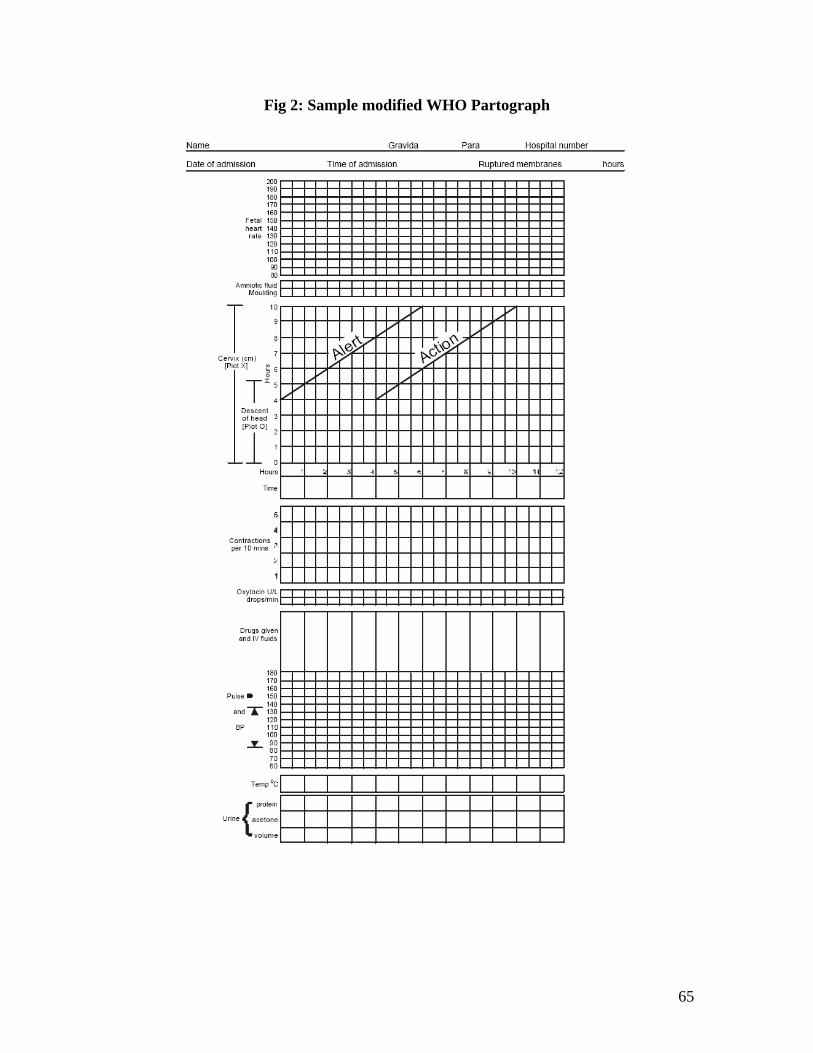
65
Fig 2: Sample modified WHO Partograph
66
Exercises on partograph: Normal Labour Exercise 1 – A sample partograph for normal labor is given below. Participants to be given the exercise written below and the sample partograph for understanding the plotting and analysis thereof: Text to the partograph:
• A primigravida was admitted in the latent phase of labor at 5 AM: - fetal head 4/5 palpable; - cervix dilated 2 cm; - 3 contractions in 10 minutes, each lasting 20 seconds; - Normal maternal and fetal condition.
Note: This information is not plotted on the partograph.
• At 9 AM: - fetal head is 3/5 palpable; - cervix dilated 5 cm;
Note: The woman was in the active phase of labor and this information is plotted on the partograph. Cervical dilatation is plotted on the alert line.
- 4 contractions in 10 minutes, each lasting 40 seconds; - Cervical dilatation progressed at the rate of 1 cm per hour.
• At 2 PM:
- fetal head is 0/5 palpable; - Cervix is fully dilated; - 5 contractions in 10 minutes each lasting 40 seconds; - Spontaneous vaginal delivery occurred at 2:20 PM.

67
Fig 3: Partograph for Exercise 1

68
Prolonged active phase of labor Exercise 2
1. The woman was admitted in active labor at 10 am. Fetal head palpable 5/5 Cervix dilated 4 cm Two contractions in 10 minutes lasting less than 20 seconds
2. At 12 pm: Fetal head still palpable 5/5 Cervix still dilated 4cm, and to the right of the alert line. No improvement in contractions.
3. At 2 pm Poor progress of labor due to inefficient uterine contractions diagnosed. Augmented labor with oxytocin 10units in 1 liter I/V fluid at 15 drops per minute. Escalated oxytocin, until a good pattern of contractions was established
4. At 7 pm: Fetal head 1/5 palpable Cervix dilated 10 cm. Four contractions per 10 min., each lasting for 45 sec.
5. Spontaneous vaginal delivery occurred at 8:10 pm. Assignment: All participants to draw a partograph on the above exercise so as to elicit all points. Exercise 3: Analyze and interpret the partograph shown below

69
Fig 4: Partograph for Exercise 3

70
Obstructed labor Exercise 4: Using the exercise given below, prepare the partograph 1. The woman was admitted in active labor at 10 AM:
- fetal head 5/5 palpable; - cervix dilated 4 cm; - Inadequate contractions (two in 10 minutes, each lasting less than 20 seconds).
2. at 2 PM:
- fetal head still 5/5 palpable; - cervix dilated 4 cm and to the right of the alert line; - membranes ruptured spontaneously and amniotic fluid is clear; - Inadequate uterine contractions (one in 10 minutes, lasting less than 20 seconds).
3. at 6 PM: - fetal head still 5/5 palpable; - cervix dilated 6 cm; - Contractions still inadequate (two in 10 minutes, each lasting less than 20
seconds).
4. at 9 PM: - fetal heart rate 80 per minute; - amniotic fluid stained with meconium; - -no further progress in labor.
5. Caesarean section was performed at 9:20 PM due to fetal distress. Exercise 5: Analyze and interpret the partograph shown below.

71
Fig 5: Partograph for Exercise 5

72
Unit 3.2 Unsatisfied Progress of Labor / Obstructed Labor Learning Objectives: At the end of the session, the participants will be able to:
• Diagnose and manage women in labor with unsatisfactory progress or obstructed labor.
3.2.1 Definition Patients admitted with h/o active labor for more than 12 hrs. (It is assumed that our own hospital's inpatients will never go to the stage of prolonged or obstructed labor) The latent phase is longer than 8 hours. Cervical dilatation is to the right of the alert line on the partograph.
3.2.2 Preventive care Inpatient of the hospital who goes into labor must be managed prospectively and expected intervention must be made based on the alert and action lines of partogram. 3.2.3 Protocol on Management of Prolonged labor/ obstructed labor A) Basic Treatment
a Assess the Feto-maternal conditions – - P/R/T/BP - Dehydration level - Uterine contractions (presence of bandl’s ring to diagnose obstructed labor) - Presenting part (PP)/ Position of fetus - Foetal Heart Rate and rhythm
b Correct dehydration and acidosis if any by IV route c Start antibiotics - Ampicillin + Gentamicin (may be omitted if membranes are
intact or recently ruptured). Third generation Cephalosporins may be started in more desperate cases.
d Start Metronidazole if anaerobic infection is suspected e P/V for:
- Status of cervical dilatation, oedema - Station of Presenting part , - Colour of liquor, - Caput on the head, - Moulding - Assessment of pelvis below the Presenting part, - Membranes present / absent, - Foul smelling vaginal discharge

73
B) Decision about the mode of delivery Indications of CS
a All cases of labor with: • CPD • Malpresentation • Foetal distress • Poor past obstetric history • Pre-Eclampsia • Obstructed labor with baby alive
b If the baby is dead:
• Senior most consultant to be called immediately • Craniotomy when the head is low and obstructed • Laparotomy in all the other cases for procedures covering:
- Only CS or - CS + repair of rupture uterus, if feasible - Ligation, if family complete - Hysterectomy - when the uterus cannot be conserved as per the
opinion of the senior consultant and if she has completed her family and given consent for this procedure

74
Unit 3.3 Fetal Distress
Learning Objectives: At the end of the session, the participants will:
• Discuss the signs and symptoms of fetal distress. • Understand the standard protocol to be followed when managing a woman in
labor and having fetal distress 3.3.1 Introduction
• Abnormal fetal heart rate (less than 100 or more than 180 beats per minute). • Thick meconium-stained amniotic fluid.
3.3.2 General management
• Prop up the woman or place her on her left side. • Stop oxytocin if it is being administered.
3.3.3 Abnormal fetal heart rate
• Diagnosed by intermittent auscultation
• If a maternal cause is identified (e.g. maternal fever, drugs), initiate appropriate
management. • If a maternal cause is not identified and the fetal heart rate remains abnormal
throughout at least three contractions, perform a vaginal examination to check for explanatory signs of distress:
(a) If there is bleeding with intermittent or constant pain, suspect
abruption placentae (b) If there are signs of infection (fever, foul-smelling vaginal discharge)
give antibiotics as for amnionitis; • If the cord is below the presenting part or in the vagina, manage
as prolapsed cord. • If fetal heart rate abnormalities persist or there are additional
signs of distress (thick meconium-stained fluid), plan delivery:
• A normal fetal heart rate may slow during a contraction but usually recovers to normal as soon as the uterus relaxes.
• A very slow fetal heart rate in the absence of contractions or persisting after contractions is suggestive of fetal distress.
• A rapid fetal heart rate may be a response to maternal fever, drugs causing rapid maternal heart rate (e.g. tocolytic drugs), hypertension or amnionitis. In the absence of a rapid maternal heart rate, a rapid fetal heart rate should be considered a sign of fetal distress.

75
• If the cervix is fully dilated and the fetal head is not more than 1/5 above the
symphysis pubis or the leading bony edge of the head is at 0 station, deliver by vacuum extraction or forceps
• If the cervix is not fully dilated or the fetal head is more than 1/5 above the symphysis pubis or the leading bony edge of the head is above 0 station, deliver by caesarean section. Confirm fetal distress by CTG, if available
3.3.4 Meconium Stained Liquor Meconium staining of amniotic fluid is seen frequently as the fetus matures and by itself is not an indicator of fetal distress. A slight degree of meconium without fetal heart rate abnormalities is a warning of the need for vigilance. Thick meconium suggests passage of meconium in reduced amniotic fluid and may indicate the need for expedited delivery and meconium management of the neonatal upper airway at birth to prevent meconium aspiration. In breech presentation, meconium is passed in labor because of compression of the fetal abdomen during delivery.
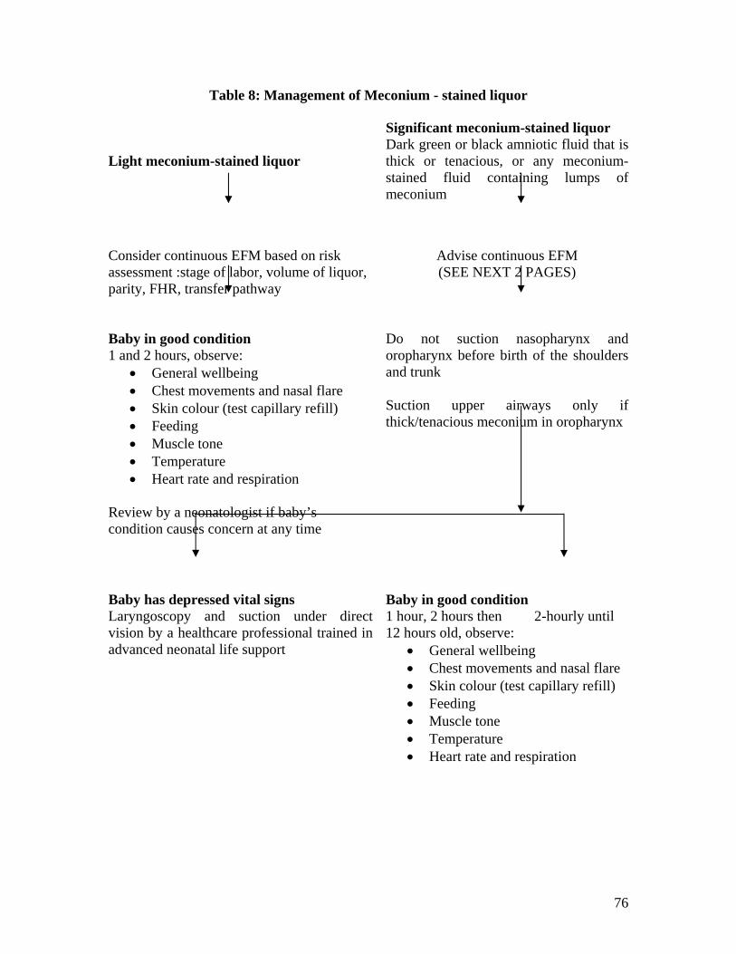
76
Table 8: Management of Meconium - stained liquor
Light meconium-stained liquor
Significant meconium-stained liquor Dark green or black amniotic fluid that is thick or tenacious, or any meconium-stained fluid containing lumps of meconium
Consider continuous EFM based on risk assessment :stage of labor, volume of liquor, parity, FHR, transfer pathway
Advise continuous EFM (SEE NEXT 2 PAGES)
Baby in good condition 1 and 2 hours, observe:
• General wellbeing • Chest movements and nasal flare • Skin colour (test capillary refill) • Feeding • Muscle tone • Temperature • Heart rate and respiration
Review by a neonatologist if baby’s condition causes concern at any time
Do not suction nasopharynx and oropharynx before birth of the shoulders and trunk Suction upper airways only if thick/tenacious meconium in oropharynx
Baby has depressed vital signs Laryngoscopy and suction under direct vision by a healthcare professional trained in advanced neonatal life support
Baby in good condition 1 hour, 2 hours then 2-hourly until 12 hours old, observe:
• General wellbeing • Chest movements and nasal flare • Skin colour (test capillary refill) • Feeding • Muscle tone • Temperature • Heart rate and respiration

77
Fig 6: Continuous Electronic Fetal Monitoring (EFM) Meconium – stained liquor
Continuous EFM Other risk factors present 3
Previous CS Pre-eclampsia Pregnancy > 42 weeks PROM > 24 hours Induced labor Diabetes Antepartum haemorrhage Other maternal medical disease Fetal growth restriction Prematurity Oligohydramnios Abnormal Doppler artery velocimetry Multiple pregnancies Breach presentation
Maternal factors that may contribute to an abnormal trace3
Woman’s position: advise her to adopt left-lateral position Woman is hypotensive Woman has just had a vaginal exam Woman has just emptied her bladder or bowel Woman has been vomiting or had a vasovagal episode Woman has just had regional analgesia sited or topped up.
With Oxytocin Suspicious trace: review; continue to increase oxytocin till 4 or 5 contractions every 10 min. Pathological trace: stop oxytocin; full assessment by obstetrician before recommencing
Normal trace with oxytocin Continue oxytocin until 4 to 5 contractions every 10 min. Reduce if more than 5 in 10
Inform that EFM will restrict woman’s mobility. Every hour take documented systematic assessment based on table 1 and 2
Real-time ultrasound assessment
Acute compromise (deceleration > 3 min)
Fetal death suspected with recordable trace Pathological
trace
If uterine hypercontractility consider 0.25 mg terbutaline subcutaneously4
Abnormal trace
Urgent Birth

78
EFM: DIFINITION AND CLASSIFICATIONS
Table 9: Definition of Normal, Suspicious and Pathological FHR traces
Category Definition Normal
Suspicious
Pathological
All four features are classified as reassuring
One feature classified as non-reassuring and the remaining features
classified as reassuring
Two or more features classified as non-reassuring or one or more
classified as abnormal
Table 10: Classification of FHR trace features
Feature Baseline
(bpm) Variability (bpm)
Decelerations Accelerations
Reassuring
Non-reassuring
Abnormal
110 – 160
100 – 109
161 – 180
< 100
> 180
Sinusoidal
pattern > 10
min
> 5
< 5 for 40 90
min
< 5 for 90
min
None
Typical variable
decelerations with over
50% of contractions, for
over 90 min
Single prolonged
deceleration for up to 3
min.
Either atypical variable
decelerations with over
50% of contractions or late
decelerations, both for over
30 min
Single prolonged
deceleration for more than
3 min
Present
The absence of
accelerations with
otherwise normal
trace is of
uncertain
significance

79
Record keeping: • Check date / time on EFM machine • Label FHR traces with mother’s name, date and hospital number. • Sign trace and record date, time and mode of birth. • None events, e.g. vaginal exam, FBS, epidural siting on trace. • Store traces securely.
Risk Management:
• Consider the time taken for instrumental vaginal birth and CS when making decisions about fetal wellbeing
• Keep FHR traces for 25 years; where possible store electronically • If the baby may suffer developmental delay, photocopy and store FHR traces
indefinitely • Use tracer systems if FHR traces stores separately from women’s records • Take paired cord blood gases only when concerned about the baby either in labor
or immediately following birth • Ensure an additional clamp for double-clamping is available at all birth settings.

80
Unit 3.4 Cord Prolapse Learning Objectives: At the end of the session we will be able to –
• Understand the clinical features and management of Cord Prolapse. NOTE: difference between cord presentation and cord prolapse is that cord presentation is the cord that has slipped down below the presenting part and is felt lying in the intact bag of membranes WHEREAS cord prolapse is the cord found lying inside the vagina or outside the vulva following rupture of membranes. 3.4.1 Protocol for cord prolapse
• The umbilical cord lies in the birth canal below the fetal presenting part. • The umbilical cord is visible at the vagina following rupture of the membranes.
General Management
• Give oxygen at 4–6 L per minute by mask or nasal canula. Specific Management PULSATING CORD
• If the cord is pulsating, the fetus is alive. • Diagnose stage of labor by an immediate vaginal examination • If the woman is in the first stage of labor, in all cases: - Put the feet up
- Wearing high-level disinfected gloves insert a FOLLEY’S CATHETER into the bladder and fill with 400 ml of normal saline
- Put a normal saline soaked pack into the vagina - If uterus is contracting, give terbutaline 500 microgram sub cutaneously/iv
(diluted and slowly) ; - Perform immediate caesarean section.
• If the woman is in the second stage of labor: - Expedite delivery with episiotomy and vacuum extraction or forceps;
If breech presentation, perform breech extraction and apply Piper or long forceps to the after-coming head
* Prepare for resuscitation of the newborn
81
CORD NOT PULSATING If the cord is not pulsating, the fetus is dead. Deliver in the manner that is safest for the woman. Case study on Cord Prolapse: Rama, 25 yrs old G3 P2+0 un booked patient (had received 2 doses of TT at the local hospital) with a full term pregnancy presented at the opd at Agra hospital with chief complaints of intermittent pain in lower abdomen for 2 hrs .her precious 2 deliveries were full term, uneventful and had been conducted at the local private hospital with pulse rate of 90bpm BP-120/70mmHg .on examination, FHS was 126 bpm, uterine contractions were mild with a frequency of 1 contraction of about 15-20 seconds coming in 10 minutes fetal presentation was cephalic, lie was longitudinal and vertex was just entering the brim. On per vaginal examination, it was found that external cervical os was patulous with internal cervical os admitting tip of finger with cervical effacement of about 50%-60%. She was admitted in the labor ward and was kept under observation for progress of labor and fetal heart monitoring. After about 3 hours the nurse monitoring the patient informed that although all her vital parameters were within normal limits with labor progressing satisfactorily at the rate of 3 contractions /10 minutes, there was an irregularity in the FHR pattern with the fetal heart rate dipping to 110bpm post contraction and this persisted for about 15 seconds post contraction. On per vaginal examination it was observed that the os was 5cm in dilatation and the vertex was still at the brim of the pelvis but a pulsating structure was present in front of the placenta with intact membranes. What is the most likely diagnosis? Most likely diagnosis in such a situation is cord presentation. How would you manage the present situation? Once the diagnosis had been made no attempt should be made to replace the cord, as it is not only ineffective but the membranes would have inevitably ruptured leading to prolapse of the cord. Since immediate vaginal delivery was not possible, CS was the best method of delivery.-during the time of preparing the patient for operative delivery, she was kept in exaggerated sim’s position to minimize cord compression. Along with this her bladder was filled with 400ml of normal saline with a Foley catheter, the balloon was inflated and the catheter was clamped. Bladder is to be emptied before a CS. (if the cord has prolapsed then just prior to making the abdominal incision, Fetal heart should be auscultated to avoid unnecessary section on a dead baby

82
Unit 3.5 Caesarean section (CS) It is the end point of a number of care pathways in obstetrics. The royal college of obstetricians and gynecologists has formulated evidence-based guidelines pertaining to the use of CS and published a guideline in April, 2004.
Common indications for a primary CS: • Failure to progress • Presumed foetal compromise • Breech presentations
Common indications for a repeat CS: • Previous CS • Failure to progress in labor • Presumed foetal compromise • Breech presentations
3.5.1 Emergency CS Emergency CS is very common in all maternity units, everywhere .The decision-delivery interval of less than30 minutes, is accepted as an audit standard for emergency services. Depending on the degree of urgency, 4 major indications are –
1. Immediate threat to life of mother/ baby: major degree of placenta praevia bleeding markedly, cord prolapse, eclampsia just controlled, and uncontrolled hypertension in severe PE with imminent eclampsia.
2. Maternal/foetal health compromise but not immediate life threatening: known major degree placenta praevia, irregular foetal heart with acidosis, uncontrolled hypertension, PE, diabetes
3. No compromise but needs early delivery to avoid complications: failed ECV, BOH, infertility
3.5.2 Planned CS It refers to a CS that is scheduled before the onset of labor for one or more specific clinical indications preferably after the completion of weeks. Indications for a planned CS:
1. Breech presentations- uncomplicated singleton breech with failed ECV cases contraindicated for ECV
2. Multiple pregnancy-if 1st twin cephalic not to be done routinely, 1st twin non cephalic common practice is CS but effects uncertain, gestational age should be less than 38 weeks if uncomplicated
3. Preterm birth –CS does not improve outcome, so CS not to be offered routinely 4. Small for dates- CS does not improve outcome, so CS not to be offered routinely 5. Placenta praevia –CS always in type 3 and type 4.
83
6. CPD-pelvimetry not useful in predicting failure of labor, so CS not done routinely. 7. MTC transmission of maternal infection is prevented by CS:
• HIV positive Women • Hepatitis B –insufficient evidence for planned CS • Hepatitis C-no CS as it does not reduce MTC • Hepatitis C + HIV – CS • HSV in 3rd trimester –CS reduces risk of NN infection • HSV recurring at birth-no routine CS , uncertain effects
3.5.3 Risks and benefits associated with each CS These are the effects of CS when compared to vaginal delivery
1. Short term effects around delivery following CS:
• Complications increased after CS Vis-a –Vis vaginal birth- abdominal pain, bladder injury, respiratory morbidity, readmission to hospital, need for further surgery , D and C , laparotomy , hysterectomy , admission to ICU , thromboembolic disease , length of hospital stay , even maternal death
• Complications not increased after CS Vis-a –Vis vaginal Birth-hemorrhage, infection, genital tract injury, initiation of micturition, neonatal morbidity/ mortality after planned CS .
• Complications decreased after CS Vis-a –Vis vaginal birth-perineal pain and trauma
Problems for neonates delivered by CS:
1. Neonatal respiratory morbidity 2. Iatrogenic prematurity 3. Laceration from surgeon’s scalpel 2% for vertex & 6% from
non-vertex. Data on Maternal Morbidity / Mortality on CS Elective CS Emergency CS Vag. Delivery Morbidity <2% Around 3% <2% Mortality – from elective CS is less than vaginal delivery
(Most reports on mat. Morbidity discounts risk of death during subsequent pregnancy) Long-term effects: Increased after CS – desire of having no more children, infertility, miscarriage, ectopic, ante partum SB, placenta praevia / abruption / adhesion in future pregnancy,

84
uterine rupture in future pregnancy, increased need for hysterectomy, incisional hernia, intestinal obstruction. No difference in effects (at 3/12 PN) : faecal incontinence, back pain, postnatal depression, dyspareunia ( fear psychosis) Decreased after CS – urinary incontinence (at 3/12 PN), UV prolapse 3.5.4 Anaesthesia for CS: • inform pt. , different anaesth./post op. analgesia • regional anaesthesia safer & results in less morbidity in mother/baby than GA • during CS under regional anaesthesia IV ephedrine/phenylephrine & volume
preload by crystalloid/colloid to reduce risk of hypotension • to reduce risk of aspiration pneumonitis , gastric volume & acidity-
antacids/H2receptors/ proton pump inhibitors are given • all CS pts. to be offered antiemetics • OT table to have 15
o tilt to reduce hypotension
3.5.5 Guidelines for surgical techniques: These apply to a term pregnancy, where the lower segment is well formed. It is also advised that the techniques may need to be modified during repeat CS or placenta praevia Guidelines: The appropriate skin incision – Appropriate skin incision is transverse Joel Cohen incision , which comprises of straight skin incision , 3 cm above SP , subsequent tissue layers opened bluntly , and , if necessary extended with scissors , not a knife. Advantages are ;
1. shorter operating time 2. Reduced postop. , febrile morbidity 3. less post operative pain 4. improved cosmetic effect
LUS incision &delivery of the baby: • Blunt extension after a transverse cut (advantages-reduces blood loss/
PPH/bl.transfusion) (Note – all women to be informed that risk of foetal tissue lacerations is about 2% with CS)
• Delivery of baby : forceps 1/ blades used in some cases , but long term neonatal morbidity uncertain

85
• Placenta removed by controlled cord traction and not MRP • Oxytocics-oxytocin 5iu slow iv drip • Caution-double gloves for CS of HIV+ cases
Repair of uterus :
• Intraperitoneally; exteriorization is not recommended as it causes more pain no improvement in blood loss/ infection rate.
• Single layer suture-effectiveness and safety uncertain. Until RCT available, repair in 2 layers recommended.
• Peritoneum repair-both visceral and parietal should not be sutured , as leaving them alone results in less postoperative analgesia and less operation time
• Rare midline abd. Incision-mass closure by slowly absorbable sutures , as it causes less incisional hernia / less dehiscence , than layered repair
Subcutaneous tissue repair: • Only if it is >2 cm:routine repair does not reduce wound infection rate • Superficial wound drain-no, as it does not reduce wound infection/wound
haematoma Prophylactic antibiotics:
• Prophylactic antibiotics are advised-first generation cephalosporins / ampicillin.
Thromboprophylaxis: Thromboprophylaxis is suggested in all obst. Cases who are at an increased risk of developing VTE, DVT.Use of graduated stockings, hydration, early mobilization, low molecularweight heparins advocated. Baby care:
• Good thermal care is very important as CS babies usually have a lower temperature
• Skin to skin contact with mother • To start breast feeding as soon as possible • To consider having 2 neonatologist for twin delivery
Care of woman after CS:
• One to one observation by skilled/ trained staff until she regains airway control, CV stability established and she is able to communicate any distress / problems. [admission to ICU is 9/1000 CS cases]
• Patient to be observed every 30 minutes in 1st2 hours then hourly until basic parameters are stable
• Pt. with intrathecal opioids hrly observation for 12 hours for diamorphine and 24 hrs. for morphine.

86
• Postop intrathecal opioids reduce analgesia need • Pain relief – for severe pain, Co-codamol + ibuprofen, in moderate pain-co
codamol only and for mild pain paracetamol only. • Add NSAID with analgesics, if not contraindicated –improves pain relief • No routine SR catheter after CS, but kept for 12 hours after last ‘top-up’
dose in regional anaesthesia • Dressing may be removed>24 hours, wound be kept clean and dry, assess
for wound infection • Routine resp. physio does not improve resp. s/s cough, phlegm, body
temperature, pulmonary changes, etc .CS increases risk of thromboembolic diseases, to note calf swellings, cough to detect early DVT/VTE.
• When to start food/ drink-any time she is thirsty / hungry, if no complications.
• Possible urinary s/s are UTI , stress incontinence[ 4% after CS] and UT injury [ 1 in 1000 CS]
• Irregular vaginal bleeding may be due to endometritis, rather than rpoc. • Hospital stay-3/4 days [vag. delivery-1/2days] , if no complaints and if
medical help available at home , she may be allowed to go home after 24 hours.

87
Unit 3.6 Vaginal Birth after Caesarean 3.6.1 Antenatal counseling • Women with a prior history of one uncomplicated lower-segment transverse
caesarean section, in an otherwise uncomplicated pregnancy at term, with no contraindication to vaginal birth, should be informed about the option of planned VBAC and the alternative of a repeat caesarean section (ERCS). Actual decision will be taken closer to the time of delivery.
• The antenatal counseling of women with a prior caesarean birth should be documented in the notes.
• A final decision for mode of birth should be agreed between the woman and her obstetrician before the expected/planned delivery date (ideally by 36 weeks of gestation).
• A plan for the event of labor starting prior to the scheduled date should be documented.
• Women considering their options for birth after a single previous caesarean should be informed that, overall, the chances of successful planned VBAC are 72–76%.
• All women who have experienced a prior caesarean birth should be counseled about the maternal and perinatal risks and benefits of planned VBAC and ERCS when deciding the mode of birth.
• The risks and benefits should be discussed in the context of the woman’s individual circumstances, including her personal motivation and preferences to achieve vaginal birth or ERCS, her attitudes towards the risk of rare but serious adverse outcomes, her plans for future pregnancies and her chance of a successful VBAC (principally whether she has previously had a vaginal birth. In addition, where possible, there should be review of the operative notes of the previous caesarean to identify the indication, type of uterine incision and any peri-operative complications.
• As up to 10% of women scheduled for ERCS go into labor before the 39th week, it is good practice to have a plan for the event of labor starting prior to the scheduled date.
3.6.2 Contraindications to vaginal birth after CS a) Previous classical or inverted T uterine scar b) Previous hysterotomy or myomectomy entering the uterine cavity c) Previous uterine rupture d) Presence of a contraindication to labor, such as placenta previa or malpresentation e) The woman declines a trial of labor after Caesarean and requests ERCS

88
3.6.3 Risk factors for unsuccessful VBAC a) Induced labor, b) No previous vaginal birth, c) Body mass index greater than 30 d) Previous caesarean section for dystocia. When all these factors are present, successful VBAC is achieved in only 40% of cases. There are numerous other factors associated with a decreased likelihood of planned VBAC success: a) VBAC at or after 41 weeks of gestation b) Birth weight greater than 4000 g c) No epidural anaesthesia d) Previous preterm caesarean birth e) Cervical dilatation at admission less than 4 cm f) Less than 2 years from previous caesarean birth g) Advanced maternal age h) Short stature Where relevant to the woman’s circumstances, this information should be shared during the antenatal counseling process to enable the woman to make the best informed choice. 3.6.4 Complications of VBAC
• There is a less than 1 % chance of rupture in case of attempted VBAC. • Women considering the options for birth after a previous caesarean should be
informed that planned VBAC compared with ERCS carries around 1% additional risk of either blood transfusion or endometritis.
• Maternal death from uterine rupture in planned VBAC occurs in less than 1/100,000 cases in the developed world;
3.6.5 Perinatal Outcome
• Absolute risk of perinatal loss is low in both groups( VBAC and ERCS), may be SLIGHTLY lower in ERCS
• Women considering the options for birth after a previous caesarean should be
informed that attempting VBAC probably reduces the risk that their baby will have respiratory problems after birth: rates are lower (2-3 %) with planned VBAC and higher (3–4%) with ERCS.

89
3.6.6 VBAC in Special Circumstances A) Preterm planned VBAC
• Women who are preterm and considering the options for birth after a previous caesarean should be informed that planned preterm VBAC has similar success rates to planned term VBAC but with a lower risk of uterine rupture.
• Twin gestation, fetal macrosomia, short inter delivery interval • A cautious approach is advised when considering planned VBAC in women with
twin gestation, fetal macrosomia and short inter delivery interval, as there is uncertainty in the safety and efficacy of planned VBAC in such situations.
B) Where and how should VBAC be conducted?
• With continuous intra partum care and monitoring • Resources available for immediate caesarean section • Availability of blood • Advanced neonatal resuscitation. • Epidural anaesthesia is not contraindicated in planned VBAC • Electronic fetal monitoring when available should be utilized, following the onset
of uterine contractions for the duration of planned VBAC. • Continuous Intrapartum care is necessary to enable prompt identification and
management of uterine scar rupture. • There is no single pathognomic clinical feature that is indicative of uterine rupture
but the presence of any of the following peripartum should raise the concern of the possibility of this event:
1. Abnormal CTG 2. Severe abdominal pain, especially if persisting between contractions 3. Chest pain or shoulder tip pain, sudden onset of shortness of breath 4. Acute onset scar tenderness 5. Abnormal vaginal bleeding or haematuria 6. Cessation of previously efficient uterine activity 7. Maternal tachycardia, hypotension or shock 8. Loss of station of the presenting part.
C) Role of induction and augmentation
• There is the two- to three-fold increased risk of uterine rupture and around • 1.5-fold increased risk of caesarean section in induced and/or augmented labor
compared with spontaneous labor. • There is a higher risk of uterine rupture with induction of labor with
prostaglandins. • DO NOT USE PROSTAGLANDINS FOR INDUCTION OF LABOR IN
VBAC EXCEPT SELECTED CASES OF CONFIRMED IUFD
90
• There should be careful serial cervical assessments, preferably by the same person, for both augmented and non-augmented labors, to ensure that there is adequate cervico metric progress, thereby allowing the planned VBAC to continue.
• The decision to induce, the method chosen, the decision to augment with oxytocin, the time intervals for serial vaginal examination and the selected parameters of progress that would necessitate and advise on discontinuing VBAC should be decided by the consultant obstetrician.
• The additional risks in augmented VBAC mean that: a Although augmentation is not contraindicated it should only be preceded by
careful obstetric assessment, maternal counseling and by a consultant-led decision
b Oxytocin augmentation should be titrated such that it should not exceed the maximum rate of contractions of four in 10 minutes; the ideal contraction frequency would be three to four in 10 minutes
c Careful serial cervical assessments, preferably by the same person, are necessary to show adequate cervico metric progress, thereby allowing augmentation to continue.
d The intervals for serial vaginal examination and the selected parameters of progress that would necessitate discontinuing VBAC labor should be consultant-led decisions.
91
MODULE 4
Care & Management of
Third Stage of Labor
Unit 4.1 Active Management of Third Stage of Labor
Unit 4.2 Post Partum Hemorrhage Unit 4.3 Post Partum Care Unit 4.4 Puerperal Sepsis

92
About this Module This Module discusses about third stage of Labour, its signs and management of any complication, which might occurs during the third stage. Unit 4.1 Active Management of Third Stage of Labor Learning Objectives At the end of the session the participants will be able to:
• Implement the different modalities for active management of the third stage of labor to prevent PPH.
• Discuss management of PPH 4.1.1 Protocol for Active management of III stage of labor in all
cases
a It is advisable for all the admitted obstetric cases in active labor to have an IV line started latest by the II stage of labor. If possible secure IV line with a intravenous canula only.
b Active management of III stage of labor in all cases. i. Prophylactic Oxytocics (Syntocinon) 10 units IM/ 20 units in 500ml IV drip
after the delivery of the baby and after excluding the presence of a second fetus in the uterus.
ii. Cord clamping after cessation of pulsation (about 2 minutes after baby is born)
iii. Controlled Cord Traction iv. Uterine massage
A) Oxytocin
• Within 1 minute of delivery of the baby, palpate the abdomen to rule out the presence of an additional baby(s) and give oxytocin 10 units IM.
• Oxytocin is preferred because it is effective 2 to 3 minutes after injection, has minimal side effects and can be given to all women. If oxytocin is not available, give ergometrine 0.2 mg IM or Misoprostol- Three tablets of 200 mcg each (total 600 mcg) may be given rectally.
• Make sure there is no additional baby(s) before giving these medications. • Do not give ergometrine to women with pre-eclampsia, eclampsia or high
blood pressure because it increases the risk of convulsions and cerebrovascular accidents.

93
B) Controlled Cord Traction • Clamp the cord close to the perineum using sponge forceps. Hold the clamped cord
and the end of forceps with one hand. • Place the other hand just above the woman’s pubic bone and stabilize the uterus by
applying counter traction during controlled cord traction. This helps prevent inversion of the uterus.
• Keep slight tension on the cord and await a strong uterine contraction (2–3 minutes).
• When the uterus becomes rounded or the cord lengthens, very gently pull downward on the cord to deliver the placenta. Do not wait for a gush of blood before applying traction on the cord. Continue to apply counter traction to the uterus with the other hand.
• If the placenta does not descend during 30–40 seconds of controlled cord traction (i.e. there are no signs of placental separation), do not continue to pull on the cord:
- Gently hold the cord and wait until the uterus is well contracted again. If necessary, use a sponge forceps to clamp the cord closer to the perineum as it lengthens;
- With the next contraction, repeat controlled cord traction with counter traction.
Never apply cord traction (pull) with out applying counter traction (push) above bone with the other hand. As the placenta delivers, the thin membranes can tear off. Hold the placenta in two hands and gently turn it until the membranes are twisted.
• Slowly pull to complete the delivery. • If the membranes tear, gently examine the upper vagina and cervix wearing
high-level disinfected gloves and use a sponge forceps to remove any pieces of membrane that are present.
• Look carefully at the placenta to be sure none of it is missing. If a portion of the maternal surface is missing or there are torn membranes with vessels, suspect retained placental fragments.
• If uterine inversion occurs, reposition the uterus. • If the cord is pulled off, manual removal of the placenta may be necessary.
C) Uterine Massage
• Immediately massage the fundus of the uterus through the woman’s abdomen until the uterus is contracted.
• Repeat uterine massage every 15 minutes for the first 2 hours. • Ensure that the uterus does not become relaxed (soft) after you stop uterine
massage.

94
4.1.2 Management for Vaginal and Perineal Tears Examination for Tears
• Examine the woman carefully and repair any tears to the cervix or vagina or repair episiotomy
Repair of vaginal and perineal tears There are four degrees of tears that can occur during delivery:
• First degree tears involve the vaginal mucosa and connective tissue. • Second degree tears involve the vaginal mucosa, connective tissue and underlying
muscles. • Third degree tears involve complete transection of the anal sphincter. • Fourth degree tears involve the rectal mucosa.
Note : It is important that absorbable sutures be used for closure. Polyglycolic sutures are preferred over chromic catgut for their tensile strength, non allergic properties and lower probability of infectious complications. Chromic catgut is an acceptable alternative, but is not ideal.
Repair of First and Second Degree Tears Most first degree tears close spontaneously without sutures.
• Review general care principles • Provide emotional support and encouragement. Use local infiltration with
Lignocaine. If necessary, use a pudendal block. • Ask an assistant to massage the uterus and provide fundal pressure. • Carefully examine the vagina, perineum and cervix. • If the tear is long and deep through the perineum, inspect to be sure there is no
third or fourth degree tear: - Place a gloved finger in the anus; - Gently lift the finger and identify the sphincter; - Feel for the tone or tightness of the sphincter.
• Change to clean, high-level disinfected gloves. • If the sphincter is injured, see the section on repair of third and fourth degree
tears. • Note: It is important that absorbable sutures be used for closure. Polyglycolic
sutures are preferred over chromic catgut for their tensile strength, non-allergenic properties and lower probability of infectious complications. Chromic catgut is an acceptable alternative, but is not ideal.
• If the sphincter is not injured, proceed with repair.

95
Fig 7: Exposing Perineal tear
Apply antiseptic solution to the area around the tear.
• Make sure there are no known allergies to Lignocaine or related drugs. • Note: If more than 40 mL of Lignocaine solution will be needed for the repair,
add adrenaline to the solution. • Infiltrate beneath the vaginal mucosa, beneath the skin of the perineum and
deeply into the perineal muscle using about 10 mL 0.5% Lignocaine solution. Note: Aspirate (pull back on the plunger) to be sure that no vessel has been penetrated. If blood is returned in the syringe with aspiration, remove the needle. Recheck the position carefully and try again. Never inject if blood is aspirated. The woman can suffer convulsions and death if IV injection of Lignocaine occurs.
• At the conclusion of the set of injections, wait 2 minutes and then pinch the area with forceps. If the woman feels the pinch, wait 2 more minutes and then retest.
Repair the vaginal mucosa using a continuous 2-0 suture:
• Start the repair about 1 cm above the apex (top) of the vaginal tear. Continue the suture to the level of the vaginal opening;
• At the opening of the vagina, bring together the cut edges of the vaginal opening; • Bring the needle under the vaginal opening and out through the perineal tear and
tie.

96
Fig 8: Repair of Perineal Tear (A)
Repair the perineal muscles using interrupted 2-0 suture. If the tear is deep, place a second layer of the same stitch to close the space.
Fig 9: Repair of Perineal Tear (B)
Repair the skin using interrupted (or subcuticular) 2-0 sutures starting at the vaginal opening.
• If the tear was deep, perform a rectal examination. Make sure no stitches are in the rectum.

97
Fig 10: Repairing the skin
Note- Recent tear should be repaired immediately following the delivery of the placenta. This reduces the chances of infection and minimizes the blood loss In cases of delay beyond 24 hrs, the repair is to be withheld. Antiseptic dressing is prescribed and the wound is allowed to heal by granulation tissue or repaired after the infection is controlled. The complete tear, however, should be repaired after 3 months if delayed beyond 24 hours.
After Care
The after care of the repaired perineal injuries is similar to that following episiotomy.
Care following repair of complete tear-
1. A low residue diet consisting of milk, eggs, biscuits, bread, fish, sweets, etc. is given from 2nd day onwards
2. Milk of magnesia 8 ml twice daily beginning on the second day and increasing the dose to 15mlon the third day is a satisfactory regime to soften the stool. If the patient fails to pass stool and is having discomfort, compound enema may be gently given by a rubber catheter
3. Intestinal antiseptics like Metronidazole 400mg thrice daily may be continued for 5-7 days

98
Unit 4.2 Post Partum Hemorrhage Learning Objectives At the end of the session, we will be able to:
• Discuss the prevention, differential diagnosis and management of post partum hemorrhage.
• Manage a case of PPH as per the protocols 4.2.1 Diagnosis of PPH
Table : Diagnosis of Post Partum Hemorrhage
Symptoms and signs typically present
Probable diagnosis
PPH (the uterus is soft and not contracted) Atonic uterus • PPH with complete/ intact placenta and /or
uterus contracted
Tears in the cervix or vagina
Placenta not delivered within 30 minutes after delivery and uterus not contracted
Retained placenta
A portion of the maternal surface of the placenta is missing or the membranes are torn
Retained placental fragments
• The uterine fundus is not felt on abdominal palpation
• Slight or intense pain • Inverted uterus apparent at the vagina
Inverted uterus
• PPH (bleeding is intra-abdominal and / or trans vaginal) with or without
• Severe abdominal pain (may decrease after rupture of the uterus)
• Shock (may be out of proportion to visible blood loss)
• Rapid maternal pulse • Tender abdomen • Uterine contour not felt.
Rupture uterus
• Secondary PPH(after 24 hrs. to 6 weeks) • Uterus softer and larger than expected for the
elapsed time • Bleeding is variable (light or heavy,
continuous or irregular), and foul-smelling • Anaemia, pyrexia, tachycardia
• secondary PPH

99
4.2.2 Prevention & Management of Post Partum Hemorrhage Definition
PPH is defined as blood loss following delivery, leading to the deterioration of the vital parameters like pulse, BP, respiration, with or without symptoms of sweating, palpitation, etc.
Prevention
IV line access: It is advisable for all the admitted obstetric cases in active labor to have an IV line latest by the II stage of labor. If possible secure IV line with a Intravenous canula only.
Active management of III stage of labor in all cases as mentioned above.
Management of PPH
a. Ask for extra help b. Rapid evaluation of general condition including vital signs c. If signs of shock appear then immediately resuscitate the patient d. Other steps are:
• Massage the uterus to contraction • Oxytocin 10units or 20 units in 500 ml IV drip • IV Infusion through a 16 gauze needle, one on each hand • Catheterization to empty bladder and note urinary output • If placenta is already expelled - examine for its intactness. • Examine the cervix & the vagina for any trauma
Management of Retained Placenta If placenta is not delivered within 30 minutes after delivery of baby, then:
a. If placenta is separated and is placed in the lower segment or vagina, it is to be removed manually with IV sedation under anaesthesia
b. Bladder to be catheterized, if not earlier done c. Start oxytocin 10 units or 20 units in 500 ml I/V drip d. If uterus is contracted - Try CCT - Failing which proceed for manual removal of
placenta (MRP) under anaesthesia e. If still bleeding continues - assess clotting status or exclude trauma.
Management of Atonic Uterus Immediately ask for Senior Consultant's help Step I
a) Continue uterine massage b) Oxytocin - 20 units in 1 ltr. 60 drops / min not more than 3 ltr. fluid with oxytocin
100
c) Injection Ergometrine: IV 0.2 mg d) Repeat after 15 minutes & every 3 hours (maximum 5 doses) e) Arrange blood transfusion
Step II
Injection 15 Methyl PGF2 Alpha
a) 0.25 mg 1M (can be given intra myometrial, one on each quadrant if required) b) Repeat every 15 minutes c) Maximum 8 doses (2 mg)
Step III
If bleeding continues - perform bimanual compression of uterus Step IV
If bleeding continues - perform compression of aorta Step V
Uterine compression sutures of Lynch or any accepted modifications Step VI
Stepwise uterine artery devascularisation –
a) Uterine artery (bilateral) ligation & utero-cervical branch if necessary b) Utero-Ovarian artery (bilateral). Save ovaries if patient is a young woman c) Unilateral internal iliac artery ligation d) Bilateral internal iliac artery ligation
Step VII Hysterectomy - The patient should be counseled and a written informed consent MUST be obtained prior to the surgery. If the patient is under GA, unconscious or unable to decide, then the same procedure has to be conducted for the woman's partner or the available next of kin.
101
4.2.3 Case Study 1 – Retained Placenta 41 year old para 5 had a rapid labor at home and delivered a male infant in the bathroom, he begins to cry spontaneously. She was brought to the hospital about 1 hour after the delivery because she had not delivered the afterbirths. She had normal vitals and there was no evidence or h/o heavy bleeding. On enquiring about her past obstetric history it was found that she had a previous CS for transverse lie 3 years back. She had received Inj. Methergine at the village local hospital. Key Points She was a grand multipara with one previous CS and may have had atonic uterus. Her pulse was 90 bpm and BP was normotensive. On per abdominal examination, uterine fundus was found to be well contracted. On local / perineal examination, no perineal or vaginal tear was found. Cervical tear was also excluded. It was noticed that she was in great pain. The diagnosis was certain that it is a case of retained placenta Blood was sent for cross matching and an intravenous line was established a gentle attempt at continuous cord traction was made but it was unsuccessful .Patient was shifted to the operation theatre for a manual removal of placenta under GA or regional block . .care must be taken to avoid uterine perforation at the site of previous CS. Risks of PPH, uterine inversion, and infection should be kept in mind and managed accordingly In this case placenta was found to be anterior and just overlying the previous scar; manual removal under GA was done successfully. The scar was found to be intact on digital examination. No PPH was reported and mother returned home the following day. 4.2.4 Case Study 2– Post Partum Hemorrhage A 22 year second gravida was admitted via casualty to the labor ward. She was previously 'un booked', but appeared to be about 36 weeks pregnant. She revealed that she started to experience abdominal pain since last 3 hour. There had been no vaginal bleeding or loss of liquor. On examination, the uterus was non-tender, the fetal heart rate was normal, and the cervix was 5 cm dilated. Urgent investigations showed a hemoglobin concentration of 8.6 g/dl, blood group 0 Rh positive. Labor progressed rapidly to full dilation 1 1/2 hours later. Artificial rupture of the membranes was then carried out, and at 03.52 hours a spontaneous vertex delivery of a live female weighing 2.4 kg occurred after a second stage of only 10 minutes. The third stage was managed actively and the placenta was removed complete. A small midline perineal tear had occurred, which was sutured with catgut. Mother and baby were transferred to the postnatal ward, but at 05.50 hours the senior house officer was asked to see her because of continued vaginal bleeding. On examination, she was pale with a pulse rate of 120 beats/min, and a blood pressure of

102
110/60 mmHg. She was complaining of lower abdominal pain, but examination revealed a well contracted uterus which was only moderately tender. The rest of the abdomen was soft and bowel sounds were normal. Questions
1. Discuss how you would manage this situation. 2. What are the common causes of primary postpartum haemorrhage? 3. What is meant by 'active management' of the third stage?
Key Points The postnatal ward is not a good place for the investigation of postpartum hemorrhage, particularly at night, the beds are not conducive to thorough examination, the lighting is often poor and staffing levels are low. The patient was therefore returned to the labor ward for investigation. Prior to transfer, an intravenous infusion of normal saline was set up, and sent for cross matching (4 units requested) Once in Labor ward, observations of pulse and blood pressure were continued, and a thorough examination was carried out in the lithotomy position. Speculum examination of the vagina allowed the evacuation of 500 ml of blood clots, with the confirmed fresh bleeding from the upper vagina. Because of the lack of patient cooperation, largely due to inadequate analgesia, a general anesthetic was administered. The whole genital tract was then examined thoroughly by inspection with a powerful light and by digital exploration. This showed that the vagina and uterus were intact, but the cervix felt ragged. Using three sponge holding forceps, advancing them round the cervix one at a time, several large cervical lacerations were demonstrated with a bleeding vessel at the base of each one. A blood transfusion was commenced, and the bleeding vessels under run with catgut sutures to obtain hemostasis. In view of the low hemoglobin on admission and an estimated loss of at least 1 liter of blood, 4 units of blood were transfused over the next 12 hours. The commonest cause of primary postpartum hemorrhage is uterine atony. This will usually respond to intravenous oxytocic. Laceration of the genital tract (vaginal, paravaginal, cervix and uterine) are the next commonest, but often overlooked. A sinister cause is failure of normal coagulation, which can occur following placental abruption, fulminating pre-eclampsia, or septicemia (inherited coagulopathies are very rare in women). Active management of the third stage involves using an oxytocic (usually 5 units of oxytocin and 0.5 mg of ergometrine intramuscularly) to accelerate contraction and retraction of uterus. To avoid trapping the placenta, controlled cord traction is necessary to remove the placenta as soon as it has separated from the uterus. (Separation is not achieved by pulling on the cord, but by contraction of the uterus. Failure to appreciate this point can result in snapping of the cord, hemorrhage and uterine inversion). This method has been show in a large prospective randomized controlled trial to minimize blood loss from postpartum hemorrhage, and to educe the need for blood transfusion by at least half compared with physiological management without oxytocics. Side effects of oxytocic are rare, with nausea and vomiting being

103
mainly due to ergometrine. Ergometrine can also cause hypertension and should not be given to women with pre-eclampsia or essential hypertension. Instead, a continuous infusion of oxytocin should be used.
4.2.5 Case Study 3- Post Partum Hemorrhage A 22-year-old primigravida was admitted to the labor ward at 39 weeks gestation with spontaneous rupture of her membranes after an uneventful pregnancy. On arrival she was noted to be draining clear liquor, but was only having weak contractions once every 20 minutes. The cervix was still about 2 centimeters long and just admitted a fingertip. The fetal heart was auscultated with a stethoscope and the rate found to be 146 beats per minute and regular. The woman was encouraged to walk about as she wished until contractions became established. Fourteen hours and good sleep later, uterine contractions were still weak and irregular, though she continued to drain clear liquor. Her general condition was good, she was apyrexial and the fetal heart rate remained normal. Vaginal examination confirmed that there had been no change in cervical dilatation. It was therefore decided that labor should be induced with an oxytocic infusion. Six hours later the cervix was effaced, moderately well applied to the presenting part and about 5 cm dilated. An epidural was inserted for analgesia and the oxytocin infusion continued. Full dilatation was reached 11 hours after the infusion was begun and after pushing for 90 minutes a forceps lift-out was performed for 'maternal exhaustion'. Syntometrine (syntocinon 5 units and ergometrine 0.5 mg) was given intramuscularly with the delivery of the anterior shoulder. After waiting for signs of separation, the placenta was delivered by continuous cord traction. Unfortunately, this was followed by a rapid haemorrhage of almost a liter of blood. Questions
1. What aspect of the labor might have contributed to the postpartum haemorrhage (PPH)?
2. How could the delivery and third stage have been managed differently? 3. The patient's haemoglobin on the third day after delivery was 8.6 g/dl. What
would you advise? Key points
1. Prolonged oxytocic infusion often predisposes to uterine atony and hence postpartum hemorrhage
2. In cases such as this the oxytocin infusion rate should be increased immediately following delivery and syntometrine is often better given as the fetal head is delivered, rather than waiting for the anterior shoulder. If there is any sign of atony, the uterus should be compressed bimanually until a further dose of ergometrine is given and / or the uterus contracts.
104
The traditional response to hemoglobin this low is blood transfusion and there is no doubt that women in this situation feel very much better for it, particularly if they intend to breast feed. Nevertheless, there is a growing resistance to transfusion on the part of the public because of the perceived risk of transmission of infectious diseases (particularly HIV) and many women will decline transfusion unless absolutely necessary. In such situations the value of the value of transfusion should be compared to the disadvantages, especially the tiredness and lethargy that may be experienced and the women herself given the final say. If transfusion is refused, iron supplements should be commenced immediately and continued until the hemoglobin level rises to normal.
105
Unit 4.3 Post Partum Care Learning Objectives: At the end of the session, we will have an understanding of:
• The relevance of post partum care. • The essential components on the delivery of post partum care.
4.3.1 Introduction Conventionally, the first 42 days (6 weeks) after delivery are taken as the postpartum period. Of this, it is the first 48 hours, followed by the first one week, which are the most crucial periods, as most of the fatal and near-fatal maternal and neonatal complications arise during this period. 4.3.2 Postnatal check-ups Ist PNC: Conducted within the first 24 hours after delivery. History Taking
• Where did the delivery take place? • Who conducted the delivery?
Maternal symptoms Ask for the following symptoms:
• H/o profuse or unusual bleeding following delivery: (to assess the presence of PPH). If she soaks a pad or cloth in <5 minutes, it indicates PPH. It requires urgent management and referral
• H/o convulsions or loss of consciousness • H/o abdominal pain • H/o fever
Maternal Examination
• Check the pulse, BP, temperature. • Look for pallor. • P/A Examination: To see if the uterus is well contracted (hard and round) and to
rule out the presence of any uterine tenderness. • Examine the vulva and the perineum for the presence of any tear, swelling or
abnormal lochia. • Examine the pad for bleeding and assess if it is heavy.

106
Neonatal symptoms and signs Check for signs of possible serious bacterial Infection in newborn and take appropriate action. IInd and IIIrd PNC: Should take place on the 3rd and 7th day respectively, following delivery. History Taking A similar history needs to be taken again except for following additional questions that should be asked
• unusual bleeding P/V –secondary PPH • H/o foul-smelling vaginal discharge: This could be indicative of puerperal sepsis.
Manage accordingly • H/o abnormal swelling (engorgement) and/or tenderness of the breasts, axillary
tail. If present, manage accordingly .Application of cream is advised for cracked nipples
• H/o pain or any problems while passing urine (dribbling or leaking). • H/o easy fatigability and "not feeling well". • H/o feeling unhappy or crying easily. This indicates postpartum depression, and
usually occurs after the first week of delivery. Examination Similar to the examination conducted during the first visit. It includes the following:
• Check the pulse, BP, temperature • Look for pallor • Examine the abdomen to see if the uterus is well contracted (hard and round) and
to rule out the presence of any uterine tenderness. • Examine the vulva and the perineum for the presence of any tear, swelling or pus
discharge. • Examine the pad for bleeding and lochia. Assess if it is profuse and whether it is
foul-smelling. • Examine the breasts for the presence of any lumps or tenderness. • Check the condition of the nipples. If they are cracked or sore application of
crème is advised Neonatal examination– same as mentioned above. 4.3.3 Counseling Diet and Rest
• Foods rich in calories (550 kcal extra per day for initial 6 months and then 400 kcal during next 6 months), proteins, iron, vitamins and other micronutrients should be advocated.
• She should be advised against food taboos, if they are harming woman and / or baby.
107
• The woman needs sufficient rest during the postpartum period to be able to regain her strength. She and her husband and other family members should be advised that she should not be allowed to do any heavy work during the postpartum period, except for looking after herself and her baby.
Resumption of Sex
• The couple should be advised to abstain from having sex during the first 6 weeks following delivery, or till the perineal / episiotomy wound heals (if present), whichever is later.
Contraception
• Remind the woman that strictly exclusive breastfeeding for first three months with no menstruation till that time may protect most women against pregnancy. Post partum contraception should be discussed with the woman. The various choices of contraceptive methods available to the couple must be told to them, so that they can make an informed choice.
Home Care and feeding of Newborn
• Advice the mother for - Exclusive Breast Feeding for 6 months, - Kangaroo Mother Care, - To attend immunization clinic for timely and complete immunization of
infant. - When to return immediately to clinic WITHOUT WAITING ( *Danger
signs to watch for) Danger Signs
• Excessive vaginal bleeding, i.e. soaking more than 2 or 3 pads in 20-30 minutes after delivery, OR bleeding increases rather than decreases after the delivery
• Convulsions • Fast or difficult breathing • Fever and weakness; inability to get out of bed • Severe abdominal pain • The woman feels ill • Swollen, red or tender breasts, or sore nipples • Dribbling of urine or painful micturition • Pain in the perineum, or pus draining from the perineal area • Foul-smelling lochia.

108
Unit 4.4 Puerperal Sepsis Learning Objectives At the end of the session we will be able to -
• Have an understanding of the differential diagnosis of fever after child birth. • Manage a case of sepsis in puerperium as per the protocols.
4.4.1 Definition Puerperal sepsis is an infection of the genital tract at any time between the onset of rupture of membranes or labor and the 42nd day following delivery or abortion in which any two or more of the following signs and symptoms are present:
• Fever of 38.5 ºC or higher, measured orally on any two occasion; • foul odour of the vaginal discharge; • Pelvic pain and uterine tenderness; • Delay in the rate of reduction of the size of the uterus (sub involution of the
uterus). 4.4.2 Management of puerperal sepsis A) Investigations : CBC , HVS-C/S , URINE-routine , microscopic , culture and
sensitivity , USG B) Start Antibiotic therapy immediately - to manage/treat the current infection and
to stop it from spreading further.
Regimen: - If the woman is not very sick (no fever or it is low grade, the pulse is not
very high, and. the consciousness is normal), you may start the woman on: Cap. Ampicillin 1 g t.d.s. orally PLUS Tab. Metronidazole 400 mg t.d.s.orally PLUS Inj. Gentamicin 80 mg IM 8 hrly , change the antibiotic regimen according to the vaginal and urine culture and sensitivity report till patient is afebrile for 48 hours.
- If the woman is very sick (very high fever, a rapid pulse, appears confused), a combination of antibiotics preferably through the parenteral route is advised.
A useful regime is:
Inj. Ampicillin 1 g IV 8 hrly, PLUS Metronidazole 400 mg IV 8 hrly, PLUS Inj. Gentamicin 80 mg IM 8 hrly ,change the antibiotic regimen according to the culture and sensitivity report. Change to oral antibiotics 48 hrs after the patient is afebrile for 5 days.

109
C) Give Plenty of fluids: The aim of this is to correct or prevent dehydration and to help bring down the fever.
4.4.3 Management in severe cases In severe cases (e.g. if the woman is in shock or is in a confused, delirious state), it is necessary to give IV fluids immediately. If the woman is conscious and there is no indication that a general anesthetic may be needed in the next few hours, she should also be given oral fluids. In mild cases, a simple increase in the oral fluid intake is sufficient. A) Rule out the presence of retained placental fragments /forgotten pack in
vagina Remove the forgotten pack from the vagina. Retained placental fragments can be a cause of puerperal sepsis. Suspect this if the uterus is soft and bulky and if the lochia is excessive and contains blood clots. Manage accordingly.. B) Tetanus Toxoid If there is a possibility that the woman was exposed to tetanus (if, for example, cow dung, mud or herbs were inserted into the vagina), and there is uncertainty about her vaccination history, then give her TT. C) Provide Skilled Nursing Care This requires careful attention. Nursing staff must be instructed to ensure the following:
• Advise the woman to take bed rest; • Monitor vital signs (temperature, pulse, BP, RR); • Measure intake and output of fluids; • Keep an accurate record of the medicines given; • Prevent the spread of infection and cross-infection.

110
References

111
References 1. Royal College of Obstetricians and Gynaecologists, Setting Standards to Improve
Women’s Health,Birth After Previous Caesarean Birth, Green Top Guidelines No. 45, February 2007.
2. Intrapartum Care, Quick Reference Guidelines, September 2007, NICE Clinical
Guidelines 55, Developed by the National Collaborative Centre for Women’s and Children Health.
3. K. Park, Park’s Textbook of Preventive and Social Medicine, 19th edition, 2007,
Bhanot Publication. 4. Integrated Management of Pregnancy and Childbirth, Pregnancy, Childbirth,
Postpartum and Newborn Care: A guide for essential practice, WHO, 2006. 5. Guidelines for Pregnancy Care and Management of Common Obstetric
Complications by Medical officers, Ministry of Health and Family welfare, Government of India, 2005.
6. D.C.Dutta, Textbooks of Obstetrics, VI edition, 2004, New Central Book Agency (P)
Ltd. 7. Royal College of Obstetricians and Gynaecologists, Setting Standards to Improve
Women’s Health, Preterm Prelabor Rupture of Membranes, Green Top Guidelines No. 44, November 2006.