obstetric trauma surgery - UroToday
94
Transcript of obstetric trauma surgery - UroToday

1

2

3
obstetric trauma surgery
art and science
setting standards by evidence-based practice
kees Ia stool fistulas
step-by-step reconstructive surgery
based on
findings and outcome
570 surgical and 900 nonsurgical procedures in 1,400 patients with kees Ia stool fistula
out of
5,100 procedures in 4,650 rectovaginal fistula patients
out of 29,000 obstetric fistula/trauma reconstructive procedures
in 25,000 patients
with full documentation
kees waaldijk

4
obstetric trauma surgery art and science
series of textbooks each with a specific topic
setting evidence-based standards
this series has been developed for setting evidence-based standards in the training and management of the obstetric trauma in all its forms in the low-, in the middle- as well as in the high-income world the name of the series has been changed from obstetric fistula into obstetric trauma surgery since the fistula is only one aspect of the complex obstetric trauma though a systematic approach is being followed this seems to be a utopia since the material is too extensive and it would take too long each time a specific topic has been finalized it will be published as a separate entity; with later on an update if needed then somewhere along the line a comprehensive summary will be produced in order to have a representative overview the emphasis is placed on the functional anatomy of the pelvis, pelvis floor and pelvis organs, the female urine and stool continence mechanisms, the mechanism of action and the principles of reconstructive and septic surgery for training reasons it will follow a step-by-step approach and repetition; together with schematic drawings and photographs the whole series is based on kees archives of obstetric trauma with so far 29,000 reconstructive and conservative procedures in 25,000 patients with a rare “complete” documentation of each procedure and results as to healing and continence by electronic reports with 150 parameters, over 100,000 pre/intra/postoperative digital photographs, 30 hours of video recordings and a comprehensive database as personal experience over a 35-year period from 1984 up till now as such it is considered to be a full scientific evidence-based report; though it has not followed the “you peer me, i peer you” doctrine it is also not following the strict protocol of the international scientific journals or the so-called established theories; since only dead fish follow the flow of the river; and strict protocols kill any creativity; the message is not in the format since it is the life work of the author it is written in his own words and in his own style writing things down helps the author in organizing his own understanding and ideas

5
foreword the literature dealing with rectovaginal fistulas is scarce and the description of specific operation techniques even scarcer more or less a terra incognita even as a trained and experienced colorectal surgeon the author found and still finds it difficult to handle the rectovaginal/stool fistula in all its forms; with falling and standing up over the years he developed a series of kees operation technique principles which he would like to describe in detail since the different types have their own general and specific characteristics with their own general and specific reconstructive surgery principles they will be addressed one by one since the rectum is a high-pressure organ and the vagina a zero- or low-pressure organ stool and/or flatus may pass from the rectum into the vagina and then to the outside therefore, once the rectum heals the posterior vagina wall will always heal; so one has to concentrate upon meticulous closure of the rectum though there is an enormous variety the kees Ia fistulas have one thing in common that they do not involve the anatomic stool continence/closing mechanism without a rectum stricture and without a circumferential defect the author made an effort to explore this terra incognita; a start for further development out of the series obstetric trauma surgery; art and science this textbook presents and outlines an evidence-based approach to the kees Ia stool fistulas which has been used by the author in a prospective way; as has been backed up by extensive documentation however, this should be used as a guideline for reconstructive surgery principles since each fistula constitutes its own unique entity needing a customized approach; it cannot beat common sense kees waaldijk md phd august 2020
6

7
table of contents
foreword 3
introduction 7
essentialsessentials kees stool fistula classification 10
essentials kees Ia stool fistulas 13
kees Ia stool fistulascharacteristics 18
operation principleskees operation principles stool fistulas 26
kees Ia fistulas 30
resultssurgery 52
spontaneous healing 54
asymptomatic 59
overall results 60
basic sciencewhat is needed 62
stool continence mechanism in the female 63
essentials pelvis anatomy 71
management principles stool fistulas 75
postscriptum 85
abbreviations 86
measurements 88
references 90

8
introduction kees Ia stool fistulas
within the kees classification the fistula which I does not involve the anatomic stool con tinence mechanism a without a rectum stricture and without a circumferential defect is a kees Ia fistula so after healing the continence rate will be (almost) 100% they are for (almost) 100% combined with a vesicovaginal fistula; unless the cause is penetration trauma, infection or a surgical complication which is rare so far, a grand total of 1,417 patients with a kees Ia stool were treated the mechanism is pressure necrosis of the soft tissues between the fetal head and the maternal sacrum bone there is a high tendency to spontaneous healing as demonstrated by the fact that 837 women healed without any surgical intervention during the first 3-6 months post partum; many of them already during the first 2 months the reconstructive surgery principles are described in a step-to-step manner; and were applied as guideline in 570 operations in 501 patients since these are proximal fistulas mostly deep inside the vagina with poor visibility, poor access and poor instrument handling, the operation procedure is complicated though the final result may be excellent as demonstrated in these 501 patients with a healing rate of 96% and a continence rate of 99% of the healed fistulas small very proximal fistulas may cause few or no symptoms at all since 79 patients did not want an operation stating once in while only flatus and/or diarrheic stool thru vagina and some even denied its existence but definitely fistula as seen during vvf-repair
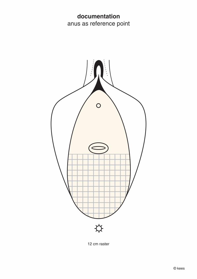
the anatomic stool continence mechanism in the female consists of the
distal 4-5 cm of the anorectum with
internal sphincter + external sphincter ani supported by
the perineal body fixed anus as reference point for measurements
kees waaldijk md phd august 2020

9
introduction kees Ia stool fistulas
within the kees classification the fistula which I does not involve the anatomic stool con tinence mechanism a without a rectum stricture and without a circumferential defect is a kees Ia fistula so after healing the continence rate will be (almost) 100% they are for (almost) 100% combined with a vesicovaginal fistula; unless the cause is penetration trauma, infection or a surgical complication which is rare so far, a grand total of 1,417 patients with a kees Ia stool fistula were treated the mechanism is pressure necrosis of the soft tissues between the fetal head and the maternal sacrum bone there is a high tendency to spontaneous healing as demonstrated by the fact that 837 women healed without any surgical intervention during the first 3-6 months post partum; many of them already during the first 2 months the reconstructive surgery principles are described in a step-to-step manner; and were applied as guideline in 570 operations in 501 patients since these are proximal fistulas mostly deep inside the vagina with poor visibility, poor access and poor instrument handling, the operation procedure is complicated though the final result may be excellent as demonstrated in these 501 patients with a healing rate of 96% and a continence rate of 99% of the healed fistulas small very proximal fistulas may cause few or no symptoms at all since 79 patients did not want an operation stating once in while only flatus and/or diarrheic stool thru vagina and some even denied its existence but definitely fistula as seen during vvf-repair
the anatomic stool continence mechanism in the female consists of the
distal 4-5 cm of the anorectum with
internal sphincter + external sphincter ani supported by
the perineal body fixed anus as reference point for measurements
kees waaldijk md phd august 2020

10

11
essentials essentials stool fistula classification kees Ia kees Ib kees Ic kees IIa kees IIb kees III postpartum stool/flatus incontinence essentials stool fistula surgery essentials kees Ia I without involvement stool continence mechanism a without rectum stricture and without circumferential defect

12
essentials kees classification of stool fistulas based on tissue loss, continence mechanism and operation technique
with consequences for prognosis
any classification is a compromise considering the enormous variety of trauma classification the following classification is presented according to the anatomic/physiologic location with consequences for operation technique only; see table I kees I fistulas not involving the continence/closing mechanism
kees II fistulas involving the continence/closing mechanism
kees III miscellaneous and of course postpartum stool/flatus incontinence without a fistula table I classification of fistulas according to anatomic/physiologic location kees I not involving continence mechanism proximal fistulas
a without rectum stricture
b with rectum stricture
c with circumferential defect
kees II involving continence mechanism distal fistulas
a without sphincter ani involvement
b with sphincter ani involvement
kees III miscellaneous, e.g. colostomy, ileouterine fistulas etc this classification is based on the progressive quantitative and qualitative amount of tissue loss and on involvement of the stool continence/closing mechanism the transition from kees I into kees II fistulas is at 4-5 cm from the anus whilst for the kees I fistulas a rectum stricture or circumferential defect has to be looked for the proximal kees I fistulas are due to pressure necrosis with anatomic tissue loss; few due to surgery most of the distal fistulas kees II are due to a cut-thru mechanism without anatomic tissue loss; including penetration trauma and surgical complications

13
a grading of involvement of the stool continence mechanism of the different types is presented in table II table II involvement of continence mechanism according to type type involvement of continence mechanism kees Ia none
kees Ib none
kees Ic none kees IIa from minimum up to moderate
kees IIb extensive kees III none results postrepair incontinence is not a major problem, though it may occur in kees IIb fistulas, whilst kees Ic fistulas have the worst results as to closure and may need a combined abdominovaginal approach; further, no clear relation to type comment so far it is the only classification with a
solid scientific background
clear operation technique principles for each type
prediction of outcome in terms of closure and continence not only the fistula has to be classified, but all the lesions/defects have to be objectively described/documented in writing to be completely transparent however, since the variety is so immense and there are no sharp demarcations but fluid transitions between the different types, this classification should be used as a compre hensive guideline since each fistula constitutes a separate unique entity and needs its own specific customized approach, and that is exactly what makes obstetric fistula surgery so intriguing and challenging since there are no identical obstetric fistulas fistula size, vagina strictures, scarring, stenosis and/or previous repair(s) are no part of any classification; it only may make the operation more complicated

14
15
essentials rectovaginal/stool fistula surgery operation principles for each type
type rectum closure special post vagina wall direction measures only half-open
adaptation kees Ia transverse (+ colpotomy) transverse kees Ib transverse + stricture transverse disruption kees Ic circumferential colpotomy transverse end-to-end (+ stricture disruption) highly complicated kees IIa common sense (+ perineal body) transverse or transverse or longitudinal longitudinal kees IIb longitudinal + sphincter ani 1x transverse
+ perineal body adaptation kees III special class of its own that needs their own specific approach comments these are only guidelines and the approach has to be customized since each fistula constitutes its own unique entity there is a clear relation between the reconstructive surgery principles and fistula type in order to reconstruct the functional anatomy there is no relation between fistula type and outcome; only that kees Ic fistulas are the most complicated with the worst outcome whilst postrepair incontinence may only occur in kees IIb fistulas kees IIb fistulas need thorough theoretic/practical knowledge of the stool continence mechanism, otherwise the results will be poor the author has never performed a colostomy for his obstetric trauma surgery; which would automatically mean at least 3 operations
16

17
essentials kees Ia stool fistulas characteristics the kees Ia fistulas comprise a group of fistulas which do not involve the anatomic stool continence mechanism; without a rectum stricture and without a circumferential defect most of them are at or fixed onto the posterior cervix lip though they may be located any where between 4-5 cm from the anus as reference point and the cervix or vault the small median ones at the cervix have a tendency to spontaneous healing if there is no infection and no outflow obstruction the lateral ones may be fixed to the lateral pelvis wall which has to be released before closure the small very proximal ones may be (almost) asymptomatic since only flatus/diarrheic stools per vaginam which is not a problem for the patients or they are even denying it on explicit asking; and as such a surprise finding intraoperatively (flatus) there is an enormous variety also within the kees Ia class and each fistula constitutes its own specific entity which needs its own customized approach nb the kees Ia fistulas are almost 100% combined with a vesicovaginal fistula = vvf mechanism of action pressure necrosis of the soft tissues in between the fetal skull and the maternal sacrum bone; so real anatomic tissue loss from minimal to extensive reconstruction the more proximal/deep the fistula the more complicated the repair since access is poor and instrument handling deep inside the vagina is difficult though other surgeons may prefer another route, for all fistulas the vagina is the route of choice since this is the most logic and least traumatic approach physiologic incision thru fistula, sharp dissection and then transverse rectum closure by a double layer (if possible) of inverting interrupted/continuous polyglycolic acid normally, a colostomy and/or colpotomy is not required; whilst a colostomy does not guarantee rectum decompression and would automatically mean three operations prognosis
good as to healing and excellent as to continence in kees Ia fistulas
18

19
kees Ia stool fistulas
I fistulas not involving continence mechanism a without rectum stricture and
without circumferential defect characteristics mechanism of action characteristics baseline data reconstructive surgery step-by step reconstruction discussion

20
kees Ia stool fistulas I fistulas not involving continence mechanism
a without rectum stricture and without circumferential defect
introduction the kees classification is based upon the quantitative and qualitative amount of tissue loss and on the involvement of the anatomic stool continence mechanism in the female with consequences for the operation technique and prognosis the fistula which I does not involve the anatomic stool continence mechanism a without a rectum stricture and without a circumferential defect is a kees Ia fistula also within this kees Ia class the variety is enormous mechanism of action pressure necrosis of the soft tissues in between the fetal skull and the maternal sacrum bone; so real anatomic tissue loss from minimal to extensive characteristics almost 100% combined with vesicovaginal fistula in kees Ia fistulas there is always anatomic tissue loss of the rectum, prerectal fascia and posterior vagina wall with possible tissue loss of the posterior cervix there may be major tissue loss with also trauma to the sacrospinous ligament, (ischio) coccygeus muscles and piriformis muscles the majority are located at the vault near or at the posterior cervix; with a fluid transition of the proximal kees Ia into kees Ib and into kees Ic stool fistulas the rest is located anywhere between the vault/cervix and the internal rectum sphincter (stool closing mechanism) with a fluid transition of distal kees Ia into kees IIa fistulas normally the fistulas are in the midline though they can be situated very laterally as well if the fistulas are near or fixed to the posterior cervix and if the cervix retracted into the abdomen or fixed towards one of the ischium spines, visualization and instrumentation will be complicated since the stool continence mechanism is not involved successful repair will ensure full stool/flatus continence

21
small kees Ia fistulas may heal spontaneously if no infection/no outflow obstruction as noticed during follow up in patients coming early for postpartum urine leakage and also passing flatus and/or stools per vaginam at first visit; and healed with full stool/flatus continence when seen later; so far, over 800 patients with spontaneous healing the very proximal kees Ia fistulas may be a surprise finding at examination at operation beginning or during operation for a vesicovaginal fistula (flatus; compression of distal rectum by speculum) since several patients say they have no complaints (maybe only when diarrhea) and some do deny its existence even upon explicit asking nb the kees Ia fistulas are almost 100% combined with a vesicovaginal fistula = vvf; isolated fistulas are rare reconstruction see special chapter since the majority are deep inside the vagina the repair poses a challenge to the fistula surgeon since access, visualization and instrumentation are complicated normally the rectum is closed transversely in line with the natural tissue forces and common sense infrequently the rectum is closed longitudinally in line with common sense seldomly an oblique fistula is closed obliquely purse string suturing was not really effectve in principle the rectum is closed by a double inverting layer; the first interrupted for strength and the second continuously for flatus proof closure the sutures are thru the prerectal fascia/muscularis resulting as well in rectum mucosa adaptation upon tying the sutures primary suturing is performed in some 20% with good results chance of healing is good; with excellent full continence of the closed/healed fistulas discussion the deeper (parts of) the fistula inside the vagina the poorer the access and the more complicated the instrumentation, especially if combined with a retracted/moving cervix and fixation of the fistula onto the sacrum; the same for fistulas not in the midline try to bring the fistula towards the outside and if not in the midline towards the midline though the (proximal) fistulas may be complicated to repair the prognosis as to closure and continence is excellent; actually, for all kees Ia fistulas there is a fluid transition from proximal kees Ia into kees Ib and into kees Ic fistulas and a fluid transition from distal kees Ia into kees IIa fistulas

22
personal experience of the author with 1,417 consecutive patients right from the beginning the high tendency to spontaneous healing was noted whilst several had no symptoms despite a fistula so that only 501 patients needed surgery spontaneous healing in 837 60% 570 operations in 501 35% few/no symptoms in 79 6% patients not bothered the baseline data for the 501 patients as operated will be presented in next chapter

23
baseline data kees Ia stool fistulas 501 patients as operated so far
as a failed system of obstetric care obstetric versus other cause obstetrics is by far the main cause of the rectovaginal fistula in 97.4% of the patients as operated for kees Ia stool fistula; as indication of a failed system of obstetric care another cause, iatrogenic, postmeasles necrotizing infection, yankan gishiri and rape accounted for only 2.6% combination with vesicovaginal fistula = vvf almost 100% are combined with a vesicovaginal fistula whilst isolated fistulas are rare accounting for only < 0.5%; nb 3 patients had a ureterosigmoidostomy as well combination with sphincter ani rupture kees IIb combined with sphincter ani rupture in 7 or 1.4% as expression of the extent of obstetric trauma in prolonged obstructed labor fistula size the majority 60% are rather small as situated at the vault at or near the cervix however, the size alone is not representative since there are extensive small fistulas and “small” extensive fistulas in terms of real obstetric trauma age at fistula the great majority of 67% were younger than 20 yr when they developed the fistula however, the age is not a contributing factor as a blunt lie by hypocritic people behind a desk look at the teenage deliveries in the united states and the united kingdom you become pregnant later you get your fistula later since these vocal fistula-illiterate people continue their blunt lies the author will continue his professional opinion not that it matters but accepting vocal political rhetoric is not in his system; the only thing acceptable is creating a network of functioning obstetric care index parity the index parity ranged from para 0 thru para 14 with majority 60% at para 1 indicating that the first delivery is a test case for the pelvis

24
place of delivery the great majority of 75% or three quarters delivered in a hospital indicating a failed system of obstetric care mode of delivery the great majority of 75% delivered vaginally spontaneously or by assisted labor whilst the rest 25% delivered by cesarean section indicating cesarean section too late to prevent (vvf+rvf) fistulas as another indication of a failed system of obstetric care operated before already 33% or one third were operated before from 1 to 4 times whilst 24 patients (5%) had a colostomy as well indicating low success rate despite claims by the verbal simple surgeons sex infant at index delivery the 3:1 male to female sex rate in rectovaginal fistulas and the 2:1 rate in vesicovagi nal fistulas cannot be explained by the heavier male birthweight; so another mechanism must be in play eluding the author who was the first to find and point this out; already in his phd study in 1989 health status infant a stillbirth rate of 97% indicating the extreme trauma to the infant once the infant has died, its head, the biggest circumference, shrinks and then the mother may be able to push the dead infant out but only if she herself has not died as well in the process duration of fistula on operation day roughly 50% with a fistula duration from 1 yr to over 30 yr indicating non-availability of expertise vagina shortening in roughly 35%, stenosis in 34% and strictures in 10% indicating what the obstetric trauma means for the pelvis organs with consequences for reconstruction of the functional female pelvis anatomy foot drop peroneal nerve trauma was found in 83% of which 316 bilateral plus 47 right foot and 42 left foot so no difference between right and left foot the obstetric trauma is more than only the obstetric fistula

25
table kees Ia stool fistula data obstetric versus other cause
obstetric 488 97.4%
nonobstetric 13 2.6% iatrogenic 5 1.2%
postmeasles necrotizing infection 5 1.0% yankan gishiri 1 0.2%
rape 1 0.2% combination with vesicovaginal fistula = vvf
combined with vvf 495 98.8% isolated 6 1.2%
combination with sphincter ani rupture kees IIb
combined with sphincter ani rupture 7 1.4% fistula size
small < 2 cm 296 59.1% medium 2-3 cm 141 28.1% large 4-5 cm 48 9.6% extensive > 6 cm 26 5.2%
age at fistula > 9 yr 6 1.2%
10-15 yr 211 42.1%
16-19 yr 118 23.6%
20-29 yr 98 19.6%
30-39 yr 57 11.4%
40-49 yr 9 1.8% index parity the index parity ranged from para 0 thru para 14 with majority 60% at para 1
para 1 291 58.1%
indicating that the first delivery is a test case for the pelvis

26
place of delivery home 100 20.5% hospital 369 75.6% not asked 19
mode of delivery
spontaneous/assisted vaginal 363 74.4% cs-delivery 125 25.6%
operated before
at least once up to 4x 164 32.7% colostomy 24 4.8% + ureterosigmoidostomy 3
sex infant at index delivery
male 368 75.4% female 120 24.6%
health status infant
sb 474 97.1% live 13 2.7%
live/died 1st day 1 0.2% duration of fistula on operation day
< 3 mth 98 19.6% 3 mth to < 1 yr 153 30.5% 1 yr to over 30 yr 240 47.9%
vagina
shortening < 9 cm 175 34.9% vagina stenosis 170 33.9% vagina stricture 49 9.8%
foot drop peroneal nerve trauma total 405 83.0%
bilateral 316 64.8% right foot only 47 9.6% left foot only 42 8.6%

27
reconstructive surgery principles
kees Ia stool fistulas
surgical principles
rectovaginal fistulas kees Ia stool fistulas step-by-step reconstruction
transverse closure longitudinal closure infrequently oblique upon indication purse string hardly discussion

28
surgical principles for rectovaginal fistulas in line with principles of septic surgery
since the vagina is never sterile introduction the main objectives of any (obstetric) fistula repair are: aa to close the fistula bb to make the patient continent and cc to preserve or to provide her with something for sexual intercourse
if these three objectives have been achieved the patient will be rehabilitated completely into her own society; this will take place spontaneously without further measures patient consent any patients is asked by the surgeon himself if she wants and agrees to be operated or not; a written consent is obtained as well timing of operation timing of operation: as soon as the wounds are clean the patients is considered to be suitable for operation unless her general condition does not permit it if overt infection sitzbaths with water and a detergent like omo (cheap and available and highly effective) 3 times daily for 20 minutes until wound is clean since the principles of septic surgery are being applied tissue inflammation itself is no contraindication preoperative bowel preparation though it is nice to have an empty rectum somehow it seems not possible to organize mechanical bowel cleansing: too early, too late or not at all or by patient to patient; or whatever, it is not functioning in severe rectum stricture the proximal loop cannot be cleansed by enema and the author has experienced serious complications by forceful enemas thru distal colostomy in case of stool impaction into the distal colostomy loop up to severe rectum stricture not noticed during the enema and then sent to operating theater with stool still impacted and contaminated enema fluid transudation thru the traumatized sigmoid wall into the peritoneal cavity therefore, the patient is instructed to stop eating the night before and to pass stools the morning of operation day routine pre-, intra- and post-operative antibiotics as prevention of what?? in septic surgery routine antibiotics are considered malpractice the author is not against the powerful working of antibiotics but only on real indication and only then it makes sense nb if routine antibiotics would really work (as predicted) one would no longer see post operative wound infection and sepsis instead this practice contributes to the ever increasing pandemic of antibiotic multi-resistance in a circulus vitiosus due to the financial lobbying of the drug makers, the fear of litigation and not to forget the vocal demand by the patients

29
manpower fistula surgery is a one-man job, and all the operations are performed by the surgeon and one assistant who is doing the instrumentation; one retractor inside the vagina is already a crowd anesthesia spinal anesthesia with a long-acting drug is the anesthesia of choice route of operation the vagina is the route of choice some surgeons prefer the combined abdominovaginal approach for kees Ic fistulas position the exaggerated lithotomy position with the buttocks over table end and legs flexed and abducted in leg holders is the position of choice though some surgeons prefer the head up/buttocks down position for kees II fistulas instruments normal (long) vaginal surgery instruments are needed together with the following special instruments: a) auvard weighted speculum for keeping the vagina open, b) long allis clamps for picking up the vagina or rectum edges, c) a pair of sharply curved thorek scissors for dissecting the posterior vagina wall from the prerectal fascia/rectum besides, a complete well-functioning hydraulic operation table is of utmost importance and a must suturing materials normal resorbable polyglycolic acid size 00 and 0 and nonabsorbable nylon sutures size 1 and 2 are needed with a strong small curved needle concentrate upon rectum closure since the rectum is a high-pressure organ and the vagina a zero- or low- pressure organ once the rectum has healed, the posterior vagina wall will always heal therefore, concentrate upon the meticulous rectum closure and only adapt the posterior vagina wall or leave it half open in line with septic surgery two-layer rectum closure in principle the rectum is closed in two layers, the first interrupted and the second continuous, by inverting sutures for strength since the rectum cannot be decompressed and for flatus- proof closure otherwise there may be contamination when flatus should pass thru the small openings in between the interrupted sutures in case of rectum distension by gas intraoperative stool contamination cleanse it with abundant clean water since the solution to pollution is dilution and leave the pvw completely open or half open after rectum closure in order to prevent abscess formation and breakdown intraoperative antibiotics on indication if there is stool contamination with large wound area or after sharp dissection of rectum stricture the author gives tinidazole orally and one shot broad-spectrum antibiotic im in order to prevent endotoxin shock/septicemia since the bacterial contamination is sucked up by the open veins into the general vascular circulation

30
transverse posterior colpotomy with opening of abdomen for the proximal kees Ia/Ib fistulas a transverse posterior colpotomy is not necessary but may facilitate the tension-free rectum closure; however, with risk of intraperitoneal contamination in kees Ic fistulas a transverse posterior colpotomy is obligatory in order to perform (adapted) circumferential dissection plus (adapted) circumferential end-to-end rectosig moidorectostomy if a colpotomy has been performed the abdomen has to be closed proximally from the repair to prevent intraperitoneal contamination if the repair should break down in severe funnel-shape shortening (ba hanya) a colpotomy is performed to facilitate the repair and to reconstruct a neovagina in the same session grafting there is no need for grafting; reconstruction of the functional pelvis anatomy will be sufficient, ie meticulous rectum closure that is the decisive factor combination rectovaginal fistula with vesicovaginal fistula in one session only if it is not too complicated and both can be done within a reasonable time frame; it is better to do them in two sessions than to compromise both in one session in the proximal kees Ia and Ib fistulas the stool fistula should be closed first in order to prevent intraoperative stool contamination kees Ic fistulas are so complicated that it is not advisable to combine them with the repair of a vesicovaginal fistula in the distal kees IIa and IIb fistulas the vesicovaginal fistula should be repaired first otherwise access to the operation field may be compromised which is excellent in the kees IIb fistulas in two sessions in principle the vvf is repaired in the first session since that is the wish of the patient in most cases and if successful the rvf can be done in the second session however, when the patient wishes it the other way the rvf is done first nb a rvf does not interfere with the healing of a vvf-repair in the author’s experience primary suturing of small kees Ia fistulas in small proximal kees Ia fistulas near or at cervix/vault a freshening is made of the fistula edge and then only pvw closure (onto posterior cevix) is performed in an everting donati manner resulting in inverting good adaptation of the rectum; with good results however, make sure there is no rectum stricture in small proximal kees Ib fistulas the same can be done; but then posterior disruption of the rectum stricture has to be performed; the results are moderate to good delicate rectum tissue the rectum tissue is rather delicate and has to be handled with care prerectal fascia + muscularis in closure of the fistula it is the prerectal fascia/muscularis which is picked up by the needle/suture whilst the mucosa will be adapted on tying the sutures theoretically and in principle the needle should not go thru the rectum lumen but that is not always avoided and actually without negative effect upon healing

31
check on rectum closure by vaginal visual inspection and intrarectal digital examination half-open posterior vagina wall adaptation in line with the principles of septic surgery since the vagina is never sterile in order to avoid abscess formation and breakdown once the high-pressure rectum has healed, the posterior wall of the low-pressure vagina will always heal large defects in the posterior vagina wall can be left open for natural secondary epithelization whereby the superficial layer of the prerectal fascia will epithelize into vagina epithelium or can be filled up by different kinds of skin flaps decompression to avoid tension on sutures/repair though after vvf-repair complete decompressions of the bladder can be ensured by an indwelling catheter it is not possible to achieve this of the rectum, even with colostomy, so from time to time there will be (high) tension on the sutures/repair by gas/flatus and stools; and stool softeners are indicated to promote smooth fecal propulsion and smooth defecation this explains the fact that the postoperative breakdown rate in rectovaginal/stool fistulas is higher than in vesicovaginal/urine fistulas colostomy = iatrogenic colocutaneous kees III fistula the rationale for colostomy in abdominal colon surgery is proximal decompression in order to prevent tension on the sutures with the possibility of breakdown with contamina tion of the peritoneal cavity as a life-threatening complication however, complete continuous decompression is not guaranteed since stool may still enter the distal colostomy loop with high pressure inside the distal loop and eventual defecation thru the anus; in combination with stool thru the functioning colostomy in rectovaginal fistula surgery where the repair and sutures are outside the abdomen the repair may break down but no stool contamination of the peritoneal cavity; so not a life-threatening complication a colostomy means automatically 3 operations: colostomy, after functioning of colosto my the rvf-repair and after objective fistula healing colostomy closure the author has never performed a colostomy in his obstetric trauma surgery, still with good results however, stool softeners are indicated to minimize straining on defecation traction on repair by fixed/moving cervix since the fixation of the prerectal fascia onto the posterior cervix is via the vault, there is hardly any traction on the repair/sutures by the cervix so this is not a factor in the healing process of a rectovaginal/stool fistula; unlike in vesi covaginal/urine fistulas principles of surgical technique(s) the vaginal approach is the route of choice with or without unilateral, median or bilateral episiotomies, spinal anesthesia is the anesthesia of choice and the (exaggerated) lithotomy position is the position of choice for kees I thru kees IIb fistulas however, kees III fistulas may need a different approach

32
reconstructive surgery kees Ia stool fistulas step by step
introduction the main objectives of any (obstetric) fistula repair are: aa to close the fistula bb to make the patient continent and cc to preserve or to provide her with something for sexual intercourse
if these three objectives have been achieved the patient will be rehabilitated completely into her own society; this will take place spontaneously without further measures step-by-step reconstruction see general principles
i anesthesia, position 000 spinal anesthesia with long-acting agent 001 the patient is placed in the exaggerated lithotomy position with the legs flexed and slightly abducted in stirrups and her buttocks over the end of the operation table; this is the position of choice if visibility is still poor the inclination of the operation table has to be increased; so more head down/buttocks up ii systematic examination under anesthesia 002 a careful inspection and systematic examination (under anesthesia!) of the whole obstetric trauma and of the fistula as to size, location and texture of the fistula in relation to the anus and the cervix or vagina vault, as to the condition of the vagina such as stricture, stenosis or even atresia, if there is a vesicovaginal fistula as well, if the fistula is accessible, if there is a stricture, circumferential defect etc 003 check pubic arch in °; if this is < 80° access to operation field and instrumentation may be complicated the narrower the pubic arch the more complicated the repair becomes 004 check vagina length in cm; if this is less than 9 cm there has been substantial vagina tissue loss 005 check position/mobility of cervix; if retracted with paradoxic movement on cough visibili ty will be poor with difficult instrumentation

33
iii kees classification 006 based upon this examination the fistula is classified, and the surgeon makes up his definite plan of action how to handle this specific fistula as its own unique entity
iv access to operation field 007 the labia minora are sutured onto the inside of the upper legs to keep the vagina open laterally 008 in order to improve the accessibility a uni- or bilateral episiotomy is performed at 4-5 and/ or 7-8 o'clock or a small median episiotomy at 6 o'clock if done within the skin grease/lines and final skin closure by intracutaneous suturing the scar will be invisible 009 then an auvard self-retaining weighted speculum is placed inside the vagina with under neath a gauze covering the anus to keep the vagina open posteriorly; no more specula
v incision and dissection 010 put one or two long allis clamps onto posterior vagina wall (or onto cervix) proximally from the fistula and have assistant pulling it upwards and towards the outside and if not in the middle towards the midline this will make the repair less complicated 011 a physiologic transverse incision is made within the ruga folds of the posterior vagina wall thru the fistula; then a circumferential incision is made at the fistula edge 012 the posterior vagina wall is dissected sharply from the prerectal fascia/anterior rectum wall using scalpel and sharply curved thorek scissors in order to execute a tension-free repair; aim for just sufficient dissection in one go to minimize wound surface and postoperative scarring; avoid the salami technique with cutting everywhere resulting in excessive scarring nb if intentionally or accidently a colpotomy has been performed during incision/dissec tion the abdomen has to be closed at operation ending proximally from the repair to prevent contamination of the peritoneal cavity if the repair should break down
vi double-layer rectum closure 013 the principles of reconstructive surgery and common sense dictate the direction of closure: longitudinal, transverse or oblique

34
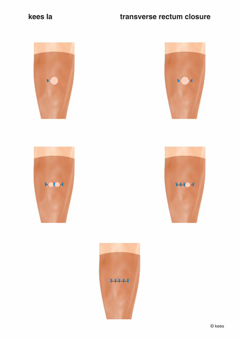
most of the time a transverse closure is the most logical in kees I fistulas as it presents itself this way and is in line with the natural tissue forces whilst longitudinal closure seems to be against these forces 014 the defect within the prerectal fascia/muscularis is closed with in the process closure of the rectum with a double layer of interrupted/continuous inverting polyglycolic acid 00 or 0 in order to obtain a flatus-proof closure transverse closure from north to south anterior to posterior closure is from bilaterally towards the midline
first inverting interrupted layer
015a start with the 2 lateral sutures
015b then the middle suture
015c if necessary complete by sutures in between the lateral and the middle suture
second inverting continuous layer if possible
015d complete the rectum closure by a second continuous layer inverting the first layer longitudinal closure from west to east side to side closure is from proximal towards distal
first inverting interrupted layer
015aa start with the most proximal (deepest) suture and work from proximal to distal
second inverting continuous layer if possible
015ab complete the rectum closure by a second continuous layer inverting the first layer oblique closure closure is from bilateral fistula edges towards the midline
first inverting interrupted layer
015aaa start with the 2 lateral sutures
015aab then the middle suture
015aac if necessary complete by sutures in between the lateral and the middle suture
second inverting continuous layer if possible
015aad complete the rectum closure by a second continuous layer inverting the first layer

35
016 good bites are taken thru the prerectal fascia/muscularis to get broad adaptation of the raw prerectal fascia/muscularis with adaptation of rectum mucosa 017 care is taken only to adapt the tissues and not to apply tension on the sutures as then they may cut through; remember sutures cannot heal, only adapt 018 care is taken not to go through the rectum mucosa as theoretically this might lead to flatus contamination thru the small needle holes but in practice this does not happen 019 cave do not cut the sutures too short since then the knot(s) will slip and loosen up nb at each step the surgeon should ask himself what am i doing exactly, which type of tissues are adapted, is it in line with the natural tissue forces and functional anatomy, and how will it look after 1 hour, 1 day, 1 week, 1 month and how ultimately after it has healed completely after 1 or 2 years
vii check result 020 intravaginal visual inspection and intrarectal digital examination
viii posterior vagina wall adaptation, episiotomy etc 021 the posterior vagina wall is only adapted by 2-4 everting absorbable or nonabsorbable sutures in donati fashion in principle in line with rectum closure (021a) if severe stool contamination give tinidazole orally and one shot of broad-spectrum antibiotic im to prevent endotoxin shock/septicemia if there is a large open wound area and only approximate the posterior vagina wall 021b if abdomen opened if for whatever reason a colpotomy has been made close the abdomen proximally from the repair to prevent intraperitoneal contamination if the repair should break down 022 if episiotomies have been performed these are adapted with final skin closure by intra cutaneous suturing 023 optional the vagina is packed tightly with gauze (soaked in antiseptic or not) to help hemostasis though normally complete hemostasis is secured 024 if the patient is in good condition she is transferred to the postoperative ward

36
ix tissue quality during the operation procedure the tissue has to be classified as good, medium or poor; this has to be entered into the operation report
x documentation since documentation is an important part of any type of surgery, analysis of technique, transparent audit and scientific process write an operation report immediately after the operation, including all the relevant data and also eventual major complications; with prediction of healing and continence on a 5% scale from 5% to 95%; so everything is documented the better the documentation the more valuable an evidence-based evaluation becomes of the technique(s) and the program comments dissection the proximal pvw is dissected in one go using scalpel or curved thorek scissors whilst the distal pvw is dissected in one go by curved thorek scissors; no salami technique with small cuts everywhere as in a sausage factory transverse closure instruments in the horizontal axis longitudinal closure instruments in the longitudinal axis one of the suture endings of the first interrupted layer is left long so the second continu ous layer can be interlocked to these suture endings fistula location the deeper the fistula inside the vagina the more complicated the reconstructive surgery becomes the more lateral the fistula away from the midline the more complicated the operation becomes the more the fistula retracted towards the sacrum the more complicated the repair position/mobility of cervix with eventual paradoxic moving the more fibrosis and the more the cervix is retracted with paradoxic moving on cough the more complicated the repair obesity the more obesity the more complicated the access and the more complicated the repair

37
presurgical data at first attempt by the author in 501 patients already 164 patients or 33% had been operated from 1 to 4 times 24 had a colostomy as well and nb 3 patients had a ureterosigmoidostomy with a still existing kees Ia stool fistula 3 patients inoperable from the start some surgical data of initial 501 repairs by the author transverse closure 445 88.8% longitudinal closure 29 5.8% oblique closure 12 2.4% purse string closure 12 2.4%
inoperable 3 0.6%
primary suturing 104 20,8% abdomen opened 37 7,3% discussion fistula surgery belongs to the most complicated reconstructive surgery the author ever encountered during his extensive surgical career simple repair of simple fistulas only exists in the simple mind of simple surgeons as simply demonstrated by the fact that already 164 or 33% out of the 501 patients had been operated by these simple surgeons from 1 to 4 times before the author started his own complicated surgery these principles are evidence-based guidelines which have to be customized to each and any fistula as its own specific unique entity residual fistulas are operated according to the same principles as if it were the first inter vention since the rectum is a high-pressure organ compared to the low-pressure vagina once the rectum has healed the vagina will always heal contrary to what many surgeons belief, grafting does not contribute to closure and/or postrepair continence and is contraindicated since it will contribute to more dissection and surgical scarring and do not think one knows it better than nature the most important is to reconstruct the functional pelvis anatomy in a straightforward way with in the process closure of the fistula so one has to concentrate on the basics which is already highly complicated considering a minimum failure rate of 10-15% even in experienced hands the more simple the solutions/operations look the more complicated they are and only experts are able to make complicated things look simple

38
sutures cannot heal and it is not the number that counts; they can only adapt tissues for a sufficiently long time so that nature can heal by natural healing processes one has to look for and then follow the natural tissue forces; by doing something against these forces the healing process may be compromised and result in severe mutilation the deeper (parts of) the fistula inside the vagina the poorer the access and the more complicated the instrumentation, especially if combined with a retracted/moving cervix and fixation of the fistula onto the sacrum the more scar tissue/fibrosis the more complicated the dissection and the more compli cated the repair the more the fistula is away from the midline the more complicated the access and the instrumentation and the repair try to bring the fistula towards the outside and if not in the midline towards the midline (severe) obesity poses a problem during any surgery and postoperative period; so also in obstetric trauma/fistula surgery so make sure everything is prepared well to ensure optimal conditions before even the incision is started since intraoperatively it may no longer be possible to correct anything as one is so concentrating on the surgery good visibility and access to the operation field can be obtained by episiotomy and by adjusting the inclination of the operating table to the individual needs of the surgeon the author likes the head down/buttocks up position; whilst other surgeons may prefer head up/buttocks down position or horizontal position though the (proximal) fistulas may be complicated to repair the prognosis as to closure and continence is excellent; actually, for all kees Ia fistulas as also demonstrated by the evidence-based and documented spontaneous healing in another 837 patients or 60% out of a total of 1,417 consecutive patients with a kees Ia stool fistula
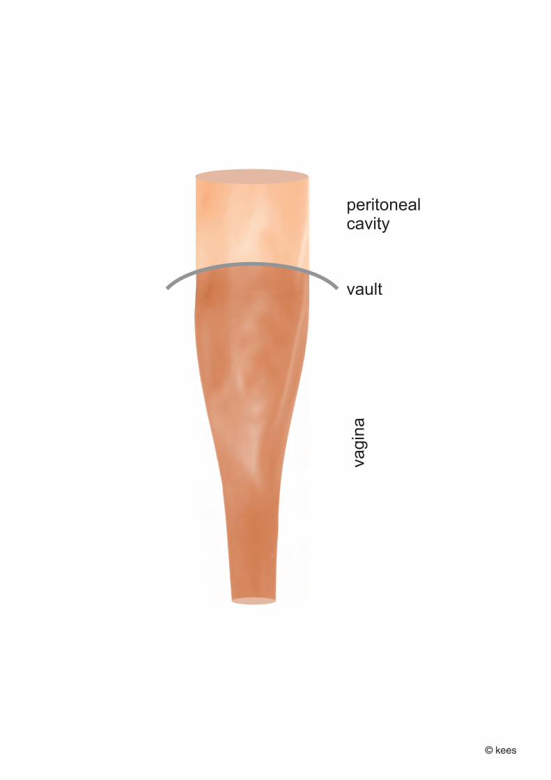
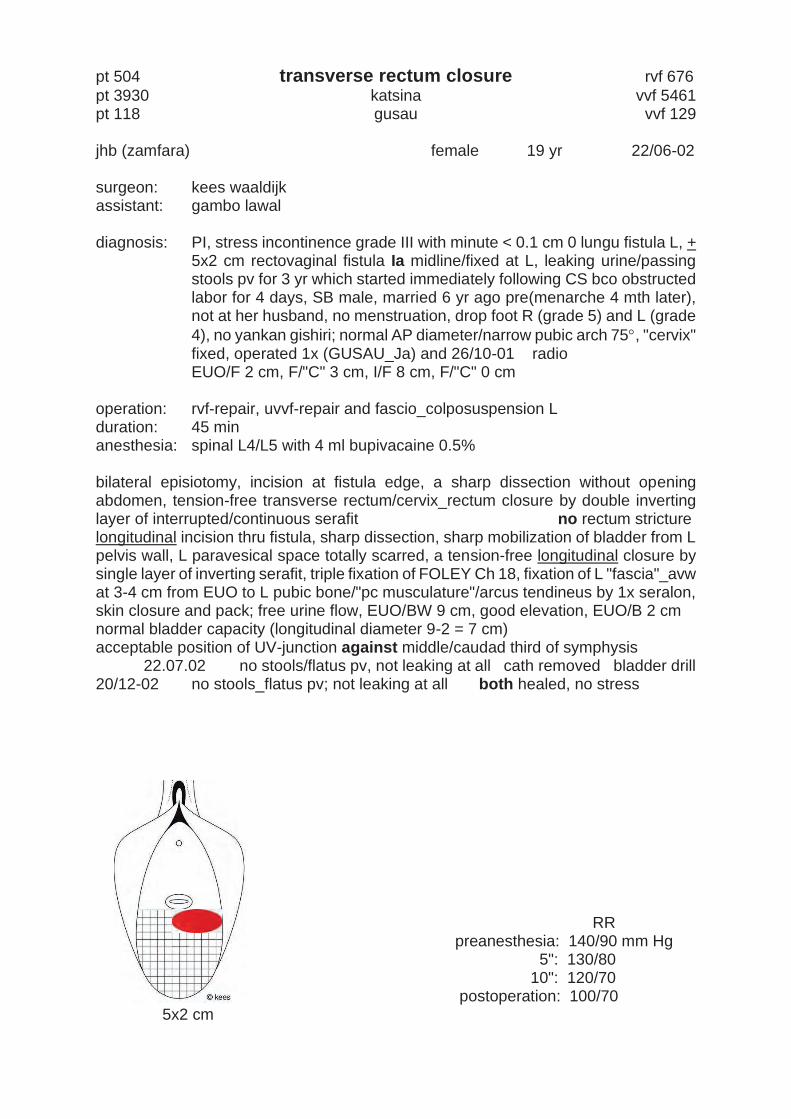
pt 504 transverse rectum closure rvf 676 pt 3930 katsina vvf 5461 pt 118 gusau vvf 129 jhb (zamfara) female 19 yr 22/06-02 surgeon: kees waaldijk assistant: gambo lawal diagnosis: PI, stress incontinence grade III with minute < 0.1 cm 0 lungu fistula L, +
5x2 cm rectovaginal fistula Ia midline/fixed at L, leaking urine/passing stools pv for 3 yr which started immediately following CS bco obstructed labor for 4 days, SB male, married 6 yr ago pre(menarche 4 mth later), not at her husband, no menstruation, drop foot R (grade 5) and L (grade 4), no yankan gishiri; normal AP diameter/narrow pubic arch 75, "cervix" fixed, operated 1x (GUSAU_Ja) and 26/10-01 radio EUO/F 2 cm, F/"C" 3 cm, I/F 8 cm, F/"C" 0 cm
operation: rvf-repair, uvvf-repair and fascio_colposuspension L duration: 45 min anesthesia: spinal L4/L5 with 4 ml bupivacaine 0.5% bilateral episiotomy, incision at fistula edge, a sharp dissection without opening abdomen, tension-free transverse rectum/cervix_rectum closure by double inverting layer of interrupted/continuous serafit no rectum stricture longitudinal incision thru fistula, sharp dissection, sharp mobilization of bladder from L pelvis wall, L paravesical space totally scarred, a tension-free longitudinal closure by single layer of inverting serafit, triple fixation of FOLEY Ch 18, fixation of L "fascia"_avw at 3-4 cm from EUO to L pubic bone/"pc musculature"/arcus tendineus by 1x seralon, skin closure and pack; free urine flow, EUO/BW 9 cm, good elevation, EUO/B 2 cm normal bladder capacity (longitudinal diameter 9-2 = 7 cm) acceptable position of UV-junction against middle/caudad third of symphysis 22.07.02 no stools/flatus pv, not leaking at all cath removed bladder drill 20/12-02 no stools_flatus pv; not leaking at all both healed, no stress
RR preanesthesia: 140/90 mm Hg 5": 130/80 10": 120/70 postoperation: 100/70 5x2 cm
39
pt 504 transverse rectum closure rvf 676 pt 3930 katsina vvf 5461 pt 118 gusau vvf 129 jhb (zamfara) female 19 yr 22/06-02 surgeon: kees waaldijk assistant: gambo lawal diagnosis: PI, stress incontinence grade III with minute < 0.1 cm 0 lungu fistula L, +
5x2 cm rectovaginal fistula Ia midline/fixed at L, leaking urine/passing stools pv for 3 yr which started immediately following CS bco obstructed labor for 4 days, SB male, married 6 yr ago pre(menarche 4 mth later), not at her husband, no menstruation, drop foot R (grade 5) and L (grade 4), no yankan gishiri; normal AP diameter/narrow pubic arch 75, "cervix" fixed, operated 1x (GUSAU_Ja) and 26/10-01 radio EUO/F 2 cm, F/"C" 3 cm, I/F 8 cm, F/"C" 0 cm
operation: rvf-repair, uvvf-repair and fascio_colposuspension L duration: 45 min anesthesia: spinal L4/L5 with 4 ml bupivacaine 0.5% bilateral episiotomy, incision at fistula edge, a sharp dissection without opening abdomen, tension-free transverse rectum/cervix_rectum closure by double inverting layer of interrupted/continuous serafit no rectum stricture longitudinal incision thru fistula, sharp dissection, sharp mobilization of bladder from L pelvis wall, L paravesical space totally scarred, a tension-free longitudinal closure by single layer of inverting serafit, triple fixation of FOLEY Ch 18, fixation of L "fascia"_avw at 3-4 cm from EUO to L pubic bone/"pc musculature"/arcus tendineus by 1x seralon, skin closure and pack; free urine flow, EUO/BW 9 cm, good elevation, EUO/B 2 cm normal bladder capacity (longitudinal diameter 9-2 = 7 cm) acceptable position of UV-junction against middle/caudad third of symphysis 22.07.02 no stools/flatus pv, not leaking at all cath removed bladder drill 20/12-02 no stools_flatus pv; not leaking at all both healed, no stress
RR preanesthesia: 140/90 mm Hg 5": 130/80 10": 120/70 postoperation: 100/70 5x2 cm

40
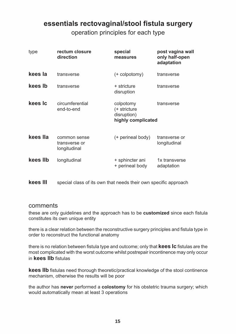
pt 362 longitudinal rectum closure rvf 464 pt 3005 katsina vvf 4134 lad (kano) female 17 yr 21/05-98 surgeon: kees waaldijk assistant: hamisu abdullahi diagnosis: PI, very extensive + 3 cm 0 urethrovesicovaginal fistula with a circum-
ferential defect/bladder base prolapse, + 1 cm 0 rectovaginal fistula Ia, leaking urine/passing stools per vaginam for 6 mth which started imme-diately following obstructed labor for 5 days, in hospital SB male, married 6 yr ago pre(menarche 2 yr later), not living with husband, no menstruation, drop foot R (grade 2-3) and L (grade 3); very narrow pubic arch, severe vagina stenosis with major pc muscle loss I/F 6 cm, F/"C" 4 cm, EUO/F 1 cm, F/"C" 0 cm 147.0 cm
operation: rvf-"repair", bilateral ureters, circumferential uvvf-"repair" as duration: 50 min minimum surgery anesthesia: spinal L3/L4 with 4 ml bupivacaine 0.5% bilateral episiotomy, incision at RVF edge, sharp dissection of pvw, sharp/ blunt bilateral mobilization of rectum, excision of scar tissue ++, tension free longitudinal rectum closure by double layer of inverting serafit the solution to pollution is dilution! bilateral ureter catheterization, incision at fistula edge, sharp circumferential minimum dissection of bladder, advancement/caudad fixation of anterior bladder onto symphysis/ "urethra", tension-free circumferential UVVF-"repair" by end-to-end vesiourethrostomy by a single layer of inverting serafit/chromic catgut, FOLEY Ch 18, suturing avw onto posterio bladder wall with 5x supramid, skin closure, vagina pack; free urine flow, EUO/BW 9 cm, good elevation, EUO/B 1.5 cm avw could not be closed! normal bladder capacity (longitudinal diameter 9-1.5 = 7.5 cm) stool pollution poor position of UV-junction against caudad third of the symphysis only the minimum has been done, everything left open deliberately 14.08.98 stools ok, urine incontinence 24/08-98 no stools_flatus pv; urine incontinence insp/ both healed, stress + 16/11-98 leaking insp/ uvvf early sex 13.02.99 operation: L ureter + uvvf-“repair” vvf 4375 10.07.99 operation: urethra/avw vvf 4531 16/06-01 no stools_flatus pv; not leaking at all both healed, no stress
RR preanesthesia: 160/100 mm Hg 5": 140/90 10": 120/80 postoperation: 100/60 1 cm
Pt 79 oblique rectum closure katsina rvf 95 pt 8044 database ba hanya 9977 vvf 9977 mmmg (katsina) female 31 yr 18/05-89 surgeon: kees waaldijk assistant: dr yushau armiyau diagnosis: PI (0 alive), + 4 cm 0 proximal rectovaginal fistula fixed to cervix, extensi
ve urethrovesicovaginal fistula, leaking urine/passing stools pv for 9 yr which started immediately following obstructed labor for 5 days, at home SB male, married 20 yr ago pre(menarche 2 yr later), not living with husband, normal menstruation, drop foot L, vagina stenosis kees Ia I/F 6 cm, F/C 0 cm
operation: rvf-repair + vaginoplasty duration: 90 min anesthesia: spinal L3/L4 with 4 ml bupivacaine 5% bilateral episiotomy, then stools per fistulam, therefore rvf-repair, incision at fistula edge, sharp dissection, sharp dissection of rectum from cervix whereby abdomen opened, tension-free oblique rectum closure by double layer of inverting interrupted chromic catgut, intrarectal check, widening/lengthening vaginoplasty by skin rotation flap from L buttock, sphincter ani dilatation, skin closure, pack 01.06 + 05.06.89 no stools/flatus pv healed 16/09-90 no stools_flatus pv, normal defecation insp/ healed 18/10-90 operation: uvvf-“repair” vvf 1666 14/11-90 not leaking, incontinence ++, normal miction stools ok
insp/ both healed, good elevation, stress incontinence ++ RR preanesthesia: 120/70 mm Hg 5': 120/70 10': 120/70 postoperation: 100/50 4 cm

41
pt 362 longitudinal rectum closure rvf 464 pt 3005 katsina vvf 4134 lad (kano) female 17 yr 21/05-98 surgeon: kees waaldijk assistant: hamisu abdullahi diagnosis: PI, very extensive + 3 cm 0 urethrovesicovaginal fistula with a circum-
ferential defect/bladder base prolapse, + 1 cm 0 rectovaginal fistula Ia, leaking urine/passing stools per vaginam for 6 mth which started imme-diately following obstructed labor for 5 days, in hospital SB male, married 6 yr ago pre(menarche 2 yr later), not living with husband, no menstruation, drop foot R (grade 2-3) and L (grade 3); very narrow pubic arch, severe vagina stenosis with major pc muscle loss I/F 6 cm, F/"C" 4 cm, EUO/F 1 cm, F/"C" 0 cm 147.0 cm
operation: rvf-"repair", bilateral ureters, circumferential uvvf-"repair" as duration: 50 min minimum surgery anesthesia: spinal L3/L4 with 4 ml bupivacaine 0.5% bilateral episiotomy, incision at RVF edge, sharp dissection of pvw, sharp/ blunt bilateral mobilization of rectum, excision of scar tissue ++, tension free longitudinal rectum closure by double layer of inverting serafit the solution to pollution is dilution! bilateral ureter catheterization, incision at fistula edge, sharp circumferential minimum dissection of bladder, advancement/caudad fixation of anterior bladder onto symphysis/ "urethra", tension-free circumferential UVVF-"repair" by end-to-end vesiourethrostomy by a single layer of inverting serafit/chromic catgut, FOLEY Ch 18, suturing avw onto posterio bladder wall with 5x supramid, skin closure, vagina pack; free urine flow, EUO/BW 9 cm, good elevation, EUO/B 1.5 cm avw could not be closed! normal bladder capacity (longitudinal diameter 9-1.5 = 7.5 cm) stool pollution poor position of UV-junction against caudad third of the symphysis only the minimum has been done, everything left open deliberately 14.08.98 stools ok, urine incontinence 24/08-98 no stools_flatus pv; urine incontinence insp/ both healed, stress + 16/11-98 leaking insp/ uvvf early sex 13.02.99 operation: L ureter + uvvf-“repair” vvf 4375 10.07.99 operation: urethra/avw vvf 4531 16/06-01 no stools_flatus pv; not leaking at all both healed, no stress
RR preanesthesia: 160/100 mm Hg 5": 140/90 10": 120/80 postoperation: 100/60 1 cm
Pt 79 oblique rectum closure katsina rvf 95 pt 8044 database ba hanya 9977 vvf 9977 mmmg (katsina) female 31 yr 18/05-89 surgeon: kees waaldijk assistant: dr yushau armiyau diagnosis: PI (0 alive), + 4 cm 0 proximal rectovaginal fistula fixed to cervix, extensi
ve urethrovesicovaginal fistula, leaking urine/passing stools pv for 9 yr which started immediately following obstructed labor for 5 days, at home SB male, married 20 yr ago pre(menarche 2 yr later), not living with husband, normal menstruation, drop foot L, vagina stenosis kees Ia I/F 6 cm, F/C 0 cm
operation: rvf-repair + vaginoplasty duration: 90 min anesthesia: spinal L3/L4 with 4 ml bupivacaine 5% bilateral episiotomy, then stools per fistulam, therefore rvf-repair, incision at fistula edge, sharp dissection, sharp dissection of rectum from cervix whereby abdomen opened, tension-free oblique rectum closure by double layer of inverting interrupted chromic catgut, intrarectal check, widening/lengthening vaginoplasty by skin rotation flap from L buttock, sphincter ani dilatation, skin closure, pack 01.06 + 05.06.89 no stools/flatus pv healed 16/09-90 no stools_flatus pv, normal defecation insp/ healed 18/10-90 operation: uvvf-“repair” vvf 1666 14/11-90 not leaking, incontinence ++, normal miction stools ok
insp/ both healed, good elevation, stress incontinence ++ RR preanesthesia: 120/70 mm Hg 5': 120/70 10': 120/70 postoperation: 100/50 4 cm

42
pt 794 primary suturing katsina rvf 1014 pt 6294 highly uncooperative examination/anesthesia/repair vvf 8004 major total circumferential trauma zak (katsina) female 22 yr 03.02.11 surgeon: kees waaldijk assistant: kabir lawal diagnosis: PV (1 alive), extensive + 2 cm 0 urethrovesicovaginal with urerhra block/
circumferential defect type IIAb, + 1 cm 0 proximal rectovaginal fistula at R type Ia, leaking urine/passing stools pv for 5 yr that started immediately following obstructed 3rd labor for 2 days, at home sb male, married 10 yr ago pre(menarche 1 yr later), not living with husband, normal menstru ation, drop foot R (grade 3) and L (grade 3-4) with bilateral gm_at contrac ture up to 95/5° dorsiflexion, no yankan gishiri, eclampsia yes; ?ap diame ter?/borderline pubic arch 80°, ar pos, bilateral atf/atl + pc_ilc_iscm loss + ssl_pm trauma, moderate vagina stenosis/shortening proximal lpl stricture euo/f 1.5 cm, f/c 1 cm, ab/au 1 cm, i/v 6 cm, a/f 6 cm, f/c 0 cm 140.0 cm
operation: ps-like uvvf-repair + ps-like rvf-repair duration: 40 min healing both 70% continence 50% anesthesia: spinal L4/L5 with 3 ml bupivacaine 0.5% episiotomy L with severing of stenosis/lpl stricture, since at deep pull pat starts moving only incision at fistula edge, without dissection ps-like avw “closure” by 4x everting seralon, then stool contamination thru rvf until operation end, triple fixation of foley ch 18 with transverse avw adaptation; free urine flow, euo/bw 10 cm, good anterior elevation, euo/b 1.5 cm vomiting food not drinking at all normal bladder capacity (longitudinal diameter 10-1.5 = 8.5 cm) poor position of uv-junction fixed against caudad third of symphysis normal-width 1.5 cm poor-quality urethra_euo in anatomic position RE/ no rectum stricture incision at rvf edge, without dissection ps-like transverse cervix/pvw “closure” by 2x everting seralon, check on hemostasis, skin closure not following any instruction: moving right from the beginning not because of operation pain but by feeling pain in shoulders; no defecation in the morning 10.03.11 stools ok, urine incontinence cath removed blad drill 22.03.11 stools ok, urine incontinence insp/ both healed, no stress 20.04 idem 18.06.11 stools ok, urine incontinence both healed, no stress RR preanesthesia: 130/80 mm Hg 5': 120/70 10': 120/70 postoperation: 120/70 1 cm

43
pt 794 primary suturing katsina rvf 1014 pt 6294 highly uncooperative examination/anesthesia/repair vvf 8004 major total circumferential trauma zak (katsina) female 22 yr 03.02.11 surgeon: kees waaldijk assistant: kabir lawal diagnosis: PV (1 alive), extensive + 2 cm 0 urethrovesicovaginal with urerhra block/
circumferential defect type IIAb, + 1 cm 0 proximal rectovaginal fistula at R type Ia, leaking urine/passing stools pv for 5 yr that started immediately following obstructed 3rd labor for 2 days, at home sb male, married 10 yr ago pre(menarche 1 yr later), not living with husband, normal menstru ation, drop foot R (grade 3) and L (grade 3-4) with bilateral gm_at contrac ture up to 95/5° dorsiflexion, no yankan gishiri, eclampsia yes; ?ap diame ter?/borderline pubic arch 80°, ar pos, bilateral atf/atl + pc_ilc_iscm loss + ssl_pm trauma, moderate vagina stenosis/shortening proximal lpl stricture euo/f 1.5 cm, f/c 1 cm, ab/au 1 cm, i/v 6 cm, a/f 6 cm, f/c 0 cm 140.0 cm
operation: ps-like uvvf-repair + ps-like rvf-repair duration: 40 min healing both 70% continence 50% anesthesia: spinal L4/L5 with 3 ml bupivacaine 0.5% episiotomy L with severing of stenosis/lpl stricture, since at deep pull pat starts moving only incision at fistula edge, without dissection ps-like avw “closure” by 4x everting seralon, then stool contamination thru rvf until operation end, triple fixation of foley ch 18 with transverse avw adaptation; free urine flow, euo/bw 10 cm, good anterior elevation, euo/b 1.5 cm vomiting food not drinking at all normal bladder capacity (longitudinal diameter 10-1.5 = 8.5 cm) poor position of uv-junction fixed against caudad third of symphysis normal-width 1.5 cm poor-quality urethra_euo in anatomic position RE/ no rectum stricture incision at rvf edge, without dissection ps-like transverse cervix/pvw “closure” by 2x everting seralon, check on hemostasis, skin closure not following any instruction: moving right from the beginning not because of operation pain but by feeling pain in shoulders; no defecation in the morning 10.03.11 stools ok, urine incontinence cath removed blad drill 22.03.11 stools ok, urine incontinence insp/ both healed, no stress 20.04 idem 18.06.11 stools ok, urine incontinence both healed, no stress RR preanesthesia: 130/80 mm Hg 5': 120/70 10': 120/70 postoperation: 120/70 1 cm

44

45

46

47

48

49

50

51

52

53
outcome kees Ia stool fistulas in
1,417 patients
surgical outcome 570 operations in 501 patients 35% of 1,417 spontaneous healing in 837 patients 60% of 1,417 minimal/no complaints in 79 patients 6% of 1,417 overall results in 1,417 patients objective subjective

54
surgery results in kees Ia fistulas 570 repairs in 501 patients
reconstructive surgery in 501 patients since there was a strong tendency to spontaneous healing only 501 patients or 35% or roughly one third needed a total of 570 repairs presurgical/surgical data and outcome of surgery are presented in table I, II and III table I presurgical data at first attempt by the author in 501 patients already 164 patients or 33% had been operated from 1 to 4 times 24 had a colostomy as well and nb 3 patients had a ureterosigmoidostomy with a still existing kees Ia stool fistula 3 patients inoperable from the start table II some surgical data of initial 501 repairs by the author transverse closure 445 88.8% longitudinal closure 29 5.8% oblique closure 12 2.4% purse string closure 12 2.4%
inoperable 3 0.6%
primary suturing 104 20,8% abdomen opened 37 7,3% table III results kees Ia reconstructive surgery 501 patients healed first attempt 448 89.4% inoperable 3 healed finally 482 96% healed with 99% continence inoperable 3 not healed 10 mortality at 17 days 1 0,2% unknown 6
incontinence 4 0.8% ureterosigmoidostomy spoiling 2

55
discussion surgery fistula surgery belongs to the most complicated reconstructive surgery the author ever encountered during his extensive surgical career simple repair of simple fistulas only exists in the simple mind of simple surgeons who will stay simple for the rest of their simple life as simply demonstrated by the fact that already 164 or 33% out of the 501 patients had been operated from 1 to 4 times by these simple surgeons before the author started his own complicated surgery these principles are evidence-based guidelines which have to be customized to each and any fistula as its own specific unique entity since the rectum is a high-pressure organ compared to the low-pressure vagina once the rectum has healed the vagina will always heal the most important is to reconstruct the functional pelvis anatomy in a straightforward way with in the process closure of the fistula so one has to concentrate on the basics which is already highly complicated considering a minimum failure rate of 10-15% even in experienced hands as demonstrated by the fact that the closure rate at first attempt by the author was 89% in this series whilst 3 fistulas were inoperable the great majority of almost 90% were closed transversely in line with the natural tissue forces since that was the logical thing to do if the repair broke down the residual fistula was operated according to the same surgical principles as the first repair six patients left the hospital after 10-14 days postoperatively before being evaluated and did not return whilst the 9 patients with a residual fistula also did not return for another repair; at least not to the author the only postoperative mortality at 17 days was due to severe gastroenteritis/dehydra tion despite rehydration measurements though the (proximal) fistulas may be complicated to repair the prognosis as to closure and continence in expert hands is excellent; actually, for all kees Ia fistulas with a final healing rate of 96% with a continence rate of 99% in this series as also demonstrated by the evidence-based and documented spontaneous healing in another 837 patients or 60% out of a total of 1,417 consecutive patients with a kees Ia stool fistula

56
spontaneous healing kees Ia stool fistulas as documented in 837
introduction there is a strong tendency to spontaneous healing in the kees Ia stool fistulas which has been documented by the author over a long period of time since 1985 when he first noticed this during immediate catheter treatment the fistula was found on vaginal examination on catheter insertion or early closure and healed on follow-up examinations; or a healed pvw “fistula” was found at/near the cervix during vvf-repair with the patient admitting that initially she had been passing stools/fla tus thru the vagina up to 2-3 months post partum and even longer the patient complained of initial passing (stool and) flatus per vaginam immediately after childbirth whereby later first passing of (diarrheic) stools stopped and then passing of flatus and then no longer flatus or stool thru the vagina whilst she is specifically asked if it is thru the vagina or thru the anus findings so far, spontaneous healing was noted in 837 patients or roughly 60% out of a total of 1,417 patients with a kees Ia stool fistula discussion spontaneous healing the natural healing potential of the human body is enormous, and our task as a surgeon is to observe and only to interfere if something goes wrong or if it does not heal spontaneous healing was found and documented in small proximal fistulas up to 2 cm an explanation for spontaneous healing is given as following since normally the rectum is empty whereby the fistula edge(s) may be in contact promo ting healing, there is intermittent filling up with distension of the rectum by stools and gas for a short time which would hinder the natural healing the (weight of the) cervix may play a role since it may bring the fistula edges into contact and/or closing off/sealing off the fistula promoting the natural healing processes whilst formed stools will not pass thru a small opening; only flatus and diarrheic stools; unless (severe) outflow obstruction in lying, sitting and standing it seems stools is sliding over/against the posterior rectum wall leaving the anterior rectum wall free and at rest promoting healing and it may be that some of the necrotic fistulas as seen on examination were not full thickness
katsina cath 1119 spontaneous healing small rvf kees Ia + vvf
zradm (katsina) female 23 yr 03.03.08 diagnosis: P ( alive), 2.5x1 + cm longitudinal urethrovesicovaginal fistula type IIAa,
small proximal rectovaginal fistula, leaking urine/passing tusa pv for 16 days which started immediately following CS bco obstructed last labor for 1 day, SB female, married 10 yr ago post(menarche 3 mth earlier), still living with husband, no menstruation, drop foot R (grade 4) and L (grade 3), no yankan gishiri; normal AP diameter/pubic arch 85°, AR pos EUO/F 2 cm, F/”C” 0 cm 159.0 cm
03.03.08 FOLEY Ch 18; free urine flow, EUO/BW 10 cm, good anterior elevation,
EUO/B 1.5 cm normal bladder capacity (longitudinal diameter 10-1.5 = 8.5 cm poor position of UV-junction against caudad third of symphysis normal-width 1.5 cm good-quality urethra_euo in anatomic position will it heal since deep necrosis
06.03.08 still leaking insp/ healing during examination tusa from proximal vagina leave catheter and wait
14.04.08 not leaking at all cath removed bladder drill 16.06.08 not leaking at all, no incontinence, normal miction tusa ok Insp/ both healed, good elevation, no stress incontinence
11.08.08 idem 06.10.08 not leaking at all, no incontinence, normal miction healed, no stress 2.5x1 cm

57
katsina cath 1119 spontaneous healing small rvf kees Ia + vvf
zradm (katsina) female 23 yr 03.03.08 diagnosis: P ( alive), 2.5x1 + cm longitudinal urethrovesicovaginal fistula type IIAa,
small proximal rectovaginal fistula, leaking urine/passing tusa pv for 16 days which started immediately following CS bco obstructed last labor for 1 day, SB female, married 10 yr ago post(menarche 3 mth earlier), still living with husband, no menstruation, drop foot R (grade 4) and L (grade 3), no yankan gishiri; normal AP diameter/pubic arch 85°, AR pos EUO/F 2 cm, F/”C” 0 cm 159.0 cm
03.03.08 FOLEY Ch 18; free urine flow, EUO/BW 10 cm, good anterior elevation,
EUO/B 1.5 cm normal bladder capacity (longitudinal diameter 10-1.5 = 8.5 cm poor position of UV-junction against caudad third of symphysis normal-width 1.5 cm good-quality urethra_euo in anatomic position will it heal since deep necrosis
06.03.08 still leaking insp/ healing during examination tusa from proximal vagina leave catheter and wait
14.04.08 not leaking at all cath removed bladder drill 16.06.08 not leaking at all, no incontinence, normal miction tusa ok Insp/ both healed, good elevation, no stress incontinence
11.08.08 idem 06.10.08 not leaking at all, no incontinence, normal miction healed, no stress 2.5x1 cm

58
kano cath 90 kees Ia rvf spontaneous closure rvf hsz (kano) female 18 yr 20.07.92 diagnosis: PII (1 alive), + 2.5x1 cm urethrovesicovaginal fistula R bladder neck IIAa,
also proximal rectovaginal fistula, leaking urine and passing stools per vaginam for 60 days which started 7 days following obstructed labor for 7 days, in hospital SB male, married 5 yr ago pre(menarche 5 mth later), still living with husband, drop foot L (grade 3), no menstruation, severe vagina stenosis; when seen 17.06.92 necrotic vagina EUO/F 3 cm, F/C 3 cm 157.0 cm
06.10.92: + 8 mm 0 residual urethrovesicovaginal fistula R bladder neck IIAa, drop
foot L (grade 3), normal menstruation, vagina stenosis/shortening spontaneous closure of proximal rvf (no longer stools per vaginam)
EUO/F 4 cm, F/C 1.5 cm 157.0 cm 06.10.92 operation: UVVF-repair vvf 457 episiotomy L, incision at fistula edge with small bilateral transverse extensions, sharp dissection of avw from scarred bladder/urethra, completely tension-free transverse closure with a single layer of interrupted inverting chromic catgut 00, transverse avw closure with everting chromic catgut and vicryl, fixation of FOLEY Ch 18, only skin closure of episiotomy, vagina pack; free urine flow, good bladder capacity (EUO/BW 12 cm), good elevation, EUO/B 4 cm normal bladder capacity (longitudinal diameter 12-4 = 8 cm) 20.10.92 not leaking at all cath removed bladder drill 17.01.93 not leaking at all, no incontinence, normal miction
insp/ healed, good elevation, no stress incontinence 19.04.93 not leaking at all, no incontinence, normal miction healed, no stress 20.07.92 06.10.92 2.5x1 cm 0.8 cm
pt 1359 katsina vvf 1645 kees Ia rvf spontaneous healing rvf rdsg (katsina) female 16 yr 04/10-90 surgeon: kees waaldijk assistant: hauwa garba diagnosis: PI, + 2 cm 0 urethrovesicovaginal fistula midline fixed to symphysis with
urethra block type IIAb, leaking urine for 4 mth that started 2 day following obstructed labor of 3 days, in hospital SB male married 4 yr ago pre(menarche 1 yr later), not with husband, no menstruation, drop foot R (grade 2) and L (grade 3), spontanous healing of proximal RVF (as noted 2 mth ago); passing stools pv stopped 2 wk ago, cervix in vault, no posterior fornix, vagina stenosis EUO/F 4 cm, F/C 6 cm
operation: UVVF-repair duration: 30 min anesthesia: spinal L3/L4 with 4 ml bupivacaine 0.5% bilateral episiotomy, an incision at fistula edge with bilateral transverse extensions, sharp dissection of avw, FOLEY Ch 18, tension-free transverse closure by single layer of inverting chromic catgut 00, transverse avw closure by everting 3x supramid/chromic catgut 0/4, skin closure, vagina pack; free urine flow good bladder capacity urethra/UV-junction/bladder neck fixed against symphysis; EUO/B 4 cm 20.10.90 not leaking at all cath removed bladder drill 28/10-90 not leaking at all, no incontinence, normal miction so
insp/ healed, good elevation, no stress incontinence new obstetric problem 09/07-99 dysuria and intermittent urine retention bladder drill 24/07-99 no complaints, not leaking, no incontinence, normal miction
insp/ healed, good elevation, no stress incontinence
RR preanesthesia: 130/80 mm Hg 5": 120/70 10": 110/60 postoperation: 110/60 1 cm 0

59
kano cath 90 kees Ia rvf spontaneous closure rvf hsz (kano) female 18 yr 20.07.92 diagnosis: PII (1 alive), + 2.5x1 cm urethrovesicovaginal fistula R bladder neck IIAa,
also proximal rectovaginal fistula, leaking urine and passing stools per vaginam for 60 days which started 7 days following obstructed labor for 7 days, in hospital SB male, married 5 yr ago pre(menarche 5 mth later), still living with husband, drop foot L (grade 3), no menstruation, severe vagina stenosis; when seen 17.06.92 necrotic vagina EUO/F 3 cm, F/C 3 cm 157.0 cm
06.10.92: + 8 mm 0 residual urethrovesicovaginal fistula R bladder neck IIAa, drop
foot L (grade 3), normal menstruation, vagina stenosis/shortening spontaneous closure of proximal rvf (no longer stools per vaginam)
EUO/F 4 cm, F/C 1.5 cm 157.0 cm 06.10.92 operation: UVVF-repair vvf 457 episiotomy L, incision at fistula edge with small bilateral transverse extensions, sharp dissection of avw from scarred bladder/urethra, completely tension-free transverse closure with a single layer of interrupted inverting chromic catgut 00, transverse avw closure with everting chromic catgut and vicryl, fixation of FOLEY Ch 18, only skin closure of episiotomy, vagina pack; free urine flow, good bladder capacity (EUO/BW 12 cm), good elevation, EUO/B 4 cm normal bladder capacity (longitudinal diameter 12-4 = 8 cm) 20.10.92 not leaking at all cath removed bladder drill 17.01.93 not leaking at all, no incontinence, normal miction
insp/ healed, good elevation, no stress incontinence 19.04.93 not leaking at all, no incontinence, normal miction healed, no stress 20.07.92 06.10.92 2.5x1 cm 0.8 cm
pt 1359 katsina vvf 1645 kees Ia rvf spontaneous healing rvf rdsg (katsina) female 16 yr 04/10-90 surgeon: kees waaldijk assistant: hauwa garba diagnosis: PI, + 2 cm 0 urethrovesicovaginal fistula midline fixed to symphysis with
urethra block type IIAb, leaking urine for 4 mth that started 2 day following obstructed labor of 3 days, in hospital SB male married 4 yr ago pre(menarche 1 yr later), not with husband, no menstruation, drop foot R (grade 2) and L (grade 3), spontanous healing of proximal RVF (as noted 2 mth ago); passing stools pv stopped 2 wk ago, cervix in vault, no posterior fornix, vagina stenosis EUO/F 4 cm, F/C 6 cm
operation: UVVF-repair duration: 30 min anesthesia: spinal L3/L4 with 4 ml bupivacaine 0.5% bilateral episiotomy, an incision at fistula edge with bilateral transverse extensions, sharp dissection of avw, FOLEY Ch 18, tension-free transverse closure by single layer of inverting chromic catgut 00, transverse avw closure by everting 3x supramid/chromic catgut 0/4, skin closure, vagina pack; free urine flow good bladder capacity urethra/UV-junction/bladder neck fixed against symphysis; EUO/B 4 cm 20.10.90 not leaking at all cath removed bladder drill 28/10-90 not leaking at all, no incontinence, normal miction so
insp/ healed, good elevation, no stress incontinence new obstetric problem 09/07-99 dysuria and intermittent urine retention bladder drill 24/07-99 no complaints, not leaking, no incontinence, normal miction
insp/ healed, good elevation, no stress incontinence
RR preanesthesia: 130/80 mm Hg 5": 120/70 10": 110/60 postoperation: 110/60 1 cm 0

60
pt 225 zaria vvf 247 kees Ia rvf spontaneous healing rvf aaaad
syk (zaria city) female 18 yr 14/07-06 surgeon: kees waaldijk assistant: kabir lawal diagnosis: PI, + 3 cm 0 urethrovesicovaginal fistula with circumferential defect IIBb,
+ 0.2 cm 0 proximal rectovaginal fistula fixed to midline cervix within 2 cm 0 pvw trauma, leaking urine/passing tusa only pv for 35 days which started immediately following destructive surgery bco obstructed labor for 2 days, SB male, married 1.5 yr ago post(menarche 3 yr earlier), not living with husband, no menstruation, drop foot R (grade 4) and L (grade 4-5), no yankan gishiri; normal AP diameter/narrow pubic arch 70, AR pos, extensive pc_ic_ic muscle loss, bare bones up to ischiac spine, empty pelvis, distal vagina stenosis with pvw_lateral stricture, major ATF loss EUO/F 1 cm, F/C 4 cm, AB/AU 2 cm wide open EUO drawn inside
operation: circumferential UVVF-repair poor-quality urethra tissue duration: 45 min healing 80% continence 50% anesthesia: spinal L3/L4 with 4 ml bupivacaine 0.5% episiotomy L with severing of stenosis_stricture, incision at fistula edge, sharp circum ferential dissection, advancement/caudad fixation of anterior bladder onto symphysis/ urethra, tension-free circumferential UVVF-repair by end-to-end vesicourethrostomy by single layer of inverting serafit, separate paraurethra fixation of ep_pc fascia, triple fixation of FOLEY Ch 18, transverse avw adaptation by 2x everting seralon, skin closure, pack; free urine flow, EUO/BW 14 cm, good anterior elevation, EUO/B 1 cm (extensive circum loss) no rectum stricture so rvf will heal spontaneously normal bladder capacity (longitudinal diameter 14-1 = 13 cm) poor position of UV-junction fixed against caudad third symphysis normal-width 1 cm urethra_EUO slightly drawn inside 11.08.06 not leaking at all cath removed bladder drill 14.09.06 not leaking at all, no incontinence, normal miction stools ok insp/ both/all healed, good elevation, no stress incontinence 15.06.07 amenorrhea for 4 mth not leaking at all instructions PII (1 alive) live male by cs 26.12.07 not leaking at all PIII (2 alive) live female by cs 27.02.09 not leaking at all RR preanesthesia: 140/90 mm Hg 5': 130/80 10': 130/80 postoperation: 120/70 3 cm 0

61
pt 225 zaria vvf 247 kees Ia rvf spontaneous healing rvf aaaad
syk (zaria city) female 18 yr 14/07-06 surgeon: kees waaldijk assistant: kabir lawal diagnosis: PI, + 3 cm 0 urethrovesicovaginal fistula with circumferential defect IIBb,
+ 0.2 cm 0 proximal rectovaginal fistula fixed to midline cervix within 2 cm 0 pvw trauma, leaking urine/passing tusa only pv for 35 days which started immediately following destructive surgery bco obstructed labor for 2 days, SB male, married 1.5 yr ago post(menarche 3 yr earlier), not living with husband, no menstruation, drop foot R (grade 4) and L (grade 4-5), no yankan gishiri; normal AP diameter/narrow pubic arch 70, AR pos, extensive pc_ic_ic muscle loss, bare bones up to ischiac spine, empty pelvis, distal vagina stenosis with pvw_lateral stricture, major ATF loss EUO/F 1 cm, F/C 4 cm, AB/AU 2 cm wide open EUO drawn inside
operation: circumferential UVVF-repair poor-quality urethra tissue duration: 45 min healing 80% continence 50% anesthesia: spinal L3/L4 with 4 ml bupivacaine 0.5% episiotomy L with severing of stenosis_stricture, incision at fistula edge, sharp circum ferential dissection, advancement/caudad fixation of anterior bladder onto symphysis/ urethra, tension-free circumferential UVVF-repair by end-to-end vesicourethrostomy by single layer of inverting serafit, separate paraurethra fixation of ep_pc fascia, triple fixation of FOLEY Ch 18, transverse avw adaptation by 2x everting seralon, skin closure, pack; free urine flow, EUO/BW 14 cm, good anterior elevation, EUO/B 1 cm (extensive circum loss) no rectum stricture so rvf will heal spontaneously normal bladder capacity (longitudinal diameter 14-1 = 13 cm) poor position of UV-junction fixed against caudad third symphysis normal-width 1 cm urethra_EUO slightly drawn inside 11.08.06 not leaking at all cath removed bladder drill 14.09.06 not leaking at all, no incontinence, normal miction stools ok insp/ both/all healed, good elevation, no stress incontinence 15.06.07 amenorrhea for 4 mth not leaking at all instructions PII (1 alive) live male by cs 26.12.07 not leaking at all PIII (2 alive) live female by cs 27.02.09 not leaking at all RR preanesthesia: 140/90 mm Hg 5': 130/80 10': 130/80 postoperation: 120/70 3 cm 0
minimal/no complaint of kees Ia stool fistulas as documented in 79 patients
introduction during some urine fistula repairs a small proximal kees Ia stool fistula was detected as a surprise by passing flatus whilst patient, only upon explicit asking, stated she had no or only minimal complaints like once in a while flatus which did not disturb her at all and even some patients denied it completely telling they never experienced anything; also on explicit asking and telling the patient there was a fistula findings a small proximal kees Ia stool fistula was detected at the beginning of or during the vvf repair as a surprise finding since during the anamnesis the patient had denied passing stool/flatus thru the vagina in 79 patients who had a vesicovaginal fistula as well longer than 1 year on explicit asking some patients admitted that once in a while they were passing flatus (and diarrheic stools) thru the vagina which did not bother them at all since it was only once in a while and considered this normal whilst some denied it completely discussion formed solid stools cannot pass a small opening in the anterior rectum wall unless there is severe outflow obstruction but that is different for liquid stools in case of diarrhea and gas unless the small fistula is closed off and sealed off by the (weight of the) cervix it seems that in standing/sitting/lying solid formed stools (?+ flatus?) is sliding over the posterior rectum wall leaving the anterior rectum wall free the passing of flatus thru a small fistula during operation is promoted by the weight of the auvard speculum compressing the distal posterior vagina wall/anterior rectum wall and so causing some kind of obstruction and narrowing so the gas escapes proximally in an otherwise symptomless fistula

62
figures and overall results for total 1,417 fistulas in
1,417 patients with kees Ia stool fistula some relevant overall figures are presented for the total number of kees Ia fistulas combination with vesicovaginal fistula almost 100% the combination is almost 100% since out of the total 1,417 stool fistulas 1,411 or over 99.5% were combined with a vesicovaginal fistula final objective results by the surgeon fistula closed 1,319 93.1% fistula not closed 89 6.3% no symptoms 79 mortality 1 0.07%
inoperable 3 0.2%
unknown 6 0.4%
final subjective results by the patient cured with continence 1,390 98,1%
healed with incontinence 4 0,3%
discussion there is a strong tendency for small rectovaginal fistulas of 60% in this series and for postpartum stool/flatus incontinence to heal spontaneously therefore, the incidence of rectovaginal/stool fistulas is higher than generally assumed as presented in this series since the spontaneously healed fistulas will not be detected unless specifically looked for at the beginning of any vaginal procedure also some patients or 6% deny its existence and say no complaints (whatsoever) so only 35% needed an operation the results are excellent, especially from the viewpoint of the patients, since 98% of the 1,417 patients reported full cure with full continence
63
basic science see textbook functional female pelvis anatomy
what is needed anatomic stool continence mechanism pelvis anatomy essentials management principles rectovaginal/stool fistulas

64
what is needed before a start is made one has to master the complicated functional anatomy of the pelvis, the pelvis organs and the pelvis floor one has to understand the functional anatomy as interaction between the different structures in order to understand the physiology of the urine and stool continence mechanisms in the female one must be able to identify the individual structures of the functional anatomy in the living female which is different from the post-mortem dead anatomy one has to study and understand the mechanism of action of the obstetric trauma, what it does to the functional anatomy of the individual structures and master the enormous variety of lesions one must be able to identify and assess the individual obstetric trauma defects in the living female one must study, understand and master the mechanisms of action of urine and stool incontinence and of prolapse in the female then one must devise a plan of action how to reconstruct the functional anatomy as customized to the individual findings and needs one must master not only the principles of general, gynecologic, urologic, colorectal and reconstructive surgery but since the vagina is never sterile also the principles of septic surgery one must understand and respect the natural tissue forces inside the human body one must master the physiologic healing processes in order to promote the enormous natural healing potential of the human body realizing it starts the moment an incision is made preferably one undergoes a practical training with a step-by-step approach where the basic skills are demonstrated in order to learn these skills though the skills can be demonstrated and be practiced step by step under strict super vision there is no automatic transfer of these skills and the ultimate responsibility and accountability for any surgery rests upon the performing surgeon
the decisive factor in surgery is the surgeon

65
female stool continence mechanism functional anatomy
introduction the functional anatomy of the female stool continence mechanism consists of a rather complicated multi-interaction of static (connective tissue) and dynamic structures (muscles; mucosa, submucous vascular plexus) and nervous innervation the anatomic stool continence mechanism is situated within the distal 4-5 cm of the anorectum, the external sphincter ani muscle and support there is an internal smooth muscle sphincter and an external striated muscle sphincter with washer effect by the mucosa and submucous vascular plexus the distal anorectum and external sphincter ani are anchored into the pierced thru punched out opening in the perineum outlet diaphragm here only a short comprehensive outline is given as a start/incentive to more extensive self-study functional anatomy anatomic stool continence mechanism anorectum-rectum junction diameter anorectal angle, normally 80°-100° anorectum length 4-5 cm shape and diameter lumen anus mucosa anal cushions = columnae anales submucous vascular plexus circular smooth muscle fibers = internal sphincter ani longitudinal smooth muscle fibers sphincter ani muscle circular striated muscle fibers around distal anorectum/anus consisting of mostly slow-twitch for tonus but also fast-twitch for emergency closure divided into three parts: subcutaneous superficial deep

66
anatomic/physiologic support rectovaginal or prerectal fascia (of denonvillier) supports the anterior anorectum perineum outlet diaphragm the anorectum with external sphincter ani complex are firmly anchored into the pierced thru punched out opening within the perineum outlet diaphragm perineal body (= centrum tendineum perinei) with transversus perinei and bulbo spongiosus muscles stabilizes the anus and sphincter ani anteriorly and laterally; in a way that is comparable to the role of the pubocervical fascia in stabilizing the posterior urethra anococcygeal ligament stabilizes the anus posteriorly levator ani muscles + levator plate anterobilaterally from pubis bone and arcus tendineus levator ani as a sling around the lateral and posterior anorectum walls and external sphincter ani, and inserting into levator plate, anococcygeal ligament and coccyx especially the puborectalis muscles, median part of pubococcygeus muscles, play a role pulling the anorectum anteriorly; these muscles are responsible for the anorectal angle; its fibers fuse with the deep portion of the external sphincter ani muscle posterior vagina wall attached to perineal body and rectovaginal or pre(ano)rectal fascia and rectum serosa perianal skin with subcutaneous tissue and constrictor ani muscle stabilizes also the anus/sphincter ani muscle intact innervation of these components autonomic sympathetic and parasympathetic (vagus) nervous system for the circular smooth muscle as internal sphincter and longitudinal smooth muscle; the sympathetic fibers for stimulation and continence against the parasympathetic fibers for relaxation and defecation; from hypogastric and pelvic plexus pudendal nerve innervating the external sphincter ani; from s2, s3, s4 levator ani nerve innervating levator ani muscles; from s3, s4 and of course the autonomic enteric nervous system discussion the stool continence mechanism must take care of gas for final sealing off the mucosa with mucosa cushions and the submucous vascular plexus are responsible

67
liquid stool for final sealing off also the mucosa with mucosa cushions and the submucous vascular plexus are responsible solid stool this is the easiest since normally there is no stool inside the rectum anorectal angle is determined by the puborectalis muscle and is normally 80°-100°; if it contracts the angle will become sharper; however, this seems to be of minor importance anorectum mucosa with mucosa cushions and submucous vascular plexus are responsible for air- and water-tight closure; washer effect water-tight closure is a problem since liquid stool inside the rectum is accompanied by a strong urge component with bowel contractions linea dentata between squamous epithelium (proctodaeum origin) with sensibility for pain and touch since innervated by pudendal nerve and cubical epithelium (hindgut origin) without sensibility since innervated by autonomic nervous system thickened circular smooth muscle = internal sphincter ani is the strongest factor and responsible for closure due to non-fatigue tonus; it is separated from the external sphincter by the longitudinal smooth muscle layer sheath longitudinal smooth muscle is playing a role in defecation since it will shorten the anorectum if contracting; it separates the internal sphincter ani muscle from the external sphincter ani muscle rectovaginal or prerectal fascia (denonvillier) bilaterally from an arcus tendineus attached to levator ani muscle fascia; this is attached to/supporting the anterior anorectum; if defective a rectocele will develop external sphincter ani circular around the distal anorectum and consists of striated muscle fibers; the slow-twitch muscle fibers are contributing to its tonus whilst the fast-twitch fibers will contribute to short-duration closure of the anus; especially in the female it is thicker posteriorly than anteriorly it consists of 3 parts: subcutaneous, superficial and deep; fibers of the puborectalis muscle fuse with the deep part bilaterally and posteriorly it is separated from the internal sphincter by the longitudinal smooth muscle sheath of the anorectum the external sphincter extends 0.5-1 cm distally from the internal sphincter (intersphinc teric groove) and protrudes slightly from the surrounding skin perineum outlet diaphragm the anorectum with the external sphincter ani complex are firmly anchored into the pierced thru punched out opening within this diaphragm stabilizing/securing these structures in their anatomic position active contraction of its striated muscle component and reflex contraction of its smooth muscle component will reinforce the stool continence mechanism

68
perineal body wedge-like connective tissue structure into which the bulbospongiosus and transversus perinei muscles radiate; attached to anterior external sphincter ani this structure stabilizes and secures the anterior sphincter ani/anorectum in its anatomic position and as such supports the stool continence mechanism transversus perinei muscles bilaterally from ischium tuberosity and uniting indirectly medially via the perineal body and prevent lateral shifting of perineal body/anus bulbospongiosus muscles bilaterally from paraclitoridally and uniting posteriorly medially via the perineal body and prevent posterior shifting of perineal body/anus levator ani muscles + levator plate a flat striated muscle sheath originating anterobilaterally from pubis bone and arcus tendineus of levator ani muscles (atflam) and like a sling around lateral vagina walls and laterally from and underneath sphincter ani/anorectum/rectum and fusing with each other and inserting posteriorly from sphincter ani/anorectum/rectum into levator ani plate, anococcygeal ligament and coccyx bone though it is one muscle it can be divided into different parts based on their origin: pubococcygeus and obturatoccygeus muscles the medial part of the pubococcygeus is called the puborectalis muscle; this portion fuses with the bilateral and posterior deep external sphincter ani muscle fibers; it is responsible for the anorectal angle and prevents posterior shifting of the anus due to its sling-like shape contraction of the levator ani muscles compresses the lateral and posterior anorectum and sharpens the anorectal angle and as such contributes to the stool continence mechanism for some investigators the (ischio)coccygeus muscle is (synonymous with) the sacro-spinous ligament posterior vagina wall covers and is attached to the perineal body and anorectum/prerectal fascia/rectum (serosa) and as such is fixed indirectly to the lateral pelvis walls anococcygeal ligament from coccyx bone to posterior sphincter ani/anus/anorectum and stabilizes/secures the external sphincter ani and anus in its posterior anatomic position and prevents anterior shifting of external sphincter ani/anus shafik mechanism surgically speaking this is difficult to check; as well this could only function if the levator ani muscles are posteriorly uniting around posterior anorectum (true), the anococcygeal ligament around anterior anorectum and/or external sphincter ani and/or perineal body (not true) and bulbospongiosus muscles unite posteriorly from anorectum (not true) besides this, as long as an anatomically correct reconstruction is performed these structures will be restored as well whatever the arrangement

69
skin with subcutaneous tissue and corrugator ani muscle the perianal skin and subcutaneous tissue in combination with corrugator ani muscle around the anus stabilizes the anus as well intact innervation of these components autonomic sympathetic, parasympathetic (vagus) nervous system for the anorectum circular smooth muscle fibers as internal sphincter and longitudinal smooth muscle fibers and for (non)sensibility of anorectum cubic mucosa up to dentate line; from hypo gastric and pelvic plexus; also complex coordination by enteric nervous system pudendal nerve innervating the external sphincter ani and for sensibility for touch/pain of perianal skin and squamous anorectum mucosa up to dentate line; from s2, s3, s4 levator ani nerve innervating levator ani muscles; from s3, s4 synergistic interaction between stool and urine continence mechanisms though the two mechanisms may function independently from each other there is a lot of analogy and synergy; for instance first flatus and then micturition (le vent avant la pluie) and first micturition before defecation, combination of cystocele and rectocele, combination of sphincter ani rupture and genuine intrinsic urine incontinence etc analogy between stool and urine continence mechanism analogy of functional anatomy: mucosa, submucous vascular plexus, internal smooth-muscle sphincter, external striated-muscle sphincter and support analogy of posterior support of urethra by endopelvic diaphragm and anterior support of sphincter ani/anorectum by perineal body with transversus perinei and bulbospongiosus muscles and by prerectal fascia analogy of innervation (pudendal nerve + autonomic nervous system) and analogy of blood supply (internal iliac artery); and for (ano)rectum also inferior mesenteric artery direct against indirect action of levator ani muscles tonus and contraction of levator ani muscles have a direct action upon the stool continence mechanism since lateral/posterior anorectum walls are being squeezed and fibers of puborectalis support deep part of sphincter ani muscle ani whilst the anorectal angle becomes sharper tonus and contraction of levator ani muscles have no direct action upon the female urine continence mechanism since there is no direct contact whatsoever between the two; but there is indirect action since the endopelvic diaphragm as attached to anterior vagina wall is moving anteriorly and cephalad by compression of lateral/posterior vagina walls which improves the support sphincter ani/perineal body complex schematic drawings of the sphincter ani/perineal body complex with transversus perinei and bulbospongiosus muscles are presented on following page. last edition august 2020

70

71

72

73
functional pelvis anatomy
essentials true pelvis cavity a confined space for the distal outlet organs of the urinary tract anteriorly, the genital tract in the middle and the digestive tract posteriorly with hydrostatic and compression pressure; normally in a continent way and divided into anterior pre_subperitoneal compartment for the distal end parts of the urinary tract: pelvic ureters, bladder and urethra median subperitoneal compartment for the (also distal end parts of) genital tract: uterus, adnexa, cervix and vagina posterior retro_subperitoneal compartment for the distal end parts of the digestive tract: rectum, anorectum and sphincter ani enclosed by parietal pelvis fascia covering pelvis wall/floor muscles and parietal peritoneum as boundary of intraperitoneal cavity as connected to each other by tela urogenitalis with corpus intrapelvinum and endopelvic diaphragm corpus intrapelvinum as dynamic 3-dimensional matrix connective tissue organ of pelvis consists of a cohesive mixture of collagen for strength, elastin for passive elasticity and plasticity and mostly smooth muscle fibers for dynamic active non-fatigue tonus in a loose, dense or condensed form as a dynamic matrix into which the organs and their supply are embedded and suspended/ connected to the pelvis wall and each other by highly specialized structures protecting the organs and their supply against trauma and stabilizing/securing them in their variable anatomic position as coordinated by intrinsic myogenic impulses and the autonomic nervous system considered to be a fluidum since no sharp demarcations between the archaic matrix and its specialized structures endopelvic diaphragm highly specialized structure of corpus intrapelvinum from symphysis anteriorly to sacrum posteriorly as connected to its bilateral arcus tendineus fasciae with cervix as centrum tendineum intrapelvinum since all musculofascia structures are connected to it as first line of counteracting intraabdominal hydrostatic pressure and supporting the urogenital continence mechanisms in their anatomic position and preventing herniation of the urinary tract, genital tract, intraperitoneal contents and distal digestive tract into the zero-pressure vagina pelvis floor as one functional unit as part of abdominopelvic wall levator ani muscles connected firmly to the perineum outlet diaphragm via perineal body and external sphincter ani muscle supporting and reinforcing each other levator ani muscles as “pelvis diaphragm” highly overrated with direct action on stool continence mechanism and only indirect action on urine continence mechanism perineum outlet diaphragm into which the end outlet organs with their striated sphincter mechanisms are anchored and supporting directly and the urine and stool continence mechanisms

74
female urine continence mechanism over in total 4-5 cm bladder neck, uv-junction and whole urethra supported by the endopelvic diaphragm and the perineum outlet diaphragm there is an internal smooth muscle sphincter and an external striated rhabdosphincter with washer effect by the mucosa and submucous vascular plexus continence potential over its whole length female genital continence mechanism over in total 3-4 cm with cervix as internal smooth muscle sphincter as anchored into endopelvic diaphragm female stool continence mechanism over in total 4-5 cm anorectum and external sphincter ani anchored within perineum outlet diaphragm there is an internal smooth muscle sphincter and an external striated sphincter ani muscle with washer effect by mucosa and submucous vascular plexus urine stress incontinence mechanism genuine and post fistula repair the anterior urethra wall is always fixed to the symphysis and cannot rotate backwards away from the symphysis whilst the posterior urethra wall is mobile as supported by the endopelvic diaphragm once this support becomes weak the posterior urethra wall rotates backwards causing funneling = vesicalization of the proximal and mid or whole urethra with a decrease in outflow resistance so that the intrinsic closing forces can no longer counteract the intra vesical expulsion forces or by defects within the anchoring into perineum outlet diaphragm; isolated or combined pelvis floor muscle exercises may have a positive effect upon the urine and stool continence mechanism since the perineum outlet diaphragm contributes to the urine and stool continence mechanism by further stabilizing the outlet parts the levator ani muscles contribute directly to the stool continence mechanism to which they are anatomically connected but only indirectly to the urine continence mechanism with no anatomic connection whatsoever with simultaneous reflex contraction of the external striated muscle sphincters with increase in tonus of smooth muscle fibers of the endopelvic diaphragm by reflex action via intrinsic myogenic impulses as modulated by the autonomic nervous system obstetric trauma due to hydrostatic pressure, dilatation of birth canal, (in)direct cutting thru, shearing and compression; and in prolonged obstructed labor due to pressure necrosis resulting in an enormous variety of defects from minimal to extensive pelvis organ prolapse herniation of adjacent high(er)-pressure organs into the zero-pressure vagina and then further prolapse thru the vagina dragging vagina wall with them as intussusception due to defects within the separating and supporting endopelvic diaphragm structures of the corpus intrapelvinum between these organs and the vagina levator ani muscles and perineum outlet diaphragm do not play a role in this process since there is no anatomic contact between those organs and these structures reconstructive surgery the science is to identify the specific defects whilst the art is to reconstruct the functional anatomy using the available dynamic autologous structures

75

76

77
pre-, intra- and postoperative management of rectovaginal/stool fistulas
the fistula is closed during the surgical process of
reconstructing the functional pelvis anatomy
the better the organization of the preoperative preparation, of the operation theater and of the postoperative care the better the outcome of fistula surgery in terms of closure and continence and the better the chance of medical, physical, mental and social rehabilitation first visit of patient extensive history clinical check-up; with vaginal examination special attention to other lesions due to obstructed labor: ulcers, foot drop etc preoperative preparation oral hematinics and high-protein diet; no antibiotics full mobilization; if needed with stick laboratory, blood bank and X-rays Hb/Ht and serum creatinine
a blood bank is complicated X-rays are not indicated
examination normal vaginal examination at first visit and day before operation
eua (examination under anesthesia) is utterly nonsense if it is not followed up im-mediately by surgery in the same session
timing of fistula management
the management has to start the moment the passing of stools is manifest then for early closure as soon as wounds are clean equipment a well functioning hydraulic operating table with 45-50° inclination is a must special surgical instruments
self-retaining weighted auvard speculum, long vaginal instruments, sharply curved thorek scissors, sharp deschamps aneurysm needle
suturing materials
polyglycolic acid and nylon; expensive atraumatic suturing materials are not re-quired

78
anesthesia spinal anesthesia with a long-acting agent, e.g. hyperbaric bupivacaine 0.5% manpower only the surgeon and one instrumentating operation theater nurse one retractor inside the vagina is already a crowd operation route
the vagina in kees I through kees IIBb fistulas; exceptionally and for type kees III fistulas other routes may be necessary the abdominal route is not advanced surgery but a lack of surgical skills
position on the operation table exaggerated lithotomy position with the legs flexed and slightly abducted in the leg holders
accessibility by median, uni- or bilateral episiotomies examination under anesthesia
this is done by any surgeon for whatever surgery at the beginning of any ope-ration; the fistula is classified and a final decision taken how to tackle this specific fistula
colostomy for decompression complete decompression not guaranteed the author has never used it for his obstetric trauma surgery prerectal fascia of endopelvic diaphragm
any defect has to be repaired meticulously the martius fibrofatty pad graft or any other kind of grafting does not contribute either to closure or to continence indwelling bladder catheter for 2-3 days foley catheter ch 18 postoperative fluid intake
high oral fluid intake vagina pack no routine vagina pack; good check on hemostasis antibiotics only on strict indications, e.g. pneumonia the indiscriminate use of antibiotics only leads to multi-resistance mobilization full mobilization the morning following surgery stool softeners
for 10-14 days

79
postoperative stool incontinence only in kees IIb fistulas if necessary rhaphy of internal sphincter with rhaphy of external sphincter with reconstruction or reinforcement of perineal body social rehabilitation only by successful repair; then it takes place spontaneously future subsequent pregnancies/deliveries
regular antenatal care with delivery in hospital by elective cesarean section since labor assistance/monitoring is very poor in most instances
tissue quality
at operation end the tissue quality is documented as good, medium or poor only for predicting prospective results; not for operation techniques
residual fistulas the same technique as if it were the first attempt principles of surgical technique(s) the vaginal approach is the route of choice with or without unilateral, median or bilateral episiotomies, spinal anesthesia is the anesthesia of choice and the (exaggerated) lithotomy position is the position of choice for kees Ia thru kees IIb fistulas; however, kees III fistulas may need a different approach the fistula is classified by careful inspection and systematic examination of the complex obstetric trauma under spinal anesthesia just before the surgery is started and a final decision taken how to handle that specific fistula an incision is made at the fistula edge, if needed with bilateral transverse extension, a sharp dissection of the rectum/prerectal fascia performed and the rectum closed without tension by two layers of inverting polyglycolic acid sutures taking good bites of the pre rectal fascia/muscularis the principles of reconstructive surgery and common sense dictate the direction of closure: longitudinal, transverse or oblique; the posterior vagina wall is only adapted or half closed, and in severe contamination only approximated, to allow free spontaneous evacuation of small blood clots, tissue debris and bacteria according to the principles of septic surgery

80
step-by-step reconstruction in line with the functional pelvis anatomy and in line with the principles of septic surgery this is divided into different parts
i anesthesia, position
ii examination under anesthesia fistula characteristics + all other obstetric trauma defects
iii classification
iv access to operation field
v physiologic incision + dissection
vi fistula closure
vii check result
viii adaptation of posterior vagina wall = pvw
ix tissue quality
x documentation
i anesthesia, position spinal anesthesia is recommended since it is effective, safe and cheap 000 spinal anesthesia with long-acting agent 001 the patient is placed in the exaggerated lithotomy position with the legs flexed and slightly abducted in stirrups and her buttocks over the end of the operation table; this is the position of choice if visibility is still poor the inclination of the operation table has to be increased; so more head down/buttocks up nb the knee-elbow position does not provide better visibility or access, is troublesome, time-consuming and may require general anesthesia; so not recommended some surgeons prefer a head up/buttocks down position for kees II fistulas ii systematic examination under anesthesia the better (all) the obstetric trauma defects are identified the better these can be corrected during the reconstructive surgery process and the better the chance of success

81
002 a careful inspection and systematic examination (under anesthesia!) of the whole obstetric trauma and of the fistula as to size, location and texture of the fistula in relation to the anus and the cervix or vagina vault, as to the condition of the vagina such as stricture, stenosis or even atresia, if there is a vesicovaginal fistula as well, if the fistula is accessible, if there is a stricture, circumferential defect etc 003 check pubic arch in °; if this is < 80° access to operation field and instrumentation may be complicated the narrower the pubic arch the more complicated the repair becomes 004 check vagina length in cm; if this is less than 9 cm there has been substantial vagina tissue loss 005 check position/mobility of cervix; if retracted with paradoxic movement on cough visibili ty will be poor with difficult instrumentation
iii kees classification the more the author uses this classification the more valuable it becomes in devising the operation principles and in prediction of the results 006 based upon this examination the fistula is classified, and the surgeon makes up his definite plan of action how to handle this specific fistula as its own unique entity
iv access to operation field good access to and good visibility of the operation field are a conditio sine qua non for any surgical operation poor access and poor visibility will lead to unnecessary complications and poor results make me shine is only by superior results, certainly not by speed/boasting 007 the labia minora are sutured onto the inside of the upper legs to keep the vagina open laterally 008 in order to improve the accessibility a uni- or bilateral episiotomy is performed at 4-5 and/ or 7-8 o'clock or a small median episiotomy at 6 o'clock if done within the skin grease/lines and final skin closure by intracutaneous suturing the scar will be invisible 009 then an auvard self-retaining weighted speculum is placed inside the vagina with under neath a gauze covering the anus to keep the vagina open posteriorly; no more specula

82
v incision and dissection
the surgical incision is an important part of any operation which should be chosen carefully in line with the natural tissue forces and executed carefully in order to obtain good access to the real operation field 010 put one or two long allis clamps onto posterior vagina wall proximally from fistula and have assistant pulling it towards the outside and if not in the middle towards the midline; this will make the repair less complicated 011 a physiologic transverse curved incision is made within the ruga folds of the posterior vagina wall thru the fistula; then a circumferential incision is made at the fistula edge 012 the posterior vagina wall is dissected sharply from the prerectal fascia/anterior rectum wall using scalpel and sharply curved thorek scissors in order to execute a tension-free repair; aim for just sufficient dissection in one go to minimize wound surface and postoperative scarring; avoid the salami technique with cutting everywhere resulting in excessive scarring
vi double-layer rectum closure the aim is to reconstruct the functional pelvis anatomy with in the process closure of the fistula the real master shows himself in his restrictions; so only do the necessary and do not think one knows it better than nature 013 the principles of reconstructive surgery and common sense dictate the direction of closure: longitudinal, transverse or oblique most of the time a transverse closure is the most logical in kees I fistulas as it presents itself this way and is in line with the natural tissue forces whilst longitudinal closure seems to be against these forces except in kees II fistulas 014 the defect within the prerectal fascia/muscularis is closed with in the process closure of the rectum with a double layer of interrupted/continuous inverting polyglycolic acid 00 or 0 in order to obtain a flatus-proof closure transverse closure from north to south anterior to posterior 015a closure is from bilaterally towards the midline, otherwise it may be very difficult to close the lateral angles and corner-corner fistulas may develop longitudinal closure from west to east side to side 015aa start with the most proximal (deepest) suture and work from proximal to distal

83
016 good bites are taken thru the prerectal fascia/muscularis in order to get broad adapta tion of the raw prerectal fascia/muscularis with adaptation of rectum mucosa 017 care is taken only to adapt the tissues and not to apply tension on the sutures as then they may cut through; remember sutures cannot heal, only adapt . 018 care is taken not to go through the rectum mucosa as theoretically this might lead to flatus contamination thru the small needle holes but in practice this does not happen 019 cave do not cut the sutures too short since then the knot(s) will slip and loosen up nb at each step the surgeon should ask himself what am i doing exactly, which type of tissues are adapted, is it in line with the natural tissue forces and functional anatomy, and how will it look after 1 hour, 1 day, 1 week, 1 month and how ultimately after it has healed completely after 1 or 2 years since anything not in line with the natural tissue forces and functional anatomy will work against the patient for life
vii check result check of the end result is part of any surgical operation so that eventual missing/wrong parts can be corrected 020 Intravaginal visual inspection and intrarectal digital examination
viii posterior vagina wall adaptation, episiotomy etc since the vagina is never sterile the posterior vagina wall is only adapted by a couple of everting sutures to allow free evacuation of blood clots, tissue debris and bacteria in line with septic surgery principles 021 the posterior vagina wall is only adapted by 2-4 everting absorbable or nonabsorbable sutures in donati fashion; if severe stool contamination only approximation 022 if episiotomies have been performed these are adapted with final skin closure by intra cutaneous suturing 023 optional the vagina is packed tightly with gauze (soaked in antiseptic or not) to help hemostasis though normally complete hemostasis is secured 024 if the patient is in good condition she is transferred to the postoperative ward

84
ix tissue quality during the operation procedure the tissue has to be classified as good, medium or poor; this has to be entered into the operation report
x documentation since documentation is an important part of any type of surgery, analysis of technique, transparent audit and scientific process write an operation report immediately after the operation, including all the relevant data and also eventual major complications; with prediction of healing and continence on a 5% scale from 5% to 95%; so everything is documented the better the documentation the more valuable an evidence-based evaluation becomes of the technique(s) and the program comments dissection the proximal pvw is dissected in one go using scalpel or curved thorek scissors whilst the distal pvw is dissected in one go by curved thorek scissors; no salami technique with small cuts everywhere as in a sausage factory transverse closure instruments in the horizontal axis longitudinal closure instruments in the longitudinal axis one of the suture endings of the first interrupted layer is left long so the second continu ous layer can be interlocked to these suture endings fistula location the deeper the fistula inside the vagina the more complicated the reconstructive surgery becomes the more lateral the fistula away from the midline the more complicated the operation becomes the more the fistula retracted towards the sacrum the more complicated the repair position/mobility of cervix with eventual paradoxic moving the more fibrosis and the more the cervix is retracted with paradoxic moving on cough the more complicated the repair obesity the more obesity the more complicated the access and the more complicated the repair

85
discussion fistula surgery belongs to the most complicated reconstructive surgery the author ever encountered during his extensive surgical career simple repair of simple fistulas are both fake information within the political blah blah blah rhetoric of the political organizations including who with a real negative impact on the functioning of professional obstetric trauma surgeons since they have to attend to the mutilation as caused by the simple surgeons these principles are evidence-based guidelines which have to be customized to each and any fistula as its own specific unique entity since the rectum is a high-pressure organ compared to the low-pressure vagina once the rectum has healed the vagina will always heal contrary to what many surgeons belief, grafting does not contribute to closure and/or postrepair continence and is contraindicated since it will contribute to more dissection and surgical scarring the most important is to reconstruct the functional pelvis anatomy in a straightforward way with in the process closure of the fistula so one has to concentrate on the basics which is already highly complicated considering a minimum failure rate of 10-15% even in experienced hands the more simple the solutions/operations look the more complicated they are and only experts are able to make complicated things look simple the deeper (parts of) the fistula inside the vagina the poorer the access and the more complicated the instrumentation, especially if combined with a retracted/moving cervix and fixation of the fistula onto the sacrum the more scar tissue/fibrosis the more complicated the dissection and the more compli cated the repair the more the fistula is away from the midline the more complicated the access and the instrumentation and the repair (severe) obesity poses a problem during any surgery and postoperative period; so also in obstetric trauma/fistula surgery so make sure everything is prepared well to ensure optimal conditions before even the incision is started since intraoperatively it may no longer be possible to correct anything as one is so concentrating on the surgery good visibility and access to the operation field can be obtained by episiotomy and by adjusting the inclination of the operating table to the individual needs of the surgeon the author likes the head down/buttocks up position; whilst other surgeons may prefer head up/buttocks down position or horizontal position

86
since the surgeon is responsible and accountable for his actions (s)he can only blame her/himself if anything goes wrong so if a repair should break down a try is made to analyze what went wrong so that this can be avoided in the repair of the residual fistula whilst a residual fistula is being operated according to the same principles as if it were the first operation remember, a surgeon cannot heal; he can only bring tissues together to the best of his/ her knowledge, skills and conscience and then leave it to nature for healing obstetric fistula surgery will bring any surgeon back with both feet solidly planted on the earth since the whole world can see (and the blind even smell) the misfortunes the surgeon can boast about his performance until the sweeper reminds him/her about the visible urine, stools and offensive smell
obstetric trauma surgery belongs to the most complicated surgery
the author encountered in his professional surgical career
calling things simple does not make things simple encouraging/licensing people to mutilate girls/women
vocal rhetoric has neither prevented nor closed a single fistula

87
postscriptum the obstetric fistula as an urban problem
against the myth besides the myth of early marriage there are many other myths as based on intentional fake information and assumption rather than on science and reality it has always been claimed that the obstetric fistula poses only a problem in rural areas due to the distance from the laboring woman to the nearest health facility however, when the author started in murtala muhammad specialist hospital in kano he noticed that the majority of the patients came from kano municipality with a population of 4-5 million people and published this in evaluation reports XXII of 2005, XXIII of 2006 and XXIV of 2007 however, without acknowledging since the myth stayed that it was a rural problem and nobody was ready to accept the bitter truth of
a failed system of obstetric care on analyzing the data the author found that roughly 4,500 women out of the 25,000 with an obstetric trauma/fistula were coming from within the municipalities of major cities with at least 1,000,000 up to over 5,000,000 inhabitants, even from lagos with far over 20,000,000 inhabitants; all with many public and private hospitals including university teaching hospitals culture has nothing to do with home deliveries only the efficiency of the obstetric care for this look at the mobile telephone; it is not in the culture but everybody has at least one, even beggars on the street, since it is functioning very well why go to a hospital and pay for the same suffering as at home with the same outcome and being abused as well in the process
so why is nobody interested in setting up
a network of functioning obstetric units
instead of persisting in vocal blah blah blah political rhetoric of
early marriage

88
abbreviations vvf = vesicovagnal fistula
rvf = rectovaginal fistula
uvvf = urethrovesicovaginal fistula
vcvf = vesicocervicovaginal fistula
vuvf = vesicouterovaginal fistula
cx = cervix
avw = anterior vagina wall
pvw = posterior vagina wall
pcmuf = pubocervical musculofascia
atf = arcus tendineus fasciae
atlam = arcus tendineus of levator ani muscle
lam = levator ani muscle
pcm = pubcoccygeus muscle
ocm = obturatococcygeus muscle
iscm = (ischio)coccygeus muscle
oim = obturator internus muscle
pm = piriformis muscle
sul = sacrouterine ligament
bl = broad ligament
cl = cardinal ligament
epd = endopelvic diaphragm
ch = charrière
g = gauge
h = hegar
p = parity
sb = stillborn
cs = cesarean section
sth = subtotal hysterectomy
tah = total abdominal hysterectomy
tvh = total vaginal hysterectomy

89
euo = external urethra opening
iuo = internal urethra opening
uv(-junction) = urethrovesical (junction)
euo/f = distance between euo and fistula
f/c = distance between fistula and cervix
f/v = distance between fistula and vagina vault
ab/au = distance between anterior bladder and anterior urethra in circumferential fistulas
euo/b = distance between euo and catheter balloon
euo/bw = distance between euo and bladder wall (fundus)
a/f = distance between anus and (rectovaginal) fistula
i/v = distance between introitus and vagina vault; vagina length
pa = pubic arch
ap = anterior to posterior pelvis diameter
ar = anal reflex
gm = gastrocnemius muscle
sm = soleus muscle
at = achilles tendon
min = minute
hr = hour
wk = week
mth = month
yr = year
R = right
L = left
reference point urine fistulas: external urethra opening = euo
reference point stool fistulas: anus opening = a

90
normal pelvis measurements vagina length 10-12 cm euo/c 6-7-8 cm anatomic urine continence mechanism 4-5 cm anatomic stool continence mechanism 4-5 cm urethra length 3.0-3.5 cm
however, during surgery it is more in the range of 2.5-3 cm; exceptionally 5 cm longitudinal bladder diameter (euo/bw minus euo/b) 7-12 cm anorectum 4-5 cm symphysis 5-6 cm broad axis inclination 30-45° as to horizontal in the upright position pubic arch 85-90° atf 7.5-8 cm inclination 25-30° as to horizontal from pubis bone to ischium spine atlam 7-7.5 cm inclination 25-30° as to horizontal from pubis bone to ischium spine angle between symphysis and atf/atlam 110-125° inter ischium spine distance 10 cm inter ischium tuberosity distance 10-11 cm pelvis inlet plane inclination 55-60° to horizontal from superior symphysis edge to
promontory in the upright position pelvis outlet 10-15° to horizontal from inferior symphysis to tip of coccyx in the
upright position anterior triangle pelvis outlet from inferior symphysis to ischium tuberosity in one plane with -10 to-15° inclination to horizontal in upright position posterior triangle pelvis outlet from ischium tuberosity to tip of coccyx in one plane with 45-50° inclination as to horizontal in upright position angle anterior perineum/posterior perineum 55-65°/115-125°

91
pelvis outlet surface 75-80 sq cm gap between levator ani ledges 25-30 sq cm diameter recta from inferior symphysis up to tip of coccyx 9-9.5 cm; up to 10.5-11cm during delivery perineum outlet spb = symphysis to perineal body 3.5-4.5 cm pb height 2-2.5 cm anus (+ sphincter) diameter 1.5-2 cm pac = anus to coccyx bone 4-5-6 cm ischium spine to upper brim ilium bone 6-7 cm area of parametrium ischium tuberosity to upper brim ilium bone 9 cm rectum 12-15 cm from upper edge S3 to anus bladder capacity by longitudinal diameter (euo/bw minus euo/b)
small < 4 cm
moderate 5-6 cm
normal 7-12 cm
transitional 13-14 cm
increased > 15 cm
reference point urine fistulas external urethra opening = euo reference point stool fistulas anus opening = a

92
references ashton-miller j a and delancey j o l functional anatomy of the female pelvic anatomy ann ny acad sci 2007; 1101: 266-296 barber d b contemporary views of female pelvic anatomy cleveland clin j med 2005 vol 72 suppl 4 S3-S11 delancey j o l why do women have stress incontinence neurourol urodyn 2010; 29:s13-s17 delancey jol and ashron-miller j a pathophysiology of adult urine incontinence gastroenterology 2004; 126: 623-632 hafferl a lehrbuch der topographischen anatomie springer verlag 1957 halban j and talbert j anatomie und aetiologe der genitalprolapse beim weibe wilhelm braumüller, 1907 hoyte l and damaser m biomechanics of the female pelvic floor elsevier 2016 lahodny j vaginale inkontinenz- und deszensuschirurgie ferdinand enke verlag stuttgart 1991 marani e and koch wfrm the pelvis: structure, gender and society springer verlag 2014 martius h die gynäkologischen operationen und ihre topographisch-anatomischen grundlagen georg thieme verlag 1960 petros p the female pelvic floor springer 2010 waaldijk k 29,000 obstetric trauma procedures as covered by 30 annual evaluation reports and as influenced by many others since the author started his medicine study in 1959 but especially by prof j m greep, prof t k a b eskes and dr med h stenkhoff

93

94