Observations on gametophytes and juvenile sporophytes of
9
INTRODUCTION Archangiopteris contains about ten species and belongs to an ancient family of ferns, the Marattiaceae (Ching, 1958; Tryon and Lugardon, 1991). The genus has been traced to the Middle Jurassic period in the fossil record (Hill and Camus, 1986) and is thought to be a lineage derived from Angiopteris (Ching, 1958; Chang, 1973). Extant species occur primarily in China, including Yunnan, Kwangsi, Kwangtung and Hainan, Tonkin and Taiwan, with high levels of endemism in local floras (Tryon and Lugardon, 1991). Achangiopteris somai Hayata is endemic to Taiwan (DeVol and Shieh, 1994) and is listed as an endangered species (Kuo, 1997). There are two populations of A. somai: one in Wulai (north Taiwan) and one in Lienhwachi (central Taiwan). Altogether, less than 1000 individuals survive. To aid conservation of this rare fern, we studied their reproductive biology, especially the gametophyte generation. In the Marattiaceae, gametophytes have been Botanical Studies (2007) 48: 205-213. *Corresponding author: E-mail: [email protected]; Tel: 886-2-23039978; Fax: 886-2-23076220. documented for a number of species, including Angiopteris evecta (Farmer, 1892; Haupt, 1940), Danaea simplicifolia (Brebner, 1896), Marattia sambucina (Stokey, 1942), Marattia douglasii (Campbell, 1894), and Macroglossum smithii (Stokey, 1942). The gametophytes of these spe- cies are large, conspicuous, and perennial. They are dark green, relatively thick, and look more like liverworts than the gametophytes of leptosporangiate ferns. The antheridia and archegonia are sunken (Nayar and Kaur, 1971). Endophytic fungi are found in some species (Haupt, 1940). The shapes of field-collected A. somai gametophytes and young sporophytes were first described, though not in great detail, by Sasaki (1928). Yang et al. (2004) successfully induced spore germination of A. somai cultured in soil collected from its natural habitat. However, the spore germination type and detailed information on gametophyte development and sporophyte formation await further study. Ploidy is the primary indicator of the reproductive mode. Most diploid and tetraploid ferns undergo sexual reproduction to form new sporophytes, but triploid ferns usually produce sporophytes apomictically (Moore et al., 2002; Chiou et al., 2006; Huang et al., 2006). In the MORPHOLOGY Observations on gametophytes and juvenile sporophytes of Archangiopteris somai Hayata (Marattiaceae), an endangered fern in Taiwan Hsueh-Mei CHOU 1 , Yao-Moan HUANG 2 , Shau-Lian WONG 3 , Tsung-Hsin HSIEH 4 , Sheng-Yuan HSU 2 , and Wen-Liang CHIOU 2, * 1 Department of Biotechnology, Yuanpei University, Hsinchu 300, Taiwan 2 Division of Forest Biology, Taiwan Forestry Research Institute, Taipei 100, Taiwan 3 Division of Interpretation and Education, Endemic Species Research Institute, Nantou 552, Taiwan 4 Department of Biological Science and Technology, National University of Tainan, Tainan 700, Taiwan (Received February 15, 2006; Accepted August 30, 2006) ABSTRACT. Archangiopteris somai is an endemic eusporangiate fern in Taiwan. It is classified as an endangered species because there are only a few small populations. In culture, spores germinated two weeks after sowing. Gametophyte development is of the “Marattia” type. Gametophyte shape changed from circular, to cordate, to branched. Antheridia and archegonia appeared 10 and 12 months, respectively, after spores were sown. Antheridia occurred in the cushion on the dorsal and ventral surfaces of gametophytes while archego- nia were observed on the ventral surface only. On hermaphroditic gametophytes, antheridia and archegonia were usually intermingled. Both types of gametangia were eusporangiate. After 13 months culture, sporo- phytes began to form. Each gametophyte had several archegonia, and only one sporophyte was produced per gametophyte. The first sporophyte frond was simple and had pinnate venation. After 3 years of culture, only 3% of the gametophytes produced a sporophyte. It took a minimum of 13 months after a spore sowing for a sporophyte to be produced by a gametophyte. Increasing the wild population will likely be slow and difficult. Keywords: Archangiopteris somai; Chromosome; Eusporangiate fern; Gametophyte; Juvenile sporophyte; Marattiaceae; Spore.
Transcript of Observations on gametophytes and juvenile sporophytes of

INTRODUCTION
Archangiopteris contains about ten species and belongs to an ancient family of ferns, the Marattiaceae (Ching, 1958; Tryon and Lugardon, 1991). The genus has been traced to the Middle Jurassic period in the fossil record (Hill and Camus, 1986) and is thought to be a lineage derived from Angiopteris (Ching, 1958; Chang, 1973). Extant species occur primarily in China, including Yunnan, Kwangsi, Kwangtung and Hainan, Tonkin and Taiwan, with high levels of endemism in local floras (Tryon and Lugardon, 1991). Achangiopteris somai Hayata is endemic to Taiwan (DeVol and Shieh, 1994) and is listed as an endangered species (Kuo, 1997). There are two populations of A. somai: one in Wulai (north Taiwan) and one in Lienhwachi (central Taiwan). Altogether, less than 1000 individuals survive. To aid conservation of this rare fern, we studied their reproductive biology, especially the gametophyte generation.
In the Marat t iaceae , gametophytes have been
Botanical Studies (2007) 48: 205-213.
* Corresponding author: E-mail: [email protected]; Tel: 886-2-23039978; Fax: 886-2-23076220.
documented for a number of species, including Angiopteris evecta (Farmer, 1892; Haupt, 1940), Danaea simplicifolia (Brebner, 1896), Marattia sambucina (Stokey, 1942), Marattia douglasii (Campbell, 1894), and Macroglossum smithii (Stokey, 1942). The gametophytes of these spe-cies are large, conspicuous, and perennial. They are dark green, relatively thick, and look more like liverworts than the gametophytes of leptosporangiate ferns. The antheridia and archegonia are sunken (Nayar and Kaur, 1971). Endophytic fungi are found in some species (Haupt, 1940).
The shapes of field-collected A. somai gametophytes and young sporophytes were first described, though not in great detail, by Sasaki (1928). Yang et al. (2004) successfully induced spore germination of A. somai cultured in soil collected from its natural habitat. However, the spore germination type and detailed information on gametophyte development and sporophyte formation await further study.
Ploidy is the primary indicator of the reproductive mode. Most diploid and tetraploid ferns undergo sexual reproduction to form new sporophytes, but triploid ferns usually produce sporophytes apomictically (Moore et al., 2002; Chiou et al., 2006; Huang et al., 2006). In the
mORphOlOgy
Observations on gametophytes and juvenile sporophytes of Archangiopteris somai hayata (marattiaceae), an endangered fern in Taiwan
Hsueh-Mei CHOU1, Yao-Moan HUANG2, Shau-Lian WONG3, Tsung-Hsin HSIEH4, Sheng-Yuan HSU2, and Wen-Liang CHIOU2,*
1Department of Biotechnology, Yuanpei University, Hsinchu 300, Taiwan2Division of Forest Biology, Taiwan Forestry Research Institute, Taipei 100, Taiwan3Division of Interpretation and Education, Endemic Species Research Institute, Nantou 552, Taiwan4Department of Biological Science and Technology, National University of Tainan, Tainan 700, Taiwan
(Received February 15, 2006; Accepted August 30, 2006)
ABSTRACT. Archangiopteris somai is an endemic eusporangiate fern in Taiwan. It is classified as an endangered species because there are only a few small populations. In culture, spores germinated two weeks after sowing. Gametophyte development is of the “Marattia” type. Gametophyte shape changed from circular, to cordate, to branched. Antheridia and archegonia appeared 10 and 12 months, respectively, after spores were sown. Antheridia occurred in the cushion on the dorsal and ventral surfaces of gametophytes while archego-nia were observed on the ventral surface only. On hermaphroditic gametophytes, antheridia and archegonia were usually intermingled. Both types of gametangia were eusporangiate. After 13 months culture, sporo-phytes began to form. Each gametophyte had several archegonia, and only one sporophyte was produced per gametophyte. The first sporophyte frond was simple and had pinnate venation. After 3 years of culture, only 3% of the gametophytes produced a sporophyte. It took a minimum of 13 months after a spore sowing for a sporophyte to be produced by a gametophyte. Increasing the wild population will likely be slow and difficult.
Keywords: Archangiopteris somai; Chromosome; Eusporangiate fern; Gametophyte; Juvenile sporophyte; Marattiaceae; Spore.

206 Botanical Studies, Vol. 48, 2007
Marattiaceae, sporophytes are either diploid (2n = 78, 80, Li, 1989; Nakato, 1988, 1996) or tetraploid Marattia (2n = 156 or 160, Manton and Sledge, 1954; Ninan, 1956; Brownlie, 1961; Ghatak, 1962; Walker, 1966). The chromosome number of Archangiopteris has not been published previously.
In this study, the sporophyte chromosome number of A. somai was determined, and the spores, gametophytes, and young sporophytes were described and imaged. Based on these findings, recommendations for conserving this endangered species ex situ are provided.
mATERIAlS AND mEThODS
plant materialsThree mature plants were randomly sampled from
Lienhwachi, Nantou County, Central Taiwan (23°54’N, 120°54’E, elevation 650-660 m). The root tips of the plants were collected to observe the number of chromosomes. Spores were collected from one, mature, spore-bearing frond from each of these three plants using the method de-scribed by Huang et al. (2003) and Yang et al. (2004) for culture.
CultureTo observe the early stages of the gametophyte, spores
were sown directly on a membrane filter (pore size 0.45 μm, 47 mm in diameter, Gelman Laboratory) lying on soil collected from the ferns’ native habitat. The num-ber of gametophytes cultured from the spores of each plant was counted once a month. After 6 months culture, gametophytes were transferred from the filter to a new medium containing soil from the native habitat. Ten gametophytes cultured for 12 months were collected to observe the development of gametangia. All cultures were maintained under white fluorescent illumination at about 24 μmole m-2s-1, 12 h/d. Temperature ranged from 20 to 28°C.
light microscopyThe root tips of three plants were pretreated for 3-4 h
with a mixture of 70 ppm cycloheximide and 250 ppm 8-hydroxyquinoline (1:1) at about 18-20°C. These root tips were fixed for 1-3 h in a mixture of 45% acetic acid and absolute ethanol (1:3) at about 20°C, and preserved in 70% ethanol at 4°C. Then they were macerated for 1-3 min in 1 N HCl at 60°C, washed for 10 s and digested for 1-2 h in 4% pectinase. Finally, the root tips were squashed in modi-fied Sharma’s solution (Sharma, 1982). The chromosomes were counted using Leica Wild M8 and Leitz Dialux 20 light microscopes (Wetzlar, Germany), and they were photographed with a Nikon Coolpix 995 digital camera (Tokyo, Japan). Moreover, gametophytes were processed through fixation (in FAA, 37% formalin: 99% propionic acid : glycerin : 95% ethanol : ddH2O v/v = 5 : 5 : 15 : 35 : 40, 2 days), dehydration (in an ethanol-acetone series), and were then embedded in Spurr’s resin (Spurr, 1969). Semi-
thin sections (1 μm) were made using an Leica Ultracut R ultramicrotome (Wien, Austria) and were stained with 0.5% toluidine blue O in Ca2CO3 buffer, pH 11.1. Sections were imaged with a Leitz Dialux 20 light microscopes (Wetzlar, Germany). The outline of each new frond and venation was observed and imaged. The cumulative rate of sporophyte production during the three years of culture was calculated.
Scanning electron microscopyTo observe the fine external feature of spores, air-
dried spores were mounted on double-stick mending tape, coated with gold, and imaged with a Hitachi Scanning Electron Microscope S2400 (Tokyo, Japan). Further, the gametangia of gametophytes were processed through fixation in FAA for 2 days, dehydration (in an ethanol-acetone series), critical point drying, being coated with gold and imaged with the same Scanning Electron Micro-scope described above.
RESUlTS
Chromosome number of sporophytesAssuming the basic chromosome number is 40, all A.
somai were tetraploid (2n = 4x = 160) (Figure 1).
External feature of sporeThe spores were globose in shape, pale to yellowish-tan
in color, and had dense unbranched spines. The proximal surface was concave and lacked an arm (Figure 2A-B).
S p o r e g e r m i n a t i o n a n d g a m e t o p hy t e development
After one week of spores sowed, the spore cell synthesized numerous chloroplasts and became distinctly green before the spore coat ruptured (Figure 2C). In two weeks culture, the first unicellular rhizoid formed from the basal cell (Figure 2D). The first gametophyte cell began to form after three weeks culture, and by four weeks it had grown into a round plate with four cells (Figure 2E). The shape of the gametophyte changed gradually from round to spatulate (Figure 2E-H). After five months culture, the gametophyte was cordate.
mature gametophyteMature gametophytes of A. somai were dark-green,
naked, and more or less cordate (Figure 3A). The midrib of the gametophyte was very thick and protruded strongly from the ventral surface. Unicellular white rhizoids, less than 5 mm long, appeared only on the ventral side of the midrib. The margin of the wings was only one cell thick while other parts of the wings were two or more cells thick. Because both the midrib and the wings are several cells thick where they join, the boundary between them is vague. Gametophytes grew vigorously for 3 years. Game-tophytes elongated with time (Figure 3B), and produced

CHOU et al. – Gametophytes and sporophytes of Archangiopteris somai 207
several branches, each with a meristem, gametangia, and rhizoids. Branches varied in shape from cordate to oblong strap-like (Figure 3C, D).
Gametophytes were potentially monoecious. An-theridia formed two months earlier than archegonia. Both gametangia were completely embedded in the gametophyte tissue. Some of the gametophytes that grew vigorously only produced archegonia while some that grew slowly only produced antheridia. On hermaphroditic gametophytes, antheridia and archegonia usually intermin-gled with each other on the ventral surfaces (Figure 3E, F). Antheridia and archegonia were distinguished by their triangular or rectangular apertures, respectively (Figure 3G-H).
The f i r s t an ther id ia formed on 10-month-o ld gametophytes, mainly on the midrib behind the apical meristem with a smaller number forming on the wings. The development of the antheridia resembled that of other Marattiaceae (Nayar and Kaur, 1971). At first, a superficial, initial cell underwent periclinal division to form an outer primary wall cell and an inner primary spermatogeneous cell. The primary wall cell then underwent three anticli-nal divisions, which formed a triangular cover cell in the central position. The inner primary spermatogeneous cell underwent a series of mitotic divisions that eventually pro-duced a large number of sperm (Figure 4A). Finally, sperm were liberated from the antheridium when the cover cell opened (Figure 4B).
In one-year of culture, the superficial cell divided into an outer primary neck cell and an inner cell on the ventral
midrib (Figure 4C). The inner cell divided periclinally to form a ventral cell and a neck canal cell. Soon, the ventral cell formed the ventral canal cell and egg. This was followed by division of the nucleus of the neck canal cell to form two nuclei that were not separated by a wall. The primary neck cell formed four neck cells by two succes-sive anticlinal divisions at right angles to each other. The neck cells increased in number, forming three tiers of four cells each (Figure 4D). Fertilization was followed by the breakdown of the ventral canal cell, neck canal cell, and neck cells (Figure 4E, F). As soon as fertilization occurred, an embryo began to develop. Although many archegonia formed on each gametophyte, only one archegonium produced the new sporophyte (Figure 5C).
Juvenile sporophyteAfter 13 months from spore sown, the embryo
protruded from the dorsal surface of the gametophyte (Figure 5A, B) and formed its first frond. At the same time, the first root appeared on the ventral surface. The first frond was simple and had a midrib and a serrated margin (Figure 5C). The first four fronds were similar in shape and venation. The fifth frond was pinnatifid. The sixth and all later fronds were pinnate (Figure 5D-L). During this three-year-study, female/hermaphroditic gametophytes totaled 1419, but only 3% (42/1419) of the gametophytes produced a sporophyte.
In summary, this observation of the culturing life cycle of A. somai included the spore germination, antheridium
Figure 1. Chromosomes of a root tip cell of A. somai. Bar = 10 μm.

208 Botanical Studies, Vol. 48, 2007
Figure 2. External feature of spores and young gametophytes of A. somai. A, Spore lateral view; B, Spore proximal view; C, Spore germination; D, The first rhizoid; E-F, Circular gametophytes; G-H, Spatulate gametophytes. A-D, Bars = 10 μm; E, Bar = 30 μm; F-H, Bars = 40 μm.

CHOU et al. – Gametophytes and sporophytes of Archangiopteris somai 209
and archegonium formation, and young sporophyte production. Those events occurred 2 weeks, 10 months, 12 months, and 13 months, respectively, after the spore had been sown.
DISCUSSION
Cytogenetic studies have found that the sporophytes of Marattiaceae, including Angiopteris, Marattia, Danaea,
are either diploid (2n = 78, 80, Li, 1989; Nakato, 1988, 1996) or tetraploid (2n = 156 or 160, Manton and Sledge, 1954; Ninan, 1956; Brownlie, 1961; Ghatak, 1962; Walker, 1966). The chromosome number of A. somai, 2n = 160, is the first number reported for this genus.
Previously, some taxonomists considered A. somai a variety of A. henryi, which grows in Yunnan, southern China (Wu and Ching, 1991). Other taxonomists treated A. somai as a separate species (Chang, 1973; DeVol, 1975;
Figure 3. Mature gametophytes in different shapes. A, Cordate; B, Elongate cordate; C-D, Branched. Bars = 1 cm; E, Ventral view of hermaphroditic gametophyte. Bar = 1 mm; F, Enlarged view of the midrib near the notch. An: antheridium, Ar: archegonium. Bars = 1 mm; G, Antheridium; H, Archegonium. Bars = 50 μm.
210 Botanical Studies, Vol. 48, 2007
DeVol and Shieh, 1994). Based on characteristics of the exine, Zhang et al. (1990) divided A. somai and A. henryi into two separate species. The exine of A. henryi spores bears branched spines (Hill and Camus, 1986). In this study, we found that the exine of A. somai spores bore unbranched spines only.
The shape o f mara t t i aceous gametophy tes i s quite uniform (Stokey, 1942). They differ from the
gametophytes of leptosporangiate ferns by having a distinctly more massive body (Campbell, 1894). The development of A. somai gametophytes (marattia-type) and young sporophytes (midribbed-type) is almost identical to that of other species of Marattiaceae (Wagner, 1952; Nayar and Kaur, 1971). Gametophytes are evidently long-lived and can achieve considerable sizes by forming multiple branches, a common phenomenon in the
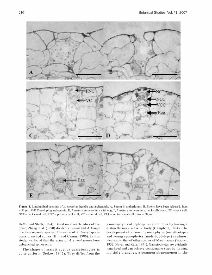
Figure 4. Longitudinal sections of A. somai antheridia and archegonia. A, Sperm in antheridium; B, Sperm have been released. Bars = 50 μm; C-F, Developing archegonia; E, A mature archegonium with egg; F, A mature archegonium, neck cells open. NC = neck cell; NCC= neck canal cell; PNC = primary neck cell; VC = ventral cell; VCC= ventral canal cell. Bars = 50 μm.

CHOU et al. – Gametophytes and sporophytes of Archangiopteris somai 211
Figure 5. Developing A. somai sporophytes. A, An embryo (dashed loop) embedded in the gametophyte; B, An embryo (dashed loop) protruding from the dorsal side of a gametophyte. Bars = 200 μm; C, A new sporophyte arising from the dorsal side of gametophyte; D, A young sporophyte with its first four fronds. Bars = 1 cm; E-F, First frond; G, Second frond; H, Third frond; I-J, Fourth fronds; K, Fifth frond; L, Sixth frond. Bar = 5 cm.

212 Botanical Studies, Vol. 48, 2007
Marattiaceae (Campbell, 1914). Endophytic fungi have been found within gametophytes of some Marattiaceae, such as Angiopteris evecta (Haupt, 1940), but none was found in A. somai during this study.
Most ferns have young leaves that lack midribs. Howev-er, ferns in the Ophioglossaceae and Gleicheniaceae, some in the Marattiaceae, and ferns in the genus Nephrolepis have midribs in juvenile leaf-blades (Wagner, 1952). A. somai also has leaves with midribs.
Some species of Marattiaceae, such as Marattia sambucina, are apomictic (Stokey, 1942). However, in this study, all A. somai sporophytes resulted from sexual reproduction. Only 3% of the gametophytes produced sporophytes. It may be that A. somai has a high genetic load that limits reproduction and contributes to its endangered status (e. g., Peck et al., 1990). However, the low reproduction observed in this study may also be due to the culture condition being ill-suited to the sporophyte formation of A. somai.
The best strategy for the long-term protection of a rare and endangered species is the preservation of natural communities and populations in the wilds, known as in situ preservation (Primack, 2004). However, if the last surviving populations are in continuous decline, and the original habitats are not protected, as is the case with A. somai, the only way to prevent extinction is to maintain individuals in culture (Primack, 2004). Sowing spores and growing gametophytes yielded few sporophytes (3%). However, a single A. somai plant may produce billions of viable spores every year, so it should be possible to obtain numerous sporophytes. Sporophytes grown in culture can be released periodically into the wild to augment the original population and maintain the existence of this rare species.
Acknowledgements. The authors thank Mr. Shih-Min Yang for taking photographs (Figure 2), and Dr. Alan Warneke for editing assistance. This research was partly supported by the National Science Council of Taiwan (NSC 91-2313-B-003-004) and Taiwan Forestry Research Institute.
lITERATURE CITED
Brebner, G. 1896. On the prothallus and embryo of Danaea simplicifolia, Rudge. Ann. Bot. 5: 109-121.
Brownlie, G. 1961. Additional chromosome numbers in New Zealand ferns. Trans. Roy. Soc. NZ Bot. 1: 1-4.
Campbell, D.H. 1894. Observations on the development of Marattia douglasii Baker. Ann. Bot. 29: 1-20.
Campbell , D.H. 1914. The s t ructure and aff ini t ies of Macroglossum alidae Copeland. Ann. Bot. 28: 651-669.
Chang, C.Y. 1973. The morphology of Archangiopteris Christ et Gies. and its relationship with Angiopteris Hoffm. Acta Bot. Sin. 15: 261-270.
Ching, R.C. 1958. A revision of the fern genus Archangiopteris
Christ & Giesenhagen. Acta Phytotax. Sin. 8: 202-224.Chiou, W.L., Y.M. Huang, T.H. Hsieh, and S.Y. Hsu. 2006.
Diplazium megaphyllum (Bak.) Christ, a rare fern in Taiwan, reproduces by apogamy. Taiwan J. For. Sci. 21: 39-47.
DeVol, C.E. 1975. Marattiaceae. In H.L. Li, T.S. Liu, T.C. Huang, T. Koyama, and C.E. DeVol (eds.), Flora of Taiwan Volume I, Epoch Publishing Co., Ltd., Taipei, Taiwan, pp. 281-301.
DeVol, C.E. and W.C. Shieh. 1994. Marattiaceae. In Editorial Committee of The Flora of Taiwan (eds.), Flora of Taiwan Volume I, Editorial Committee of The Flora of Taiwan, Taipei, Taiwan, pp. 74-79.
Farmer, J.B. 1892. On the embryogeny of Angiopteris evecta Hoffm. Ann. Bot. 6: 265-270.
Ghatak, J. 1962. Observations on the cytology and taxonomy of some ferns from Indian. Nucleus 5: 95-114.
Haupt, A.W. 1940. Sex organs of Angiopteris evecta. Bull. Torry Club 67: 125-129.
Hill, C.R. and J.M. Camus. 1986. Evolutionary cladistics of marattialean ferns. Bull. Br. Mus. Nat. Hist. (Bot.) 14: 219-300.
Huang, Y.M., H.M. Chou, T.H. Hsieh, J.C. Wang, and W.L. Chiou. 2006. Cryptic characteristics distinguish diploid and triploid varieties of Pteris fauriei (Pteridaceae). Can. J. Bot. 84: 261-268.
Huang, Y.M., S.L. Wong, and W.L. Chiou. 2003. The collection and storage of pteridophytes spore. Taiwan J. For. Sci. 18: 75-79.
Li, J.W. 1989. Chromosome numbers of some species in the genus Angiopteris Hoffm. from Yunnan, China. In K.H. Shing and K.U. Kramer (eds.), Proc. Int. Symp. Systematic Pteridology, Science and Technology Press, Beijing, China, pp. 109-110.
Kuo, C.M. 1997. Archangiopteris somai Hayata. In S.Y. Lu and W.L. Chiou (eds.), Rare and Endangered Plants in Taiwan, Volume II, Council of Agriculture, Taipei, Taiwan, pp. 7-8.
Manton, I. and W.A. Sledge. 1954. Observations on the cytology and taxonomy of the Pteridophyte flora of Ceylon. Phiols. Trans. Roy. Soc. London. B 238: 127-185.
Moore, S.J., T.H. Hsieh, Y.M. Huang, and W.L. Chiou. 2002. Diplazium maonense Ching, a poorly known species of the Athyriaceae (Pteridophyta) in Taiwan. Taiwan J. For. Sci. 17: 113-118.
Nakato, N. 1988. Notes on chromosomes of Japanese pterido-phytes (2). J. Jpn. Bot. 63: 214-218.
Nakato, N. 1996. Notes on chromosomes of Japanese pterido-phytes (4). J. Jpn. Bot. 71: 163-167.
Nayar, B.K. and S. Kaur. 1971. Gametophytes of homosporous ferns. Bot. Rev. 37: 296-396.
Ninan, C.A. 1956. Studies on the cytology and phylogeny of the pteridophytes. I. Observations on the Marattiaceae. J. Ind Bot. Soc. 35: 233-239.
Peck, J.H., C.J. Peck, and D.R. Farrar. 1990. Influences of life
CHOU et al. – Gametophytes and sporophytes of Archangiopteris somai 213
history attributes on formation of local and distant fern population. Amer. Fern J. 80: 126-142.
Primack, R.B. 2004. A Primer of Conservation Biology (third edition). Sinauer. Sunderland, Massachusetts.
Sasaki, S. 1928. List of plants of Formosa. Trans. Nat. Hist. Soc. Formosa 19: 306-307.
Sharma, H.C. 1982. A technique for somatic counts from root tips of cereal seedlings raised by embryo culture. Curr. Sci. 51: 143-144.
Spurr, A.R.J. 1969. A low-viscosity epoxy resin embedding me-dium for electron microscopy. J. Ultrastruct. Res. 26: 31-43.
Stokey, A.G. 1942. Gametophytes of Marattia sambucina and Macroglossum smithii. Bot. Gaz. 103: 559-569.
Tryon, A.F. and B. Lugardon. 1991. Spores of the Pteridophyta. Springer-Verlag, New York.
Wagner, W.H.Jr. 1952. Types of foliar dichotomy in living ferns. Amer. J. Bot. 39: 578-592.
Walker, T.G. 1966. A cytotaxonomic survey of the pteridophytes of Jamaica. Trans. Roy. Soc. Edinb. 66: 169-237.
Wu, S.H. and R.C. Ching. 1991. Fern Families and Genera of China. Science Press, Beijing.
Yang, S.M., P.Y. Tasi, W.S. Ho, S.L. Wong, and H.M. Chou. 2004. Key factors for the spore germination of Archangi-opteris somai Hayata, a rare and endemic fern in Taiwan. BioFormosa 39: 33-39.
Zhang, Y.L., J.T. Zhang, N.Q. Du, Z.C. Kong, Y.Z. Xi, G.Z. Gao, and X.G. Sun. 1990. Spore Morphology of Chinese Pteridophytes. Science Press, Beijing.
一種瀕臨滅絕的蕨類 — 台灣原始觀音座蓮(觀音座蓮舅科) — 配子體與幼孢子體的觀察
周雪美1 黃曜謀2 翁韶良3 謝宗欣4 徐昇圓2 邱文良2
1 元培科技大學生物技術學系 2 農委會林業試驗所森林生物組 3 特有生物研究保育中心解說教育組 4 國立台南大學生物科學技術學系
台灣原始觀音座蓮是一台灣產特有的厚囊蕨類。由於其族群與數量稀少,被歸類為瀕臨滅絕的種
類。培養時其孢子在灑播二週後開始發芽;配子體以「觀音座蓮舅(Marattia)」型發育,配子體生長過程由圓形轉心臟形再形成分支。藏精器與藏卵器分別在孢子灑播 10 及 12 月後發生;藏精器長在中肋的背面及腹面,藏卵器只在腹面發生;兩性配子體的藏精器與藏卵器混生一起,兩者均屬厚囊蕨型。孢
子體在經 13 個月的培養後開始產生,每個配子體雖然有許多的藏卵器,但只產生一株孢子體,第一片孢子體的葉片為單葉羽狀脈。經過三年的培養,只有 3% 的配子體產生孢子體。由於孢子體的生成至少需 13 個月,以及其低生成率,可能導致其野外族群之擴增極為緩慢且困難。
關鍵詞:台灣原始觀音座蓮;染色體;厚囊蕨類;配子體;幼孢子體;觀音座蓮舅科;孢子。