Obey 10 User Manual Rev. 3 - CHAUVET® Lighting
12
Resuscitation (2008) 76, 431—442 available at www.sciencedirect.com journal homepage: www.elsevier.com/locate/resuscitation EXPERIMENTAL PAPER Improving neurological outcomes post-cardiac arrest in a rat model: Immediate hypothermia and quantitative EEG monitoring , Xiaofeng Jia a,∗ , Matthew A. Koenig b,c , Hyun-Chool Shin a,e , Gehua Zhen c , Carlos A. Pardo b,d , Daniel F. Hanley b , Nitish V. Thakor a , Romergryko G. Geocadin b,c a Department of Biomedical Engineering, Johns Hopkins University School of Medicine, Baltimore, MD 21205, USA b Department of Neurology, Johns Hopkins University School of Medicine, Baltimore, MD 21205, USA c Department of Anesthesiology-Critical Care Medicine, Johns Hopkins University School of Medicine, Baltimore, MD 21205, USA d Department of Pathology, Johns Hopkins University School of Medicine, Baltimore, MD 21205, USA e Department of Electronic Engineering, Soongsil University, Seoul, Republic of Korea Received 25 June 2007; received in revised form 23 July 2007; accepted 2 August 2007 KEYWORDS Cardiac arrest; Electroencephalography; Hypothermia; Functional outcome; Brain ischemia Summary Objectives: Therapeutic hypothermia (TH) after cardiac arrest (CA) improves outcomes in a fraction of patients. To enhance the administration of TH, we studied brain electrophysiolog- ical monitoring in determining the benefit of early initiation of TH compared to conventional administration in a rat model. Methods: Using an asphyxial CA model, we compared the benefit of immediate hypothermia (IH, T = 33 ◦ C, immediately post-resuscitation, maintained 6 h) to conventional hypothermia (CH, T = 33 ◦ C, starting 1 h post-resuscitation, maintained 12 h) via surface cooling. We tracked quantitative EEG using relative entropy (qEEG) with outcome verification by serial Neurological Deficit Score (NDS) and quantitative brain histopathological damage scoring (HDS). Thirty-two rats were divided into 4 groups based on CH/IH and 7/9-min duration of asphyxial CA. Four sham rats were included for evaluation of the effect of hypothermia on qEEG. Results: The 72-h NDS of the IH group was significantly better than the CH group for both 7-min (74/63; median, IH/CH, p < 0.001) and 9-min (54/47, p = 0.022) groups. qEEG showed greater A Spanish translated version of the summary of this article appears as Appendix in the final online version at 10.1016/j.resuscitation.2007.08.014. Portions of this work were previously presented in 4th Annual Meeting of the Neurocritical Care Society in Baltimore, MD (November 2006) and at the 36th Annual Meeting of the Society for Neuroscience at Atlanta, Georgia (October 2006) where it was selected for the lay language summary press book. ∗ Corresponding author at: CRB II Building 3M-South, 1550 Orleans Street, Johns Hopkins University School of Medicine, Baltimore, MD 21231, USA. Tel.: +1 410 502 2820; fax: +1 410 502 7869. E-mail address: [email protected] (X. Jia). URL: http://www.jhu.edu/xjia (X. Jia). 0300-9572/$ — see front matter © 2007 Elsevier Ireland Ltd. All rights reserved. doi:10.1016/j.resuscitation.2007.08.014
Transcript of Obey 10 User Manual Rev. 3 - CHAUVET® Lighting

Resuscitation (2008) 76, 431—442
avai lab le at www.sc iencedi rec t .com
journa l homepage: www.e lsev ier .com/ locate / resusc i ta t ion
EXPERIMENTAL PAPER
Improving neurological outcomes post-cardiac arrestin a rat model: Immediate hypothermia andquantitative EEG monitoring�,��
Xiaofeng Jiaa,∗, Matthew A. Koenigb,c, Hyun-Chool Shina,e, Gehua Zhenc,Carlos A. Pardob,d, Daniel F. Hanleyb, Nitish V. Thakora,Romergryko G. Geocadinb,c
a Department of Biomedical Engineering, Johns Hopkins University School of Medicine, Baltimore, MD 21205, USAb Department of Neurology, Johns Hopkins University School of Medicine, Baltimore, MD 21205, USAc Department of Anesthesiology-Critical Care Medicine, Johns Hopkins University School of Medicine,Baltimore, MD 21205, USAd Department of Pathology, Johns Hopkins University School of Medicine, Baltimore, MD 21205, USAe Department of Electronic Engineering, Soongsil University, Seoul, Republic of Korea
Received 25 June 2007; received in revised form 23 July 2007; accepted 2 August 2007
KEYWORDSCardiac arrest;Electroencephalography;Hypothermia;Functional outcome;Brain ischemia
SummaryObjectives: Therapeutic hypothermia (TH) after cardiac arrest (CA) improves outcomes in afraction of patients. To enhance the administration of TH, we studied brain electrophysiolog-ical monitoring in determining the benefit of early initiation of TH compared to conventionaladministration in a rat model.Methods: Using an asphyxial CA model, we compared the benefit of immediate hypothermia(IH, T = 33 ◦C, immediately post-resuscitation, maintained 6 h) to conventional hypothermia
◦
(CH, T = 33 C, starting 1 h post-resuscitation, maintained 12 h) via surface cooling. We trackedquantitative EEG using relative entropy (qEEG) with outcome verification by serial NeurologicalDeficit Score (NDS) and quantitative brain histopathological damage scoring (HDS). Thirty-tworats were divided into 4 groups based on CH/IH and 7/9-min duration of asphyxial CA. Foursham rats were included for evaluation of the effect of hypothermia on qEEG.Results: The 72-h NDS of the IH group was significantly better than the CH group for both 7-min(74/63; median, IH/CH, p < 0.001) and 9-min (54/47, p = 0.022) groups. qEEG showed greater� A Spanish translated version of the summary of this article appears as Appendix in the final online version at10.1016/j.resuscitation.2007.08.014.
�� Portions of this work were previously presented in 4th Annual Meeting of the Neurocritical Care Society in Baltimore, MD (November2006) and at the 36th Annual Meeting of the Society for Neuroscience at Atlanta, Georgia (October 2006) where it was selected for thelay language summary press book.
∗ Corresponding author at: CRB II Building 3M-South, 1550 Orleans Street, Johns Hopkins University School of Medicine, Baltimore, MD21231, USA. Tel.: +1 410 502 2820; fax: +1 410 502 7869.
E-mail address: [email protected] (X. Jia).URL: http://www.jhu.edu/xjia (X. Jia).
0300-9572/$ — see front matter © 2007 Elsevier Ireland Ltd. All rights reserved.doi:10.1016/j.resuscitation.2007.08.014

432
recovery with IH (p < 0.001) and siversus CH: 33.2 ± 4.4%, p = 0.006).(p < 0.05) and 72-h behavioral subthe sham group.Conclusions: Immediate but shooutcome in rats after 7- and 9-min
ing anAll ri
I
Aocaocd
hotifnhTocftr
eiW1coit(foptram
rtloWhmqmi
M
Tkw
E
Taoio
sbiFwoowuae
cm1F1Tsbfeai2
sat
E
electrophysiological monitor© 2007 Elsevier Ireland Ltd.
ntroduction
pproximately 400,000 out-of-hospital cardiac arrests (CA)ccur in the United States each year.1 Despite advances inardiopulmonary resuscitation (CPR) and critical care, over-ll survival from out-of-hospital CA remains poor, averagingnly 5—8% in most centers.2 Among survivors, neurologicalomplications represent the leading cause of morbidity andisability.3,4
Two recent clinical trials demonstrated that inducedypothermia to 32—34 ◦C improves survival and functionalutcomes in comatose survivors of CA.5,6 While a break-hrough treatment for brain injury after cardiac arrest,ts full beneficial therapeutic effect has not yet beenully realized. A meta-analysis showed that the number-eeded-to-treat to allow one additional patient to leave theospital with favorable neurological recovery was 4—13.7
he optimal initiation time, duration of therapy, and depthf hypothermia have not been defined. Early initiation ofooling has been suggested but it appears to be success-ul even if delayed by 4—6 h.8 Most clinical studies delayhe initiation of hypothermia by 2 or more hours afteresuscitation.5,6,9,10
Recent animal studies have shown that mild to mod-rate hypothermia (33—34 ◦C) mitigates brain injury whennduced before,11 during,11,12 or after resuscitation.12—14
e demonstrated previously that initiation of hypothermiah after CA significantly improved neurological outcomesompared to normothermic controls.15,24 Despite the usef hypothermia to ameliorate brain injury, no direct mon-toring is undertaken to assess the brain’s response toherapy. We have previously validated quantitative EEGqEEG) monitoring in a rodent model after resuscitationrom CA.17,18 This entropy-based qEEG provides real-time,bjective tracking of neurological injury, recovery and earlyrognostication after CA.17—23 This method is also sensi-ive to temperature-related modulation of brain injury, withapid and sustained improvement of qEEG measures in thosenimals treated with hypothermia compared to normother-ic controls.15,16,24
In this study, we tested the hypothesis that neu-omonitoring techniques track brain recovery and responseo brain-directed therapy in real-time. Clinical trans-ation of these techniques may optimize the deliveryf hypothermia to a neuro-electrophysiological endpoint.e examined the impact of immediate initiation of 6-h
ypothermia (IH) compared to 12-h conventional hypother-ia (CH) initiated at 1 h post-resuscitation with real-timeEEG tracking followed by functional outcome assess-ent and histological assessment of the brain corticalnjury.
WetS
X. Jia et al.
gnificantly less neuronal cortical injury by HDS (IH: 18.9 ± 2.5%The 1-h post-resuscitation qEEG correlated well with 72-h NDSgroup of NDS (p < 0.01). No differences in qEEG were noted in
rter hypothermia compared to CH leads to better functionalCA. The beneficial effect of IH was readily detected by neuro-d histological changes supported the value of this observation.
ghts reserved.
aterial and methods
he experimental protocol was approved by the Johns Hop-ins Animal Care and Use Committee and all proceduresere compliant with NIH guidelines.
xperimental asphyxia—–CA model
hirty-six adult male Wistar rats (360 ± 20 g) were assignedt random to 4 groups based on CH/IH and 7/9-min durationf asphyxial CA (7CH, 7IH, 9CH, 9IH, n = 8). Four rats werencluded as a sham control group for evaluation of the effectf hypothermia on qEEG in the absence of CA injury.
Our group and others have used this rodent model totudy calibrated brain injury after asphyxial CA.15,18,25 Inrief (detail described by Jia et al. 15), rats were mechan-cally ventilated with 1.0% halothane in N2/O2 (50/50%).ive minutes of baseline recording was followed by 5-minashout to ensure no significant residual effect of halothanen qEEG.17 CA was initiated via asphyxia with cessationf mechanical ventilation for periods of 7 or 9 min. CPRas performed with sternal chest compressions (200 min−1)ntil return of spontaneous circulation (ROSC). Sedative andnesthetic agents were avoided to minimize confoundingffects on EEG.26
CH was induced 1 h after ROSC by surface cooling withold mist to achieve the target core temperature of 33 ◦Conitored by an intraperitoneal temperature sensor within
5 min and was maintained between 32 and 34 ◦C for 12 h.15
or the IH groups, cooling was initiated immediately (within5 min) after ROSC and was maintained at 32—34 ◦C for 6 h.he 6-h duration was chosen based on a pilot group of ratshowing rapid recovery from unresponsiveness to exploringehavior in IH animals. Then rats were gradually re-warmedrom 33.0 to 37.0 ◦C over 2 h using a warming blanket. Tonsure no post-resuscitation spontaneous hypothermia,27 allnimals were then kept inside a neonatal incubator (Isolettenfant incubator, Air-shields Inc, Pennsylvania) for the first4 h post-ROSC.
Continuous EEG was undertaken in four anesthetizedham rats which underwent identical surgical preparationnd subjected to normothermic baseline for 30 min, andhen induced hypothermia for 1 h.
EG recording and qEEG analysis
e have used qEEG analysis previously to track brain recov-ry after CA.15,16,20—24 EEGs were recorded from baselinehrough re-warming periods using DI700 Windaq system.15
erial 30-min EEG recordings were then performed at 24,

rrest 433
of counted cells] × 100) representing the degree of necroticcell death per region.35
Statistics
Univariate analysis was performed for parametric data(reported as mean ± S.E.M.) with the use of the Student’st-test for continuous variables and the Chi-square test forcategorical variables. Non-parametric analysis of variance
Figure 1 Temperature recording of conventional hypother-mia (CH) and immediate hypothermia (IH) rats in the (A) 7-minasphyxia group and (B) 9-min asphyxia group. The dark line isCH and light line is IH. The solid heavy line is mean temper-ature and the field with lighter shading is S.E.M. For the firstperiod (a), no temperature difference was noted during base-line and cardiac arrest periods (36.7 ± 0.0/36.3 ± 0.0 (A(a)) for7-min group (CH/IH, mean ± S.E.M.) and 36.7 ± 0.1/36.5 ± 0.0(B(a)) for 9-min group) in (A(a)) and (B(a)). In the secondperiod (b), the mean temperature during the CH group washigher than IH (36.2 ± 0.1/33.2 ± 0.1 (A(b)) for the 7-min groupand 35.8 ± 0.2/32.9 ± 0.1 (B(b)) for the 9-min group) (bothp < 0.001). The temperature was within the 32—34 ◦C rangeduring hypothermia (period (c)) (32.7 ± 0.0/32.7 ± 0.0 (A(c))for the 7-min group and 32.2 ± 0.0/32.8 ± 0.0 (B(c)) for the9-min group). In period (d), animals in the IH group were re-
Immediate hypothermia and quantitative EEG post-cardiac a
48, and 72 h after ROSC in each group. In this study weemployed entropy to analyze nonstationary EEG signals. Theinformation quantity (IQ) is calculated using a sliding tempo-ral window technique from a window block of EEG signal forthe entire data set.15,24 Sub-band IQ (SIQ, referred below asqEEG) is the average value of IQ within different frequenciesbands. The details of this analysis method are provided inAppendix A. We chose 9 segments from EEG data in each ratand calculated qEEG: baseline, CA, 30-min, 1-h, 4-h, 6-h,24-h, 48-h, and 72-h.
Neurological evaluation
The Neurological Deficit Scale (NDS) was patterned after theneurological examination in humans10 and functional out-come scales for global cerebral ischemia in animals.11,13,25
The previously validated15,18 NDS ranges from 80 indicatinga functionally normal rat to a score of 0 for brain or car-diac death prior to conclusion of the study. Behavioral testsincluding gait coordination, balance beam walking, rightingreflex, negative geotaxis, visual placing and turning alleytests were analyzed as a subgroup of NDS17,18 (see AppendixA for details). NDS was determined by a trained examinerblinded to temperature groups after the re-warming periodat 2 h post-hypothermia on the first day, and then repeatedat 24, 48, and 72 h after ROSC. The primary outcome mea-sure was defined as the 72-h NDS score.
Quantitative brain histopathological damagescoring (HDS)
Ten �m paraffin-embedded sections were stained with Cre-syl violet28,29 which was used to quantify ischemic changesusing a standard rat brain atlas30 in each hemisphereunder ×400 magnification with an Olympus BX51 micro-scope (Olympus, Center Valley, PA). Cresyl violet stainingwas chosen based on its reliability and consistency inthe evaluation of cytoplasmic and nuclear morphologicalchanges as compared with H&E, and its better suitabilityfor stereological techniques to identify nuclear and nucle-olar structures that are required as part of the assessmentof cell viability.31—34 Ischemic neurons were identified usingstandard criteria29,35—37: pyknosis, karyorrhexis, karyolysisand cytoplasmic changes in form and color.
After de-identifying the histologic slides of clinical data,two investigators, supervised by a neuropathologist, quanti-fied ischemic neurons in a standardized region of temporalcortex layers 4 and 5 anterior to the rhinal fissure andadjacent to the hippocampus, approximating the tempo-ral cortex areas Te1 and Te3,30 along with CA1 of thehippocampus. These areas were chosen because CA1 isselectively vulnerable to global cerebral ischemia in ani-mals and humans studies and the cerebral cortex is closelyrelated to functional outcome and EEG changes.11,18,26,38,39
Stereologic technique was performed in the predefinedset of random, non-overlapping microscope fields using
Adreas Stereo Investigator software 5.05.4 (MicroBright-Field, Williston, VT) to estimate the degree of neuronalinjury. In addition to ischemic neurons, normal-appearingneurons were counted in each field. These data were used tocalculate a percentage of injury ([injured cells/total numberwarmed while hypothermia was maintained for 6 more hours inthe CH group (32.7 ± 0.0/35.4 ± 0.1 (A(d)) for 7-min group and32.4 ± 0.0/35.2 ± 0.1 (B(d)) for 9-min group) (both p < 0.001).After 12 h of hypothermia in the CH group, normothermia wasmaintained similar to the IH group.

4
wrutaFayss1
R
Ba
Tbd(
gF1tt
u(n(b
E
Wa
34
as used to test for differences in rank order NDS as aepeated measure. Multivariate General Linear Model wassed for advanced comparison of aggregate data to con-rol for influencing factors such as hypothermia methodnd asphyxia time. The mortality rate was analyzed byisher’s exact test (crosstabs) and survival was analyzed byKaplan—Meier test. Pearson correlation of bivariate anal-
sis was used to analyze the correlation between 72-h NDScore with serial qEEG and HDS. Statistical significance waset at p < 0.05. Statistical analysis was performed with SPSS4.0 (SPSS Inc., Chicago, IL).
esults
aseline characteristics, temperature monitoringnd ABG data
here are no significant difference between groups of theaseline characteristics including body weights, anesthesiauration for preparation, heart rates and blood pressurep > 0.05). The temperature recording for 7- and 9-min
ii1(r
Table 1 Arterial blood gas and MAP data in cardiac arrest experi
7CHa 7IHb
BaselinepH 7.48 ± 0.03 7.42 ± 0.02PCO2 (mmHg) 36 ± 2 39 ± 4PO2 (mmHg) 323 ± 51 383 ± 25HCO3 (mmol/l) 26 ± 1 24 ± 2O2SAT (%) 100 ± 0 100 ± 0MAP (mmHg) 100 ± 7 90 ± 11
10 min post-CApH 7.34 ± 0.03 7.35 ± 0.02PCO2 (mmHg) 42 ± 3 36 ± 2PO2 (mmHg) 442 ± 39 403 ± 52HCO3 (mmol/l) 22 ± 1 19 ± 1O2SAT (%) 100 ± 0 100 ± 0MAP (mmHg) 123 ± 9 123 ± 10
20 min post-CApH 7.36 ± 0.04 7.37 ± 0.01PCO2 (mmHg) 38 ± 2 35 ± 2PO2 (mmHg) 434 ± 25 496 ± 19HCO3 (mmol/l) 21 ± 1 20 ± 1O2SAT (%) 100 ± 0 100 ± 0MAP (mmHg) 85 ± 10 99 ± 8
40 min post-CApH 7.40 ± 0.04 7.31 ± 0.02PCO2 (mmHg) 42 ± 4 44 ± 4PO2 (mmHg) 483 ± 29 455 ± 39HCO3 (mmol/l) 25 ± 1 22 ± 1O2SAT (%) 100 ± 0 100 ± 0MAP (mmHg) 90 ± 9 110 ± 10
* Statistically significant difference was noted but was minimal to caPCO2 at 10 and 20 min in 9IH48,49 while CO2 elevation is due to normothin IH group may be a reflection of the ongoing systemic beneficial effe
a Conventional hypothermia.b Immediate hypothermia.
X. Jia et al.
roups during the hypothermia experiment is shown inigure 1. Cooling to target temperature was achieved in1.1 ± 4.6 min and re-warming in 116.8 ± 21.4 min to reachhe target range of 36.5—37.5 ◦C. All animals were main-ained at 37 ± 0.5 ◦C after re-warming during the first 24 h.
ABG data including arterial pH, HCO3−, PO2 and O2 sat-
ration, were similar between groups at all time pointsbaseline, 10, 20, and 40 min after ROSC). There are sig-ificant differences of PCO2 at 10 min (p < 0.01) and 20 minp < 0.01) and of blood pressure at 40 min (p < 0.05) existedetween groups (data shown in Table 1).
EG-bursting analysis and qEEG analysis
ithin seconds of CA, the normal EEG signal was reduced ton isoelectric tracing, which was followed by periodic activ-
ty resembling burst-suppression of variable duration. Thenterval between CA and the first burst in the 4 groups was6.3 ± 0.7 min (7CH), 20.3 ± 1.7 min (9CH), 16.3 ± 1.2 min7IH) and 19.6 ± 2.8 min (9IH), respectively, where CAefers to the starting period of asphyxia and the intervalment among 4 groups
9CH 9IH p value of 4 groups
7.53 ± 0.06 7.45 ± 0.01 0.22040 ± 0 38 ± 1 0.929
323 ± 26 391 ± 46 0.59828 ± 0 25 ± 1 0.683
100 ± 0 100 ± 0 0.76288 ± 5 91 ± 7 0.644
7.35 ± 0.04 7.35 ± 0.03 0.93638 ± 2 30 ± 2 0.009*
434 ± 49 491 ± 20 0.38720 ± 2 17 ± 0 0.022*
100 ± 0 100 ± 0 0.522119 ± 12 150 ± 12 0.191
7.22 ± 0.00 7.38 ± 0.02 0.15550 ± 0 29 ± 2 0.009*
595 ± 0 451 ± 26 0.08220 ± 0 18 ± 1 0.165
100 ± 0 100 ± 0 0.86288 ± 16 102 ± 15 0.753
7.43 ± 0.04 7.36 ± 0.02 0.16439 ± 3 32 ± 3 0.089
424 ± 59 440 ± 30 0.84124 ± 1 19 ± 1 0.003*
100 ± 0 100 ± 0 0.47462 ± 5 93 ± 15 0.020*
use any significant change in pH. Hypothermia may decrease theermia in 9CH animals.49,50 The relative increase in MAP at 40 minct hypothermia.5,51,52

Immediate hypothermia and quantitative EEG post-cardiac arrest 435
Figure 2 Raw EEG data and qEEG of representative 9-min asphyxia animals in the first 4 h. (A) 9-min conventional hypothermiain),
of qE
a6rbs
F
Nmsicgg((
M
T
(9CH); (B) 9-min immediate hypothermia (9IH): (I) Baseline (0 m4 h after CA—hypothermia maintenance period; (C) comparisonafter ROSC.
from CA to the first burst is the duration of isoelectricEEG.
There was a statistically significant difference between7-min CA (16.3 ± 0.7) and 9-min CA groups (20.0 ± 0.9)(p = 0.004) while no significant difference existed betweenIH and CH rats. No significant difference was observed inqEEG of sham rats between the periods of hypothermia(0.59 ± 0.02) and normothermia (0.60 ± 0.02) (p = 0.921).
Using visual assessment by comparing the evolution ofburst frequency and amplitude from baseline of the rawEEG signal alone, qualitative differences between the groupswere not evident (Figure 2A and B). Subtle differences inEEG, however, were readily discerned using qEEG analysis(Figure 2C). Similar to the raw EEG data, the aggregateqEEG decreased from baseline (qEEG = 1) to the lowest pointrapidly after CA, then gradually recovered close to baseline.
Mean qEEG relative entropy values over the study periodwere not different (p = 0.08) between 7IH rats (0.78 ± 0.03)and 7CH rats (0.75 ± 0.04); while 9IH rats (0.73 ± 0.03)had significantly greater mean qEEG entropy than 9CH rats(0.63 ± 0.04) (p < 0.001) (Figure 3A and B). Aggregate anal-ysis showed mean qEEG entropy was greater in the IH rats
(0.76 ± 0.02) compared to CH controls (0.70 ± 0.03) over the72-h study period (p < 0.001).The predictive capacity of early qEEG for 72-h NDS bybivariate analyses, using aggregate qEEG and NDS data(n = 28), revealed significant correlations between 72-h NDS
1r0da
(II) Cardiac arrest (CA) period (19 min), (III) 1 h after CA, (IV)EG in CH and IH, CH started at 1 h and IH started immediately
nd qEEG values showing statistical significance at 1, 4,and 24 h (p < 0.05). The value of early qEEG to predict
ecovery of behavioral and cognitive function, evaluatedy comparing qEEG against the behavioral subgroup of NDS,howed a significant correlation at 1 and 4 h (p < 0.05).
unctional recovery by NDS
DS analysis results are shown in Figure 4. IH ani-als had persistently better functional recovery by NDS
cores at all time periods with statistical significancen the 7-min groups (IH/CH (median, 25—75th per-entile): 74, 60.75—74/63, 50.25—70; p = 0.001) and 9-minroups (IH/CH: 54, 49—61.75/47, 39—60; p = 0.022). Aggre-ate analysis showed statistically significant differencesp = 0.001) between CH group (54, 42—66) and IH group61.5, 51.25—74).
ortality and qEEG
he mortality rates were 0% (0/8) in 7IH, 12.5% (1/8) in 7CH,
2.5% (1/8) in 9IH, and 25% (2/8) in 9CH group, with an oddsatio (OR) of mortality in the CH group of 3.667 (95% CI:.3—42.9). No statistically significant differences in meanuration of survival hours existed between the 7IH (72.0 h)nd 7CH (63.8 h) groups or between the 9IH (67.5 h) and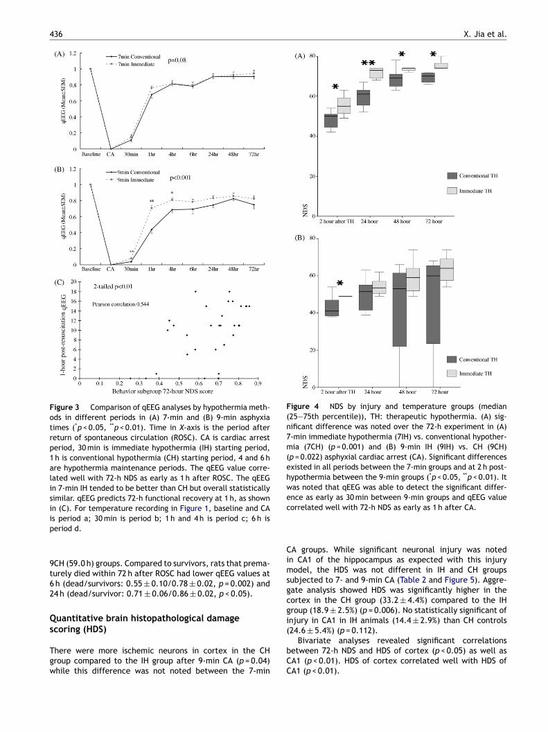
436 X. Jia et al.
Figure 3 Comparison of qEEG analyses by hypothermia meth-ods in different periods in (A) 7-min and (B) 9-min asphyxiatimes (*p < 0.05, **p < 0.01). Time in X-axis is the period afterreturn of spontaneous circulation (ROSC). CA is cardiac arrestperiod, 30 min is immediate hypothermia (IH) starting period,1 h is conventional hypothermia (CH) starting period, 4 and 6 hare hypothermia maintenance periods. The qEEG value corre-lated well with 72-h NDS as early as 1 h after ROSC. The qEEGin 7-min IH tended to be better than CH but overall statisticallysimilar. qEEG predicts 72-h functional recovery at 1 h, as shownin (C). For temperature recording in Figure 1, baseline and CAip
9t62
Qs
Tgw
Figure 4 NDS by injury and temperature groups (median(25—75th percentile)), TH: therapeutic hypothermia. (A) sig-nificant difference was noted over the 72-h experiment in (A)7-min immediate hypothermia (7IH) vs. conventional hypother-mia (7CH) (p = 0.001) and (B) 9-min IH (9IH) vs. CH (9CH)(p = 0.022) asphyxial cardiac arrest (CA). Significant differencesexisted in all periods between the 7-min groups and at 2 h post-hypothermia between the 9-min groups (*p < 0.05, **p < 0.01). Itwas noted that qEEG was able to detect the significant differ-ence as early as 30 min between 9-min groups and qEEG valuec
Cimsgcgi
s period a; 30 min is period b; 1 h and 4 h is period c; 6 h iseriod d.
CH (59.0 h) groups. Compared to survivors, rats that prema-urely died within 72 h after ROSC had lower qEEG values ath (dead/survivors: 0.55 ± 0.10/0.78 ± 0.02, p = 0.002) and4 h (dead/survivor: 0.71 ± 0.06/0.86 ± 0.02, p < 0.05).
uantitative brain histopathological damagecoring (HDS)
here were more ischemic neurons in cortex in the CHroup compared to the IH group after 9-min CA (p = 0.04)hile this difference was not noted between the 7-min
(
bCC
orrelated well with 72-h NDS as early as 1 h after CA.
A groups. While significant neuronal injury was notedn CA1 of the hippocampus as expected with this injuryodel, the HDS was not different in IH and CH groups
ubjected to 7- and 9-min CA (Table 2 and Figure 5). Aggre-ate analysis showed HDS was significantly higher in theortex in the CH group (33.2 ± 4.4%) compared to the IHroup (18.9 ± 2.5%) (p = 0.006). No statistically significant ofnjury in CA1 in IH animals (14.4 ± 2.9%) than CH controls
24.6 ± 5.4%) (p = 0.112).Bivariate analyses revealed significant correlationsetween 72-h NDS and HDS of cortex (p < 0.05) as well asA1 (p < 0.01). HDS of cortex correlated well with HDS ofA1 (p < 0.01).
Immediate hypothermia and quantitative EEG post-cardiac arrest 437
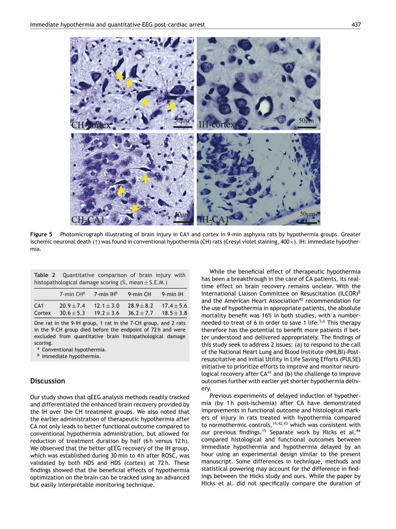
Figure 5 Photomicrograph illustrating of brain injury in CA1 and cortex in 9-min asphyxia rats by hypothermia groups. Greaterischemic neuronal death (↑) was found in conventional hypothermia (mia.
Table 2 Quantitative comparison of brain injury withhistopathological damage scoring (%, mean ± S.E.M.)
7-min CHa 7-min IHb 9-min CH 9-min IH
CA1 20.9 ± 7.4 12.1 ± 3.0 28.9 ± 8.2 17.4 ± 5.6Cortex 30.6 ± 5.3 19.2 ± 3.6 36.2 ± 7.7 18.5 ± 3.8
One rat in the 9-IH group, 1 rat in the 7-CH group, and 2 ratsin the 9-CH group died before the endpoint of 72 h and wereexcluded from quantitative brain histopathological damagescoring.
htIatmntttoriloe
mietocihour using an experimental design similar to the presentmanuscript. Some differences in technique, methods and
a Conventional hypothermia.b Immediate hypothermia.
Discussion
Our study shows that qEEG analysis methods readily trackedand differentiated the enhanced brain recovery provided bythe IH over the CH treatment groups. We also noted thatthe earlier administration of therapeutic hypothermia afterCA not only leads to better functional outcome compared toconventional hypothermia administration, but allowed forreduction of treatment duration by half (6 h versus 12 h).We observed that the better qEEG recovery of the IH group,which was established during 30 min to 4 h after ROSC, was
validated by both NDS and HDS (cortex) at 72 h. Thesefindings showed that the beneficial effects of hypothermiaoptimization on the brain can be tracked using an advancedbut easily interpretable monitoring technique.siH
CH) rats (Cresyl violet staining, 400×). IH: immediate hypother-
While the beneficial effect of therapeutic hypothermiaas been a breakthrough in the care of CA patients, its real-ime effect on brain recovery remains unclear. With thenternational Liaison Committee on Resuscitation (ILCOR)8
nd the American Heart Association40 recommendation forhe use of hypothermia in appropriate patients, the absoluteortality benefit was 16% in both studies, with a number-
eeded-to-treat of 6 in order to save 1 life.5,6 This therapyherefore has the potential to benefit more patients if bet-er understood and delivered appropriately. The findings ofhis study seek to address 2 issues: (a) to respond to the callf the National Heart Lung and Blood Institute (NHLBI)-Post-esuscitative and initial Utility in Life Saving Efforts (PULSE)nitiative to prioritize efforts to improve and monitor neuro-ogical recovery after CA41 and (b) the challenge to improveutcomes further with earlier yet shorter hypothermia deliv-ry.
Previous experiments of delayed induction of hypother-ia (by 1 h post-ischemia) after CA have demonstrated
mprovements in functional outcome and histological mark-rs of injury in rats treated with hypothermia comparedo normothermic controls,14,42,43 which was consistent withur previous findings.15 Separate work by Hicks et al.44
ompared histological and functional outcomes betweenmmediate hypothermia and hypothermia delayed by an
tatistical powering may account for the difference in find-ngs between the Hicks study and ours. While the paper byicks et al. did not specifically compare the duration of

4
hamNesa
tiroathoawt
hbiedHtsfso
ricibsr
ceWtontimHgtqb((
ACapa
(ifgtnggmdwhnies
lpaahot(ri
tEtrtiwrwgqctathiqcdtsm
iebia
38
ypothermia as we did, the potential advantage of earlierdministration was suggested with the immediate hypother-ia group showing a trend toward better outcomes in theDS, histological score, and heat shock protein levels. Ourxperiment lends further support to the theory that coolinghould begin as soon as possible after ROSC, similar to othernimal models.12,13,45
With electrophysiological markers, this study suggestshat earlier initiation of hypothermia may have a greatermpact during the early period of recovery and injured neu-ons that are immediately treated have a better chancef recovering. These observations, as supported by qEEGnd histology, preclude the need for longer treatment dura-ion and suggest the need to re-evaluate the timing ofypothermia initiation in human subjects. Given the risksf coagulopathy and immune suppression, need for sedationnd paralysis, expense, and high resource use associatedith hypothermia,5,6,9,10 these results are potentially impor-
ant.As human studies have shown, with this degree of
ypothermia (33 ± 1 ◦C), the detrimental effects have noteen significantly different than normothermia. Given thedentical temperature range in IH and CH, we did notxpect significant differences in detrimental effects, so weid not look at the detrimental effects in much detail.owever, considering the commonly reported detrimen-al side effects, we observed no significant hemorrhage,eizure activity, or arrhythmias. We did not look specificallyor pneumonia or renal failure. The enhanced recovery ofhorter IH over longer CH is most likely the result of rapidnset rather than a decrement in adverse effects.
Translational research verification may justify changingesuscitation strategies such that paramedics begin cool-ng in the field or ambulance en route to the hospital. Theost of longer periods of hypothermia centers on need forntensive nursing care. And the cost of rapid inductions maye dependent on technologies; however chilled saline infu-ion is definitely an economical and effective way to achieveapid hypothermia induction.46
While functional outcome by NDS was our primary out-ome measure and qEEG was a tracking tool, we alsomployed histology for additional verification of outcomes.e acknowledge that previous consensus47 showed that his-
ological outcomes do not readily translate into clinicalutcomes, as reflected in the 7-min CA group. The significanteuronal injury observed in CA1 of the hippocampus reflectshe high susceptibility of this area to global ischemia, butts lack of direct influence on arousal and cortical activityay account for the lack of concurrence with EEG findings.istological injury in the cortex, a significant site for EEGeneration and modulation, demonstrated stronger correla-ion with qEEG differences. The minor difference betweenEEG in the 7IH and 7CH groups despite NDS differences maye due to the fact that 7 min is a minimal injury for cortexrepresented by EEG), despite significant subcortical injuryreflected in NDS).
As part of our protocol,15 we attempted to normalize the
BG variables, especially pH to limit the injury primarily toA. In order to achieve this, we adjusted ventilator ratess needed.17 At 10 min post-CA, all rats showed a decline inH from the asphyxial cardiac arrest. These values however,re a result of the 10-min post-ROSC hyperventilation periodohtmA
X. Jia et al.
tidal volumes 10 ml/kg, respiration rate 65 min−1 and pos-tive expiratory end pressure 6 cm H2O) that we employedor all animals in this model.15 Although, at some point, 9CHroup appears to have a larger metabolic acidosis comparedo 9IH, there were no statistically significant differencesoted in pH. Hypothermia has not been started for the 9CHroup at 20 min while hypothermia has been reached in 9IHroup (average cooling time to target is 11.1 min). Hypother-ia may decrease the PCO2 in 9IH while CO2 elevation isue to normothermia in 9CH animals, which is consistentith other recent publications.48—50 By 40 min, hypothermiaas been ongoing in the IH group for ∼30 min while it hasot been started in CH group. The relative increase in MAPs probably a reflection of the ongoing systemic beneficialffects of hypothermia and may be due to an increase inystemic vascular resistence.5,51,52
From a neuromonitoring perspective, this study high-ights the importance of the immediate post-resuscitationeriod when brain injury may be most amendable to ther-peutic interventions. We have shown previously that qEEGnalysis detected the therapeutic benefit of conventionalypothermia compared to normothermic controls.15,16 Thisbservation is carried over into the present study. Addi-ionally, we also showed that it is not the temperature32—34 ◦C) itself that causes the change in EEG but theesponse of the injured brain to hypothermia as manifestedn the qEEG.
As in our previous studies, we observed that animalshat proceed more quickly from a flat EEG to a continuousEG pattern and have higher qEEG values achieved bet-er functional outcomes. We have reported previously theapid EEG response within 10—90 min18,19,38 and this reflectshe ability of EEG to be a sensitive real-time measure ofnjury and recovery. The qEEG as a reflection of brain injuryas also further crystallized by the ability of our algo-
ithms to prognosticate functional outcomes and mortalityithin the first few hours after resuscitation. Using aggre-ate data that included the entire animal population, theEEG measure was able to accurately predict neurologi-al outcome defined by the NDS and cognitive-behavioralests as early as 1—4 h after ROSC, a time in which thenimal remains comatose. As a measure of coma recovery,he NDS is weighted toward brain stem function, which isighly preserved in all but the most severe global ischemicnjury.53 The principle improvement in animals with higherEEG, however, was seen in more advanced behavioral andoordination tests, such as balance beam walking, gait coor-ination, and righting reflexes. These functions, especiallyhe neuro-behavioral assessment, may require more focusedtudy to fully document the long-term effects of hypother-ia on these animals.While the manual interpretation of continuous raw EEG
s laborious, subjective, and requires specialized experi-nce, entropy-based qEEG can be readily used to trackrain recovery. Our results suggest that early qEEG mon-toring may assist clinicians in tracking recovery after CAnd the therapeutic response to hypothermia. The devel-
pment of accurate neuromonitoring techniques duringypothermia is particularly important for the evaluation ofherapeutic response, because clinical neurological assess-ent is obscured by sedative and paralytic medications.s a continuous, real-time, and non-invasive methodology,
Immediate hypothermia and quantitative EEG post-cardiac arrest 439
qEEG monitors the response to potential neuroprotec-tive strategies by translating complicated and subjectivewaveform analysis into an objective measure. Similaruse of qEEG analysis has been successfully incorporatedin hypothermia treatment in neonates with global brainischemia.54
Conclusions
In conclusion, our qEEG analysis method is able to detectthe brain’s response to therapeutic benefits of hypother-mia, and it is able to predict recovery of arousal, functionaloutcome, and survival. The neurological recovery appearsto be better under immediate induction, but shorter dura-tion, of hypothermia after resuscitation. These experimentshave the potential to develop brain monitoring and guide theoptimum effect of therapeutic hypothermia.
Conflict of interest
There are no conflicts of interest in this study.
Acknowledgment
This research was supported by NIH Grants R01 HL071568and R21 NS054146.
Appendix A
A.1
The NDS and its components can be found in Table A.1
Table A.1 Neurodeficit scoring for rats (normal = 80; braindead = 0)
(A) General behavioraldeficit
Total score: 19
Consciousness Normal 10/stuporous5/comatose or unresponsive 0
Arousal Eyes open spontaneously3/eyes open to pain 1/no eyeopening 0
Respiration Normal 6/abnormal (hypo orhyperventilation) 3/absent 0
(B) Brain-stem function Total score: 21Olfaction: response to
smell of foodPresent 3/absent 0
Vision: head movementto light
Present 3/absent 0
Pupillary reflex:pupillary light reflex
Present 3/absent 0
Corneal reflex Present 3/absent 0Startle reflex Present 3/absent 0Whisker stimulation Present 3/absent 0Swallowing: swallowing
liquids or solidsPresent 3/absent 0
Table A.1 ( Continued )
(C) Motor assessment Total score: 6Strength Normal 3/stiff or weak 1/no
movement or paralyzed 0Left and right side tested andscored separately
(D) Sensory assessment Total score: 6Pain Brisk withdrawal with pain
3/weak or abnormal response(extension or flexion posture)1/no withdrawal 0Left and right side tested andscored separately
(E) Motor behavior Total score: 6Gait coordination Normal 3/abnormal 1/absent 0Balance on beam Normal 3/abnormal 1/absent 0
(F) Behavior Total score: 12Righting reflex Normal 3/abnormal 1/absent 0Negative geotaxis Normal 3/abnormal 1/absent 0Visual placing Normal 3/abnormal 1/absent 0Turning alley Normal 3/abnormal 1/absent 0
(G) Seizures (convulsiveor non-convulsive)
Total score: 10No seizure 10/focal seizure5/general seizure 0
Balance beam testing is normal if the rat can cross a 2 cm wideby 1 m long beam suspended 0.5 m above the floor. Abnormalis scored if the rat attempts and does not continue or staysmomentarily and falls. Absent is scored when the rat falls offimmediately upon placement on the beam. Other behaviorreflex subscores evaluated the following: (1) righting reflex(animal placed on its back is able to correct to upright position);(2) turning alley (the animal is made to walk and turn backat the end of a 15 cm × 0.5 m alley); (3) visual placing (the
A
Tytmr
aotgdti
S
wm
animal is lifted and is able to visually orient itself to objectsand depth); (4) negative geotaxis (animal placed on its back ona plane angled at 45◦ corrects itself and moves up the incline).
.2. Technique detail of qEEG analysis
he development of this novel, entropy-based EEG anal-sis, which has shown promising results in objectivelyracking the EEG recovery under hypothermia and nor-othermia after cardiac arrest, has been previously
eported.15,55,24,56—58
Entropy is a method to quantify the order/disorder oftime series. It is calculated from the distribution of one
f the signal parameters, such as amplitude, power, orime—frequency representation. The Shannon entropy (SE)ives useful criteria for analyzing and comparing probabilityistribution and provides a good measure of the informa-ion content.59 The classical Shannon entropy is expressedn:
E = −M∑
p(m) log p(m)
m=12
here p(m) is the probability of finding the system in theth microstate with 0 ≤ p(m) ≤ 1 and
∑M
m=1p(m) = 1. To

4
aomt
Ns
W
wgw
W
dcptiU
S
Bcc
W
NTW
W
Swo
I
wtoodoisdt
R
1
1
1
1
1
1
1
1
40
nalyze nonstationary EEG signals, the temporal evolutionf SE was determined by an alternative time-dependent SEeasure based on application of a sliding temporal window
echnique.Let {s(i): i = 1, . . ., N} denote the raw sampled EEG signal.
ow we define a sliding temporal window w ≤ N, and theliding step � ≤ w. Then sliding windows are defined by
(n; w; �) = {s(i), i = 1 + n�, . . . , w + n�}
here n = 0, 1, . . . , [n/�] − w + 1 and [x] denotes the inte-er part of x. To calculate the probability, pn(m) within eachindow W(n; w; �), we introduce intervals such that
(n; w; �) =M⋃
m=1
Im
Next, wavelet analysis of the signal is carried out toecompose the EEG signals into wavelet subbands, whichan be interpreted as frequency subbands. The probabilityn(m) that the sampled signal belongs to the interval Im ishe ratio between the number of the signals found withinnterval Im and the total number of signals in W(n; w; �).sing pn(m), SE(n) is defined as
E(n) = −M∑
m=1
pn(m) log2pn(m)
ased on the above arguments, the information quantity (IQ)an be defined. First the discrete wavelet transform (DWT)oefficients within each window are obtained as:
C(r; n; w; �) = DWT[W(n; w; �)].
ext, wavelet coefficients are obtained from the DWT.o calculate pwc
n (m) within each transformed windowC(r; n; w; �), intervals in W(n; w; �) are modified
C(r; n; w; �) =M⋃
m=1
Iwcm
imilar with pn(m) in SE, the probability, pwcn (m) within each
indow WC(r; n; w; �) is calculated. and finally the IQ isbtained as:
Q(n) = −M∑
m=1
pwcn (m) log2pwc
n (m).
here pn(m) is an estimated probability that the wavelet-ransformed signal belongs to mth bin and M is the numberf bin. IQ is calculated from a temporal sliding window blockf EEG signal. Thus, we explore the IQ evolution of the wholeata {s(i): i = 1, . . ., N}. In short, IQ is the Shannon entropy
55
f the decorrelated entire EEG data set. Sub-band IQ (SIQ)s the average value of IQ within different frequencies bandsuch as 0—2, 2—4, 4—8, 8—16, and 16—32 Hz. SIQ has betteristinction capacity and separately characterizes recoveryrends in different bands.16,601
X. Jia et al.
eferences
1. State-specific mortality from sudden cardiac death—–UnitedStates, 1999. MMWR Morb Mortal Wkly Rep. 2002;51:123—6.
2. Nichol G, Laupacis A, Stiell IG, et al. Cost-effectiveness anal-ysis of potential improvements to emergency medical servicesfor victims of out-of-hospital cardiac arrest. Ann Emerg Med1996;27:711—20.
3. Berek K, Jeschow M, Aichner F. The prognostication of cerebralhypoxia after out-of-hospital cardiac arrest in adults. Eur Neurol1997;37:135—45.
4. Vaagenes P, Ginsberg M, Ebmeyer U, et al. Cerebral resusci-tation from cardiac arrest: pathophysiologic mechanisms. CritCare Med 1996;24:S57—68.
5. Bernard SA, Gray TW, Buist MD, et al. Treatment of comatosesurvivors of out-of-hospital cardiac arrest with inducedhypothermia. N Engl J Med 2002;346:557—63.
6. The Hypothermia after Cardiac Arrest Study Group. Mild ther-apeutic hypothermia to improve the neurologic outcome aftercardiac arrest. N Engl J Med. 2002;346:549—56.
7. Holzer M, Bernard SA, Hachimi-Idrissi S, Roine RO, Sterz F, Mull-ner M. Hypothermia for neuroprotection after cardiac arrest:systematic review and individual patient data meta-analysis.Crit Care Med 2005;33:414—8.
8. Nolan JP, Morley PT, Hoek TL, Hickey RW. Therapeutic hypother-mia after cardiac arrest. An advisory statement by theAdvancement Life support Task Force of the InternationalLiaison committee on Resuscitation. Resuscitation 2003;57:231—5.
9. Bernard SA, Jones BM, Horne MK. Clinical trial of inducedhypothermia in comatose survivors of out-of-hospital cardiacarrest. Ann Emerg Med 1997;30:146—53.
0. Zeiner A, Holzer M, Sterz F, et al. Mild resuscitative hypothermiato improve neurological outcome after cardiac arrest. A clinicalfeasibility trial. Hypothermia After Cardiac Arrest (HACA) StudyGroup. Stroke 2000;31:86—94.
1. Xiao F, Safar P, Radovsky A. Mild protective and resuscitativehypothermia for asphyxial cardiac arrest in rats. Am J EmergMed 1998;16:17—25.
2. Kuboyama K, Safar P, Radovsky A, Tisherman SA, StezoskiSW, Alexander H. Delay in cooling negates the beneficialeffect of mild resuscitative cerebral hypothermia after cardiacarrest in dogs: a prospective, randomized study. Crit Care Med1993;21:1348—58.
3. Ao H, Tanimoto H, Yoshitake A, Moon JK, Terasaki H. Long-termmild hypothermia with extracorporeal lung and heart assistimproves survival from prolonged cardiac arrest in dogs. Resus-citation 2001;48:163—74.
4. Colbourne F, Corbett D. Delayed and prolonged post-ischemichypothermia is neuroprotective in the gerbil. Brain Res1994;654:265—72.
5. Jia X, Koenig MA, Shin HC, et al. Quantitative EEG andneurological recovery with therapeutic hypothermia afterasphyxial cardiac arrest in rats. Brain Res 2006;1111:166—75.
6. Jia X, Koenig MA, Shin H-C, et al. Earlier initiation of therapeu-tic hypothermia increases neurological recovery after asphyxialcardiac arrest in rats. In: 4th Annual Meeting of the NeurocriticalCare Society. 2006.
7. Geocadin RG, Ghodadra R, Kimura T, et al. A novel quantitativeEEG injury measure of global cerebral ischemia. Clin Neurophys-
iol 2000;111:1779—87.8. Geocadin RG, Muthuswamy J, Sherman DL, Thakor NV, Han-ley DF. Early electrophysiological and histologic changes afterglobal cerebral ischemia in rats. Mov Disord 2000;15(Suppl.1):14—21.

rres
4
4
4
4
4
4
4
4
4
4
5
5
5
5
5
5
5
5
Immediate hypothermia and quantitative EEG post-cardiac a
19. Geocadin RG, Sherman DL, Christian Hansen H, et al. Neurolog-ical recovery by EEG bursting after resuscitation from cardiacarrest in rats. Resuscitation 2002;55:193—200.
20. Bezerianos A, Tong S, Thakor N. Time-dependent entropy esti-mation of EEG rhythm changes following brain ischemia. AnnBiomed Eng 2003;31:221—32.
21. Thakor NV, Tong S. Advances in quantitative electroen-cephalogram analysis methods. Annu Rev Biomed Eng 2004;6:453—95.
22. Tong S, Bezerianos A, Malhotra A, Zhu Y, Thakor NV. Parameter-ized entropy analysis of EEG following hypoxic—ischemic braininjury. Phys Lett A 2003;314:354—61.
23. Tong S, Bezerianos A, Paul J, Zhu Y, Thakor NV. Nonextensiveentropy measure of EEG following brain injury from cardiacarrest. Phys A: Stat Mech Appl 2002;305:619—28.
24. Jia X, Koenig MA, Shin HC, Zhen G, Geocadin RG, Thakor NV.Detection and Monitoring of Brain Recovery after TherapeuticHypothermia in a Post-cardiac Arrest Rodent Model: A Quanti-tative EEG Study. Circ Res 2006;99:E45P156.
25. Katz L, Ebmeyer U, Safar P, Radovsky A, Neumar R. Outcomemodel of asphyxial cardiac arrest in rats. J Cereb Blood FlowMetab 1995;15:1032—9.
26. Luft AR, Buitrago MM, Paul JS, et al. Early restitution of elec-trocorticogram predicts subsequent behavioral recovery fromcardiac arrest. J Clin Neurophysiol 2002;19:540—6.
27. Hickey RW, Kochanek PM, Ferimer H, Alexander HL, Garman RH,Graham SH. Induced hyperthermia exacerbates neurologic neu-ronal histologic damage after asphyxial cardiac arrest in rats.Crit Care Med 2003;31:531—5.
28. Derugin N, Wendland M, Muramatsu K, et al. Evolution of braininjury after transient middle cerebral artery occlusion in neona-tal rats. Stroke 2000;31:1752—61.
29. Garcia JH, Liu KF, Ho KL. Neuronal necrosis after middle cere-bral artery occlusion in Wistar rats progresses at differenttime intervals in the caudoputamen and the cortex. Stroke1995;26:636—42 [discussion 643].
30. Paxinos G, Watson C. The rat brain in stereotaxic coordinates.2nd ed. San Diego, California: Academic Press; 1986.
31. Hattori K, Lee H, Hurn PD, Crain BJ, Traystman RJ, DeVries AC.Cognitive deficits after focal cerebral ischemia in mice. Stroke2000;31:1939—44.
32. Schroeter M, Jander S, Stoll G. Non-invasive induction offocal cerebral ischemia in mice by photothrombosis of corti-cal microvessels: characterization of inflammatory responses. JNeurosci Methods 2002;117:43—9.
33. Kilic E, Kilic U, Bassetti CL, Hermann DM. Intravenously admin-istered recombinant tissue-plasminogen activator attenuatesneuronal injury after mild focal cerebral ischemia in mice. Neu-roreport 2004;15:687—9.
34. Mathews VP, Monsein LH, Pardo CA, Bryan RN. Histologic abnor-malities associated with gadolinium enhancement on MR in theinitial hours of experimental cerebral infarction. AJNR Am JNeuroradiol 1994;15:573—9.
35. Buitrago MM, Luft AR, Thakor NV, Blue ME, Hanley DF. Effects ofsomatosensory electrical stimulation on neuronal injury afterglobal hypoxia-ischemia. Exp Brain Res 2004;158:336—44.
36. Farber JL, Chien KR, Mittnacht Jr S. Myocardial ischemia: thepathogenesis of irreversible cell injury in ischemia. Am J Pathol1981;102:271—81.
37. Eke A, Conger KA, Anderson M, Garcia JH. Histologic assess-ment of neurons in rat models of cerebral ischemia. Stroke1990;21:299—304.
38. Geocadin RG, Malhotra AD, Tong S, et al. Effect of acute hypoxic
preconditioning on qEEG and functional recovery after cardiacarrest in rats. Brain Res 2005;1064:146—54.39. Rodriguez MJ, Ursu G, Bernal F, Cusi V, Mahy N. Perinatal humanhypoxia-ischemia vulnerability correlates with brain calcifica-tion. Neurobiol Dis 2001;8:59—68.
5
t 441
0. 2005 American Heart Association Guidelines for Cardiopul-monary Resuscitation and Emergency Cardiovascular Care.Circulation 2005;112:IV1—203.
1. Becker LB, Weisfeldt ML, Weil MH, et al. The PULSEinitiative: scientific priorities and strategic planning forresuscitation research and life saving therapies. Circulation2002;105:2562—70.
2. Colbourne F, Li H, Buchan AM. Indefatigable CA1 sector neu-roprotection with mild hypothermia induced 6 hours aftersevere forebrain ischemia in rats. J Cereb Blood Flow Metab1999;19:742—9.
3. Colbourne F, Corbett D. Delayed postischemic hypothermia: asix month survival study using behavioral and histological assess-ments of neuroprotection. J Neurosci 1995;15:7250—60.
4. Hicks SD, DeFranco DB, Callaway CW. Hypothermia duringreperfusion after asphyxial cardiac arrest improves functionalrecovery and selectively alters stress-induced protein expres-sion. J Cereb Blood Flow Metab 2000;20:520—30.
5. Carroll M, Beek O. Protection against hippocampal CA1 cell lossby post-ischemic hypothermia is dependent on delay of initia-tion and duration. Metab Brain Dis 1992;7:45—50.
6. Kim F, Olsufka M, Longstreth WT Jr, et al. Pilot randomizedclinical trial of prehospital induction of mild hypothermia inout-of-hospital cardiac arrest patients with a rapid infusion of4 degrees C normal saline. Circulation 2007;115:3064—70.
7. Gladstone DJ, Black SE, Hakim AM. Toward wisdom from failure:lessons from neuroprotective stroke trials and new therapeuticdirections. Stroke 2002;33:2123—36.
8. Jia J, Lin YQ, Liu WF, et al. Study of the effects of mildhypothermia on cerebral PO2 , PCO2 and pH and body tempera-ture in patients with acute severe head injury. Chin J Traumatol2005;8:138—41.
9. Palmer C, Vannucci RC, Christensen MA, Brucklacher RM.Regional cerebral blood flow and glucose utilization dur-ing hypothermia in newborn dogs. Anesthesiology 1989;71:730—7.
0. Hawkins JA, Paape KL, Adkins TP, Shaddy RE, Gay Jr WA. Extra-corporeal circulation in the fetal lamb. Effects of hypothermiaand perfusion rate. J Cardiovasc Surg (Torino) 1991;32:295—300.
1. Kanstrup IL, Poulsen TD, Hansen JM, et al. Blood pressure andplasma catecholamines in acute and prolonged hypoxia: effectsof local hypothermia. J Appl Physiol 1999;87:2053—8.
2. Shankaran S, Laptook A, Wright LL, et al. Whole-body hypother-mia for neonatal encephalopathy: animal observations as a basisfor a randomized, controlled pilot study in term infants. Pedi-atrics 2002;110:377—85.
3. Cerchiari EL, Sclabassi RJ, Safar P, Hoel TM. Effects of com-bined superoxide dismutase and deferoxamine on recovery ofbrainstem auditory evoked potentials and EEG after asphyxialcardiac arrest in dogs. Resuscitation 1990;19:25—40.
4. Higgins RD, Raju TN, Perlman J, et al. Hypothermia and peri-natal asphyxia: executive summary of the National Instituteof Child Health and Human Development workshop. J Pediatr2006;148:170—5.
5. Shin H-C, Tong S, Yamashita S, Jia X, Geocadin RG, Thakor NV.Quantitative EEG and effect of hypothermia on brain recoveryafter cardiac arrest. IEEE Trans Biomed Eng 2006;53:1016—23.
6. Shin H-C, Jia X, Thakor NV. Effects of ischemic brain injury onthalamo-cortical and cortico-thalamic interaction: neural elec-trical perspectives. In: The 36th Annual Meeting of the Societyfor Neuroscience. 2006.
7. Shin H-C, Jia X, Thakor NV. Quantitative analysis of neural elec-
trical activities in thalamus and cortex after ischemic braininjury. In: World Congress on Medical Physics and BiomedicalEngineering. 2006.8. Shin H-C, Tong S, Jia X, Geocadin RG, Thakor NV. Quan-titative EEG assessment of brain injury and hypothermic

4
5
42
neuroprotection after cardiac arrest. In: 28th IEEE EMBS AnnualInternational Conference. 2006.
9. Shannon CE. A mathematical theory of communication. Bell SystTechnol J 1948;27:623—56.
6
X. Jia et al.
0. Jia X, Shin H-C, Koenig MA, et al. The effect of immediatehypothermia therapy on neurological recovery after cardiacarrest in rats. In: The 36th Annual Meeting of the Society forNeuroscience. 2006.