Nutritional Value and Hypoglycemic Effect of Prickly...
22
Australian Journal of Basic and Applied Sciences, 5(10): 356-377, 2011 ISSN 1991-8178 Corresponding Authors: Fatma Hassan Abd El-Razek, Department of Biochemistry & Nutrition, Women’s College, Ain Shams Univ., Cairo, E-mail: [email protected] 356 Nutritional Value and Hypoglycemic Effect of Prickly Cactus Pear (Opuntia Ficus-Indica) Fruit Juice in Alloxan-Induced Diabetic Rats 1 Fatma Hassan Abd El-Razek and 2 Amal A. Hassan 1 Department of Biochemistry a n d Nutrition, Women’s College, Ain Shams Univ., Cairo, Egypt. 2 Food Sciences Department, Faculty of Agriculture, Ain Shams Univ., Cairo, Egypt. Abstract: The present study was carried out to investigate the nutritional value, antioxidant activity and the effect of cactus pear (Opuntia ficus-indica) fruit juice on biochemical parameters, enzyme activities and lipid peroxidation in alloxan-induced diabetic rats. Alloxan was administrated as a single dose (130 mg/Kg BW) to induce diabetes. A single or repeated dose of cactus fruit juice (5 ml/once, twice, three or four times/rat) was orally administrated daily to alloxan-induced diabetic rats for five weeks. The levels of glucose, cholesterol, urea, creatinine, aspartate aminotransferase (AST), alanine aminotransferase (ALT), alkaline phosphatase (ALP) and malondialdehyde (MDA) were significantly (P < 0.05) increased, while levels of superoxide dismutase (SOD), reduced glutathione (GSH), HDL- cholesterol, protein, hemoglobin and liver glycogen were significantly decreased in serum of alloxan- induced diabetic rats. Treatment of the diabetic rats with single or repeated dose of cactus fruit juice could restore the changes of the above parameters to their normal levels. Histopathological observations revealed that treatment with cactus juice could protect (restorative) the tissues of liver, kidney and pancreas and regenerates the toxic effect of alloxan. On the other hand, data showed that cactus fruit juice was rich in bioactive compounds (total phenols, flavonoids, carotenoids, dietary fibers, betalains, taurine and linoleic acid), vitamins (C, E, group-B and β-carotene), minerals (potassium, calcium, phosphorus and selenium), and free amino acids (proline, phenylalanine, alanine, lysine and histidine). In addition, fresh cactus fruit juice recorded higher scores in all of the sensory attributes. From the above results, it could be concluded that cactus fruit juice possesses antioxidant, hypoglycemic, hypocholesterolemic and antiatherogenic properties and consequently positively affects the body’s redox balance, decrease oxidative damage to lipid and improve antioxidant status in diabetic rats. This effect may be due to its antioxidant activity, bioactive compounds or its high content of selenium which was proved in this study or due to a combination of all of these compounds producing synergistic effects. Key words: Cactus juice, Nutritional value, Alloxan, Diabetic rats, Hypoglycemic effect, Sensory evaluation, Antioxidant activity. INTRODUCTION Diabetes mellitus (DM) is the most common serious metabolic disorder in the endocrine system and is one of the three causes of death in the world (Islam and Choi, 2009). It is a growing health problem in most countries and its incidence is considered to be high (4%-5%) all over the world (Tharkar et al., 2010). DM is a multifactorial disease which generally involves absolute or relative insulin deficiency (type 1) and/or insulin resistance (type 2) and ultimately leads to hyperglycemia. Both the major types of diabetes mellitus are characterized by hyperglycemia, abnormal lipid and protein metabolism along with specific long-term complications affecting the retina, kidney and nervous system (American Diabetes Association, 2007). Oxidative stress has also been postulated to be the main metabolic abnormality causing microvascular complications including retinopathy, nephropathy and neuropathy as a result of hyperglycemia and diabetes (Ginsberg, 2000). The pharmacological agents currently used for treatment of type 2 diabetes produce serious side effects (Suba et al., 2004) and fail to significantly alter the course of diabetic complications and are not safe for use during pregnancy (Atta-Ur-Rahman and Zaman, 1989). The prickly cactus pear (Opuntia ficus-indica) is a member of the Cactaceae family and is widely distributed in Mexico, much of Latin America, South Africa and the Mediterranean area. It has been used in traditional folk medicine because of its role in treating a number of diseases and conditions, including anti-inflammatory effects (Park et al, 1998), hypoglycemic effects (Frati et al, 1990), inhibition of stomach ulceration (Galati et al., 2003), neuroprotective effects (Dok-Go et al., 2003). through antioxidant actions and also used for treating diabetes, burns, bronchial, asthma and indigestion in many countries over the world (Kim et al, 2006).
Transcript of Nutritional Value and Hypoglycemic Effect of Prickly...

Australian Journal of Basic and Applied Sciences, 5(10): 356-377, 2011 ISSN 1991-8178
Corresponding Authors: Fatma Hassan Abd El-Razek, Department of Biochemistry & Nutrition, Women’s College, Ain
Shams Univ., Cairo, E-mail: [email protected]
356
Nutritional Value and Hypoglycemic Effect of Prickly Cactus Pear (Opuntia Ficus-Indica) Fruit Juice in Alloxan-Induced Diabetic Rats
1Fatma Hassan Abd El-Razek and 2Amal A. Hassan
1Department of Biochemistry a n d Nutrition, Women’s College, Ain Shams Univ., Cairo, Egypt. 2Food Sciences Department, Faculty of Agriculture, Ain Shams Univ., Cairo, Egypt.
Abstract: The present study was carried out to investigate the nutritional value, antioxidant activity and the effect of cactus pear (Opuntia ficus-indica) fruit juice on biochemical parameters, enzyme activities and lipid peroxidation in alloxan-induced diabetic rats. Alloxan was administrated as a single dose (130 mg/Kg BW) to induce diabetes. A single or repeated dose of cactus fruit juice (5 ml/once, twice, three or four times/rat) was orally administrated daily to alloxan-induced diabetic rats for five weeks. The levels of glucose, cholesterol, urea, creatinine, aspartate aminotransferase (AST), alanine aminotransferase (ALT), alkaline phosphatase (ALP) and malondialdehyde (MDA) were significantly (P < 0.05) increased, while levels of superoxide dismutase (SOD), reduced glutathione (GSH), HDL-cholesterol, protein, hemoglobin and liver glycogen were significantly decreased in serum of alloxan-induced diabetic rats. Treatment of the diabetic rats with single or repeated dose of cactus fruit juice could restore the changes of the above parameters to their normal levels. Histopathological observations revealed that treatment with cactus juice could protect (restorative) the tissues of liver, kidney and pancreas and regenerates the toxic effect of alloxan. On the other hand, data showed that cactus fruit juice was rich in bioactive compounds (total phenols, flavonoids, carotenoids, dietary fibers, betalains, taurine and linoleic acid), vitamins (C, E, group-B and β-carotene), minerals (potassium, calcium, phosphorus and selenium), and free amino acids (proline, phenylalanine, alanine, lysine and histidine). In addition, fresh cactus fruit juice recorded higher scores in all of the sensory attributes. From the above results, it could be concluded that cactus fruit juice possesses antioxidant, hypoglycemic, hypocholesterolemic and antiatherogenic properties and consequently positively affects the body’s redox balance, decrease oxidative damage to lipid and improve antioxidant status in diabetic rats. This effect may be due to its antioxidant activity, bioactive compounds or its high content of selenium which was proved in this study or due to a combination of all of these compounds producing synergistic effects. Key words: Cactus juice, Nutritional value, Alloxan, Diabetic rats, Hypoglycemic effect, Sensory
evaluation, Antioxidant activity.
INTRODUCTION
Diabetes mellitus (DM) is the most common serious metabolic disorder in the endocrine system and is one of the three causes of death in the world (Islam and Choi, 2009). It is a growing health problem in most countries and its incidence is considered to be high (4%-5%) all over the world (Tharkar et al., 2010). DM is a multifactorial disease which generally involves absolute or relative insulin deficiency (type 1) and/or insulin resistance (type 2) and ultimately leads to hyperglycemia. Both the major types of diabetes mellitus are characterized by hyperglycemia, abnormal lipid and protein metabolism along with specific long-term complications affecting the retina, kidney and nervous system (American Diabetes Association, 2007). Oxidative stress has also been postulated to be the main metabolic abnormality causing microvascular complications including retinopathy, nephropathy and neuropathy as a result of hyperglycemia and diabetes (Ginsberg, 2000).
The pharmacological agents currently used for treatment of type 2 diabetes produce serious side effects (Suba et al., 2004) and fail to significantly alter the course of diabetic complications and are not safe for use during pregnancy (Atta-Ur-Rahman and Zaman, 1989). The prickly cactus pear (Opuntia ficus-indica) is a member of the Cactaceae family and is widely distributed in Mexico, much of Latin America, South Africa and the Mediterranean area. It has been used in traditional folk medicine because of its role in treating a number of diseases and conditions, including anti-inflammatory effects (Park et al, 1998), hypoglycemic effects (Frati et al, 1990), inhibition of stomach ulceration (Galati et al., 2003), neuroprotective effects (Dok-Go et al., 2003). through antioxidant actions and also used for treating diabetes, burns, bronchial, asthma and indigestion in many countries over the world (Kim et al, 2006).
Aust. J. Basic & Appl. Sci., 5(10): 356-377, 2011
357
The fruit is a fleshy berry, varying in shape, size and color. The fruit pulp is rich in vitamin C, minerals (calcium and magnesium), free amino acids (proline, taurine, glutamine, serine), polysaccharides, polyphenolic compounds (Quercetin, kaempferol, isorhamnetin and their derivatives), pigments (betaxanthins and betacyanins responsible for yellow and red color, respectively) and flavor compounds (Salim et al., 2009). Cactus pear fruit, usually consumed fresh or in processed form such as beverages, syrups, candies, jellies, marmalades, barbecue sauces, natural sweeteners, dehydrated sheets, nectars (Sáenz and Sepúlveda, 2001). Being high in nutritional and bioactive phytochemicals, cactus pear fruit can be used both as a potential source of natural antioxidants and as a direct functional food (Siriwardhana and Jeon, 2004).
One of the most frequently utilized fruit and vegetable technologies is juice production. Juices, in general, are a good source of sugars, vitamins and minerals; all valuable components to human health. The current food trend toward healthier diets makes juice consumption an important natural food alternative, and improve the availability of its nutritive compounds. Fruit and vegetable juices could play an important role in enhancing human health. In some countries , e.g., Chile, cactus pear juice is consumed at home, in vegetarian restaurants, or in local health-food store. However, due to certain technological problems associated with its production, no commercial products are produced at the industrial level (Sáenz and Sepúlveda, 2001).
The fruits of O. ficus-indica and O. dillenii, have anti-inflammatory and analgesic effects (Park et al., 2001), anti-hyperglycemia and hypocholesterolemic effects (Roman-Ramos et al., 1995 and Perfumi and Tacconi, 1996). Butera et al (2002) reported that prickly pear (O. ficus-indica) white fruit extracts showed the highest protective effects of all models of lipids oxidation due to its high content of betalains, which contributes to the antioxidant activity of prickly pear fruit. Kanner et al., (2001) also specified betalain as a new class of dietary cationized antioxidant. The nutraceutical benefits of O. ficus fruits are believed to their antioxidant properties related to ascorbic acid, phenolics and a mixture of betaxanthin and betacyanin pigments (Tesoriere et al., 2003). Shedbalkar et al., (2010) found that the pulp of prickly pears contained phenolics and other antioxidants such as biothiol and concluded that they had a positive effect in the redox balance of humans mainly due to reduced LDL hydroperoxides levels. The nutraceutical benefits have been attributed to the synergistic effects of betalains and flavonoids (Stintzing et al., 2005).
Recent investigations showed that the effectiveness of polysaccharides derived from Opuntia spp. as well as taurine against H2O2-induced damage, free radical-scavenging, antidiabetic, and blood lipid-lowering effects (Huang et al., 2008 and Zhao et al., 2011).
The aim of this study was to evaluate the nutritional value and the hypoglycemic effect of prickly cactus pear (Opuntia ficus-indica) fruit juice administration in alloxan-induced diabetic rats, as well as the effect of administration of cactus fruit juice on biomarkers of oxidative stress and antioxidant status in serum of diabetic rats.
MATERIALS AND METHODS Plant Material and Juice Preparation:
The orange-yellow prickly cactus pear (Opuntia ficus-indica) fruits were purchased from a local market in Cairo (Egypt) during summer season, 2010. Whole fresh fruits were sorted, washed with tap water, manually peeled then the juice was extracted from the whole edible pulp using a food processor (Moulinex, 750 W, type 5000, France) with no addition of water, and strained through cheesecloth. Samples of freshly prepared single strength juice were kept frozen at-20○C for the subsequent analytical determinations and biological experiment. Chemicals.
Folin-Ciocalteu reagent, gallic acid, rutin, ascorbic acid, 2, 2-diphenyl-1-picryl-hydrazl (DPPH), linoleic fatty acid, β-carotene, 2, 6-dichlorophenol indophenol reagent, alloxan, sodium carbonate, aluminum chloride, butylated hydroxy anisole (BHA), and tert-butyl hydroquinone (TBHQ) were obtained from Sigma-Aldrich Co. Ltd. (St. Louis, MO, USA). All kits were purchased from Gama Tried Co. (El-Mohandessen, Cairo). The chemicals used including the solvents, were of analytical grade.
Animals. Thirty six adult female albino rats of Wister strain weighing 130-150 g were obtained from the Research
Institute of Ophthalmology, Giza, Egypt. The rats were housed in clean polypropylene cages and kept in the animal house of the Research Institute of Ophthalmology under a controlled environment (temperature 22 ± 1○C; relative humidity 55 ± 5%), with a 12h light and a 12h dark cycle. The rats were fed with a commercial diet and water ad libitum then acclimatized for these conditions for one week before starting the experiment. Animals were cared according to the guidelines and protocol in the Guide for the Care and Use of Laboratory Animals (National Research Council, 1996). Induction of Diabetes in Rats:
Diabetes was induced in rats by using a single intraperitoneal injection of alloxan monohydrate (130 mg/Kg body weight) dissolved in sterile distilled water immediately before use, whereas non-treated rats

Aust. J. Basic & Appl. Sci., 5(10): 356-377, 2011
358
received the same amount of sterile water without alloxan. Alloxan-treated animals exhibited massive glycosuria and hyperglycemia within few days. Diabetes was confirmed in alloxan-treated rats by measuring the fasting blood glucose concentration after fifth day post injection of alloxan monohydrate. Rats with fasting glucose greater than 250 mg/dl were considered as diabetic and used for the experiment. Experimental design.
Rats were randomly divided into six groups having six rats in each group as follows: Group 1. Normal control group, rats received distilled water (negative control group).
The other groups comprised rats with alloxan-induced diabetes (group 2-6). Group 2. Diabetic control group, rats received distilled water (positive control group). Rats in groups (3-6) were treated with a single or repeated oral dose of cactus fruit juice (5 ml/once, twice, three or four times daily/rat). Group 3. Rats received a single oral dose of cactus juice (5 ml/once daily/rat). Group 4. Rats received a repeated oral dose of cactus juice (5ml/ twice daily/rat). Group 5. Rats received a repeated oral dose of cactus juice (5 ml/three times daily/rat). Group 6. Rats received a repeated oral dose of cactus juice (5 ml/four times daily/rat).
The experiment continued for 5 weeks and the body weight of the animals was recorded before and after experiment.
Samples Collection:
At the end of the experimental period, rats were fasted overnight, anesthetized with ether and blood samples were withdrawn from orbital plexus venous by using fine capillary glass tubes and aliquots of the blood were taken for reduced glutathione determination, then the rest of the blood was allowed to clot and serum was separated by centrifugation at 3000 rpm for 10 min for measurement of some serum biochemical parameters. Chemical analysis.
Moisture, crude protein, crude fat, crude fiber, ash, total titratable acidity and dietary fibers were determined according to the methods described by the (AOAC, 2000). The pH value was determined in the cactus fruit juice by a pH meter (Jenway, 3510, UK). The total soluble solids expressed as ºBrix was measured using a manual refractometer (Abbe, 60, refractometer, Wells, England). Total sugars and reducing sugars were determined using phenol sulfuric acid method as described by DuBois et al (1956), while non-reducing sugars were determined by differences between total sugars and reducing sugars. Juice color was measured with Hunter Lab colorimeter (Hunter Lab Scan XE-Reston VA, USA) and expressed in CIE L*, a* and b* values. Hue and color index (E) were calculated from the following equations (Chávez-Santoscoy et al, 2009): Hue =
TAN-1(b/a), E = (L2+a2+b2)1/2, respectively. Minerals content was determined using an inductively coupled plasma atomic emission spectrometer
(Perkin Elmer, Optima 2000 DV, Optical Emission Spectrometer, USA) according to the method of Sahari et al (2007). The soluble sugars (glucose, fructose and sucrose) and taurine were determined by High Performance Liquid Chromatography (HPLC) as described by Míguez Bernárdez et al., (2004) and Aranda and Morlock (2006), respectively, while fatty acids composition was determined by Gas Chromatography (GC) according to the method of Ennouri et al., (2005).
Vitamins B-group, E and β-carotene contents were determined in the fresh juice by HPLC according to the methods described by (Aranda and Morlock,2006, Lee et al, 2000 and Danish official, 1996, respectively). While vitamin C or ascorbic acid content was measured using the 2,6-dichlorophenol indophenol titrimetric method (AOAC, 2000). Vitamin C content was expressed as mg/100g fresh weight cactus fruit juice. Amino acids composition was carried out by Amino Acid Analyzer (LC 3000, Germany).
The free-radical scavenging effect of cactus fruit juice as well as BHA and TBHQ was estimated by the method described by Sánchez-Moreno et al., (1998), the results were expressed by the proportion of DPPH degradation (%) compared with the control. While the antioxidant activity of cactus fruit juice was determined according to the method of Emmons et al., (1999), the antioxidant activity was expressed as percent inhibition of linoleic acid oxidation relative to the control after 60 min incubation. The betalains content was determined by the colorimetric method described by Stintzing et al., (2005). The extinction coefficient of betain (betacyanins) and indicaxanthin (betaxanthins) were obtained from Herbach et al., (2007).
Total phenolics content was determined using Folin-Ciocalteu reagent (Singleton et al, 1999). Gallic acid was used as a standard and the results were expressed as mg gallic acid equivalents (GAE)/100g fresh weight cactus juice. Total flavonoids content was determined by the colorimetric method described by the colorimetric method described by Zhishen et al (1999), using rutin as a standard. The results were expressed as mg rutin equivalents (RE)/100g fresh weight cactus juice. Total carotenoids content was measured spectrophotometrically using the method described by Dere et al (1998). The results were expressed as mg/100g fresh weight cactus juice.
Aust. J. Basic & Appl. Sci., 5(10): 356-377, 2011
359
Sensory Evaluation: An acceptance test with 50 consumer suffering from diabetes mellitus type 2 was carried out using a 9-
point hedonic scale (1 = “disliked extremely”; 9 = “liked extremely”) for color, taste, appearance, flavor and overall acceptability evaluation (Wakeling and MacFie, 1995). Biochemical analysis.
Serum glucose was determined by the method of Trinder (1969), serum total cholesterol was assayed by the method of Allain et al., (1974), serum HDL-cholesterol was determined according to Lopes-Virella et al., (1977), serum total protein was estimated by the method of Doumas (1975), serum urea was assayed according to Fawcett and Scott (1960), serum creatinine was determined as described by Bartles et al., (1972), serum hemoglobin was estimated by the method of Drabkin, (1949).
Serum aspartate aminotransferase (AST) and alanine aminotransferase (ALT) were measured according to Reitman and Frankel., (1957). Glycogen content in liver was determined as described by the method of Carroll et al (1956). Serum superoxide dismutase activity (SOD) was assayed by the method of Marklund and Marklund (1974), alkaline phosphatase was estimated by the method of Tietz et al., (1983). While reduced glutathione in blood was determined by the method of Beutler et al (1963). Serum lipid peroxidation as malondialdehyde (MDA) was estimated according to the method of Draper and Hadley, (1990). Histopathological examination.
The animals were sacrificed and organs (liver, kidney and pancreas) were excised immediately and thoroughly washed with ice-cold physiological saline then specimens from l i v e r , k i d n e y a n d pancreas tissues were fixed immediately in 10 % neutral buffered formalin, dehydrated in ascending grades of alcohol, cleared in xylene and embedded in paraffin wax. Sections of 5 μm in thickness were prepared and stained with haematoxylin and eosin (Bancroft et al., 1996) and examined microscopically. Statistical Analysis:
All data were expressed as mean values ± SE for six rats in each group. Statistical analysis was performed using one way analysis of variance (ANOVA). Differences among means were compared using the Least Significant Difference (LSD) test with a level of significance of P < 0.05. Relationships among measurement variables were studied using Pearson correlation, R being the correlation factor. Statistical analysis was conducted with the Statistical Analysis System (SAS, 1996).
RESULTS AND DISCUSSION Physical and Chemical Characteristics of Cactus Fruit Juice:
The physico-chemical characteristics of cactus fruit juice are presents in Table (1). Data showed that cactus fruit juice had the moisture content of 89.75 ± 0.05% with total solids amounted to 10.25 ± 0.05% and the total soluble solids (TSS) content was 14.67±0.17○Brix.
Table 1: Phesico-chemical characteristics of prickly cactus pear fruit juice (% w/w, fresh weight).
Characteristic Content (%) Characteristic Content (%) Moisture
Total soluble solids ºBrix
89.75 ± 0.05 14.67 ± 0.17
Crude fat Crude fiber
0.048 ± 0.002 0.435 ± 0.002
Total solids (TS) Total titratable acidity*
10.25 ± 0.05 0.055 ± 0.003
Ash ºBrix / Acidity ratio
0.39 ± 0.01 268.13 ± 14.14
pH value 6.16 ± 0.02 Color attributes, L* 23.14 ± 0.12 Total sugars 12.65 ± 0.13 a* 19.11 ± 0.06 Reducing sugars 7.33 ± 0.09 b* 37.42 ± 0.22 Non-reducing sugars 5.32 ± 0.05 E 47.97 ± 0.25 Crude protein 0.23 ± 0.001 Hue 62.95 ± 0.07
Data are means ± standard error of three separate determinations. *Total titratable acidity as citric acid.
This TSS level compares well with that recommended for cactus pear fruits (13-15 ºBrix) (Kuti, 1992). The total solids and total soluble solids contents are important factors in the production of fruit juice. It is well established that the higher the total solids the better is the quality of juice. The pH value was 6.16 ± 0.02 and the titratable acidity was 0.055% (as citric acid), thus cactus juice characterized as a low-acid food (pH > 4.5). The amount of total sugars, reducing and non-reducing sugars were 12.65 ± 0.13%, 7.33 ± 0.09% and 5.32 ± 0.05%, respectively.
Cactus juice contained low levels of protein (0.23 ± 0.001%), fat (0.048 ± 0.002%), ash (0.39 ± 0.01%) and crude fiber (0.435 ± 0.002%). The high sugar content of the cactus fruit juice resulted in high ○Brix/Acidity ratio (268.13 ± 14.14), which was responsible for the blend taste and therefore, far from a sensory pleasant ratio of 10 to 18 (Stintzing and Carle, 2006). These results are consistent with those of Chávez-Santoscoy et al (2009) who found that ○Brix /Acidity ratios of nine Mexican Opuntia prickly juice samples were ranged from 33.5 to 470.
Aust. J. Basic & Appl. Sci., 5(10): 356-377, 2011
360
El-Samahy et al., (2008) reported that the cactus pear pulp had ○Brix/Acid ratio 150.83. Mosshammer et al., (2006) mentioned that the sugar : acid ratios within the range of 90 : 1up to 490 : 1 for cactus pears far exceed the sensorial criterion of a pleasant sweet-sour taste, as a consequence, cactus fruit juices should be blended with high acid fruits or acidified to correct for their low acidity. The lightness (L*), redness (a*) and yellowness (b*) values of cactus fruit juice are illustrated in Table (1). Cactus fruit juice showed high (L*), (b*), (a*) and Hue values (23.14, 37.42, 19.11 and 62.95, respectively). These results are within the range reported by Chávez- Santoscoy et al., (2009).
Nutritional Value of Cactus Fruit Juice:
Minerals content :The minerals content of cactus fruit juice is summarized in Table (2). Cactus fruit juice showed to be rich source of potassium, calcium, phosphorus and magnesium (167.3, 25.49, 23.81 and 19.47 mg/100 g fresh weigh, respectively), but contained low amount of sodium (10.62 mg/100 g fresh weight). Among microelements, zinc content was found to be higher than iron, copper and manganese content. Cactus fruit juice had the Na/K and Ca/P ratios (0.06 and 1.07, respectively) close to recommended ratios, thus cactus fruit juice is considered an advantage for people with renal and blood pressure problems.
Table 2: Minerals content of prickly cactus pear fruit juice.
Mineral
Concentration mg/100g fresh weight
RDI* for adults amount/day
% RDI of 100g of cactus juice
Iron (Fe) 0.903 8-18 mg 5.02-11.29Zinc (Zn) 1.019 8-11 mg 9.26-12.74Copper (Cu) 0.072 0.9 mg 8Manganese (Mn) 0.351 1.8-2.3 mg 15.26-19.5Calcium (Ca) 25.49 1000-1200 mg 2.12-2.55Sodium (Na) 10.62 1300-1500 mg 0.71-0.82Potassium (K) 167.3 4700 mg 3.56Magnesium (Mg) 19.47 320-420 mg 4.64-6.08Phosphorus (P) 23.81 700 mg 3.40Selenium (Se) 0.602 0.055 mg 1094.54
*RDI = Recommended daily intake (Food & Nutrition Board, 2004).
These results are in harmony with those of Feugang et al., (2006) and El-Samahy et al., (2008), and higher
than those obtained by Guzmán-Maldonado et al., (2010) and approach to Shedbalkar et al., (2010) and lower than those of Piga, (2004). The mineral pattern depends on fruit origin i.e. the edaphic factors at the site of cultivation, differences in soil calcium contents and other minerals, thus explaining the conflicting literature data. It could be noticed that the contributions to intakes of calcium, phosphorus, potassium and magnesium were nearly moderate representing approximately 2%, 3%, 4% and 5%, respectively of the recommended dietary intakes. Whereas important contribution to the intake of selenium was observed for the consumption of 100 g of cactus fruit juice. Selenium is an essential nutrient trace element for the human body. It has been associated with protection against oxidative damage of biological membranes due to the presence of free radicals.
Therefore, cactus fruit juice could contribute to the intake of antioxidant substances such as ascorbic acid and phenols. Whilst high levels of calcium, magnesium and potassium are used for energy and sports drinks to uphold the mineral pool during periods of physical exhaustion, low level of sodium and chloride are preferred for preventing high blood pressure. Mineral fortification of any fruit or vegetable preparation can thus be attained easily by simply adding cactus pear juice or concentrates (Stintzing et al., 2001). Vitamins Content:
Table (3) revealed that cactus fruit juice contained appreciable amounts of vitamins B1 and B6 representing approximately (29%-42%), of the recommended dietary intake, respectively. While it contained large quantity of B12, B2 and niacin representing approximately 73% and much higher than 100% of the recommended dietary intake, respectively. These vitamins play an important role in metabolism, particularly the metabolism of carbohydrates, proteins and fats.
The content of vitamin C was 20.07 mg/100 g fresh weight representing 22%-27% of the recommended dietary intake. These results are in agreement with those of Feugang et al., (2006), El-Samahy et al., (2008) and Shedbalkar et al., (2010).
On the other hand, the amount of fat-soluble vitamins in cactus fruit juice (β-carotene and vitamin E) were 17.54 and 125 μg/100 g fresh weight, respectively and representing only (1.9% to 2.5%) and 0.83% of the recommended dietary intake, respectively. These results are higher than those obtained by Feugang et al., (2006) who mentioned that fruit pulp of cactus pear contained β-carotene and vitamin E amounted to (1.2-3.0 μg/100 g fresh weight) and (111-115 μg/100 g fresh weight), respectively and lower than those reported by Sáenz, (1996) who reported that cactus pear pulp contained 0.53 mg/100 g fresh weight of β-carotene.

Aust. J. Basic & Appl. Sci., 5(10): 356-377, 2011
361
Amino Acids Profile: The amino acids composition of cactus fruit juice is given in Table (4). Data revealed that free amino acids
comprised most of the essential amino acids. Interestingly, proline constituted the main amino acid, amounting 150.1 mg/100 g fresh weight followed by phenylalanine (48.12 mg/ 100 g fresh weight).
Table 3: vitamins content of prickly cactus pear fruit juice.
Vitamin Content mg/100g fresh weight
RDI* for adult amount/day
% RDI of 100g of cactus juice
B1 0.352 1.1-1.2 mg 29.3-32 B2 7.646 1.1-1.3 mg 588.2-695.1 B6 0.622 1.7 mg 36.59-41.47 B12 1.751 2.4 mg 72.96 Niacin 26.82 14-16 mg 167.6-191.6 C 20.07 75-90 mg 22.3-26.8 E 125 μg 15 mg 0.83 β-carotene 17.54 μg 0.7-0.9 mg 1.9-2.5
*RDI = Recommended daily intake (Food & Nutrition Board, 2004). Table 4: Amino acid content of prickly cactus pear fruit juice (mg/100g fresh weight).
Amino acid Concentration Amino acid Concentration Aspartic acid 14 Tyrosine 4.78 Serine 4.9 Phenylalanine 48.12 Glutamic acid 14.1 Proline 150.1 Alanine 19.5 Lysine 18.4 Methionine 9.6 Histidine 16.7 Leucine 10.88 Cysteine 8.2
Cactus fruit juice also contained appreciable amounts of aspartic acid, glutamic acid, alanine, lysine and
histidine (14, 14.1, 19.5, 18.4 and 16.7 mg/100 g fresh weight, respectively).While it contained moderate levels of serine, methionine, tyrosine and cysteine. These results are matching with those of Piga, (2004) and Feugang et al., (2006). Fatty Acids Composition:
Fatty acids composition of lipid of cactus fruit juice are listed in Table (5). Palmitic acid was the major component (33.86%), followed by linoleic acid (26.46%), stearic acid (20.55%) and oleic acid (19.12%). Lipid of cactus fruit juice was found to be rich source in unsaturated fatty acids representing 45.58% of the total fatty acids. These results are in accordance with those of Feugang et al (2006) who mentioned that both seed and fruit oils were rich source of essential fatty acids and sterols. Linoleic acid as well as beta-sitosterol and campesterol (90% of the total sterols) were the major constituents of the fatty acid and sterol fractions. Additionally, in cactus pear pulp oil, linoleic acid was reported to be the dominating fatty acid, followed by Palmitic and oleic acids, the polyunsaturated fatty acids γ-linolenic and α-linolenic acids were detected in higher amounts (Ramadan and Mörsel, 2003). Sugars Composition:
Table (5) shows the sugars composition of cactus fruit juice. Data revealed that fructose was the predominant reducing sugar amounted to 103.38 mg/100 g fresh weight followed by glucose (43.5 mg/100 g fresh weight). Sucrose, on the other hand, was found in low amounts (27.96 mg/100 g fresh weight). These results are consistent with those obtained by Sáenz, (1996). El-Kossori et al., (1998) mentioned that the presence of glucose and fructose in the pulp and skin makes these natural carbohydrate sources of sweetness for food preparations. Furthermore, Sáenz, (2000); Stintzing et al., (2003) and Piga, (2004) reported that glucose and fructose being the predominant sugars in a ratio of about 1:1 depending on invertase activity. Table 5: Fatty acids and sugars composition of prickly cactus pear fruit juice.
Fatty acid Concentration % (g/100g of total fatty acids) Sugar Concentration mg/100g fresh weight
Palmitic C16:0 33.86 Sucrose 27.96Stearic C18:0 20.55 Glucose 43.50Oleic C18:1 19.12 Fructose 103.38Linoleic C18:2 26.46
DPPH Radical Scavenging and Antioxidant Activities of Cactus Fruit Juice:
Results of free radical scavenging activity of cactus fruit juice are given in Table (6). DPPH radical scavenging activity was increased with increasing concentration of tested cactus fruit juice from 50 to 600 μl/ml. Data in the same Table indicated that the reaction was followed a concentration dependent pattern, where the DPPH scavenging activities (%) were increased significantly with increasing the concentration of the

Aust. J. Basic & Appl. Sci., 5(10): 356-377, 2011
362
methanolic extract of cactus fruit juice from 50 to 600 μl/ml (r = 0.99 at P < 0.001). It was also observed that the methanolic extract of cactus fruit juice at concentration of 300 μl/ml exhibited free radical scavenging activity approach to the synthetic butylated hydroxyl anisole (BHA) at concentration of 200 ppm (48.82 ± 0.94%) while at concentration of 450 μl/ml, the free radical scavenging activity was close to the synthetic tert- butyl hydro quinone (TBHQ) at concentration of 200 ppm (63.09 ± 0.10%). It is well known BHA, BHT and TBHQ are the most common synthetic antioxidants used in food industry, however, can not be used beyond a concentration of 200 ppm, while for antioxidants from natural sources, there is no such limit (Suja et al., 2005). Table 6: DPPH radical scavenging and antioxidant activities of different concentration of prickly cactus pear fruit juice.
Concentration of cactus juice (μl/ml)
DPPH scavenging activity (%)
Concentration of cactus juice (μl/ml)
Inhibition of linoleic acid oxidation (%)
50 19.34 ± 0.73 h 100 13.30 ± 0.46 f 100 28.68 ± 0.46 g 200 54.93 ± 0.60 e 150 32.35 ± 0.31 f 300 66.90 ± 0.37 d 300 48.82 ± 0.94 400 81.60 ± 0.34 c 450 63.09 ± 0.10 c 500 86.90 ± 0.46 b 600 92.59 ± 0.14 a 600 91.46 ± 0.55 a * BHA (200 ppm) 58.30 ± 0.22 d * * TBHQ (200 ppm) 64.70 ± 0.49 bLSD 1.519 1.366
Data are means ± standard error of three separate determinations. * BHA: Butylated hydroxyanisole. **TBHQ; tert-butyl hydroquinone.
The same trend was also observed for the antioxidant activity of the cactus fruit juice using the β-carotene bleaching test. As shown in Table (6) the methanolic extract of cactus fruit juice inhibited the oxidation of linoleic acid in a concentration dependent manner. Cactus fruit juice showed high correlation between its antioxidant activity and concentration (r = 0.92 at P < 0.001). The free-radical scavenging activity and the inhibition of linoleic acid oxidation of the cactus fruit juice may be related to its high contents of both phenols and flavonoids. Galati et al., (2003) attributed the antioxidant capacity of Sicilian Opuntia ficus- indica fruit juice to ferulic acid, rutin and isorhamnetin flavonol glycosides.
Bioactive Substance Contents of Cactus Fruit Juice:
Table (7) showed that total phenolic content was 228.5 ± 0.74 mg gallic acid equivalents/100 g fresh weight of cactus fruit juice. These results are higher than those obtained by Días Medina et al., (2007) and Fernández-López et al., (2010) who reported that total phenolics content ranged from 117 to 218.8 mg GAE/100 g fresh weight and Chávez-Santoscoy et al., (2009) who found that the total phenolics content of fruit juices extracted from nine Mexican prickly pears varied from 2.23 to 22.6 mg GAE/100 g. The content of flavonoids was found to be 26.95 ± 0.07 mg rutin equivalents/100g fresh weight of cactus juice. These results are within the range reported by Chang et al., (2008) and Chávez-Santoscoy et al., (2009) who found that total flavonoids of juice extracted from nine Mexican prickly pears ranged from 9.58 to 37.43 mg quercetin equivalents/100g. Table 7: Total phenolics, total flavonoids, carotenoids, betalains, total, soluble, insoluble dietary fibers and taurine of prickly cactus pear fruit juice.
parameter Cactus fruit juice Total phenolics (mg gallic acid equivalents/100g fresh weight juice, GAE) 228.5 ± 0.74 Total flavonoids (mg rutin equivalents/100g fresh weight juice, RE) 26.95 ± 0.07 Total carotenoids (mg/100g fresh weight juice) 3.98 ± 0.14 Betacyanins (mg betanin/100g fresh weight juice) 7.55 ± 0.08 Betaxanthins (mg indicaxanthin/100g fresh weight juice) 2.09 ± 0.06 Betalains (betacyanins+betaxanthins, mg/100g fresh weight juice) 9.65 ± 0.12 Total dietary fibers (g/100g fresh weight juice) 5.16 ± 0.01 Soluble dietary fibers (g/100g fresh weigh juice) 2.74 ± 0.01 Insoluble dietary fibers (g/100gfresh weight juice) 2.42 ± 0.01
Taurine (mg/100g fresh weight juice) 18.12 ± 0.01
Data are means ± standard error of three separate determinations.
Carotenoids content was assessed in cactus fruit juice and was found to be 3.98 ± 0.14 mg/100 g fresh weight. These results are within the range reported by Hernández-Pérez et al., (2005) who mentioned that carotenoids contents in the pulp of three prickly pear varieties were ranged from 4 to 85 mg/100 g fresh weight. While Kuti, (2004) and Fernández-López et al., (2010) found that total carotenoids contents of the red-skinned Opuntia cactus pear fruits ranged from 2.58 to 23.7 μg/g fresh weight. Dietary guidelines recommended increased consumption of carotenoids-rich fruits to combat the incidence of human chronic diseases (Rao and Rao, 2007).

Aust. J. Basic & Appl. Sci., 5(10): 356-377, 2011
363
Prickly cactus fruit juice is considered a rich source of yellow-orange betaxanthins and red-violet betacyanins. The content of betalains of cactus fruit juice was 9.65 ± 0.12 mg/100 g fresh weight, while there was great difference in terms of betaxanthins and betacyanins contents (Table, 7). Cactus fruit juice contained at least 3 times more betacyanins (7.55 ± 0.08 mg/100 g fresh weight) compared to betaxanthins (2.09 ± 0.06 mg/100 g fresh weight). These results are in agreement with those obtained by Chávez-Santoscoy et al., (2009) and Coria Cayupán et al., (2011), they found that betaxanthins, betacyanins and total betalains concentration ranged from (0.31 to 15 mg/100 g), (0.08 to 34.4 mg/100g) and (0.39 to 48.4 mg/100 g), respectively. While these results were lower than those reported by Fernández-López et al., (2010).
As shown in Table (7), total dietary fibers (TDF) content of cactus fruit juice was 5.16 ± 0.01% (g/100 g fresh weight) and a slight increase was observed as regard soluble dietary fibers (SDF, 2.74 ± 0.01) compared to insoluble dietary fibers (IDF, 2.42 ± 0.01). The soluble / insoluble dietary fibers ratio is an important nutritional parameter, like total dietary fibers content, because of the different physiological effects. Soluble dietary fibers are usually constituted by compounds with high water holding capacity, which are substrates for intestinal microorganisms, contributing to health status. About 53.1% of the total dietary fibers in the cactus fruit juice was soluble whereas the remaining (46.9%) was insoluble.
Dietary fibers together with other functional phytochemicals may contribute to the prevention of chronic diseases (Roehrig, 1988). These results are matching with those of Bensadón et al., (2010) who found that total dietary fibers content of cactus pear fruit was 4.01 g/100 g fresh weight, while Hernández-Pérez et al., (2005) reported that total dietary fibers content in pulp of three prickly pear varieties was ranged from 4.2 to 7.8 % (g/100 g dry weight). As shown in Table (7) the taurine content of cactus fruit juice was found to be 18.12 ± 0.01 mg/100 g fresh weight. These results are higher than those obtained by Fernández-López et al., (2010) who found that the red-skinned cactus fruit contain taurine (7.7 to 11.2 mg/100 g fresh weight) at the same level of Sicilian cultivars of Opuntia ficus-indica (Tesoriere et al., 2005), but lower than that reported for American and African cultivars (32.36 to 57.2 mg/100 ml) (Stintzing et al, 2001 and Feugang et al., 2006). Taurine is a nonessential sulfur-containing amino acid that functions a neuro-inhibitory transmitter and is considered a cell protective compound. Despite the fact that health effects of taurine are largely unknown, taurine has become a popular supplement and ingredient in energy drinks in recent years. Evidence from animal studies has revealed that the main biological actions of taurine include regulate blood pressure and act as a potent antioxidant agent (Wójcik et al., 2010). Sensory Evaluation of Cactus Fruit Juice:
The organoleptic characteristics of fresh cactus fruit juice (taste, color, flavor, appearance and overall acceptability) were evaluated by 50 individual of Egyptian consumers suffering from diabetes mellitus type 2. The obtained results indicated that all tested attributes recorded scores above 7 (Table 8), which revealed that fresh cactus juice was organoleptically accepted by people suffering from diabetes mellitus type 2. These results are in harmony with those of Essa (2009). Table 8: Sensory evaluation of prickly cactus pear fruit juice.
Characteristic cactus fruit juice Taste 8.04 ± 0.14 Color 8.48 ± 0.01 Appearance 8.04 ± 0.12 Flavor 8.04 ± 0.13 Overall acceptability 8.09 ± 0.11
Data are means ± standard error (n=50).
Effect of Oral Administration of Prickly Cactus Pear Fruit Juice on Body Weight and Body Weight Gain of Rat:
As shown in Table (9) all rats in the six groups have almost same initial body weight. After five weeks, the mean body weight of the alloxan-induced diabetic rats was 180.5 ± 2.81 g and was significantly (P < 0.05) lower than that of the rats in the normal control group (206.5 ± 8.02 g). The body weight of the diabetic rats treated with cactus fruit juice at repeated dose of 5 ml (three and four times daily/rat, G5 and G6, respectively) were significantly higher (P < 0.05) than those of the diabetic rats in the positive control group, but no significant difference (P > 0.05) was found between the body weights of the diabetic rats (G2) and the diabetic rats treated with cactus fruit juice at dose of 5 ml/once and twice daily/rat (G3and G4), respectively. Similar trend was also observed for the body weight gain.
These results are in agreement with those obtained by Punithavathi et al., (2008) and Fernandes et al., (2009). Cactus fruit juice administration to diabetic rats particularly at repeated dose (5 ml/three and four times daily /rat) improved the body weight and this could be due to a better control of the hyperglycemic state in diabetic rats. These results are in harmony with those of Abdallah, (2008) who reported that intraperitoneal injection of rats with streptozotocin caused highly significant reduction in body weight gain %
Aust. J. Basic & Appl. Sci., 5(10): 356-377, 2011
364
compared to the normal control group and oral administration of Opuntia dillenii Haw fruit juice to diabetic rats induced significant improvement in body weight gain %. Liu et al., (2010) found that administration of extracts of cactus pear fruit polysaccharide to streptozotocin–induced diabetic rats significantly increase the body weight and treatment with polysaccharides from Opuntia dillenii plant resulted in restore the body weight in streptozotocin–induced diabetic mice, Zhao et al., (2011).
Table 9: Change in body weight and body weight gain in control and experimental groups.
Groups IBW (g) FBW (g) BWG (g/5 weeks) G1 142±3.06 a 206.5±8.02 a 64.5±6.62 a G2 140±3.65 a 180.5±2.81 b 40.5±5.16 b G3 140±3.42 a 181.0±3.45 b 41.0±2.37 G4 142±3.32 a 198.6±5.43 ab 56.7±4.21 ab G5 140±2.89 a 207.0±8.62 a 67.0±6.37 a G6 140±2.89 a 210.0±7.16 a 70.0±8.88 a LSD 9.284 18.239 17.219
Each value represents mean ± SE of 6 rats. Means in the same column followed by different letters differ significantly at P<0.05. IBW: initial body weight; FBW: final body weight; BWG: body weight gain. Group1. Normal control rats (negative control); Group2. Alloxan-induced diabetic rats (positive control); Group3. Diabetic rats received a single dose of cactus juice (5 ml/rat/once daily); Group4. Diabetic rats received repeated dose of cactus juic (5 ml/rat/twice daily); Group5. Diabetic rats received repeated dose of cactus juice (5 ml/rat/ three times daily); group6. Diabetic rats received repeated dose of cactus juice (5 ml/rat/four times daily).
Effect of Oral Administration of Prickly Pear Cactus Fruit Juice on Serum Glucose, Total Cholesterol, HDL- Cholesterol, TC/HDL-C Ratio, HTR% and aI of Rats:
Table (10) shows alterations in serum glucose, total cholesterol (TC), high density lipoprotein cholesterol (HDL-C), total cholesterol/high density lipoprotein cholesterol (TC/HDL-C) ratio, HTR% [(HDL-C/TC) X 100] and Atherogenic index (TC-HDL-C/HDL-C) of rats. Alloxan-induced diabetic rats showed significant (P < 0.05) increases in the levels of serum glucose, total cholesterol, TC/HDL-C ratio, Atherogenic index (AI) and significant decrease in HDL-C and HTR% compared with normal control rats (negative control group, G 1).
A significant positive correlations were found between total cholesterol levels, AI and TC/HDL-C ratio and increase in serum glucose levels (r = 0.898, 0.955 and 0.955 at P < 0.01, respectively) while a significant negative correlations were found between HDL-C levels, HTR% and increase in serum glucose levels (r = - 0.877 and-0.874 at P < 0.01, respectively). The increase in total cholesterol level and decrease in HDL-C level in diabetic rats might be due to stimulation of lipolysis in adipose tissue which cause hyperlipidemia.
Treatment of diabetic rats with single or repeated dose of cactus fruit juice significantly (P < 0.05) decreased the levels of serum glucose and total cholesterol by 49.11%,50.13%, 50.30%, and 51.16%; 20.12%, 22.26%, 25.26% and 27.44%, respectively.
Hypoglycemic effect of plants may be due to presence of insulin-like substances in plants (Gray and Flatt, 1999), stimulation of β-cells to produce more insulin (Khan et al., 1990), improving insulin action and binding (Khan et al., 1990), increasing glucose metabolism (Broadhurst, 1997), high level of fiber which interfere with carbohydrate absorption (Nelson et al., 1991) or regenerative effect of plants on pancreatic tissue (Shanmugasundaram et al., 1990).
Table 10: Effect of oral administration of different levels of prickly cactus pear fruit juice on serum glucose, total cholesterol, HDL- cholesterol, TC/HDL-C, HTR%, atherogenic index (AI) in diabetic rats.
Groups Glucose mg/dl
Total cholesterol mg/dl
HDL-C mg/dl
TC/HDL-C ratio
HTR %
AI
G1 119.66±2.80 c 158.24±3.33 cd 63.47±1.87 d 2.51±0.12 b 40.26±1.71 c 1.51±0.12 b G2 281.49±9.58 a 208.58±1.85 a 31.86±0.77 e 6.56±0.14 a 15.27±0.32 d 5.56±0.14 a G3 143.24±3.49 b 166.61±3.19 b 70.79±2.13 c 2.37±0.09 bc 42.59±1.68 c 1.37± 0.09 bc G4 140.39±1.17 b 162.15±1.65 bc 75.28±0.59 b 2.15±0.03 cd 46.45±0.56 b 1.15±0.03 cd G5 139.90±2.74 b 155.89±4.08 cd 74.68±0.81 b 2.09±0.05 de 48.05±1.16 b 1.09±0.05 de G6 137.49±2.42 b 151.34±1.25 d 79.69±0.75 a 1.9±0.02 e 52.66±0.43 a 0.9±0.02 e LSD 13.262 7.961 3.766 0.248 3.270 0.248
Each value represents mean ± SE of 6 rats. Means in the same column followed by different letters differ significantly at P<0.05. HTR% = (HDL-C/Total cholesterol) X 100; AI = (Total cholesterol-HDL-C)/HDL-C. Group1. Normal control rats (negative control); Group2. Alloxan-induced diabetic rats (positive control); Group3. Diabetic rats received a single dose of cactus juice (5 ml/rat/once daily); Group4. Diabetic rats received repeated dose of cactus juice (5 ml/rat/twice daily); Group5. Diabetic rats received repeated dose of cactus juice (5 ml/rat/ three times daily); group6. Diabetic rats received repeated dose of cactus juice (5 ml/rat/four times daily).
Serum HDL-cholesterol level was higher (P<0.05) for group 6, 4 and 5 and lowest in diabetics. Results of
HDL-C shown in Table (10) indicated that the best treatment was accomplished with repeated dose of cactus

Aust. J. Basic & Appl. Sci., 5(10): 356-377, 2011
365
fruit juice (5 ml/four times daily/rat, G6). That is an advantage , since HDL-cholesterol is responsible for the transportation of cholesterol from peripheric tissues to the liver for metabolization. Cactus fruit juice thus had the potential to prevent the formation of atherosclerosis and coronary heart disease which are the secondary diabetic complications of severe diabetes mellitus (Nigdikar et al., 1998).
In the present study, the total cholesterol/high density lipoprotein-cholesterol (TC/HDL-C) ratio was highest (P < 0.05) for the alloxan-induced diabetic rats (6.56 ± 0.14) and was lowest (1.9 ± 0.02) for the diabetic rats treated with repeated dose of cactus fruit juice (G 6) however, cactus fruit juice administration to diabetic rats at single or repeated dose attenuated the TC/HDL-C ratio and brought it back to the normal control level (G1).
As shown in Table (10) the HTR% showed a significant (P < 0.05) decrease (15.27 ± 0.32%) whereas, AI was significantly (P < 0.05) increased (5.56 ± 0.14) in diabetic rats (G2). Cactus fruit juice administration to diabetic rats resulted in a significant increase in HTR% and decline in AI in all treated groups. Effect of Oral Administration of Prickly Cactus Pear Fruit Juice on Hemoglobin, Liver Glycogen, Total Protein, Urea and Creatinine of Rats: Data in Table (11) revealed that blood hemoglobin level and liver glycogen content in diabetic rats (G2) exhibited significant (P < 0.05) decrease compared to normal control group (G1). Statistical analysis showed a significant negative correlation (r = -0.772 and-0.872 at P < 0.01) between hemoglobin levels, liver glycogen contents and increase in serum glucose levels, respectively. Under conditions of severe oxidative stress, free radical generation leads to protein modification. In diabetes mellitus, a variety of proteins are subjects to non-enzymic glycation and are thought to contribute to the long-term complications of the disease (Vlassara et al., 1981). Punithavathi et al., (2008) found that the glycated hemoglobin levels increased in diabetic rats with subsequent decrease in the levels of total hemoglobin. Agents with antioxidant or free radical scavenging activity may inhibit oxidative reactions associated with glycation (Elgawish et al, 1996).
Table 11: Effect of oral administration of different levels of prickly cactus pear fruit juice on serum emoglobin, liver glycogen, total
protein, urea and creatinine in diabetic rats.
Groups Hemoglobin g/dl
Liver glycogen mg/g wet tissue
Total protein g/dl
Urea g/dl
Creatinine mg/dl
G1 13.58±0.38 a 13.24±0.62 b 5.76±0.25 b 43.43±1.33 de 2.08±0.11 b G2 6.44±0.34 e 6.78±0.36 c 3.33±0.05 c 84.92±2.48 a 2.95±o.17 a G3 8.65±0.31 d 13.31±0.31 b 7.61±0.72 a 68.42±1.97 b 2.91±0.17 a G4 11.98±0.27 c 14.08±0.50 ab 6.78±0.36 ab 50.62±2.82 c 2.90±0.18 a G5 12.59±0.27 bc 14.36±0.14 ab 6.53±0.33 ab 50.22±2.15 cd 2.81±0.15 a G6 13.33±0.44 ab 14.69±0.60 a 6.86±0.47 ab 40.11±3.25 e 2.81±0.19 a LSD 0.979 1.316 1.202 6.966 0.473
Each value represents mean ± SE of 6 rats. Means in the same column followed by different letters differ significantly at P<0.05. Group1. Normal control rats (negative control); Group2. Alloxan-induced diabetic rats (positive control); Group3. Diabetic rats received a single dose of cactus juice (5 ml/rat/once daily); Group4. Diabetic rats received repeated dose of cactus juice (5 ml/rat/twice daily); Group5. Diabetic rats received repeated dose of cactus juice (5 ml/rat/ three times daily); group6. Diabetic rats received repeated dose of cactus juice (5 ml/rat/four times daily).
The present results revealed that cactus fruit juice is rich in antioxidant agents such as polyphenols,
flavonoids, vitamins C, B, E and β-carotene, betalains and taurine, thus inhibited the oxidation reactions associated with glycation as reported by Elgawish et al., (1996) and consequently increased the total hemoglobin levels in cactus fruit juice-treated diabetic rats. In addition, the hepatic glycogen levels in diabetic rats with cactus fruit juice were similar to (P > 0.05) or higher than (P < 0.05) that in the normal control group (G1). Restoration of hepatic glycogen by cactus fruit juice treatment could be possibly due to stimulation of insulin release from β-cells that activate the glycogen synthase system (Kamalakkannan and Prince, 2005).
A significant (P < 0.05) decrease in total protein level (3.33 ± 0.05 g/dl) was observed in serum of alloxan- induced diabetic rats compared with normal control rats (5.76 ± 0.25 g/dl). Administration of cactus fruit juice to diabetic rats restored the protein levels in diabetic rats similar (P > 0.05) to or higher than (P < 0.05) that in the normal control group (Table 11). In diabetic conditions, insulin deficiency leads to various metabolic aberration in the animals such as decreased protein content. Insulin deficiency causes excessive catabolism of protein and the amino acids released are used for gluconeogensis.
Generally, insulin has anabolic effect on protein metabolism in that it stimulate protein synthesis and retard protein degradation which may be responsible for the increased levels of hemoglobin and protein in cactus fruit juice-treated groups. These results are in line with those obtained by Jothivel et al., (2007). The diabetic hyperglycemia induces elevation of serum levels of urea and creatinine which are considered as significant markers of renal dysfunction. The results in Table (11) showed significant (P < 0.05) increase in the level of serum urea and creatinine in diabetic rats (G2) by 95.53% and by 41.83% of normal control level, respectively. These results indicated that diabetes could lead to renal dysfunction. While, after treatment of alloxan-induced

Aust. J. Basic & Appl. Sci., 5(10): 356-377, 2011
366
diabetic rats with cactus fruit juice (G3-G6) the level of urea was significantly (P < 0.05) decreased in serum by 19.43%, 40.39%, 40.86% and 52.77%, respectively compared to mean value of diabetic positive group, but no significant difference (P > 0.05) was found between the serum creatinine level for the diabetic group and treated groups (G3, G4, G5 and G6).
Alloxan-induced diabetic rats were reported to have significantly higher levels lipid peroxides in the plasma, urine and renal proximal tubules suggesting increased oxidative stress in diabetic kidneys (Sharma et al, 2006). Cactus fruit juice treatment counteracted the hyperglycemia-induced oxidative stress as well as renal dysfunction. Effect of Oral Administration of Prickly Cactus Pear Fruit Juice on Serum Enzymes (AST, ALT, ALP, SOD), Blood Reduced Glutathione (GSH) and Malondialdehyde (MDA) of Rats:
Elevated activities of serum aminotransferases are a common sign of liver and cardiovascular diseases and are observed more frequently among people with diabetes than in general population (Arkkila et al, 2001). Results of serum enzymes (AST, ALT, ALP, and SOD), blood reduced glutathione (GSH) and malondialdehyde (MDA) are summarized in Table (12).
Table 12: Effect of oral administration of different levels of prickly cactus pear fruit juice on serum AST, ALT, ALP, SOD, MDA and
blood GSH in diabetic rats. Groups AST
U/L ALT U/L
ALP U/L
SOD U/ml
GSH mg/dl
MDA nmol/ml
G1 39.04±1.09 c 20.76±0.46 de 48.48±2.42 c 2.04±0.02 a 98.29±1.93 a 2.92±0.02 bG2 48.29±1.06 a 29.58±0.48 a 97.57±4.13 a 0.61±0.01 e 43.79±0.74 e 3.90±0.12 aG3 46.25±1.11 ab 26.25±0.38 b 66.32±4.01 b 0.91±0.01 d 56.40±0.99 d 2.84±0.04 bG4 44.91±0.56 b 24.21±0.40 c 63.99±2.36 b 1.02±0.01 c 77.24±0.82 c 2.45±0.02 cG5 40.53±0.60 c 21.82±0.39 d 50.58±0.74 c 1.68±0.07 b 79.27±1.29 c 2.35±0.06 cG6 38.15±0.49 c 20.22±0.40 e 47.58±2.75 c 1.95±0.02 a 83.77±1.07 b 2.00±0.18 dLSD 2.492 1.218 8.556 0.091 3.492 0.273
Each value represents mean ± SE of 6 rats. Means in the same column followed by different letters differ significantly at P<0.05. Group1. Normal control rats (negative control); Group2. Alloxan-induced diabetic rats (positive control); Group3. Diabetic rats received a single dose of cactus juice (5 ml/rat/once daily); Group4. Diabetic rats received repeated dose of cactus juice (5 ml/rat/twice daily); Group5. Diabetic rats received repeated dose of cactus juice (5 ml/rat/ three times daily); group6. Diabetic rats received repeated dose of cactus juice (5 ml/rat/four times daily).
Activities of AST, ALT and ALP were increased significantly (P < 0.05) in alloxan-induced diabetic rats in comparison with normal control group (G1). A significant positive correlations were found between the levels of serum AST, ALT, ALP and increase in serum glucose levels (r = 0.613, 0.789 and 0.861 at P < 0.01, respectively). Treatment with cactus fruit juice to diabetic rats significantly (P < 0.05) decreased the elevated AST, ALT and ALP almost to normal levels. Comparing data (AST, ALT and ALP) of groups 1, 5 and 6, no significant (P > 0.05) difference was found between them.
The increase in activities of serum AST, ALT and ALP (Table 12) indicates that diabetes may induce hepatic dysfunction. Supporting our findings it has been found by Ohaeri (2001) that liver was necrotized in diabetic rats. Therefore, the increase in the activities of AST, ALT and ALP in serum may be mainly due to the leakage of these enzymes from the liver cytosol into the blood stream (Concepción et al, 1993) which gives an indication on hepatotoxic effect of alloxan, which leads to liver damage. However, treatment of alloxan-induced diabetic rats with cactus fruit juice at a single or repeated dose for 5 weeks caused reduction in the activity of these enzymes in serum to their normal levels. A possible explanation for the restoration of serum AST, ALT and ALP to their normal levels after the treatment may be due to revival of insulin secretion as suggested by Jothivel et al., (2007). Similar results are also obtained by Fernandes et al., (2009).
Diabetic rats exhibited a significant (P < 0.05) decrease (0.61 ± 0.01 U/ml) in antioxidant superoxide dismutase (SOD) activity when compared with normal control group (2.04 ± 0.02 U/ml). SOD levels had a significant negative correlation (r = - 0.674 at P < 0.01) with increase in serum glucose levels. The decrease in activity of SOD in diabetic rats may be due to increased production of reactive oxygen radicals that can themselves reduce the activity of this enzyme (Wohaieb and Godin, 1987). SOD is an important defense enzyme which converts superoxide to H2O2. The reduction of this enzyme in diabetic rats may lead to number of deleterious effects. Administration of cactus fruit juice (G3, G4, G5 and G6) restored the activity of this enzyme compared to the diabetic group (G2) and may help to avoid the deleterious effects of free radicals generating during diabetes (Table 12).
The same trend was also observed in blood reduced glutathione (GSH). A significant negative correlation (r = - 0.784 at P < 0.01) between the GSH contents and the increase of serum glucose levels was found. The GSH content in diabetic rats was significantly decreased (43.79 ± 0.74 mg/dl) when compared with normal control group (98.29 ± 1.93 mg/dl). Treatment of diabetic rats with cactus fruit juice resulted in significant improve (P < 0.05) in GSH content when compared with diabetic group (G2).
Aust. J. Basic & Appl. Sci., 5(10): 356-377, 2011
367
GSH is a major non-protein thiol in living organisms which plays a central role in coordinating the body’s antioxidant defense process. It is involved in the maintenance of normal cell structure and function, probably through its redox and detoxification reactions. GSH levels were lowered in diabetic rats, and they were near to normal on treatment of diabetic rats with cactus fruit juice. Several studies support the hypothesis that an increased polyol pathway during hyperglycemia which is due to increase in aldose reductase activity which in turn reduces glucose to sorbitol by consuming NADPH. As well as aldose reductase is also been reported to detoxify of lipid peroxidation products in the form of GSH-aldehyde adducts which results in decrease in GSH and subsequently increases oxidative stress (Srivastava et al, 1998).
Thus this reduces the effectiveness of the glutathione redox cycle in scavenging free radicals. A possible explanation of this process in that cactus fruit juice lowered serum glucose in diabetic rats so NADPH/NADP ratio goes up resulting in increased activity of glutathione reductase, which in turn elevated the availability of GSH, the substrate for glutathione peroxidase (GPx), so the activity of GPx increased which in turn scavenged H2O2. Thus current results demonstrated that cactus fruit juice may play a protective role in diabetes mellitus. Administration of diabetic rats with cactus fruit juice appeared to attenuate hyperglycemia and their susceptibility to oxygen free radicals by reducing the influx of glucose through the polyol pathway, thus maintain GSH at optimal concentrations (Table 12).
Data in the present study showed a significant increase (P < 0.05) in malondialdehyde (MDA) level (3.90 ± 0.12 nmol/ml) in diabetic rats compared to the normal control group (2.92 ± 0.02 nmol/ml) and a significant positive correlation (r = 0.749 at P < 0.01) was found between the MDA levels and the increase in serum glucose levels. MDA which is a secondary product of lipid peroxidation is known to cause cross-linkage of membrane components containing amino groups and make the membrane fragile (Cameron and Cotter, 1994). This is in accordance with the observation of Maritim et al., (2003) and Ravi et al., (2004), who reported that induction of diabetes in rats with alloxan results in an increase in lipid peroxidation (as MDA), an indirect evidence of intensified free radical production.
Treatment of diabetic rats with cactus juice could significantly (P < 0.05) lower the elevated MDA levels in serum compared with diabetic rats (Table 12). These results are matching with those of Abdallah, (2008) and Vivian and Smilee, (2010) who reported that MDA level was significantly elevated in non-independent diabetes mellitus (NIDDM) patients, while a highly significant decrease in GSH and SOD in NIDDM patients compared to control subjects.
The present study revealed that cactus fruit juice was rich of dietary fibers, polyphenols, flavonoids, betalains, vitamins C, E, B and β-carotene, taurine and minerals such as potassium, phosphorus, manganese, selenium and they work synergistically to reduce oxidative damage and improve the oxidative stress status in
alloxan-induced diabetic rats.
Histopathological Investigation: The results revealed that the significant decrease of serum glucose level in cactus fruit juice treated diabetic rats. The Histopathological investigations of the liver, kidney and pancreas tissues are illustrated in Figures (1-3). The liver histopathological data of alloxan-induced diabetic rats showed marked structural alterations in the liver as a result of absence of insulin. The major alterations were severe dilatation of the portal vein with fibrosis, cellular infiltration and slight sinusoids dilatation (Fig. 1B). While, treatment with fruit juice (5 ml/four times daily/rat) showed improvement, since there was restoration of normal architecture of liver tissue with slight dilatation and congestion of the central and portal vein (Fig. 1F).
Kidney section of alloxan-induced diabetic rats showed various cellular injuries which could be due to oxidative stress induced by hyperglycemia (Fig. 2B). Hyperglycemia causes increase the advanced glycation end products and the result of this reaction is facilitation in production the free radicals by disorder in produce the reactive oxygen species. Therefore, the protective role of cactus fruit juice was for reduce of blood sugar and free radical scavenging effect. The excellent recovery of renal function expected with treatment of cactus fruit juice (particularly repeated dose, 5 ml/rat/four times daily) could be explained by the regeneration capability of the renal tubules (Fig. 2F).
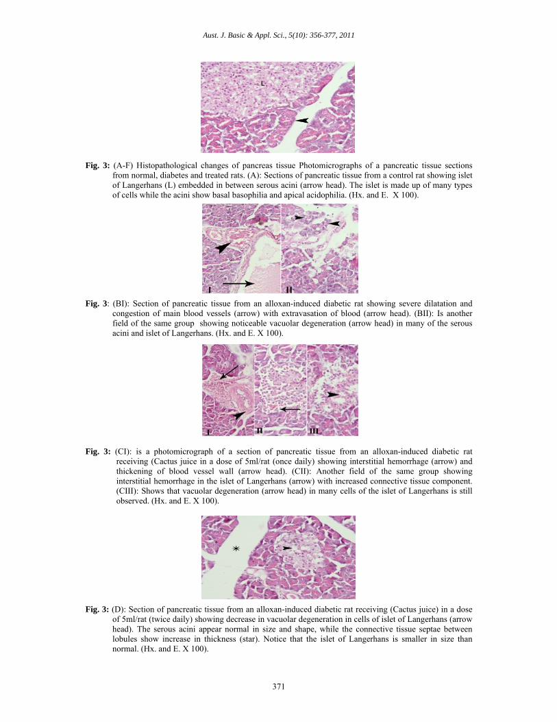
The histopathological of pancreas tissue in alloxan-induced diabetic rats showed severe dilatation and congestion of main blood vessels with degeneration in many of the serous acini and islets of Langerhans (Fig. 3B). This result is in agreement with those of (Ragavan and Krishrakumari, 2006). Oral administration of cactus fruit juice, particularly, repeated dose (5 ml/four times daily/rat) to alloxan-induced diabetic rats improved the previous changes and brought back the normal architecture of the pancreatic tissue, as the islets of Langerhans increased in size and the serous acini appeared normal in size and shape and the connective tissue septae were close to normal (Fig. 3F). These results are in accordance with those of Abdallah (2008) and Essa (2009) who reported that oral administration of Opuntia dillenii Haw fruit juice to streptozotocin-induced diabetic rats or fed of alloxan-induced diabetic rats on 2.5% and 5% prickly pear seedless pulp improved the effect damage of streptozotocin or alloxan as the majority of the islets of Langerhans tended to be normal or with moderate expansion of pancreatic islets.

Aust. J. Basic & Appl. Sci., 5(10): 356-377, 2011
368
Fig. 1: (A-F) Histopathological changes of liver tissue. Photomicrographs of liver tissue sections from normal,
diabetes and treated rats. (A): section of liver tissue from a control rat showing the hepatocytes radiating from the central vein (Cv) and separated from each other by equal-sized blood sinusoids containing kupffer cells (arrow). (Hx. and E. X 100).
Fig. 1: (BI): Section of liver tissue from an alloxan-induced diabetic rat showing dilatation of the portal vein
(Pv) with fibrosis and cellular infiltration around extending in between the hepatocytes (arrow). (BII): Is another field from the same group showing severe dilatation of the portal vein (Pv) with appearance of abnormal vessels in fibrous strands that extend between hepatocytes (arrow). Blood sinusoids show slight dilatation. (Hx. and E. X 100).
Fig. 1: (C): Section of liver tissue from an alloxan-induced diabetic rat receiving (Cactus juice) in a dose of 5 ml/rat (once daily) showing dilatation and congestion of central vein (Cv). Focal areas of necrosis with or without cellular infiltration (arrow) around are seen beside the dilated central vein. (Hx. and E. X 100).
Fig. 1: (D): Section of liver tissue from an alloxan-induced diabetic rat receiving (Cactus juice) in repeated dose
of 5ml/rat (twice daily) showing noticeable decrease in dilatation of portal vein (Pv), although congestion is still present. Also, marked decrease in fibrosis (arrow) at the portal area is observed. Most of the central veins appeared normal in size and shape (at the top of the figure) but some show moderate dilatation (at the bottom of the figure). (Hx. and E. X 100).

Aust. J. Basic & Appl. Sci., 5(10): 356-377, 2011
369
Fig. 1: (E): Section of liver tissue from an alloxan-induced diabetic rat receiving (Cactus juice) in repeated dose of 5ml/rat (three times daily) showing mild dilatation and congestion of central vein (Cv) and blood sinusoids (arrow). (Hx. and E. X 100).
Fig. 1: (F): Section of liver tissue from an alloxan-induced diabetic rat receiving (Cactus juice) in repeated dose
of 5ml/rat (four times daily) showing restoration of normal architecture of liver tissue and marked decrease of fibrosis (arrow). The main blood vessels (central and portal vein) show slight dilatation and congestion (arrow head). (Hx. and E. X 100).
Fig. 2: (A-F) Histopathological changes of renal tissue hotomicrographs of renal tissue sections from normal,
diabetes and treated rats. (A): Section of renal tissue from the control rat showing the normal structure of this tissue being formed of glomeruli (G) embedded in between tubules (arrow). (Hx. and E. X 200).
Fig. 2: (BI): Section of renal tissue from an alloxan-induced diabetic rat showing severe interstitial hemorrhage
between tubules (arrow). (BII): Is another field from the same group showing marked vacuolar degeneration (arrow head) in the epithelial lining of the tubules and cellular debris in the lumen (arrow). The interstitial hemorrhage is less severe than that in the previous field. (Hx. and E. X 200).

Aust. J. Basic & Appl. Sci., 5(10): 356-377, 2011
370
Fig. 2: (C): Section of renal tissue from an alloxan-induced diabetic rat received (Cactus juice) in a dose of
5ml/rat (once daily) showing dilatation with congestion of blood vessels (arrow), thickening of the parietal wall of the Bowman’s capsule (arrow head) and widening in the urinary space (star) denoting edema. Signs of vacuolar degeneration (V) are still present in the epithelial lining of many tubules. (Hx. and E. X 200).
Fig. 2: (D): Section of renal tissue from an alloxan-induced diabetic rat received (Cactus juice) in repeated dose of 5mg/rat (twice daily) showing that neither hemorrhage nor congestion is observed, although thickening of the parietal layer of Bowman’s capsule is still noticed (arrow head). Most of the tubules show more or less normal epithelium. (Hx. and E. X 200).
Fig. 2: (E): Section of renal tissue from an alloxan-induced diabetic rat received (Cactus juice) in reported dose
of 5 ml/rat (three times daily) showing marked decrease of the thickening of Bowman’s capsule wall but with lobulation of the glomeruli (arrow head). Some tubules show widened lumen and atrophied epithelial lining denoting edema (star). (Hx. and E. X 200).
Fig. 2: (F): Section of renal tissue from an alloxan-induced diabetic rat received (Cactus juice) in repeated dose of 5 ml/rat (four times daily) showing normal shaped and sized tubules and glomeruli, however, the glomeruli show lobulation (arrow). (Hx. and E. X 200).
Aust. J. Basic & Appl. Sci., 5(10): 356-377, 2011
371
Fig. 3: (A-F) Histopathological changes of pancreas tissue Photomicrographs of a pancreatic tissue sections from normal, diabetes and treated rats. (A): Sections of pancreatic tissue from a control rat showing islet of Langerhans (L) embedded in between serous acini (arrow head). The islet is made up of many types of cells while the acini show basal basophilia and apical acidophilia. (Hx. and E. X 100).
Fig. 3: (BI): Section of pancreatic tissue from an alloxan-induced diabetic rat showing severe dilatation and congestion of main blood vessels (arrow) with extravasation of blood (arrow head). (BII): Is another field of the same group showing noticeable vacuolar degeneration (arrow head) in many of the serous acini and islet of Langerhans. (Hx. and E. X 100).
Fig. 3: (CI): is a photomicrograph of a section of pancreatic tissue from an alloxan-induced diabetic rat
receiving (Cactus juice in a dose of 5ml/rat (once daily) showing interstitial hemorrhage (arrow) and thickening of blood vessel wall (arrow head). (CII): Another field of the same group showing interstitial hemorrhage in the islet of Langerhans (arrow) with increased connective tissue component. (CIII): Shows that vacuolar degeneration (arrow head) in many cells of the islet of Langerhans is still observed. (Hx. and E. X 100).
Fig. 3: (D): Section of pancreatic tissue from an alloxan-induced diabetic rat receiving (Cactus juice) in a dose
of 5ml/rat (twice daily) showing decrease in vacuolar degeneration in cells of islet of Langerhans (arrow head). The serous acini appear normal in size and shape, while the connective tissue septae between lobules show increase in thickness (star). Notice that the islet of Langerhans is smaller in size than normal. (Hx. and E. X 100).

Aust. J. Basic & Appl. Sci., 5(10): 356-377, 2011
372
Fig. 3: (E): Section of pancreatic tissue from an alloxan-induced diabetic rat receiving (Cactus juice) in a dose
of 5ml/rat (three times daily) showing decrease in vacuolar degeneration in cells of islet of Langerhans (arrow head). The serous acini appear normal in size and shape, while the connective tissue septae between lobules show increase in thickness (star). Notice that the islet of Langerhans is smaller in size than normal. (Hx. and E. X 100).
Fig. 3: (F): is a photomicrograph of a section of pancreatic tissue from an alloxan-induced diabetic rat receiving (Cactus juice) in a dose of 5ml/rat (four times daily) showing restoration of the normal architecture of the pancreatic tissue, where the islet of Langerhans (L) is increased in size, the serous acini appear normal in size and shape and the connective tissue septa are close to normal (Hx. and E. X 100).
Furthermore, Vessal et al., (2003) and Coskun et al., (2005) mentioned that chemicals with antioxidant properties and free radical scavengers such as quercetin might help in the regeneration of β-cells and protect pancreatic islets against the cytotoxic effects of streptozotocin. The obtained results confirmed that cactus fruit juice had antioxidant and free radical scavenging properties and contained many of bioactive compounds such as polyphenols, flavonoids, betalains and taurine.
Taurine, as a potent antioxidant, has been shown to have a protective on the pancreas by preventing or scavenging free radicals (consequently reduction in the incidence of apoptosis in islets cells) which involve decreasing of nitric oxide through inhibition of nitric oxide synthesis within β-cells. Thus supplementation of taurine (0.05%) in drinking water resulted in a significant increase in the size and number of the islets of Langerhans. These histological effects of taurine are consistent with the hypoglycemic effects of taurine indiabetes (El Idrissi et al., 2009). Signs of regeneration of β-cells, potentiating of insulin secretion from urviving β-cells of the islets of Langerhans and decrease of blood glucose have been reported following consumption of some plant extracts (Yadav et al., 2008).
The role of cactus fruit juice in reversing the diabetic state at the cellular level besides the metabolic normalization further proves its potential as a hypoglycemic agent.
Conclusions: The results in the present study showed that cactus fruit juice has a strong antioxidant property and can scavenge reactive oxygen species which resulted in markedly reduced hyperglycemia, hypercholesterolemia, lipid peroxidation (as MDA), elevated levels of urea, improves the activities of antioxidant enzyme (SOD), blood reduced glutathione (GSH), serum aminotransferases (AST & ALT), serum ALP, and increased the hemoglobin, protein and liver glycogen content in diabetic rats. In addition histopathological examination of pancreas tissue showed evidence which could be signs of regeneration of β-cells in group of rats that treated with cactus fruit juice particularly at repeated dose (5 ml/four times daily/rat). Thus it could be concluded that administration of cactus fruit juice positively affects the body’s redox balance, decrease oxidative damage to lipid and improves antioxidant status in diabetic rats.

Aust. J. Basic & Appl. Sci., 5(10): 356-377, 2011
373
REFERENCES Abdallah, Inas, Z.A., 2008. Evaluation of hypoglycemic activity of Opuntia dillenii Haw fruit juice in
streptozotocin-induced diabetic rats. The Egypt. J. Hospital Med., 33: 544-558. Allain, C.C., L.S. Poon, C.S.G. Chan, W. Richmond and C. Fu, 1974. Enzymatic determination of total
serum cholesterol. Clin. Chem., 20(4):470-475. American Diabetes Association, 2007. Diagnosis and classification of diabetes mellitus. Diabetes Care, 30: S42-S47. AOAC, 2000. Official Methods of Analysis of the Association of Official Analytical Chemists, 17th ed. Gaithersburg, Maryland, USA.
Aranda, M. and G.Aranda, M. and G. M o r l o c k , 2006. Simultaneous determination of riboflavin, pyridoxine, nicotinamid, caffeine and taurine in energy drinks by planar chromatography-multiple detection with confirmation by electrospray ionization mass spectrometry. J. Chromatogr. A, 1131(1-2): 253-260.
Arkkila, P.E.T., P.J. Koskinen, I.M. Kantola, T. Rӧ nnemaa, E. Seppänen and J.S. Viikari, 2001. Diabetic complications are associated with liver enzyme activities in people with type 1 diabetes. Diabetes Res. Clin Pract., 52 (2): 113-118.
Atta-Ur-Rahman and K. Zaman, 1989. Medicinal plants with hypoglycemic activity. J. Ethnopharmacol, 26(1): 1-55.
Bancroft, J . D., A. S t e v e n s and D . R . T u r n e r , 1996. Theory and Practice of Histological
Techniques, 4th ed., Churchill Livingstone, Edinburgh, London, Melbourne, pp: 47-67. Bartles, H., M. Bӧ hmer and C. Heierli, 1972. Serum creatinine determination without protein
precipitation. Clinica Chimica Acta, 37: 193-197. Bensadón, S., D. Hervert-Hernández, S.G. Sáyago-Ayerdi and I. Goñi, 2010. By-products of Opuntia
ficus-indica as a source of antioxidant dietary fiber. Plant Foods Hum. Nutr., 65 (3): 210-216. Beutler, E., O. Duron and B.M. Kelly, 1963. Improved method for the determination of blood
glutathione. J. Lab. Clin. Med., 61: 882-888. Broadhurst, C. L., 1997. Nutrition and non-insulin dependent diabetes mellitus from an anthropological
perspective. Alt. Med. Rev., 2 (5): 378-399. Butera, D., L. Tesoriere, F. Di Gaudio, A. Bongiorno, M. Allegra, A.M. Pintaudi, R. Kohen and M.A.
Livrea, 2002. Antioxidant activities of Sicilian prickly pear (Opuntia ficus-indica) fruit extracts and reducing properties of its betalains: betanin and indicaxanthin. J. Agric. Food Chem., 50 (23): 6895-6901.
Cameron, N.E and M.A. Cotter, 1994. The relationship of vascular changes to metabolic factors in diabetes mellitus and their role in the development of peripheral nerve complications. Diabetes Metab. Rev., 10 (3): 189-224.
Carroll, N.V., R.W. Longley and J.H. Roe, 1956. The determination of glycogen in liver and muscle by use of anthrone reagent. J. Biol.Chem., 220(2): 583-593.
Chang, S.-F., C.-L. Hsieh, and G.-C. Yen, 2008. The protective effect of Opuntia dillenii Haw fruit against low-density lipoprotein peroxidation and its active compounds. Food Chem., 106 (2): 569-575.
Chávez-Santoscoy, R.A., J.A. Gutiérrez-Uribe and S.O. Serna-Saldívar, 2009. Phenolic composition, antioxidant capacity and in vitro cancer cell cytotoxity of nine prickly pear (Opuntia spp.) juices. Plant Foods Hum. Nutr., 64 (2): 146-152.
Concepción, N.M., M.M. Pilar, A. Martin, J. Jiménez and U.M. Pilar, 1993. Free radical scavenger and antihepatotoxic activity of Rosmarinus tomentosus. Plant Med., 59 (4): 312-314.
Coria Cayupán, Y.S., M.J. Ochoa and M.A. Nazareno, 2011. Health-promoting substances and antioxidant properties of Opuntia sp. fruits. Changes in bioactive-compound contents during ripening process. Food Chem., 126 (2): 514-519.
Coskun, O., M. Kanter, A. Korkmaz and S. Oter, 2005. Quercetin, a flavonoid antioxidant, prevents and protects streptozotocin-induced oxidative stress and β-cell damage in rat pancreas. Pharmacol. Res., 51(2): 117- 123.
Danish Official, 1996. β-carotene determination. HPLC Method No. AF 255.1. 3rd ed. Authorized by National Food Agency of Denmark Ministry of Health. Institute of Food Chemistry and Nutrition.
Dere, Ş., T. Güneş, and R. Sivaci, 1998. Spectrophotometric determination of chlorophyll - a, b and total carotenoids contents of some algae species using different solvents. Tr. J. Botany, 22: 13-17.
Días Medina, E.M., E.M. Rodríguez Rodríguez and C. Días Romero, 2007. Chemical characterization of Opuntia dillenii and Opuntia ficus–indica fruits. Food Chem., 103(1): 38-45.
Dok-Go, H., K.H. Lee, H.J. Kim, E.H. Lee, J. Lee, Y.S. Song, Y.H. Lee, C. Jin, Y.S. Lee and J . C h o , 2003. Neuroprotective effects of antioxidative flavonoids, quercetin, (+)-dihydroquercetin and quercetin-methyl ether, isolated from Opuntia ficus-indica var. saboten. Brain Res., 965(1-2): 130-136.

Aust. J. Basic & Appl. Sci., 5(10): 356-377, 2011
374
Doumas, B.T., 1975. Colorimetric determination of total proteins in serum or plasma. Clin. Chem., 21(8): 1159-1166.
Drabkin, D.L., 1949. The standardization of hemoglobin measurement. Am. J. Med. Sci., 217(6): 710. Draper, H.H. and M. Hadley, 1990. Malondialdehyde determination as i n d e x of lipid peroxidation. Methods Enzymol., 186: 421-431.
DuBois,M., K.A. Gilles, J.K. Hamilton, P.A. Rebers and F. Smith, 1956. Colorimetric methods for determination of sugars and related substances. Anal. Chem., 28(3): 350-356.
El Idrissi, A., L. Boukarrou and W. L’Amoreaux, 2009. Taurine supplementation and pancreatic remodeling. In: Taurine 7, Advances in Experimental Medicine and Biology, Azuma, J, T.Ito, and S.W.Schaffer (eds.), vol. 643, part VI, pp. 353-358. Springer publisher. DOI: 10.1007/978-0-387-75681-3-36.
Elgawish, A., M. Glomb, M. Friedlander and V.M. Monnier, 1996. Involvement of hydrogen peroxide in collagen cross- linking by high glucose in vitro and in vivo. J. Biol. Chem., 271(22): 12964-12971.
El-Kossori, R.L, C. Villaume, E. El-Boustani, Y. Sauvaire and L. Méjean, 1998. Composition of pulp, skin and seeds of prickly pears fruit (Opuntia ficus-indica sp.). Plant Foods Hum. Nutr., 52: 263-270.
El-Samahy, S.K., H.A. El-Mansy, H.E. Bahlol, A.I. El-Desouky and A.E. Ahmed, 2008. Thermal process time and sensory evaluation for canned cactus pear nectar. J. Prof. Assoc. Cactus Dev., 10: 85-107.
Emmons, C.L., D.M. Peterson and G.L. Paul, 1999. Antioxidant capacity of oat (Avena sativa L.) extracts. 2. In vitro antioxidant activity and contents of phenolic and tocol antioxidants. J. Agric. Food Chem., 47(12): 4894-4898.
Ennouri, M., B. Evelyne, M. Laurence and A. Hamadi, 2005. Fatty acid composition and rheological behavior of prickly pear seed oils. Food Chem., 93(3): 431-437.
Essa, Rowida, Y., 2009. Chemical and technological studies on Opuntia ficus-indica and its effect on diabetics, pp.53-57. M.Sc. Thesis, Food Tech. Dept. Fac. of Agric., Kafr El-Sheikh Univ., Egypt.
Fawcett, J.K and J.E. Scott, 1960. A rapid and precise method for the determination of urea. J. Clin. Pathol., 13 (2): 156-159.
Fernandes, A.A.H., E.L.M. Novelli, A.F. Junior, and C.M. Galhardi, 2009. Effect of naringerin on biochemical parameters in the streptozotocin-induced diabetic rats. Braz. Arch. Biol. Technol., 52(1): 51-59.
Fernández-López, J.A., L. Almela, J.M Obón and R. Castellar, 2010. Determination of antioxidant constituents in cactus pear fruits. Plant Foods Hum. Nutr., 65(3): 253-259.
Feugang, J.M., P. Konarski, D. Zou, F.C. Stintzing and C. Zou, 2006. Nutritional and medicinal use of cactus pear (Opuntia spp.) cladodes and fruits. Front. Biosci., 11: 2574-2589.
Frati, A.C., E. Jiménez and C.R. Ariza, 1990. Hypoglycemic effect of Opuntia ficus-indica in non-insulin dependent diabetes mellitus patients. Phytother. Res., 4(5): 195-197.
Galati, E.M., M.R. Mondello, D. Giuffrida, G. Dugo, N. Miceli, S. Pergolizzi and M.F. Taviano, 2003. Chemical characterization and biological effects of Sicilian Opuntia ficus-indica (L.) Mill. Fruit juice: antioxidant and antiulcerogenic activity. J. Agric. Food Chem., 51(17): 4903-4908.
Ginsberg, H.N., 2000. Insulin resistance and cardiovascular disease. J. Clin. Invest., 106(4): 453-458. Gray, A.M and P.R. Flatt, 1999. Insulin-releasing and insulin-like activity of the traditional anti- diabetic
plant coriander (Coriandrum sativum). Br. J. Nutr., 81(3): 203-208. Guzmán-Maldonado, S.H., G. Herrera-Hernández, D. Hernández-López, R. Reynoso-Camacho, A.
Guzmán-Tovar, F. Vaillant and P. Brat, 2010. Physicochemical, nutritional and functional characteristics of two underutilized fruit cactus species (Myrtillocactus) produced in central Mexico. Food Chem., 121(2): 381-386.
Herbach, K.M., C. Maier, F.C. Stintzing and R. Carle, 2007. Effects of processing and storage on juice color and betacyanin stability of purple pitaya (Hylocereus polyrhizus) juice. Eur. Food Res. Technol., 224 (5): 649-658.
Hernández-Pérez, T., A. Carrillo-López, F. Guevara-Lara, A. Cruz-Hernández and O. Paredes-López, 2005. Biochemical and nutritional characterization of three prickly pear species with different ripening behavior. Plant Foods Hum. Nutr., 60(4): 195-200.
Huang, X., Q. Li, L. Guo and Z. Yan, 2008. Protection of cactus polysaccharide against H2O2- induced damage in the rat cerebral cortex and hippocampus differences in time of administration. Neural Regen. Res., 3 (1): 4-18.
Islam, M.S and H. Choi, 2009. Antidiabetic effect of Korean traditional baechu (Chinese cabbage) kimchi in a type 2 diabetes model of rats. J. Med. Food, 12(2): 292-297.
Jothivel, N., S.P. Ponnusamy, M. Appachi, S. Singaravel, D. Rasilingam, K. Deivasigamani and S. Thangavel, 2007. Anti-diabetic activity of methanol leaf extract of Costus pictus D. Don in alloxan-induced iabetic rats. J. Health Sci., 53(6): 655-663.
Kamalakkannan, N. and P.S. Prince, 2005. The effect of Aegle marmelos fruit extract in streptozotocin diabetes: a histopathological study. J. Herb. Pharmacother., 5(3): 87-96.
Kanner, J., S. Harel and R. Granit, 2001. Betalains-a new class of dietary cationized antioxidants. J. Agric. Food Chem., 49(11): 5178-5185.

Aust. J. Basic & Appl. Sci., 5(10): 356-377, 2011
375
Khan, A., N.A. Bryden, M.M. Polansky and R.A. Anderson, 1990. Insulin potentiating factor and chromium content of selected food and spices. Biol. Trace. Elem. Res., 24(3): 183-188.
Kim, J.H., S.M. Park, H.J. Ha, C.J. Moon, T.K. Shin, J.M. Kim, N.H. Lee, H.C. Kim, K.J. Jang and M.B. Wie, 2006. Opuntia ficus-indica attenuates neuronal injury in in vitro and in vivo models of cerebral ischemia. J. Ethnopharmacol., 104(1-2): 257-262.
Kuti, J.O., 1992. Growth and compositional changes during the development of prickly pear fruits. J. Hort. Sci., 67: 861-868.
Kuti, J.O., 2004. Antioxidant compounds from four Opuntia cactus pear fruit varieties. Food Chem., 85 (4): 527-533.
Lee, J.; L. Ye, W.O. Landen and R.R. Eitenmiller, 2000. Optimization of an extraction procedure for the quantification of vitamin E in tomato and broccoli using response surface methodology. J. Food Compos. Anal., 13(1): 45-57.
Liu, H.G., Q.Y. Liang, H.L. Meng and H.X. Huang, 2010. Hypoglycemic effect of extracts of cactus pear fruit polysaccharide in rats. J. Chinese Med. Mater., 33(2): 240-242.
Lopes-Virella, M.F., P. Stone, S. Ellis and J.A. Colwell, 1977. Cholesterol determination in high- ensity lipoproteins separated by three different methods. Clin. Chem., 23(5): 882-884.
Maritim, A.C., R.A. Sanders and J.B. Watkins, 2003. Diabetes, oxidative stress, and antioxidants: a review. J. Biochem. Mol. Toxicol., 17(1): 24-38.
Marklund, S and G. Marklund, 1974. Involvement of the superoxide anion radical in the autoxidation of pyrogallol and a convenient assay for superoxide dismutase. Eur. J. Biochem., 47(3): 469-474.
Mígues Bernárdez, M., J. De la Montaña Míguelez and J. García Queijeiro, 2004. HPLC determination of sugars in varieties of chestnut fruits from Galicia (Spain). J. Food Compos. Anal., 17: 63-67.
Mosshammer, M.R., F.C. Stintzing, and R. Carle, 2006. Cactus pear fruits (Opuntia spp.): a review of rocessing technologies and current uses. J. Prof. Assoc. Cactus Dev., 8: 1-25.
National Research Council., 1996.Veterinary medical care. In Guide for the Care and Use of Laboratory Animals, pp. 56-70. Washington, DC, The National Academies press.
Nelson, R.W., S.L. Ihle, L.D. Lewis, S.K. Salisbury, T. Miller, V. Bergdall and G.D Bottoms, 1991. Effects of dietary fiber supplementation on glycemic control in dogs with alloxan-induced diabetes mellitus. Am. J. Vet. Res., 52(12): 2060-2066.
Nigdikar, S.V., N.R. Williams, B.A. Griffin and A.N. Howard, 1998. Consumption of red wine polyphenols reduces the susceptibility of low-density lipoproteins to oxidation in vivo. Am. J. Clin. Nutr., 68(2): 258-265.
Ohaeri, O.C., 2001. Effect of garlic oil on the levels of various enzymes in the serum and tissue of streptozotocin diabetic rats. Biosci. Rep., 21(1): 19-24.
Park, E.H., J.H. Kahng and E.A. Paek, 1998. Studies on the pharmacological action of cactus: identification its anti-inflammatory effect. Arch. Pharm. Res., 21(1): 30-34.
Park, E.H., J.H. Kahng, S.H. Lee and K.H. Shin, 2001. An anti-inflammatory principle from cactus. Fitoterapia, 72(3): 288-290.
Perfumi, M. and R. Tacconi, 1996. Anti-hyperglycemic effect of fresh Opuntia dillenii fruit from Tenerife (Canary Islands). Intern. J. Pharmacog., 34(1): 41-47.
Piga, A., 2004. Cactus Pear: a fruit of nutraceutical and functional importance. J. Prof. Assoc. Cactus Dev., 6: 9-22.
Punithavathi, V.R., R. Anuthama and P.S. Prince, 2008. Combined treatment with naringin and vitamin C ameliorates streptozotocin-induced diabetes in male Wister rats. J. Appl. Toxicol., 28(6): 806-813.
Ragavan, B. and S. Krishnakumari, 2006. Effect of Terminalia arjuna stem bark extract on histopathology of liver, kidney and pancreas of alloxan-induced diabetic rats. Afr. J. Biomed. Res., 9(3): 189-197.
Ramadan, M.F. and J.T. Mӧ rsel, 2003. Lipid profile of prickly pear pulp fractions. J. Food Agric. Environ.,1(2): 66-70.
Rao, A.V and L.G. Rao, 2007. Carotenoids and human health. Pharmacol. Res., 55(3): 207-216. Ravi, K., B. Ramachandran and S. Subramanian, 2004. Effect of Eugenia jambolana seed kernel on
antioxidant defense system in streptozotocin-induced diabetes in rats. Life Sci., 75(22): 2717-2731. Reitman, S. and S. Frankel, 1957. A colorimetric method for the determination of serum glutamic
oxaloacetic acid and glutamic pyruvic transaminases. Am. J. Clin. Pathol., 28(1): 56-63. Roehrig, K.L.,1988. The physiological effects of dietary fiber (a review). Food Hydrocolloids, 2(1): 1-18. Roman-Ramos, R., J. L. Flores-Saenz and F. J. Alarcon-Aguilar, 1995. Anti-hyperglycemic effect of some
edible plants. J. Ethnopharmacol., 48(1): 25-32. Sáenz, C., 1996. Food products from cactus pear (Opuntia ficus-indica). Food Chain., 18: 10-11. Sáenz, C., 2000. Processing technologies: an alternative for cactus pear (Opuntia spp.) fruits and cladodes.
J. Arid Environ., 46(3): 209-225. Sáenz, C and E. Sepúlveda, 2001. Cactus-pear juices. J. Prof. Assoc. Cactus Dev., 4: 3-10. Sahari, M.A., M. Barzegar and R. Radfar, 2007. Effect of varieties on the composition of dates (Phoenix

Aust. J. Basic & Appl. Sci., 5(10): 356-377, 2011
376
dactylifera L.). Food Sci. Technol. Intern., 13(4): 269-275. Salim, N., C. Abdelwaheb, C. Rabah and B. Ahcene, 2009. Chemical composition of Opuntia ficus- indica
(L.) fruit . Afr. J. Biotechnol., 8(8): 1623-1624. Sánchez-Moreno, C., J.A. Larrauri and F. Saura-Calixto, 1998. A procedure to measure the antiradical
efficiency of polyphenols. J. Sci. Food Agric., 76(2): 270-276. SAS, 1996. Statistical Analysis System for windows. In: SAS/STAT user’s guide, version 4.10,
release 6.12. SAS Institute Inc. Cary, NC, USA. Shanmugasundaram, E.R., K.L. Gopinath, S.K. Radha and V.M. Rajendran, 1990. Possible regeneration of
the islets of Langerhans in streptozotocin-diabetic rats given Gymnema sylvestre leaf extract. J. Ethnopharmacol., 30(3): 265-279.
Sharma, S., S.K. Kulkarni and K. Chopra 2006. Curcumin, the active principle of turmeric (Curcuma longa), ameliorates diabetic nephropathy in rats. Clin. Exp. Pharmacol. Physiol., 33(10): 940-945.
Shedbalkar, U.U., V.S. Adki, J.P. Jadhav and V.A. Bapat, 2010. Opuntia and other cacti: applications and biotechnological insights. Tropical plant Biol., 3(3): 136-150.
Singleton, V.L., R. Orthofer. and R.M. Lamuela-Raventós,1999. Analysis of total phenols and other oxidation substances and antioxidants by means of folin- ciocalteau reagent. Methods in Enzymol., 299:
152-178. Siriwardhana, N and Y.J. Jeon, 2004. Antioxidative effect of cactus pear fruit (Opuntia ficus-indica) extract
on lipid peroxidation inhibition in oils and emulsion model systems. Eur. Food Res. Technol., 219(4): 369-376. Srivastava, S., A. Chandra, N.H. Ansari, S.K. Srivastava and A. Bhatnagar, 1998. Identification of cardic
oxidoreductase (s) involved in the metabolism of the lipid peroxidation derived aldehyde -4-hydroxnonenal. Biochem J., 329(pt3): 469-475.
Stintzing, F.C and R. Carle, 2006. Cactus fruits – more than color. Fruit processing, 16(3): 166-171. Stintzing, F.C., A. Schieber and R. Carle, 2001. Phytochemical and nutritional significance of cactus pear.
Eur. Food Res. Technol., 212(4): 396-407. Stintzing, F.C., A. Schieber and R. Carle, 2003. Evaluation of color properties and chemical quality
parameters of cactus juice. Eur. Food Res. Technol., 216(4): 303-311. Stintzing, F.C., K.M. Herbach, M.R. Mosshammer, R. Carle, W. Yi, S. Sellappan, C.C. Akoh, R. Bunch
and P. Felker, 2005. Color, betalain pattern, and antioxidant properties of cactus pear (Opuntia spp.) clones. J. Agric. Food Chem., 53(2): 442-451.
Suba, V., T. Murugesan, R.B. Rao, L . Ghosh, M. Pal, S.C. Mandal and B.P. Saha, 2 004. Antidiabetic potential of Barleria lupulina extract in rats. Fitoterapia, 75(1): 1-4.
Suja, K.P., A. Jayalekshmy and C. Arumughan, 2005. Antioxidant activity of sesame cake extract. Food Chem., 91(2): 213-219.
Tesoriere, L., D. Butera, D. D’Arpa, F. Di Gaudio, M. Allegra, C. Gentile and M.A. Livrea, 2003. Increased resistance to oxidation of betalain-enriched human low density lipoproteins. Free Radic. Res., 37 (6): 689-696.
Tesoriere, L., M. Fazzari, M. Allegra and M.A. Livrea, 2005. Biothiols, taurine, and lipid-soluble antioxidants in the edible pulp of Sicilian cactus pear (Opuntia ficus-indica) fruits and changes of bioactive juice components upon industrial processing. J. Agric. Food Chem., 53(20): 7851-7855.
Tharkar, S., A. Devarajan, S. Kumpatla and V. Viswanathan, 2010. The socioeconomics of diabetes from a developing country: a population based cost of illness study. Diabetes Res. Clin. Pract., 89(3): 334-340.
Tietz, N.W., C.A. Burtis, P. Duncan, K. Ervin, C.J. Petitclerc, A.D. Rinker, D. Shuey and E.R. Zygowicz, 1983. A reference method for measurement of alkaline phosphatase activity in human serum. Clin. Chem., 29 (5): 751.
Trinder, P., 1969. Determination of glucose in blood using glucose oxidase with alternative oxygen acceptor. Ann. Clin. Biochem., 6: 24-27.
Vessal, M., M. Hemmati and M. Vasei, 2003. Antidiabetic effects of quercetin in streptozotocin-induced diabetic rats. Comp. Biochem. Physiol. C Toxicol. Pharmcol., 135C(3): 357-364.
Vivian, S.T. and J.S. Smilee, 2010. Evaluation of lipid peroxidation and antioxidant status in diabetes with and without complications. J. Biomed. Sci. Res., 2(3): 162-166.
Vlassara, H., M. Brownlee and A. Cerami, 1981. Non-enzymatic glycosylation of peripheral nerve protein in diabetes mellitus. Proc. Natl. Acad. Sci. USA, 78(8): 5190-5192.
Wakeling, I.N and H. J.H. MacFie, 1995. Designing consumer trials balanced for first and higher orders of carry-over effect when only a subset of K samples from t may be tested . Food Qual. Prefer., 6(4): 299-308.
Wohaieb, S.A and D.V. Godin, 1987. Alterations of free radical tissue-defense mechanisms in streptozotocin-induced diabetes rats. Effects of insulin treatment. Diabetes, 36(9): 1014-1018.
Wójcik, O.P., K.L. Koenig, A. Zeleniuch-Jacquotte, M. Costa and Y. Chen, 2010. The potential protective effects of taurine on coronary heart disease. Atherosclerosis, 208(1): 19-25.
Aust. J. Basic & Appl. Sci., 5(10): 356-377, 2011
377
Yadav, J.P., S. Saini, A.N. Kalia, and A.S. Dangi, 2008. Hypoglycemic activity of ethanolic extract of Salvadora oleoides in normal and alloxan-induced diabetes rats. Indian J. Pharmacol., 40(1): 23-27.
Zhao, L.Y., Q.J. Lan, Z.C. Huang, L.J. Ouyang and F.H. Zeng, 2011. Antidiabetic effect of a newly identified component of Opuntia dillenii polysaccharides. Phytomedicine, 18(8-9): 661-668.
Zhishen, J., T. Mengcheng and W. Jianming, 1999. The determination of flavonoids contents in mulberry and their scavenging effects on superoxide radicals. Food Chem., 64(4): 555-559.