EGG. EGG STRUCTURE & COMPOSITION 1.Egg yolk 2.Albumen (white egg) 3.Shell membrane 4.Egg Shell.

ORIGINAL ARTICLE
Nurse egg consumption and intracapsular developmentin the common whelk Buccinum undatum (Linnaeus 1758)
Kathryn E. Smith • Sven Thatje
Received: 29 November 2011 / Revised: 12 April 2012 / Accepted: 17 April 2012 / Published online: 3 May 2012
� Springer-Verlag and AWI 2012
Abstract Intracapsular development is common in mar-
ine gastropods. In many species, embryos develop along-
side nurse eggs, which provide nutrition during ontogeny.
The common whelk Buccinum undatum is a commercially
important North Atlantic shallow-water gastropod. Devel-
opment is intracapsular in this species, with individuals
hatching as crawling juveniles. While its reproductive
cycle has been well documented, further work is necessary
to provide a complete description of encapsulated devel-
opment. Here, using B. undatum egg masses from the south
coast of England intracapsular development at 6 �C is
described. Number of eggs, veligers and juveniles per
capsule are compared, and nurse egg partitioning, timing of
nurse egg consumption and intracapsular size differences
through development are discussed. Total development
took between 133 and 140 days, over which 7 ontogenetic
stages were identified. The number of both eggs and veli-
gers were significantly related to capsule volume, with
approximately 1 % of eggs developing per capsule. Each
early veliger consumed nurse eggs rapidly over just
3–7 days. Within each capsule, initial development was
asynchronous, but it became synchronous during the veli-
ger stage. No evidence for cannibalism was found during
development, but large size differences between embryos
developing within each capsule were observed, and occa-
sionally ‘empty’ veligers were seen, which had not
successfully consumed any nurse eggs. These results
indicate a high level of competition for nurse eggs within
each capsule during development in the common whelk.
The initial differences observed in nurse egg uptake may
affect individual predisposition in later life.
Keywords Intracapsular development � Buccinum
undatum � Nurse egg partitioning � Competition �Reproduction
Introduction
Many marine gastropods undergo intracapsular development
inside egg capsules (Thorson 1950; Natarajan 1957; D’Asaro
1970; Fretter and Graham 1985). Embryos develop within the
protective walls of a capsule that safeguards against factors
such as physical stress, predation, infection and salinity
changes (Thorson 1950; Pechenik 1983, 1999; Strathmann
1985; Rawlings 1995, 1999). Periods of encapsulation vary;
some species are released as veligers and undergo a plank-
tonic stage before reaching adult life (mixed development),
while others display direct development, hatching from cap-
sules as crawling juveniles (Natarajan 1957; D’Asaro 1970;
Pechenik 1979). When direct development occurs, embryos
are often accompanied in a capsule by nurse eggs, non-
developing food eggs, which provide nutrition during devel-
opment (Thorson 1950; Spight 1976b; Rivest 1983; Lahbib
et al. 2010). These are usually indistinguishable from
embryos in the very early stage of ontogeny and are consumed
during development, potentially increasing size of juveniles
at hatching (Thorson 1950). In some species, nutrition may
also be provided by intracapsular fluid or protein from capsule
walls (Bayne 1968; Stockmann-Bosbach 1988; Moran 1999;
Ojeda and Chaparro 2004).
Generally speaking, nurse egg consumption occurs over
a period of several weeks or months. It commences some
Communicated by Martin Thiel.
K. E. Smith (&) � S. Thatje
Ocean and Earth Science, University of Southampton,
National Oceanography Centre, Southampton,
European Way, Southampton SO14 3ZH, UK
e-mail: [email protected]
123
Helgol Mar Res (2013) 67:109–120
DOI 10.1007/s10152-012-0308-1

weeks into development as embryos form, and nurse eggs
are then slowly consumed throughout much of develop-
ment (Chaparro and Paschke 1990; Ilano et al. 2004;
Lahbib et al. 2010). The number of nurse eggs consumed
during this period varies across species. Ratios range from
1.7 nurse eggs per embryo in the Pacific shallow-water
muricid Acanthinucella spirata (Spight 1976a), to between
50,000 and 100,000 nurse eggs per embryo in the North
Atlantic deep-sea buccinid Volutopsius norwegicus (Thor-
son 1950). Often, within a species, the nurse egg to embryo
ratio varies from capsule to capsule within one clutch
(Thorson 1950; Spight 1976a). For example, Rivest (1983)
found this ratio in the buccinid Lirabuccinum dirum to vary
from 11 to 46 across capsules. Similar differences have
been reported for other gastropods (Natarajan 1957; Spight
1976a). Within a capsule however, there is usually little
variation in the number of nurse eggs ingested by each
embryo, with all embryos generally being equal in their
ability to consume. Any differences observed are minimal,
and juveniles hatching from each capsule are normally of a
very similar size (Natarajan 1957; Spight 1976a; Rivest
1983; Chaparro and Paschke 1990; Chaparro et al. 1999;
Lloyd and Gosselin 2007). Large size differences amongst
capsulemates are unusual, but have been reported in some
species of muricid gastropod (Gallardo 1979; Gonzalez and
Gallardo 1999; Cumplido et al. 2011). In gastropods, the
number of eggs inside a capsule is usually positively
related to capsule size. Within a species, larger capsules
hold more eggs and more developing embryos (Gallardo
1979; Pechenik et al. 1984; Miloslavich and Dufresne
1994). The relationship between capsule size and number
of eggs (including nurse eggs) has, however, previously
been shown to be stronger than the relationship between
capsule size and number of developing embryos (Spight
1976b). In some cases, the number of developing embryos
within a capsule has been found to be independent of
capsule volume. This suggests that embryos are distributed
at random, while nurse eggs are regularly placed amongst
capsules (Rivest 1983; Chaparro et al. 1999).
Intracapsular development and nurse egg and embryo
partitioning have been investigated in several species of
marine gastropod (Natarajan 1957; D’Asaro 1970; Spight
1976a; Rivest 1983; Cumplido et al. 2011). While some
attempts have been made to examine encapsulated devel-
opment in the common whelk Buccinum undatum (Portmann
1925; Fretter and Graham 1985; Nasution 2003), it has not
yet been fully described. Nasution (2003) gives the most in-
depth account of development to date, but his descriptions
are incomplete and his reports of nurse egg consumption do
not match our observations. Descriptions from Portmann
(1925) better fit our observations but lack detail. There are
also gaps in the current literature, and very limited knowl-
edge exists on nurse egg partitioning and intracapsular
embryo size ranges through development. The common
whelk is a scavenger found widespread in coastal areas in the
North Atlantic. It is generally found from the shallow sub-
tidal down to a few hundred metres of water depth (Valen-
tinsson et al. 1999; Valentinsson 2002; Rosenberg 2009),
with a latitudinal range from 38�N to 79�N spanning the
North Atlantic and Arctic Oceans (OBIS http://iobis.org/
mapper/?taxon=Buccinumundatum). Buccinum undatum is
an important commercial species, providing locally valuable
fisheries in several areas around the North Atlantic including
the UK, the USA and Canada (Hancock 1967; Morel and
Bossy 2004). It has been suggested as a good candidate for
aquaculture (Nasution and Roberts 2004) and globally,
demand for it is continuously increasing (Department of
Marine Resources www.maine.gov/dmr/rm/whelks.html).
Its reproductive cycle has been well documented across its
range (Hancock 1967; Martel et al. 1986a, b; Kideys et al.
1993; Valentinsson 2002). Females group to deposit small
creamy coloured spherical egg capsules (Martel et al.
1986a). Each lays approximately 80–150, which collectively
can create large egg masses of hundreds to thousands of
capsules (Fretter and Graham 1985; Valentinsson 2002). The
time of year for spawning varies in this species across its
distribution. In coastal waters of the UK, egg capsules are
laid during the autumn and winter months (predominantly
late November–January) as annual water temperatures drop
below 9 �C (Hancock 1967; Kideys et al. 1993). In the
northwest Atlantic, egg laying instead takes place in spring
(late May to mid July) as water temperatures warm
(approximately 2–3 �C) (Martel et al. 1986a). Intracapsular
development takes between 2.5 and 9 months across the
species range (Fretter and Graham 1985; Martel et al. 1986a;
Kideys et al. 1993; Nasution 2003). Given the widespread
distribution of B. undatum, its current commercial impor-
tance and its potential as a future candidate for aquaculture, it
is important to understand fully the development in this
species.
Here, we examine intracapsular development in B. und-
atum using a population from the south coast of England, at
the southern end of the species distribution. Number of eggs
and number of developing veligers and juveniles are exam-
ined through development. Ontogenetic stages are described
in detail including nurse egg partitioning, nurse egg con-
sumption and intracapsular ranges in embryo sizes.
Materials and methods
Embryonic development
In order to study the intracapsular development in
B. undatum, 150 adults were collected from Viviers UK in
late November 2009 (www.fishmarketportsmouth.co.uk).
110 Helgol Mar Res (2013) 67:109–120
123

Adults were originally gathered from the Solent, UK
(50�390 N, 001�370 W) from approximately 15 m water
depth by Viviers using whelk traps. They were taken to the
aquarium at the National Oceanography Centre, South-
ampton, and placed in a large outdoor tank with continuous
seawater flow through. Whelks were fed scrap fish ad libi-
tum 3 times a week, and the tank was checked daily for
laying activity. Egg laying occurred between early
December 2009 and early February 2010, predominantly
when water temperatures fell below 8 �C. All egg masses
were laid on aquarium walls within a few centimetres of
the water line.
Three egg masses laid in early January were removed for
examination through development. Each was left undis-
turbed for 24 h after egg laying had ceased before being
removed from the aquarium walls and maintained in 1 lm
filtered seawater at 6 �C. This was close to local water
temperatures, which ranged 4.0–8.3 �C between January
and March 2010 (local temperature data obtained brambl-
emet (www.bramblemet.co.uk/) and CEFAS (www.cefas.
defra.gov.uk/our-science/observing-and-modelling/monitor
ing-programmes/sea-temperature-and-salinity-trends/presen
tation-of-results/station-22-fawley-ps.aspx) databases). Each
week 3 capsules were randomly selected and dissected from
each egg mass (Fig. 1a). For each egg mass, the outer layer of
egg capsules was removed prior to any examination as these
are often empty or hold a very small number of eggs. The
contents of each capsule were examined, ontogenetic stage
was described and eggs or embryos were measured along their
longest axis using an eyepiece graticule. When a capsule
contained loose eggs, approximately 20 were measured per
capsule. When embryos were present of any age, all were
measured (on average 9–11). From the trochophore stage and
for the duration of nurse egg feeding, 3 capsules per egg mass
were examined daily to determine the duration of short
ontogenetic stages and the time taken to consume nurse eggs.
Each egg mass was also examined non-invasively each week.
Transparency of the capsule wall allowed approximate onto-
genetic stage to be determined, and the percentage of the mass
at each developmental stage was estimated (Fig. 1b). From
this, embryonic development was described, including onto-
genetic stages, developmental timing, change in embryo size,
nurse egg partitioning and intracapsular size differences dur-
ing development. Ontogenetic stages were defined as egg,
trochophore, early veliger, veliger, pediveliger, pre-hatching
juvenile and hatching juvenile (see below for descriptions).
Intracapsular contents through development
In order to investigate the intracapsular contents, B. und-
atum egg masses were collected from Southampton Water
(Southampton, UK, 50�500 N, 001�190 W) from approxi-
mately 10 m water depth between January and March,
2009 and 2010. Seawater temperatures ranged from 4 to
10 �C during these periods. Collection took place using
beam trawls deployed by the University of Southampton
research vessel RV Callista. In total, 35 egg masses were
collected, all of which were fixed in 4 % formalin for later
investigation.
Capsules were selected at random from all 35 egg
masses. As above, the outer layer of each egg mass was
removed prior to this. Buccinum undatum egg capsules are
relatively ellipsoid in shape, with a convex/concave face
(Fig. 1a, b). Each capsule was measured in three dimen-
sions (length, width, depth; ±0.01 mm) using digital cali-
pers (Absolute digimatic caliper, Mitutoyo (UK) Ltd,
Andover, UK). From these measurements, the volume of
each egg capsule was estimated using an adaptation of
equations used by Pechenik (1983), Rawlings (1990). The
following equation was used.
V ¼ p abð Þ � c
where a = length/2, b = width/2 and c = depth.
Each capsule was then dissected, number of embryos
was counted (using a bogorov counting chamber) and
ontogenetic stage determined under a compound-micro-
scope. To investigate the relationship between capsule
volume and number of eggs or veligers within a capsule,
approximately 160 capsules at egg stage (i.e. prior to any
development occurring; 15 egg masses; 10–11 capsules
from each) and 160 capsules at veliger stage were exam-
ined (18 egg masses, 8–9 capsules from each). Capsules
ranging from 5.15 to 10.49 mm length (39.0–287.5 mm3
volume) were compared. Regression analyses were carried
out to examine the relationship between capsule volume
and number of eggs, and capsule volume and number of
veligers.
Change in number of embryos per capsule during
development was investigated by examining 100 capsules
at veliger stage (12 egg masses, 8–9 capsules from each)
and 100 capsules at pre-hatching juvenile stage (9 egg
masses, 11–12 capsules from each). Since the number of
eggs and embryos per capsule is related to capsule size, for
this comparison, capsules of a narrower size range (length
6–8 mm, volume 52.4–146.2 mm3) were used. This elimi-
nated the possibility of any change in number of embryos
per capsule to be influenced by capsule size. Only veligers
containing nurse eggs were counted; it was presumed
veligers with no nurse eggs would not develop success-
fully. An unpaired t test was carried out to compare number
of veligers per capsule to number of pre-hatching juveniles
per capsule.
Helgol Mar Res (2013) 67:109–120 111
123

Results
Ontogenetic stages
Seven ontogenetic stages were identified. These are
described below.
Egg
Each capsule contains 475–2,639 (mean 1,094) small
spherical eggs with no definition. Eggs are cream or yellow
in colour and have an average diameter of 234 lm. Within
a capsule, egg diameter varies on average by 36 lm.
Approximately 1 % of these eggs are developing embryos.
The remaining are nurse eggs. At this stage, both devel-
oping and nurse eggs are identical (Fig. 2a; Table 1). Egg
capsules remain at this stage on average for 49 days.
Trochophore
After 42–56 days developing embryos become globular
shaped with a non-circular translucent membrane around
the darker embryo. A cilia band (prototroch) is present
around approximately one-third to half of the outer cir-
cumference of the membrane (Fig. 2b). Each trochophore
is a little larger than an egg, with an average length of
321 lm. Each embryo remains at the trochophore stage for
just 2–3 days (Table 1).
Early veliger
As the early veliger stage is reached, the prototroch extends
laterally to form paired velar lobes with marginal cilia
around a central simple mouth. Velar lobes are used for
collection of eggs and locomotory movement. Each early
veliger is mobile but lacks obvious intentional direction.
Behind each lobe and just in front of the main body of the
early veliger, paired larval kidneys develop, slightly opaque
in colour. Whole (generally nurse) eggs are manipulated into
the mouth section using the cilia. These are engulfed and
stored in the midgut (Portmann 1925), which forms a cir-
cular ball directly behind the mouth section, surrounded by a
thin outer membrane. There is some asynchrony in the early
development of the embryos from individual capsules. In
total, between 2 and 35 veligers develop per capsule (aver-
age 11). Each embryo consumes nurse eggs for 3–7 days (at
6 �C). Total consumption by all embryos within a capsule
occurs during the early veliger stage, over 4–10 days. Eggs
are not damaged during consumption but are stored in
the midgut, conserved for later nutritional use. Whole,
Fig. 1 a Egg mass of B. undatum showing individual capsules. b A large individual egg capsule showing many developed embryos inside, post
nurse egg consumption. Scale bars represent 5 mm. c capsule, e embryo
112 Helgol Mar Res (2013) 67:109–120
123
undamaged nurse eggs can be seen inside the each early
veliger. Early veligers average 1.46 mm across their longest
axis. Within one capsule, embryo size may vary by as much
as 0.85 mm. These size differences continue to be observed
throughout development. Once all nurse eggs are con-
sumed, early veligers, veligers and even pediveligers are
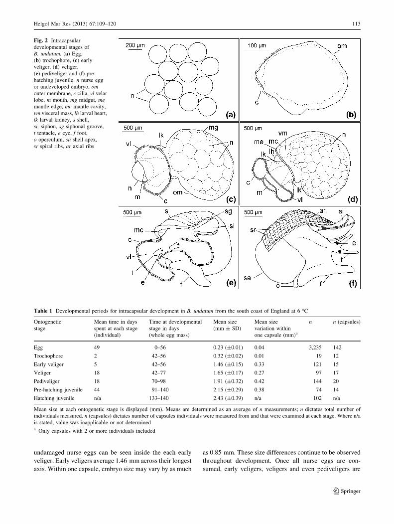
Fig. 2 Intracapsular
developmental stages of
B. undatum. (a) Egg,
(b) trochophore, (c) early
veliger, (d) veliger,
(e) pediveliger and (f) pre-
hatching juvenile. n nurse egg
or undeveloped embryo, omouter membrane, c cilia, vl velar
lobe, m mouth, mg midgut, memantle edge, mc mantle cavity,
vm visceral mass, lh larval heart,
lk larval kidney, s shell,
si, siphon, sg siphonal groove,
t tentacle, e eye, f foot,
o operculum, sa shell apex,
sr spiral ribs, ar axial ribs
Table 1 Developmental periods for intracapsular development in B. undatum from the south coast of England at 6 �C
Ontogenetic
stage
Mean time in days
spent at each stage
(individual)
Time at developmental
stage in days
(whole egg mass)
Mean size
(mm ± SD)
Mean size
variation within
one capsule (mm)a
n n (capsules)
Egg 49 0–56 0.23 (±0.01) 0.04 3,235 142
Trochophore 2 42–56 0.32 (±0.02) 0.01 19 12
Early veliger 5 42–56 1.46 (±0.15) 0.33 121 15
Veliger 18 42–77 1.65 (±0.17) 0.27 97 17
Pediveliger 18 70–98 1.91 (±0.32) 0.42 144 20
Pre-hatching juvenile 44 91–140 2.15 (±0.29) 0.38 74 14
Hatching juvenile n/a 133–140 2.43 (±0.39) n/a 102 n/a
Mean size at each ontogenetic stage is displayed (mm). Means are determined as an average of n measurements; n dictates total number of
individuals measured. n (capsules) dictates number of capsules individuals were measured from and that were examined at each stage. Where n/a
is stated, value was inapplicable or not determineda Only capsules with 2 or more individuals included
Helgol Mar Res (2013) 67:109–120 113
123

occasionally found in a capsule, which have consumed no
nurse eggs at all (Figs. 1b, 2c, 3a, b; Table 1).
Veliger
In the veliger, the mantle edge thickens and a thin larval
shell becomes visible around the midgut, creating a trans-
parent layer. The midgut appears important in dictating the
dimensions of this shell. The velar lobes become more
separated, and distinct and the larval kidneys continue to be
seen, often with a central yellow spot. The central mouth
section becomes more opaque, early foot development
begins and no further nurse egg consumption is possible.
The mantle edge and the visceral mass (white in colour)
beneath it become obvious. A transparent pulsating mem-
brane located dorsolaterally in front of the mantle edge
becomes evident; this is often named the larval heart
(Hughes 1990; Khanna and Yadav 2004). Nurse eggs
stored beneath the mantle are still clearly individually
discernible at this stage and even going into the pediveliger
stage (Figs. 2d, 3b, c; Table 1). It is possible to break the
mantle or shell on the back of the veliger or pediveliger and
find nurse eggs still inside, which are not degraded and
have not yet been digested. Embryos remain at the veliger
stage for approximately 14–21 days. During this period,
development within a capsule becomes synchronised.
Fig. 3 Early development in B. undatum. (a) Early pediveliger stage
with empty midgut indicating few or no nurse eggs were consumed.
(b) Veligers of varying sizes developing alongside each other; within
one capsule and following nurse egg consumption. (c) Early
pediveliger stage with individual nurse eggs still clearly discernible
under the shell. (d) Well-developed mid pediveliger stage with velar
lobes still present. Growth lines can be observed on shell. n nurse egg,
vl velar lobe, mg midgut, vm visceral mass, s shell, sg siphonal
groove, t tentacle, e eye, f foot, gl growth lines. Scale bars represent
500 lm
114 Helgol Mar Res (2013) 67:109–120
123

Pediveliger
At the pediveliger stage, the shell thickens and becomes
increasingly apparent. The mantle cavity is initially visible
beneath the mantle edge and the siphonal groove begins to
form. The foot, eyes, tentacles and siphon appear. The
velum and cilia, which are large at the beginning of this
stage, begin to shrink back. They disappear by the end of
the pediveliger stage. The larval kidneys and larval heart
also disappear. Embryos remain at this stage for approxi-
mately 14–21 days (Figs. 2e, 3c, d; Table 1).
Pre-hatching juvenile
Shell growth continues and spiral and axial ribs begin to
develop in the shell as the pre-hatching juvenile stage is
reached. The shell thickens and colours brown (becomes
pigmented). The first whorl becomes obvious and the shell
shape elongates. Head, foot, tentacle and siphon features
become more prominent and the operculum appears. The
feeding proboscis also develops internally during this time.
Pre-hatching juveniles complete development over a further
35–49 days before hatching commences. Pre-hatching juve-
nile size ranges from 1.57 to 3.06 mm. (Fig. 2f; Table 1).
Hatching juvenile
The features described for pre-hatching juveniles become
more prominent. The juvenile emerges from the egg cap-
sule through an opening created through radular scraping.
They remain on the egg mass for a few days before moving
off to feed. Overall hatching size ranged from 1.70 to
3.45 mm (Table 1).
Embryonic development
Each egg mass took between 9 and 11 days to be laid, with
complete intracapsular development taking 133–140 days
(19–20 weeks) at 6 �C. Within each egg mass, development
was asynchronous by up to 14 days throughout the devel-
opmental period. Within each capsule, development was
initially asynchronous; both trochophore and early veliger
stages, and early veliger and veliger stages were observed
together in capsules. By late veliger stage development
within a capsule was synchronous. Following an initial
increase in embryo size as nurse egg consumption occurred,
individual size (measured as change in length) increased at a
steady rate throughout the remainder of the encapsulated
period (Figs. 4, 5; Table 1). Within each capsule, large size
differences were observed between embryos at all stages of
development. Whole, undamaged nurse eggs were visible
inside embryos throughout the veliger and pediveliger
stages. Occasional early veligers, veligers and pediveligers
were found, which had not consumed any nurse eggs. Apart
from the absence of nurse eggs, these embryos were com-
pletely normal in their development (Fig. 3a–c; Table 1).
Intracapsular contents through development
Relationship between capsule volume and number
of embryos per capsule
Egg capsule volume ranged from 39.0 to 287.5 mm3 (capsule
length 5.15–10.49 mm). Overall, number of eggs per capsule
averaged 1,094 and number of veligers per capsule averaged
11. Regression analysis showed there to be a significant
relationship between capsule volume and number of eggs
(r2 = 0.7646; p \ 0.001), and capsule volume and number of
veligers (r2 = 0.5615; p \ 0.001). As a percentage of total
eggs, on average 1 %, develop into veligers (Fig. 6a, b).
Fig. 4 Developmental time (days) for B. undatum from Southampton
Water (UK) at 6 �C. Times shown represent development across
whole egg masses
Fig. 5 Change in size of individuals (measured as length along
longest axis) during intracapsular development. Size displayed is
average length of individual at each stage in lm. Nurse egg
consumption occurs between trochophore and early veliger stages.
The average size displayed for early veliger is taken post nurse egg
consumption. Error bars indicate ±1 SD
Helgol Mar Res (2013) 67:109–120 115
123

Change in number of embryos per capsule through
development
When examining capsules ranging from 6 to 8 mm in
length (volume 52.4–146.2 mm3), number of developing
veligers per capsule ranged from 3 to 21 (average 9) and
number of pre-hatching juveniles per capsule ranged from
2 to 20 (average 9). An unpaired t test showed there to be
no difference between the two groups (p = 0.772).
Discussion
Embryonic development and intracapsular contents
data
The distribution of B. undatum extends from the southern
coast of the UK, northwards up into the North Atlantic and
Arctic oceans, across a temperature range of -1.5 to 22 �C
(Bramblemet; CEFAS; Martel et al. 1986a). For the pop-
ulation used in the present study, annual temperatures vary
seasonally from approximately 4–22 �C, and egg laying
and development normally occur in water temperatures
ranging 4–10 �C. With temperatures maintained at 6 �C,
the duration of intracapsular development (4.5–5 months)
was similar to previous estimates of B. undatum develop-
ment in British waters (Kideys et al. 1993; Valentinsson
2002). Longer and shorter periods have been reported
across the species distribution (e.g. Martel et al. 1986a;
Nasution 2003). The observed differences in duration of
development can be attributed to the known effects of
temperature on metabolic rates in ectotherms.
In the present study, the number of eggs per capsule
averaged 1,094 and the number of developing veligers
averaged 11. While egg numbers were similar to those
indicated in previous studies (Table 2), veliger numbers
were similar to figures reported by Hancock (1967), but
lower than other estimates (Portmann 1925; Martel et al.
1986a). Since number of veligers is often significantly
related to capsule volume (Gallardo 1979; Pechenik et al.
1984; Valentinsson 2002), it is likely that larger capsules
were examined in the latter studies. Results indicate
approximately 1 % of eggs developed, giving a ratio of 109
nurse eggs per embryo, almost identical to the 110 eggs per
embryo reported by Portmann (1925). The percentage of
eggs developing was also comparative to other previous
estimates for B. undatum (Martel et al. 1986a; Valentinsson
2002; Nasution 2003). Similar results have been reported
for other buccinids including 1.1–2 % for Buccinum isa-
otakki (Ilano et al. 2004), 0.2–1.8 % for Buccinum cyane-
um (Miloslavich and Dufresne 1994) and 1 % for Colus
stimpsoni (West 1979).
Past studies provide conflicting views on the occurrence
of intracapsular cannibalism in B. undatum (Table 2).
Portmann (1925) indicated a reduction in number of indi-
viduals per capsule during development (from early veli-
gers to veligers and pre-hatching juveniles), which was
suggested to be due to cannibalism (Fretter and Graham
1985). Contrary to this, other studies have shown the
number of developing embryos per capsule to remain
constant during development, indicating no cannibalism
(Hancock 1967; Martel et al. 1986a). Our results were in
agreement with these latter studies. Similarly, no canni-
balism during development was reported in the buccinids
B. cyaneum (Miloslavich and Dufresne 1994) and B. isa-
otakki (Ilano et al. 2004), and only very rarely was it
observed in the buccinid L. dirum (Rivest 1983). It has,
however, been reported in some other gastropods including
Crucibulum quiriquinae (Veliz et al. 2001), Crepidula
coquimbensis (Veliz et al. 2003; Brante et al. 2009) Tro-
phon geversianus (Cumplido et al. 2011) and a vermetid
gastropod (Strathmann and Strathmann 2006).
Capsule size or volume has previously been shown to be
a good indicator of number of eggs and veligers within a
capsule. In the current study, these figures were both sig-
nificantly related to capsule volume. Number of eggs was
more closely related to volume than number of veligers,
suggesting eggs are more regularly distributed amongst
(a)
(b)
Fig. 6 Relationship between capsule volume and (a) number of eggs,
(b) number of veligers in egg masses of B. undatum. Both
relationships are significant to p \ 0.001. The r2 values are displayed
116 Helgol Mar Res (2013) 67:109–120
123

capsules than are developing embryos. This pattern has
been reported before for both B. undatum (Valentinsson
2002; Nasution et al. 2010) and other gastropods, including
B. cyaneum (Miloslavich and Dufresne 1994), B. isaotakki
(Ilano et al. 2004), Hexaplex (Trunculariopsis) trunculus
(Lahbib et al. 2010), Acanthina monodon (Gallardo 1979),
Nucella lapillus (Pechenik et al. 1984) and Nucella lam-
ellosa (Spight 1976b). Contrary to this, number of eggs has
been found to be related to, but number of veligers to be
independent of capsule size in the buccinid L. dirum
(Rivest 1983), the calyptraeid Crepipatella dilatata
(Chaparro et al. 1999) and the muricid Nucella ostrina
(Lloyd and Gosselin 2007).
An initial rapid increase in embryo size was observed at
the early veliger stage in the present investigation. This
was followed by a relatively linear increase in size for the
remainder of intracapsular development. Similar changes
in size during development have been reported for
B. cyaneum (Miloslavich and Dufresne 1994) and B. isa-
otakki (Ilano et al. 2004). For both, however, the initial
increase was slower than was observed in this investiga-
tion. In B. isaotakki, it is likely that this is reflective of the
slower nurse egg consumption rate previously observed in
this species (Ilano et al. 2004). Probably, nurse eggs are
also taken up at a slower rate in B. cyaneum.
Previous hatching sizes for B. undatum have been
reported ranging from 1.0 to 3.1 mm (e.g. Fretter and
Graham 1985; Nasution et al. 2010). These are similar to
hatching sizes observed in the present investigation, which
averaged just below 2.5 mm in length.
Nurse egg partitioning
Life history theories suggest parental fitness is maximised
by investing equally into all offspring (Smith and Fretwell
1974). Traditionally, resource partitioning (in the form of
nurse eggs) during intracapsular development follows this
trend. Embryos compete for nurse eggs, but within a cap-
sule competitiveness is normally equal. As a result, nurse
eggs are consumed quite evenly by all embryos. This does
not mean hatchlings are always of a similar size; within
one species, or even one clutch, the ratio of nurse eggs to
developing embryo may vary greatly between capsules,
resulting in large differences in offspring size. This is
usually believed to be due to irregular distribution of
embryos amongst capsules (Thorson 1950; Rivest 1983;
Spight 1976a; Miloslavich and Dufresne 1994). Within a
capsule however, generally only small differences in off-
spring size are reported. For example, Spight (1976a)
examined 2 species of muricid gastropod (Nucella ema-
rginata and A. spirata) and found that although embryo
size varied considerably between capsules, within a
capsule large differences were rare. Previous studiesTa
ble
2R
epro
du
ctiv
eb
iolo
gy
of
B.
un
da
tum
fro
mp
rese
nt
and
pre
vio
us
stu
die
s
Stu
dy
Loca
tion
Dev
elopm
ent
tem
per
ature
(�C
)
Tim
eto
hat
chin
g
(mo
nth
s)
Cap
sule
size
(len
gth
mm
)
No
.o
f
egg
sp
er
cap
sule
Eg
g
dia
met
er
(lm
)
%o
f
egg
sth
at
dev
elo
p
No
.o
f
vel
iger
sp
er
capsu
le
No
.h
atch
ing
juv
enil
esp
er
cap
sule
Len
gth
of
shel
lat
hat
chin
g
(mm
)
Po
rtm
ann
(19
25)
Ro
sco
ff,
Fra
nce
5–
9n
/an
/a5
0–[
2,0
00
n/a
n/a
av.
30
av.
10
n/a
Han
cock
(19
67)
Bu
rnh
amo
nC
rouch
,U
Kn
/a3
–4
n/a
B3
,000
n/a
n/a
13
–1
4n
/an
/a
Fre
tter
and
Gra
ham
(19
85)
n/a
n/a
3–
96
–1
25
00
–3
,000
20
0–3
00
n/a
av.
30
3–
10
1–
1.4
Mar
tel
etal
.(1
98
6a)
Gu
lfo
fS
tL
awre
nce
,C
anad
a2
–3
5–
8n
/a2
,700
n/a
1.1
0av
.3
0av
.3
03
Kid
eys
etal
.(1
99
3)
Do
ug
las,
Isle
of
Man
n/a
3–
5n
/an
/an
/an
/an
/an
/an
/a
Val
enti
nss
on
(20
02)
Sk
ager
rak
,S
wed
en4
–8
3–
47
–9
.57
00
–2
,300
24
5–2
85
0.2
0–
1.2
0n
/a2
–1
61
.9–
2.8
Nas
uti
on
(20
03)
Iris
hS
ea,
No
rth
ern
Irel
and
8–
11
2.5
–3
n/a
av.
2,3
60
a3
40
0.4
7–
1.6
5n
/an
/aN
earl
y2
b
Nas
uti
on
etal
.(2
01
0)
Iris
hS
ea,
No
rth
ern
Irel
and
10
3n
/a5
58
–4
,196
n/a
0.4
7–
1.6
5n
/an
/a2
.1–
3.1
Pre
sen
tst
ud
yS
ou
tham
pto
nW
ater
,U
K6
4.5
–5
5–
10.5
47
5–2
,639
20
0–2
60
1.0
1av
.9
–1
1av
.9
1.7
0–
3.4
5
Au
tho
r,p
erso
nal
ob
serv
atio
ns
Bre
idaf
jord
ur,
Icel
and
Ap
pro
x.
3n
/a7
–1
08
04
–1
,441
n/a
1.4
8av
.1
7n
/an
/a
n/a
no
tav
aila
ble
inst
ud
y,
av.
aver
age,
no
.n
um
ber
aR
ange
stat
edin
jou
rnal
was
no
tp
oss
ible
bN
oac
cura
teval
ue
was
stat
edin
publi
cati
on
Helgol Mar Res (2013) 67:109–120 117
123

examining development in B. undatum have indicated
similar results, and comparable observations have also
been reported for the gastropods L. dirum (Rivest 1983)
and C. dilatata (Chaparro and Paschke 1990; Chaparro
et al. 1999). In contrast, the present study found nurse egg
partitioning to be quite different to that previously descri-
bed for B. undatum or other buccinids. Large size differ-
ences were continually observed between embryos from
any one capsule, and regularly individuals were found
alongside a capsulemate four times their size (Fig. 3b).
Although to our knowledge, variations in nurse egg con-
sumption have not previously been reported in other buc-
cinids, such intracapsular differences have been described
for a small number of gastropods, predominantly from the
muricidae family. These include A. monodon (Gallardo
1979), Chorus giganteus (Gonzalez and Gallardo 1999)
and T. geversianus (Cumplido et al. 2011). In A. monodon
and C. giganteus, intracapsular size differences continue to
be evident at hatching, presumed to be related to earlier
nurse egg consumption (Gallardo 1979; Gonzalez and
Gallardo 1999). In T. geversianus, sibling cannibalism
(which can also affect offspring size) occurs during later
developmental stages, and it is not clear whether hatching
sizes vary (Cumplido et al. 2011).
It is widely assumed that offspring quality increases
with size (e.g. Thorson 1950; Spight 1976a; Rivest 1983;
Gosselin and Rehak 2007; Lloyd and Gosselin 2007;
Przeslawski 2011). Larger hatchlings are less likely to be
affected by factors such as physical stress, predation and
starvation. While intracapsular size differences are gener-
ally believed to be due to competition (Gallardo 1979;
Gonzalez and Gallardo 1999), in the present investigation,
they are probably enhanced by a combination of asyn-
chrony in development and short nurse egg consumption
periods. We found nurse egg feeding to be very rapid, with
each early veliger consuming eggs for just 3–7 days. This
relates to 2–5 % of the developmental period. In compar-
ison, in most gastropods, nurse egg consumption occurs
over a large proportion of intracapsular development
(Table 3). Even the shortest uptake periods previously
reported (8–20 % of the developmental period) (Rivest
1983) are still more than double the length of the con-
sumption period observed by us. Within a capsule, the
potential to take up nurse eggs is limited by the amount
already consumed by earlier developers. Thus, while
intracapsular asynchrony in early development is not
uncommon (e.g. Vasconcelos et al. 2004; Fernandez et al.
2006; Lahbib et al. 2010), when it is combined with the
short nurse egg consumption period seen in B. undatum, it
follows that even a 24-h lag in initial embryonic develop-
ment will put individuals at a distinct disadvantage. Rapid
nurse egg consumption in B. undatum is consistent with
findings by Portmann (1925), but contradictory to those of
Nasution (2003). Additionally, 6 �C is towards the lower
end of the temperature range that southern populations of
B. undatum naturally develop in. Nurse egg consumption is
even faster at warmer temperatures (Authors, unpublished
data). This may lead to larger intracapsular size differences
during development, and with predicted sea temperature
elevations, intracapsular size ranges may increase.
Normal veligers and pediveligers that had not success-
fully consumed any nurse eggs were occasionally found
within a capsule in the present investigation (Fig. 3a). It is
likely that these individuals reached the feeding stage after
all resources had been consumed. Since no further feeding
occurs between nurse egg consumption and hatching, these
embryos had no nutrition available to them for development
and we assumed they did not survive. This in itself is very
unusual and even in the few reported cases of large intra-
capsular size differences between embryos (Gallardo 1979;
Table 3 Periods of development and nurse egg consumption times for different species of gastropods
Species Temperature (�C) Duration of
intracapsular
development
(days)a
Duration of
nurse egg
consumption
(days)a
Percentage of
development over
which nurse eggs
are consumed (%)
Authors
B. isaotakii 2.5–10.2 200 40 20 Ilano et al. (2004)
B. undatum 8–11 70 28 40 Nasution (2003)
B. undatum 6 133–140 3–7 2–5 Present study
C. dilatata 17 18–26 Up to 26 100 Chaparro and Paschke (1990)
Hexaplex(Trunculariopsis)trunculus
22–24 49 35 71 Lahbib et al. (2010)
L. dirum 12 84–98 7–21 8–20 Rivest (1983)
T. geversianus 12–14 112 38 34 Cumplido et al. (2011)
All species included are direct developersa Some timings have been converted from weeks stated in original study
118 Helgol Mar Res (2013) 67:109–120
123

Gonzalez and Gallardo 1999; Cumplido et al. 2011), to our
knowledge completely ‘empty’ embryos have not been
observed.
In the current study, it was noted that for several weeks
following consumption, individual nurse eggs could still be
observed through the thin veliger mantle and early shell
(Fig. 3c). Throughout this period, if the mantle or shell was
broken, whole eggs would spill out. This indicated that
although eggs were rapidly consumed, they were not
immediately utilised but instead were stored for later
nutritional use. This phenomenon was also noted by Port-
mann (1925), who recognised that nurse eggs stayed intact
inside B. undatum veligers for long periods of time. In
comparison, he found they disintegrated directly after
consumption in N. lapillus. Nurse eggs have also been
shown to be visible internally throughout the feeding per-
iod in A. monodon (Gallardo 1979), L. dirum (Rivest 1983)
and C. dilatata (Chaparro and Paschke 1990). In each case
however, the literature suggests nurse eggs begin to be
assimilated shortly following consumption. In other species
such as T. geversianus, nurse eggs break down prior to
consumption by embryos (Cumplido et al. 2011).
The range in size of embryos within a capsule and the
occurrence of ‘empty’ embryos observed in this investiga-
tion indicates that a higher level of competition is occurring
in B. undatum than is normally observed during intracapsular
development in gastropods. While large intracapsular size
differences have been observed in some muricid gastropods,
to our knowledge, competition for nurse eggs to the degree
that some embryos are left with no nutrition for development
has never previously been reported.
Acknowledgments Thanks are given to the skipper and crew of RVCallista for their help with sample collection. This work was sup-
ported by grants from the Total Foundation (Abyss2100) to ST and
the Malacological Society to KS.
References
Bayne CJ (1968) Histochemical studies on the egg capsules of eight
gastropod molluscs. Proc Malacol Soc Lond 38:199–212
Brante A, Fernandez M, Viard F (2009) Limiting factors to
encapsulation: the combined effects of dissolved protein and
oxygen availability on embryonic growth and survival of species
with contrasting feeding strategies. J Exp Biol 212:2287–2295
Chaparro OR, Paschke KA (1990) Nurse egg feeding and energy
balance in embryos of Crepidula dilatata (Gastropoda: Calypt-
raeidae) during intracapsular development. Mar Ecol Prog Ser
65:183–191
Chaparro OR, Oyarzun RF, Vergara AM, Thompson RJ (1999)
Energy investment in nurse eggs and egg capsules in Crepiduladilatata Lamarck (Gastropoda, Calyptraeidae) and its influence
on the hatching size of the juvenile. J Exp Mar Biol Ecol
232:261–274
Cumplido M, Pappalardo P, Fernandez M, Averbuj A, Bigatti G
(2011) Embryonic development, feeding and intracapsular
oxygen availability in Trophon geversianus (Gastropoda:
Muricidae). J Moll Stud 77:429–436
D’Asaro CN (1970) Egg capsules of prosobranch mollusks from south
Florida and the Bahamas and notes on spawning in the labora-
tory. Bull Mar Sci 20:414–440
Fernandez M, Pappalardo P, Jeno K (2006) The effects of temperature
and oxygen availability on intracapsular development of Acanth-ina monodon (Gastropoda: Muricidae). Rev Chile Hist Nat
79:155–167
Fretter V, Graham A (1985) The prosobranch molluscs of Britain and
Denmark. Part 8. Neogastropoda. J Moll Stud [Suppl] 15:
435–556
Gallardo CS (1979) Developmental pattern and adaptations for
reproduction in Nucella crassilabrum and other muricacean
gastropods. Biol Bull 157:453–463
Gonzalez KA, Gallardo CS (1999) Embryonic and larval develop-
ment of the muricid snail Chorus giganteus (Lesson 1829) with
an assessment of the developmental nutrition source. Ophelia
51:77–92
Gosselin LA, Rehak R (2007) Initial juvenile size and environmental
severity: the influence of predation and wave exposure on
hatching size in Nucella ostrina. Mar Ecol Prog Ser 339:
143–155
Hancock DA (1967) Whelks. MAFF Laboratory Leaflet No. 15.
Fisheries Laboratory, Burnham-upon-Crouch, Essex
Hughes RN (1990) Larval development of Morum oniscus (L.)
(Gastropoda: Harpidae). J Moll Stud 56:1–8
Ilano AS, Fujinaga K, Nakao S (2004) Mating, development and
effects of female size on offspring number and size in the
neogastropod Buccinum isaotakii (Kira, 1959). J Moll Stud
70:277–282
Khanna DR, Yadav PR (2004) Biology of mollusca. Discovery
Publishing House, Delhi
Kideys AE, Nash RDM, Hartnoll RG (1993) Reproductive cycle and
energetic cost of reproduction of the neogastropod Buccinumundatum in the Irish Sea. J Mar Biol Assoc UK 73:391–403
Lahbib Y, Abidli S, Trigui El, Menif N (2010) Laboratory studies of
the intracapsular development and juvenile growth of the banded
murex, Hexaplex trunculus. J World Aquac Soc 41:18–34
Lloyd MJ, Gosselin LA (2007) Role of maternal provisioning in
controlling interpopulation variation in hatching size in the
marine snail Nucella ostrina. Biol Bull 213:316–324
Martel A, Larrivee DH, Himmelman JH (1986a) Behavior and timing
of copulation and egg-laying in the neogastropod Buccinumundatum. J Exp Mar Biol Ecol 96:27–42
Martel A, Larrivee DH, Klein KR, Himmelman JH (1986b) Repro-
ductive cycle and seasonal feeding activity of the neogastropod
Buccinum undatum. Mar Biol 92:211–222
Miloslavich P, Dufresne L (1994) Development and effect of female
size of egg and juvenile production in the neogastropod
Buccinum cyaneum from the Saguenay Fjord. Can J Fish Aquat
Sci 51:2866–2872
Moran AL (1999) Intracapsular feeding by embryos of the gastropod
genus Littorina. Biol Bull 196:229–244
Morel GM, Bossy SF (2004) Assessment of the whelk (Buccinumundatum L.) population around the Island of Jersey, Channel
Isles. Fish Res 68:283–291
Nasution S (2003) Intra-capsular development in marine gastropod
Buccinum undatum (Linnaeous 1758). J Nat Indones 5:124–128
Nasution S, Roberts D (2004) Laboratory trials on the effects of
different diets on growth and survival of the common whelk,
Buccinum undatum L. 1758, as a candidate species for aquacul-
ture. Aquac Int 12:509–521
Nasution S, Roberts D, Farnsworth K, Parker GA, Elwood RW (2010)
Maternal effects on offspring size and packaging constraints in
the whelk. J Zool 281:112–117
Helgol Mar Res (2013) 67:109–120 119
123

Natarajan AV (1957) Studies on the egg masses and larval
development of some prosobranchs from the Gulf of Mannar
and the Palk Bay. Proc Indian Acad Sci 46:170–228
Ojeda JA, Chaparro OR (2004) Morphological, gravimetric, and
biochemical changes in Crepidula fecunda (Gastropoda: Calypt-
raeidae) egg capsule walls during embryonic development. Mar
Biol 144:263–269
Pechenik JA (1979) Role of encapsulation in invertebrate life
histories. Am Nat 114:859–870
Pechenik JA (1983) Egg capsules of Nucella lapillus (L.) protect
against low-salinity stress. J Exp Mar Biol Ecol 71:165–179
Pechenik JA (1999) On the advantages and disadvantages of larval
stages in benthic marine invertebrate life cycles. Mar Ecol Prog
Ser 177:269–297
Pechenik JA, Chang SC, Lord A (1984) Encapsulated development of
the marine prosobranch gastropod Nucella lapillus. Mar Biol
78:223–229
Portmann A (1925) Der Einfluss der Nahreier auf die Larven-
Entwicklung von Buccinum und Purpura. Z Morphol Okol Tiere
3:526–541
Przeslawski R (2011) Notes on the egg capsule and variable
embryonic development of Nerita melanotragus (Gastropoda:
Neritidae). Moll Res 31:152–158
Rawlings TA (1990) Associations between egg capsule morphology
and predation among populations of the marine gastropod,
Nucella emarginata. Biol Bull 179:312–325
Rawlings TA (1995) Direct observation of encapsulated development
in muricid gastropods. Veliger 38:54–60
Rawlings TA (1999) Adaptations to physical stresses in the intertidal
zone: the egg capsules of neogastropod molluscs. Am Zool
39:230–243
Rivest BR (1983) Development and the influence of nurse egg
allotment on hatching size in Searlesia dira (Reeve, 1846)
(Prosobranchia: Buccinidae). J Exp Mar Biol Ecol 69:217–241
Rosenberg G (2009) A database of Western Atlantic marine mollusca.
Malacol 4.1.1 URL http://www.malacolog.org/
Smith CC, Fretwell SD (1974) The optimal balance between size and
number of offspring. Am Nat 108:499–506
Spight TM (1976a) Ecology of hatching size for marine snails.
Oecologia 24:283–294
Spight TM (1976b) Hatching size and the distribution of nurse eggs
among prosobranch embryos. Biol Bull 150:491–499
Stockmann-Bosbach R (1988) Early stages of the encapsulated
development of Nucella lapillus (Linnaeus) (Gastropoda,
Muricidae). J Moll Stud 54:181–196
Strathmann RR (1985) Feeding and nonfeeding larval development
and life-history evolution in marine invertebrates. Ann Rev Ecol
Syst 16:339–361
Strathmann MF, Strathmann RR (2006) A vermetid gastropod with
complex intracapsular cannibalism of nurse eggs and sibling
larvae and a high potential for invasion. Pac Sci 60:97–108
Thorson G (1950) Reproductive and larval ecology of marine bottom
invertebrates. Biol Rev 25:1–45
Valentinsson D (2002) Reproductive cycle and maternal effects on
offspring size and number in the neogastropod Buccinumundatum (L.). Mar Biol 140:1139–1147
Valentinsson D, Sjodin F, Jonsson PR, Nilsson P, Wheatley C (1999)
Appraisal of the potential for a future fishery on whelks
(Buccinum undatum) in Swedish waters: CPUE and biological
aspects. Fish Res 42:215–227
Vasconcelos P, Gaspar MB, Joaquim S, Matias D, Castro M (2004)
Spawning of Hexaplex (Trunculariopsis) trunculus (Gastropoda:
Muricidae) in the laboratory: description of spawning behavior,
egg masses, embryonic development, hatchling and juvenile
growth rates. Invertebr Rep Biol 46:125–138
Veliz D, Guisado C, Winkler FM (2001) Morphological, reproduc-
tive, and genetic variability among three populations of Crucib-ulum quiriquinae (Gastropoda: Calyptraeidae) in northern Chile.
Mar Biol 139:527–534
Veliz D, Winkler FM, Guisado C (2003) Development and genetic
evidence for the existence of three morphologically cryptic
species of Crepidula in northern Chile. Mar Biol 143:131–142
West DL (1979) Nutritive egg determination in Colus stimpsoni(Prosobranchia: Buccinidae). Am Zool 19:851–1015
120 Helgol Mar Res (2013) 67:109–120
123