Nunc Cryopreservation Manual
16
Safety first Nunc Cryopreservation Manual TM
Transcript of Nunc Cryopreservation Manual

Safety first
NuncCryopreservationManual
TM

Contents
Introduction . . . . . . . . . . . . . . . . . . . . . . . . . . . . . . . . . . . . . . . . . . . . . . . . . . . . . . 3
Seed Lot System . . . . . . . . . . . . . . . . . . . . . . . . . . . . . . . . . . . . . . . . . . . . . . . . . . . 4
Cryoprotective Agents . . . . . . . . . . . . . . . . . . . . . . . . . . . . . . . . . . . . . . . . . . . . . . 4-5
Preparation of Cells . . . . . . . . . . . . . . . . . . . . . . . . . . . . . . . . . . . . . . . . . . . . . . . . 5-7
Equilibration . . . . . . . . . . . . . . . . . . . . . . . . . . . . . . . . . . . . . . . . . . . . . . . . . . . . . . 7
Ampoules and Cryotubes . . . . . . . . . . . . . . . . . . . . . . . . . . . . . . . . . . . . . . . . . . . . 7-8
Rate of Cooling . . . . . . . . . . . . . . . . . . . . . . . . . . . . . . . . . . . . . . . . . . . . . . . . . . . 8-9
Storage . . . . . . . . . . . . . . . . . . . . . . . . . . . . . . . . . . . . . . . . . . . . . . . . . . . . . . . . . 9-10
Reconstitution (Thawing) . . . . . . . . . . . . . . . . . . . . . . . . . . . . . . . . . . . . . . . . . . . . 10
Determination of Recovered Cells . . . . . . . . . . . . . . . . . . . . . . . . . . . . . . . . . . . . . 10
Inventory Control . . . . . . . . . . . . . . . . . . . . . . . . . . . . . . . . . . . . . . . . . . . . . . . . . 11
Culture Collection Practices . . . . . . . . . . . . . . . . . . . . . . . . . . . . . . . . . . . . . . . . . . 11
Safety Considerations . . . . . . . . . . . . . . . . . . . . . . . . . . . . . . . . . . . . . . . . . . . . . . . 12
Step-by Step . . . . . . . . . . . . . . . . . . . . . . . . . . . . . . . . . . . . . . . . . . . . . . . . . . . . . 12
Selected References . . . . . . . . . . . . . . . . . . . . . . . . . . . . . . . . . . . . . . . . . . . . . . . . 13
2
Cryopreservation Manual

To ensure reproducible results and continuity in research and biomedical processes, today’s scientists are faced with the task of genetically stabilizing living cells. Serial subculturing is time consuming and can lead to contamination or genetic drift as smaller and smaller portions of a population are selected. However, a population of cells can be stabilized by subjecting them to cryogenic temperatures which, for all practical purposes, stops time. Stabilizing cells at cryogenic temperatures is called cryopreservation, an applied aspect of cryobiology, or the study of life at low temperatures. Advances in cryopreservation technology have led to methods that allow low-temperature maintenance of a variety of cell types. Techniques are available for the preservation of microorganisms, isolated tissue cells, small multicellular organisms, and even more complex organisms such as embryos.
The freezing process involves complex phenomena that, even after decades of research, are not fully understood. Cryobiological studies have led to speculation on what occurs during the freezing of living cells and how adverse phenomena can be overcome. Since water is the major component of all living cells and must be available for the chemical processes of life to occur, cellular metabolism stops when all water in the system is converted to ice.Ice forms at different rates during the cooling process. During slow cooling, freezing occurs external to the cell before intracellular ice begins to form1. As ice forms,
water is removed from the extracellular environment and an osmotic imbalance occurs across the cell membrane leading to water migration out of the cell. The increase in solute concentration outside the cell, as well as intracellularly, can be detrimental to cell survival1. If too much water remains inside the cell, damage due to ice crystal formation and recrystallization during warming can occur.
The rate of cooling has a dramatic effect on these phenomena. Rapid cooling minimizes the solute concentration effects as ice forms uniformly, but leads to more intracellular ice. Slow cooling, on the other hand, results in a greater loss of water from the cell and less internal ice, but increases the solution effects. Cell permeability affects the rate of water loss; more permeable cells are able to tolerate rapid cooling better than less permeable cells2. Mazur et.al.3 have postulated that ice crystal formation and solution effects both play a role in cell inactivation, and that an optimum cooling rate minimizes the effect of each. With few exceptions, a cooling rate of 1°C per minute is preferred. Using cryoprotective additives or chemicals that protect the cells during freezing can also minimize the detrimental effects of increased solute concentration and ice crystal formation. The most commonly used cryoprotective agents are dimethylsulfoxide (DMSO) and glycerol. Additionally, maintaining frozen cells at the proper storage temperature and using an appropriate warming rate will minimize damage to frozen cells.
3
Introduction
Fast cooling rate Slow cooling rate
More intracellular iceLess osmotic imbalance
Less intracellular iceMore osmotic imbalance
Fig. 1
Use the cooling rate of 1°C/minute and a cryo-protective agent to minimize damage due to osmotic imbalance and ice crystal formation.
Cryopreservation Manual
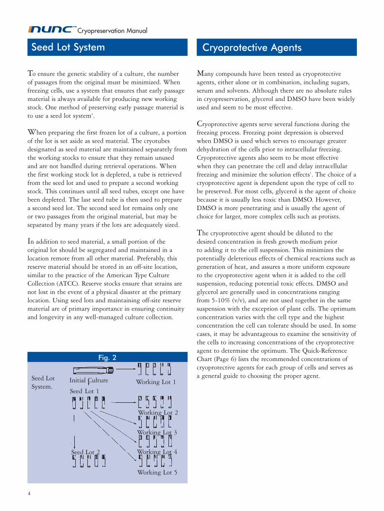
To ensure the genetic stability of a culture, the number of passages from the original must be minimized. When freezing cells, use a system that ensures that early passage material is always available for producing new working stock. One method of preserving early passage material is to use a seed lot system4.
When preparing the first frozen lot of a culture, a portion of the lot is set aside as seed material. The cryotubes designated as seed material are maintained separately from the working stocks to ensure that they remain unused and are not handled during retrieval operations. When the first working stock lot is depleted, a tube is retrieved from the seed lot and used to prepare a second working stock. This continues until all seed tubes, except one have been depleted. The last seed tube is then used to prepare a second seed lot. The second seed lot remains only one or two passages from the original material, but may be separated by many years if the lots are adequately sized.
In addition to seed material, a small portion of the original lot should be segregated and maintained in a location remote from all other material. Preferably, this reserve material should be stored in an off-site location, similar to the practice of the American Type Culture Collection (ATCC). Reserve stocks ensure that strains are not lost in the event of a physical disaster at the primary location. Using seed lots and maintaining off-site reserve material are of primary importance in ensuring continuity and longevity in any well-managed culture collection.
Many compounds have been tested as cryoprotective agents, either alone or in combination, including sugars, serum and solvents. Although there are no absolute rules in cryopreservation, glycerol and DMSO have been widely used and seem to be most effective.
Cryoprotective agents serve several functions during the freezing process. Freezing point depression is observed when DMSO is used which serves to encourage greater dehydration of the cells prior to intracellular freezing. Cryoprotective agents also seem to be most effective when they can penetrate the cell and delay intracellular freezing and minimize the solution effects1. The choice of a cryoprotective agent is dependent upon the type of cell to be preserved. For most cells, glycerol is the agent of choice because it is usually less toxic than DMSO. However, DMSO is more penetrating and is usually the agent of choice for larger, more complex cells such as protists.
The cryoprotective agent should be diluted to the desired concentration in fresh growth medium prior to adding it to the cell suspension. This minimizes the potentially deleterious effects of chemical reactions such as generation of heat, and assures a more uniform exposure to the cryoprotective agent when it is added to the cell suspension, reducing potential toxic effects. DMSO and glycerol are generally used in concentrations ranging from 5-10% (v/v), and are not used together in the same suspension with the exception of plant cells. The optimum concentration varies with the cell type and the highest concentration the cell can tolerate should be used. In some cases, it may be advantageous to examine the sensitivity of the cells to increasing concentrations of the cryoprotective agent to determine the optimum. The Quick-Reference Chart (Page 6) lists the recommended concentrations of cryoprotective agents for each group of cells and serves as a general guide to choosing the proper agent.
Cryoprotective Agents
Working Lot 5Seed Lot 2
Fig. 2
Seed LotSystem.
Working Lot 2
Working Lot 3
Working Lot 4
Seed Lot 1
Initial Culture Working Lot 1
4
Seed Lot System
Cryopreservation Manual
Working Lot 5
Seed Lot 2

Several factors must be considered when preparing cells for cryopreservation. These include the type of cell, cell viability, growth conditions, physiological state of the cells, the number of cells, and how the cells are handled. When preparing the initial seed stock of a new isolate or cell line, the culture should be examined for identity and contaminating microorganisms. This examination should be repeated after preservation and each time a new lot of the culture is prepared.
Microorganisms Microbial cells, particularly bacteria and yeast, grown under aerated conditions demonstrate a greater resistance to the detrimental effects of cooling and freezing than non-aerated cells5. T. Nei et.al.2 have demonstrated that cell permeability is greater in aerated cultures, and that the aerated cells dehydrate faster during cooling than non-aerated cells. Microbial cells harvested from late log or early stationary cultures also demonstrate greater resistance to the freezing process than younger or older cells6.
Generally, the greater the number of cells present initially, the greater the recovery. For most bacteria and yeast, approximately 107/ml cells are required to ensure adequate recovery7. These can be conveniently harvested from agar slants or plates, or when greater quantities
Preparation of Cells
Caution must be observed when handling DMSO as it is quickly absorbed into the body through the skin and may transport harmful substances into the body with it.
Glycerol and DMSO should be of reagent grade or better, sterilized prior to use, and examined for undesirable properties. Each lot should be examined for toxic properties by exposing sensitive cells to concentrations previously used with success. Glycerol may be sterilized by autoclaving for 15 minutes at 121°C and 15 psig. Glycerol should be protected from light during storage. DMSO must be sterilized by filtration using a 0.2-micron nylon syringe filter or a Teflon* PTFE syringe filter which has been pre-washed with alcohol and rinsed with DMSO. Cryoprotective agents should be prepared in single-use volumes to minimize the risk of contamination and moisture introduction with repeated use from one container.
* Teflon is a registered trademark of DuPont
are required, grown in broth culture and harvested by centrifugation. In either case, cells are generally suspended in fresh growth medium containing the cryo-protective agent. Protists can also be concentrated by centrifugation, but are often suspended in the used medium and then diluted by adding an equal volume of fresh growth medium containing the cryoprotective agent7.
Spore-forming fungi require harvesting of spores and suspension of the spores in fresh growth medium con-taining the cryoprotective agent. When freezing fungal spores, care must be taken not to delay the freezing process too long to ensure that germination does not occur prior to freezing. For fungi that do not form spores, special procedures for harvesting mycelia prior to freezing must be utilized. For fungi with tough mycelia, the culture is harvested from agar growth by cutting and removing agar plugs containing the mycelia and placing the plugs into fresh growth medium containing the cryoprotective agent. Tough mycelia that do not adhere well to agar cultures are grown in broth culture and the mycelial mass is blended prior to freezing7.
Genetically modified organisms can be preserved in a manner similar to the unmodified host cell in most cases8,9.
Cryoprotective Agents
5
Cryopreservation Manual

Animal Cell LinesWhen preparing animal cells for cryopreservation, cell populations need to be adjusted to levels that ensure adequate recovery without unnecessarily growing large numbers of cells. For most mammalian cells, a starting population between 106 to 107 pt cells/ml is optimum10. The cell suspension should initially be prepared at a concentration twice that desired for preservation so that an equal volume of cryoprotectant (2 x cryoprotective agent + medium) can be added. Alternatively, the cell pellet can be resuspended in the cryoprotectant (1 x cryoprotective agent + medium) to the desired cell concentration. Gentle handling during cell harvesting and concentration procedures will ensure healthy cells prior to subjecting them to cold stress. Vigorous pipeting and high-speed centrifugation should be avoided if possible7. Where appropriate, the pH should be maintained by gassing with 5% or 10% C02.
Animal cell cultures are especially susceptible to contamination by other cells, such as HeLa cells11, and contaminating microorganisms. The species of origin of cell lines should be verified by isoenzyme analysis, karyotyping, and/or immunological assays. These should be performed prior to and following preservation. Contamination by viruses and Mycoplasma sp. is of particular concern12. A good characterization program for animal cell lines should include a check for contamination by bacteria, fungi, appropriate viruses, mycoplasma, and in some cases, protozoa.
Plant Cell LinesPlant cells respond to cryopreservation in a manner similar to other cells13. The stage in the growth cycle from which they are harvested can affect their recovery, most optimum being late log phase. Also, cell density may
The viability and an estimate of recovery should be determined both before and after freezing the culture. Viability is a measure of the culture’s ability to grow and reproduce. For some material, such as protozoan cultures, this should include several passages to ensure stability. An estimate of the number of cells recovered can be made by several means including serial dilution, plate counts, or direct cell counting. A comparison of the counts prior to and after freezing gives an indication of the degree of recovery or the success of the preservation procedure.
Footnotes to Quick-Reference Chart:* While -60°C is adequate for most organisms in the groups noted, some sensitive cells may not survive long periods of storage at this temperature.† Mycelial masses are prepared for freezing of the hyphae of fungi without regard to numbers of cells.** Plant cells are generally packed to 3-20% cell volume for freezing.‡ The number of infectious particles has little effect on the recovery of viruses and bacteriophage.
Quick-Reference Chart
Preparation of Cells
play a role in recovery, the optimum cell density depending on the species being preserved.
(To be used as a general guide only)
Number Cryoprotective MinimumCell Type of Cells Agent Storage Temp.
Bacteria 107/ml Glycerol (10%) - 60°C*
Bacteriophage 108 pfu/ml Glycerol (10%) - 60°C
Fungi
Hyphae __† Glycerol (10%) -150°C
Spores 106/ml Glycerol (10%) - 60°C
Yeast 107/ml Glycerol (10%) -150°C
Protozoa l05-107/ml DMSO (5-10%) or -150°C Glycerol (10-20%)
Algae l05-107/ml Methanol (5-10%) -150°C or DMSO (5-10%)
Plant Cells __** DMSO (5 -10%) -150°C +Glycerol (5-10%)
Animal Cells 106-107/ml DMSO (5-10%) -150°C or Glycerol (5-10%)
Hybridomas 107/ml DMSO (5-10%) -150°C + Serum (20%)
Plant viruses __‡ None -60°C
Animal viruses
Cell Free __‡ -60°C
Infected Cells 106/ml DMSO (7%)+ -150°C Fetal Bovine Serum (10%)
Plasmids 106/ml Glycerol (10%) -150°C
Phage Libraries __‡ Glycerol (10%) -150°C
Cryopreservation Manual

Equilibration Ampoules and Cryotubes
The period of time between mixing the cryoprotectant with the cell suspension and the beginning of the cooling process is called the equilibration period. For most cells, equilibration should occur for at least 15 minutes, but no longer than 45-60 minutes. The cryoprotective agent may be toxic to the cells if the equilibration time is too long.
Equilibration, which should take place at ambient temperature, allows time for the cryoprotective agent to penetrate the cells, with larger and less permeable cells requiring a longer equilibration period. During this period of time the cell suspension may be dispensed into cryotubes and otherwise manipulated in preparation for freezing. The optimal equilibration time should be determined empirically for the cells being cryopreserved to maximize later recovery.
A variety of small containers such as flame-sealed glass ampoules and screw-cap plastic tubes can be used for storing cells at ultra-low temperatures. The most commonly used sizes are 1.0- to 1.8-ml tubes which maximize storage capacity but retain some ease of handling. Generally, 0.5-1.0 ml of the cell suspension is placed into each container. Several factors must be considered when selecting a container, including its cryotolerance, storage conditions, type of cells to be stored, and safety considerations.
For temperatures above -100°C where low-temperature mechanical stresses are less severe, a variety of containers may be used. However, when storing material at liquid nitrogen temperatures, containers specifically designed to withstand cryogenic temperatures must be used. A variety of containers specifically designed for cryogenic use are available. Plastic tubes have screw-on closures with external or internal threads. The rate of warming may be affected by the type of container used since plastic tubes
Combinations of cryoprotective agents are more effective than agents used singly. The cooling rate is important, and in many cases, a two-step cooling process where the cells are held at -30°C to -40°C for a period of time before cooling to liquid nitrogen temperatures, is beneficial. This process enhances the dehydration of the cytoplasm prior to freezing. Rapid thawing is preferred, but there is evidence that slow warming is just as effective in some cases.
VirusesMost viruses can be frozen as cell-free preparations without difficulty and do not require controlled cooling7. The exceptions are those viruses cultured in viable infected cells which require controlled cooling. Plant viruses can be preserved either in infected plant tissue or as purified virus preparations.
Preparation of Cells
Screw cap for tubes with external thread.
Stopper with silicone gasket for tubes with internal thread.
7
Cryopreservation Manual
Fig. 3

Once the cells and the cryoprotectant have been com-bined and dispensed into tubes, the next step is to cool the suspension. The rate of cooling is important since it affects the rate of formation and size of ice crystals, as well as the solution effects that occur during freezing. Different types of cells may require different cooling rates, however a uniform cooling rate of 1°C per minute from ambient temperature is effective for a wide variety of cells.
Generally, the larger the cells, the more critical slow cooling becomes. Most bacteria and spore-forming fungi will tolerate less-than-ideal cooling rates and can be frozen by placing the material at -60°C for a period of time. More fastidious bacteria and non-sporulating fungi require more uniform rates of cooling. Protists, animal cells and plant cells often require even greater control of the cooling rate including special manipulation to minimize the detrimental effects of undercooling and the heat liberated during freezing.
Despite the control applied to the cooling of cells, most of the water present will freeze at approximately -2°C to -5°C. The change in state from liquid to crystalline form results in the release of energy in the form of heat; this is known as the latent heat of fusion. Warming of the sample occurs until the equilibrium freezing point is reached, at which temperature ice continues to form. To minimize the detrimental effects of this phenomenon, undercooling must be minimized by artificially inducing the formation of ice. This can be accomplished by seeding the suspension with ice or some other nucleating agent, or by rapidly dropping the temperature of the external environment to encourage ice crystal formation.
8
If plastic cryotubes are stored in the liquid phase of liquid nitrogen, the use of NUNC™ CryoFlex™ is strongly recommended (NUNC Cat. No. 343958).
See Warning above.
usually require a longer warming period for complete thawing than glass ampoules. This difference in warming rate may be significant for some fastidious cells, but for most cells does not contribute to a loss of viability. Glass ampoules may be flame sealed.
Storage by immersion in liquid nitrogen is not advised. Improperly sealed glass ampoules may have micro-channels14 that lead to liquid nitrogen penetration over time. When these are retrieved from liquid nitrogen to ambient temperature, rapid conversion of the liquid nitrogen to vapor inside the ampoule can result in explosion of the ampoule. Plastic cryotubes with screw-top closures are also susceptible to liquid nitrogen pene-tration15, but do minimize the explosion potential. However, during warming of plastic cryotubes, liquid can spray from the cap/tube interface with potential dissemination of the cryotube contents.
Rate of Cooling
Fig. 5Fig. 4
The ideal cell cooling rate and cooling rate provided by NALGENE® “Mr. Frosty” 1°C frezing container, NALGENE Cat. No. 5100-0001.
Cooling Rates
Ampoules and Cryotubes
Cryopreservation Manual
WARNINGDo not use tubes for storage in liquid phase of liquid nitrogen unless correctly sealed in NUNCTM CryoFlex Tubing (Cat. No. 343958). Improper use may cause entrapment of liquified nitrogen inside the tube and lead to pressure build-up, resulting in possible explosion or biohazard release. Use appropriate safety procedures as outlined in this manual when handling and disposing of tubes.

9
Rate of Cooling
To achieve uniform, controlled cooling rates, use a programmable-rate cell freezing apparatus. Simple units allow only the selection of a single cooling rate for the entire temperature range. More sophisticated units, however, allow a selection of variable rates for different portions of the cooling curve. Less costly and easier-to-use systems are available for simulating a controlled-rate cooling process by placing the cryotubes in a mechanical freezer at -60°C to -80°C. In order to accomplish a uniform rate of cooling, the cryotubes must be placed in specially designed containers.
NALGENE “Mr. Frosty” 1°C freezing container (Cat. No. 5100-0001) provides a simple-to-use system designed to achieve a rate of cooling very close to 1°C per minute. Typical cooling rates for homemade freezing systems lead to uncontrolled cooling that averages 1°C per minute but the cells actually experience more rapid rates of cooling during some parts of the cooling curve16. Homemade freezing systems are also non-repeatable. “Mr. Frosty” eliminates the need for direct immersion in an alcohol bath. This feature eliminates the potential for contamination due to wicking of the alcohol, as well as the presence of residual alcohol on the exterior of the cryotubes. During handling at colder temperatures, the presence of alcohol on the cryotubes makes the cryotubes colder to the touch and extremelyslippery.
NALGENE® “Mr. Frosty”
Storage
When the sample has been frozen for 48 hours, a cryotube should be thawed to determine whether the cells are viable and able to establish a cell population, i.e., survive the freezing procedure. See Determination of Recovered Cells.
The temperature at which frozen preparations are stored affects the length of time after which cells can be recovered. The lower the storage temperature, the longer the viable storage period. Ultimate stability of frozen cells cannot be assured unless the material is maintained below -130°C 17. Some bacteria and spore-forming fungi may tolerate storage temperatures of -60°C to -80°C. However, more fastidious cells, such as mammalian tissue cultures, must be maintained below -130°C.
For ultimate security, living cells should be stored at liquid nitrogen temperatures. However, there are risks in storing cryotubes directly in liquid nitrogen, as discussed previously. Liquid nitrogen units that provide all-vapor storage are ideal as long as the working temperature at the opening of the unit remains below -130°C. To assure that a liquid nitrogen freezer maintains the proper working temperature, the volume of liquid nitrogen in the unit should be adjusted to a level that results in a temperature of -150°C just above the stored material when the lid of the unit is removed. This working temperature can be attained in most liquid nitrogen freezers. However, the design of some models
means the amount of liquid nitrogen necessary to attain the proper working temperature will reduce the amount of usable storage space. Mechanical freezers that go down to -150ºC are also available.
Improper handling of material maintained at cryogenic temperatures can have a detrimental effect on the viability of frozen cells. Each time a frozen cryotube is exposed to a warmer environment, even briefly, it experiences a change in temperature. Storage systems should be designed to minimize exposure of stored material to warmer
Fig. 6
Recommended temperatures in liquid-nitrogen storage system.
Cryopreservation Manual

For most cells, warming from the frozen state should occur as rapidly as possible until complete thawing is achieved. To achieve rapid warming, place the frozen tube into a 37°C water bath. Remember, material frozen in plastic tubes will take longer to thaw than that in glass ampoules, and sometimes gentle agitation of the tube during warming will accelerate the thawing process. Care must be taken, however, not to vigorously agitate tubes containing fragile cells such as protists and mammalian cells. As soon as the contents of the tube have been thawed, remove the tube from the water bath. To minimize the risk of contamination during reconstitution, disinfect the external surface of the tube by wiping with alcohol-soaked gauze prior to opening.
Immediately transfer the contents of the tube to fresh growth medium following thawing to minimize exposure to the cryoprotective agent. For most cultures, the entire contents of the tube may be placed into fresh media. However, further dilution may be necessary for cell lines. It is recommended that the suspension be centrifuged at 100 x g for 10 minutes after initial dilution, the supernate be removed, and the cells resuspended into fresh growth medium to remove residual cryoprotective agents.
temperatures, as well as minimizing prolonged exposure of personnel during specimen retrieval. Box stacking systems (i.e. stainless steel racks) necessitate exposure of boxes at the top to warmer temperatures when retrieving boxes at lower temperatures. When stainless steel racks are used, maintain a small number of tubes of each preparation in the top box of the rack and store the remaining tubes of each preparation in lower boxes. By doing this, a tube of one preparation can be retrieved without exposing all tubes of any particular culture or lot.
To maximize the available space in liquid nitrogen freezers and minimize exposure of material during retrieval, use small storage boxes or aluminum canes. Press the tubes onto the canes, putting no more than one lot of one culture on each cane. Canes provide a flat surface for coding their position and easy identification during retrieval. Place the canes into cardboard or plastic sleeves to eliminate the potential for tubes to fall from the canes. When retrieving vials from canes, the cane should be lifted only to a level that exposes the first available vial, without removing the remaining vials from the working temperature of the freezer.
Determination of Recovered Cells
Methods used to estimate the number of viable cells recovered following freezing depend on the type of material preserved. Visual inspection alone can be deceptive, and although staining and dye exclusion are effective in determi-ning the presence of viable cells for most mammalian cells, they do not indicate an ability to establish the cell popula-tion. For microbial cells, serial dilution and plate counts are effective in quantifying the population of cells recovered. Although there may be some tube-to-tube variation within a given lot, with constant storage conditions, the number of recovered cells will generally be the same in all tubes. Tube-to-tube variation may be an indication of problems occurring during storage and handling.
Reconstitution (Thawing)
Storage
10
Fig. 7
Clear plastic sleeve allows identification of vials in an aluminium cane.
Cryopreservation Manual

11
Despite its use in stabilizing living material, low-tempe-rature preservation can stress cells. Care must be taken to ensure that preserved material remains unchanged following preservation. A good preservation program should include effective characterization and cataloging programs, both of which combined, provide optimum culture collection practices. There is little use in preserving material that is of little value, inadequately characterized, contaminated or misidentified.
The first step in maintaining a collection of cultures is to assess the material to be preserved to ensure that it is of use and is worth keeping. This practice should continue throughout the collection process to minimize the accumulation of preserved material to unmanageable levels. To avoid duplication of collection materials, a system of identification of each item should be established. This can be done by devising unique numbering systems or number/ letter combinations. Each identification number should be cross-referenced to other information about the culture material.
Preservation of living cells ensures stability but does not correct any problems already present in the material. All material to be preserved should be examined thoroughly for contamination, proper identification, and other key characteristics unique to the cells, prior to preservation. Since freezing can stress cells and handling exposes cells to the risks of contamination, characterization must continue after successful preservation is accomplished. Each time a new lot of frozen material is prepared, complete characterization of the material should be carried out.
Cataloging and data record keeping are important aspects of all culture collection programs. Cataloging ensures that duplication of material does not occur and is especially useful when collection material is to be made available to others. Maintaining records on data generated during the characterization and preservation of collection materials ensures that any future problems can be adequately addressed.
Inventory Control Culture Collection Practices
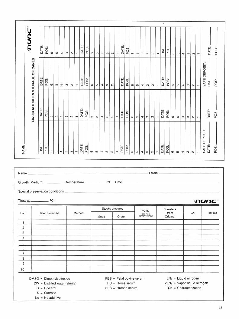
Appropriate record keeping is important in any laboratory. There are a number of possible methods for keeping records on cryopreserved materials. When establishing your own method, keep in mind that there are major classes of information which will be important in the future: (a) the preservation methodology used, (b) the location and identification of the stored material, (c) preservation date, and (d) number of passages.
Identification begins with proper labeling of the tubes. The label information should include an identification code for the frozen material, as well as a lot number. This label information should be kept with the records that include the location code for each tube. These records can be main-tained on inventory cards (one for each item) or in computer files. Duplicate inventory records should be maintained in a location separate from working records. Locator codes should be specific enough to allow rapid and easy retrieval for a specific lot and should include freezer unit number, a code for a freezer section or stainless steel rack, a box or canister number, and possibly even a grid spot within the box or a cane number when appropriate. Detailed locator codes minimize hunting for material which risks warming the freezer unit, exposure of other materials to warmer temperatures, and prolonged exposure of laboratory personnel to extreme cold.
Accurate record keeping specifically for cryopreserved material. Copies of these records are printed at end of this manual and may be duplicated.
Fig. 8
Cryopreservation Manual

1. Harvest cells from late log or early stationary growth. Scrape cells from the growth surface if they are anchorage dependent. Centrifuge broth or anchorage independent cultures to obtain a cell pellet, if desired.
2. Prepare presterilized DMSO or glycerol in the concen- tration desired in fresh growth medium. When mixing with a suspension of cells, prepare the cryoprotective agents in twice the desired final concentration.
3. Add the cryoprotectant solution to the cell pellet or mix the solution with the cell suspension. Begin timing the equilibration period.
4. Gently dispense the cell suspension into tubes.
5. Begin cooling the cells after the appropriate equilibra- tion time. a. Uncontrolled cooling—place the tubes on the bottom of a -60°C freezer for 90 minutes. b. Semi-controlled cooling—use Mr. Frosty freezing container to freeze the tubes in a -70°C freezer. c. Controlled cooling—use a programmable cooling unit to cool the cells at 1°C per minute to -40°C.
6. Remove the cells from the cooling unit and place them at the appropriate storage temperature.
7. To reconstitute, remove a tube from storage and place into a water bath at 37°C. When completely thawed, gently transfer the entire contents to fresh growth medium.
Safety precautions must be observed throughout the preservation and maintenance process. All work with hazardous cultures should be performed under proper containment, and U.S. Public Health Service Biosafety Level guidelines should be adhered to at all times18.
Animal cells may contain adventitious viral agents that require special handling, and all animal cells that have not been thoroughly characterized should be handled at Biosafety Level II. At this level, laboratory staff must have training in handling pathogenic agents and work under the direction of a competent scientist. Access to the laboratory must be limited and biological safety cabinets must be used for large-volume work or when aerosols are generated18.
Low-temperature storage of cells presents unique hazards that necessitate safety precautions. Cryogenic tempera-tures can result in exposure of personnel to extremely cold conditions, and precautions must be taken to protect personnel during operations in liquid nitrogen freezers. Insulated gloves and long-sleeved laboratory coats or other garb protect the skin from exposure. It is extremely im-portant to wear a full face and neck shield when working in the liquid portion of a liquid nitrogen freezer. As noted previously, improperly sealed glass ampoules may explode when retrieved from liquid nitrogen. To minimize the risk of potential explosions, place the tube in the vapor phase for 24 hours. A face shield that provides neck protection should be mandatory when retrieving tubes from liquid nitrogen. The use of NUNC™ CryoFlex™ is also strongly recommended. See Warning below.
Special precautions must be taken when working with hazardous biological materials at liquid nitrogen temperatures. Always thaw and open tubes containing hazardous material inside a biological safety cabinet. Be prepared for exploding and leaking ampoules/tubes. Broken ampoules in a liquid nitrogen freezer are a potential source of contamination19 and contaminants may survive, despite the extremely cold temperatures. When a liquid nitrogen freezer becomes contaminated, the entire unit should be decontaminated after warming to room temperature. When closing down a liquid nitrogen freezer that is not obviously contaminated, remove all material to be retained, warm the unit to room temperature and disinfect it prior to further handling.
Safety Considerations Step by Step
WARNINGDo not use tubes for storage in liquid phase of liquid nitrogen unless correctly sealed in NUNCTM CryoFlex Tubing (Cat. No. 343958). Improper use may cause entrapment of liquified nitrogen inside the tube and lead to pressure build-up, resulting in possible explosion or biohazard release. Use appropriate safety procedures as outlined in this manual when handling and disposing of tubes.
12
For more information on Cryopreservation and Cryoware Products, visit us at www.nuncbrand.com
Cryopreservation Manual

1. Farrant, J. 1980. General observations on cell preservation. In: M.J. Ashwood-Smith and J. Farrant, Eds. Low Temperature Preservation in Medicine and Biology, Pitman Medical Limited, Kent, England, p. 1-18.
2. Nei, T., T. Araki and T. Matsusaka.1969. Freezing injury to aerated and non-aerated cultures of Escherichia coli. In T. Nei, Ed. Freezing and Drying of Microorganisms. University of Tokyo Press, Tokyo, Japan.
3. Mazur, P., S.P. Leibo and E.H.Y. Chu. 1972. A two factor hypothesis of freezing injury. Experimental Cell Research 71: 345-355.
4. Hay, R.J. 1988. The seed stock concept and quality control for cell lines. Analytical Biochemistry 171: 225-237.
5. Alexander, M.T. and F.P. Simione. 1980. Factors affecting the recovery of freeze-dried Saccharomyces cerevisiae. Abstracts of the Annual Meeting of the American Society for Microbiology, p. 86.
6. Heckly, R.J. 1978. Preservation of microorganisms. Advances in Applied Microbiology 24: 1-53.
7. ATCC Preservation Methods: Freezing and Freeze Drying. Ed. F.P. Simione and E.M. Brown, American Type Culture Collection, Rockville, Maryland, 1991.
8. Nierman, W.C. and T. Feldblyum. 1985. Cryopreservation of cultures that contain plasmids. Dev. Ind. Microbiol. 26: 423-434.
9. Nierman, W.C., C. Trypus and L.L. Deaven. 1987. Preservation and stability of bacteriophage lambda libraries by freezing in liquid nitrogen. Biotechniques 5: 724-727.
10. Hay, R.J. 1978. Preservation of cell culture stocks in liquid nitrogen. TCA Manual 4: 787-790.
11. Lavappa, K.S. 1978. Survey of ATCC stocks of human cell lines for HeLa contamination. In Vitro 14: 469-475.
12. McGarrity, G.J. 1982. Detection of mycoplasmal infection of cell cultures. Adv. Cell Culture 2: 99-131.
13. Withers, L.A. 1985. Cryopreservation of cultured plant cells and proto- plasts. In: K.K. Kartha, Ed. Cryopreservation of Plant Cells and Organs, CRC Press, Inc., Boca Raton, Florida.
14. Greiff, D., H. Melton and T.W. Rowe. 1975. On the sealing of gas-filled glass ampoules. Cryobiology 12: 1-14.
15. Simione, F.P., P.M. Daggett, M.S. MacGrath and M.T. Alexander. 1977. The use of plastic ampoules for freeze preservation of microorganisms. Cryobiology 14: 500-502.
16. Simione, F.P. and P.M. Daggett. 1977. Recovery of a marine dinoflagellate following controlled and uncontrolled freezing. Cryobiology 14: 362-366.
17. Mazur, P. 1984. Freezing of living cells: mechanisms and implications. Am J. Physiol. 247: 125-142.
18. Biosafety in Microbiological and Biomedical Laboratories. U.S. Department of Health and Human Services, Public Health Service, Centers for Disease Control and NIH, Bethesda, Maryland, 1988.
19. Shafer, T.W., J. Everett, G.H. Silver, and P.E. Came. 1976. Biohazard: Virus contaminated liquid nitrogen. Science 191: 24-26.
COMMENTS:
Selected References
13
Cryopreservation Manual

14
15

If it is worth preservingit deserves the best
N50026 (1103) 5M © 2003 Nalge Nunc International Printed in USA
www.nuncbrand.comNalge Nunc International · USA · Tel +1 585 586 8800 · [email protected] · www.nuncbrand.com
Nunc A/S · Denmark · Tel +45 4631 2000 · [email protected] · www.nuncbrand.com
Nunc GmbH & Co. KG · Germany · Tel +49 6111 86740 · [email protected] · www.nunc.de
Nalge Nunc International KK · Japan · Tel +81 3381 63355 · [email protected] · www.nalgenunc.co.jp