Nucleotide ampicillin Escherichia plasmid pBR322 - pnas.org · Proc. Nati. Acad.Sci. USA Vol. 75,...
5
Proc. Nati. Acad. Sci. USA Vol. 75, No. 8, pp. 3737-3741, August 1978 Biochemistry Nucleotide sequence of the ampicillin resistance gene of Escherichia coli plasmid pBR322 (protein sequence/secretion signal/,-lactamase/DNA chemistry) J. GREGOR SUTCLIFFE* The Biological Laboratories, Harvard University, Cambridge, Massachusetts 02138 Communicated by Walter Gilbert, June 14, 1978 ABSTRACr I have determined the nucleotide sequence of the ampicillin resistance gene of pBR322, an Escherichia coli plasmid that encodes a penicillin fl-lactamase. This gene codes for a protein of 286 amino acid residues. The first 23 amino acids presumably form a signal for secretion, because they do not appear in the mature enzyme, whose partial amino acid se- quence has been determined independently, [Ambler, R. P. & Scott, G. K. (1978) Proc. NatL Acad Sci. USA 75, 3732-3736]. Recent advances in direct DNA sequencing methods have greatly increased the scope of the DNA sequences that are ac- cessible. As large amounts of sequence data are accumulated, it is important to know how reliable the data are likely to be, as well as how quickly a given sequence may be determined. The ampicillin resistance (ampr) gene provided a sequence whose accuracy could be tested;t determining this sequence was also a desirable goal in its own right. The ampr gene of Escherichia coli and other Gram-negative bacteria codes for a fl-lactamase (penicillin amido-jl-lac- tamhydrolase, EC 3.5.2.6) of approximately 27,000 daltons that catalyzes the hydrolysis of penicillins to penicilloic acids. Ra- tional design of penicillins that are resistant to this penicillinase requires the elucidation of the structure of the enzyme's active site. To this end a crystallographic study of the protein is in progress (1), and mutations that alter the kinetic properties of the enzyme (presumably by altering the active site) have been generated (2). The amino acid sequence of fl-lactamase is es- sential to solving the structure of the enzyme. The ampr gene is easily accessible because it is carried along with tetracycline resistance on the plasmid vector pBR322. pBR322 is superior to other cloning vectors, and any informa- tion about this plasmid is likely to be useful, especially because pBR322 has been approved by the National Institutes of Health as an EK2 vector for cloning in E. coli. The plasmid RI (origi- nally designated R7268) was isolated from the wild in London (1963) from Salmonella paratyphi B (3). The ampr gene was transposed to pBR312 from RI via pSF2124 (4-6). Molecular rearrangements of pBR312 produced pBR322 (7). The purified ,B-lactamase of RI has been shown to be of type RTEM 1 (8). Plasmid DNA was prepared by chloramphenicol amplifi- cation (9) in hosts RR1 (lacking restriction and modification specificity of K-12) (6) or GM1l9 (dam-3) (10). The ampr gene was thought to cover the unique site for the restriction enzyme Pst I (6, 7), and I confirmed this by opening theDNA circlewith Pst I, then treating the opened plasmid sequentially with S1 nuclease and DNA ligase. The single-strand DNase SI was ex- pected to chew back the tetranucleotide 3' extension charac- teristic of Pst I cleavage (11), and the ligase was expected to reseal the circle. The DNA so treated was used to transform E. coil, and of 26 transformants selected for tetracycline resistance, 20 were found to be ampicillin sensitive. A lesion at the Pst I site, therefore, inactivates the penicillinase. Thus, at the onset of this project I had two pieces of information: (i) that the ampr gene covered the unique Pst I site, and (ii) that the size of the fl-lactamase protein was about 27,000 daltons. This meant I needed a sequence of at least 729 base pairs overlapping the Pst I site. The plasmid has unique restriction sites for the enzymes EcoRI and Sal I (as well as Pst I) and, in addition, two sites for the enzyme HindII, one of which is the same as the Sal I site (7). The EcoRl site is about 750 nucleotide pairs from the Pst I site (determined by gel electrophoresis); the Sal I site is remote from the Pst I site; and the other HindII site lies about midway between the EcoRI and Pst I site. I compared restriction digests generated by one of the frequently cutting enzymes (Hae III, Hpa II, Alu I, Hinfl, Tag I, Tha I) with double digests gener- ated by that enzyme and a rarely cutting enzyme (EcoRI, Sal I, Pst I, HindII), and thereby determined which restriction fragments lie between the EcoRI and Pst I sites. The largest Taq I fragments of pBR322 contained the Pst I site, and this was examined to determine which sites for other restriction enzymes it contained. When the enzyme Mbo I was used, the DNA had to be prepared in the host GM1i9, which lacks the deoxade- nosine methylase (10). Mbo I cuts unmethylated but not methylated DNA. These results were sufficient for making a crude restriction map of the ampr gene. I selected relevant fragments and se- quenced them, using the method of Maxam and Gilbert (12). Fig. 1 is an example of an autoradiogram, which is the data from which a sequence is directly read. I ensured the continuity of all adjacent restriction fragments by sequencing across all junctions on overlapping fragments. All sequences were de- termined several times and, whenever it was experimentally convenient, both strands were sequenced. Fig. 2 shows the se- quences that I determined. The Sequence. Fig. 3 represents the sequence of the DNA from the unique EcoRI site through the end of the structural gene for ,-lactamase. At position 750-755 is the sequence C- T-G-C-A-G, the recognition sequence of Pst I. Of the six pos- * Current Address: Department of Cellular and Developmental Im- munology, Scripps Clinic and Research Foundation, 10666 North Torrey Pines Road, La Jolla, CA 92037. t This project was first considered on Feb. 8, 1977, and I began se- quencing in March. Soon thereafter R. P. Ambler and G. K. Scott sent their partial amino acid sequence data for the penicillin fl-lactamase of E. coli to Jeremy Knowles at Harvard University, who held the data until the final DNA sequence was presented to him. On Sept. 8, 1 considered the data to be unambiguous and presented them to Walter Gilbert, who, after interpreting a subset of the autoradio- grams, concurred with the sequence. The comparison of the DNA sequence with the partial amino acid sequence occurred at tea on Sept. 25, 1977. 3737 The costs of publication of this article were defrayed in part by the payment of page charges. This article must therefore be hereby marked "advertisenent" in accordance with 18 U. S. C. §1734 solely to indicate this fact. Downloaded by guest on January 5, 2020
Transcript of Nucleotide ampicillin Escherichia plasmid pBR322 - pnas.org · Proc. Nati. Acad.Sci. USA Vol. 75,...

Proc. Nati. Acad. Sci. USAVol. 75, No. 8, pp. 3737-3741, August 1978Biochemistry
Nucleotide sequence of the ampicillin resistance geneof Escherichia coli plasmid pBR322
(protein sequence/secretion signal/,-lactamase/DNA chemistry)
J. GREGOR SUTCLIFFE*The Biological Laboratories, Harvard University, Cambridge, Massachusetts 02138
Communicated by Walter Gilbert, June 14, 1978
ABSTRACr I have determined the nucleotide sequence ofthe ampicillin resistance gene of pBR322, an Escherichia coliplasmid that encodes a penicillin fl-lactamase. This gene codesfor a protein of 286 amino acid residues. The first 23 amino acidspresumably form a signal for secretion, because they do notappear in the mature enzyme, whose partial amino acid se-quence has been determined independently, [Ambler, R. P. &Scott, G. K. (1978) Proc. NatL Acad Sci. USA 75, 3732-3736].Recent advances in direct DNA sequencing methods havegreatly increased the scope of the DNA sequences that are ac-cessible. As large amounts of sequence data are accumulated,it is important to know how reliable the data are likely to be,as well as how quickly a given sequence may be determined.The ampicillin resistance (ampr) gene provided a sequencewhose accuracy could be tested;t determining this sequence wasalso a desirable goal in its own right.The ampr gene of Escherichia coli and other Gram-negative
bacteria codes for a fl-lactamase (penicillin amido-jl-lac-tamhydrolase, EC 3.5.2.6) of approximately 27,000 daltons thatcatalyzes the hydrolysis of penicillins to penicilloic acids. Ra-tional design of penicillins that are resistant to this penicillinaserequires the elucidation of the structure of the enzyme's activesite. To this end a crystallographic study of the protein is inprogress (1), and mutations that alter the kinetic properties ofthe enzyme (presumably by altering the active site) have beengenerated (2). The amino acid sequence of fl-lactamase is es-sential to solving the structure of the enzyme.The ampr gene is easily accessible because it is carried along
with tetracycline resistance on the plasmid vector pBR322.pBR322 is superior to other cloning vectors, and any informa-tion about this plasmid is likely to be useful, especially becausepBR322 has been approved by the National Institutes of Healthas an EK2 vector for cloning in E. coli. The plasmid RI (origi-nally designated R7268) was isolated from the wild in London(1963) from Salmonella paratyphi B (3). The ampr gene wastransposed to pBR312 from RI via pSF2124 (4-6). Molecularrearrangements of pBR312 produced pBR322 (7). The purified,B-lactamase of RI has been shown to be of type RTEM 1 (8).
Plasmid DNA was prepared by chloramphenicol amplifi-cation (9) in hosts RR1 (lacking restriction and modificationspecificity of K-12) (6) or GM1l9 (dam-3) (10). The ampr genewas thought to cover the unique site for the restriction enzymePst I (6, 7), and I confirmed this by opening theDNA circlewithPst I, then treating the opened plasmid sequentially with S1nuclease and DNA ligase. The single-strand DNase SI was ex-pected to chew back the tetranucleotide 3' extension charac-teristic of Pst I cleavage (11), and the ligase was expected toreseal the circle. The DNA so treated was used to transform E.
coil, and of 26 transformants selected for tetracycline resistance,20 were found to be ampicillin sensitive. A lesion at the Pst Isite, therefore, inactivates the penicillinase. Thus, at the onsetof this project I had two pieces of information: (i) that the amprgene covered the unique Pst I site, and (ii) that the size of thefl-lactamase protein was about 27,000 daltons. This meant Ineeded a sequence of at least 729 base pairs overlapping the PstI site.The plasmid has unique restriction sites for the enzymes
EcoRI and Sal I (as well as Pst I) and, in addition, two sites forthe enzyme HindII, one of which is the same as the Sal I site(7). The EcoRl site is about 750 nucleotide pairs from the PstI site (determined by gel electrophoresis); the Sal I site is remotefrom the Pst I site; and the other HindII site lies about midwaybetween the EcoRI and Pst I site. I compared restriction digestsgenerated by one of the frequently cutting enzymes (Hae III,Hpa II, Alu I, Hinfl, Tag I, Tha I) with double digests gener-ated by that enzyme and a rarely cutting enzyme (EcoRI, SalI, Pst I, HindII), and thereby determined which restrictionfragments lie between the EcoRI and Pst I sites. The largest TaqI fragments of pBR322 contained the Pst I site, and this wasexamined to determine which sites for other restriction enzymesit contained. When the enzyme Mbo I was used, the DNA hadto be prepared in the host GM1i9, which lacks the deoxade-nosine methylase (10). Mbo I cuts unmethylated but notmethylated DNA.
These results were sufficient for making a crude restrictionmap of the ampr gene. I selected relevant fragments and se-quenced them, using the method of Maxam and Gilbert (12).Fig. 1 is an example of an autoradiogram, which is the datafrom which a sequence is directly read. I ensured the continuityof all adjacent restriction fragments by sequencing across alljunctions on overlapping fragments. All sequences were de-termined several times and, whenever it was experimentallyconvenient, both strands were sequenced. Fig. 2 shows the se-quences that I determined.The Sequence. Fig. 3 represents the sequence of the DNA
from the unique EcoRI site through the end of the structuralgene for ,-lactamase. At position 750-755 is the sequence C-T-G-C-A-G, the recognition sequence of Pst I. Of the six pos-
* Current Address: Department of Cellular and Developmental Im-munology, Scripps Clinic and Research Foundation, 10666 NorthTorrey Pines Road, La Jolla, CA 92037.
t This project was first considered on Feb. 8, 1977, and I began se-quencing in March. Soon thereafter R. P. Ambler and G. K. Scott senttheir partial amino acid sequence data for the penicillin fl-lactamaseof E. coli to Jeremy Knowles at Harvard University, who held thedata until the final DNA sequence was presented to him. On Sept.8, 1 considered the data to be unambiguous and presented them toWalter Gilbert, who, after interpreting a subset of the autoradio-grams, concurred with the sequence. The comparison of the DNAsequence with the partial amino acid sequence occurred at tea onSept. 25, 1977.
3737
The costs of publication of this article were defrayed in part by thepayment of page charges. This article must therefore be hereby marked"advertisenent" in accordance with 18 U. S. C. §1734 solely to indicatethis fact.
Dow
nloa
ded
by g
uest
on
Janu
ary
5, 2
020
Proc. Natl. Acad. Sci. USA 75 (1978)
A+G G C C+T
.i
.i
op.. ..a
FIG. 1. Maxam-Gilbert sequence ladderepresents an autoradiogram of a polyacry](20% acrylamide/0.67% bisacrylamide/7 M(pH 8.3)/1 mM EDTA) was run for 40 hr at 1four lanes on the right side of the photograpi32p_ labeled 5' end for this sequence run is tat position 996 (see Fig. 3). The reactions u:ref. 12 except that magnesium acetate was onrstop solutions. Reactions are, from left, A,only, and C + T. The first 40 bases have bee
G C C+T codon (ATG) corresponds to the first methionine residue. Acontinuous polypeptide of 286 amino acids can be. read, ter-minating at an ochre codon (TAA). The sequence of the amprgene was determined in the absence of any knowledge of pro-* 1 X9teinsequence.The amino terminus of purified -lactamase was determined
*a" W * to be His-Pro-Glu-Thr-Leu by Alan Hall (personal communi-cation). This sequence matches the residues 24-28 of thehypothetical protein decoded from the DNA sequence. Thepartial protein sequence of Ambler and Scott (13) totally agreeswith the DNA sequence, with one exception. The details of thatcomparison are chronicled in the accompanying paper, alongwith the details of the protein sequencing, but a few featureswill be discussed here. The Gln-37 of the pBR322 f3-lactamasediffers from Lys at that position in the Edinburgh sequence.It is likely that both assignments are correct. In the pBR322 case
* - a single base change (CAG AAG) would allow Gln Lys;wSet* however, such a change in the DNA sequence would remove
a restriction site for the enzyme Mbo I (G-A-T-C G-A-T-A).The restriction site is definitely present, as is demonstrated byoverlapping sequence as well as by sequence using the 5' endsgenerated by the enzyme. Also, I have shown that Mbo I willnot cut at G-A-T-A (such a site at position 175 is not cut by MboI, while the G-A-T-C site at 315 is cut). The Edinburgh se-quence comes from the enzyme expressed by the plasmid R6K.The Lys must be correct because it specifies the end of a trypticfragment. If both assignments are correct, the pBR322 and R6Kproteins should differ in p1.J
Glutamic acid/glutamine and aspartic acid/asparaginedifferences are hard to pick up by protein sequencing tech-niques, as are tryptophan residues. The DNA sequencing doesnot have these difficulties. The potential problems in the in-
W *-aW. terpretation of DNA sequencing data are independent of thoseof protein sequencing.
-~ite°a The result of the comparison of the hypothetical proteinpredicted by the DNA sequence and the partial protein se-quence is that the DNA sequence is probably totally correct.Errors, it they exist, are confined to degeneracies of the geneticcode. The ampr gene sequence was solved without consultingthe protein data so that it could be determined if the DNA se-quence analysis could stand alone. A distance of 789 nucleotides
er from ampr gene. This with no detectable errors shows that, in fact, distance is no ob-lamide slab gel. The gel stacle for sequence determination. Confidence can be placedurea/50 mM Tris borate in carefully sequenced regions, even if no cross-check with300 V. The loading of the protein is available. As a corollary, if some data about a protein,h was delayed 12 hr. The such as the composition of its tryptic fragments, is available,the top strand Hinfl site then DNA sequence may be even more easily obtained. Fewersed were as described in than 7 mo were required both to learn all the techniques of
> G. alternate G oy, C DNA sequencing and to obtain the 1100 base pairs of confirmeden run off the bottom of sequence presented here.
the gel in the left lanes. The pattern can easily be read further than150 bases from the 5' end (the A > G track is better resolved in lighterexposures). The alternate G only reaction was used because it de-creases the ambiguity that confuses the G > A reaction at purinesdistal to the 5' end. The light ghost purine bands that show up in thepyrimidine tracks are routinely observed, but in general are not asource of confusion. It can be seen that the chemistry is working fine150 bases from the end, but that this particular gel system no longerresolves well. Variations in electrophoresis conditions can increasethe resolution such that autoradiograms can be read 200-300 basesfrom the 5'-end label (A. Maxam, R. Tizard, G. Sutcliffe, and W.Gilbert, unpublished results).sible translation reading frames (three in each orientation) thatcover the Pst I site, only one frame can code for a hypotheticalpolypeptide of appropriate length without encountering anin-phase nonsense codon. The amino acids encoded in thisframe are also indicated in Fig. 3. The first in-phase initiator
* This point is the source of some confusion. Ri and R6K penicillinaseshave each been classified as RTEM 1 by isoelectric focusing. Twoclasses exist, TEM 1 having a pI of 5.4 andTEM 2 of 5.6. Either thepBR322 penicillinase has an even lower pI than RTEM 1 or else therehas been a mix-up in the history of R6K. A. Hall and J. Knowles(personal communication) report that their "R6K," obtained fromthe Microbiological Research Establishment, Porton, England (whichalso supplied Ambler and Scott, and Knox's group), carries resistanceto tetracycline, but not to streptomycin. These phenotypes differ fromthose reported by Heffron et al. (14). I suggest that the pBR322 en-zyme is RTEM 1 and the "R6K" enzyme of Ambler and Scott, Halland Knowles, and probably Knox et al. is actually RTEM 2. Thedifference between TEM 1 and 2 would be explained by the Gly/Lysdifference discussed in the text. Nicause the two types of enzymehave the same activity spectrum against various penicillins andcephalosporins, the altered residue must not be involved in the en-zymatic activity.
3738 Biochemistry: Sutcliffe
A+(
.'.W
am
Om
Dow
nloa
ded
by g
uest
on
Janu
ary
5, 2
020

Proc. Natl. Acad. Sci. USA 75 (1978) 3739
Mbo I(GATC)A/u I(AGCT)Tha I
(CGCG)Hpa II
(CCGG)Hae III
(GGCC)I )nimipr
A_-I Is I1o00 200 300 400 1 500
.coRI Taq HindIIkATTC) (TCGA) (GTYRAC)
I I i I
600 700 T 800Pst I
(CTGCAG)
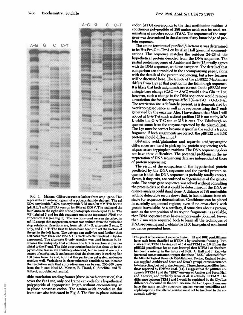
FIG. 2. Restriction map and sequencing strategy for pBR322 ampr gene. The numbers along the abscissa represent base pairs and correspondto the DNA sequence of Fig. 3. Each horizontal strip represents the cleavage map for a different restriction enzyme; that enzyme and its cuttingsequence are indicated at the end of the strip. The bottom strip (labeled unique) represents the map for enzymes that cut only once in the amprregion. The arrows within the strips show the extent of individual sequencing runs. The tail of the arrow is at the 5' end of the sequenced fragment.Two features of the sequencing allowed complete confidence in the cumulative results. Seventy percent of the gene sequence was determinedon both strands. Occasional regions of autoradiograms show peculiar band-to-band spacing, a phenomenon that can be accounted for by intra-molecular secondary structure of the DNA during electrophoresis. When both strands have been sequenced, the possibility for errors in filmreading is reduced because even if both strands show the peculiarity, it will be located at opposite regions of the sequence such that each portionof the DNA is clearly read on at least one strand. The second feature is that the films were subjected to multiple readings. It is the nature of thedata that they consist of long strings of meaningless symbols. Clerical errors can be rampant.
It is a feature of DNA that it has more to say than just whatprotein it encodes. The first 23 amino acids predicted by theDNA sequence do not appear in the mature, secreted protein.These 23 amino acids are predominantly hydrophobic, If bothtranscription and translation are colinear with the DNA, thenthe f3-lactamase carries a "leader" of 23 amino acids. The signalhypothesis of Blobel's group (15) ascribes the ability of proteinsthat must pass through or become part of the cell membraneto the presence of a hydrophobic amino terminus on nascentpolypeptides. This signal functions in the processes of transportand secretion and then is cleaved from the primary translationproduct to form the active molecule. The RTEM #l-lactamasefits nicely with this hypothesis. The genetic information for thesignal is contiguous with that for the structural enzyme. it is notknown if the signal functions by virtue of specific features ofits sequence or because of the character that it endows on therest of the protein. The observation that some secreted proen-
zymes (e.g., Bacillus licheniformis penicillinase) (16) have fullenzymatic activity argues that the signal need not inhibit thisactivity. Presumably the removal of the signal relegates theprotein to the external cell compartment. The penicillinase onpBR322 should provide a good model system for using re-combinant DNA technology to study the compartmentalizationof prokaryotic proteins.
As more gene sequences become known, a picture willemerge as to how the genetic code is utilized by the organism.Table 1 shows the distribution of codons used to assemble theamino acids of fl-lactamase. Although the source of this genein the wild was Salmonella, it has functioned well in laboratoryE. colt for over 10 years. A few asymmetries in codon usageappear for ampr. However, when the composite of all availablegenes for which a sequence is known [lac i, cro, and the proteinsof phages cX174 and Ms2 (17-20)] is surveyed, the only no-ticeable asymmetry is in arginine codons. CGY is used morefrequently than CGR or AGR.
Table 1. Codon distribution for pBR322 fl-lactamasePhe TTT 9Phe TTC 1Leu TTA 5Leu TTG 4
Leu CTT 9Leu CTC 2Leu CTA 5Leu CTG 8
Ile ATT 5Ile ATC 7Ile ATA 5Met ATG 10
Val GTT 6Val GTC 2Val GTA 5Val GTG 2
Ser TCT 3Ser TCC 3Ser TCA 2Ser TCG 1
Pro CCT 2Pro CCC 3Pro CCA 6Pro CCG 3
Thr ACT 6Thr ACC 3Thr ACA 4Thr ACG 7
Ala GCT 8Ala GCC 8Ala GCA 9Ala GCG 4
Tyr TAT 1Tyr TAC 3
TA -
TAG
His CAT 4His CAC 3Gln CAA 5Gln CAG 4
Asn AAT 3Asn AAC 5Lys AAA 6Lys AAG 5
Asp GAT 11Asp GAC 5Glu GAA 11Glu GAG 9
Cys TGT 1Cys TGC 2
TGATrp TGG 4
Arg CGT 7Arg CGC 7Arg CGA 2Arg CGG 1
Ser AGT 5Ser AGC 2Arg AGA 2Arg AGG 0
Gly GGT 8Gly GGC 4Gly GGA 4Gly GGG 5
E
(GA
I 1900 1000 1100
Hinf I(GANTC)
-~~~~~~~~~~~~~~~~~~~~~~-.1 -
The triplet codons for the amino acids are represented 5'-3' in DNA. The numbers represent how many times the particular triplet appearsin phase in the DNA sequence. AGG is never used for ,B-lactamase. TAA, TAG, and TGA are nonsense codons whose appearance would indicatepolypeptide chain termination. TAA is the single terminator used for f-lactamase.
7-, !, ti .I
I
w i=oI
0 4
-F..r I
----II .I i N I is IIl-r
Biochemistry: Sutcliffe
vrilqut:
I
Dow
nloa
ded
by g
uest
on
Janu
ary
5, 2
020

3740 Biochemistry: Sutcliffe Proc. Nati. Acad. Sci. USA 75 (1978)
GA^TTCTT"G^^ACGA^AAGGGCCTCGTGATACGCCTATTTTTATAGGTTAATGTCATGATAATAATGGTTTCTTAGACGTCAGGTGGCACTTTTC"G66AATGTG20 4'0 60 -0 100
CTTAAGAACTTCTGCTTTCCCGGAGCACTATGCGGAT^AAAAATATCCAATTACAGTACTATTATTACCAAAGAATCTGCAGTCCACCGTGAAAAGCCCCTTTACAC
CGCGGAACCCCTATTTGTTTATTTTTCT^AATACATTCAAATATGTATCCGCTCATGAGACAAT^ACCCTGATAAATGCTTCAATMATATTGAAA GGAAG120 140 160 160 200
GCGCCTTeGGGATAAACAAATAAAAGATTTATGTAAGTTTATACATAGGCGAGTACTCTGTTATTGGGACTATTTACGAAGTTATTATAACTTTTTCCTTC
10 20 30
SletserI1<eG~nHisPheArg~alfA aLeudI ZeProPhePheA 1aAlaPheCyaLeu.ProValPheAltaf~isPro~tuThrLeuValLys~alLysAspAGTATGAGTATTCAACATTTCCGTGTCGCCCTTATTCCCTTTTTTGCGGCATTTTGCCTTCCTGTTTTTGCTCACCCAGAAACGCTGGTGAAAGTAAAAG6AT
220 2140 260 280 300TCATACTCATAAGTTGTAAAGGCACAGCGGGAATAAGGGAAAAAACGCCGTAsAAACGGAAGGACAAAAACGAGTGGGTCTTTGCGACCACTTTCATTTTCTA
4 0 550 60AZaC ZuA pC InLew tyA IaAr-gVa ItGIty TyrI IteC tuLeuAl jpLeuA Jn~erGtyLy# IIZe LeuC 1fuSerPheArgProC IuCluA rgPhePro~e tMe tGCTGAAGATCAGTTGGGTGCACGAGTGGGTTACATCGAACTGGATCTCAACAGCGGTAAGATCCTTGAGAGTTTTCGCCCCGAAGAACGTTTTCCAATGATG
320 340 360 380 4 00CGACTTCTAGTCAACCCACGTGCTCACCCAATGTAGCTTGACCTAGAGTTGTCGCCATTCTAGGAACTCTCAAAMGCGGGGCTTCTTGCAAMAGGTTACTAC
70 80 90 100
SerThrPheLys Va t~euLeuCyeG lyA la Va ILeuSerA rg Va IA#pA taClyZyIn~G t nLeuG I yA rgA rgIIt*Hi#TyrSerG I nAanAspLeuzVa IAGCACTTTTAAAGTTCTGCTATGTGGCGCGGTATTATCCCGTGTTGACGCCGGGCAAGAGCAMCTCGGTCGCCGCATACACTATTCTCAGA^TGACTTGGTT
4120 4.4.0 4160 4180 500TCGTG^AAATTTCAAGACGATACACCGCGCCATAATAG66CACAACTGCGGCCCGTTCTCGTTGAGCCAGCGGCGTATGTGATAAGAGTCTTACTGAACCAA
110 120 130GtuTyrSerPro~a IThrG Iuky cHiaLeuThrA~pGyMe tThrtta1Arq I uLeuCy StrA IaA IaIeThr~e tSerAspAonThrA taA taAsanLeu6A6TACTCACCA6TCACAGAAACATCTTAC66AT6CAT6ACAGTAA6AGATTATGCAGT6GCTGCATA.ACCATGATGT6fAACACTGCGGCCAACTTA
520 5.0 s60 560 600CTCATGAGTGGTCAGTGTCTTTTCGTAGAATGCCTACCGTACTGTCATTCTCTTAATACGTCAC6ACGGTATTGGTACTCACTATTGTGACGCCGGTTGAT
1140 1 50 160
LeuLeiThrThrItleGZGlMProLGICtuL*uThrA IaPheLeuhiiAaenMe t (,I: .AafrHi8 Va IThrArgLekAapA rqTrpGluProGCuLeuAanGtIuCTTCT6ACAACGTCG"G"6CCG^^GGAGCTAACC6CTTTTTTGCACAACAT6G6G6ATCATGTAACTCGCCTTGATCGTTGB6^ACCGGAGCTGAATGAA
620 640 660 660 700GAAGACTGTTGCTAGCCTCCTGGCTTCCTCGATTGGCGAAAAAACGTGTTGTACCCCCTAGTACATTGAGCG6AACTAGCAACCCTTGGCCTCGACTTACTT
170 160 190 200A LaILeProA*nA*pGlCuArgAUpThrThrMetProA laA taMetA laThrlihrLeuArgLyaLeuLeuThrGCtyGlCLeuLcuThrLeuA taSerArgGinGCCATACCAAAC"C6A6CGTGACACCACGATGCCTGCAGCAATGGCAACAACGTTGCGCAAACTATTAACTGGCGAACTACTTACTCTAGCTTCCCGGCAA
720 740 760 760 6ooCGGTATGGTTTGCTGCTCGCACTGTGGTGCTACGGACGTCGTTACCGTTGTTGCAACGCGTTTGATAATTGACCGCrtGATGAATGAGATCGAAGGGCCGTT
210 220 230
GtnLeuxt*AspfrpM~et~uA taAspLys ValZA laC yProLeuLeuA rgSerA ZaLeuProAzaC l y TrpPhellteAl1faAspLy'Ser~tyA taC tyC tCAATTAATAGACTGGATGGAGGCGGATAAAGTTGCAGGACCACTTCTGCGCTCGGCCCTTCCGGCTGGCTGGTTTATTGCTGATAATCTGGAGCCGGTGAG
620 840 860 660 900GTTAATTATCTGACCTACCTCCGCCTATTTCAACGTCCTGGTGAAGACGCGAGCCGGGAAGGCCGACCGACCAAATAACGACTATTTAGACCTCGGCCACTC
2140 250 260 270
ArgGtyS*rArgGCty6eItZeAZaA laLeu4GlyProAepGCtyLysProSerArgIZeVaZVatIZeTyrThrThrGlCySerCGtnAlaThrMetA8pGtuArgCGTGGGTCTCGCGGTATCATTGCAGCACTGGGGCCAGATGGTAAGCCCTCCCGTATCGTAGTTATCTACAC6ACGGGGAGTCAGGCAACTATGGAT6AACGA920 940 960 980 1000 1020GCACCCAGAGCGCCATAGTAACGTCGTGACCCCGGTCTACCATTCGGGAGGGCATAGCATC ATAGATGTGCTGCCCCTCAGTCCGTTGATACCTACTTGCT
280 ae6AenArgGtnIreA taGluIlteGtyA taSerLeulteLbysHiaTrpAATAGACAGATCGCTGAGATAGGTGCCTCACTGATTAAGCATTGGTAACTGTCAGACCAAGTTTACTCATATATACTTTAGA
1040 1060 1060 1100TTATCTGTCTAGCGACTCTATCCACGGAGTGACTAATTCGTAACCATTGACAGTCTGGTTCAATGAGTATATATGAMATCT
FIG. 3. The sequence of the ampr gene. The top strand of sequence is 5'-3'; the bottom strand is 3'-5', complementary to the top strand.The running count between the strands (every 20 base pairs) starts with the first T in the top strand (the middle of the EcoRI site). The three-lettercodes for the amino acids of fl-lactamase appear directly over their three-base codons and they are numbered (every 10 amino acids) startingfrom the first methionine.
Dow
nloa
ded
by g
uest
on
Janu
ary
5, 2
020

Proc. Natl. Acad. Sci. USA 75 (1978) 3741
A few other features of the sequence merit attention, al-though no supporting biological data currently exist. Frompositions 189 to 195 is the sequence A-A-T-A-T-T-G, a 5 of 7match with the canonical "Pribnow box" (21). There is a goodcorrespondence to the complement of the 3' ends of the 16Sribosomal RNA (22) in positions 199-203. These two regionsare likely to represent the recognition sequence for RNApolymerase and the ribosome binding sequence, respectively.Further sequence analysis has located the inverted comple-mentary repeat of the ampicillin transposon Tn3 downstreamfrom the sequence presented here.The sequence presented in this paper establishes that direct
DNA sequence analysis is capable of providing accurate resultsover extensive regions in a very short time relative to proteinsequencing. Fundamentally, DNA sequence analysis can standon its own merits, but when combined with data about trans-lation or transcriptional products, the results can be essentiallyinfallible.
I thank A. Maxam, P. Farabaugh, A. Hall, D. Hourcade, L. Johnsrud,D. McConnell, W. McClure, R. Ogata, U. Siebenlist, R. Simpson, J.Sims, and R. Tizard for discussions and enzymes. I thank W. Gilbertand J. Knowles for conceiving this exercise and R. P. Ambler and G.K. Scott for their enthusiastic, unselfish participation. This work wassupported by National Institutes of Health Grant GM 21514-3 toWalter Gilbert.1. Knox, J. R., Kelly, J. A., Moews, P. C. & Murthy, N. S., (1976) J.
Mol. Biol. 104,865-875.2. Hall, A. & Knowles, J. R. (1976) Nature 264,803-804.3. Datta, N. and Kontomichalou, P. (1965) Nature 208, 239-
241.
4. Meynell, E. & Datta, N. (1967) Nature 214, 885-887.5. So, M., Gill, R. & Falkow, S. (1975) Mol. Gen. Genet. 142,
239-249.6. Bolivar, F., Rodriguez, R. L., Betlach, M. C. & Boyer, H. W.
(1977) Gene 2,75-93.7. Bolivar, F., Rodriquez, R. L., Greene, P. J., Betlach, M. C.,
Heyneker, H. L. & Boyer, H. W. (1977) Gene 2,95-113.8. Matthew, M. & Hedges, R. W. (1976) J. Bacteriol. 125, 713-
718.9. Tanaka, T. & Weisblum, B. (1975) J. Bacteriol. 121, 354-362.
10. Marinus, M. G. & Morris, N. R. (1975) Mutat. Res. 28,15-26.11. Brown, N. L. & Smith, M. (1976) FEBS Lett. 65,284-287..12. Maxam, A. M. and Gilbert, W. (1977) Proc. Natl. Acad. Sdi. USA
74,560-564.13. Ambler, R. P. & Scott, G. K. (1978) Proc. Natl. Acad. Sci. USA
75,3732-3736.14. Heffron, F., Sublett, R., Hedges, R. W., Jacob, A. & Falkow, S.
(1975) J. Bacteriol. 122, 250-256.15. Blobel, G. & Dobberstein, B. (1975) J. Cell. Biol. 67,835-851.16. Dancer, B. N. & Lampen, J. 0. (1975) Blochem. Biophys. Res.
Commun. 66,1357-1364.17. Farabaugh, P. J. (1978) Nature, in press.18. Roberts, T. M., Shimtake, H., Brady, C. & Rosenberg, M. (1977)
Nature 270, 274-275.19. Sanger, F., Air, G. M., Barrell, B. G., Brown, N. L., Coulson, A.
R., Fiddes, J. C., Hutchison, C. A., Slocombe, P. M. & Smith, M.(1977) Nature 265,687-695.
20. Fiers, W. (1975) in RNA Phages, ed. Zinder, N. (Cold SpringHarbor Laboratory, Cold Spring Harbor, New York), pp.353-396.
21. Pribnow, D. (1975) Proc. Nati. Acad. Sci. USA 72,784-788.22. Shine, J. & Dalgarno, L. (1974) Proc. Natl. Acad. Sci. USA 71,
1342-1346.
Biochemistry: Sutcliffe
Dow
nloa
ded
by g
uest
on
Janu
ary
5, 2
020


![Microscopic theoryof irreversible processes · Proc. Natl. Acad.Sci. USA74(1977) 4153 Thequadraticcharacterofthefunctional [1.3] permitsusto link theconstructionofMtothedeterminationofanewrep-](https://static.fdocuments.in/doc/165x107/601c715594813961252adeeb/microscopic-theoryof-irreversible-processes-proc-natl-acadsci-usa741977-4153.jpg)