Nucleic acid peptide nanogels for the treatment of ...
15
Nanoscale PAPER Cite this: Nanoscale, 2020, 12, 17411 Received 20th April 2020, Accepted 19th July 2020 DOI: 10.1039/d0nr03095c rsc.li/nanoscale Nucleic acid peptide nanogels for the treatment of bacterial keratitis† Sybil Obuobi, ‡ a Venkatesh Mayandi, b Nurul Azlyn Mohd Nor, a Benedict Jiasheng Lee, a Rajamani Lakshminarayanan a,b and Pui Lai Rachel Ee * a,c Cage-shaped nucleic acid nanocarriers are promising molecular scaffolds for the organization of poly- peptides. However, there is an unmet need for facile loading strategies that truly emulate nature’s host– guest systems to drive encapsulation of antimicrobial peptides (AMPs) without loss of biological activity. Herein, we develop DNA nanogels with rapid in situ loading of L12 peptide during the thermal annealing process. By leveraging the binding affinity of L12 to the polyanionic core, we successfully confine the AMPs within the DNA nanogel. We report that the thermostability of L12 in parallel with the high encapsu- lation efficiency, low toxicity and sustained drug release of the pre-loaded L12 nanogels can be translated into significant antimicrobial activity. Using an S. aureus model of infectious bacterial keratitis, we observe fast resolution of clinical symptoms and significant reduction of bacterial bioburden. Collectively, this study paves the way for the development of DNA nanocarriers for caging AMPs with immense significance to address the rise of resistance. 1. Introduction To emulate nature’s precise spatial co-location of bioactive molecules, scientists are adapting the elegance of rational design and self-assembly to create addressable, nanoscale bio- logically inspired containers. 1–4 These nanocarriers are attrac- tive candidates for drug delivery applications given their large loading capabilities, excellent in vivo targeting and enhanced stability features. 5–7 Notably, the non-toxicity, size homogen- eity and biodegradability of biomimetic nanocontainers favor their use over synthetic nanoscale objects. 8–10 To date, a vast majority of self-assembled biological containers reported hitherto are fabricated by coupling highly ordered protein architectures, which may comprise thousands of copies of different proteins and are challenged by chemical/thermal instability or pose immunogenicity issues. 11–15 Moreover, there is an unmet need to develop biomimetic cages where spatial control over drug entrapment can easily be achieved. As a burgeoning field in nanomedicine, advances in nucleic acid nanotechnology provide an unparalleled rich toolbox for the bottom-up construction of deoxyribonucleic acid (DNA) nanostructures of variable sizes, mechanical rigidity and envel- oping capability. 16–19 DNA nanostructures can be reproducibly and rapidly prepared in high yield using minimal sequence composition, and their negative surface potential confers sig- nificantly greater stability than their component oligonucleotides. 20–24 Owing to their unprecedented control over size, shape and presentation of targeting ligands, cage- shaped DNA polyhedral structures have emerged as promising molecular scaffolds for the organization of biomolecules. 25,26 To this end, a large number of studies exploit cargo entrap- ment via chemical or supramolecular interactions for mole- cular payloads such as proteins 27–29 and anticancer therapies. 30,31 Chemical crosslinking strategies are challenged by the formation of heterogeneous product mixtures and can introduce permanent cargo modifications which can interfere with activity. 32 On the other hand, while non-covalent strat- egies have demonstrated enhanced efficacy, many rely on post- loading strategies which may be challenged by long incubation times. 33 Recently, de Vries and colleagues reported on novel DNA amphiphiles wherein the corona was extended with DNA aptamer binding kanamycin B or ribonucleic acid (RNA) aptamer binding neomycin B for antimicrobial drug delivery. 34 While fabrication was achieved rapidly, drug loading requires † Electronic supplementary information (ESI) available. See DOI: 10.1039/ d0nr03095c ‡ Present address: Drug Transport and Delivery Research Group, Department of Pharmacy, Faculty of Health Sciences, UiT-The Arctic University of Norway, N-9037, Tromsø, Norway. a Department of Pharmacy, National University of Singapore, 18 Science Drive 4, Singapore 117543. E-mail: [email protected]; Fax: +65 6779 1554; Tel: +65 6516 2653 b Singapore Eye Research Institute, The Academia, Discovery Tower Level 6, 20 College Road, Singapore 169857 c NUS Graduate School for Integrative Sciences and Engineering, 21 Lower Kent Ridge Road, Singapore 119077 This journal is © The Royal Society of Chemistry 2020 Nanoscale, 2020, 12, 17411–17425 | 17411 Open Access Article. Published on 14 August 2020. Downloaded on 4/4/2022 11:24:40 AM. This article is licensed under a Creative Commons Attribution-NonCommercial 3.0 Unported Licence. View Article Online View Journal | View Issue
Transcript of Nucleic acid peptide nanogels for the treatment of ...

Nanoscale
PAPER
Cite this: Nanoscale, 2020, 12, 17411
Received 20th April 2020,Accepted 19th July 2020
DOI: 10.1039/d0nr03095c
rsc.li/nanoscale
Nucleic acid peptide nanogels for the treatment ofbacterial keratitis†
Sybil Obuobi, ‡a Venkatesh Mayandi,b Nurul Azlyn Mohd Nor,a
Benedict Jiasheng Lee,a Rajamani Lakshminarayanan a,b andPui Lai Rachel Ee *a,c
Cage-shaped nucleic acid nanocarriers are promising molecular scaffolds for the organization of poly-
peptides. However, there is an unmet need for facile loading strategies that truly emulate nature’s host–
guest systems to drive encapsulation of antimicrobial peptides (AMPs) without loss of biological activity.
Herein, we develop DNA nanogels with rapid in situ loading of L12 peptide during the thermal annealing
process. By leveraging the binding affinity of L12 to the polyanionic core, we successfully confine the
AMPs within the DNA nanogel. We report that the thermostability of L12 in parallel with the high encapsu-
lation efficiency, low toxicity and sustained drug release of the pre-loaded L12 nanogels can be translated
into significant antimicrobial activity. Using an S. aureus model of infectious bacterial keratitis, we observe
fast resolution of clinical symptoms and significant reduction of bacterial bioburden. Collectively, this
study paves the way for the development of DNA nanocarriers for caging AMPs with immense significance
to address the rise of resistance.
1. Introduction
To emulate nature’s precise spatial co-location of bioactivemolecules, scientists are adapting the elegance of rationaldesign and self-assembly to create addressable, nanoscale bio-logically inspired containers.1–4 These nanocarriers are attrac-tive candidates for drug delivery applications given their largeloading capabilities, excellent in vivo targeting and enhancedstability features.5–7 Notably, the non-toxicity, size homogen-eity and biodegradability of biomimetic nanocontainers favortheir use over synthetic nanoscale objects.8–10 To date, a vastmajority of self-assembled biological containers reportedhitherto are fabricated by coupling highly ordered proteinarchitectures, which may comprise thousands of copies ofdifferent proteins and are challenged by chemical/thermalinstability or pose immunogenicity issues.11–15 Moreover, there
is an unmet need to develop biomimetic cages where spatialcontrol over drug entrapment can easily be achieved.
As a burgeoning field in nanomedicine, advances in nucleicacid nanotechnology provide an unparalleled rich toolbox forthe bottom-up construction of deoxyribonucleic acid (DNA)nanostructures of variable sizes, mechanical rigidity and envel-oping capability.16–19 DNA nanostructures can be reproduciblyand rapidly prepared in high yield using minimal sequencecomposition, and their negative surface potential confers sig-nificantly greater stability than their componentoligonucleotides.20–24 Owing to their unprecedented controlover size, shape and presentation of targeting ligands, cage-shaped DNA polyhedral structures have emerged as promisingmolecular scaffolds for the organization of biomolecules.25,26
To this end, a large number of studies exploit cargo entrap-ment via chemical or supramolecular interactions for mole-cular payloads such as proteins27–29 and anticancertherapies.30,31 Chemical crosslinking strategies are challengedby the formation of heterogeneous product mixtures and canintroduce permanent cargo modifications which can interferewith activity.32 On the other hand, while non-covalent strat-egies have demonstrated enhanced efficacy, many rely on post-loading strategies which may be challenged by long incubationtimes.33 Recently, de Vries and colleagues reported on novelDNA amphiphiles wherein the corona was extended with DNAaptamer binding kanamycin B or ribonucleic acid (RNA)aptamer binding neomycin B for antimicrobial drug delivery.34
While fabrication was achieved rapidly, drug loading requires
†Electronic supplementary information (ESI) available. See DOI: 10.1039/d0nr03095c‡Present address: Drug Transport and Delivery Research Group, Department ofPharmacy, Faculty of Health Sciences, UiT-The Arctic University of Norway,N-9037, Tromsø, Norway.
aDepartment of Pharmacy, National University of Singapore, 18 Science Drive 4,
Singapore 117543. E-mail: [email protected]; Fax: +65 6779 1554;
Tel: +65 6516 2653bSingapore Eye Research Institute, The Academia, Discovery Tower Level 6, 20
College Road, Singapore 169857cNUS Graduate School for Integrative Sciences and Engineering, 21 Lower Kent Ridge
Road, Singapore 119077
This journal is © The Royal Society of Chemistry 2020 Nanoscale, 2020, 12, 17411–17425 | 17411
Ope
n A
cces
s A
rtic
le. P
ublis
hed
on 1
4 A
ugus
t 202
0. D
ownl
oade
d on
4/4
/202
2 11
:24:
40 A
M.
Thi
s ar
ticle
is li
cens
ed u
nder
a C
reat
ive
Com
mon
s A
ttrib
utio
n-N
onC
omm
erci
al 3
.0 U
npor
ted
Lic
ence
.
View Article OnlineView Journal | View Issue

specific aptamer binding sequences and the externally boundpost-loaded antibiotics may compromise the surface potentialdue to the presence of positively charged amino acid groups,induce mammalian cell toxicity or even distort the structuralfeatures of the nanocarrier. Therefore, non-covalent universalstrategies that rapidly immobilize and retain antimicrobialswithin DNA nanocarriers in vitro without loss of biologicalactivity, structural integrity or compatibility are highly desir-able for in vivo applications against infectious diseases.
In this context, specific binding sites within pristine DNAnanocarriers comprise of abundant negatively charged phos-phate pools.33 Therefore, taking full advantage of the polyanio-nic backbone of DNA, cationic antimicrobial cargos can easilybe encapsulated within DNA nanocontainers via electrostaticinteractions. Herein, a facile strategy that fabricates anti-microbial peptide (AMP) encapsulated DNA nanogels whereinthe thermal annealing process proceeds in the presence of thepeptides without loss in biological activity is reported. Weleverage electrostatic interactions between the DNA monomersand the charged residues of the AMPs to spontaneously encap-sulate the peptide during nanogel self-assembly. Fabricationof the DNA nanogels was achieved under physiological con-ditions and rapid immobilization of the AMP was visibly con-served within the caged structures. Converse to other drugloading strategies with long incubation times, we demonstraterapid encapsulation, high encapsulation efficiency, peptide-dependent modulation of nanogel physicochemical featuresalongside improved cell viability. Furthermore, with thepeptide loaded DNA nanogels, in vitro time-dependent anti-microbial potency against Staphylococcus aureus infections wasobserved. Finally, we demonstrate that the efficient caging ofthe AMP can be translated into significant in vivo anti-microbial efficacy against S. aureus keratitis in mice(Scheme 1).
2. Materials and methods2.1. Materials
Synthetic oligonucleotides (see ESI Table 1† for sequences)and L12 antimicrobial peptides (non-FITC and FITC-labeled:>95%) were obtained from Integrated DNA Technologies, Inc(CA, USA) and GL Biochem Ltd (Shanghai, China) respectively.Staphylococcus aureus (ATCC 29737) and Pseudomonas aerugi-nosa (ATCC 25922) were obtained from American Type CultureCollection (DC, USA). Human dermal fibroblast (HDF) cellswere purchased from Lonza (Basel, Switzerland) and humandermal keratinocytes (HaCaT) were a kind gift from Dr LifengKang (Sydney, Australia). Tris base and ethylenediaminetetraa-cetic acid (EDTA) pH 8.0 were purchased from Axil Scientific(Singapore). Sodium chloride was purchased from Uni-ChemChemical Reagents (Serbia, EU). Sodium bicarbonate, sodiumhydroxide, boric acid and hydrochloric acid, cation-adjustedMueller Hinton Broth (MHB) and Dulbecco’s Modified Eagle’sMedium (DMEM) were purchased from Sigma-Aldrich (MO,USA). Fetal bovine serum (FBS) and penicillin–streptomycin
were purchased from HyClone Laboratories (UT, USA). 0.5%trypsin–EDTA was purchased from Life Technologies (CA,USA). Triton X-100 and phosphate-buffered saline (PBS) solu-tion were purchased from Bio-Rad Laboratories (CA, USA).MTS colorimetric cell proliferation CellTiter 96® AQueous OneSolution Reagent was purchased from Promega (WI, USA). Allother analytical grade chemicals were obtained from commer-cial sources and used as purchased.
2.2. Preparation of the monomers
To prepare the Y-shaped monomer A (YMA), Y-shapedmonomer B (YMB) and L-shaped linking unit (DL), stoichio-metric quantities of the oligonucleotides (Table S1†) wereadded into either sodium borate (SB, 10 mM NaOH and36 mM boric acid pH 8.8), Tris/borate/EDTA (TBE, 0.89 mMTris base, 0.89 mM boric acid, 0.02 M EDTA pH 8.5) or Trisbuffered saline (TBS, 20 mM Tris-HCl and 100 mM NaCl pH7.5) buffer solutions and made up to a final volume of 100 μLwith grade 1 water. YMA and DL solutions were heated at95 °C for 5 min, 60 °C for 30 min, 50 °C for 30 min, 37 °C for30 min, 25 °C for 30 min and cooled for 2 h at 4 °C. YMB washybridized at 95 °C for 5 min, 51 °C for 30 min, 50 °C for30 min, 37 °C for 30 min, 25 °C for 30 min and cooled for 2 hat 4 °C. The annealing process was performed in a DNAEngine Tetrad® Thermal Cycler (BioRad, CA, USA).
2.3. Preparation of blank and L12 loaded DNA nanogels
Equal volumes of 4 μM YMA, 1 μM YMB and 6.5 μM DL weremixed together and subjected to hybridization (95 °C for5 min, 25 °C for 30 min and cooled for 3.5 h at 4 °C) to formthe blank DNA nanogels. To prepare the L12 loaded nanogelsvia the post-loading method, 30 μL of blank DNA nanogels wasadded into 30 μL of L12 solution and gently mixed to yield L12nanogel formulations with final peptide concentration of5.8 μM, 11.5 μM, 23 μM or 46 μM. Conversely, nanogels pre-pared via the pre-loading method were formed by adding10 μL YMA, 10 μL YMB and 10 μL DL into 30 μL of L12. Themixtures were annealed (95 °C for 5 min, 25 °C for 30 min andcooled for 3.5 h at 4 °C) to form the pre-loaded L12 nanogelswith final peptide concentrations of 5.8 μM, 11.5 μM, 23 μM or46 μM.
2.4. Characterization of the blank and L12 loaded DNAnanogels
Gel electrophoretic measurements were conducted at 100 V, 25°C for 45 min using 10 μM of each sample and with 3% w/vagarose gels in 1× TBE buffer solutions. For the run, YMA,YMB, DL monomers and the blank DNA nanogels were madeup to volume in 5% v/v glycerol solutions. Agarose gels werestained with SYBR safe dye (Invitrogen) and scanned using theBiorad Gel Doc XR system for visualization.
The hydrodynamic diameter and zeta potential of the DNAnanogels was carried out using a Litesizer™ 500 (Anton Paar,Austria). The hydrodynamic sizes of the DNA nanogels andL12 nanogel samples was determined on a Kalliope™ (AntonPaar, Austria) software using a quartz low volume cuvette (105-
Paper Nanoscale
17412 | Nanoscale, 2020, 12, 17411–17425 This journal is © The Royal Society of Chemistry 2020
Ope
n A
cces
s A
rtic
le. P
ublis
hed
on 1
4 A
ugus
t 202
0. D
ownl
oade
d on
4/4
/202
2 11
:24:
40 A
M.
Thi
s ar
ticle
is li
cens
ed u
nder
a C
reat
ive
Com
mon
s A
ttrib
utio
n-N
onC
omm
erci
al 3
.0 U
npor
ted
Lic
ence
.View Article Online

251-85-40) (Hellma Analytics, Germany) operating at a 90 °Cangle (side scatter). Surface potential measurements were alsocarried out using omega zeta potential cuvettes (Anton Paar,Austria). The effect of temperature (4 °C and 25 °C), pH (7.5,8.5 and 8.8) and ionic strength (100 mM and 200 mM NaCl)was examined using DLS measurements. All reported datawere expressed as mean ± standard deviation (n = 3).
The external morphologies of the freeze-dried DNA nano-gels was imaged using the JEOL JEM 2010F HRTEM (JEOL Ltd,Tokyo, Japan) for cryo-EM examination. Briefly, DNA nanogelswere concentrated by centrifugation and thereafter flash-frozen in liquid nitrogen prior to examination with the elec-tron microscope. High resolution TEM imaging was also per-
formed using JEOL TEM 2200FS operated at 200 kV (JEOL Ltd,Tokyo, Japan). Air-dried nanogel samples on Tedpella carboncoated copper grids (200 mesh) were stained with UranylessEM stain (Electron Microscopy Sciences, USA) solutions for 3 hat RT, washed with grade 1 water and air-dried overnight priorto examination with the electron microscope.
To examine the L12 nanogels using confocal microscopy,DNA nanogels were labelled with Alexa-594 and the L12peptide was labelled with FITC. L12 loaded nanogels (20 μL)were added onto polylysine slides and secured in positionusing cover slips and nail varnish. Conventional confocalmicroscopy was performed using the Olympus FluoView™instrument (FV1000, Tokyo, Japan) equipped with a solid laser
Scheme 1 Utility of AMP loaded DNA nanogels for treatment of topical bacterial keratitis. We leveraged electrostatic interactions between self-assembled DNA nanostructures and cationic AMP to spontaneously cage the α-helical L12 AMP (a–d). Following ophthalmic application in vivo,inherent DNase promotes the degradation of the nanogel to release of the encapsulated AMPs, thereby driving rapid and potent bactericidal activity.
Nanoscale Paper
This journal is © The Royal Society of Chemistry 2020 Nanoscale, 2020, 12, 17411–17425 | 17413
Ope
n A
cces
s A
rtic
le. P
ublis
hed
on 1
4 A
ugus
t 202
0. D
ownl
oade
d on
4/4
/202
2 11
:24:
40 A
M.
Thi
s ar
ticle
is li
cens
ed u
nder
a C
reat
ive
Com
mon
s A
ttrib
utio
n-N
onC
omm
erci
al 3
.0 U
npor
ted
Lic
ence
.View Article Online
diode 594 nm and 490 nm. SIM imaging was performed with aDeltaVision OMX Imaging System (GE Healthcare).
2.5. Encapsulation efficiency
To quantify the encapsulation efficiency of the formulations,pre-loaded L12 nanogels containing 92 μM of L12 were pre-pared. The nanogels were centrifuged at 13 000 rpm for30 min and the supernatant was subjected to fluorescencequantification. Using the indirect method, encapsulationefficiency (EE%) of L12 was calculated using the followingequation:
2.6. In vitro release of L12
In vitro release of FITC labelled L12 from the pre-loaded nano-gels was examined in TBS buffer, pH 7.5 by monitoring changesin fluorescence intensity. The excitation and emission wave-lengths were set at 490 nm and 535 nm respectively. L12 loadednanogels containing 46 μM (n = 3) of L12 were dispersed in TBSbuffer, pH 7.5 at 37 °C with mild shaking. At pre-determinedtime points, samples were collected from the solutions and cen-trifuged (13 000 rpm, 15 minutes). The supernatant containingreleased L12 was collected and analyzed. The released concen-trations of L12 was determined using a calibration curvedobtained from standard solutions in TBS.
2.7. Stability of L12 nanogels
Changes in hydrodynamic diameter of the L12 nanogels wasexamined over 7 days. Pre-loaded L12 nanogels (n = 3) contain-ing 20 μM L12 were prepared in TBS and stored at 4 °C, 25 °Cand 37 °C. Size measurements was monitored over 7 days foreach group and reported as the average hydrodynamic dia-meters ± standard deviation. All readings were performed intriplicates.
2.8. In vitro biocompatibility of the nanogels
Biocompatibility of the blank nanogels was evaluated againstHDF and HaCaT cells. HDF and HaCaT cells were seeded into96-well plate at a density of 3000 cells or 4000 per well respect-ively. After 24 h, the culture medium was aspirated andreplaced with 50 μL DMEM added to 50 μL of the blank nano-gels prepared using monomer ratios (μM) of 4/1/6.5, 8/2/13 or16/4/26 (YMA/YMB/DL) in 1× TBS buffers. 1× TBS solutionsserved as controls. The cells were incubated for 72 h and cellviability determined using MTS solutions as described.
The toxicity of the pre-loaded L12-loaded nanogels was eval-uated against HDF and HaCaT cells using the MTS colori-metric cell proliferation assay. HDF and HaCaT cells wereseeded into 96-well plate at a density of 3000 or 6000 cells perwell respectively. The cells were cultivated in 200 μL cellculture media (DMEM – high glucose (D1152-10X1L) sup-plemented with 10% FBS and 1% penicillin–streptomycinsolution) at 37 °C and 5% CO2 in the incubator. After 24 h, thecell culture media was aspirated and replaced with fresh
media (50 μL) together with 50 μL L12 nanogels (L12 final con-centration range: 11.5 μM, 23 μM and 46 μM). Peptide treatedcells served as controls. After 24 h, 20 μL of CellTiter 96®AQueous One solution was added into each well and incubatedfor 2 h. 100 μL of the media was added into 96 well plates intriplicates and the absorbance was read (490 nm) using HIDEXmicroplate reader (HIDEX, Turku, Finland).
2.9. In vitro antibacterial activity
MIC of L12 was tested against antibiotic susceptible and resist-ant pathogens i.e. Staphylococcus aureus (S. aureus; ATCC
29737), methicillin resistant Staphylococcus aureus (MRSA) andPseudomonas aeruginosa (P. aeruginosa; ATCC 9027). Bacteriastrains were cultured on TSB agar plates overnight and singlecolonies were inoculated into cation-adjusted Mueller HintonBroth until the cultures reached mid-log phase. Opticaldensity at 600 nm (OD600) of the cultures was measured,adjusted to 0.07 (108 CFU ml−1) and further diluted to a finalconcentration of 105 CFU ml−1. L12 test solutions subjected tonanogel hybridizing conditions (95 °C for 5 min, 25 °C for30 min and 4 °C for 3.5 h) were added to the inoculum in2-fold serial dilutions to give peptide concentration rangingbetween 2.5–32 μM. Non-thermocycled peptide solutions andpre-loaded L12 nanogels were added to the bacteria inoculumin a similar fashion. Growth media containing only bacteriaand the blank nanogels served as negative controls. The plateswere incubated at 37 °C with constant agitation at 100 rpm.OD600 was used as an indicator for growth and measured after18 h using the HIDEX microplate reader (HIDEX, Turku,Finland). Minimum inhibitory concentration (MIC90) wasdefined as the lowest concentration of antimicrobial peptidewhich resulted in 90% growth inhibition as compared to theuntreated control. All MIC readings were performed intriplicates.
In vitro killing kinetics of the pre-loaded L12 nanogel for-mulations was performed against S. aureus and P. aeruginosa.Briefly, tubes containing 500 μL of MHB with L12 nanogelscontaining 0.5×, 1×, 2× and 4× MIC L12 was inoculated withS. aureus and P. aeruginosa at an inoculum density of 105–106
CFU mL−1 and incubated at 37 °C for 24 h under continuousagitation (100 rpm). At pre-determined time points, aliquotswere removed and serially diluted in MHB. The dilutedsamples were then plated in triplicates on MHB agar platesand further incubated at 37 °C for 24 h. The total bacterialcounts was determined by manual counting and resultsexpressed as the CFU mL−1 ± standard deviation.
2.10. Corneal wound infection model
Pathogen free 6–8 weeks old female mice (wild type C57BL/6)with average body weight of 19 g were used to study the in vivoefficacy of the L12 nanogels. All animal procedures were per-formed in accordance with the Guidelines for Care and Use of
Encapsulation efficiencyðEE%Þ ¼ L12 concentration innanogels� concentration of L12 in supernatantL12 concentration in nanogels
� 100%
Paper Nanoscale
17414 | Nanoscale, 2020, 12, 17411–17425 This journal is © The Royal Society of Chemistry 2020
Ope
n A
cces
s A
rtic
le. P
ublis
hed
on 1
4 A
ugus
t 202
0. D
ownl
oade
d on
4/4
/202
2 11
:24:
40 A
M.
Thi
s ar
ticle
is li
cens
ed u
nder
a C
reat
ive
Com
mon
s A
ttrib
utio
n-N
onC
omm
erci
al 3
.0 U
npor
ted
Lic
ence
.View Article Online

Laboratory Animals of the Singapore Eye Research Instituteand approved by the Animal Ethics Committee known as theSing-Health Institutional Animal Care and Use Committee(IACUC) (protocol no. 2016/SHS/1204). Additionally, all theanimals were handled in accordance with the guidelines ofAssociation for Research in Vision and Ophthalmology. Priorto wounding and infection, all mice eyes were examined byslit-lamp photography and AS-OCT to ensure that no cornealaberrations such as vascularization or other ocular defectswere present. Mice were anesthetized by intraperitoneal injec-tion of xylazine (10 mg kg−1, Troy Laboratories, Smithfield,Australia) and ketamine (80 mg kg−1, Parnell Laboratories,Australia) under a dissecting microscope (Zeiss, Stemi-2000 C).The mice corneal epithelium was then scratched and removedusing a sterile Beaver6400 Mini-Blade to create a superficialwound without damaging the stroma. A drop of 1–5% ligno-caine hydrochloride was topically applied as an anesthesiaagent and cornea infection was induced immediately by apply-ing 15 μL of bacteria suspension (S. aureus, ATCC 29213) con-taining 1–5 × 10⁶ CFU mL−1 on the corneal surface. Eachgroup was assigned 5–6 mice and after 6 h post-infection, micewere treated with 15 μL pre-loaded L12 nanogel (0.15% or0.3% L12) test compounds which was topically applied everyone and half hours for 3 days. Zymar eye drops containing0.3% gatifloxacin served as a positive control for the S. aureuskeratitis and the blank nanogels served as negative controltreatments. The eyes were examined daily using slit-lamp andAS-OCT and the animal were sacrificed on day 3 for bacteriaquantification analysis. Changes in corneal thickness (CT) wasmeasured pre-inoculation and post-inoculation over the treat-ment period.
2.11. Quantification of viable bacteria from infected micecornea
Three days after treatment with the blank nanogels, pre-loadedL12 nanogel and gatifloxacin, the infected eyes were enu-cleated and used for bacterial quantification. Briefly, the micecorneas were dissected and homogenized in sterile PBS usingsterile plastic pestles (Z359971, Sigma) followed by fine hom-ogenization with sterile glass beads (2 mm). Homogenizedmixtures were vortexed and 10-fold serial dilutions were pre-pared using sterile saline and inoculated onto Tryptic Soy Agar(TSA) plates. The plates were incubated at 37 °C for 48 h andthe number of colonies were counted and expressed as thelog10 number of CFU/cornea.
2.12. Statistical evaluation
All statistical analyses were performed with software GraphpadPrism (Version 7.0, GraphPad Software Inc., CA, USA). Betweentwo groups with statistical difference, the student’s t-test wasused. To compare experimental results from three or moregroups, one-way Anova or two-way Anova with Turkey’s post hocanalysis was performed. Results were represented as mean ±standard deviation and p values of <0.05 were consideredsignificant.
3. Results and discussion3.1. Synthesis and characterization of the blank DNAnanogels
In this work, we fabricated a DNA nanogel platform (Fig. 1a)which features three monomers (Y-shaped monomer A: YMA,Y-shaped monomer B: YMB and L-shaped linking unit: DL)that are terminated by complementary ‘sticky end’ units(Table S1†) as previously reported.35 The terminating unitYMB, which comprises of one sticky end unit allows thenanogel formation process to be controlled and its sequencescan easily be modified to incorporate fluorophores or bacteriaspecific aptamer sequences. Moreover, inherent enzymes (e.g.DNases) in the ocular fluid and infection milieu (e.g. S. aureus)can facilitate sustained release of encapsulated therapeutics.
While fabrication of rigid DNA nanostructures (e.g. tetrahe-drons) and nucleic acid hybrids (e.g. micelles using amphiphi-lic DNA) has been demonstrated to enhance nuclease stability,the reported nuclease responsiveness of other pristine nucleicacid nanostructures such as nanogels can be exploited toenhance in vivo responsiveness.36,37 Fabrication of the blanknanogels is facile and controllable. First, the DNA nano-structures are individually hybridized via thermal annealingand then mixed together to self-assemble. Although previousstudies have shown that the size of the nanogels can be con-trolled by varying the concentration of the monomers, exploit-ing this platform for drug loading has not been performed.For drug entrapment, a post-loading (Fig. 1a [i]) and pre-loading method (Fig. 1a [ii]) was adopted using our previouslypublished L12 peptide44 (Fig. 1d) to fabricate the antimicrobialnanogel system. Using agarose gel electrophoresis, a distinctband was observed for lanes 1, 2 and 3 (Fig. 1b) demonstratingthat stable assembled nanostructures were formed. Aftermixing the three nanostructures, the bands were replaced byan intense band in the sample loading well (Fig. 1b; lane 4)which corresponds with an increase in molecular weight andconfirms the formation of a new product of larger size. Theproduct was characterized using cryo-scanning electronmicroscopy to visualize its morphology and confirm the for-mation of the nanogels. As shown in Fig. 1c, we observed asmooth surface and spherical appearance of the productswhich provides clear evidence of the formation of thenanogels.
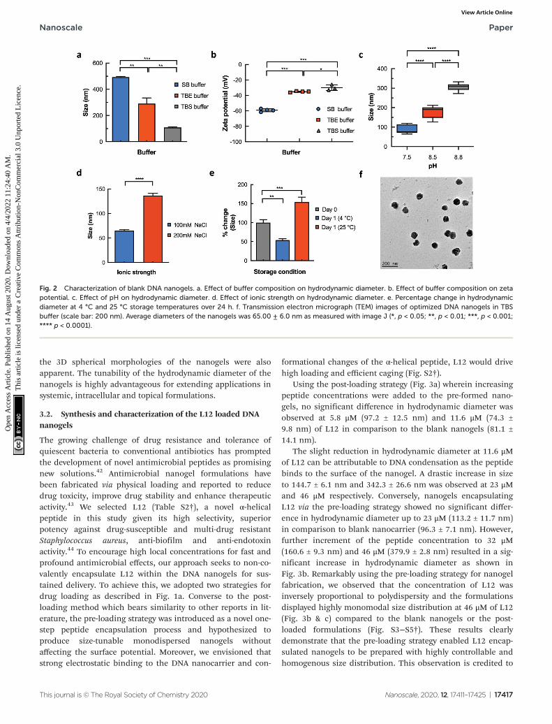
The impact of buffer identity and composition on thephysicochemical features of the nanogels was investigatedusing dynamic light scattering (DLS) and zeta potential experi-ments. Significant variation in hydrodynamic diameter andzeta potential was observed for nanogels prepared in the threedifferent buffer systems. For instance, DLS revealed largerhydrodynamic diameters (493.4 ± 10.2 nm) and a highly nega-tive surface potential (−59.2 ± 1.8 mV) for nanogels preparedin sodium borate (SB) buffer (Fig. 2a & b). Conversely, weobserved significantly smaller hydrodynamic diameters of 290± 68.5 nm (p < 0.01) and 108.8 ± 5.3 nm (p < 0.001) for nano-gels prepared in Tris/borate/EDTA (TBE) and Tris bufferedsaline (TBS) buffers respectively. This reduction in size corre-
Nanoscale Paper
This journal is © The Royal Society of Chemistry 2020 Nanoscale, 2020, 12, 17411–17425 | 17415
Ope
n A
cces
s A
rtic
le. P
ublis
hed
on 1
4 A
ugus
t 202
0. D
ownl
oade
d on
4/4
/202
2 11
:24:
40 A
M.
Thi
s ar
ticle
is li
cens
ed u
nder
a C
reat
ive
Com
mon
s A
ttrib
utio
n-N
onC
omm
erci
al 3
.0 U
npor
ted
Lic
ence
.View Article Online

sponded with the observed changes in surface potential to−35.1 ± 0.7 mV (TBE buffer; p < 0.001) and −29.9 ± 3.3 mV(TBS buffer; p < 0.001).
To identify the properties of the buffer systems that controlthe hydrodynamic diameter and net charge of the DNA nano-gels, DLS measurements were performed on nanogels pre-pared in TBS buffers of varying pH and ionic strength. Asshown in Fig. 2c, changing the pH of the buffer from 7.5 to 8.8steadily increased the hydrodynamic diameter of the nanogels.The average hydrodynamic diameter of the nanogels waslowest at pH 7.5 (86.98 ± 20.78 nm). We observed an increasein the measured size to 177.4 ± 29.6 nm at pH 8.5 and 305.9 ±19.8 nm at pH 8.8. The considerable size increase at higher pHis likely attributed to the increased ionic strength of the bufferwhich contributes to the growth of the nanogel.
To confirm this, the effect of ionic strength on the hydro-dynamic diameter was determined under different sodiumchloride concentrations. Increasing the salt concentration ofthe external buffer solution from 100 mM to 200 mM resultedin a significant increase in size from 64.7 ± 5.0 nm (100 mM)to 136.5 ± 10.0 nm (200 mM) as shown in Fig. 2d. This can beattributed to nanogel instability or charge screening due to
increasing counterions that mediates an effective electrostaticattraction between the DNA strands or nanogels.38–40
Subsequently, temperature effects were determined by measur-ing percentage changes in hydrodynamic diameter of theblank nanogels at storage temperatures of 4 °C and 25 °C over24 h. We found that at low temperatures (4 °C), the hydrodyn-amic diameter of the blank nanogels significantly reduced to53.8 ± 10.6% of its initial value (Fig. 2e) and remained stablefor 5 days (Fig. S1†). On the other hand, increasing the temp-erature of the buffer solution to 25 °C resulted in a significantpercentage increase in the hydrodynamic diameter (153.8 ±37.3%). These results demonstrate that the crosslinked blankDNA nanogels display reversible volume transition in responseto pH, ionic strength and temperature. These features justifytheir characterization as nanogels and are central for develop-ing nanocarriers with controlled loading and release character-istics.41 Accordingly, nanogels prepared in TBS buffer at pH7.5 and salt concentration of 100 mM were used for all sub-sequent experiments as the optimal conditions. TEM micro-graph images (Fig. 2f) of the blank nanogels prepared in TBSbuffers displayed average diameters of 65.00 ± 6.0 nm whichcorrelated well with DLS measurements (64.7 ± 5.0 nm) and
Fig. 1 Schematic illustration of L12 nanogels and L12 peptide, hybridization and morphology of blank nanogels. a. Synthesis of L12 nanogels via (i)post-loading and (ii) pre-loading strategies. b. Agarose gel electrophoresis to verify self-assembly of DNA nanostructures (lane 1 = YMA; lane 2 =YMB; lane 3 = DL) and blank nanogels (lane 4). c. Cryo-electron microscopy image of the blank DNA nanogels (scale bar: 100 nm). d. Schematicillustration of L12 antimicrobial peptide (sequence: LKKLLKKLLKKL).
Paper Nanoscale
17416 | Nanoscale, 2020, 12, 17411–17425 This journal is © The Royal Society of Chemistry 2020
Ope
n A
cces
s A
rtic
le. P
ublis
hed
on 1
4 A
ugus
t 202
0. D
ownl
oade
d on
4/4
/202
2 11
:24:
40 A
M.
Thi
s ar
ticle
is li
cens
ed u
nder
a C
reat
ive
Com
mon
s A
ttrib
utio
n-N
onC
omm
erci
al 3
.0 U
npor
ted
Lic
ence
.View Article Online
the 3D spherical morphologies of the nanogels were alsoapparent. The tunability of the hydrodynamic diameter of thenanogels is highly advantageous for extending applications insystemic, intracellular and topical formulations.
3.2. Synthesis and characterization of the L12 loaded DNAnanogels
The growing challenge of drug resistance and tolerance ofquiescent bacteria to conventional antibiotics has promptedthe development of novel antimicrobial peptides as promisingnew solutions.42 Antimicrobial nanogel formulations havebeen fabricated via physical loading and reported to reducedrug toxicity, improve drug stability and enhance therapeuticactivity.43 We selected L12 (Table S2†), a novel α-helicalpeptide in this study given its high selectivity, superiorpotency against drug-susceptible and multi-drug resistantStaphylococcus aureus, anti-biofilm and anti-endotoxinactivity.44 To encourage high local concentrations for fast andprofound antimicrobial effects, our approach seeks to non-co-valently encapsulate L12 within the DNA nanogels for sus-tained delivery. To achieve this, we adopted two strategies fordrug loading as described in Fig. 1a. Converse to the post-loading method which bears similarity to other reports in lit-erature, the pre-loading strategy was introduced as a novel one-step peptide encapsulation process and hypothesized toproduce size-tunable monodispersed nanogels withoutaffecting the surface potential. Moreover, we envisioned thatstrong electrostatic binding to the DNA nanocarrier and con-
formational changes of the α-helical peptide, L12 would drivehigh loading and efficient caging (Fig. S2†).
Using the post-loading strategy (Fig. 3a) wherein increasingpeptide concentrations were added to the pre-formed nano-gels, no significant difference in hydrodynamic diameter wasobserved at 5.8 μM (97.2 ± 12.5 nm) and 11.6 μM (74.3 ±9.8 nm) of L12 in comparison to the blank nanogels (81.1 ±14.1 nm).
The slight reduction in hydrodynamic diameter at 11.6 μMof L12 can be attributable to DNA condensation as the peptidebinds to the surface of the nanogel. A drastic increase in sizeto 144.7 ± 6.1 nm and 342.3 ± 26.6 nm was observed at 23 μMand 46 μM respectively. Conversely, nanogels encapsulatingL12 via the pre-loading strategy showed no significant differ-ence in hydrodynamic diameter up to 23 μM (113.2 ± 11.7 nm)in comparison to blank nanocarrier (96.3 ± 7.1 nm). However,further increment of the peptide concentration to 32 μM(160.6 ± 9.3 nm) and 46 μM (379.9 ± 2.8 nm) resulted in a sig-nificant increase in hydrodynamic diameter as shown inFig. 3b. Remarkably using the pre-loading strategy for nanogelfabrication, we observed that the concentration of L12 wasinversely proportional to polydispersity and the formulationsdisplayed highly monomodal size distribution at 46 μM of L12(Fig. 3b & c) compared to the blank nanogels or the post-loaded formulations (Fig. S3−S5†). These results clearlydemonstrate that the pre-loading strategy enabled L12 encap-sulated nanogels to be prepared with highly controllable andhomogenous size distribution. This observation is credited to
Fig. 2 Characterization of blank DNA nanogels. a. Effect of buffer composition on hydrodynamic diameter. b. Effect of buffer composition on zetapotential. c. Effect of pH on hydrodynamic diameter. d. Effect of ionic strength on hydrodynamic diameter. e. Percentage change in hydrodynamicdiameter at 4 °C and 25 °C storage temperatures over 24 h. f. Transmission electron micrograph (TEM) images of optimized DNA nanogels in TBSbuffer (scale bar: 200 nm). Average diameters of the nanogels was 65.00 ± 6.0 nm as measured with image J (*, p < 0.05; **, p < 0.01; ***, p < 0.001;**** p < 0.0001).
Nanoscale Paper
This journal is © The Royal Society of Chemistry 2020 Nanoscale, 2020, 12, 17411–17425 | 17417
Ope
n A
cces
s A
rtic
le. P
ublis
hed
on 1
4 A
ugus
t 202
0. D
ownl
oade
d on
4/4
/202
2 11
:24:
40 A
M.
Thi
s ar
ticle
is li
cens
ed u
nder
a C
reat
ive
Com
mon
s A
ttrib
utio
n-N
onC
omm
erci
al 3
.0 U
npor
ted
Lic
ence
.View Article Online

the highly cationic and hydrophobic nature of L12 which actsas a driving force for secure encapsulation within the nanogelcore. Subsequently, TEM images confirmed the spherical mor-phology of the peptide loaded nanogel formulations as shownin Fig. 3d.
Next, we investigated the surface potential of the pre-loadedand post-loaded L12 nanogel formulations. As shown in Fig. 3e,post-loading L12 resulted in a steady increase in zeta potentialwith a drastic change from the highly anionic surface charge of−36.9 ± 1.5 mV for the blank nanogel to +3.9 ± 0.1 mV and +5.6± 0.1 mV at L12 concentrations of 23 μM and 46 μM respectively.This observation is attributable to complete neutralization ofthe negative charge on the surface of the DNA nanogels as aresult of electrostatic interaction and binding of L12 and corres-ponds with DLS measurements. Conversely, we observed no sig-nificant difference in zeta potential using the pre-loadingmethod at 23 μM (−30.0 ± 0.8 mV) and 46 μM (−34.4 ± 0.9 mV)L12. These results clearly demonstrate that pre-loading L12does not influence the surface potential of the DNA nanogelsand serve as a prime indication of peptide caging in contrast tothe post-loading strategy wherein surface bound peptidesaccount for the highly positive zeta potential.
Having demonstrated the peptide-dependent variation innanogel size, we sought to illustrate flexibility of drug loading
by increasing monomer ratio of the nanostructures to drivehigher loading capacity (Fig. 3f). Doubling the monomer ratioof YMA : YMB : DL from 4 : 1 : 6.5 to 8 : 2 : 13, resulted in a sig-nificant reduction (p < 0.1) in hydrodynamic diameter from1182.8 ± 156.9 nm to 584.9 ± 85.8 nm for pre-loaded nanogelscontaining 92 μM of L12. Similarly, while a significant increasein hydrodynamic diameter at 4 : 1 : 6.5 monomer ratio (3337.3 ±669.8 nm) was seen at 184 μM L12, further increment of themonomer ratio to 8 : 2 : 13 and 16 : 4 : 26 subsequently resultedin drastic reduction to 2436.4 ± 774.3 nm and 789.8 ± 116.6 nmrespectively. This may be attributable to the increase in polya-nionic DNA monomers for L12 binding during nanogel for-mation. We confirmed this by showing that by increasing themonomer ratio of the nanostructures, a significant increase inthe number (kcount per s) of blank nanogels can be quantified(Fig. S6†). Taken together, these results demonstrate that, theloading capacity of the DNA nanogels can be expanded bytuning the monomer ratio of the nanostructures.
The thermal stability of the pre-loaded L12 nanogel formu-lations was examined over 7 days by monitoring the changesin hydrodynamic diameter below dehybridization conditions.We prepared pre-loaded L12 nanogels loaded with 20 μM L12in TBS buffer and stored the samples at 4 °C, 25 °C and 37 °C.As shown in Fig. 3g, we observed no apparent change in the
Fig. 3 Characterization of L12 nanogels. a. Effect of post-loading L12 peptide on hydrodynamic diameter of DNA nanogels. b. Effect of pre-loadedL12 peptide on hydrodynamic diameter of DNA nanogels. c. Size distribution of pre-loaded L12 nanogels. d. TEM images of L12 nanogels. Scale baris 200 nm. e. Effect of pre-loading and post-loading L12 peptide on zeta potential (**** p < 0.0001). f. Effect of increasing monomer ratio andpeptide concentration on hydrodynamic diameter (*, p < 0.05; **, p < 0.01; **** p < 0.0001) of the pre-loaded L12 nanogels. g. Effect of temperaturechanges on hydrodynamic diameter of pre-loaded L12 nanogels for 7 days.
Paper Nanoscale
17418 | Nanoscale, 2020, 12, 17411–17425 This journal is © The Royal Society of Chemistry 2020
Ope
n A
cces
s A
rtic
le. P
ublis
hed
on 1
4 A
ugus
t 202
0. D
ownl
oade
d on
4/4
/202
2 11
:24:
40 A
M.
Thi
s ar
ticle
is li
cens
ed u
nder
a C
reat
ive
Com
mon
s A
ttrib
utio
n-N
onC
omm
erci
al 3
.0 U
npor
ted
Lic
ence
.View Article Online

hydrodynamic diameter when the L12 nanogels were stored at4 °C for 7-days. At 25 °C, no difference in hydrodynamic dia-meter was shown after 24 h. However, on longer storage, alarge increase in the average hydrodynamic diameter of thenanogels by 62.2 ± 26.7% (day 3) and 148.1 ± 73.8% (day 7)was observed. It was found that, heating the L12 nanogels to37 °C under mild agitation resulted in rapid increase in theaverage hydrodynamic diameter by 87.1 ± 27.7% (day 1), 189.9± 86.2% (day 3) and 241.4 ± 32.4% (day 7). The observed dra-matic increase in hydrodynamic diameter may be attributableto the temperature responsiveness of the peptide loaded DNAnanogels and highlight their volume phase transition at phys-iological conditions which is hypothesized to drive peptiderelease. Moreover, these results also clearly demonstrate thatthe peptide loaded nanogels do not aggregate during storageat 4 °C and remained stable for at least 7-days.
Visualized TEM images of the formulations could not dis-tinguish between the encapsulated peptide and the DNA nano-gels while using negative stain. Thus, to detect the encapsu-lated peptide within the formulations, we performed confocal
microscopy using alexa-594 labelled DNA nanogels containingthe FITC labelled L12. As shown in Fig. 4a, spherical swollennetworks were observed in pre-loaded L12 nanogel formu-lations that were homogenously dispersed in the aqueousbuffer solution. Analysis of the superimposed images revealeda yellow core surrounded by a red corona which clearly con-firms FITC-labelled peptide caging within the nanogels(Fig. 4a). The post-loaded L12 nanogels however demonstratedattachment of L12 to the nanogel surface which was visualizedas yellow cores surrounded by green coronas and is in goodagreement with surface potential measurements (Fig. 4b). Toimprove the resolution of the nanogels and to visualize theentrapped L12 peptide within, super resolution structured illu-mination (SIM) confocal microscopy was used and showedthat the DNA nanogels displayed a well-organized, highlyporous internal structure when loaded with the unlabeled andlabelled L12 peptide (Fig. 4c & d). Within the matrix of the L12nanogels, we observed that the FITC labelled L12 peptide wasstrictly bound to the self-assembled DNA nanostructures,assumed its architecture and was evenly distributed within the
Fig. 4 Visualization of the preloaded L12 nanogels. DNA nanogels are labelled with Alexa-594 and L12 peptide is labelled withFITC. a. Conventional confocal microscopy of pre-loaded L12 nanogels. b. Conventional confocal microscopy of post-loaded L12nanogels. c. Structured illumination microscopy (SIM) images of DNA nanogels containing unlabeled (UL) antimicrobial L12 peptide. DNA nanogelsare labelled with Alexa-594 and L12 peptide is unlabeled. d. SIM confocal image of the L12 nanogels. Individual frames of Z-stacks (step size:0.125 μm) are shown in the right panel to demonstrate distribution of L12 within the nanogels. DNA nanogels are labelled with Alexa-594 and L12peptide is labelled with FITC.
Nanoscale Paper
This journal is © The Royal Society of Chemistry 2020 Nanoscale, 2020, 12, 17411–17425 | 17419
Ope
n A
cces
s A
rtic
le. P
ublis
hed
on 1
4 A
ugus
t 202
0. D
ownl
oade
d on
4/4
/202
2 11
:24:
40 A
M.
Thi
s ar
ticle
is li
cens
ed u
nder
a C
reat
ive
Com
mon
s A
ttrib
utio
n-N
onC
omm
erci
al 3
.0 U
npor
ted
Lic
ence
.View Article Online

Alexa-594 labeled DNA nanogels as shown in Fig. 4d. Converseto the post-loaded formulation, the pre-loaded L12 nanogelsdisplayed homogeneity, stability of the surface potential withincreasing concentration of L12 and capacity to entrap thepeptide fully into its core. Thus, we therefore chose the pre-loaded formulation for further in vitro and in vivo evaluations.
Finally, quantification of the encapsulation efficiency of thepre-loaded L12 nanogels revealed high encapsulation efficien-cies of 92.1 ± 0.4% as determined by fluorescence spec-troscopy. These results are comparable to the reported encap-sulation efficiency of other antimicrobial peptides in naturaland synthetic nanocarriers.45–47 Taken together, the observedhomogenous distribution and strict association of the peptidewithin the pre-loaded nanogel matrix in parallel with the highencapsulation efficiency corroborates our hypothesis that theencapsulation of L12 is facilitated by non-covalent electrostaticinteractions. While there is concern that there is limited roomto increasing the loading of cationic cargos without distortingthe nanostructures, we have demonstrated in this present workthat by simply optimizing the monomer concentration, we canpotentially overcome this issue (Fig. 3f). Indeed, the cost ofDNA in comparison to other biopolymers e.g. natural polymersis one of the barriers to clinical translation. Nevertheless, com-pared to bulk gel systems (e.g. hydrogel), the lower amount (%w/w) of DNA required for fabricating nanogels may translate tooverall lower cost. Of interest, a recent report indicates that an
estimated cost of less than €10 per dose of DNA nano-structured formulations is perceivable without incorporatingcosts associated with sterilization, purity and batch-to-batchconsistency.48 Ultimately, large scale synthesis (multi kg range)is expected to reduce overall cost of production, drive fairlymoderate competitive market prices that are comparable toother approved formulations (e.g. monoclonal antibodies).This would render the development of nucleic acid systemspromising for future therapeutics.48
3.3. Antibacterial activity, in vitro drug release and cellulartoxicity of DNA nanogels
We have previously reported L12 as a highly potent syntheticpeptide against Gram positive and Gram negative pathogens.44
Having demonstrated high encapsulation efficiency and hom-ogenous distribution of L12 within the DNA nanogels, time-kill kinetic studies were performed to monitor the time-courseof the antimicrobial effect of the L12 nanogels againstS. aureus. Rapid release of L12 is essential to ensure that lethalantibacterial concentrations are available to avert undesirablecomplications attributable to the circulation of bacterial endo-toxins or exotoxins.49,50 The pre-loaded L12 nanogels displayedrapid onset of action, reducing bacterial inoculum by 89.2%(0.96 log reduction) at 4× MIC within 2 h of treatment(Fig. 5a). After 4 h, 2.27 log, 2.65 log and 2.87 log reduction ofS. aureus colonies was attained for cells treated with 1×, 2× and
Fig. 5 In vitro antimicrobial activity, drug release and cytotoxicity evaluations. a. Time kill kinetics of pre-loaded L12 nanogels againstStaphylococcus aureus (106 CFU mL−1). b. In vitro release of pre-loaded L12 nanogels. c. Toxicity of pre-loaded L12 nanogels against HDF cells over24 h (*, p < 0.05; ****, p < 0.0001). d. Toxicity of blank nanogels against HDF and HaCaT cells over 72 h treated with YMA/YMB/DL monomer ratiosof 1× (4/1/6.5), 2× (8/2/13), 4× (16/4/26) and 8× (32/8/52) to prepare the DNA nanogel.
Paper Nanoscale
17420 | Nanoscale, 2020, 12, 17411–17425 This journal is © The Royal Society of Chemistry 2020
Ope
n A
cces
s A
rtic
le. P
ublis
hed
on 1
4 A
ugus
t 202
0. D
ownl
oade
d on
4/4
/202
2 11
:24:
40 A
M.
Thi
s ar
ticle
is li
cens
ed u
nder
a C
reat
ive
Com
mon
s A
ttrib
utio
n-N
onC
omm
erci
al 3
.0 U
npor
ted
Lic
ence
.View Article Online

4× MIC of the L12 nanogel formulations. A strong and pro-longed antibacterial activity was observed over 24 h for cellstreated with 2× (6.7 log reduction) and 4× (6.7 log reduction)MIC concentrations of the L12 nanogels.
Notably, the empty nanogel control group had no antibac-terial effect on S. aureus in vitro leading to the conclusion thatL12 is rapidly released in vitro to exert an antibacterial effectdespite being securely loaded and bound to the nanogel core.Similar efficacy was observed against P. aeruginosa (Fig. S10†).Thereafter, in vitro release of L12 from the pre-loaded DNAnanogels incubated at 37 °C was monitored over 24 h by centri-fuging aliquots of the formulations at pre-determined intervalsand measuring the concentration of FITC-labelled L12 in thesupernatant. As shown in Fig. 5b, 45.59% of L12 was releasedwithin 2 h which is in good agreement with the rapid onset ofantibacterial activity. By 24 h, 88.13% of L12 was released fromthe L12 nanogels. The initial rapid release of L12 from thenanogel system is likely due to simple diffusion of the cationiccargo from the nanostructures.51,52 Conversely, the sustainedrelease profile between 2 h and 24 h can potentially be attribu-ted to swelling of the nanogels under elevated temperatures asshown in Fig. 3f. The release profile of the L12 nanogels corre-lated well with the time-kill kinetics and the observed anti-microbial efficacy in vitro.
Nanocarriers that prolong the release of encapsulated drugshave the advantage of maintaining peptide concentrationswithin the therapeutic window over an extended period, mini-mizing underexposure and reducing drug toxicity. Thus, wequantified the ability of the pre-loaded L12 nanogels toimprove toxicity of L12 over 24 h. Against HDF cells, weobserved a 32 ± 1.2% increase in the viability (p < 0.01) of cellstreated with L12 nanogels at 46 μM compared to the peptidecontrols (Fig. 5c). A 20.60 ± 4.4% and 22.3 ± 5.7% increase incell viability (p < 0.01) was observed for HaCaT cells treatedwith 23 μM and 46 μM of L12 nanogels respectively comparedto the peptide controls (Fig. S11†). These results are caused bythe sustained release of L12 from the nanogels and not associ-ated with the toxicity of the DNA nanogel itself. To demon-strate the biocompatibility of the blank DNA nanogels, cell pro-liferation studies were performed. HDF and HaCaT cells weretreated with DNA nanogels prepared using four differentmonomer ratios up to 8-fold increase. As shown in Fig. 5d,blank DNA nanogels and their degradation products did notinduce cell death after 72 h and were biocompatible withmammalian cells. Overall, these results underscore the advan-tage of using DNA-based materials as nanocarriers for drugdelivery and clearly highlight the ability of the DNA nanogelsto reduce the toxicity of encapsulated peptides.
3.4. In vivo testing of the L12 nanogels in a corneal infectionwound model
Corneal infections caused by S. aureus are the leading causeof blinding microbial keratitis.53 Recent documented cases ofkeratitis caused by MRSA have shown resistance against firstline antimicrobials such as fluoroquinolones, highlightingthe need for new therapies.54,55 Moreover, the promising
in vitro activity of the formulation warrants further in vivostudies. Thus, using a mouse model of bacterial keratitis, wesought to test whether the nanogels could be used as anti-microbial delivery systems by assessing the efficacy of the L12nanogels in vivo. Superficial wounds created by carefullyremoving the corneal epithelium were inoculated withS. aureus to create the corneal infection wound model.Following clinical diagnosis of ocular infection, initial treat-ment usually involves intensive topical antibiotics,56 thus wecompared intensive ophthalmic application of a clinicallyapproved formulation (0.3% gatifloxacin) with our pre-loadedL12 nanogels and monitored the progression of the infectionusing Slit-lamp (SL) biomicroscopy and anterior-segmentoptical coherence tomography (AS-OCT). The mice weretreated with gatifloxacin (0.3% w/v), L12 nanogels (0.15% w/v)or L12 nanogels (0.3% w/v) for 48 h (Fig. 6a). After 48 h, micewere sacrificed and the infected eyes were enucleated for bac-terial quantification.
Slit lamp (SL) examination of S. aureus infected corneatreated with various groups displayed different degrees ofcorneal opacity (Fig. 6b).57 SL images of infected corneatreated with nanogel indicated dense opacities on cornealsurface over pupil. 0.3% L12 nanogel treated cornea, however,contained readily detectable opacity that was lower than 0.15%L12 nanogel. Infected cornea treated with gatifloxacinappeared clear and normal and superior clinical presentationamong all the groups, confirming potency of the antibiotics inresolving S. aureus infections. Consistent with the SL examin-ation, AS-OCT images indicated the presence of hyper-reflec-tive materials and edematous cornea in the nanogel treatedgroups.
The baseline corneal thickness of mice eye was ranged from80–98 μm (mean thickness 92.0 ± 5.8 μm). Within 24 h ofS. aureus wound infection, the mean corneal thickness in thecontrol group increased to 153.6 ± 21.8 μm (Fig. 6c), suggestingexaggerated inflammatory response. Conversely, a significantreduction in the mean corneal thickness was observed for the0.15% pre-loaded L12 nanogel (105.6 ± 39.0 μm) and 0.3% pre-loaded L12 nanogel (104.0 ± 28.0 μm) treated mice over 24 h.However, no significant difference in mean corneal thicknesswas observed for the 0.3% gatifloxacin (127.9 ± 22.6 μm)infected eyes. The significant decrease in corneal thickness forthe L12 nanogel treated group at 24 h highlight fast resolutionof infection-induced inflammation which is potentially attribu-table to the anti-inflammatory action of the L12 peptide andthe DNA nanostructures as previously reported.26,44,58 At 48 hpost-inoculation the mean corneal thickness continued toincrease for the control group (194.4 ± 27.4 μm) which indi-cates increased severity of the infection.59 An even moreintense reduction in the average corneal thickness was seen inthe 0.15% pre-loaded L12 nanogel (112.7 ± 29.6 μm), 0.3% pre-loaded L12 nanogel (117.2 ± 8.7 μm) and 0.3% gatifloxacin(124.3 ± 18.7 μm) treated groups after 48 h.
A severe clinical course of ocular infection in the controlgroup was observed along with increased bacterial bioburdenin infected mice treated with the blank nanogels.
Nanoscale Paper
This journal is © The Royal Society of Chemistry 2020 Nanoscale, 2020, 12, 17411–17425 | 17421
Ope
n A
cces
s A
rtic
le. P
ublis
hed
on 1
4 A
ugus
t 202
0. D
ownl
oade
d on
4/4
/202
2 11
:24:
40 A
M.
Thi
s ar
ticle
is li
cens
ed u
nder
a C
reat
ive
Com
mon
s A
ttrib
utio
n-N
onC
omm
erci
al 3
.0 U
npor
ted
Lic
ence
.View Article Online

Microbiological determination of bacterial viability indicateda significant reduction in bacterial bioburden for micetreated with 0.15% pre-loaded L12 nanogel (p < 0.01) (Fig. 6d)at 48 h. However, a more substantial decrease in the numberof viable bacterial cells was recovered from corneal woundstreated with 0.3% pre-loaded L12 nanogels (p < 0.0001) and0.3% gatifloxacin (p < 0.0001). The slightly better efficacy ofgatifloxacin than 0.3% pre-loaded L12 nanogels could beattributed to the higher molar concentrations of the fluoro-
quinolone drug. Alongside the in vitro antimicrobial activityof the formulation, these results establish the excellent anti-microbial properties of pre-loaded L12 nanogels againstS. aureus keratitis. Notably, the topical application of the for-mulations over 48 h demonstrated excellent ocular toleranceand no significant changes in mice weight or behavior wasobserved. These results corroborate the in vitro biocompat-ibility of the nanogels and are in good agreement with otherreports on DNA nanocarriers.
Fig. 6 Analysis of in vivo antimicrobial efficacy in murine corneal wounds infected with S. aureus. a. Schematic representation of experimentaldesign for the bacterial keratitis wound model of S. aureus and timescale of topical treatment. A total of 14 doses of pre-loaded L12 nanogels andgatifloxacin was administered over 48 h and the bacterial bioburden from the infected eyes post-treatment was compared to the blank nanogelcontrol (n = 5–7 infected eyes from 5–7 mice per group was used). b. Slit lamp images of the murine corneal wounds after treatment with the blanknanogels, 0.15% pre-loaded L12 nanogels, 0.3% pre-loaded L12 nanogels and 0.3% gatifloxacin formulations. c. Time dependent changes in cornealthickness measurement at 24 h and 48 h post-inoculation by AS-OCT for mice treated with the blank nanogels, 0.15% pre-loaded L12 nanogels,0.3% pre-loaded L12 nanogels and 0.3% gatifloxacin formulations. Results are reported as the means ± standard deviations for 5–7 corneas pergroup. d. Quantitative S. aureus burden in infected corneas following treatment with blank nanogel, pre-loaded L12 nanogels (0.15% and 0.3% L12)and 0.3% gatifloxacin (Zymar) at 48 h. Results are reported as the means ± standard deviations for 5–7 corneas per group (ns, no statistical signifi-cance; *, p < 0.05; **p < 0.01; ***, p < 0.001; **** p < 0.0001).
Paper Nanoscale
17422 | Nanoscale, 2020, 12, 17411–17425 This journal is © The Royal Society of Chemistry 2020
Ope
n A
cces
s A
rtic
le. P
ublis
hed
on 1
4 A
ugus
t 202
0. D
ownl
oade
d on
4/4
/202
2 11
:24:
40 A
M.
Thi
s ar
ticle
is li
cens
ed u
nder
a C
reat
ive
Com
mon
s A
ttrib
utio
n-N
onC
omm
erci
al 3
.0 U
npor
ted
Lic
ence
.View Article Online

4. Conclusion
Our work highlights a facile strategy to cage antimicrobial pep-tides within DNA nanocarriers in a tunable manner withoutloss of biological activity or structural integrity for local drugdelivery applications. We demonstrate that the sustainedrelease of encapsulated peptides can be translated into signifi-cant in vitro and in vivo antimicrobial efficacy against S. aureus.The progressive decrease in corneal thickness and quantitativeanalysis of bacterial viability demonstrate that the topicalapplication of L12 nanogels can reduce the clinical symptomsand bacterial bioburden in S. aureus models of infectious bac-terial keratitis. Considering that S. aureus remains the leadingcause of blinding bacterial keratitis, the observed fast resolu-tion of infection-induced inflammation and comparableefficacy of topical 0.3% L12 nanogels to gatifloxacin is clini-cally advantageous. Moreover, these nanocarriers are biocom-patible against mammalian cells, significantly improve toxicityof encapsulated peptides and possessed excellent ocular toler-ance in vivo. For future applications, targeted delivery againstpathogenic bacteria and transport across ocular barriersshould be explored.
Author contributions
S.O. and P.L.R.E. conceived the project; S.O., V.M., R.L. and P.L.R.E. designed the experiments; S.O., V.M., N.A.M.N. and B.L.J. performed experiments and collated the experimentalresults; S.O. wrote the manuscript with editing and sugges-tions from V.M., N.A.M.N., R.L. and P.L.R.E. Schematic dia-grams were prepared by N.A.M.N and S.O. All authorsapproved the final version of the manuscript.
Conflicts of interest
The authors declare that no competing interest exists.
Acknowledgements
The authors would like to acknowledge research funding andfacilities provided by the National University of Singapore andMinistry of Education Academic Research Fund(R148000240114) awarded to P. L. R. Ee and the SingaporeInternational Graduate Award (SINGA) to S. O. R. L. would liketo thank funding support from the Singapore Ministry ofHealth’s National Medical Research Council under its CentreGrant Programme – Optimization of Core PlatformTechnologies for Ocular Research (INCEPTOR)-NMRC/CG/M010/2017_SERI and the SingHealth Foundation (SHF/FG663P/2017). We would like to thank the A*STAR MicroscopyPlatform for technical guidance in the SIM studies. Art workacknowledgement to N. A. M. N and S. O.
References
1 R. M. Putri, J. J. Cornelissen and M. S. Koay, Self-assembledcage-like protein structures, ChemPhysChem, 2005, 16, 911–918.
2 N. P. King and Y. T. Lai, Practical approaches to designingnovel protein assemblies, Curr. Opin. Struct. Biol., 2013, 23,632–638.
3 S. Hurtley, Spatial cell biology. Location, location, location.Introduction, Science, 2009, 326, 1205.
4 A. H. Chen and P. A. Silver, Designing biological compart-mentalization, Trends Cell Biol., 2012, 22, 662–670.
5 S. Deshayes and R. Gref, Synthetic and bioinspired cagenanoparticles for drug delivery, Nanomedicine, 2014, 9,1545–1564.
6 M. Uchida, et al., Biological containers: Protein cages asmultifunctional nanoplatforms, Adv. Mater., 2007, 19,1025–1042.
7 J. G. Heddle, S. Chakraborti and K. Iwasaki, Natural andartificial protein cages: design, structure and therapeuticapplications, Curr. Opin. Struct. Biol., 2017, 43, 148–155.
8 M. Rother, M. G. Nussbaumer, K. Renggli and N. Bruns,Protein cages and synthetic polymers: a fruitful symbiosisfor drug delivery applications, bionanotechnology andmaterials science, Chem. Soc. Rev., 2016, 45, 6213–6249.
9 D. Jiang, C. G. England and W. Cai, DNA nanomaterials forpreclinical imaging and drug delivery, J. Controlled Release,2016, 239, 27–38.
10 Y. Jin, Z. Li, et al., Biodegradable, multifunctionalDNAzyme nanoflowers for enhanced cancer therapy, NPGAsia Mater., 2017, 9, 1–10.
11 Q. Luo, C. Hou, Y. Bai, R. Wang and J. Liu, ProteinAssembly: Versatile Approaches to Construct Highly OrderedNanostructures, Chem. Rev., 2016, 116, 13571–13632.
12 J. F. Ross, et al., Decorating Self-Assembled Peptide Cageswith Proteins, ACS Nano, 2017, 11, 7901–7914.
13 W. M. Aumiller, M. Uchida and T. Douglas, Protein cageassembly across multiple length scales, Chem. Soc. Rev.,2018, 47, 3433–3469.
14 E. Sasaki, et al., Structure and assembly of scalable porousprotein cages, Nat. Commun., 2017, 8, 14663.
15 C. R. Kaiser, et al., Biodistribution studies of protein cagenanoparticles demonstrate broad tissue distribution andrapid clearance in vivo, Int. J. Nanomed., 2007, 2, 715–733.
16 R. Chhabra, J. Sharma, Y. Liu, S. Rinker and H. Yan, DNAself-assembly for nanomedicine, Adv. Drug Delivery Rev.,2010, 62, 617–625.
17 C. Angell, S. Xie, L. Zhang and Y. Chen, DNANanotechnology for Precise Control over Drug Delivery andGene Therapy, Small, 2016, 12, 1117–1132.
18 Y. Zhang, H. F. Chan and K. W. Leong, Advanced materialsand processing for drug delivery: the past and the future,Adv. Drug Delivery Rev., 2013, 65, 104–120.
19 S. H. Um, J. B. Lee, N. Park, S. Y. Kwon, C. C. Umbach andD. Luo, Enzyme-catalused assembly of DNA hydrogel, Nat.Mater., 2006, 5, 104–120.
Nanoscale Paper
This journal is © The Royal Society of Chemistry 2020 Nanoscale, 2020, 12, 17411–17425 | 17423
Ope
n A
cces
s A
rtic
le. P
ublis
hed
on 1
4 A
ugus
t 202
0. D
ownl
oade
d on
4/4
/202
2 11
:24:
40 A
M.
Thi
s ar
ticle
is li
cens
ed u
nder
a C
reat
ive
Com
mon
s A
ttrib
utio
n-N
onC
omm
erci
al 3
.0 U
npor
ted
Lic
ence
.View Article Online

20 G. D. Hamblin, K. M. Carneiro, J. F. Fakhoury, K. E. Bujoldand H. F. Sleiman, Rolling circle amplification-templatedDNA nanotubes show increased stability and cellpenetration ability, J. Am. Chem. Soc., 2012, 134, 2888–2891.
21 J. W. M. Conway, C. K. McLaughlin, K. J. Castor andH. Sleiman, DNA nanostructure serum stability: Greaterthan the sum of its parts, Chem. Commun., 2013, 49, 1172–1174.
22 J. W. Keum and H. Bermudez, Enhanced resistance of DNAnanostructures to enzymatic digestion, Chem. Commun.,2009, 7036–7038.
23 R. P. Goodman, R. M. Berry and A. J. Turberfield, Thesingle-step synthesis of a DNA tetrahedron, Chem.Commun., 2004, 12, 1372–1373.
24 R. P. Goodman, et al., Rapid chiral assembly of rigid DNAbuilding blocks for molecular nanofabrication, Science,2005, 310, 1661–1665.
25 A. S. Walsh, H. Yin, C. M. Erben, M. J. Wood andA. J. Turberfield, DNA cage delivery to mammalian cells,ACS Nano, 2011, 5, 5427–5432.
26 S. Obuobi, H. K. Tay, N. D. T. Tram, V. Selvarajan,J. S. K. Khara, Y. Wang and P. L. R. Ee, Facile and Efficientencapsulation of antimicrobial peptides via crosslinkedDNA nanostructures and their application in woundtherapy, J. Controlled Release, 2019, 313, 120–130.
27 C. M. Erben, R. P. Goodman and A. J. Turberfield, Single-molecule protein encapsulation in a rigid DNA cage,Angew. Chem., Int. Ed., 2006, 45, 7414–7417.
28 R. Crawford, et al., Non-covalent single transcription factorencapsulation inside a DNA cage, Angew. Chem., Int. Ed.,2013, 52, 2284–2288.
29 Z. Zhao, et al., Nanocaged enzymes with enhanced catalyticactivity and increased stability against protease digestion,Nat. Commun., 2016, 7, 10619.
30 V. Kumar, et al., DNA Nanotechnology for Cancer Therapy,Theranostics, 2016, 6, 710–725.
31 H. V. P. Thelu, et al., Size controllable DNA nanogels fromthe self-assembly of DNA nanostructures through multi-valent host-guest interactions, Nanoscale, 2018, 10, 222–230.
32 A. Sprengel, et al., Tailored protein encapsulation into aDNA host using geometrically organized supramolecularinteractions, Nat. Commun., 2017, 8, 14472.
33 E. Kim, et al., One-Pot Synthesis of Multiple Protein-Encapsulated DNA Flowers and Their Application inIntracellular Protein Delivery, Adv. Mater., 2017, 29,1701086.
34 J. Willem de Vries, et al., DNA nanoparticles for ophthalmicdrug delivery, Biomaterials, 2018, 157, 98–106.
35 J. Li, et al., Self-assembly of DNA nanohydrogels with con-trollable size and stimuli-responsive property for targetedgene regulation therapy, J. Am. Chem. Soc., 2015, 137, 1412–1415.
36 A. M. Rush, M. P. Thompson, E. T. Tatro andN. C. Gianneschi, Nuclease-resistant DNA via high-density
packing in polymeric micellar nanoparticle coronas, ACSNano, 2013, 7, 1379–1387.
37 Y. Ma, H. Liu, Q. Mou, D. Yan, X. Zhu and C. Zhang,Floxuridine-containing nucleic acid nanogels for anti-cancer drug delivery, Nanoscale, 2018, 10, 8367–8371.
38 Y. Burak, G. Ariel and D. Andelman, Onset of DNA aggrega-tion in presence of monovalent and multivalent counter-ions, Biophys. J., 2003, 85, 2100–2110.
39 G. C. L. Wong and L. Pollack, Electrostatics of StronglyCharged Biological Polymers: Ion-Mediated Interactionsand Self-Organization in Nucleic Acids and Proteins, Annu.Rev. Phys. Chem., 2010, 61, 171–189.
40 S. Kewalramani, G. I. Guerrero-Garcia, L. M. Moreau,J. W. Zwanikken, C. A. Mirkin, M. O. de la Cruz andM. J. Bedzyk, Electrolyte-Mediated Assembly of ChargedNanoparticles, ACS Cent. Sci., 2016, 2, 219–224.
41 S. Bontha, A. V. Kabanov and T. K. Bronich,Polymer micelles with cross-linked ionic cores for deliveryof anticancer drugs, J. Controlled Release, 2006, 114, 163–174.
42 A. G. McArthur, et al., The comprehensive antibiotic resis-tance database, Antimicrob. Agents Chemother., 2013, 57,3348–3357.
43 J. P. Silva, Delivery of LLKKK18 loaded into self-assemblinghyaluronic acid nanogel for tuberculosis treatment,J. Controlled Release, 2016, 235, 112–124.
44 J. S. Khara, et al., Disruption of drug-resistant biofilmsusing de novo designed short alpha-helical antimicrobialpeptides with idealized facial amphiphilicity, ActaBiomater., 2017, 57, 103–114.
45 M. Fumakia and E. A. Ho, Nanoparticles Encapsulated withLL37 and Serpin A1 Promotes Wound Healing andSynergistically Enhances Antibacterial Activity, Mol.Pharm., 2016, 13, 2318–2331.
46 A. M. Piras, Chitosan nanoparticles loaded with the anti-microbial peptide temporin B exert a long-term antibacter-ial activity in vitro against clinical isolates ofStaphylococcus epidermidis, Front. Microbiol., 2015, 6, 372.
47 K. K. Chereddy, et al., PLGA nanoparticles loaded with hostdefense peptide LL37 promote wound healing, J. ControlledRelease, 2014, 194, 138–147.
48 A. Keller and V. Linko, Challenges and Perspectives of DNANanostructures in Biomedicine, Angew. Chem., Int. Ed.,2020, 59, 2–18.
49 G. A. Pankey and L. D. Sabath, Clinical relevance of bacter-iostatic versus bactericidal mechanisms of action in thetreatment of Gram-positive bacterial infections, Clin. Infect.Dis., 2004, 38, 864–870.
50 A. Ebbensgaard, et al., Comparative Evaluation of theAntimicrobial Activity of Different Antimicrobial Peptidesagainst a Range of Pathogenic Bacteria, PLoS One, 2015, 10,e0144611.
51 G. Zhu, et al., Self-assembled, aptamer-tethered DNA nano-trains for targeted transport of molecular drugs in cancertheranostics, Proc. Natl. Acad. Sci. U. S. A., 2013, 110, 7998–8003.
Paper Nanoscale
17424 | Nanoscale, 2020, 12, 17411–17425 This journal is © The Royal Society of Chemistry 2020
Ope
n A
cces
s A
rtic
le. P
ublis
hed
on 1
4 A
ugus
t 202
0. D
ownl
oade
d on
4/4
/202
2 11
:24:
40 A
M.
Thi
s ar
ticle
is li
cens
ed u
nder
a C
reat
ive
Com
mon
s A
ttrib
utio
n-N
onC
omm
erci
al 3
.0 U
npor
ted
Lic
ence
.View Article Online

52 D. Mathur and I. L. Medintz, The Growing Development ofDNA Nanostructures for Potential Healthcare-RelatedApplications, Adv. Healthcare Mater., 2019, 8, e1801546.
53 R. J. O’Callaghan, The Pathogenesis of Staphylococcusaureus Eye Infections, Pathogens, 2018, 7, 1–22.
54 A. Lichtinger, et al., Shifting trends in bacterial keratitis inToronto: an 11-year review, Ophthalmology, 2012, 119,1785–1790.
55 N. Ni, et al., Seasonal, geographic, and antimicrobial resis-tance patterns in microbial keratitis: 4-year experience ineastern Pennsylvania, Cornea, Cornea, 2015, 34, 294–302.
56 X. W. Tan, et al., Effectiveness of antimicrobial peptideimmobilization for preventing perioperative cornea
implant-associated bacterial infection, Antimicrob. AgentsChemother., 2014, 58, 5229–5238.
57 L. D. Hazlett, M. M. Moon, M. Strejc and R. S. Berk,Evidence for N-acetylmannosamine as an ocular receptorfor P. aeruginosa adherence to scarified cornea, Invest.Ophthalmol. Visual Sci., 1987, 28, 1978–1985.
58 Q. Zhang, et al., Anti-inflammatory and AntioxidativeEffects of Tetrahedral DNA Nanostructures via theModulation of Macrophage Responses, ACS Appl. Mater.Interfaces, 2018, 10, 3421–3430.
59 M. Venkatesh, et al., Antimicrobial Activity and CellSelectivity of Synthetic and Biosynthetic Cationic Polymers,Antimicrob. Agents Chemother., 2017, 61, e00469–e00417.
Nanoscale Paper
This journal is © The Royal Society of Chemistry 2020 Nanoscale, 2020, 12, 17411–17425 | 17425
Ope
n A
cces
s A
rtic
le. P
ublis
hed
on 1
4 A
ugus
t 202
0. D
ownl
oade
d on
4/4
/202
2 11
:24:
40 A
M.
Thi
s ar
ticle
is li
cens
ed u
nder
a C
reat
ive
Com
mon
s A
ttrib
utio
n-N
onC
omm
erci
al 3
.0 U
npor
ted
Lic
ence
.View Article Online
![Calix[8]arene Functionalized Polyglycerol Nanogels for ...8]arene... · Calix[8]arene Functionalized Polyglycerol Nanogels for Encapsulation and Stabilization of Fluorescent Dyes](https://static.fdocuments.in/doc/165x107/5afee8f27f8b9a444f8f7955/calix8arene-functionalized-polyglycerol-nanogels-for-8arenecalix8arene.jpg)