Nucleation of Biomimetic Hydroxyapatite · new materials. Biomimetic method was found suitable for...
9
7 Serbian Dental Journal, vol. 58, N o 1, 2011 ORIGINAL ARTICLE / ORIGINALNI RAD UDC: 616.314.16-089.27:616.311.2-002-089 DOI: 10.2298/SGS1101007C Nucleation of Biomimetic Hydroxyapatite Božana Čolović 1 , Dejan Marković 2 , Vukoman Jokanović 1 1 Laboratory for Radiation Chemistry and Physics, Institute of Nuclear Sciences “Vinča”, Belgrade, Serbia; 2 Clinic for Pediatric and Preventive Dentistry, School of Dentistry, University of Belgrade, Belgrade, Serbia SUMMARY Introduction The aim of the study was to assess the formation of biomimetic calcium hydroxyapatite (HAP) on the surface of different substrates. Material and Methods Silica coated stainless steel tapes and thin polymer films (alginate, cellulose, poly lactide-co- glycolide – PLGA) deposited on hydroxyapatite scaffold were used as substrate. Supersaturated simulated body fluid (SBF) and SBF combined with Fetal Calf Serum (FCS) or Eagle’s Minimum Essential Medium (EMEM) were used as bioactive liquid medium where biomimetic nucleation of HAP occurred. Infrared spectroscopy with Fourier trans- formation was used to analyze the formed phases, while scanning electron microscopy indicated the morphology of nucleated phase. Results The results of measuring the mass with volume adjustments done by the BET method showed that the thick- ness of the film of nucleated calcium hydroxyapatite depended on the time that samples spent soaked in SBF-in as well as the type of selected biomimetic medium. Conclusion Biomimetic calcium hydroxyapatite is possible to produce by self nucleation on different substrates in the presence of simulating body fluid. Keywords: biomimetic method; bioactive thin films; biomimetic hydroxyapatite; nanostructure design INTRODUCTION Nature has created materials with excellent functional characteristics, and science has focused on developing new methods of synthesis, which are able to mimic natu- ral processes. The aim is not only to imitate the biological systems, but to use ideas inspired by them, to synthesize new materials. Biomimetic method was found suitable for synthesis modern materials of complex forms and hierar- chical organization structure, adequate for use in biolog- ical systems [1, 2]. Biomimetic method can be used to form bioactive coat- ings on metallic implants that allow formation of appro- priate bonds with surrounding bone [3-6]. Using biomi- metic method, it is possible to produce apatite with similar structural and morphological characteristics as natural bone on the surface of implant. Since this method uses solutions with similar ionic composition as blood plasma, the conditions for hydroxyapatite (HAP) nucleation are similar to the natural [7-11]. The formation of HAP on the surface of metal can be accelerated by previous coating with thin layer of oxide (SiO 2 , TiO 2 , with built OH groups within the oxide layer). Built OH groups serve as activa- tors of heterogeneous apatite nucleation during deposi- tion of calcium and phosphate ions from oversaturated simulated body fluid (SBF) [12, 13]. The aim of this study was to investigate the mecha- nism of HAP nucleation on the stainless steel tapes coated with a layer of SiO 2 , using SBF and SBF in combination with Fetal Calf Serum (FCS) or Eagle’s Minimum Essential Medium (EMEM) with positive effect on the kinetics of HAP nucleation due to the presence of amino groups. An additional objective was to examine the application of biomimetic methods in designing porous apatite carri- ers. Adequate nanotopology of interior walls of porous carriers is essential for normal cell growth, and it could be achieved by biomimetic treatment of these carriers in SBF, previously synthesized by any method. Polymers (PLGA, cellulose and alginate) were used for the function- alization of the walls of porous carrier in order to achieve a specific nanodesign during nucleation of HAP on their surface [14]. MATERIALS AND METHODS The experimental part of the research included the prepara- tion of simulating body fluid, preparation of the substrate, forming biomimetic HAP and characterization of nucle- ated HAP. Preparation of SBF SBF was prepared using slightly different instruction, in comparison to the original SBF, where the concentrations of individual ions were: c(Cl - )=54 mmol/dm 3 ; c(Na + )=54.2 mmol/dm 3 ; c(Ca 2+ )=2.5 mmol/dm 3 ; c(PO 4 3- )=1 mmol/ dm 3 ; c(Mg 2+ )=0.3 mmol/dm 3 ; c(NO 3 - )=0.6 mmol/dm 3 and c(K + )=1,4 mmol/dm 3 [10]. Concentration of PO 4 3- and Address for correspondence: Vukoman JOKANOVIĆ, Institute for Nuclear Sciences “Vinča”, Mike Petrovića Alasa 12-14, 11001 Belgrade, Serbia; [email protected]
Transcript of Nucleation of Biomimetic Hydroxyapatite · new materials. Biomimetic method was found suitable for...

7Serbian Dental Journal, vol. 58, No 1, 2011ORIGINAL ARTICLE / ORIGINALNI RAD
UDC: DOI: 10.2298/SGS1004185SUDC: 616.314.16-089.27:616.311.2-002-089 DOI: 10.2298/SGS1101007C
Nucleation of Biomimetic Hydroxyapatite
Božana Čolović1, Dejan Marković2, Vukoman Jokanović1
1Laboratory for Radiation Chemistry and Physics, Institute of Nuclear Sciences “Vinča”, Belgrade, Serbia;2Clinic for Pediatric and Preventive Dentistry, School of Dentistry, University of Belgrade, Belgrade, Serbia
SUMMARYIntroduction The aim of the study was to assess the formation of biomimetic calcium hydroxyapatite (HAP) on the surface of different substrates.Material and Methods Silica coated stainless steel tapes and thin polymer films (alginate, cellulose, poly lactide-co-glycolide – PLGA) deposited on hydroxyapatite scaffold were used as substrate. Supersaturated simulated body fluid (SBF) and SBF combined with Fetal Calf Serum (FCS) or Eagle’s Minimum Essential Medium (EMEM) were used as bioactive liquid medium where biomimetic nucleation of HAP occurred. Infrared spectroscopy with Fourier trans-formation was used to analyze the formed phases, while scanning electron microscopy indicated the morphology of nucleated phase. Results The results of measuring the mass with volume adjustments done by the BET method showed that the thick-ness of the film of nucleated calcium hydroxyapatite depended on the time that samples spent soaked in SBF-in as well as the type of selected biomimetic medium.Conclusion Biomimetic calcium hydroxyapatite is possible to produce by self nucleation on different substrates in the presence of simulating body fluid.
Keywords: biomimetic method; bioactive thin films; biomimetic hydroxyapatite; nanostructure design
INTRODUCTION
Nature has created materials with excellent functional characteristics, and science has focused on developing new methods of synthesis, which are able to mimic natu-ral processes. The aim is not only to imitate the biological systems, but to use ideas inspired by them, to synthesize new materials. Biomimetic method was found suitable for synthesis modern materials of complex forms and hierar-chical organization structure, adequate for use in biolog-ical systems [1, 2].
Biomimetic method can be used to form bioactive coat-ings on metallic implants that allow formation of appro-priate bonds with surrounding bone [3-6]. Using biomi-metic method, it is possible to produce apatite with similar structural and morphological characteristics as natural bone on the surface of implant. Since this method uses solutions with similar ionic composition as blood plasma, the conditions for hydroxyapatite (HAP) nucleation are similar to the natural [7-11]. The formation of HAP on the surface of metal can be accelerated by previous coating with thin layer of oxide (SiO2, TiO2, with built OH groups within the oxide layer). Built OH groups serve as activa-tors of heterogeneous apatite nucleation during deposi-tion of calcium and phosphate ions from oversaturated simulated body fluid (SBF) [12, 13].
The aim of this study was to investigate the mecha-nism of HAP nucleation on the stainless steel tapes coated with a layer of SiO2, using SBF and SBF in combination with Fetal Calf Serum (FCS) or Eagle’s Minimum Essential
Medium (EMEM) with positive effect on the kinetics of HAP nucleation due to the presence of amino groups. An additional objective was to examine the application of biomimetic methods in designing porous apatite carri-ers. Adequate nanotopology of interior walls of porous carriers is essential for normal cell growth, and it could be achieved by biomimetic treatment of these carriers in SBF, previously synthesized by any method. Polymers (PLGA, cellulose and alginate) were used for the function-alization of the walls of porous carrier in order to achieve a specific nanodesign during nucleation of HAP on their surface [14].
MATERIALS AND METHODS
The experimental part of the research included the prepara-tion of simulating body fluid, preparation of the substrate, forming biomimetic HAP and characterization of nucle-ated HAP.
Preparation of SBF
SBF was prepared using slightly different instruction, in comparison to the original SBF, where the concentrations of individual ions were: c(Cl-)=54 mmol/dm3; c(Na+)=54.2 mmol/dm3; c(Ca2+)=2.5 mmol/dm3; c(PO43-)=1 mmol/dm3; c(Mg2+)=0.3 mmol/dm3; c(NO3-)=0.6 mmol/dm3
and c(K+)=1,4 mmol/dm3 [10]. Concentration of PO43- and
Address for correspondence: Vukoman JOKANOVIĆ, Institute for Nuclear Sciences “Vinča”, Mike Petrovića Alasa 12-14, 11001 Belgrade, Serbia; [email protected]
8 Čolović B. et al. Nucleation of Biomimetic Hydroxyapatite
Ca2+ ions were slightly higher, and concentrations of Na+ and Cl- ions slightly lower than in the original SBF, while the pH value was adjusted to 7.4.
Preparation of substrate
SiO2 substrate
Silica films were deposited on the tapes of stainless steel (Sandvik OC 404, thickness 35 μm, width 3 cm, length 10 cm) using dual fluid dispensing nozzle. The solution of SiO2 in concentration of 13.5% was used as a precursor. The thickness of the deposited SiO2 film was 22.5 μm [15].
Polymer substrate
Substrate for biomimetic deposition of HAP was prepared as follows: a thin layer of granules of porous HAP (size 300 μm) obtained by polymeric foam was applied on the glass plates and a thin film of alginate, cellulose or PLGA was applied over the apatite layer. Before application, alginate and cellulose were dissolved in water and PLGA in chloro-form, in order to obtain solutions of concentration 1% w/w.
Formation of biomimetic HAP
Stainless steel tapes with deposited SiO2 film were cut into pieces, the first part was put in SBF, the second part in SBF with addition of Fetal Calf Serum (FCS), while the third part was soaked in SBF with addition of Eagle’s Minimum Essential Medium (EMEM). The samples were soaked in SBF for 10, 14, 20, 33 and 43 days. During that time, a self nucleation of HAP occurred on the surface of submerged parts. Tiles with inflicted HAP and polymer films were then immersed in SBF and kept for 6 weeks at room temperature. After removing from the medium, the
samples were rinsed with deionized water and obtained phases were analyzed.
Methods of characterization of nucleated HAP
Phases nucleated on the surface of both types of substrate were analyzed by FTIR-ATR method (Nicollet 380 FT-IR, Thermo Electron Corporation). The morphology and structure of films of nucleated HAP were analyzed by SEM microscopy.
BET method showed that the thickness of the film of nucleated calcium hydroxyapatite depended on time that samples spent in SBF, and addition of FCS and EMEM to SBF medium increased the speed of HAP nucleation.
RESULTS AND DISCUSSION
FTIR analysis
SiO2 substrate
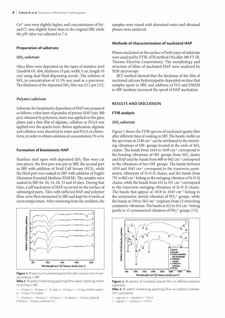
Figure 1 shows the FTIR spectra of nucleated apatite film after different time of soaking in SBF. The bands visible on the spectrum at 2340 cm-1 can be attributed to the stretch-ing vibrations of OH- groups located at the ends of SiO2 chains. The bands from 1643 to 1649 cm-1 correspond to the bending vibrations of OH- groups from SiO2 chains and HAP and the bands from 600 to 662 cm-1 correspond to the vibrations of free OH- groups. The bands between 1018 and 1043 cm-1 correspond to the transverse asym-metric vibrations of Si-O-Si chains, and the bands from 791 to 802 cm-1 belong to the swinging vibration of Si-O-Si chains, while the bands from 432 to 451 cm-1 correspond to the transverse swinging vibrations of Si-O-Si chains. The bands that appear at 1018 to 1043 cm-1 belong to the asymmetric stretch vibration of PO43- groups, while the bands at 550 to 563 cm-1 originate from ν2 stretching symmetric vibrations. The bands at 432 to 451 cm-1 belong partly to ν2 symmetrical vibration of PO43- groups [15].
Figure 2. IR spectra of nucleated apatite film on different polymer substratesSlika 2. IR spektri nukleiranog apatitnog filma na različitim polimer-nim supstratima1 – alginate; 2 – cellulose; 3 – PLGA1 – alginat; 2 – celuloza; 3 – PLGA
Figure 1. IR spectra of nucleated apatite film after various time of sam-ple soaking in SBFSlika 1. IR spektri nukleiranog apatitnog filma nakon različitog vreme-na držanja u SBF1 – 10 days; 2 – 20 days; 3 – 33 days; 4 – 43 days; 5 – 14 days, EMEM added; 6 – 14 days, FCS added
1 – 10 dana; 2 – 20 dana; 3 – 33 dana; 4 – 43 dana; 5 – 14 dana, dodatak EMEM; 6 – 14 dana, dodatak FCS

9Stomatološki glasnik Srbije. 2011;58(1):7-15
Polymer substrate
By analyzing the FTIR spectra, it was concluded that the phase biomimetically formed on the surface of HAP/polymer composite is hydroxyapatite with shifted stei-chiometry (Figure 2). All three IR spectra in Figure 2 show the characteristic bands for HAP: a weak peak that corresponds to the stretching OH- groups was observed at about 3570 cm-1; the bands at about 1640 cm-1 corre-spond to the bending vibrations of OH- groups, and the bands at 600 to 650 cm-1 correspond to the vibrations of free OH- groups; the bands that were reported at 970-1090 cm-1 belong to the asymmetric stretch vibrations of PO43- groups, while the bands at 550 to 640 cm-1 originate from the symmetric stretching vibrations of PO43--groups; the bands at 430 to 450 cm-1 belong partly to ν2 symmetric vibrations of PO43- groups.
The bands characteristic for each polymer were also present in the corresponding IR spectra:
• AlginateThe bands present at about 3500-3700 cm-1 are attrib-
uted to the stretching OH-groups; the bands from 1615 to 1420 cm-1 correspond to the asymmetric and symmet-ric stretching of COO- groups, respectively; the bands at about 1640 cm-1 can be attributed to water bonded to the polymer; the bands at about 1300 cm-1 correspond to the vibrations of the polymer chain; the bands that belong to
the asymmetric stretching of C-O-C at about 1080-1020 cm-1 are partially hidden by the bands that correspond to the apatite; the bands present at about 820 cm-1 are attrib-uted to the combination of the following vibrations: bend-ing vibrations -CCO, and –CCH groups and twisted vibra-tions of C=O group [16, 17].
• CelluloseThe bands at 2920 and 2940 cm-1 correspond to the
stretching of C=O and C-H groups, respectively; the bands around 2900 cm-1 belong to O-H stretching; while the bands at 1640 cm-1 correspond to O-H bending of absorbed water; the bands at 1590 cm-1 confirm the presence of COO- groups; the bands at about 1430 cm-1 are attributed to HCH and OCH bending deformation in the plane, while the band at about 1380 cm-1 corresponds to C-H bending; the bands at about 900 cm-1 belong to the COC, CCO, and CCH defor-mation and stretched vibrations, which correspond to the shifts of C-5 and C-6 atoms [18, 19].
• PLGAThe bands recorded at 2995 cm-1 correspond to asym-
metric C-H stretching while the bands at about 2950 and 2880 cm-1 belong to C-H symmetric stretching; the expressed band at 1760 cm-1 is attributed to C=O stretch-ing; the bands at about 1100-1280 cm-1 correspond to C-O-C stretching, the bands observed from 1400 to 1500 cm-1 correspond to CH2, CH3, and CH deformations; the band at about 750 cm-1 belongs to the swinging vibrations of long CH2 chain [20, 21].
SEM study
SiO2 substrate
Figures 3a and 3b clearly show the nucleation of apatite by sedimentation of the layers, one on another up to the final thickness of apatite film. Clear morphology of nucleated particles, shaped as dandelion flower and mean diame-ter of about 1 μm is observed.
Polymer substrates
SEM images showed strongly developed morphology of HAP particles (Figures 4a-b, 5a-b and 6a-b).
As shown in Figures 4a and 4b, HAP particles nucleated on alginate are present in two forms-the form of dande-lion flower and a polygonal shape. Particles of polygo-nal shape are smaller; the diameter of the smallest parti-cle is about 1 μm, while the greatest dimension is about 5 μm. Size distribution of the particles of dandelion shape is from 3 to 5 μm. Width of needle-shaped structures on the surface of these particles is only 18-25 nm, while the distance between them is 150-500 μm.
The diverse morphology of hydroxyapatite particles nucleated on cellulose is shown in Figures 5a and 5b. Mean particle size is about 3 μm while the smallest particle size is about 1 μm. Needle and plate- shaped structures are visible on the surface of particles and their width is from 30 to 150 nm.
Figure 3a-b. SEM micrographs of nucleated apatite film on SiO2Slika 3a-b. SEM snimci nukleiranog apatitnog filma na SiO2
a
b

10 Čolović B. et al. Nucleation of Biomimetic Hydroxyapatite
Apatite nucleated on the surface of PLGA film is shown on SEM images on Figures 6a and 6b. Particles are shaped as dandelion flower with a highly developed surface morphology and the petal-shaped structure. The parti-cle size is generally 2.5-3.5 μm, but the finer particles are also present (0.7-1.5 μm), as well as a lower number of larger particles (4.3-8.5 μm). Width of petal-shaped struc-ture on the surface of the particles is 15-20 nm.
Figure 4a-b. SEM micrographs of nucleated apatite film on alginateSlika 4a-b. SEM snimci nukleiranog apatitnog filma na alginatu
a b
Figure 5a-b. SEM micrographs of nucleated apatite film on celluloseSlika 5a-b. SEM snimci nukleiranog apatitnog filma na celulozi
a b
Figure 6a-b. SEM micrographs of nucleated apatite film on PLGASlika 6a-b. SEM snimci nukleiranog apatitnog filma na PLGA
a b
Film thickness of nucleated HAP
Based on measurements of weight and pore volume using Lecloux and Pirard method, the thickness of nucleated apatite films was determined [15]. The thickness of nucle-ated apatite film depended on the duration of soaking in SBF, as well as the type of used medium (Table 1).

11Stomatološki glasnik Srbije. 2011;58(1):7-15
Table 1. Thicknesses of self nucleated apatite film after various periods of nucleationTabela 1. Debljina samonukleiranog apatitnog filma posle različitih perioda nukleacije
Thickness of SiO2 film (μm)Debljina SiO2
filma (μm)
MediumMedijum
Time of apatite nucleation (days)Vreme nukleacije
apatita (dani)
The mass of apatite film (mg)Masa apatitnog
filma (mg)
Specific area (m2/g)
Specifična površina (m2/g)
Volume of pore (cm3/g)
Zapremina pora (cm3/g)
Thickness of apatite film (μm)
Debljina apatitnog filma (μm)
22.5
SBF
10 0.2 - - -
20 0.6 94 0.116 2.7
33 1.2 102 0.107 4.9
43 1.4 112 0.097 6.2
SBF+EMEM 14 1.3 110 0.100 4.8
SBF+FCS 14 1.1 105 0.110 4.1
The mechanism of HAP formation
SiO2 substrate
When stainless steel tapes with SiO2 coatings are sunk in SBF, HAP self nucleation happens on their surface. It is caused by presence of Na+ ions in SBF, which incorporate in nanopores of SiO2 film leading to local pH rise on the film surface, supporting the heterogeneous nucleation of HAP by the reaction [22]:
10Ca2+(aq)+6PO43-(aq) + 2OH-(aq) = Ca10(PO4)6(OH)2 (s)
Firstly, positive Ca2+ ions are attracted to the surface of SiO2 films due to the presence of Si-OH groups, and then, they are attracted to negatively charged PO43- ions, which lead to the formation of calcium phosphate. When HAP is formed on the surface of the SiO2 film, further nucleation occurs spontaneously because the activation energy for heterogeneous nucleation on the surface is less than the activation energy for homogeneous nucle-ation in the solution [23]. The nucleated apatite film is growing by simultaneous attraction of calcium and phos-phate ions from SBF.
Addition of FCS and EMEM to SBF, beside nucleation which mechanism is already described, gave a significant influence of amino group in FCS and sections of charged proteins in EMEM. They acted as additional and very effective nucleation centers due to their polar effect, and according to the mechanism “PILP” (polymer induced liquid precursor) caused rapid nucleation of apatite through the process of binding Ca2+ in the given active centers, and then simultaneous binding of PO43- ions.
Polymer substrates
Carboxyl/hydroxyl groups of alginate, cellulose and PLGA chains act as active centers which initiate nucleation. These negatively charged groups, in the initial stage of nucleation, were attracted by Ca2+ ions from SBF, which were then attached on the polymer surface. In the next step, PO43- ions were attracted to the positive Ca2+ ions. The process of calcium phosphate nucleation occured by synchro-nized attaching Ca2+ ions on active sites of correspond-ing polymer and simultaneous attachment of PO43- ions
on Ca2+ ions. As shown in some studies, the concentration of carboxyl/hydroxyl groups on the polymer surface the most likely had a significant effect on speed and mecha-nism of calcium phosphate nucleation [24, 25]. The density of nucleation sites can also affect the size of agglomerates and their morphology, as confirmed by the results in the current study. The highest density of nucleated parti-cles was observed with alginate (Figure 4a-b), which was probably caused by high density of carboxyl and hydroxyl groups in alginate chains. In the case of nucleation on PLGA (Figure 6a-b) which contains significantly lower number of carboxylic groups than alginate, the density of nucleated particles was also lower. The spatial distri-bution of hydroxyl groups in cellulose chains revealed that they were relatively close to each other. Therefore, the agglomerates of neighboring nucleation sites bonded to each other and formed elongated structures, as shown in Figures 5a and 5b. In the samples where alginate and cellulose were used, the smaller particles (1-5 μm) were formed, possibly due to larger number of nucleation sites. In the case of PLGA, larger agglomerates were obtained (some bigger than 8 μm), which can be attributed to the smaller number of nucleation sites that caused further growth of apatite nucleus.
CONCLUSION
This study demonstrated the possibility of forming biomi-metic calcium hydroxyapatite by self nucleation on differ-ent substrates in the presence of SBF. When SiO2 or apatite porous carriers coated with polymer films were used as substrate, after a certain time spent in SBF, the presence of HAP on their surface was confirmed by FTIR-ATR method. SEM microscopy determined the structure of self nucleated apatite film. By measuring the mass with volume adjustments using BET method, it was shown that the thickness of nucleated film depended on the time that tapes were soaked in SBF. The addition of FCS and EMEM increased the velocity of apatite self nucleation.
ACKNOWLEDGMENT
This work was funded by the Ministry of Science and Technological Development of Serbia (Project no. 172026).
12 Čolović B. et al. Nucleation of Biomimetic Hydroxyapatite
REFERENCES
1. Xu AW, Ma Y, Cölfen H. Biomimetic mineralization. J Mater Chem. 2007; 17:415-49.
2. Dujardin E, Mann S. Bio-inspired materials chemistry. Adv Mater. 2002; 14:775-88.
3. Niinomi M. Recent metallic materials for biomedical applications. Metall Mater Trans A. 2002, 33:477-86.
4. Webster TJ. Nanophase ceramics: the future of orthopedic and dental implant material. In: Ying JY. Nanostructured Materials. New York: Academic Press; 2001. p.125-166.
5. Kokubo T, Takadama H. How useful is SBF in predicting in vivo bone bioactivity? Biomaterials. 2006; 27:2907-15.
6. Le Geros RZ. Properties of osteoconductive biomaterials: calcium phosphates. Clin Orthop Relat Res. 2002; 395:81-98.
7. Tas AC. Synthesis of biomimetic Ca-hydroxyapatite powders at 37°C in synthetic body fluids. Biomaterials. 2000; 21:1429-38.
8. Barrere F, van der Valk CM, Dalmeijer RAJ, van Blitterswijk CA, de Groot K, Layrolle P. In vitro and in vivo degradation of biomimetic octacalcium phosphate and carbonate apatite coatings on titani-um implants. J Biomed Mater Res. 2003; 64:378-87.
9. Uchida M, Kim HM, Kokubo T, Nakamura T. Bonelike apatite for-mation induced on zirconia gel in simulated body fluid and its modified solutions. J Am Ceram Soc. 2001; 84:2041-4.
10. Oyane A, Kim HK, Furuya T, Kokubo T, Miyazaki T, Nakamura T. Preparation and assessment of revised simulated body fluids. J Biomed Mater Res. 2003; 65A:188-95.
11. Bigi A, Boanini E, Bracci B, Facchini A, Panzavolta S, Segatti F, et al. Nanocrystalline hydroxyapatite coatings on titanium: a new fast biomimetic method. Biomaterials. 2005; 26:4085-9.
12. Hench LL. In: Yamamoro T, Hench LL, Wilson J, editors. CRC Handbook of Bioactive Ceramics. Vol. 1. Boca Raton, FL: CRC; 1990. p.7.
13. Li P, Ohtsuki C, Kokubo T, Nakanishi K, Soga N, Nakamura T, et al. A role of hydrated silica, titania and alumina in forming biologically ac-tive bone-like apatite on implant. J Biomed Mater Res. 1994; 28:7-15.
14. Kokubo T. Apatite formation on surfaces of ceramics, metals and polymers in body environment. Acta Mater. 1998; 46:2519-27.
15. Čolović B, Jokanović V, Marković-Todorović B, Marković Z. AFM investigations of calcium hydroxyapatite thin films on the surface
of thin silica films. Journal of Optoelectronics and Advanced Materials. 2009; 11:70-5.
16. Dupuy B, Arien A, Minnot AP. FT-IR membranes made with algi-nate/polylysine complexes. Variations with mannuronic or guluron-ic content of the polysaccharides. Art Cells Blood Substit Immobil Biotechnol. 1994; 22:71-82.
17. Lawrie G, Keen I, Drew B, Chandler-Temple A, Rintoul L, Fredericks P, et al. Interactions between alginate and chitosan biopolymers char-acterized using FTIR and XPS. Biomacromolecules. 2007; 8:2533-41.
18. Zhbankov RG, Firsov SP, Buslov DK, Nikonenko NA, Marchewka MK, Ratajczak H. Structural physico-chemistry of cellulose mac-romolecules. Vibrational spectra and structure of cellulose. J Mol Struct. 2002; 614:117-25.
19. Oh SY, Yoo DI, Shin Y, Seo G. FTIR analysis of cellulose treated with sodium hydroxide and carbon dioxide. Carbohydr Res. 2005; 340:417-28.
20. Petricca SE, Marra KG, Kumta PN. Chemical synthesis of poly(lactic-co-glycolic acid)/hydroxyapatite composites for orthopedics ap-plications. Acta Biomater. 2006; 2:277-86.
21. Tai H, Upton CE, White LJ, Pini R, Storti G, Mazzotti M, et al. Studies on the interactions of CO2 with biodegradable poly(DL-lactic acid) and poly(lactic acid-co-glycolic acid) copolymers using high pres-sure ATR-IR and high pressure rheology. Polymer. 2010; 51:1425-31.
22. Cheng X, Nie B, Kumar S. Preparation and bioactivity of SiO2 func-tional films on titanium by PACVD. Trans Nonferrous Met Soc China. 2008; 18:627-30.
23. Li P, Kangasniemi I, de Grot K, Kokubo T. Bonelike hydroxyapa-tite induction by a gel-derived titania on titanium substrate. J Am Ceram Soc. 1994; 17:1307-12.
24. Nge T, Sugiyama J. Surface functional group dependent apatite for-mation on bacterial cellulose microfibrils network in a simulated body fluid. J Biomed Mater Res A. 2007; 81:124-34.
25. Hashizume M, Horii H, Kikuchi JI, Kamitakahara M, Ohtsuki C, Tanihara M. Effects of surface carboxylic acid groups of cerasomes, morphologically stable vesicles having a silica surface, on biomimet-ic deposition of hydroxyapatite in body fluid conditions. J Mater Sci Mater Med. 2010; 2:111-9.
Received: 05/11/2010 • Accepted: 09/02/2011

13Stomatološki glasnik Srbije. 2011;58(1):7-15
Nukleacija biomimetskog hidroksiapatita
Božana Čolović1, Dejan Marković2, Vukoman Jokanović1
1Laboratorija za radijacionu hemiju i fiziku, Institut za nuklearne nauke „Vinča”, Beograd, Srbija;2Klinika za dečju i preventivnu stomatologiju, Stomatološki fakultet, Univerzitet u Beogradu, Beograd, Srbija
KRATAK SADRŽAJUvod Cilj ovog ra da je bio da se is pi ta for mi ra nje bi o mi met skog kal ci jum-hi drok si a pa ti ta na po vr ši na ma raz li či tih sup stra ta.Ma te ri jal i me to de ra da Kao sup stra ti su ko ri šće ni tan ki fil mo vi si li ci jum-di ok si da na ne ti na če lič ne tra ke i tan ki po li mer ni fil mo-vi (al gi nat, ce lu lo za, po li lak tid ko gli ko lid) na ne ti na vi so ko po ro zni hi drok si a pa tit ni no sač. Si mu li ra na te le sna teč nost (engl. si mu la-ted body fluid – SBF), kao i SBF kom bi no van sa se ru mom iz fe tu sa go ve če ta ili Iglo vim me di ju mom ko ri šće ni su kao bi o ak tiv ni teč-ni me di jum u ko jem se od vi ja bi o mi met ska nu kle a ci ja apa ti ta. Za ana li zu for mi ra nih fa za ko ri šće na je in fra cr ve na spek tro sko pi ja sa Fu ri je o vom tran sfor ma ci jom, dok je ske ni ng elek tron ska mi kro sko pi ja uka zi va la na mor fo lo gi ju nu kle i ra ne fa ze.Re zul ta ti Me re nje ma se uz ko rek ci ju za pre mi ne pri me nom me to de BET po ka za lo je da de blji na fil ma nu kle i ra nog kal ci jum-hi-drok si a pa ti ta za vi si od vre me na dr ža nja uzo ra ka u SBF, kao i od vr ste iza bra nog bi o mi met skog me di ju ma.Za klju čak Do bi ja nje bi o mi met skog kal ci jum-hi drok si a pa ti ta mo gu će je sa mo nu kle a ci jom na raz li či tim sup stra ti ma i pri me nom SBF.
Ključ ne re či: bi o mi met ska me to da; bi o ak tiv ni tan ki fil mo vi; bi o mi met ski hi drok si a pa tit; di zaj ni ra nje na no struk tu re
UVOD
Pri ro da je stvo ri la ma te ri ja le s od lič nim funk ci o nal nim oso bi na-ma, a na u ka se usme ri la ka raz vi ja nju no vih na či na sin te ze ko ji tre ba da opo na ša ju pri rod ne pro ce se. Glav ni cilj ni je sa mo u to-me da se imi ti ra ju od re đe ni bi o lo ški si ste mi, već da se is ko ri ste nji ma in spi ri sa ne ide je, kako bi se stvo ri li no vi ma te ri ja li. Bi o-mi met ska me to da se po ka za la po god nom za sin te zu sa vre me nih ma te ri ja la slo že nog ob li ka i hi je rar hij ske struk tur ne or ga ni za ci-je, ko ji su ade kvat ni za pri me nu u bi o lo škim si ste mi ma [1, 2].
Bi o mi met ska me to da mo že da se is ko ri sti kao jed na od me-to da za do bi ja nje bi o ak tiv nih pre vla ka na me tal nim im plan ta ti-ma ko je omo gu ću ju for mi ra nje ade kvat ne ve ze s okol nom ko-sti [3-6]. Bi o mi met skom me to dom se na po vr ši ni me tal nog im-plan ta ta do bi ja apa tit slič nih struk tur nih i mor fo lo ških od li ka kao i pri rod na kost, s ob zi rom na to da je ova me to da za sno-va na na ko ri šće nju ras tvo ra slič nog jon skog sa sta va kao krv na pla zma, pa su i uslo vi nu kle a ci je hi drok si a pa ti ta (HAP) slič ni pri rod nim [7-11]. For mi ra nje HAP na po vr ši ni me ta la mo že se ubr za ti pret hod nim pre vla če njem po vr ši ne me ta la tan kim slo-jem ok si da (SiO2, TiO2, sa ugra đe nim OH-gru pa ma unu tar ok-sid nog slo ja). Ugra đe ne OH-gru pe slu že kao ak ti va to ri he te ro-ge ne nu kle a ci je apa ti ta pri li kom ta lo že nja kal ci ju mo vih i fos-fat nih jo na iz pre si će ne si mu li ra ne te le sne teč no sti (engl. si mu-la ted body fluid – SBF) [12, 13].
Cilj ovog ra da je bio da se pro u či me ha ni zam nu kle a ci je HAP na če lič nim tra ka ma pre svu če nim slo jem SiO2 ko ri ste ći SBF, kao i SBF u kom bi na ci ji sa fe tu som go ve če ta (engl. fe tal calf se rum – FCS) ili Iglo vim me di ju mom (engl. Eagle’s Mi ni mum Es sen-tial Me di um – EMEM), ko ji usled za stu plje no sti ami no-gru pa po volj no uti ču na ki ne ti ku nu kle a ci je HAP. Po red na ve de nog, do dat ni cilj je bio da se is pi ta pri me na bi o mi met ske me to de za do dat no di zaj ni ra nje po ro znih apa tit nih no sa ča. Ade kvat na na no to po lo gi ja unu tra šnjih zi do va po ro znih no sa ča je bit na za nor ma lan rast će li ja, i ona bi se mo gla po sti ći bi o mi met skim tret ma nom ovih no sa ča u SBF, pret hod no sin te ti sa nih bi lo ko-jom me to dom. Po li me ri (ce lu lo za, al gi nat, po li lak tid ko gli ko lid – PLGA) su ko ri šće ni za funk ci o na li za ci ju zi do va po ro znih no-sa ča ka ko bi se po sti gao spe ci fi čan na no di zajn pri li kom nu kle-i ra nja HAP na nji ho vu po vr ši nu [14].
MATERIJAL I METODE RADA
Eks pe ri men tal ni deo is tra ži va nja je ob u hva tio pri pre mu SBF, pri pre mu sup stra ta, for mi ra nje bi o mi met skog HAP, od no sno ka rak te ri za ci ju nu kle i ra nog HAP.
Priprema SBF
SBF je pri pre mljen po ne što iz me nje noj re cep tu ri u od no su na ori gi nal ni SBF, pri če mu su kon cen tra ci je po je di nih jo na bi le: c(Cl-)=54 mmol/dm3; c(Na+)=54,2 mmol/dm3; c(Ca2+)=2,5 mmol/dm3; c(PO43-)=1 mmol/dm3; c(Mg2+)=0,3 mmol/dm3; c(NO3-)=0,6 mmol/dm3 i c(K+)=1,4 mmol/dm3 [10]. Kon cen-tra ci je PO43- i Ca2+ jo na su bi le ne što ve će, a kon cen tra ci je Na+ i Cl- jo na ne što ma nje ne go kod ori gi nal nog SBF, dok je pH vred-nost SBF po de še na na 7,4.
Priprema supstrata
SiO2 supstrat
Fil mo vi si li ci jum-di ok si da de po no va ni su na tra ke od ner đa ju-ćeg če li ka (San dvik OC 404, de blji na 35 μm, ši ri na 3 cm, du ži-na 10 cm) ko ri ste ći dvo flu id nu mla zni cu za ras pr ša va nje. Kao pre kur sor je ko ri šćen ras tvor SiO2 u kon cen tra ci ji od 13,5%. De-blji na de po no va nog SiO2 fil ma bi la je 22,5 μm [15].
Polimerni supstrat
Sup strat za bi o mi met sku de po zi ci ju HAP pri pre mljen je na sle-de ći na čin: na sta kle ne plo či ce na ne sen je ta nak sloj gra nu la po-ro znog HAP (di men zi ja 300 μm), do bi je nog me to dom po li mer-nih pe na, a za tim su tan ki fil mo vi al gi na ta, ce lu lo ze ili PLGA na ne ti pre ko apa tit nog slo ja. Pre na no še nja, al gi nat i ce lu lo za su ras tvo re ni u vo di, a PLGA u hlo ro for mu, ta ko da su do bi jeni ras tvo ri kon cen tra ci je od 1%.

14 Čolović B. et al. Nucleation of Biomimetic Hydroxyapatite
Formiranje biomimetskog HAP
Če lič ne tra ke sa de po no va nim SiO2 fil mom su ise če ne na de-li će, od ko jih je je dan deo po to pljen u SBF, dru gi deo u SBF sa do dat kom FCS, dok je tre ći deo po to pljen u SBF sa do dat kom EMEM. Uzor ci su osta vlje ni da od sto je u SBF do is te ka od re-đe nog vre me na (10, 14, 20, 33 i 43 da na), pri če mu je do la zi lo do sa mo nu kle a ci je HAP na nji ho voj po vr ši ni. Plo či ce sa na ne-tim HAP i po li mer nim fil mo vi ma su po tom po to plje ne u SBF i osta vlje ne da sto je šest ne de lja na sob noj tem pe ra tu ri. Na kon va đe nja iz me di ju ma uzor ci su is pra ni de jo ni zo va nom vo dom, a do bi je ne fa ze su ana li zi ra ne.
Metode karakterizacije nukleiranog HAP
Nu kle i ra ne fa ze na po vr ši ni obe vr ste sup stra ta ana li zi ra ne su me to dom FTIR-ATR (Ni col let 380 FT-IR, Ther mo Elec tron Cor-po ra tion). Mor fo lo gi ja i struk tu ra fil mo va nu kle i ra nog HAP ana li zi ra ne su ske ning-elek tron skom mi kro sko pi jom (SEM).
BET me to dom je po ka za no da de blji na fil ma nu kle i ra nog HAP za vi si od vre me na sta ja nja uzo ra ka u SBF, kao i od to ga što do da tak FCS i EMEM u SBF me di ju mu uti če na po ve ća nje br zi ne nu kle a ci je HAP.
REZULTATI I DISKUSIJA
FTIR analiza
SiO2 supstrat
Na sli ci 1 pri ka za ni su FTIR spek tri nu kle i ra nog apa tit nog fil ma na kon raz li či tog vre me na dr ža nja uzo ra ka u SBF. Tra ke uoč lji-ve na spek tru na 2340/cm mo gu se pri pi sa ti is te žu ćim vi bra ci-ja ma OH-gru pa ko je se na la ze na kra je vi ma SiO2 la na ca. Tra-ke od 1643/cm do 1649/cm od go va ra ju sa vi ja ju ćim vi bra ci ja ma OH-gru pa i iz SiO2 la na ca i iz HAP, a tra ke na 600/cm do 662/cm slo bod nim vi bra ci ja ma OH-gru pa. Tra ke iz me đu 1018/cm i 1043/cm od go va ra ju tran sver zal nim asi me trič nim vi bra ci ja ma
Si-O-Si la na ca, tra ke od 791/cm do 802/cm pri pa da ju lju lja ju-ćim vi bra ci ja ma Si-O-Si la na ca, dok tra ke od 432/cm do 451/cm od go va ra ju tran sver zal nim lju lja ju ćim vi bra ci ja ma ovo ga la na-ca. Tra ke ko je se po ja vlju ju na 1018/cm do 1043/cm pri pa da-ju asi me trič nim is te žu ćim vi bra ci ja ma PO43- gru pa, dok tra ke od 550/cm 563/cm po ti ču od ν2 si me trič nih is te žu ćih vi bra ci-ja. Tra ke od 432/cm do 451/cm de lom pri pa da ju ν2 si me trič-nim vi bra ci ja ma PO43- gru pa [15].
Polimerni supstrat
Ana li zi ra njem FTIR spek tra za klju če no je da je fa za for mi ra na bi o mi met ski na po vr ši ni HAP – po li mer kom po zi ta, hi drok si-a pa tit po me re ne ste hi o me tri je (Sli ka 2). Sva tri IR spek tra po-ka zu ju tra ke ti pič ne za HAP: slab vr hu nac ko ji od go va ra is te-za nju OH-gru pa uočen je na oko 3570/cm; tra ke na oko 1640/cm od go va ra ju sa vi ja ju ćim vi bra ci ja ma OH- gru pa, a tra ke na 600/cm do 650/cm slo bod nim vi bra ci ja ma OH- gru pa; tra ke ko je se ja vlja ju na 970/cm do 1090/cm pri pa da ju asi me trič nim
is te žu ćim vi bra ci ja ma PO43- gru pa, dok tra ke od 550/cm do 640/cm po ti ču od si me trič nih is te žu ćih vi bra ci ja PO43- gru pa; tra ke od 430/cm do 450/cm de lom pri pa da ju ν2 si me trič nim vi-bra ci ja ma PO43- gru pa.
Tra ke ti pič ne za sva ki od po li me ra se ta ko đe na la ze na od-go va ra ju ćim IR spek tri ma:
• Al gi natTra ke ko je se na la ze na 3500-3700/cm pri pi su ju se is te za-
nju OH-gru pa; tra ke na 1615/cm i 1420/cm od go va ra ju asi-me trič nom i si me trič nom is te za nju COO- gru pa; tra ke na oko 1640/cm se mo gu pri pi sa ti vo di ve za noj za po li mer; tra ke na oko 1300/cm od go va ra ju vi bra ci ja ma po li mer nog lan ca; tra ke ko je pri pa da ju asi me trič nom is te za nju C-O-C na 1080-1020/cm su de li mič no skri ve ne tra ka ma ko je od go va ra ju apa ti tu; tra ke ko je se na la ze na oko 820/cm pri pi su ju se kom bi na ci-ji sa vi ja ju će vi bra ci je -CCO i -CCH gru pa i uvi ja ju će vi bra ci-je C=O gru pe [16, 17].
• Ce lu lo zaTra ke na 2920/cm i 2940/cm od go va ra ju is te za nju C=O i C-H
gru pa; tra ke oko 2900/cm pri pa da ju O-H is te za nju, dok tra ke na 1640/cm od go va ra ju sa vi ja nju O-H iz ap sor bo va ne vo de; tra-ke na 1590/cm po tvr đu ju po sto ja nje COO-gru pe; tra ke na oko 1430/cm se pri pi su ju HCH i OCH sa vi ja ju ćim de for ma ci ja ma u rav ni, dok tra ka na oko 1380/cm od go va ra C-H sa vi ja nju; tra-ke na oko 900/cm pri pa da ju COC, CCO i CCH de for ma ci ja ma i is te žu ćim vi bra ci ja ma ko je od go va ra ju po me ra nji ma od C-5 i C-6 ato ma [18, 19].
• PLGATra ke re gi stro va ne na 2995/cm od go va ra ju C-H asi me trič-
nom is te za nju, dok tra ke na oko 2950/cm i 2880/cm pri pa da ju C-H si me trič nom is te za nju; iz ra že na tra ka na 1760/cm se pri-pi su je C=O is te za nju; tra ke na 1100-1280/cm od go va ra ju C-O-C is te za nju, dok tra ke uoče ne od 1400/cm do 1500/cm od go va ra-ju CH2, CH3 i CH de for ma ci ja ma; tra ka na oko 750/cm pri pa da lju lja ju ćim vi bra ci ja ma du gog CH2 lan ca [20, 21].
SEM ispitivanja
SiO2 supstrat
Na sli ka ma 3a i 3b ja sno se vi di na čin nu kle a ci je apa ti ta se di men-ti ra njem slo je va do za vr šne de blji ne apa tit nog fil ma. Uoča va se ve o ma raz vi je na mor fo lo ška struk tu ra nu kle i ra nih če sti ca, ko-je su u ob li ku cve ta ma slač ka i sred njeg preč ni ka od oko 1 μm.
Polimerni supstrati
SEM snim ci su po ka za li ve o ma raz vi je nu mor fo lo šku struk tu-ru če sti ca HAP (Sli ke 4a-b, 5a-b i 6a-b).
Kao što se uoča va na sli ka ma 4a i 4b, če sti ce HAP nu kle i ra-nog na al gi na tu su u ob li ku cve ta ma slač ka i po li go nal nom ob-li ku. Ove po to nje su ma nje, preč ni ka 1-5 μm. Preč nik če sti ca u ob li ku ma slač ka je 3-5 μm. Ši ri na igli ča stih struk tu ra na po-vr ši ni ovih če sti ca je sve ga 18-25 nm, dok je ra sto ja nje iz me đu njih 150-500 μm.
Na sli ka ma 5a i 5b za pa ža se ra zno li kost u mor fo lo škoj struk-tu ri če sti ca HAP nu kle i ra nog na ce lu lo zi. Sred nji preč nik če-sti ca je oko 3 μm, dok su naj ma nje če sti ce ve li či ne oko 1 μm.

15Stomatološki glasnik Srbije. 2011;58(1):7-15
Igli ča ste i ta nji ra ste struk tu re se uoča va ju na po vr ši ni če sti ca, a nji ho va ši ri na je 30-150 nm.
Apa tit nu kle i ran na po vr ši ni PLGA fil ma pri ka zan je na sli-ka ma 6a i 6b. Če sti ce su u ob li ku cve ta ma slač ka s ve o ma raz-vi je nom mor fo lo škom struk tu rom po vr ši ne, na ko joj se na la-ze la ti ča ste struk tu re. Ve li či na če sti ca je uglav nom 2,5-3,5 μm, ali one ne ka da mo gu bi ti i sit ni je (0,7-1,5 μm), od no sno krup-ni je (4,3-8,5 μm). Ši ri na la ti ča stih struk tu ra na po vr ši ni če sti-ca je 15-20 nm.
Debljina filma nukleiranog HAP
Na osno vu me re nja ma se i ukup ne za pre mi ne po ra me to dom Le klua (Lec lo ux) i Pi ra ra (Pi rard), od re đe na je de blji na nu kle-i ra nih apa tit nih fil mo va [15]. De blji na nu kle i ra nog apa tit nog fil ma za vi si od vre me na dr ža nja tra ka u SBF i od vr ste me di-ju ma (Ta be la 1).
Mehanizam formiranja HAP
SiO2 supstrat
Ka da se če lič ne tra ke sa SiO2 pre vla ka ma po to pe u SBF, na nji-ho voj po vr ši ni do la zi do sa mo nu kle a ci je HAP. To je uslo vlje-no pri su stvom Na+ jo na u SBF, ko ji se in kor po ri ra ju u na no po-re SiO2 fil ma, do vo de ći do po ve ća nja lo kal ne pH vred no sti na po vr ši ni fil ma, što po spe šu je he te ro ge nu nu kle a ci ju HAP pre-ma re ak ci ji [22]:
10 Ca2+(aq) + 6 PO43-(aq) + 2 OH-(aq) = Ca10(PO4)6(OH)2 (s).
Naj pre po zi tiv ni Ca2+ jo ni bi va ju pri vu če ni na po vr ši nu SiO2 fil ma usled po sto ja nja Si-OH gru pa, a za tim pri vu če ni i ne ga-tiv no na e lek tri sa ni PO43- jo ni, što do vo di do for mi ra nja kal ci-jum-fos fa ta. Ka da se HAP stvo ri na po vr ši ni SiO2 fil ma, da lja nu kle a ci ja se de ša va spon ta no, jer je ener gi ja ak ti va ci je za he-te ro ge nu nu kle a ci ju na po vr ši ni ma nja od ener gi je ak ti va ci je za ho mo ge nu nu kle a ci ju u ras tvo ru [23]. Da lje do la zi do ra sta nu kle i ra nog apa tit nog fil ma uz isto vre me no pri vla če nje kal ci-ju mo vih i fos fat nih jo na iz SBF.
Do dat kom FCS i EMEM u SBF, po red nu kle a ci je, či ji je me-ha ni zam već opi san, zna ča jan je i uti caj ami no-gru pa u FCS i de lo va na e lek tri sa nih pro te i na u EMEM. Oni de lu ju kao do dat-ni i vr lo efi ka sni cen tri nu kle a ci je svo jim po lar nim dej stvom, i po me ha ni zmu PILP (engl. polymer in du ced li qu id pre cur sor – teč ni pre kur sor in du ko van po li me rom) uslo vlja va ju ubr za-nu nu kle a ci ju apa ti ta kroz pro ces ve zi va nja Ca2+ na da tim ak-tiv nim cen tri ma, a po tom si mul ta nim ve zi va njem PO43- jo na.
Polimerni supstrati
Kar bok sil ne i hi drok sil ne gru pe iz al gi nat nih, ce lu lo znih i la-na ca PLGA de lu ju kao ak tiv ni cen tri ko ji ini ci ra ju nu kle a ci ju. Ove ne ga tiv no na e lek tri sa ne gru pe, u po čet noj fa zi nu kle a ci-je pri vla če Ca2+ jo ne iz SBF, ko ji po tom bi va ju na ka če ni na po-vr ši nu po li me ra. U sle de ćem ko ra ku PO43- jo ni bi va ju pri vu če-ni od po zi tiv nih Ca2+ jo na. Pro ces nu kle a ci je kal ci jum-fos fa ta
da lje se od i gra va „ka če njem” Ca2+ jo na na ak tiv na me sta od-go va ra ju ćeg po li me ra i si mul ta nim „ka če njem” PO43- jo na na Ca2+ jo ne. Kao što je po ka za no u ne kim stu di ja ma, kon cen tra-ci ja kar bok sil nih i hi drok sil nih gru pa na po vr ši ni po li me ra naj-ve ro vat ni je zna čaj no uti če na br zi nu i me ha ni zam nu kle a ci je kal ci jum-fos fa ta [24, 25].
Gu sti na nu kle a ci o nih me sta mo že ta ko đe da uti če na ve li-či nu aglo me ra ta i nji ho vu mor fo lo šku struk tu ru, što po tvr đu-ju i re zul ta ti do bi je ni u ovom ra du. Naj ve ća gu sti na nu kle i ra-nih če sti ca uoče na je pri ko ri šće nju al gi na ta (Sli ka 4a-b), što je ve ro vat no uslo vlje no ve li kom gu sti nom kar bok sil nih i hi-drok sil nih gru pa u al gi nat nim lan ci ma. U slu ča ju nu kle a ci je na PLGA (Sli ka 6a-b), ko ji sa dr ži znat no ma nji broj kar bok sil-nih gru pa ne go al gi nat, re la tiv no je ma nja gu sti na nu kle i ra nih če sti ca. Po sma tra ju ći pro stor ni ras po red hi drok sil nih gru pa u ce lu lo znim lan ci ma, mo že se vi de ti da su re la tiv no bli zu jed na dru goj. Sto ga se ve ro vat no aglo me ra ti sa su sed nih nu kle a ci o-nih me sta spa ja ju for mi ra ju ći iz du že ne struk tu re (Sli ka 5a-b). Ta ko đe, u uzor ci ma gde su ko ri šće ni al gi nat i ce lu lo za do bi je-ne su ma nje če sti ce (1-5 μm), što mo že bi ti po sle di ca ve li kog bro ja nu kle a ci o nih me sta, dok su u slu ča ju PLGA do bi je ni ve-ći aglo me ra ti (ne ki preč ni ka ve ćeg od 8 μm), što se mo že pri-pi sa ti ma njem bro ju nu kle a ci o nih me sta ko ja uslo vlja va ju da-lji rast apa tit nog nu kle u sa.
ZAKLJUČAK
U ovom ra du po ka za na je mo guć nost do bi ja nja bi o mi met skog HAP sa mo nu kle a ci jom na raz li či tim sup stra ti ma uz za stu plje-nost SBF. I u slu ča ju SiO2 kao sup stra ta, i u slu ča ju ka da su ko-ri šće ni po li mer ni fil mo vi na ne se ni na po ro zni apa tit ni no sač, FTIR-ATR me to dom po tvr đe no je po sto ja nje HAP na nji ho voj po vr ši ni po sle od re đe nog vre me na dr ža nja u SBF. SEM ana li-zom utvr đe na je struk tu ra sa mo nu kle i ra nog apa tit nog fil ma. Me re njem ma se uz ko rek ci je za pre mi ne me to dom BET po ka-za no je da de blji na nu kle i ra nog fil ma za vi si od vre me na dr ža-nja tra ka u SBF, kao i da do da tak FCS i EMEM me di ju ma ubr-za va pro ces sa mo nu kle a ci je apa tit nog fil ma.
NAPOMENA
Iz ra du ovo ga ra da fi nan sij ski je po dr ža lo Mi ni star stvo za na u-ku i teh no lo ški raz voj Re pu bli ke Sr bi je (Pro je kat br. 172026).


![Deconvolutional Networks - matthewzeiler · 2018-12-07 · Amit and Geman [1] and Jin and Geman [10] apply hierar-chical models to deformed Latex digits and car license plate recognition.](https://static.fdocuments.in/doc/165x107/5f2a11ec5a6f393d51414616/deconvolutional-networks-matthewzeiler-2018-12-07-amit-and-geman-1-and-jin.jpg)