Nuclear Magnetic Resonance - · PDF fileNuclear Magnetic Resonance For students of HI...
106
Nuclear Magnetic Resonance For students of HI 6001-125 “Computational Structural Biology” Willy Wriggers, Ph.D. http://biomachina.org/courses/structures/06.html T H E U N I V E R S I T Y of T E X A S S C H O O L O F H E A L T H I N F O R M A T I O N S C I E N C E S A T H O U S T O N
-
Upload
duongkhanh -
Category
Documents
-
view
216 -
download
0
Transcript of Nuclear Magnetic Resonance - · PDF fileNuclear Magnetic Resonance For students of HI...

Nuclear Magnetic Resonance
For students of HI 6001-125“Computational Structural Biology”
Willy Wriggers, Ph.D.
http://biomachina.org/courses/structures/06.html
T H E U N I V E R S I T Y of T E X A S
S C H O O L O F H E A L T H I N F O R M A T I O N
S C I E N C E S A T H O U S T O N

Introduction / Medical Applications

NMR History
© 2002, Michael Sattler http://www.embl.de/nmr/sattler/teaching

Nuclear MagneticMoment
Nucleus with SPINe.g. proton
Bar magnet
N
S
Magnetic Moment
Spin and Magnetic Moment
© 2003, Peter Cole http://www.liv.ac.uk/~iop/PTC/TechMedicImag.ppt

Zero External Magnetic Field
Point in random directions.
Effect of External Field
© 2003, Peter Cole http://www.liv.ac.uk/~iop/PTC/TechMedicImag.ppt
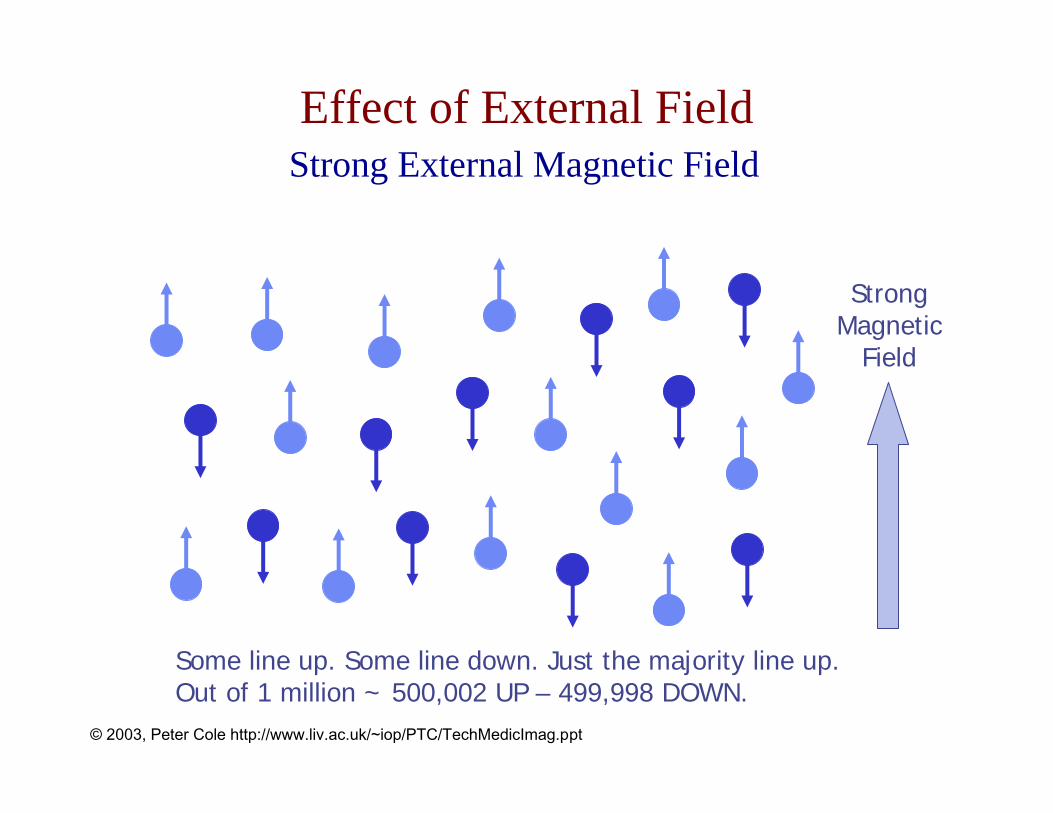
Strong External Magnetic Field
StrongMagnetic
Field
Some line up. Some line down. Just the majority line up.Out of 1 million ~ 500,002 UP – 499,998 DOWN.
Effect of External Field
© 2003, Peter Cole http://www.liv.ac.uk/~iop/PTC/TechMedicImag.ppt

Hydrogen Nucleus
The proton.
Biggest nuclear magnetic moment of any stable nucleus.
Most abundant nucleus in the human body.
Water and lipid (fat).
MRI gives a distribution of water and fat in the patient.
Magnetic Resonance Imaging (MRI)
© 2003, Peter Cole http://www.liv.ac.uk/~iop/PTC/TechMedicImag.ppt

Flipping Spins
BulkMagnetisation
‘M’
Mainmagnetic
field (~ 1.5 T)
RadiofrequencyPulse
N
S
Wobbling‘gyroscope’motion.Precession
EMFsinduced
To computer
Magnetic Resonance Imaging (MRI)
© 2003, Peter Cole http://www.liv.ac.uk/~iop/PTC/TechMedicImag.ppt

Larmor Frequency
Rate of ‘wobbling’ depends on big magnetic field strength.
ω = γ B
γ = gyromagnetic ratio(42.57 MHz per Tesla for protons)
1 Tesla ≈ 10,000 x Earth’s magnetic field.
Magnetic Resonance Imaging (MRI)

Frequency Encoding of Spatial Dimensions
X
|B|
No gradient
X
|B|
θ
With gradient
3 blobs of protons
All 3 ‘see’ the same B& wobble at same rate
Each ‘see’ a different B& wobble at 3 different rates
Knownslope
Magnetic Resonance Imaging (MRI)
© 2003, Peter Cole http://www.liv.ac.uk/~iop/PTC/TechMedicImag.ppt
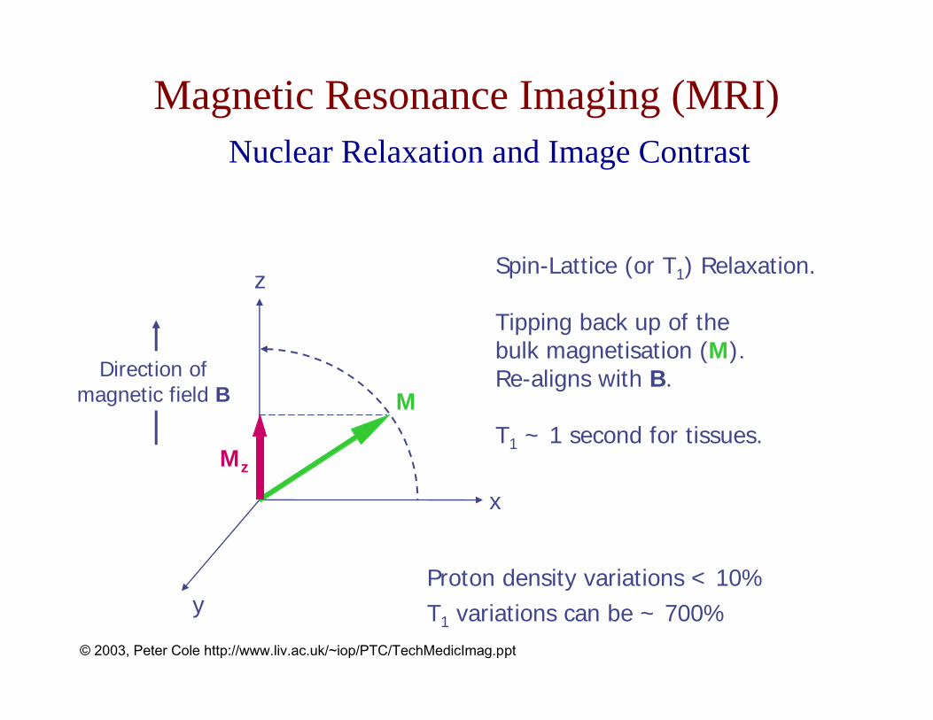
Nuclear Relaxation and Image Contrast
x
y
z
M
Mz
Spin-Lattice (or T1) Relaxation.
Tipping back up of thebulk magnetisation (M).Re-aligns with B.
T1 ~ 1 second for tissues.
Direction ofmagnetic field B
Proton density variations < 10%T1 variations can be ~ 700%
Magnetic Resonance Imaging (MRI)
© 2003, Peter Cole http://www.liv.ac.uk/~iop/PTC/TechMedicImag.ppt

T1-weighted T2-weighted Proton densityweighted
Axial Brain ImagesMagnetic Resonance Imaging (MRI)
© 2003, Peter Cole http://www.liv.ac.uk/~iop/PTC/TechMedicImag.ppt

Big superconductingmagnet (~ 1.5 tesla).
Gradient coils.
Radiofrequency coils.
MRI Scanner
© 2003, Peter Cole http://www.liv.ac.uk/~iop/PTC/TechMedicImag.ppt
Why Biomolecular NMR?
© 2002, Michael Sattler http://www.embl.de/nmr/sattler/teaching

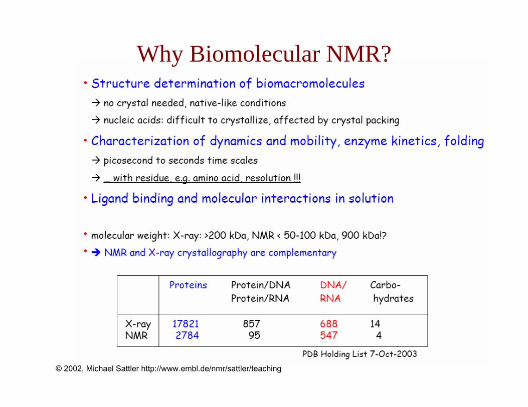
Why Biomolecular NMR?
© 2002, Michael Sattler http://www.embl.de/nmr/sattler/teaching

Basic Physics Concepts

Angular Momentum
Right hand rule
http://instruct1.cit.cornell.edu/courses/biobm730
A rotating object possesses angular momentum

Ltot = [J(J + 1)]1/2ħ
ħ ≈ 1.054 x 10⁻³⁴ Js
Angular Momentum is Quantized
Diatomic moleculehttp://instruct1.cit.cornell.edu/courses/biobm730
Example: Rotational energy of a moleculeAt the level of atoms and molecules, only specific rotational states are “allowed”

Spin Angular Momentum
• really an intrinsic property (not due to rotation)• is quantized• particles with spin I have 2I + 1 sublevels
(degenerate without B or E field)• bosons = particles with integer spin• fermions = particles with half-integer spin• arises from quantizing the electromagnetic field
(Dirac)

Neutrons and Protons
3 quarks, stuck together by gluons
http://instruct1.cit.cornell.edu/courses/biobm730

Nuclear Spin Energy Levels
Ground state nuclear spin ~ empirical property of each isotope
no magnetic field
http://instruct1.cit.cornell.edu/courses/biobm730

Determining Spin of Isotopes
mass number atomic number (Z) I NMR detectableodd even or odd 1/2, 3/2, 5/2 ... yeseven even 0 noeven odd 1, 2, 3 ... yes
Possible number of spin states = 2I + 11H: I = 1/2 2(1/2) + 1 = 2 m = ±1/214N: I = 1 2(1) + 1 = 3 m = -1, 0, 1

NMR-Active Nuclei in Proteins
N C’
OH
Cβ
H
H
H
H
HH
OH
H
Cα
PO3
Naturally abundant1H, spin ½31P, spin ½
Enriched via bacterial expression(isotope labeling)
2H, spin 113C, spin ½15N, spin ½

The Gyromagnetic RatioFor spin angular momentum of the nucleus,
Defining the “gyromagnetic ratio” of µ and I:
the relationship between angular momentum and magnetic moment becomes:
Hence, the angular momentum and magnetic moment vectors associated with nuclear spin are pointed in the same direction and are related by a constant.
N Ng Iµµ =
N Ng µ γ=
Iµ γ=
where gN is the nuclear g-factor and µN is the nuclear magneton

Gyromagnetic Ratio, (
http://instruct1.cit.cornell.edu/courses/biobm730
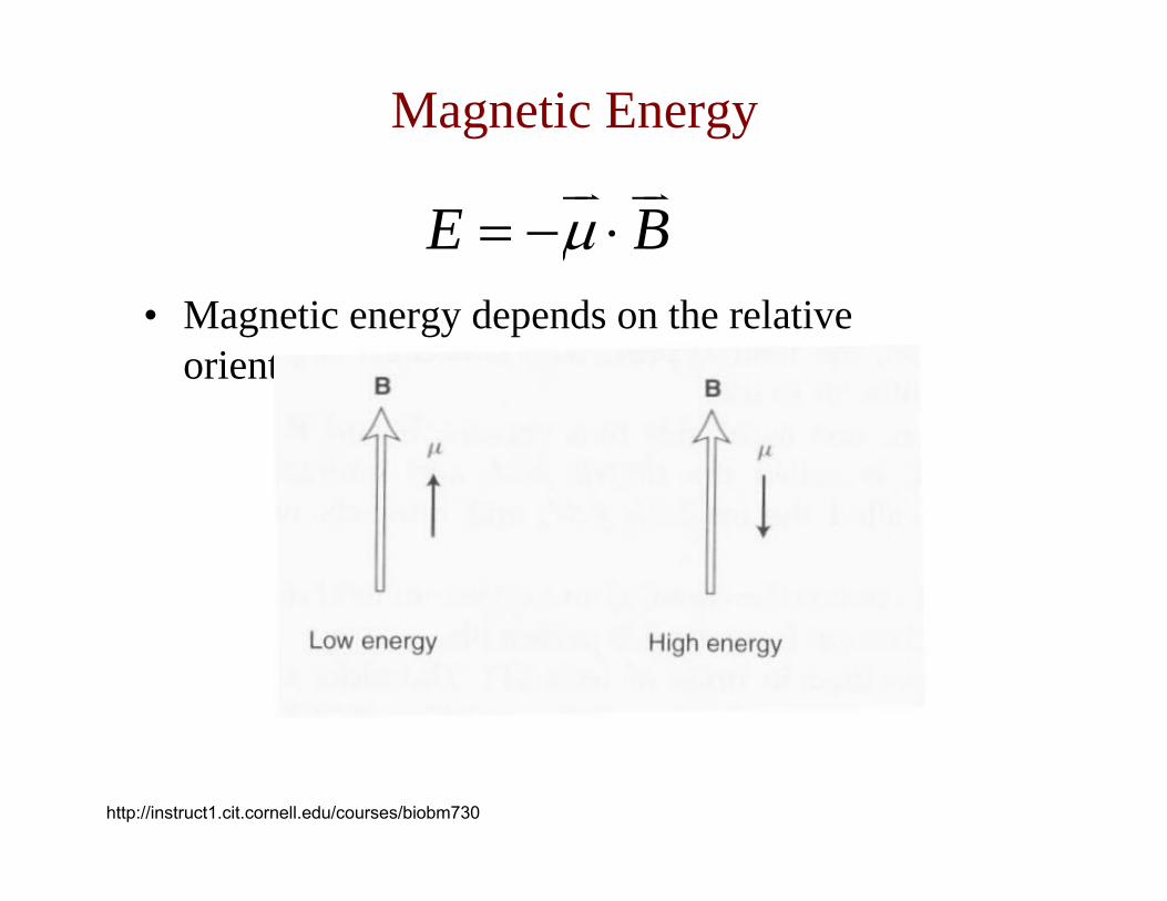
• Magnetic energy depends on the relative orientations of µ and B:
Magnetic Energy
E Bµ= − ⋅
http://instruct1.cit.cornell.edu/courses/biobm730

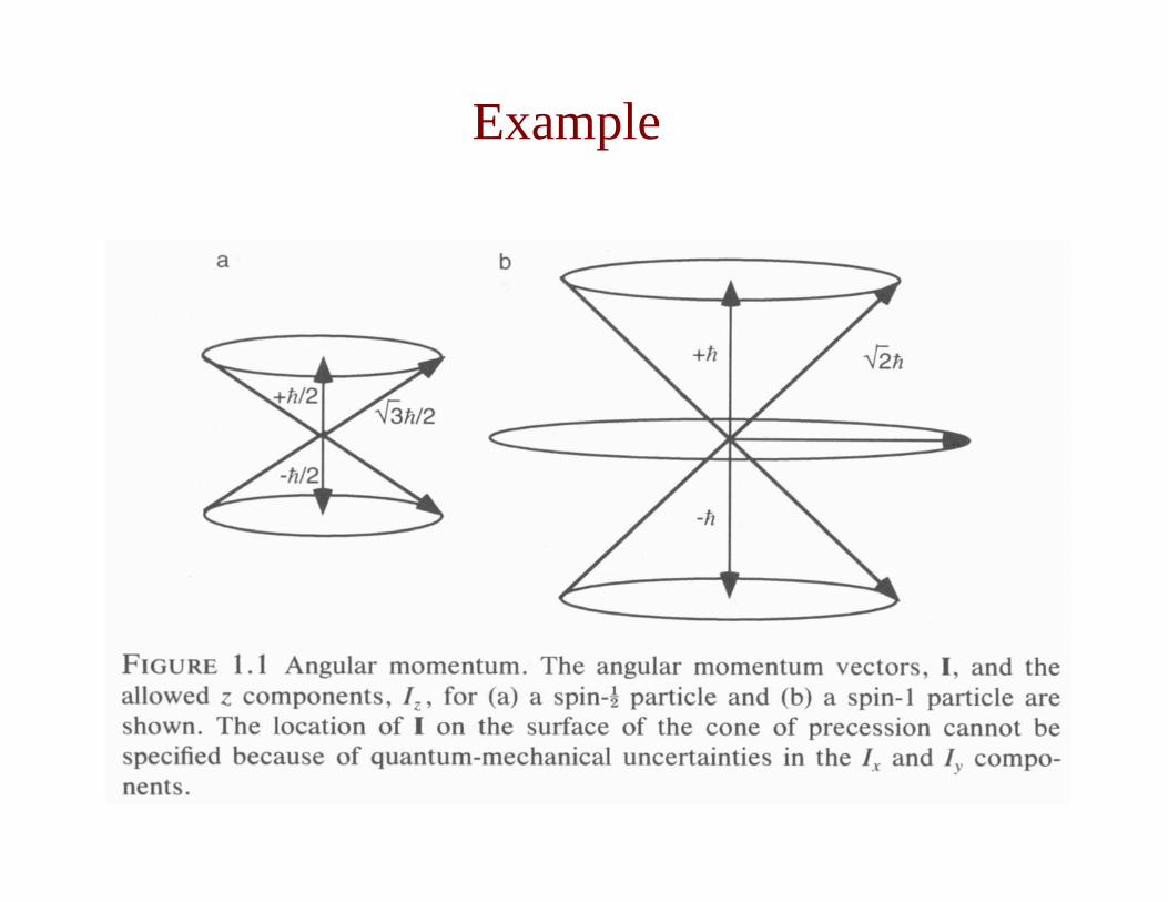
Angular Momentum and Projection Quantum Number
Magnitude of the angular momentum vector is fixed by the value of the nuclear spin quantum number
and that the z-component of the angular momentum vector is given by
where m is the magnetic quantum number:m = (-I, -I+1, ..., I-1, I)
Iz has 2I+1 possible values
I = I(I +1)
zI m=
Example

• No magnetic field:(2I+1) spin states are degenerate (i.e. they all have the
same energy).
• With magnetic field:Spin states separate in energy (larger values of m have
lower energy)
• The separation of energy levels in a magnetic field is called the nuclear Zeeman effect. The energy of a spin state is given by:
Effect of an External Magnetic Field
; E B Iµ µ γ= − ⋅ =

Magnetic Quantum Number and Interaction Energy
Em = −mBoγ
I I(I 1); zI m= + =
Thus, the discrete values of Iz are always smaller than |I|. The minimum energy occurs when the projection of µ onto B is the greatest. Hence, the energies of the m allowed spin states are proportional to their projection onto Bo:
where:Em = Energy of the statem = magnetic quantum numberBo = magnetic field strengthγ = gyromagnetic ratioħ = Planck’s constant/2π
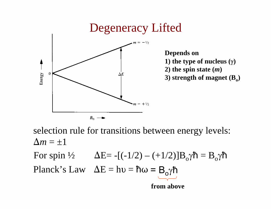
For spin ½ )E= -[(-1/2) – (+1/2)]Boγħ = BoγħPlanck’s Law )E = hL = ħT
selection rule for transitions between energy levels: )m = ±1
Depends on1) the type of nucleus (γ)2) the spin state (m) 3) strength of magnet (Bo)
from above
= Boγħ
Degeneracy Lifted

Energy Levels and Populations
© 2002, Michael Sattler http://www.embl.de/nmr/sattler/teaching
The Boltzmannequation tells us the population of a state if we know its energy:
TkEE
BeNN αβ
β
α
−
=

Interaction with RF Radiation

Electromagnetic radiation is composed of magnetic and electronic waves:
From: R.S. Macomber (1988) NMR spectroscopy: Essential Theory and Practice
• The frequency is defined as ν = 1/to, where to is the peak-to-peak time.
• A wave travels λ (distance) in to, so that the speed of the radiation (c, the speed of light, 3x10⁸ m/s) is defined as:
related inversely arefrequency and wavelength ∴== λνλ
otc
Electromagnetic Radiation
http://instruct1.cit.cornell.edu/courses/biobm730

Radiofrequency energy (∆E for nuclear spin state transitions):
λ = 1011 to 3 x 10⁷ nmν = 106 to 1010 Hz
By setting the frequency of electromagnetic radiation (ν, or equivalently ω) to the resonance condition, transitions between nuclear spin states can be induced
(i.e. one can do NMR spectroscopy!).
ων ==∆ hE
allowed spin states
Electromagnetic Radiation
http://instruct1.cit.cornell.edu/courses/biobm730
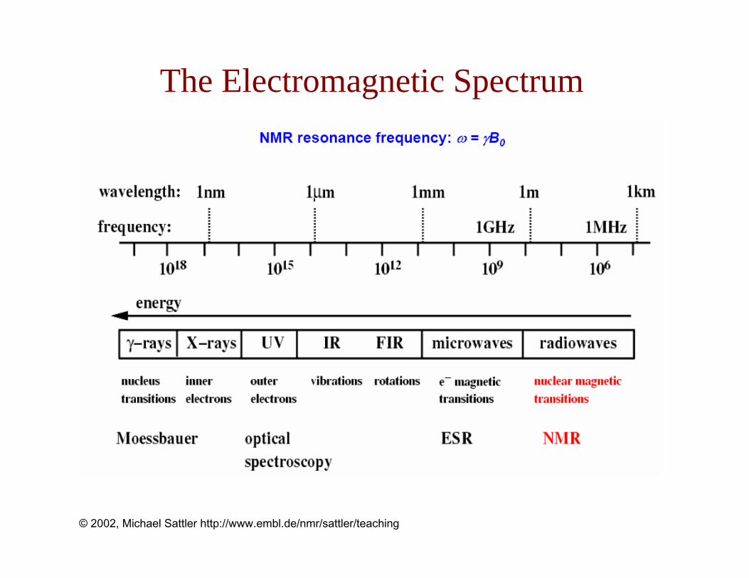
The Electromagnetic Spectrum
© 2002, Michael Sattler http://www.embl.de/nmr/sattler/teaching

Resonance (To), Bo and γ
Resonance condition: ∆E = hν = ħω = Boγħ
Resonance (Larmor) frequency for exciting nuclear spin transition:
ωo = Boγ
ωo = Boγ
∆Ε = ħωo
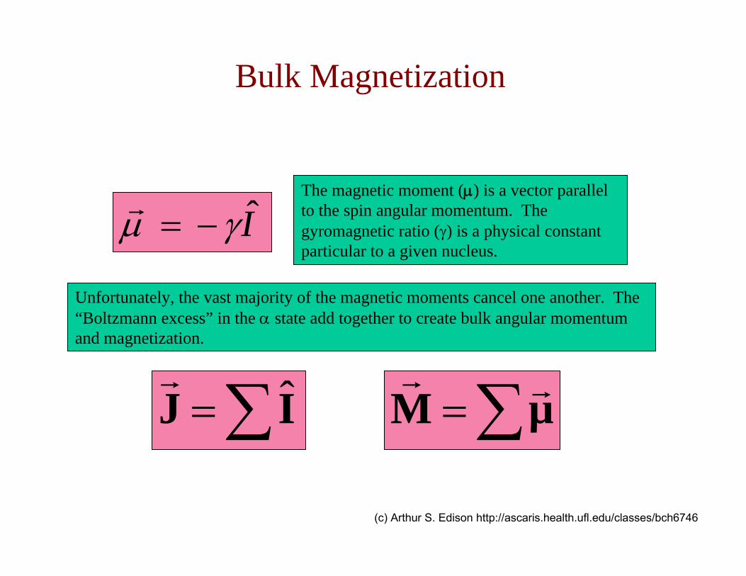
The magnetic moment (µ) is a vector parallel to the spin angular momentum. The gyromagnetic ratio (γ) is a physical constant particular to a given nucleus.
Bulk Magnetization
Unfortunately, the vast majority of the magnetic moments cancel one another. The “Boltzmann excess” in the α state add together to create bulk angular momentum and magnetization.
∑= µM∑= IJ ˆ
Iγµ −=
(c) Arthur S. Edison http://ascaris.health.ufl.edu/classes/bch6746

Individual magnetic moments:
µ MBulk Magnetization:
0B 0B
∑= µM
Iγµ −=(c) Arthur S. Edison http://ascaris.health.ufl.edu/classes/bch6746
Bulk Magnetization

Classical physics tells us about the motion of a magnet in a magnetic field
This precession is very similar to the motion of a spinning gyroscope or top in a gravitational field
d tdt
mL r g( )= ×
L(t) is the gyroscope’s angular momentum, r its radius from the fixed point of rotation, m its mass and g the force of gravity.
The change in angular momentum per unit time is torque (τ)BM
dtd
×=J
(c) Arthur S. Edison http://ascaris.health.ufl.edu/classes/bch6746
Classical Motion of a Magnet
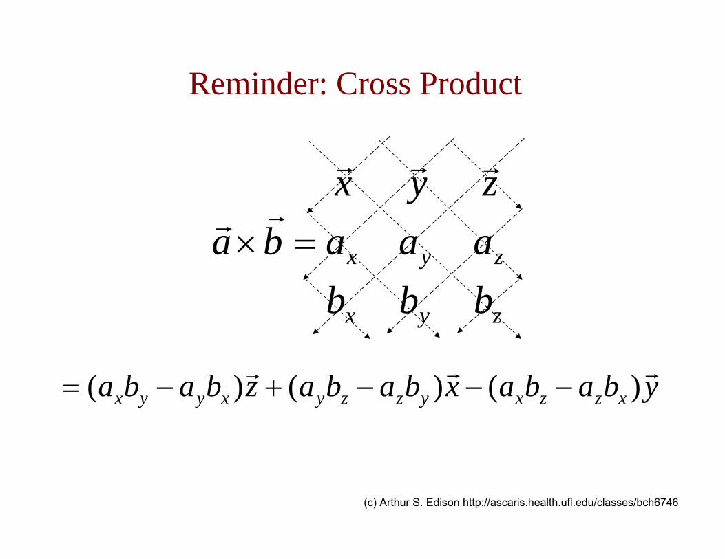
Reminder: Cross Product
zyx
zyx
bbbaaazyx
ba =×
ybabaxbabazbaba xzzxyzzyxyyx )()()( −−−+−=
(c) Arthur S. Edison http://ascaris.health.ufl.edu/classes/bch6746

Direction of Precession
http://instruct1.cit.cornell.edu/courses/biobm730
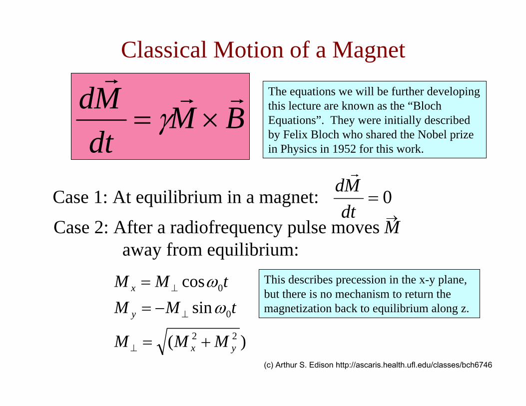
BMdtMd
×= γ
Case 1: At equilibrium in a magnet: 0=dtMd
Case 2: After a radiofrequency pulse moves M away from equilibrium:
)(
sincos
22
0
0
yx
y
x
MMM
tMMtMM
+=
−==
⊥
⊥
⊥
ωω
→
This describes precession in the x-y plane, but there is no mechanism to return the magnetization back to equilibrium along z.
The equations we will be further developing this lecture are known as the “Bloch Equations”. They were initially described by Felix Bloch who shared the Nobel prize in Physics in 1952 for this work.
(c) Arthur S. Edison http://ascaris.health.ufl.edu/classes/bch6746
Classical Motion of a Magnet

In order to allow the system to return to equilibrium, Felix Bloch made the following modifications to the basic equation
d tdt
t t t MM M B R M( ) ( ) ( ) ( ( ) )= × − −γ 0
Empirical modification in which a “relaxation matrix” R acts on magnetization that is different from the equilibrium state, M0 (cannot be justified with classical physics, need QM).
(c) Arthur S. Edison http://ascaris.health.ufl.edu/classes/bch6746
Bloch Equations

d tdt
t t t MM M B R M( ) ( ) ( ) ( ( ) )= × − −γ 0
This equation is easiest to understand broken into its matrix components:
1
0
T)()]()()([)( MtMtBMtBtM
dttdM z
xyyxz −
−−= γ Magnetization along the z-axis
2T)()]()()([)( tMtBMtBtM
dttdM x
yzzyx −−= γ Magnetization
along the x-axis
2T)(
)]()()([)( tM
tBMtBtMdt
tdM yzxxz
y −−= γ Magnetization along the y-axis
(c) Arthur S. Edison http://ascaris.health.ufl.edu/classes/bch6746
Bloch Equations

1
011 T
)()]()()([)( MtMtBMtBtMdt
tdM zxy
yx
z −−−= γ B1 refers to the rf
field in the rotating frame
21 T
)()()()( tMtBMtMdt
tdM xyzy
x −−∆−= γω
21 T
)()()(
)( tMMtBtM
dttdM y
xx
zy −∆+= ωγ
Substituting ∆ω=-γB0-ωrf (where B0=Bz and is not time-dependent) into the Bloch equations yields:
(c) Arthur S. Edison http://ascaris.health.ufl.edu/classes/bch6746
Bloch Equations in the Rotating Frame

1
0
T)()()()()()(
11
MtMtBtMtBtMdt
tdM zxy
yx
z −−−= γγ
y-axis pulse x-axis pulse
2T)(
)()()()(
1
tMtMtBtM
dttdM y
xx
zy −∆+= ωγ
x-axis pulse
2T)()()()()(
1
tMtBtMtMdt
tdM xyzy
x −−∆−= γω
y-axis pulse
In the Bloch equations, magnetic fields along the x and y axes create B1 fields or pulses. These are typically applied for short durations, and the length of time the pulse is turned on is adjusted to give a desired rotation (such as 90 or 180 degrees).
(c) Arthur S. Edison http://ascaris.health.ufl.edu/classes/bch6746
Bloch Equations

Populations of Spin States and RF Pulses
90˚ and 180˚pulses
From: J. Cavanagh et al. (1996) Protein NMR spectroscopy
equilibrium non-equilibrium non-equilibrium
Transverse relaxation Longitudinal relaxation
http://instruct1.cit.cornell.edu/courses/biobm730

1 2 3 4 5
-0.5
0.5
1
1 2 3 4 5
-0.75
-0.5
-0.25
0.25
0.5
0.75
1
MxMy
0.2
0.4
0.6
0.8Mz
-1-0.5
0
0.51
-0.5
0
0.5
1
0
0.25
0.5
0.75
1
-1-0.5
0
0.51
Mz
Mx
Myt
t t
In most NMR experiments, the pulses are short and the relaxation times are relatively long. We mainly worry about relaxation after the pulses are applied.
T1
T2 T2
(c) Arthur S. Edison http://ascaris.health.ufl.edu/classes/bch6746
Precession and Relaxation

Longitudinal Relaxation (T1)
• first order rate processd Mz (t)
dt=
Mo − Mz t( )( )T1
Mz (t) = Mo − Mo −Mz 0( )( )e−t T1
Mo = total magnetization
Mz(0) = magnetization along the z axis at t = 0
Bo
M
x
y
z
http://instruct1.cit.cornell.edu/courses/biobm730
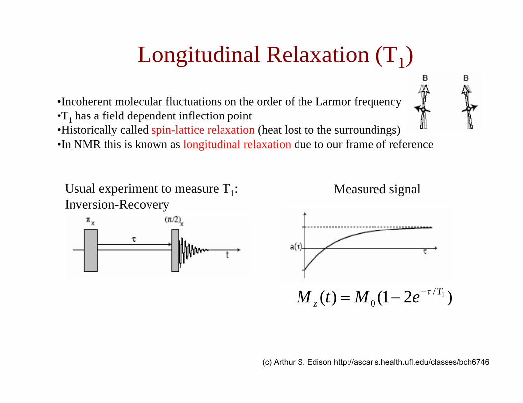
•Incoherent molecular fluctuations on the order of the Larmor frequency•T1 has a field dependent inflection point•Historically called spin-lattice relaxation (heat lost to the surroundings)•In NMR this is known as longitudinal relaxation due to our frame of reference
)21()( 1/0
Tz eMtM τ−−=
Usual experiment to measure T1: Inversion-Recovery
Measured signal
(c) Arthur S. Edison http://ascaris.health.ufl.edu/classes/bch6746
Longitudinal Relaxation (T1)

Buildup
Decay
http://instruct1.cit.cornell.edu/courses/biobm730
Longitudinal Relaxation (T1)
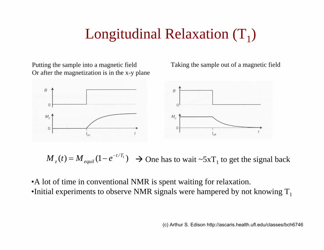
Putting the sample into a magnetic fieldOr after the magnetization is in the x-y plane
Taking the sample out of a magnetic field
)1()( 1/Ttequilz eMtM −−= One has to wait ~5xT1 to get the signal back
•A lot of time in conventional NMR is spent waiting for relaxation.•Initial experiments to observe NMR signals were hampered by not knowing T1
(c) Arthur S. Edison http://ascaris.health.ufl.edu/classes/bch6746
Longitudinal Relaxation (T1)
Relaxation back to equilibrium
Bo
M
x
y
z
http://instruct1.cit.cornell.edu/courses/biobm730
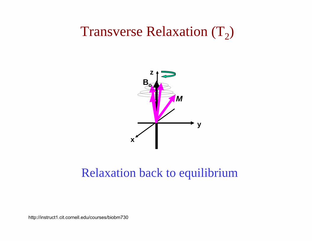
Transverse Relaxation (T2)

Inhomogeneous broadening: variations in the macroscopic magnetic field
Homogeneous broadening: fluctuating microscopic magnetic fields
•Molecular dynamics and spin-spin interactions more details later•Chemical exchange•Historically called spin-spin relaxation•In NMR we call it transverse relaxation loss of signal in the x-y plane
•Instrument limitations•Magnetic susceptibility
(c) Arthur S. Edison http://ascaris.health.ufl.edu/classes/bch6746
Transverse Relaxation (T2)

http://instruct1.cit.cornell.edu/courses/biobm730
Transverse Relaxation (T2)

2/)cos()( Ttoox etMtM −= ω
2/)sin()( Ttooy etMtM −= ω
Free Induction Decay
http://instruct1.cit.cornell.edu/courses/biobm730
Transverse Relaxation (T2)

Decreasing T2 (increasing relaxation)Broadening of peak in spectrum
(c) Arthur S. Edison http://ascaris.health.ufl.edu/classes/bch6746
Transverse Relaxation (T2)
The Biomolecular NMR Experiment

Hardware
© 2002, Michael Sattler http://www.embl.de/nmr/sattler/teaching
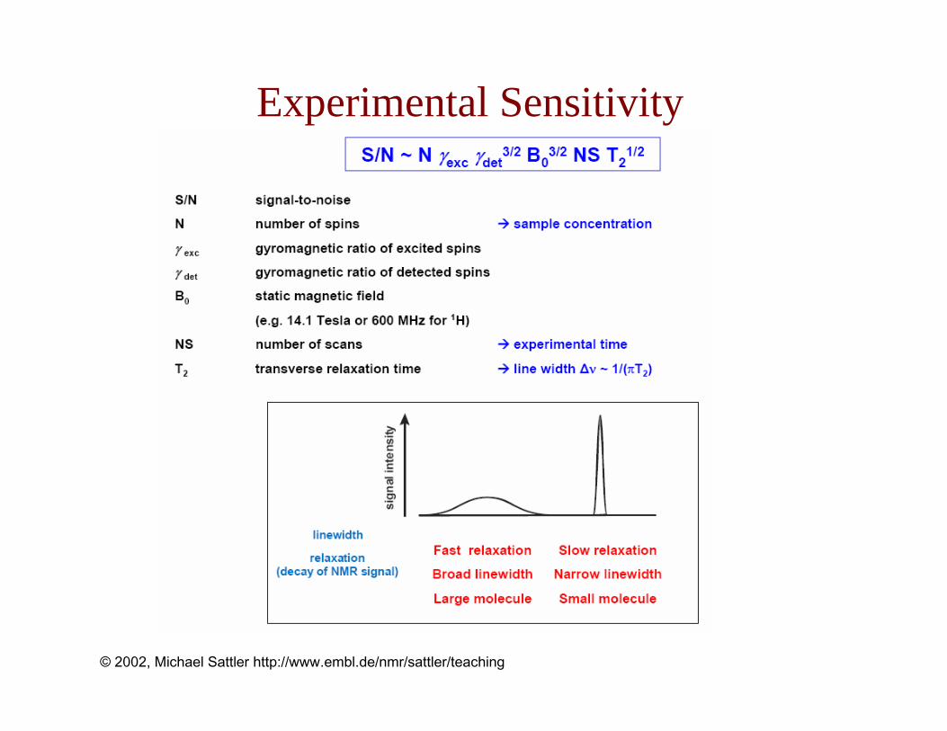
Experimental Sensitivity
© 2002, Michael Sattler http://www.embl.de/nmr/sattler/teaching
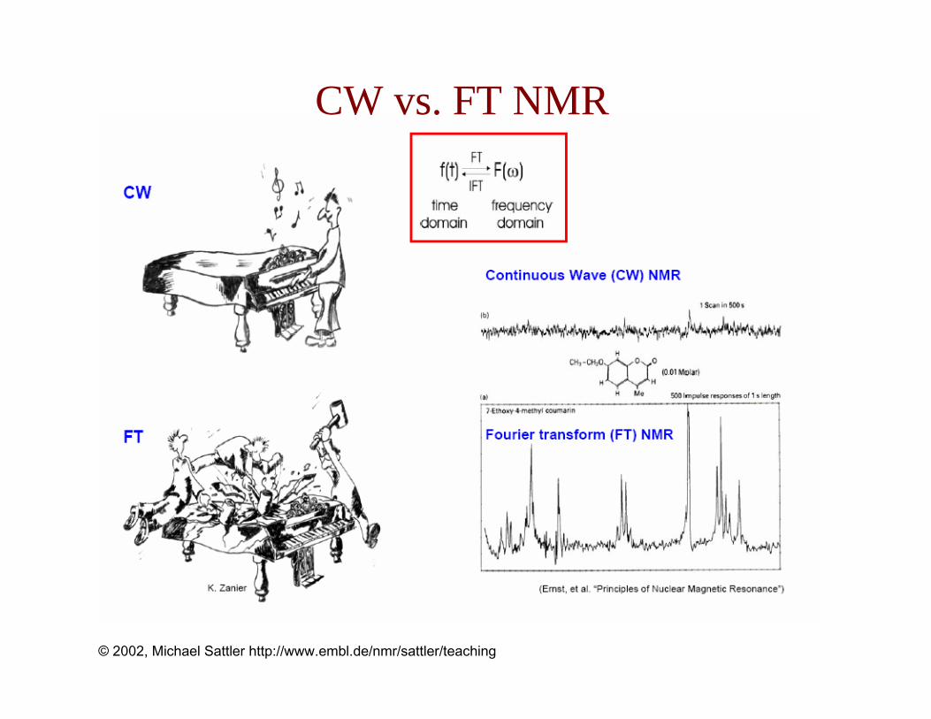
CW vs. FT NMR
© 2002, Michael Sattler http://www.embl.de/nmr/sattler/teaching
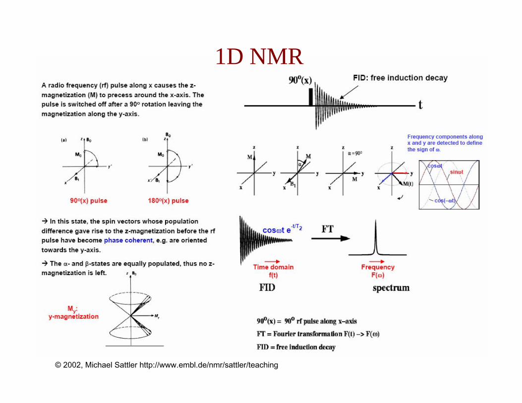
1D NMR
© 2002, Michael Sattler http://www.embl.de/nmr/sattler/teaching

1D Spectrum of a Protein
© 2002, Michael Sattler http://www.embl.de/nmr/sattler/teaching

Chemical Shift
• A molecule may contain multiple protons that exist in unique electronic environments.
• Therefore not all protons are shielded to the same extent.
• Resonance differences in protons are very small (ppm).
• Measure differences in resonance energy relative to a reference.
• Tetramethylsilane (CH3)4Si (TMS) provides highly shielded reference (set to 0ppm).
Origin: Nuclear Shielding
• Nuclei are shielded by electrons.
• Induced field associated with orbiting electrons.
• Require stronger magnetic field than H0.
• Increased shielding requires greater applied field strength to achieve resonance.
http://mason.gmu.edu/~bbishop1/chem318.1stlecture.ppt
Chemical Shift (δ, ppm) = Observed chemical shift from TMS (Hz)Sptectrometer frequencey (MHz)
= ppmSpectrometer frequency (MHz)

• Hypothetical NMR spectra.• Shows TMS reference.• Chemical shifts (δ, ppm) given relative to TMS
http://mason.gmu.edu/~bbishop1/chem318.1stlecture.ppt
Chemical Shift
A b s o r b a n c e
0123456789101112
Increasing magnetic field strength
δ, ppm
TMS
CH
HH
TMS as reference is set to 0 ppm
Representative peak,3 equivalent protons
Increased sheilding of nuclei

• Protons in the same environment will have the same chemical shift.
• Protons in different environments have different chemical shifts.
• Protons with the same chemical shift are referred to as chemically equivalent.
• Integrated area of peak is proportional to the number of protons.
H
H
H
H
H
HC
C
H
H
H
H
H
H
H
C
H
H
H
H
H
H
H
C
C
H
H
H
H
C
H
H H
H
http://mason.gmu.edu/~bbishop1/chem318.1stlecture.ppt
Chemical Shift: Equivalency

Chemical shifts are influenced by the electronic environment. Therefore, they are diagnostic for particular types of molecular structures. The following figure indicates average ranges of chemical shifts for protons in different types of molecules.
(c) http://www.cem.msu.edu/~reusch/OrgPage/nmr.htm
Chemical Shift

Chemical Shift: Summary
© 2002, Michael Sattler http://www.embl.de/nmr/sattler/teaching

Scalar / J-Coupling
© 2002, Michael Sattler http://www.embl.de/nmr/sattler/teaching

Levitt
(c) Arthur S. Edison http://ascaris.health.ufl.edu/classes/bch6746
Scalar / J-Coupling

J-Coupling and Chemical Shift: Example
1H NMR 1D spectra of Methyl α-D-Arabinofuranoside in CD3CN. Collected at 11.7 T by Jim Rocca in AMRIS.
All of the splittings are J couplings
(c) Arthur S. Edison http://ascaris.health.ufl.edu/classes/bch6746
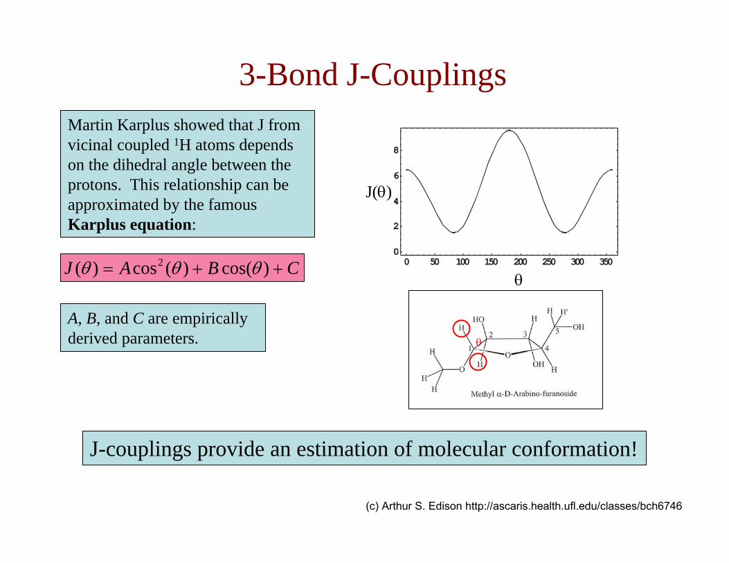
3-Bond J-CouplingsMartin Karplus showed that J from vicinal coupled 1H atoms depends on the dihedral angle between the protons. This relationship can be approximated by the famous Karplus equation:
CBAJ ++= )cos()(cos)( 2 θθθ
A, B, and C are empirically derived parameters.
J-couplings provide an estimation of molecular conformation!
J(θ)
θ
θ
(c) Arthur S. Edison http://ascaris.health.ufl.edu/classes/bch6746

Karplus Relation and Peptide Torsion Angle Φ
Levitt
(c) Arthur S. Edison http://ascaris.health.ufl.edu/classes/bch6746
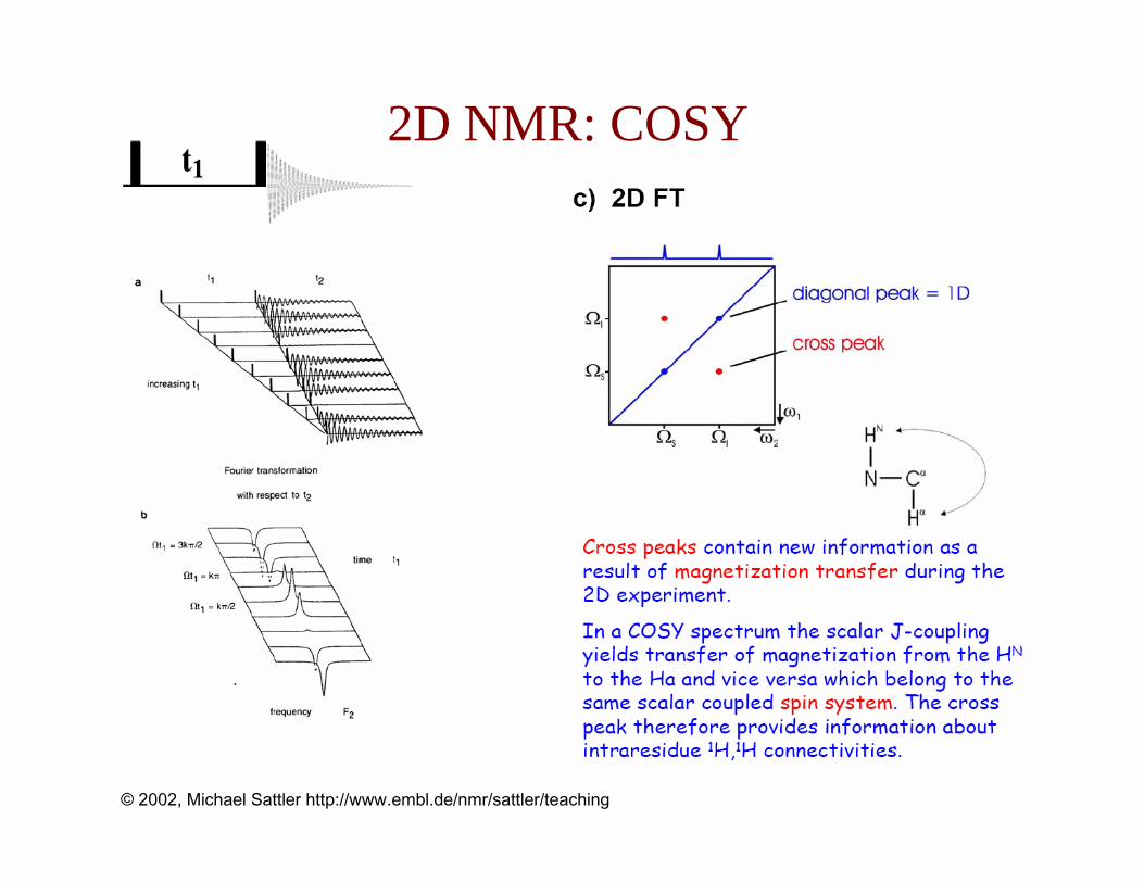
2D NMR: COSY
© 2002, Michael Sattler http://www.embl.de/nmr/sattler/teaching

•The nuclear Overhauser effect (NOE) is in incoherent process in which two nuclear spins “cross-relax”. Recall that a single spin can relax by T1(longitudinal or spin-latice) or T2 (transverse or spin-spin) mechanisms. Nuclear spins can also cross-relax through dipole-dipole interactions and other mechanisms. This cross relaxation causes changes in one spin through perturbations of the other spin.
•The NOE is dependent on many factors. The major factors are molecular tumbling frequency and internuclear distance. The intensity of the NOE is proportional to r-6 where r is the distance between the 2 spins.
•Since protons have a higher polarization than carbons and the same sign of gamma they increase the observed carbon intensities.
(c) Arthur S. Edison http://ascaris.health.ufl.edu/classes/bch6746
Nuclear Overhauser Effect (NOE)

Two nuclear spins within about 5 Åwill interact with each other through space. This interaction is called cross-relaxation, and it gives rise to the nuclear Overhauser effect (NOE).
Two spins have 4 energy levels, and the transitions along the edges correspond to transitions of one or the other spin alone. W2 and W0 are the cross-relaxation pathways, which depend on the tumbling of the molecule.
(c) Arthur S. Edison http://ascaris.health.ufl.edu/classes/bch6746
Nuclear Overhauser Effect (NOE)

The NOE only can measure distances up to about 5 Å, because the effect depends on r-6 where r is the distance between the two interacting protons.
1/r6
r
(c) Arthur S. Edison http://ascaris.health.ufl.edu/classes/bch6746
Nuclear Overhauser Effect (NOE)

When two nuclear spins are within 5 Å, they will cross-relax. If one spin (S) is saturated (red lines along the edge), the system is not in equilibrium anymore. Magnetization will either flow from the top to the bottom (W2 active) or from the right to left (W0 active). The difference in energy between ββ and αα is twice the spectrometer frequency, and molecular motions about that frequency are required for the transition. The difference between αβ and βα is very small, and very slow molecular motions (e.g. proteins) will excite that transition.
(c) Arthur S. Edison http://ascaris.health.ufl.edu/classes/bch6746
Nuclear Overhauser Effect (NOE)

Residual Dipolar Couplings
© 2002, Michael Sattler http://www.embl.de/nmr/sattler/teaching
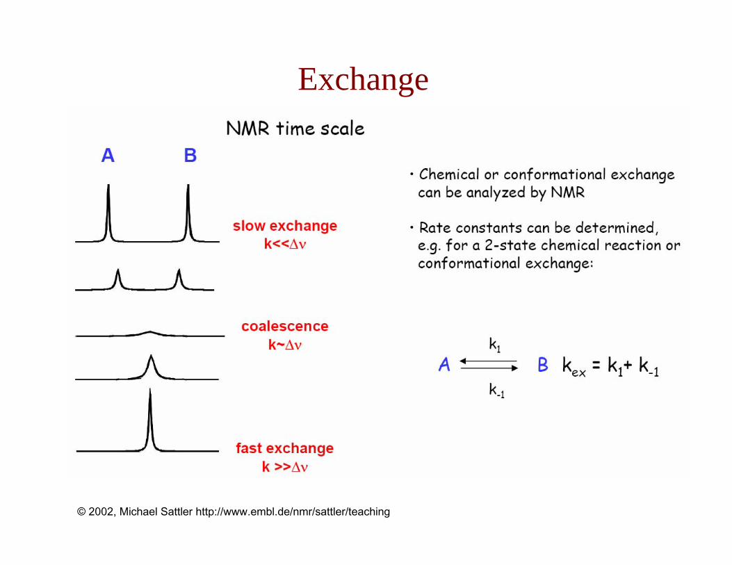
Exchange
© 2002, Michael Sattler http://www.embl.de/nmr/sattler/teaching

NMR Time Scales
© 2002, Michael Sattler http://www.embl.de/nmr/sattler/teaching

NMR Observables
© 2002, Michael Sattler http://www.embl.de/nmr/sattler/teaching

Structure Determination
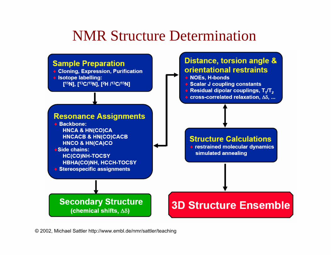
NMR Structure Determination
© 2002, Michael Sattler http://www.embl.de/nmr/sattler/teaching

NMR Structure Determination
© 2002, Michael Sattler http://www.embl.de/nmr/sattler/teaching
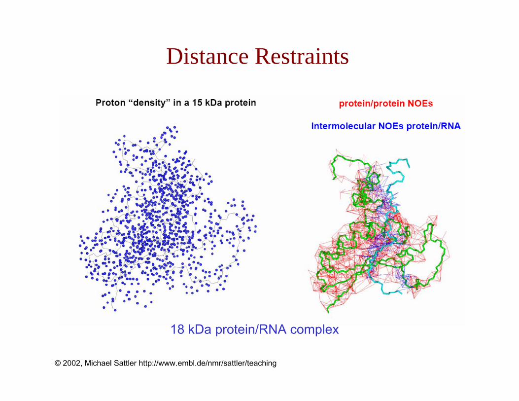
Distance Restraints
© 2002, Michael Sattler http://www.embl.de/nmr/sattler/teaching
18 kDa protein/RNA complex
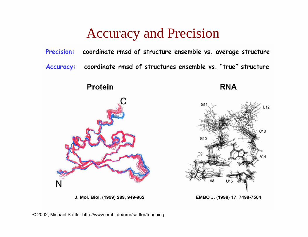
Accuracy and Precision
© 2002, Michael Sattler http://www.embl.de/nmr/sattler/teaching

Problems with Higher Molecular Weights
© 2002, Michael Sattler http://www.embl.de/nmr/sattler/teaching
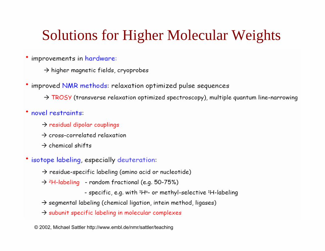
Solutions for Higher Molecular Weights
© 2002, Michael Sattler http://www.embl.de/nmr/sattler/teaching

TROSY and 2H-Labeling
© 2002, Michael Sattler http://www.embl.de/nmr/sattler/teaching

Increase in Molecular Weight
© 2002, Michael Sattler http://www.embl.de/nmr/sattler/teaching

NMR Tools for Protein-Ligandand Protein-Protein Interactions

Two-Site Exchange
© 2002, Michael Sattler http://www.embl.de/nmr/sattler/teaching

NMR Titrations
© 2002, Michael Sattler http://www.embl.de/nmr/sattler/teaching

NMR in Drug Research
© 2002, Michael Sattler http://www.embl.de/nmr/sattler/teaching

GroEL/ES Subunit Labeling
© 2002, Michael Sattler http://www.embl.de/nmr/sattler/teaching

Molecular Interface Mapping
© 2002, Michael Sattler http://www.embl.de/nmr/sattler/teaching

Molecular Interface Mapping
© 2002, Michael Sattler http://www.embl.de/nmr/sattler/teaching
Characterizing Protein Dynamics

Backbone Dynamics – Multidomain Proteins
© 2002, Michael Sattler http://www.embl.de/nmr/sattler/teaching

Enzyme Dynamics During Catalysis
© 2002, Michael Sattler http://www.embl.de/nmr/sattler/teaching

Enzyme Dynamics During Catalysis
© 2002, Michael Sattler http://www.embl.de/nmr/sattler/teaching
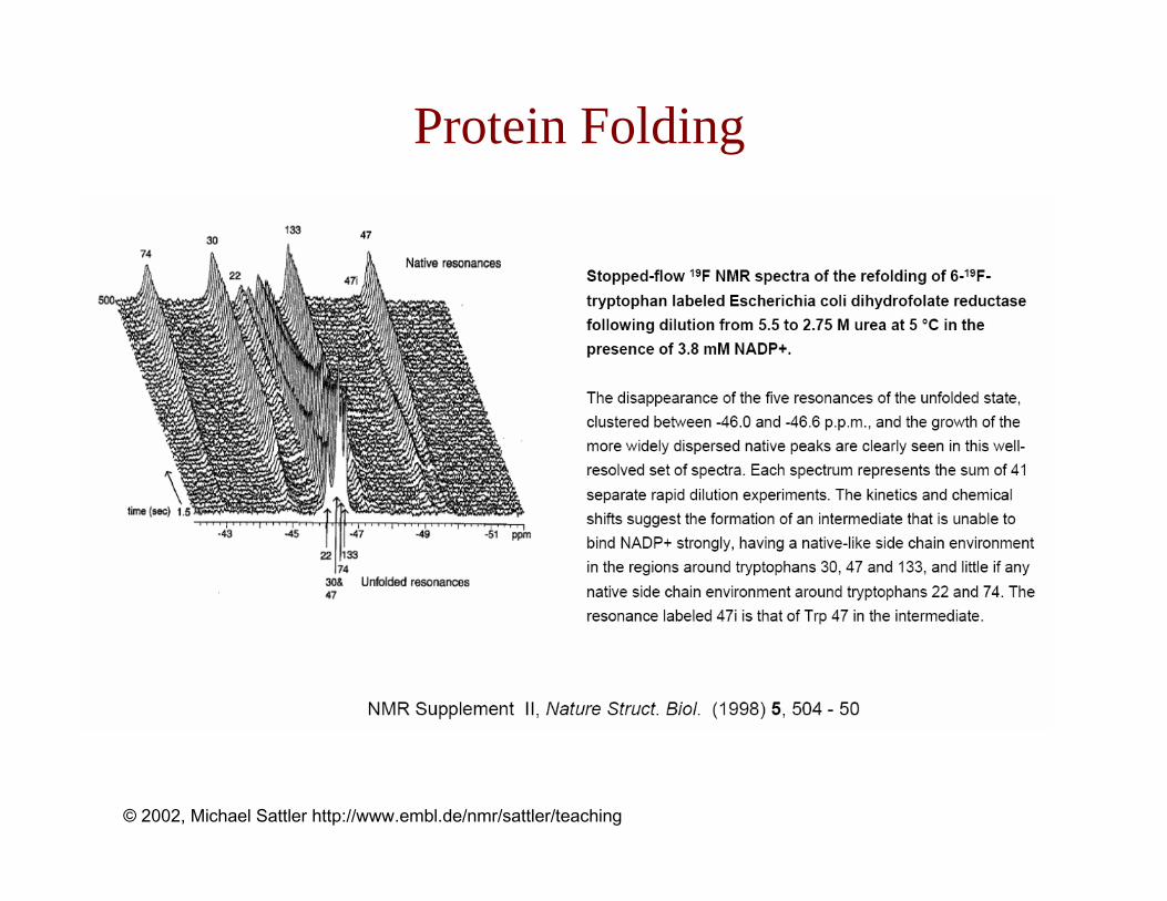
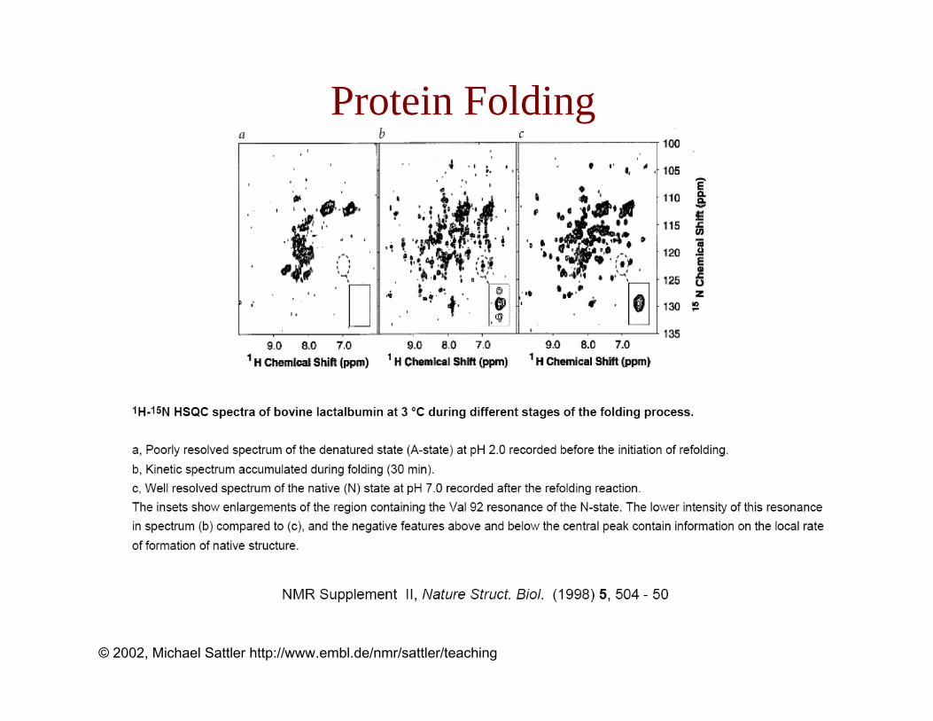
Protein Folding
© 2002, Michael Sattler http://www.embl.de/nmr/sattler/teaching
Protein Folding
© 2002, Michael Sattler http://www.embl.de/nmr/sattler/teaching

Resources and Further ReadingWWW:http://www.embl.de/nmr/sattler/teaching
NMR theory:• Spin dynamics - basics of nuclear magnetic resonanceMalcolm H. Levitt, Wiley 2001• Protein NMR spectroscopy – Principles and Practice. Cavanagh, Fairbrother, PalmerIII, Skelton. Academic Press (1996)• Multidimensional NMR in liquids - Basic principles and experimental methods. van de Ven, VCH (1995)• Nuclear Magnetic Resonance Spectroscopy. Harris. Longman (1983)• Principles of NMR in one and two dimensions. Ernst, Bodenhausen, Wokaun. Oxford (1989)
Biomolecular NMR:• NMR of Proteins and Nucleic Acids. Wüthrich. Wiley (1986)• Nature Struct. Biol. (1997) 4, 841-865 & 5, 492-522 (NMR supplement I & II)• NMR spectroscopy of large molecules and multimolecular assemblies in solution. Wider, Wüthrich Curr. Op. Struct. Biol. (1999) 9, 594-601