Nuclear integration of positive Dpp signals, antagonistic ... · (Azpiazu et al., 1996) of a stage...
14
1429 Introduction Tissues and organs develop from primordial cells that arise in precisely defined spatial and temporal patterns within a particular germ layer of the early embryo. These patterns are typically generated by combinatorial cues, whose restricted domains of activity intersect at specific positions within a larger field of cells. In order to understand early organogenesis, it is necessary to identify these cues and to determine the mechanisms by which they cooperate at the developmental and molecular level to elicit their responses. Tissue development in the Drosophila mesoderm has been a favorable system in which to study these events. Upon the spreading of the mesodermal cell layer underneath the embryonic ectoderm, the progenitor cells of different organs, such as the dorsal vessel, somatic and visceral muscles, are generated at stereotyped locations within the mesoderm. Specifically, in the dorsal region of the mesoderm (which has been studied in most detail), the progenitors of cardioblasts, pericardial cells, specific dorsal somatic muscles and circular midgut muscles are generated (Campos-Ortega and Hartenstein, 1997; Frasch and Nguyen, 1999). Signals from the dorsal ectoderm mediated by Dpp are required, but not sufficient, for the induction of all of these progenitor cells in the dorsal mesoderm (Staehling-Hampton et al., 1994; Frasch, 1995). Importantly, the Dpp signals need to act in concert with mesoderm-intrinsic regulators, which make the mesodermal cells competent to respond. One of the key regulators intrinsic to the mesoderm is the NK homeobox gene tinman, which, like dpp, is required for the induction of all dorsal mesodermal cell types (Azpiazu and Frasch, 1993; Bodmer, 1993; Yin and Frasch, 1998). tinman itself is initially activated in the early mesoderm by twist and, just prior to cell specification events, its expression is prolonged by Dpp signals specifically in the dorsal mesoderm (Bodmer et al., 1990; Frasch, 1995; Yin et al., 1997). In addition to these dorsal cues, differentially active cues modulate the specific responses in the mesoderm along the anteroposterior (AP) axis. Notably Wg, which is expressed in transversely striped domains within the A compartments of the ectoderm, is required in combination with Dpp for the Tissue induction during embryonic development relies to a significant degree on the integration of combinatorial regulatory inputs at the enhancer level of target genes. During mesodermal tissue induction in Drosophila, various combinations of inductive signals and mesoderm-intrinsic transcription factors cooperate to induce the progenitors of different types of muscle and heart precursors at precisely defined positions within the mesoderm layer. Dpp signals are required in cooperation with the mesoderm-specific NK homeodomain transcription factor Tinman (Tin) to induce all dorsal mesodermal tissue derivatives, which include dorsal somatic muscles, the dorsal vessel and visceral muscles of the midgut. Wingless (Wg) signals modulate the responses to Dpp/Tin along anteroposterior positions by cooperating with Dpp/Tin during dorsal vessel and somatic muscle induction while antagonizing Dpp/Tin during visceral mesoderm induction. As a result, dorsal muscle and cardiac progenitors form in a pattern that is reciprocal to that of visceral muscle precursors along the anteroposterior axis. Our present study addresses how positive Dpp signals and antagonistic Wg inputs are integrated at the enhancer level of bagpipe (bap), a NK homeobox gene that serves as an early regulator of visceral mesoderm development. We show that an evolutionarily conserved bap enhancer element requires combinatorial binding sites for Tin and Dpp-activated Smad proteins for its activity. Adjacent binding sites for the FoxG transcription factors encoded by the Sloppy paired genes (slp1 and slp2), which are direct targets of the Wg signaling cascade, serve to block the synergistic activity of Tin and activated Smads during bap induction. In addition, we show that binding sites for yet unknown repressors are essential to prevent the induction of the bap enhancer by Dpp in the dorsal ectoderm. Our data illustrate how the same signal combinations can have opposite effects on different targets in the same cells during tissue induction. Key words: Drosophila, Dpp, Wg Summary Nuclear integration of positive Dpp signals, antagonistic Wg inputs and mesodermal competence factors during Drosophila visceral mesoderm induction Hsiu-Hsiang Lee* and Manfred Frasch † Brookdale Department of Molecular, Cell and Developmental Biology, Box 1026, Mount Sinai School of Medicine, New York, NY 10029, USA *Present address: Genetics, Development and Behavioral Sciences Building, 1550 4th Street, UCSF, San Francisco, CA 94158, USA † Author for correspondence (e-mail: [email protected]) Accepted 6 January 2005 Development 132, 1429-1442 Published by The Company of Biologists 2005 doi:10.1242/dev.01687 Research article Development
Transcript of Nuclear integration of positive Dpp signals, antagonistic ... · (Azpiazu et al., 1996) of a stage...

1429
IntroductionTissues and organs develop from primordial cells that arise inprecisely defined spatial and temporal patterns within aparticular germ layer of the early embryo. These patterns aretypically generated by combinatorial cues, whose restricteddomains of activity intersect at specific positions within alarger field of cells. In order to understand early organogenesis,it is necessary to identify these cues and to determine themechanisms by which they cooperate at the developmental andmolecular level to elicit their responses.
Tissue development in the Drosophila mesoderm has beena favorable system in which to study these events. Upon thespreading of the mesodermal cell layer underneath theembryonic ectoderm, the progenitor cells of different organs,such as the dorsal vessel, somatic and visceral muscles, aregenerated at stereotyped locations within the mesoderm.Specifically, in the dorsal region of the mesoderm (which hasbeen studied in most detail), the progenitors of cardioblasts,pericardial cells, specific dorsal somatic muscles and circularmidgut muscles are generated (Campos-Ortega and
Hartenstein, 1997; Frasch and Nguyen, 1999). Signals from thedorsal ectoderm mediated by Dpp are required, but notsufficient, for the induction of all of these progenitor cells inthe dorsal mesoderm (Staehling-Hampton et al., 1994; Frasch,1995). Importantly, the Dpp signals need to act in concert withmesoderm-intrinsic regulators, which make the mesodermalcells competent to respond. One of the key regulators intrinsicto the mesoderm is the NK homeobox gene tinman, which, likedpp, is required for the induction of all dorsal mesodermal celltypes (Azpiazu and Frasch, 1993; Bodmer, 1993; Yin andFrasch, 1998). tinman itself is initially activated in the earlymesoderm by twist and, just prior to cell specification events,its expression is prolonged by Dpp signals specifically in thedorsal mesoderm (Bodmer et al., 1990; Frasch, 1995; Yin etal., 1997).
In addition to these dorsal cues, differentially active cuesmodulate the specific responses in the mesoderm along theanteroposterior (AP) axis. Notably Wg, which is expressed intransversely striped domains within the A compartments ofthe ectoderm, is required in combination with Dpp for the
Tissue induction during embryonic development relies toa significant degree on the integration of combinatorialregulatory inputs at the enhancer level of target genes.During mesodermal tissue induction in Drosophila, variouscombinations of inductive signals and mesoderm-intrinsictranscription factors cooperate to induce the progenitors ofdifferent types of muscle and heart precursors at preciselydefined positions within the mesoderm layer. Dpp signalsare required in cooperation with the mesoderm-specific NKhomeodomain transcription factor Tinman (Tin) to induceall dorsal mesodermal tissue derivatives, which includedorsal somatic muscles, the dorsal vessel and visceralmuscles of the midgut. Wingless (Wg) signals modulate theresponses to Dpp/Tin along anteroposterior positions bycooperating with Dpp/Tin during dorsal vessel and somaticmuscle induction while antagonizing Dpp/Tin duringvisceral mesoderm induction. As a result, dorsal muscleand cardiac progenitors form in a pattern that is reciprocalto that of visceral muscle precursors along the
anteroposterior axis. Our present study addresses howpositive Dpp signals and antagonistic Wg inputs areintegrated at the enhancer level of bagpipe (bap), a NKhomeobox gene that serves as an early regulator of visceralmesoderm development. We show that an evolutionarilyconserved bap enhancer element requires combinatorialbinding sites for Tin and Dpp-activated Smad proteinsfor its activity. Adjacent binding sites for the FoxGtranscription factors encoded by the Sloppy paired genes(slp1 and slp2), which are direct targets of the Wg signalingcascade, serve to block the synergistic activity of Tin andactivated Smads during bap induction. In addition, weshow that binding sites for yet unknown repressors areessential to prevent the induction of the bap enhancer byDpp in the dorsal ectoderm. Our data illustrate how thesame signal combinations can have opposite effects ondifferent targets in the same cells during tissue induction.
Key words: Drosophila, Dpp, Wg
Summary
Nuclear integration of positive Dpp signals, antagonistic Wg inputsand mesodermal competence factors during Drosophila visceralmesoderm inductionHsiu-Hsiang Lee* and Manfred Frasch†
Brookdale Department of Molecular, Cell and Developmental Biology, Box 1026, Mount Sinai School of Medicine, New York,NY 10029, USA*Present address: Genetics, Development and Behavioral Sciences Building, 1550 4th Street, UCSF, San Francisco, CA 94158, USA†Author for correspondence (e-mail: [email protected])
Accepted 6 January 2005
Development 132, 1429-1442Published by The Company of Biologists 2005doi:10.1242/dev.01687
Research article
Dev
elop
men
t

1430
specification of the progenitors of cardioblasts, pericardial cellsand dorsal somatic muscles (Baylies et al., 1995; Wu et al.,1995). Conversely, the precursors of the midgut visceralmesoderm are induced by Dpp but suppressed by Wg (Frasch,1995; Azpiazu et al., 1996) (see http://www.eurekah.com/abstract.php?chapid=2028&bookid=162&catid=20). Hence,visceral mesoderm precursors arise in domains thatare alternating with those of cardiac and somatic muscleprogenitors along the AP axis in the dorsal mesoderm.Additional cues, which include signals through variousreceptor tyrosine kinases (RTKs) and the FGF receptorHeartless, then generate further subdivisions within thevisceral mesoderm as well as diverse identities among theprogenitors of cardiac and somatic muscle tissues (Carmena etal., 1998; Michelson et al., 1998; Englund et al., 2003; Lee etal., 2003). Mutual repression among induced regulatory genesalso plays a role (Han et al., 2002; Jagla et al., 2002). Thecombined actions of these regulators results in the spatiallyrestricted transcriptional activation of target genes, whichdrive genetic programs controlling the specification and/ordifferentiation of individual cells.
Recently, significant progress has been made in resolving theissue of how combinatorial inputs are integrated at the level ofenhancers of target genes in this system. A relatively simplesituation exists for the Dpp-responsive enhancer of tin, whichdoes not receive any differential inputs along the AP axis. Thisenhancer has been shown to contain several copies of bindingsites for Smads, which function as nuclear Dpp signalingeffectors, as well as binding sites for Tin protein. Each of thetwo types of binding sites are essential for enhancer activity(Xu et al., 1998). Thus, it appears that combinatorial bindingof Dpp-activated Smads and mesoderm-intrinsic Tin, togetherwith protein interactions between Smads and Tin (Zaffran etal., 2002), provides the synergism required for the active stateof the enhancer. A more complex situation, when comparedwith tin, is found for even-skipped (eve), a homeobox gene thatis induced in specific segmentally repeated progenitors ofpericardial cells and dorsal somatic muscles within the dorsalmesoderm (Frasch et al., 1987; Su et al., 1999). This patternof eve expression requires not only Dpp but also Wg signalsand RTK signals that are active in smaller areas within thefields where Dpp and Wg intersect (Frasch, 1995; Wu et al.,1995; Azpiazu et al., 1996; Carmena et al., 1998). As for theinduction of tin, these external signals require Tin as amesoderm-intrinsic activity for the induction of eve (Azpiazuand Frasch, 1993; Bodmer, 1993). Consistent with theseidentified inputs, the corresponding enhancer region of eve hasbeen found to contain functionally important binding sites forTin, Smads, the Wg effector dTCF/Lef-1 and Ets-domainprotein-binding sites that are presumed targets of RTK signals(Halfon et al., 2000; Knirr and Frasch, 2001; Han et al., 2002).A comparison between the situation in the eve versus the tinenhancer raises the question: why are the Smad and Tin sitesin the tin enhancer sufficient for its induction when the eveenhancer requires additional inputs from Wg via the dTCF/Lef-1 sites? Additional functional studies have provided answersto this question. A model was proposed, in which bounddTCF/Lef-1 acts as a repressor that abrogates the activity ofthe bound Tin and Smad proteins in the absence of Wg signals,whereas in the presence of Wg signals the repressive activityof dTCF/Lef-1 is abolished (Knirr and Frasch, 2001).
Consequently, Wg signals allow Tin/Smads (together withRTK signal effectors) to induce eve in segmentally repeatedclusters of cells within the dorsal mesoderm.
Herein, we define the distinct molecular inputs into a thirdenhancer, namely that of the NK-homeobox gene bagpipe(bap), which is induced by Dpp in the early dorsal mesodermduring the same period when tin and eve are being induced(Staehling-Hampton et al., 1994; Frasch, 1995). bap is a crucialregulator of the development of the trunk visceral mesodermand, hence, of midgut muscle development (Azpiazu andFrasch, 1993; Zaffran et al., 2001) (see http://www.eurekah.com/abstract.php?chapid=2028&bookid=162&catid=20). bapis induced in metameric clusters of cells within the dorsalmesoderm that alternate with those expressing eve and otherearly cardiac and dorsal muscle markers along the AP axis (seeFig. 1). This pattern is explained by the finding that the activityof Dpp to induce bap is abrogated by Wg signals (Azpiazu etal., 1996), which contrasts with the situation for eve, where Wgsynergizes with Dpp. Recent studies have shown that Wgsignals act indirectly during this process and function byinducing the forkhead domain genes sloppy paired 1 andsloppy paired 2 (slp1 and slp2) in striped domains within themesoderm (Lee and Frasch, 2000). Slp proteins, in turn, act assegmental repressors of bap induction (Riechmann et al., 1997;Lee and Frasch, 2000) (see Fig. 1). In common with eve (andtin), the induction of bap expression by Dpp signals alsorequire synergism with mesodermal tin (Azpiazu and Frasch,1993) (see Fig. 1). Our functional dissection of thecorresponding bap enhancer reveals interesting similarities anddifferences to the mesodermal tin and eve enhancers.Specifically, we show that a 267 bp element, bap3.2, whichrecapitulates the endogenous bap pattern in the dorsal trunkmesoderm, includes combinatorial binding sites for Tin andSmad proteins that are essential for its induction. By contrast,
Development 132 (6) Research article
Fig. 1. Summary of signaling and transcriptional pathways duringthe induction of trunk visceral mesoderm. Shown are threeparasegments of the mesoderm (divided into P and A domains)(Azpiazu et al., 1996) of a stage 10 embryo stained for bap mRNA(purple) and Eve protein (brown). The expression domains of Tin(schematically shown in blue) and the relevant Slp domains (red) arewithin the mesoderm, whereas Dpp (yellow) and Wg (brown) aresecreted from the overlying ectoderm. Inductive signals arerepresented by hatched arrows and transcriptional interactions arerepresented by solid arrows.
Dev
elop
men
t

1431Drosophila visceral mesoderm induction
binding sites for Slp are required for the segmental repressionof bap enhancer activity. We also show that the Slp-bindingsites exert an additional, positive function during bapinduction, in part through binding of Biniou (Bin), a forkheaddomain protein that is activated downstream of bap butprovides positive feedback on bap (Zaffran et al., 2001).Finally, we show that this bap enhancer includes elements thatprevent the induction of bap by Dpp in the dorsal ectodermand, thereby, contribute to the observed germ layer specificresponse. Similar elements were previously found in the Dpp-responsive enhancer of tin (Xu et al., 1998), and we providedata to indicate that the same repressing mechanism preventsinduction of both tin and bap in the dorsal ectoderm.Altogether, the data presented illustrate how Wg signals caneither antagonize or cooperate with Dpp signals at themolecular level. More generally, they extend our knowledge ofhow the molecular integration of combinatorial signals at thelevel of target enhancers generates mesoderm-specific outputswith precisely defined spatial patterns, which prefigure specifictissue primordia.
Materials and methodsConstruction of P-transformation plasmidsThe bap gene of Drosophila virilis was isolated from a genomiclibrary obtained from W. Hanna-Rose through J. D. Licht (Hanna-Rose et al., 1997) using bap cDNA of Drosophila melanogaster as aprobe. For the P-transformation constructs, upstream and downstreamDNA fragments of bap (D. melanogaster and D. virilis) were clonedinto the pCaSpeRhs43-βgal vector with reversed orientations relativeto the basal promoters, unless mentioned otherwise. The followingDNA constructs of D. melanogaster were made and tested in vivo:For bapDS3.5, a 3.5 kb SalI/HincII DNA downstream fragment; forbapH2-1.2, a 1.2 kb HincII/HincII DNA downstream fragment; andfor bap3, a 460 bp HincII/XhoI DNA downstream fragment werecloned into the vector. Constructs bap1 (432 bp), bap2 (460 bp),bap3.1 (255 bp), bap3.2 (267 bp) and bap3.2.1 (180 bp) weregenerated by PCR reaction with BamHI/EcoRI site-containingprimers and bapH2-1.2 as a template, and cloned into the vector. ForD. virilis, the following constructs were generated and tested in vivo:for bapUS3.5-R, a 3.5 kb SalI/SalI DNA upstream DNA fragment; forbapDS2.7-R, a 2.7 kb EcoRI/BamHI downstream fragment; forbapDS2.7-F, the same 2.7 kb fragment was cloned into the vector withnative (forward) orientation relative to the basal promoters; forbapDS4.6-R, a 4.6 kb EcoRI/EcoRI downstream fragment was clonedwith inverted and for bapDS4.6-F the same fragment with native(forward) orientation into the vector. Constructs bapV1 (545 bp) andbapV2 (165 bp) were made by PCR with primers containingBamHI/EcoRI sites and bapDS2.7 as a template.
For site-directed mutagenesis and deletion within the bap3.2.1DNA fragment, PCR reactions were performed with bap3.2.1 as atemplate and with different primer sets, which were designed tointroduce new restriction sites to mutate or delete specific sites.Detailed information on the sequences of primers used for PCR canbe obtained upon request. All constructs were sequenced to confirmthat only the intended mutations were introduced, and were thencloned into the transformation vector. The mutated DNA sequencesof each construct are shown in Figs 5, 7.
To identify the DNA elements mediating the ectodermal repression,the following constructs were made by PCR, cloned into vectors andtested in embryos: for bap3.2∆R3, a 237 bp DNA fragmentfrom nucleotides 1-237 of bap3.2 (for sequences see Fig. 5); forbap3.2∆R1-2, a 210 bp DNA fragment from nucleotides 58-267 ofbap3.2; and for bap3.2∆R1, a 248 bp DNA fragment from nucleotides
20-267 of bap3.2. For bap3.2R1-3mut, PCR reactions were performedwith primers to mutate the core sequences of R1, R2 and R3 sites.PstI, NsiI and BglII were introduced to mutate R1, R2 and R3,respectively (for R1 site, CGTCCCGctgcaGATGG; for R2 site,GAGGAGGAtgcatAACGG; for R3 site, TGTGCCCAGatctAATTG;for wild-type sequence, see Fig. 8). For bap3.2.1-tinD1.5′ andbap3.2.1-tinD1.3′, the DNA fragments around tinD1a and tinD1b(sequences shown in Fig. 8B) were added to the 5′- and the 3′-endsof bap3.2.1, respectively.
Embryo stainingsIn situ hybridizations were performed as described by Lo and Frasch(Lo and Frasch, 1997), antibody stainings as described by Azpiazu etal. (Azpiazu et al., 1996) and double fluorescent staining as describedby Knirr et al. (Knirr et al., 1999). Rabbit anti-βGal antibodies(Cappel) and guinea pig anti-Slp antibody (Kosman et al., 1998) wereused in this study.
DNase I footprinting assaysFootprinting assays were performed essentially as described by Yinet al. (Yin et al., 1997) with single-end-labeled bap3.2.1 probes.Different amounts of GST-Tin, GST-Mad, GST-Medea (Xu et al.,1998), GST-Bin (Zaffran et al., 2001), GST-Bap and GST-Slp wereadded to the reaction. For producing GST-Bap, a SspI/EcoRI DNAfragment from the bap cDNA (filled in by Klenow reaction),containing the full coding sequences, was cloned into the SmaI siteof pGEX3X. For GST-Slp, a DNA fragment (EcoRV/NotI) from a slp1cDNA (a gift from L. Pick) containing the full protein-codingsequence was cloned into the SmaI site of pGEX3X.
ResultsFunctional and positional conservation of bapenhancer elements in DrosophilaTypically both the coding and regulatory sequences ofdevelopmentally important genes are conserved throughevolution among related species. For example, the genes andenhancer elements of tin and Mef2 are highly conservedbetween Drosophila melanogaster (D. melanogaster) andDrosophila virilis (D. virilis), two related species that separatedevolutionarily about 60 million years ago (Yin et al., 1997;Cripps et al., 1998). As shown in Fig. 2A,B, the embryonicexpression pattern of bap is also fully conserved between thesetwo Drosophila species, suggesting that the correspondingregulatory elements of bap are also conserved. DNA sequencecomparisons of these regulatory elements between the twospecies would be expected to facilitate the identification ofimportant regulatory sites, as these should display the highestdegrees of sequence conservation. Based upon this premise,flanking genomic sequences were isolated from both D.melanogaster and D. virilis, and tested for their ability todrive lacZ reporter gene expression in embryos. Constructswith upstream sequences of the bap gene, baplac4.5 in D.melanogaster and bapUS3.5-R in D. virilis, direct metamericlacZ expression within the visceral mesoderm from stage 11,after the segmented bap-expressing cell clusters have mergedinto continuous visceral mesoderm bands (Fig. 3, ‘late TVM’)(Azpiazu and Frasch, 1993) (data not shown). In addition,the flanking genomic DNA fragments downstream of bap,bapDS3.5 in D. melanogaster and bapDS4.6-R in D. virilis, areable to activate lacZ expression strongly in the primordia offoregut and hindgut visceral mesoderm (FVM and HVM) fromstage 11 until late embryogenesis, and very weakly in the trunk
Dev
elop
men
t

1432
visceral mesoderm (Fig. 2C-F, Fig. 3). The focus of our studiespresented herein is on the regulation of bap expression in theearly trunk visceral mesoderm (TVM) precursors, which isdriven by regulatory sequences within a 1.2 kb genomic DNAfragment, bapH2-1.2, downstream of the bap-coding region inD. melanogaster (Fig. 3A). This enhancer, as well as atruncated version of it, bap3 (460 bp), is active in 11 metamericdomains on either side of the dorsal mesoderm at stage 10-11(Fig. 2G, Fig. 3A; data not shown). A similarly active element,bapDS2.7-R, was also found downstream of bap in D. virilis(Fig. 2H, Fig. 3B). Hence, the activity of these elementsrecapitulates the early endogenous bap expression pattern ofsegmented domains in the dorsal mesoderm, i.e. in thepresumptive trunk visceral mesoderm (TVM). Overall, thesimilar spatial and temporal activities of the different bapregulatory elements as well as their genomic arrangementswith respect to the coding sequences in D. melanogaster andD. virilis illustrate the high degree of evolutionarilyconservation of bap regulatory elements and suggest that theregulatory mechanisms between the two species are alsoconserved.
To further dissect this early bap regulatory element, bap3,from D. melanogaster, several overlapping shorter constructswere made to examine their enhancer activities in embryos. Aminimal 267 bp DNA fragment, termed bap3.2, was ableto drive reporter expression similar to the endogenous bapexpression pattern in the TVM precursors (Fig. 2I, Fig. 3A).DNA sequence alignments of the 267 bp bap3.2 element of D.melanogaster with the 2.7 kb bapDS2.7-R element of D. virilis,as well as with corresponding genomic DNA sequences fromD. yakuba, D. pseudoobscura and D. ananassae (obtainedfrom http://hgsc.bcm.tmc.edu/projects/drosophila/), revealedhigh levels of similarities within a stretch of ~150 to 200 bp ofgenomic sequences in all five species (Fig. 5). In particular, theDNA sequences from D. melanogaster and D. pseudoobscurashare ~90% identity within a DNA stretch from nucleotide 85to 230 of the D. melanogaster bap3.2 element (Fig. 5).Consistent with this strong sequence conservation, a 540 bpgenomic DNA fragment within the bapDS2.7-R element,bapV1 of D. virilis, which contains the highly conserved 150bp sequences in its center, was capable of driving lacZexpression in the dorsal mesoderm similar to bap3.2-lacZ fromD. melanogaster (Fig. 2J, Fig. 3B).
Repressing sequences prevent ectopic induction ofbap enhancer in the dorsal ectodermA shorter regulatory DNA fragment, bap3.2.1, was derivedfrom the bap3.2 regulatory element by removing the first 57 bp
Development 132 (6) Research article
Fig. 2. Conserved bap expression and activities of bap enhancersfrom D. melanogaster and D. virilis. (A-H) Lateral views, (I-L)dorsal views, (M-O) cross-sections and (P) ventral view.(I-P) Arrows indicate the dorsal mesoderm and arrowheads indicatethe dorsal ectoderm. (A) Stage 10 D. melanogaster embryohybridized with a D. melanogaster bap probe, which detects bapmRNA expression in the trunk visceral mesoderm (TVM) andhindgut visceral mesoderm (HVM) primordia (foregut/FVMexpression out of focus). (B) Hybridization of a stage 10 D. virilisembryo with a D. virilis bap probe shows an identical expressionpattern as in D. melanogaster. (C) D. melanogaster bapDS3.5-lacZ(stage 10) recapitulating bap expression in HVM and FVM. Lowlevels of TVM expression are also seen. (D) D. virilis bapDS4.6-R-lacZ activity is nearly identical to that of D. melanogaster bapDS3.5-lacZ. (E,F) Stage 14 embryos carrying the same constructs asembryos in C,D, respectively, show foregut and hindgut visceralmesoderm expression. (G) D. melanogaster bap3-lacZ recapitulatesthe TVM pattern of bap mRNA expression during stage 10. (H) D.virilis bapDS2.7-R-lacZ activity is similar to D. melanogaster bap3-lacZ activity. (I) D. melanogaster bap3.2-lacZ activity in stage 11embryo is largely confined to the TVM primordia, although there aretraces of ectopic activity in the dorsal ectoderm (arrowhead). (J) D.virilis bapV1-lacZ activity is similar to D. melanogaster bap3.2-lacZactivity. (K) D. melanogaster bap3.2.1-lacZ embryo (stage11)showing ectopic segmented enhancer activity in the dorsal ectodermin addition to normal mesodermal expression. (L) D. virilis bapV2-lacZ showing ectopic ectodermal and largely normal mesodermalenhancer activity, similar to D. melanogaster bap3.2.1. (M) Cross-sectioned bap3-lacZ embryo (stage 10) showing exclusive dorsal-mesodermal enhancer activity. (N) bap3.2-lacZ with largelymesodermal expression but weak ectopic ectodermal expression.(O) bap3.2.1-lacZ showing equally strong activity in dorsalmesoderm and dorsal ectoderm. (P) Stage 9 embryo stained for bapmRNA (green) and phospho-Mad (red), showing coincidence of theventral borders of nuclear pMad and bap mRNA expression.
Dev
elop
men
t
1433Drosophila visceral mesoderm induction
RBS S R
bapUS3.5-Rlate TVM+
bapDS2.7-F -
bapDS4.6-FFVM & HVM+
bapDS4.6-Rlate TVM,FVM & HGM+
bapDS2.7-Rearly TVM+
1 Kb
bapDS2.7-R
bapV1+ ear ly TVM
bapV2+ ear ly TVM
ms & ec200 bp
lacZ expression
bap
Early TVM FVM & HVMLate TVM
D. virilis
RB H SRR BB H
baplac4.5+ late TVM
bapH2-1.21 Kb+ ear ly TVM
bapH2-1.2
bapDS3.5+ late TVM,
FVM & HVM
bap1 -
bap2-
bap3+ ear ly TVM
bap3.1 -
bap3.2+
ear ly TVMms & ec
lacZ expression
+ ear ly TVMms & ecbap3.2.1100 bp
Early TVM FVM & HVMLate TVM
D. melanogasterA
B
bap
bap
bap
Fig. 3. bap enhancer regions and reporter constructs from D. melanogaster (A) and D. virilis (B). A restriction map of the genomic locus includingthe bagpipe (bap) transcription unit (5′ to the left) is shown at the top of each panel. Shown below are the genomic fragments tested in reporterconstructs, with the arrowhead indicating their orientations in the construct (arrowheads point towards basal promoter). bapH2-1.2 (D.melanogaster), bapDS2.7-R (D. virilis) and their respective subfragments are shown at higher magnification. The in vivo reporter gene expressionpatterns are indicated on the right-hand side. Identified enhancer regions are summarized at the bottom of each panel. B, BamHI; R, EcoRI; H,HincII; S, SalI; ec, ectoderm; ms, mesoderm; FVM, foregut visceral mesoderm; HVM, hindgut visceral mesoderm; TVM, trunk visceral mesoderm.
Dev
elop
men
t
1434
from the 5′-end and the last 30 bp from the 3′-end of bap3.2(see Fig. 3A, Fig. 5). bap3.2.1 drives expression in the samesegmented pattern as bap3.2; interestingly, however, thisexpression occurs not only in the dorsal mesoderm but also inthe dorsal ectoderm (Fig. 2K, compare with 2I). The lacZexpression patterns of bap3.2.1-lacZ in dorsal ectoderm andmesoderm can largely be superimposed onto one another. Theonly major difference between the two germ layers is observedin parasegments 13 and 14, where bap3.2.1-lacZ produces twoadditional expression clusters in the ectoderm that is neitherseen with any of the reporter constructs nor with endogenousbap (Fig. 2K, see also 2A). Likewise, a 165 bp genomic DNAfragment, bapV2 from D. virilis, which corresponds to thebap3.2.1 element of D. melanogaster (see Fig. 3B and Fig. 5),also displays enhancer activity in both dorsal mesoderm anddorsal ectoderm with this pattern (Fig. 2L). The presence ofectopic lacZ expression in the ectoderm with bap3.2 derivativeswas further confirmed in embryo cross-sections. Whereasbap3-lacZ embryos show no detectable lacZ expression in theectoderm (Fig. 2M), there are traces of ectodermal lacZexpression in bap3.2-lacZ embryos (Fig. 2N) and strong dorsalectodermal lacZ expression in bap3.2.1-lacZ embryos (Fig.2O).
Altogether, these observations imply that the first 57 bp andthe last 30 bp of the bap3.2 regulatory element have a key rolein the repression of bap enhancer induction in the dorsal
ectoderm and show that the mechanism of repression ofectodermal bap induction is evolutionarily conserved. Thesimilar patterns of enhancer activity in both germ layersupon deletion of these repressor sequences support thenotion, based on our genetic data, that the major spatialinputs regulating bap expression in the mesoderm are alsoactive in the ectoderm (Azpiazu et al., 1996; Lee andFrasch, 2000). Indeed, one of these candidate inputs fromthe ectoderm, namely Dpp, leads to the activation of Madin a dorsal domain in the mesoderm (and ectoderm) theventral border of which coincides with the ventral bordersof bap induction (Fig. 2P).
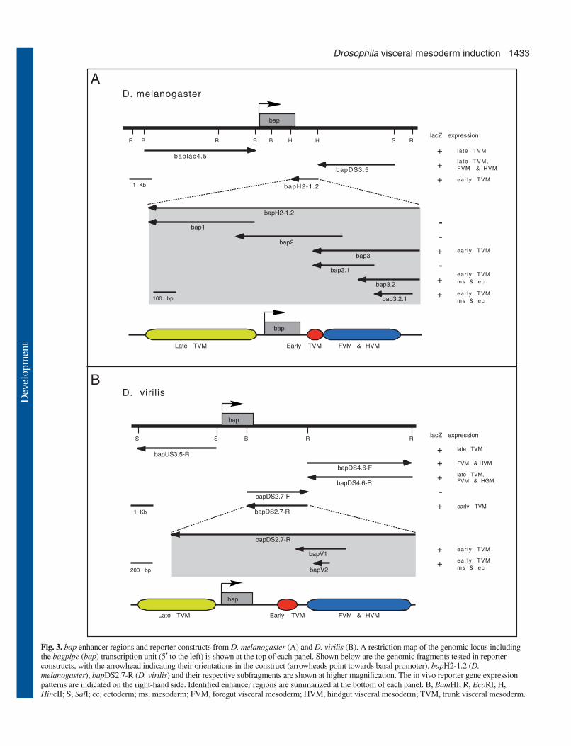
Early TVM enhancer of bap containscombinatorial binding sites for key signalingeffectors and mesodermal regulatorsTo investigate whether the bap regulators identifiedgenetically, including tin, dpp, slp (downstream of wg) andbin, can act directly on the early TVM regulatory elementof bap, DNaseI protection experiments with recombinantTin, Bap, Smad (Mad and Medea), Slp and Bin proteinswas performed on the 180 bp bap3.2.1 DNA sequencefrom D. melanogaster. The DNA footprinting resultsdemonstrate that both Tin and Bap proteins can bind to thepredicted Tin-binding site, which includes a perfect matchto the canonical Tin-binding motif TCAAGTG (Fig. 4;
Fig. 5) (Chen and Schwartz, 1995; Gajewski et al., 1997). Inaddition to the Tin-binding site, a site with a TAAG core motifcan strongly bind Bap but not Tin (CTTA in opposite strand;Fig. 4 and Fig. 5; note that the same core motif is found inbinding sites of a Bap ortholog, Nkx3.2) (Kim et al., 2003).With regard to Dpp signaling mediators, there are five Mad-protected regions, three of which are also protected byrecombinant Medea (Mad/Medea-1 to -3; Fig. 4 and Fig. 5).Site 1 includes an AGAC motif that was initially identified asa Smad binding motif in vertebrates (Zawel et al., 1998; Shi etal., 1998) whereas sites 3-5 contain GC-rich sequences withCGGC motifs that were first shown to bind Smad proteins inDrosophila (Kim et al., 1997; Shi, 2001). Site 2 may be acombination of the two types (TGAC motif and CG-richsequences). We do not observe a clear correlation of either typeof site with the binding of Mad versus Medea. Finally,recombinant Slp proteins protect a wide stretch that includesan inverted repeat of core binding motifs for forkheadtranscription factors (TAAACA) (Pierrou et al., 1994;Kaufmann et al., 1995), but extends further downstream (Fig.5 and Fig. 4). During the course of our work, it was reportedthat Slp can bind to tandem repeats of CAAA sequences, whichare present in three copies in the 3′ region of the protectedregion (Andrioli et al., 2002). Gel mobility shift andcompetition assays with Slp using wild-type oligonucleotidesand a version in which the TAAACA motifs were mutated
Development 132 (6) Research article
Fig. 4. DNAseI protection experiments with candidatetransacting factors on bap3.2.1 enhancer DNA. γ32P-labelledprobe was tested with two different amounts (1� and 3�, seeMaterials and methods) of bacterially expressed GST-fusionproteins of Bap, Tin, Slp, Mad, Medea and Bin, as well as BSAas a control. C+T sequencing ladder is shown on the left of eachblot and a schematic drawing of protected regions on the right.
Dev
elop
men
t

1435Drosophila visceral mesoderm induction
1 42
1 9
1 9
1 6
C A A T A T C G T C C C G G G T G G G A T G G A G G A G G A G G A G G A G G A G G A
G G A G G A G G A
G G A T G A G G C
G A C G C T
C T T T T G G C A C A G A T C
43 82
10 27
10 27
7 25
1 37
G T G G G A A C G - - G G A T T G G G A T C G G A A T T A G G A T C G G A A T T G G
G T G G G A A C G - - G G A T C G G G A - - - - - - - - - - - - - - - - - - - - - -
- T G G G G T A T - G G G A A G G G G A - - - - - - - - - - - - - - - - - - - - - -
G T G G G A A A A - A G G A T C G A G A - - - - - - - - - - - - - - - - - - - - - -
G C C G G G C G G G C A G A C C A G G A C A
G G
83 124
28 67
28 67
26 65
38 79
G A A C A T A G A C A T G T C C T T A G C G G C A C A G T T G G T G G C T G A C G A
- - A C A T A G A C A T G T C C T T A G C A G C A C A G T T G G T G G C T G A C G A
- - A C A T A G A C A T G T C C T T A G C A G C A C A G T T G G T G G C T G A C G A
- - A C A T A G A C A T G T C C T T A G C A G C A C A G T T G G T G G C T G A C G A
A C A T G C C G A G G T C C T T A G G C G C A C A G C T G G C G G C C G A C G A
125 165
68 108
68 109
66 107
80 103
C A - C A A G G A C C T G C C T C A A G T G C C C G G C G G C G A C A A T T C T A A
C A - C A A G G A C A T G A C T C A A G T G C C C G G C G G C G A C A A T T C T A A
C A A C A A G G A C A T G C C T C A A G T G C T T G T C G G C G A C A A T T T T A A
C A A C A A G G A T A C T T G T C A A G T G C C T G G C G G C G A C A A A T T C A A
C A A C A A G G A C A C G G C T C A A G T G T T - - - - - - - - - - - - - - - - - -
166 207
109 150
110 148
108 145
104 125
C G G C C G G C C G A C C T G T C G A T G T T T A T A A A C A A A C A A A C A A A T
C G G C C G G G C G A C C A G G C G A T G T T T A T A A A C A A A C A A A C C A A T
C G G A C C G - - - A C G A G G C G A T G T T T A T A A A C A A A C A A A C A A A T
C G G C C C G - - - - C T A G G C G A T G T T T A T A A A C A A A C A C A C A A A T
- - - - - - - - - - - - - - - - C C A T G T T T A T A A A C A A A C A - -- -A A T
208 237
151 188
149 182
146 175
126 154
A G A A T C A C C A A C T G A - - - - - - - - C G C C C C G G - - A T C G - - G A A
A G A A T C A C C A A C T G A T C A C G A G A T C A C C A G G - - A T G A - - G A A
A G A A T C A C A A G C T G A T - - - - - - - - G C C C A A G C G A T C C T T G G A
A G A A T T A C A A A C C G A - - - - - - - - C G G C C A G G - - A T C A - - G A A
G G G A - - - C A A A A T G T G C- - - - - -- - G A C A A G T C G T C C - - A A A
238 267
189 219
183 216
176 211
155 165
- - A A- - -- - G G A C A T G T G C C C A G T G G A A A T T G T C G A C
- - A A- -- - A G G A C A T G T G C A C A G T G G G A A A T G A T C A G
C T A G G C C G G A G C C A G G-- A C G A A - G G C A T T T C T A G G A
T C A A T A C A A G G A C A T G C G A C C A C T G G A A A A T T C - - A A
A C G A A T G A A A A
D. mel.
D. yak.
D. pse.
D. ana.
D. vir.
D. mel.
D. yak.
D. pse.
D. ana.
D. vir.
D. mel.
D. yak.
D. pse.
D. ana.
D. vir.
D. mel.
D. yak.
D. pse.
D. ana.
D. vir.
D. mel.
D. yak.
D. pse.
D. ana.
D. vir.
D. mel.
D. yak.
D. pse.
D. ana.
D. vir.
D. mel.
D. yak.
D. pse.
D. ana.
t t t t- - aaMad/Medea-1 Bap
a gg c a t t -- -a a aMad/Medea-2C1
t t t ta- - -
aTin/Bap
g cMad/Medea-3
Mad-4 Slp/Bin
Mad-5
bap3.2
bapV2
bap3.2.1
bap3.2
bap3.2.1
bapV2
R3
R2
R1
C2
T G T C C A- A G T G G A
- -
- -
- -
- -
G C A A C T G T G T A A300 323
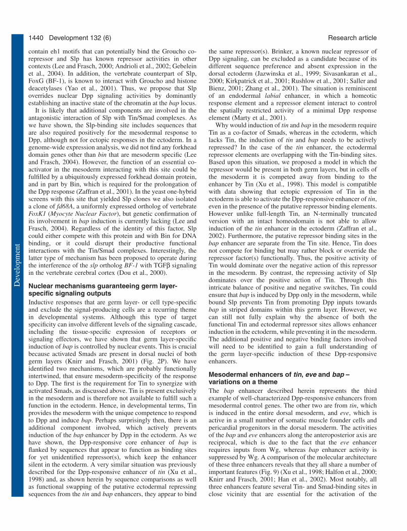
Fig. 5. Evolutionary conservation of bap enhancer sequences and binding motifs. Shown are alignments of enhancer sequences of bap3.2 (D.melanogaster), bapV2 (D. virilis) and corresponding genomic sequences from D. yakuba (D. yak.), D. pseudoobscura (D. pse.) and D.ananassae (D. ana.). Colored boxes above the sequences with unbroken lines indicate the extent of DNAseI footprints on D. melanogastersequences, boxes with black broken lines delineate highly conserved DNA stretches (C1 and C2), and colored boxes within the sequencesdenote core binding motifs for the respective binding factors. Nucleotides altered by in vitro mutagenesis for in vivo testing of binding siteactivities are shown on top of the D. melanogaster sequence (for Slp/Bin site mutations, see Fig. 7). For R1-R3 motifs, see text and Fig. 8. TheR1 sequence is not readily detectable in the other species but the 5′ region of bap3 (not shown) contains additional R-related motifs that areconserved and may have functionally redundant activities.
Dev
elop
men
t

1436
indicated that Slp can bind to both the TAAACA and theCAAA motifs with roughly equal affinity (data not shown). Inaddition, the FoxF family protein Bin binds to the TAAACAinverted repeat region, but less well to the CAAA repeat regionwhen compared with Slp (Fig. 4). Taken altogether, thesebinding data are consistent with the hypothesis that the knownmesodermal regulators of bap, namely Tin and Bin (andpossibly autoregulatory Bap), as well as the signaling inputsfrom Dpp and Wg (through Smads and Slp, respectively) areintegrated via direct binding to the early TVM enhancer of bap.
Binding sites for Tin, Smads, and additionalconserved sequences are required for activation ofthe bap TVM enhancer in the mesodermThe very high degree of sequence conservation of the bindingsites for Tin, Bap, Slp/Bin and Smads (except for Mad site 5)in different Drosophila species (Fig. 5) is indicative of thebiological importance of these sites. In addition, two othersequence stretches, C1 and C2, without any known candidatesfor binding factors, are also highly conserved (Fig. 5). To testfor the relevance of these sites in vivo, transgenes carryingeither mutations or deletions of these sequences were examinedfor their enhancer activities in embryos. These assays wereperformed within the context of the 180 bp regulatory elementbap3.2.1, which shows ectopic activity in stripes within thedorsal ectoderm (Fig. 2K, Fig. 6B). The use of bap3.2.1 ratherthan bap3.2 allowed us to determine whether any particular siteis required for receiving inputs from a mesoderm-specificfactor or a factor active in both germ layers.
Mutations at the Bap-specific binding site result in a delayedonset of lacZ expression in the mesoderm and reduced levelsof expression at early stage 11 (bap3.2.1-bap-m; Fig. 5, Fig.6A-C), but by late stage 11 an almost normal expression leveland pattern is observed in the TVM (data not shown). Thereduced activity of bap3.2.1-bap-m in the mesoderm at earlystages suggests a contribution of Bap to the regulation of bapenhancer activity in the TVM precursors. The full contributionof Bap autoregulation may be masked by the presence of anintact Tin/Bap-binding site in this construct. Mis-expression ofbap in the whole mesoderm does not result in any expansionof bap3.2.1-lacZ expression, suggesting that Bap alone is notsufficient to autoactivate its own enhancer in the mesoderm(data not shown).
The construct bap3.2.1-tin-m with a mutated Tin/Bap-binding site completely fails to activate lacZ expression in thedorsal mesoderm (Fig. 6A,D). By contrast, and as predictedbecause there is no Tin, the ectodermal lacZ expressionremains unaffected (Fig. 6D; the ectodermal expression is alsounperturbed in bap3.2.1-bap-m, Fig. 6C). twist-driven ectopicexpression of tin in the whole mesoderm does not cause anyectopic expression of bap or bap3.2.1-lacZ ventrally (data notshown), suggesting that Tin binding to this bap enhancerelement is essential, but also not sufficient to activate bap inthe mesoderm. Most likely, Tin needs to cooperate with otherlocalized activators that bind to the same regulatory element toactivate bap expression in the mesoderm. However, bap3.2.1enhancer activity in the dorsal ectoderm, which lacks Tin, doesnot require an intact Tin-binding site. These results point to anintricate molecular mechanism that makes bap3.2.1 enhanceractivity differentially sensitive to regulators in the mesodermversus ectoderm.
To study the in vivo function of Smad-binding sites in theearly bap regulatory element, a series of mutation or deletionconstructs were generated. A reporter with bap3.2.1-Smad1-m,with mutations in the AGAC core sequence of the 5′ mostMad/Medea-binding site (Mad/Medea-1), does not display anylacZ expression in either mesoderm or ectoderm (Fig. 6A,E).
Development 132 (6) Research article
Fig. 6. In vivo requirements for binding sites of Smad, Tin and Bapproteins, and for other conserved sequences. (A) Schematicrepresentations of bap3.2.1 and its mutated derivatives with asummary of their in vivo activities (ms, dorsal mesoderm; ec, dorsalectoderm). (B-I) Dorsal views of stage 11 embryos. Arrow indicatesmesodermal layer and arrowhead indicates ectodermal layer.(B) Activity of the parental bap3.2.1-lacZ construct. (C) Mutations inthe Bap-binding site cause a slight and transient reduction ofmesodermal enhancer activity. (D) Mutations in the Tin-binding sitecause a loss of enhancer activity in the mesoderm. (E) Mutations inthe Mad/Medea-binding site 1 cause a loss of enhancer activity inboth ectoderm and mesoderm. (F) Mutations in the Mad/Medea-binding site 2 nearly abolish enhancer activity in both ectoderm andmesoderm. (G) Deletion of DNA sequences containing theMad/Medea-binding sites 3 and 4 causes a strong reduction ofectodermal and mesodermal enhancer activity. (H,I) Mutationswithin the conserved sequence C1 of the enhancer from D.melanogaster (H) and D. virilis (I) cause a loss of enhancer activityin both ectoderm and mesoderm. (The observed expression withinsingle ectodermally derived cells in each hemisegment is an artificialeffect from the transformation vector.)
Dev
elop
men
t

1437Drosophila visceral mesoderm induction
Hence, this Smad-binding site (Mad/Medea-1) is essential forbap3.2.1 enhancer activity in both mesoderm and ectoderm. Inaddition, mutations in the second Mad/Medea-binding site(Mad/Medea-2; derivative bap3.2.1-Smad2-m) result in a lossof lacZ expression in the ectoderm and a near-loss ofexpression in the mesoderm (Fig. 6A,F). The above results andthe highly conserved sequences of Mad/Medea-1 and -2 amongdifferent Drosophila species (Fig. 5) suggest that both Smad-binding sites have essential and non-redundant functions inregulating bap expression during embryogenesis. Deletion ofboth the third and fourth Smad-binding sites (Mad/Medea-3and Mad-4; derivative bap3.2.1-Smad3,4-d) causes weak lacZexpression in both mesoderm and ectoderm (Fig. 6A,G). Thus,these two Smad-binding sites are not quite as crucial as sites 1and 2, but have additive or synergistic effects in inducing highlevels of enhancer activity. Consistent with this notion, thesetwo Smad-binding sites are absent from the homologous bapregulatory element of D. virilis (Fig. 5). The most 3′ Smad-binding site (Mad-5) does not closely match the GC-richMad/Medea-binding motif and is not well conserved amongthe five Drosophila species (Fig. 5). Deletion of this Smad-binding site (derivative bap3.2.1-Smad5-d) does not cause anychange of lacZ expression in either mesoderm or ectoderm(data not shown), implying that it does not have an essentialfunction in vivo even though Mad can bind to it in vitro.
Mutations in the C1 region from D. melanogaster (bap3.2.1-C1-m) and, likewise, within the bapV2 element from D. viriliswere also examined for their effects in embryos. In both cases,lacZ expression is absent in both mesoderm and ectoderm (Fig.6H,I), suggesting that the highly conserved C1 sequence playsan essential role in mediating the function of bap activators thatfunction in conjunction with Dpp in both germ layers. Bycontrast, mutation of the C2 region does not affect enhanceractivity (data not shown).
From the above data we conclude that the activation of bapin the mesoderm normally requires combinatorial binding ofmesodermal Tin and Dpp-activated Smad proteins. Binding ofBap (and Bin, see below) increases enhancer activity via afeedback regulatory loop. In addition, yet unidentifiedactivating binding factors are required, potentially as generalDNA-binding Smad co-activators.
The Slp-binding site mediates segmental repressionof bap enhancer induction and overlaps with anessential mesodermal activation siteBased on the observation that Slp represses bap expressionwithin the slp-expressing domains of the mesoderm(Riechmann et al., 1997; Lee and Frasch, 2000), we predictedthat mutations made at the Slp-binding site would result inuniform lacZ expression along the AP axis, similar to theendogenous bap expression in slp mutant embryos. However,several different mutations, particularly within the forkheaddomain consensus sites, caused a complete loss (bap3.2.1-slp-m1, Fig. 7A-C) or severe reduction (bap 3.2.1-slp-m2, Fig.7A,D; bap 3.2.1-slp-m3, Fig. 7A,E) of lacZ expression in themesoderm. The levels of ectodermal enhancer activity are notaffected by these mutations. These observations show thatthere are one or several activators that require this site andwhose function is mesoderm specific. These activators arelikely to include Bin, which is needed for prolonged expressionof bap at stage 11 and binds to this site (Fig. 4) (Zaffran et al.,
2001). However, there must be at least one additional, yetunidentified, binding factor that is required for initiation ofenhancer activity through this site.
In spite of this complication, the observed ectopicectodermal expression of these enhancer derivatives and the
Fig. 7. Functional dissection of the Slp binding sites in the bapenhancer. (A) Wild-type and mutated sequences within the regionprotected by Slp. The inverted repeat of canonical forkhead domain-binding motifs is in black boxes and the CAAA type of Slp-bindingmotifs are underlined in red. Unaltered sequences are represented bydashes below, and deleted sequences are indicated as a bracketedunbroken line. (B) Activity of the parental bap3.2.1-lacZ constructused as a control. (C) bap3.2.1-slp-m1-lacZ is not active in themesoderm, while in the dorsal ectoderm it is active in metamericdomains and there is weak ectopic activity between these domains.(D) bap3.2.1-slp-m2-lacZ shows very weak activity in the mesodermand similar ectodermal activity as with bap3.2.1-slp-m1-lacZ.(E) bap3.2.1-slp-m3-lacZ shows weakened activity in the mesodermand similar ectodermal activity as with bap3.2.1-lacZ. (F) bap3.2.1-slp-m4-lacZ activity is similar to that of the parental bap.3.2.1-lacZ(mesodermal clusters have physically merged at this slightly laterstage). (G) bap3.2.1-slp-m5-lacZ shows lack of mesodermal activityand largely uniform dorsal ectodermal activity along theanteroposterior axis. (H,I) Fluorescent double staining for Slp (red)and βGal (green) in stage 10 embryos. bap3.2.1-lacZ expression (H)is complementary to that of Slp, whereas bap3.2.1-slp-d1-lacZexpression (I) overlaps with Slp.
Dev
elop
men
t

1438
known presence of Slp in the same pattern in both mesodermand ectoderm enabled us to study the potential repressiveactivity of the Slp-binding sequences further. With bap3.2.1-slp-m1, in which both of the canonical forkhead domain-binding sites are mutated, reporter gene expression in theectoderm is expanded only slightly along the AP axiscompared with the strictly segmented bap3.2.1-lacZexpression (Fig. 7B,C). As Slp is able to bind CAAA sequencerepeats (Andrioli et al., 2002), which are present in three copieswithin the 3′ half of the protected sequence stretch, it waspossible that Slp can still bind to bap3.2.1-slp-m1 andrepress lacZ expression in the ectoderm. Indeed,electrophoresis mobility shift assays (EMSA) withrecombinant Slp proteins and bap3.2.1-slp-m1 DNAoligo probes showed that Slp was still able to bind,presumably through the CAAA sequence motifs (datanot shown). No segmental de-repression is observedwhen only one of the two canonical forkhead domainsites is mutated (bap3.2.1-slp-m2, Fig. 7D; bap3.2.1-slp-m3, Fig. 7E), presumably because of the unaffectedbinding of Slp to the intact site and/or the CAAAsequence motifs. Similarly, mutations in the CAAArepeats still allow segmental repression in bothectoderm and mesoderm (Fig. 7F and data not shown).Presumably, Slp is able to repress enhancer activitythrough binding to the canonical forkhead domain sitesin this situation, which can also bind the unknownmesodermal activator. By contrast, the introduction ofmutations in both types of Slp-binding sequences(bap3.2.1-slp-m5, Fig. 7A) or the deletion of the entiresequence protected by Slp (bap3.2.1-slp-d1, Fig. 7A),results in almost complete segmental de-repression ofreporter gene expression in the ectoderm (Fig. 7G-I).The inability of Slp to repress these enhancerderivatives is further confirmed by the observed co-expression of enhancer-driven lacZ with Slp in thedorsal ectoderm (Fig. 7I, compare with the normalmutually exclusive expression, 7H). Taken together, theabove results suggest that in the normal context, theSlp-protected DNA fragment mediates segmentalrepression by Slp proteins in the mesoderm boththrough the canonical forkhead domain sites and theSlp-specific CAAA motifs. Conversely, the activationof the enhancer in the mesoderm requires binding ofBin and a yet unidentified activator to the canonicalforkhead domain sites or sequences overlapping withthem.
Characterization of sequence elementspreventing the induction of bap and tin in thedorsal ectodermInformation from several different enhancer derivativeshas shown that the germ layer-specific induction ofbap, as well as of tin, relies in part on repressivesequences that prevent ectopic induction of both genesin the dorsal ectoderm. For example, the bap enhancerderivative bap3.2.1, which differs from bap3.2 by theabsence of 57 bp from the 5′-end and 30 bp from the3′-end, is induced ectopically in the dorsal ectodermwith the same pattern as its normal expression in thedorsal mesoderm (Fig. 2K, Fig. 6B, Fig. 8D). Similarly,
a shortened version of the bap enhancer from D. virilis, bapV2,drives segmental lacZ expression ectopically in the dorsalectoderm, in contrast to a longer version, bapV1, which islargely mesoderm specific (Fig. 2J,L). An analogous situationwas previously described for tin. In this case, it was observedthat the deletion of two identical sequence motifs, tinD1a andtinD1b, within the Dpp-responsive enhancer of tin causesectopic enhancer induction by Dpp in the dorsal ectoderm (Xuet al., 1998). Together, these observations suggest that themechanisms for the repression of ectopic induction of
Development 132 (6) Research article
Fig. 8. Sequences required for preventing the induction of bap enhanceractivity in the dorsal ectoderm. (A) Summary of tested enhancer derivativesand their activities in the dorsal mesoderm (ms) and ectoderm (ec). R1, R2and R3 denote native motifs with putative repressing activities, whereastinD1a and tinD1b denote related sequence motifs from the tinD enhancer oftinman. (B) Sequence alignments of motifs thought to confer ectodermalrepression from the bap3.2 and tinD enhancers. (C-H) Dorsal views of earlystage 11 βGal-stained embryos carrying various reporter constructs (arrow,mesoderm; arrowhead, ectoderm). (C) bap3.2-lacZ shows almost completerepression in the dorsal ectoderm. (D) bap3.2.1-lacZ shows complete de-repression in the dorsal ectoderm. (E) bap3.2∆R3-lacZ shows low levels ofde-repression in the dorsal ectoderm (small arrow; see comment in Fig. 6legend regarding large ectodermal cells). (F) bap3.2∆R1-2-lacZ and (G)bap3.2R1-3mut-lacZ show strong de-repression of enhancer activities in thedorsal ectoderm. (H) The addition of tinD1 sequences to bap3.2.1 preventsectopic induction in the ectoderm.
Dev
elop
men
t

1439Drosophila visceral mesoderm induction
mesodermal genes by Dpp in the ectoderm have beenconserved in different Drosophila species, and that differentDpp targets in the mesoderm use closely related mechanismsto ensure their germ layer-specific induction.
DNA sequence comparison among the 5′-57 bp and the 3′-30 bp sequences of bap3.2, as well as tinD1a and tinD1b fromthe tin enhancer, identified three regions, termed R1, R2 andR3, in bap3.2 that showed sequence similarities with oneanother and with tinD1a and tinD1b (Fig. 8B). Four to fiveadditional copies of this type of motifs are found at conservedpositions within ~200 bp of sequences upstream of bap3.2in different Drosophila species (data not shown). Theseobservations raise the possibility that these motifs mightrepresent binding sites for a yet unknown repressor preventingmesodermal gene induction in the ectoderm. To furthercharacterize the DNA elements mediating the ectodermalrepression, various derivatives of bap3.2 were generated thatwere either truncated or contained mutated or swappedsequences in the identified repressing regions (Fig. 8A;Materials and methods). All three putative binding sites for theectodermal repressor were found to contribute to therepression, albeit with slightly different degrees of inhibitoryactivities. Comparisons of the ectodermal enhancer activitiesof bap3.2∆R3, bap3.2∆R1-2 and bap3.2∆R1 suggest that thethree sites have partially redundant activities, with R1 havingthe strongest effect in ectodermal repression (Fig. 8E,F; datanot shown). When mutations are introduced all three sites, R1,R2 and R3, within bap3.2 (bap3.2R1-3mut), the resulting de-repression in the ectoderm is almost as complete as withdeletions of these sequences (Fig. 8G, compare with 8D).Hence, the identified sequence motifs appear to be largelyresponsible for the repression of induction in the ectoderm.The sequence similarities of tinD1a and tinD1b, and theiranalogous biological activities within the tin enhancer wouldsuggest that these sequences are able to replace the R1, R2 andR3 sequences functionally within the bap enhancer. To test thispossibility, both tinD1a and tinD1b were added to either the 5′-or the 3′-end of bap3.2.1 (bap3.2.1-tinD1.5′ and bap3.2.1-tinD1.3′, respectively). As predicted, tinD1a and tinD1bstrongly repress bap3.2.1 enhancer activity in the ectoderm,without affecting it in the mesoderm (Fig. 8H and data notshown). These results suggest that similar mechanisms,probably via binding of identical repressor factor(s), areinvolved in preventing the ectopic induction of bap and tin inthe ectoderm. The ectopic ectodermal expression of theenhancers of bap and tin is directly controlled by Dpp signals,as shown with mutations of Smad-binding sites, which preventthe induction in the dorsal ectoderm of the enhancers that lackthe repressing sequences (Fig. 6E-G) (Xu et al., 1998).Consequently, the putative repressors must normally interferewith Dpp signaling outputs at the level of the target enhancers.
DiscussionIt has become evident that enhancers of target genes providecrucial platforms for the integration of diverse signaling inputsand germ layer or tissue-specific nuclear factors duringinductive events in development. As a result, specifictranscriptional responses of combinatorial signals are triggeredin restricted domains within a target tissue, but usually not inthe cells that send the signals. In several systems, particularly
during Drosophila and Xenopus embryogenesis, TGFβ orBMP signals have been shown to act in concert with Wntsignals to achieve particular inductive responses of this type.Although in most cases described to date, TGFβ/BMP and Wntsignals act in a synergistic manner, there are also a few knownsituations in which Wnt signals antagonize TGFβ or BMPsignals (Hazelett et al., 1998; Yu et al., 1998; Kopp et al., 1999;Nishita et al., 2000; Morata, 2001; Waltzer et al., 2001; Zaffranand Frasch, 2002). However, the molecular basis of thesesignal interactions, particularly of antagonistic ones, is largelyunknown.
The induction of mesodermal tissues in Drosophila is aprocess during which Wg signals can modulate the responsesto Dpp signals by either synergizing with them or antagonizingthem. Whereas previous studies have described the functionalarchitecture of mesodermal enhancers that are targeted eitherby Dpp alone or by synergistic Dpp and Wg signals, ourpresent study describes an example of an enhancer whoseresponse to Dpp is suppressed by Wg signals. A comparisonof the functional organization of these enhancers provides newinsight into molecular strategies of nuclear signal integrationto produce differential developmental responses.
Nuclear Dpp signaling outputs and theirsuppression by Wg-induced SlpOur data show that bap is a direct target of Dpp signals. Thus,we can rule out an indirect pathway of bap being activatedsolely by tin, whose mRNA expression is known to depend onDpp inputs during the time of bap activation (Azpiazuand Frasch, 1993). Rather, tin acts simultaneously andsynergistically with Dpp. In fact, recent data with tin alleleslacking the Dpp-responsive enhancer show that bap can beinduced in the absence of Dpp-induced tin products, as long asthe twist-activated tin products are present (S. Zaffran andM.F., unpublished). We show that the molecular basis for thisobserved synergism of tin and dpp relies on the combinatorialbinding of Tin and Dpp-activated Smad proteins to the bapenhancer. Several possible molecular mechanisms couldunderlie the strict requirement for combinatorial binding of Tinand Smads. For example, the relatively low binding affinity andspecificity of Smads might be enhanced by bound Tin, whichcan engage in protein interactions with Mad and Medea(Zaffran et al., 2002). The combined presence of Tin andSmads in close vicinity or in complexes may also be aprerequisite for the assembly of higher order complexes withtranscriptional co-activators such as CBP/p300 (Liu et al.,1997; Feng et al., 1998; Janknecht et al., 1998; Pouponnot etal., 1998; Waltzer and Bienz, 1999). In addition, Tin maycounteract the function of yet unknown repressors of nuclearDpp signaling activity so that they can only repress in theectoderm.
Unlike Dpp, Wg signals act indirectly upon the early bapenhancer. Previous genetic and molecular data showed that Wginduces the expression of the forkhead domain-encoding geneslp via crucial dTCF/Lef-1 binding sites in both mesoderm andectoderm (Lee and Frasch, 2000). slp, in turn, functions as arepressor of bap (Riechmann et al., 1997; Lee and Frasch,2000). Our present data show that slp products exert thisfunction by direct binding to the Dpp-responsive bap enhancer,which obviously results in a suppression of the synergisticactivity of bound Tin and Smad complexes. Slp proteins
Dev
elop
men
t
1440
contain eh1 motifs that can potentially bind the Groucho co-repressor and Slp has known repressor activities in othercontexts (Lee and Frasch, 2000; Andrioli et al., 2002; Gebeleinet al., 2004). In addition, the vertebrate counterpart of Slp,FoxG (BF-1), is known to interact with Groucho and histonedeacetylases (Yao et al., 2001). Thus, we propose that Slpoverrides nuclear Dpp signaling activities by dominantlyestablishing an inactive state of the chromatin at the bap locus.
It is likely that additional components are involved in theantagonistic interaction of Slp with Tin/Smad complexes. Aswe have shown, the Slp-binding site includes sequences thatare also required positively for the mesodermal response toDpp, although not for ectopic responses in the ectoderm. In agenome-wide expression analysis, we did not find any forkheaddomain genes other than bin that are mesoderm specific (Leeand Frasch, 2004). However, the function of an essential co-activator in the mesoderm interacting with this site could befulfilled by a ubiquitously expressed forkhead domain protein,and in part by Bin, which is required for the prolongation ofthe Dpp response (Zaffran et al., 2001). In the yeast one-hybridscreens with this site that yielded Slp clones we also isolateda clone of fd68A, a uniformly expressed ortholog of vertebrateFoxK1 (Myocyte Nuclear Factor), but genetic confirmation ofits involvement in bap induction is currently lacking (Lee andFrasch, 2004). Regardless of the identity of this factor, Slpcould either compete with this protein and with Bin for DNAbinding, or it could disrupt their productive functionalinteractions with the Tin/Smad complexes. Interestingly, thelatter type of mechanism has been proposed to operate duringthe interference of the slp ortholog BF-1 with TGFβ signalingin the vertebrate cerebral cortex (Dou et al., 2000).
Nuclear mechanisms guaranteeing germ layer-specific signaling outputsInductive responses that are germ layer- or cell type-specificand exclude the signal-producing cells are a recurring themein developmental systems. Although this type of targetspecificity can involve different levels of the signaling cascade,including the tissue-specific expression of receptors orsignaling effectors, we have shown that germ layer-specificinduction of bap is controlled by nuclear events. This is crucialbecause activated Smads are present in dorsal nuclei of bothgerm layers (Knirr and Frasch, 2001) (Fig. 2P). We haveidentified two mechanisms, which are probably functionallyintertwined, that ensure mesoderm-specificity of the responseto Dpp. The first is the requirement for Tin to synergize withactivated Smads, as discussed above. Tin is present exclusivelyin the mesoderm and is therefore not available to fulfill such afunction in the ectoderm. Hence, in developmental terms, Tinprovides the mesoderm with the unique competence to respondto Dpp and induce bap. Perhaps surprisingly then, there is anadditional component involved, which actively preventsinduction of the bap enhancer by Dpp in the ectoderm. As wehave shown, the Dpp-responsive core enhancer of bap isflanked by sequences that appear to function as binding sitesfor yet unidentified repressor(s), which keep the enhancersilent in the ectoderm. A very similar situation was previouslydescribed for the Dpp-responsive enhancer of tin (Xu et al.,1998) and, as shown herein by sequence comparisons as wellas functional swapping of the putative ectodermal repressingsequences from the tin and bap enhancers, they appear to bind
the same repressor(s). Brinker, a known nuclear repressor ofDpp signaling, can be excluded as a candidate because of itsdifferent sequence preference and absent expression in thedorsal ectoderm (Jazwinska et al., 1999; Sivasankaran et al.,2000; Kirkpatrick et al., 2001; Rushlow et al., 2001; Saller andBienz, 2001; Zhang et al., 2001). The situation is reminiscentof an endodermal labial enhancer, in which a homeoticresponse element and a repressor element interact to controlthe spatially restricted activity of a minimal Dpp responseelement (Marty et al., 2001).
Why would induction of tin and bap in the mesoderm requireTin as a co-factor of Smads, whereas in the ectoderm, whichlacks Tin, the induction of tin and bap needs to be activelyrepressed? In the case of the tin enhancer, the ectodermalrepressor elements are overlapping with the Tin-binding sites.Based upon this situation, we proposed a model in which therepressor would be present in both germ layers, but in cells ofthe mesoderm it is competed away from binding to theenhancer by Tin (Xu et al., 1998). This model is compatiblewith data showing that ectopic expression of Tin in theectoderm is able to activate the Dpp-responsive enhancer of tin,even in the presence of the putative repressor binding elements.However unlike full-length Tin, an N-terminally truncatedversion with an intact homeodomain is not able to allowinduction of the tin enhancer in the ectoderm (Zaffran et al.,2002). Furthermore, the putative repressor binding sites in thebap enhancer are separate from the Tin site. Hence, Tin doesnot compete for binding but may rather block or override therepressor factor(s) functionally. Thus, the positive activity ofTin would dominate over the negative action of this repressorin the mesoderm. By contrast, the repressing activity of Slpdominates over the positive action of Tin. Through thisintricate balance of positive and negative switches, Tin couldensure that bap is induced by Dpp only in the mesoderm, whilebound Slp prevents Tin from promoting Dpp inputs towardsbap in striped domains within this germ layer. However, wecan still not fully explain why the absence of both thefunctional Tin and ectodermal repressor sites allows enhancerinduction in the ectoderm, while preventing it in the mesoderm.The additional positive and negative binding factors involvedwill need to be identified to gain a full understanding ofthe germ layer-specific induction of these Dpp-responsiveenhancers.
Mesodermal enhancers of tin, eve and bap –variations on a themeThe bap enhancer described herein represents the thirdexample of well-characterized Dpp-responsive enhancers frommesodermal control genes. The other two are from tin, whichis induced in the entire dorsal mesoderm, and eve, which isactive in a small number of somatic muscle founder cells andpericardial progenitors in the dorsal mesoderm. The activitiesof the bap and eve enhancers along the anteroposterior axis arereciprocal, which is due to the fact that the eve enhancerrequires inputs from Wg, whereas bap enhancer activity issuppressed by Wg. A comparison of the molecular architectureof these three enhancers reveals that they all share a number ofimportant features (Fig. 9) (Xu et al., 1998; Halfon et al., 2000;Knirr and Frasch, 2001; Han et al., 2002). Most notably, allthree enhancers feature several Tin- and Smad-binding sites inclose vicinity that are essential for the activation of the
Development 132 (6) Research article
Dev
elop
men
t

1441Drosophila visceral mesoderm induction
enhancer in the mesoderm. Each enhancer includes both typesof known Smad-binding motifs, which have ‘AGAC’ and‘CG’-rich cores, respectively. Hence, the basic activationmechanisms of each of the three enhancers downstream of Dppare likely to be closely related. As discussed above, in theenhancers of both tin and bap, binding sites for a nuclearrepressor of Dpp signals are key for the germ layer specificityof the inductive response. Although we do not know whetherthe same repressive mechanism operates at the eve enhancer,we note that motifs related to the presumed repressor bindingmotifs are present and their function can now be tested in vivo(M.F., unpublished). As in the case of bap, the tin enhancerincludes also additional sites that are required for Dpp-inducible enhancer activity, which may bind essential Smad co-factors. However, based upon the divergent sequences of thesesites (C1 site in the bap and ‘CAATGT’ motifs in the tinenhancer) (Xu et al., 1998), they appear to bind different typesof factors in each case.
On top of this basic arrangement that allows the enhancer tobe active in the dorsal mesoderm, the enhancers from bap andeve, but not tin, include binding sites that make them respondto Wg inputs in an opposite fashion (Fig. 9). In the case of bap,Wg-induced Slp binds and dominantly suppresses the activityof bound Smad effectors. For the eve enhancer we haveproposed that there is an analogous repressive activity;however, in this case, it is exerted by bound Wg signal
effectors, i.e. dTCF/Lef-1, in the absence of Wg signals (Knirrand Frasch, 2001). In the domains with active Wg signaling,the repressive activity of dTCF/Lef-1 is neutralized by the Wgsignaling cascade, which allows the Dpp effectors to be activeat the eve enhancer (as it lacks Slp binding sites). Throughthese switches, the bap and eve enhancers become induced inreciprocal AP patterns. In addition, the eve enhancer includesbinding sites for activators and repressors downstream ofreceptor tyrosine kinases and Notch, respectively, which serveto restrict its activity to specific subsets of cells within thedomains of overlapping Dpp and Wg activities (Halfon et al.,2000). Clearly, many of the molecular details still need to beclarified. Nevertheless, we are now beginning to understand thebasic principles of how differential inputs from inductivesignals and tissue-specific activities can be integrated at theenhancer level to achieve distinct patterns of target geneexpression during early tissue induction in the Drosophilamesoderm.
We thank Stephane Zaffran for the footprint with Bin protein andZhizhang Yin for performing the initial round of bap enhancerdissections. We also appreciate receiving antibodies from John Reinitz(αSlp) and Carl-Henrik Heldin (αP-Smad1). This work was supportedby grants from the NIH (HD30832 and DK59406).
ReferencesAndrioli, L., Vasisht, V., Theodosopoulou, E., Oberstein, A. and Small, S.
(2002). Anterior repression of a Drosophila stripe enhancer requires threeposition-specific mechanisms. Development 129, 4931-4940.
Azpiazu, N. and Frasch, M. (1993). tinman and bagpipe: two homeo boxgenes that determine cell fates in the dorsal mesoderm of Drosophila. GenesDev. 7, 1325-1340.
Azpiazu, N., Lawrence, P., Vincent, J.-P. and Frasch, M. (1996).Segmentation and specification of the Drosophila mesoderm. Genes Dev.10, 3183-3194.
Baylies, M., Martinez Arias, A. and Bate, M. (1995). wingless is requiredfor the formation of a subset of muscle founder cells during Drosophilaembryogenesis. Development 121, 3829-3837.
Bodmer, R. (1993). The gene tinman is required for specification of the heartand visceral muscles in Drosophila. Development 118, 719-729.
Bodmer, R., Jan, L. Y. and Jan, Y. N. (1990). A new homeobox-containinggene, msh-2, is transiently expressed early during mesoderm formation ofDrosophila. Development 110, 661-669.
Campos-Ortega, J. A. and Hartenstein, V. (1997). The EmbryonicDevelopment of Drosophila melanogaster. Berlin: Springer Verlag.
Carmena, A., Gisselbrecht, S., Harrison, J., Jimenez, F. and Michelson, A.(1998). Combinatorial signaling codes for the progressive determination ofcell fates in the Drosophila embryonic mesoderm. Genes Dev. 15, 3910-3922.
Chen, C. and Schwartz, R. (1995). Identification of novel DNA bindingtargets and regulatory domains of a murine tinman homeodomain factor,nkx-2.5. J. Biol. Chem. 270, 15628-15633.
Cripps, R., Black, B., Zhao, B., Lien, C., Schulz, R. and Olson, E. (1998).The myogenic regulatory gene Mef2 is a direct target for transcriptionalactivation by Twist during Drosophila myogenesis. Genes Dev. 12, 422-434.
Dou, C., Lee, J., Liu, B., Liu, F., Massague, J., Xuan, S. and Lai, E. (2000).BF-1 interferes with transforming growth factor beta signaling byassociating with Smad partners. Mol. Cell. Biol. 20, 6201-6211.
Englund, C., Loren, C. E., Grabbe, C., Varshney, G. K., Deleuil, F.,Hallberg, B. and Palmer, R. H. (2003). Jeb signals through the Alkreceptor tyrosine kinase to drive visceral muscle fusion. Nature 425, 512-516.
Feng, X. H., Zhang, Y., Wu, R. Y. and Derynck, R. (1998). The tumorsuppressor Smad4/DPC4 and transcriptional adaptor CBP/p300 arecoactivators for Smad3 in TGF-beta-induced transcriptional activation.Genes Dev. 12, 2153-2163.
Frasch, M. (1995). Induction of visceral and cardiac mesoderm by ectodermalDpp in the early Drosophila embryo. Nature 374, 464-467.
Fig. 9. Molecular switching of Dpp-responsive enhancer activities inthe dorsal mesoderm. The schematic diagram summarizes themolecular basis for the reciprocal activities of the bap and eveenhancers in the A and P domains of the dorsal mesoderm. In the Adomains, the differential activities are due to the Wg-dependent reliefof dTCF-associated co-repressors at the eve enhancer and to the Wg-dependent loading of Slp/co-repressor complexes at the bapenhancer. Conversely, in the P domains where Wg signaling is absentslp is not induced, which allows the bap enhancer to be active, whiledTCF/co-repressor complexes keep the eve enhancer ‘off’. Owing tothe lack of Wg-responsive sequences, the tin enhancer is ‘on’ in bothdomains of the dorsal mesoderm. For simplicity, only one site ofeach type is depicted and various sites binding yet unknown factorsinvolved in activation or ectodermal repression are not depicted.
Dev
elop
men
t

1442
Frasch, M. and Nguyen, H. T. (1999). Genetic control of mesodermpatterning and differentiation during Drosophila embryogenesis. Adv. Dev.Biochem. 5, 1-47.
Frasch, M., Hoey, T., Rushlow, C., Doyle, H. J. and Levine, M. (1987).Characterization and localization of the even-skipped protein of Drosophila.EMBO J. 6, 749-759.
Gajewski, K., Kim, Y., Lee, Y., Olson, E. and Schulz, R. (1997). D-mef2 isa target for Tinman activation during Drosophila heart development. EMBOJ. 16, 515-522.
Gebelein, B., McKay, D. J. and Mann, R. S. (2004). Direct integration ofHox and segmentation gene inputs during Drosophila development. Nature431, 653-659.
Halfon, M., Carmena, A., Gisselbrecht, S., Sackerson, C., Jimenez, F.,Baylies, M. and Michelson, A. (2000). Ras pathway specificity isdetermined by the integration of multiple signal-activated and tissue-restricted transcription factors. Cell 103, 63-74.
Han, Z., Fujioka, M., Su, M., Liu, M., Jaynes, J. B. and Bodmer, R. (2002).Transcriptional integration of competence modulated by mutual repressiongenerates cell-type specificity within the cardiogenic mesoderm. Dev. Biol.252, 225-240.
Hazelett, D. J., Bourouis, M., Walldorf, U. and Treisman, J. E. (1998).decapentaplegic and wingless are regulated by eyes absent and eyegone andinteract to direct the pattern of retinal differentiation in the eye disc.Development 125, 3741-3751.
Jagla, T., Bidet, Y., da Ponte, J. P., Dastugue, B. and Jagla, K. (2002).Cross-repressive interactions of identity genes are essential for properspecification of cardiac and muscular fates in Drosophila. Development 129,1037-1047.
Janknecht, R., Wells, N. J. and Hunter, T. (1998). TGF-beta-stimulatedcooperation of Smad proteins with the coactivators CBP/p300. Genes Dev.12, 2114-2119.
Jazwinska, A., Rushlow, C. and Roth, S. (1999). The role of brinker inmediating the graded response to Dpp in early Drosophila embryos.Development 126, 3323-3334.
Kaufmann, E., Muller, D. and Knöchel, W. (1995). DNA recognition siteanalysis of Xenopus winged helix proteins. J. Mol. Biol. 248, 239-254.
Kim, D. W., Kempf, H., Chen, R. E. and Lassar, A. B. (2003).Characterization of Nkx3.2 DNA binding specificity and its requirement forsomitic chondrogenesis. J. Biol. Chem. 278, 27532-27539.
Kim, J., Johnson, K., Chen, H., Carroll, S. and Laughon, A. (1997).Drosophila Mad binds to DNA and directly mediates activation of vestigialby Decapentaplegic. Nature 388, 304-308.
Kirkpatrick, H., Johnson, K. and Laughon, A. (2001). Repression of dpptargets by binding of Brinker to Mad sites. J. Biol. Chem. 276, 18216-18222.
Knirr, S. and Frasch, M. (2001). Molecular integration of inductive andmesoderm-intrinsic inputs governs even-skipped enhancer activity in asubset of pericardial and dorsal muscle progenitors. Dev. Biol. 238, 13-26.
Kopp, A., Blackman, R. K. and Duncan, I. (1999). Wingless,Decapentaplegic and EGF receptor signaling pathways interact to specifydorso-ventral pattern in the adult abdomen of Drosophila. Development 126,3495-3507.
Lee, H. and Frasch, M. (2000). Wingless effects mesoderm patterning andectoderm segmentation events via induction of its downstream target sloppypaired. Development 127, 5497-5508.
Lee, H. H. and Frasch, M. (2004). A survey of fork head domain encodinggenes in the Drosophila genome: Classification and expresssion patterns.Dev. Dyn. 229, 357-366.
Lee, H. H., Norris, A., Weiss, J. B. and Frasch, M. (2003). Jelly belly proteinactivates the receptor tyrosine kinase Alk to specify visceral musclepioneers. Nature 425, 507-512.
Liu, F., Pouponnot, C. and Massagué, J. (1997). Dual role of theSmad4/DPC4 tumor suppressor in TGFβ-inducible transcriptionalcomplexes. Genes Dev. 11, 3157-3167.
Marty, T., Vigano, M. A., Ribeiro, C., Nussbaumer, U., Grieder, N. C. andAffolter, M. (2001). A HOX complex, a repressor element and a 50 bpsequence confer regional specificity to a DPP-responsive enhancer.Development 128, 2833-2845.
Michelson, A. M., Gisselbrecht, S., Zhou, Y., Baek, K. H. and Buff, E. M.(1998). Dual functions of the Heartless fibroblast growth factor receptor indevelopment of the Drosophila embryonic mesoderm. Dev. Genet. 22, 212-229.
Morata, G. (2001). How Drosophila appendages develop. Nat. Rev. Mol. Cell.Biol. 2, 89-97.
Nishita, M., Hashimoto, M. K., Ogata, S., Laurent, M. N., Ueno, N.,
Shibuya, H. and Cho, K. W. (2000). Interaction between Wnt and TGF-beta signalling pathways during formation of Spemann’s organizer. Nature403, 781-785.
Pierrou, S., Hellqvist, M., Samuelsson, L., Enerback, S. and Carlsson, P.(1994). Cloning and characterization of seven human forkhead proteins:binding site specificity and DNA bending. EMBO J. 13, 5002-5012.
Pouponnot, C., Jayaraman, L. and Massague, J. (1998). Physical andfunctional interaction of SMADs and p300/CBP. J. Biol. Chem. 273, 22865-22868.
Riechmann, V., Irion, U., Wilson, R., Grosskortenhaus, A. and Leptin, M.(1997). Control of cell fates and segmentation in the Drosophila mesoderm.Development 124, 2915-2922.
Rushlow, C., Colosimo, P., Lin, M.-C., Xu, M. and Kirov, N. (2001).Transcriptional regulation of the Drosophila gene zen by competing Smadand Brinker inputs. Genes Dev. 15, 340-351.
Saller, E. and Bienz, M. (2001). Direct competition between Brinker andDrosophila Mad in Dpp target gene transcription. EMBO Rep. 2, 298-305.
Shi, Y. (2001). Structural insights on Smad function in TGFbeta signaling.BioEssays 23, 223-232.
Shi, Y., Wang, Y. F., Jayaraman, L., Yang, H., Massague, J. and Pavletich,N. P. (1998). Crystal structure of a Smad MH1 domain bound to DNA:insights on DNA binding in TGF-beta signaling. Cell 94, 585-594.
Sivasankaran, R., Vigano, M., Muller, B., Affolter, M. and Basler, K.(2000). Direct transcriptional control of the Dpp target omb by the DNAbinding protein Brinker. EMBO J. 19, 6162-6172.
Staehling-Hampton, K., Hoffmann, F. M., Baylies, M. K., Rushton, E. andBate, M. (1994). dpp induces mesodermal gene expression in Drosophila.Nature 372, 783-786.
Su, M., Fujioka, M., Goto, T. and Bodmer, R. (1999). The Drosophilahomeobox genes zfh-1 and even-skipped are required for cardiac-specificdifferentiation of a numb-dependent lineage decision. Development 126,3241-3251.
Waltzer, L. and Bienz, M. (1999). A function of CBP as a transcriptional co-activator during Dpp signalling. EMBO J. 18, 1630-1641.
Waltzer, L., Vandel, L. and Bienz, M. (2001). Teashirt is required fortranscriptional repression mediated by high Wingless levels. EMBO J. 20,137-145.
Wu, X., Golden, K. and Bodmer, R. (1995). Heart development inDrosophila requires the segment polarity gene wingless. Dev. Biol. 169, 619-628.
Xu, X., Yin, Z., Hudson, J., Ferguson, E. and Frasch, M. (1998). Smadproteins act in combination with synergistic and antagonistic regulators totarget Dpp responses to the Drosophila mesoderm. Genes Dev. 12, 2354-2370.
Yao, J., Lai, E. and Stifani, S. (2001). The winged-helix protein Brain Factor1 interacts with Groucho and Hes proteins to repress transcription. Mol. Cell.Biol. 21, 1962-1972.
Yin, Z. and Frasch, M. (1998). Regulation and function of tinman duringdorsal mesoderm induction and heart specification in Drosophila. Dev.Genet. 22, 187-200.
Yin, Z., Xu, X.-L. and Frasch, M. (1997). Regulation of the Twist target genetinman by modular cis-regulatory elements during early mesodermdevelopment. Development 124, 4871-4982.
Yu, X., Riese, J., Eresh, S. and Bienz, M. (1998). Transcriptional repressiondue to high levels of Wingless signalling. EMBO J. 17, 7021-7032.
Zaffran, S. and Frasch, M. (2002). Early signals in cardiac development.Circ. Res. 91, 457-469.
Zaffran, S., Küchler, A., Lee, H. H. and Frasch, M. (2001). biniou (FoxF),a central component in a regulatory network controlling visceral mesodermdevelopment and midgut morphogenesis in Drosophila. Genes Dev. 15,2900-2915.
Zaffran, S., Xu, X., Lo, P. C., Lee, H. H. and Frasch, M. (2002).Cardiogenesis in the Drosophila model: control mechanisms during earlyinduction and diversification of cardiac progenitors. Cold Spring HarborSymp. Quant. Biol. 67, 1-12.
Zawel, L., Dai, J. L., Buckhaults, P., Zhou, S., Kinzler, K. W., Vogelstein,B. and Kern, S. E. (1998). Human Smad3 and Smad4 are sequence-specifictranscription activators. Mol. Cell 1, 611-617.
Zhang, H., Levine, M. and Ashe, H. (2001). Brinker is a sequence-specifictranscriptional repressor in the Drosophila embryo. Genes Dev. 15, 261-266.
Development 132 (6) Research article
Dev
elop
men
t