
NTP REPORT ON CARCINOGENS BACKGROUND DOCUMffiNTfurTAMOXWEN ... · NTP REPORT ON CARCINOGENS...
32
NTP REPORT ON CARCINOGENS BACKGROUND DOCUMffiNTfurTAMOXWEN FINAL MARCH 1999 Prepared for the October 30-31, 1997, Meeting of the Report on Carcinogens Subcommittee of the NTP Board of Scientific Counselors Prepared by Integrated Laboratory Systems Post Office Box 13501 Research Triangle Park, North Carolina 27709 NIEHS Contract No. N01-ES-25346
Transcript of NTP REPORT ON CARCINOGENS BACKGROUND DOCUMffiNTfurTAMOXWEN ... · NTP REPORT ON CARCINOGENS...
NTP REPORT ON CARCINOGENS BACKGROUND DOCUMffiNTfurTAMOXWEN
FINAL MARCH 1999
Prepared for
the October 30-31, 1997, Meeting of the Report on Carcinogens Subcommittee
of the NTP Board of Scientific Counselors
Prepared by
Integrated Laboratory Systems Post Office Box 13501
Research Triangle Park, North Carolina 27709 NIEHS Contract No. N01-ES-25346
NTP Report on Carcinogens 1997 Background Document for Tamoxifen
TABLE OF CONTENTS
NTP Report on Carcinogens Listing for Tamoxifen ..................................... 1 Listing Criteria from the Report on Carcinogens, Eighth Edition ••.•..••••••••.• 4
1.0 CHEMICAL PROPERTmS•••••••••••••••••••••••••••••••••••••••••••••••••••••••••••••••••••• 5 1.1 Chemical Identification ..•.••.••••••••••••.••.............•.••••.•......•.........•••. S
1.1.1 Tamoxifen [10540-29-1 ] •••••••••.•••••.•••••••••••••••••••••••••••••••.•••• S 1.1.2 Tamoxifen citrate [54965-24-1 ] •••.•••••.••.••••••••••.•...•••.••••••••• 5
1.2 Physical-Chemical Properties...•.••••••....................•..•••.................. 6 1.~.1 1rSlmoxi1ren•••••••••••••••••••••••••••••••••••••••••••••••••••••••••••••••••••••••• fi 1.2.2 Tamoxifen Citrate..••.•••.••••.••..••..•.....••••.••...••••.......•.......... 6
~.0 ~E~OS~ .•••••••.•••.••••••••.••••••••.•••••••••.••••••••••••.•••.••••••.•••••••••••• ~ 2.1 Use •••••.••••••••.•••••••••••••••••••••••.••••••••••••••••••••••••••••••••••••••••••••••••••••••• 7 2.2 Production Process and Volume ................................................... 7 2.3 Environmental Exposure •••.•••••••••••••.•••••••••....•..••...•.••.•...••..•..•••••.• 7 2.4 Occupational Exposure •••••••••••••••••••••••••••••••••••.•.•...•...•.•.••••••••••••••• 8 2.5 Regulations and Criteria.••••.••..••••••••..••.....••••••••••••••••...••.••.••••••..•• 8
3.() ~~1£1(]][)~~ ••••••••••••••••••••••••••••••••••••••••••••••••••••••••••••••••••••••••••••••••• ~
3.1 Human Studies Reviewed by IARC (1996)•.••.•••••.••.••.•••..•••••••••••••. 9 3.2 Human Studies Published Post IARC (1996) •..•••••••••.••••.••.••••••••••. 9 Table 3-1 Summary of Randomized Clinical Trial of Adjuvant
Use of Tamoxifen (not reviewed by IARC, 1996): Endometrial Cancers in Patients Treated for Breast Cancer..•.•.••.••••••••.••••.•10
4.0 EXPERIME-NTAL CARCIN"OGENESIS •••••.•••••.••••.••..•••••••.•.••.•••..•.••••••••11
:5.() ~El'J01rO~C:~••.••••••••.•.•.•••••••.•....•.••••..••..•..•..•..•......•.•.•....•.•..•.•.•.•...ll Table 5-1 Summary of Additional ~enotoxicity Studies.........•...........13
6.0 ABSORPTION, DISTRIBUTION, METABOLISM, AND EXCRETION •••••••••••••.•..••••••••••..•.••••••••••••••.••..•.••.••••••.•......•...••.••••••.•17 6.1 Absorption, Distribution, and Excretion •••••.•.••••••..•..•.••••••••••.••••.•17 6.2 Metabolites ••..•..•.•••••••.••••••..•••..••..••..•.•...•••.•....••••.•.••••••••.•••.•••.•••17 6.3 Structure-Activity Relationships .................................................18
iii
NTP Report on Carcinogens 1997 Background Document for Tamoxifen
7.0 ~CHANISMS ••••••..•.•.•••...••...•••....••...•••••.•.•...•..••••.•..•••••.••....••..•..•••.••.18 7.1 Genotoxicity' ..............................................................................18 7.2 Tamoxifen-Estrogen Receptor Interactions ••••.•••••••••••••••••••••••••••19
~.() ~~~ ......................................................................................!~
APPENDIX A - Excerpts from the IARC Monograph on the Evaluation
APPENDIX C - Report on Carcinogens (RoC), 9th Edition
Of the Carcinogenic Risks of Chemicals to Humans Volume 66 (Some Pharmaceutical Drugs) Tamoxifen, pp. 253-365, 1996 •••••••••••A-1
APPENDIX B- Description of Online Searches for Tamoxifen ••••••.•••••.•••• B-1
Review Summary ...........................................................................C-1
iv
NTP Report on Carcinogens 1997 Background Document for Tamoxifen
NTP Report on Carcinogens Listing for Tamoxifen
Carcinogenicity Tamoxifen is known to be a human carcinogen based on studies in humans that indicate a
causal relationship between exposure to tamoxifen and cancers of the uterine endometrium. However, there is also conclusive evidence that tamoxifen therapy reduces the risk of contralateral breast cancer in women with a previous diagnosis ofbreast cancer. IARC recently evaluated the carcinogenic risks of tamoxifen to humans and reached the same conclusion (IARC, v .66, 1996).
The potential effect of tamoxifen in increasing the risk ofendometrial cancer has been reported in one adequate cohort study, four adequate case-control studies, and 14 randomized clinical trials.
The cohort study (Curtis et al., 1996) examined the effect oftamoxifen on risk of endometrial cancer in 87,323 women with breast cancer reported to the Surveillance, Epidemiology and End Results (SEER) program in the United States and found a statistically significant elevation of endometrial cancer in women who had received tamoxifen therapy. In two of the four case-control studies (Sasco et al., 1996; van Leeuwen et al., 1994), a nonsignificant elevation of risk for endometrial cancer was found, with a significant increase in risk with increasing duration of therapy in one of these studies (van Leeuwen et al., 1994). In the U.S. case-control study (Cook et al., 1995), no increase was seen, but a shorter duration oftamoxifen use was reported. In the fourth case-control study (Bardell, 1988b; cited by IARC V.66, 1996), increased risk ofendometrial cancer for tamoxifen use was found, but confounding factors could not be eliminated.
In the two largest randomized clinical trials (Fisher et al., 1994; Rutqvist et al., 1995), there was a strong and statistically significant association between risk for endometrial cancer and use of tamoxifen. In the 12 other smaller trials, no statistically significant increases in endometrial cancer were seen, although 29 endometrial cancers were reported in tamoxifen-treated individuals and 14 in controls when these 12 studies were combined.
In 32 case studies, 102 cases ofendometrial cancer were reported in women who received tamoxifen for breast cancer. One case series reported significantly more high-grade endometrial tumors in tamoxifen-treated breast cancer patients than in patients without tamoxifen use (Magriples et al., 1993); this difference, however, was not seen in six other studies.
MacMahon (1997) concluded that published results were suggestive of a causal association between tamoxifen use and endometrial cancer but were not conclusive because of confounding factors such as prior hysterectomy and/or hormone replacement therapy. Examining the same confounding factors, an IARC Working Group concluded that there is a positive association between tamoxifen use and endometrial cancer and cited several studies in support of this conclusion; the same potential confounders were considered unlikely to have a major effect on the reported relative risks (IARC, V.66, 1996).
Experimental animal studies also provide evidence oftamoxifen's carcinogenic effects. The IARC Working Group (IARC, V .66, 1996) reviewed experimental studies reported prior to 1996 and reached a similar conclusion. Tamoxifen, administered orally, was evaluated in one mouse study and eight rat studies. In mice, the incidences ofbenign ovarian and testicular tumors
1
NTP Report on Carcinogens 1997 Background Document for Tamoxifen
were significantly increased after 3 months of treatment. In eight rat studies that varied in treatment lengths, tamoxifen induced preneoplastic liver lesions and benign or malignant liver tumors. One rat study reported a decrease in tumors in hormone-dependent tissues, but reduced weight gain may have been a contributing factor. In one additional study where tamoxifen was given by subcutaneous administration, mammary tumor development was inhibited in intact and ovariectomized mice (reviewed in IARC V.66, 1996).
Uterine abnormalities including endometrial carcinoma have also been reported in experimental animals exposed to tamoxifen. Rats receiving tamoxifen daily by oral gavage for 20 to 52 weeks were reported to have squamous cell metaplasia, dysplasia, and squamous cell carcinoma of the uterus while no comparable lesions were seen in controls (MfultyHi et al., 1996). Although not included in the IARC monograph, short-term developmental exposure to tamoxifen on days l to 5 of neonatal life has recently been reported to significantly increase the incidence of reproductive tract abnormalities in both female and male mice, including uterine carcinoma and seminal vesicle tumors (Newbold et al., 1996 abstr., 1997).
Other Information Relating to Carcinogenesis or Possible Mechanisms of Carcinogenesis
Several studies reviewed by IARC (V.66, 1996) described tumor initiation/promotional and co-carcinogenicity attributes oftamoxifen. In mice, tamoxifen inhibited 3methylcholanthrene-induced cervical cancer and virus-induced leukemia. In several studies with male and female rats, it enhanced liver tumors induced by N-nitrosodiethylamine. In one rat study, it enhanced the development ofN-nitrosodiethylamine-induced kidney tumors; but in a number of other studies, it inhibited 7,12-dimethyl[a]benzanthracene-induced mammary tumors. In hamsters, two studies reported the inhibition of kidney and liver tumors induced by 17~estradiol.
Several reports in the literature (IARC V .21, 1979) demonstrate that women receiving estrogen replacement therapy unopposed by progesterone have a highly elevated risk for endometrial cancer. Because ofthese data, conjugated estrogens are considered known human carcinogens (IARC V.21, 1979; NTP, 1998 [Report on Carcinogens, 8'h ed]). Unlike the breast, where tamoxifen is an anti-estrogen (used to treat breast cancer because of this property), it acts as an estrogen agonist in the uterus. Therefore, tamoxifen would likely produce the same effects as conjugated estrogens in the uterus. Available data strongly indicate that endometrial cancer following exposure to estrogens is caused by estrogen receptor-mediated responses. DNA adducts have not been detected in human samples (IARC V.66, 1996) with one exception where low levels of DNA adducts were seen in leukocytes and endometrial tissue of breast cancer patients receiving tamoxifen (Hemminki et al., 1996, 1997).
In animal and in vitro experiments, tamoxifen readily forms DNA adducts in several tissues and cells, and either these adducts or the estrogenic activity of tamoxifen could be responsible for liver cancer observed in rodents exposed to tamoxifen.
Although tamoxifen is not mutagenic in bacteria, it is positive for micronuclei formation in human cells in vitro (Otto et al., 1996). In vivo, it increases aneuploidy and chromosomal aberrations in the livers of female Sprague-Dawley rats (Sargent et al., 1996).
2

NTP Report on Carcinogens 1997 Background Document for Tamoxifen
Available data indicate that the receptor-mediated mechanisms involved in the carcinogenic actions of tamoxifen are operative in humans. Genotoxic mechanisms may also be operative in people, but preliminary studies suggest that they are quantitatively less than in rodents.
3
NTP Report on Carcinogens 1997 Background Document for Tamoxifen
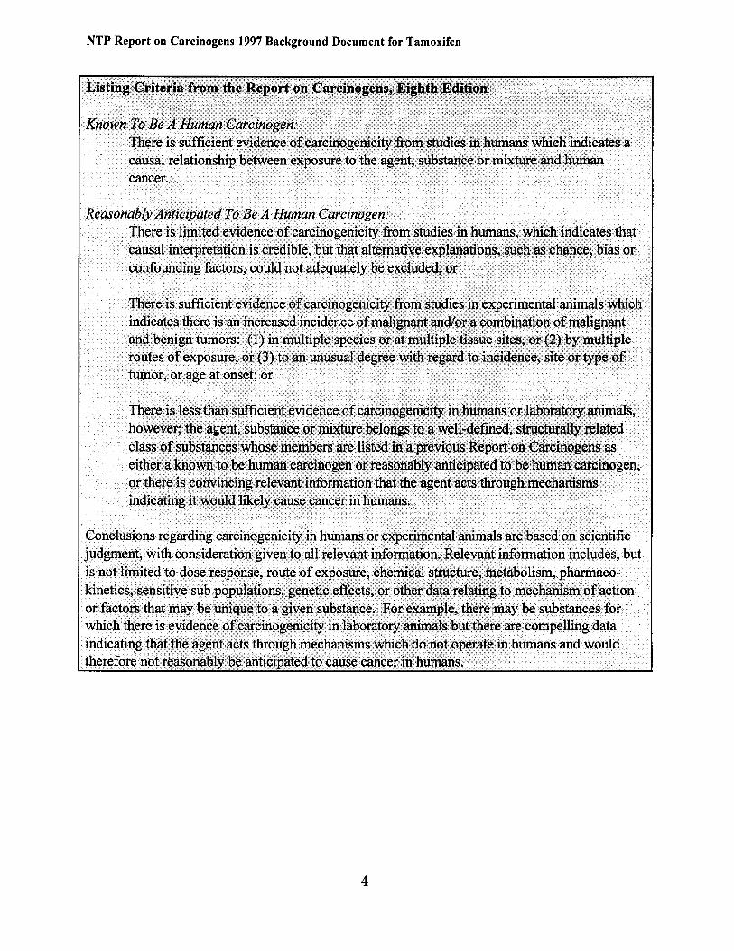
Known ToBe A Human Carcinogen: There is suffiCient evidence ofcarcinogenicity from studies in humans whiclfindicates a causal relationship between exposure to the agent~ substance or mixture and human cancer.
Reasonably Anticipated To Be A Human Carcinogen: There is limited evidence ofcarcinogenicity from studies in humans, which indicates that causal interpretation is credible, but that alternative explanations,.such as chance, bias or confounding factors, could notadequately be excluded, or
There is sufficient evidence ofcarcinogenicity from studies in experimental·animals which indicates there is an increased incidence ofmalignant and/or a combination ofmalignant and benign tumors: (1) in multiple species or atmultiple tissue sites, or {2) by multiple routes ofexposure, or (3) to an unusual degree with regard to incidence, site or type of tumor, or age at onset; or
There is less than sufficient evidence ofcarcinogenidityin humans orlaboratory animals, however; the agent, substance or mixture belongs to a welh·defmed, structurally related class ofsubstances whose members are listed in a .Previous Report on Carcinogens as either a known to be human carcinogen or reasonably anticipated to be human carcinogen, or there is convincing relevant information that the agent acts through mechanisms indicating it would likely cause cancer in humans.
Conclusions regarding carcinogenicity in humans or experimental animals are based on scientific judgment, with consideration given to all·relevant information. Relevant information includes, but is not limited to dose response, route of exposure, chemicaLstructure, metabolism, pharmaco· kinetics, sensitive subpqpulations, genetic effects, or other data relating to mechanism ofaction or factors thatmay be unique to a given substance. For example, there may be substances for which there is evidence ofcarcinogenicity in laboratory animals but there are compelling data indicating that the agent acts through mechanisms which do not operate in humans and would therefore not reasonably be anticipated to cause cancer.in humans.
4
NTP Report on Carcinogens 1997 Background Document for Tamoxifen
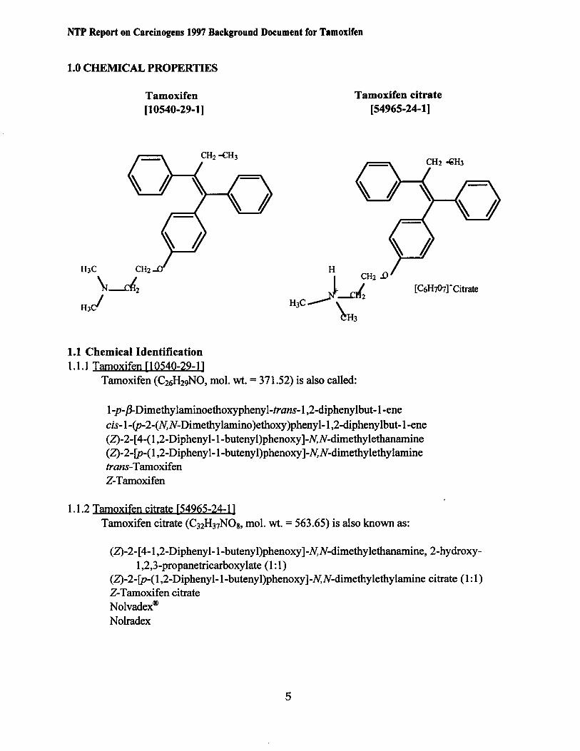
1.0 CHEMICAL PROPERTIES
Tamoxifen Tamoxifen citrate (10540-29-1] [54965-24-1]
1.1 Chemical Identification 1.1.1 Tamoxifen [10540-29-1]
Tamoxifen (C26H29NO, mol. wt. =371.52) is also called:
1-p-,8-Dimethy laminoethoxyphenyl-trans-1 ,2-diphenylbut-1-ene
cis-1-(p-2-(N,N-Dimethylamino )ethoxy)phenyl-1 ,2-diphenylbut-1-ene (Z)-2-[ 4-(1 ,2-Diphenyl-1-butenyl)phenoxy ]-N,N-dimethylethanamine (Z)-2-fp-(1,2-Diphenyl-1-butenyl)phenoxy]-N,N-dimethylethylamine trans-Tamoxifen Z-Tamoxifen
1.1.2 Tamoxifen citrate [54965-24-1] Tamoxifen citrate (C32H37N08, mol. wt. = 563.65) is also known as:
(Z)-2-[ 4-1 ,2-Diphenyl-1-butenyl)phenoxy ]-N,N-dimethylethanamine, 2-hydroxy1 ,2,3-propanetricarboxylate (1: 1)
(Z)-2-fp-(1 ,2-Diphenyl-1-butenyl)phenoxy ]-N,N-dimethylethylamine citrate (1: 1) Z-Tamoxifen citrate Nolvadex® Nolradex
5
NTP Report on Carcinogens 1997 Background Document for Tamoxifen
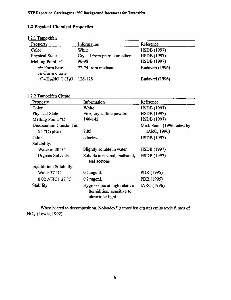
1.2 Physical-Chemical Properties
1.2.1 Tamoxifen
Property Information Reference Color Physical State
Melting Point, °C
cis-Form base cis-Form citrate
C26H29NO.C6HsO
1.2.2 Tamoxifen Citrate
White Crystal from petroleum ether 96-98
72-74 from methanol
126-128
HSDB (1997) HSDB (1997) HSDB (1997)
Budavari (1996)
Budavari (1996)
ProEertl Information Reference Color Physical State Melting Point, °C
Dissociation Constant at 25 oc (pKa)
Odor Solubility:
Water at 20 oc Organic Solvents
Equilibrium Solubility: Water 37 oc 0.02 NHCl 37 oc
Stability
White Fine, crystalline powder 140-142
8.85
odorless
Slightly soluble in water
Soluble in ethanol, methanol, and acetone
0.5mglmL
0.2mglmL
Hygroscopic at high relative humidities, sensitive to ultraviolet light
HSDB (1997) HSDB (1997) HSDB (1997)
Med. Econ. (1996; cited by IARC, 1996)
HSDB (1997)
HSDB (1997)
HSDB (1997)
PDR (1995)
PDR (1995)
IARC (1996)
When heated to decomposition, Nolvadex® (tamoxifen citrate) emits toxic fumes of NOx (Lewis, 1992).
6
NTP Report on Cal'eiaepr11 l997 Background Document for Tamoxifen
2.0 HUMAN EXPOSURE 2.1 Use
Tamoxifen has proven to be a successful palliative therapy for advanced breast cancer yielding response rates similar to those seen with other endocrine treatments, but with few side effects. It has been commonly used as the citrate as a primary therapy for breast cancer in elderly women who are considered poor candidates for surgery. Tamoxifen has been the adjuvant therapy of choice for postmenopausal, node-positive, and estrogen or progesterone receptorpositive women since the mid-1980s, and for postmenopausal, node-negative, and estrogen or progesterone receptor-positive women since the early 1990s. It is also being used in many cases of node-negative and receptor-positive premenopausal women. A high proportion (40-60%) of all women who undergo potentially curative surgery for breast cancer now receive adjuvant tamoxifen therapy for a period of 2 to 5 years (IARC, 1996).
First approved for pharmaceutical use in the United Kingdom in 1973 and in the United States in 1977 (Diogenes, 1997), tamoxifen is presently registered in 97 countries. Tamoxifen use has been estimated at more than 7 million patient-years. The usual dose in the United States and the United Kingdom is 20 mg/day for 1 to 2 years whereas in continental Europe, usual doses are 30 to 40 mg/day (IARC, 1996).
2.2 Production Process and Volume Tamoxifen is produced by treating 4-P,.dimethylaminoethoxy-a-ethy1desoxy1benzoin
with phenylmagnesium bromide or phenyllithium to form 1-(4-P,.dimethy1aminoethoxyphenyl)1,2-diphenylbutanol. Dehydration of the product yields a mixture oftamoxifen and its E-isomer, (E)-2-[4-(1,2-diphenylbut-1-enyl)phenoxy]ethyldimethylamine, which may be separated with petroleum ether. For pharmaceutical preparations, tamoxifen is converted to the 1:1 citrate (Gennaro, 1995; cited by IARC, 1996).
The U.S. and British pharmacopoeias limit theE-isomer to not more than 0.3% and 1%, respectively, in tamoxifen and tamoxifen citrate (IARC, 1996).
Tamoxifen in pharmaceutical formulations is present as its citrate salt. Tamoxifen citrate is available as 15.2-, 30.4-, and 45.6-mg tablets. These correspond to 10, 20, and 30 mg of tamoxifen (IARC, 1996).
Two suppliers of tamoxifen citrate are listed in the Chemcyclopedia 1997 (Strum, 1996). The product Nolvadex® is marketed by Zeneca Pharmaceuticals (PDR, 1995).
Production oftamoxifen citrate worldwide increased from approximately 15,000 lb [7.0 metric tons (Mg)] in 1989 to 19,000 lb (8.5 Mg) in 1991, 22,300 lb (10.1 Mg) in 1993, and 22,700 lb (1 0.3 Mg) in 1995 (IARC, 1996).
2.3 Environmental Exposure Tamoxifen is not known to occur as a natural product (IARC, 1996).
7
NTP Report on Carcinogens 1997 Background Document for Tamoxifen
2.4 Occupational Exposure A U.S. National Institute of Occupational Safety and Health (NIOSH) National
Occupational Exposure Survey (NOES) for 1981-1983 indicated that 350 employees were potentially exposed to tamoxifen in the workplace. Additionally, 2100 employees were potentially exposed to tamoxifen citrate (IARC, 1996).
2.5 Regulations and Criteria Tamoxifen citrate was first allowed on the U.S. market in 1977 (equivalent to 10 mg
base). The June 1997 edition of the New Drug Application List (NDL) lists both 10-mg and 20mg base forms with indications for the treatment of metastatic breast cancer in premenopausal women as an alternative to oophorectomy or ovarian irradiation and for the treatment of panic disorder, with or without agoraphobia. In 1986, it was allowed in postmenopausal women as a single agent to delay breast cancer recurrence following total mastectomy and axillary dissection. In 1989, it was allowed in premenopausal women as an alternative to oophorectomy or ovarian irradiation. In 1990, it was allowed in women with axillary node negative breast cancer. In 1993, tamoxifen was permitted to be used for the treatment of metastatic breast cancer in males. In 1994, the FDA established a new strength (20 mg) and dosage regimen (once or twice daily) (Diogenes, 1997).
California listed tamoxifen as a carcinogen in May 1995. The expert committee, established for Proposition 65, decided to let the public know that tamoxifen use is likely to cause endometrial cancer. Zeneca Pharmaceuticals, the supplier ofNolvadex®, did not challenge
these findings (Mack, 1995).
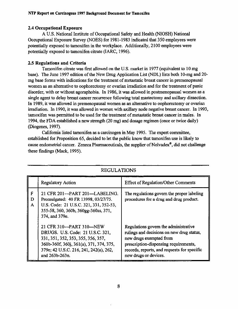
REGULATIONS
Regulatory Action Effect ofRegulation/Other Comments
F D A
21 CFR 201-PART 201-LABELING. Promulgated: 40 FR 13998, 03/27/75. U.S. Code: 21 U.S.C. 321, 331, 352-53, 355-58, 360, 360b, 360gg-360ss, 371, 374, and 379e.
21 CFR 310-PART 310-NEW DRUGS. U.S. Code: 21 U.S.C. 321, 331,351,352,353,355,356,357, 360b-360f, 360j, 361(a), 371, 374, 375, 379e; 42 U.S.C. 216, 241, 242(a), 262, and 263b-263n.
The regulations govern the proper labeling procedures for a drug and drug product.
Regulations govern the administrative rulings and decisions on new drug status, new drugs exempted from prescription-dispensing requirements, records, reports, and requests for specific new drugs or devices.
8
NTP Report on Carcinogens 1997 Background Document for Tamoxifen
3.0 HUMAN STUDIES 3.1 Human Studies Reviewed by IARC (1996)
IARC (1996, pp. 260-280; see Appendix A) reviewed descriptive studies of single cases and case series, case-control studies, cohort studies, and randomized clinical trials involving tamoxifen (invariably as tamoxifen citrate) reported prior to 1996. Based on 1 adequate cohort study, 4 adequate case-control studies, and 14 randomized clinical trials, IARC (1996) concluded that tamoxifen use increased the risk ofendometrial cancer. The positive case-control studies were considered evidence ofcancer because potential confounders, while generally acknowledged, were not regarded as important relative to the magnitude of reported relative risks. Two of the 14 randomized clinical trials were considered particularly important because of the strong and statistically significant association between the use of tamoxifen and risk ofendometrial cancer. The relative risk (odds ratio) for endometrial cancer was 5.6 (95% confidence interval or CI = 1.916.2) in the randomized clinical trial reported by Rutqvist et al. (1995; cited by IARC, 1996), and 7.5 (95% CI = 1.7-32.7) in the randomized clinical trial reported Fisher et al. (1994; cited by IARC, 1996).
3.2 Human Studies Published Post IARC (1996) In a recent review on the association between tamoxifen use and increased risk of
endometrial cancer, MacMahon (1997) concluded that the published results (including those reviewed by IARC, 1996), while suggestive of an association, were not conclusive. MacMahon (1997) based this conclusion on the fact that a positive association was not seen in all randomized clinical trials, that a deficit of endometrial cancer appears to have been present in the comparison groups of two of the most important studies, that none of the studies adequately addressed the problems of confounding by hormone replacement therapy and/or prior hysterectomy, and that none of the studies addressed the issue of detection bias. These same potential confounders were considered by the IARC Working Group and discounted as having a major effect on the reported relative risks (IARC, 1996).
A recent study (Rubagotti et al., 1996), which was not reviewed by IARC, does not show increased risk of endometrial or other cancer among breast cancer patients treated with tamoxifen (Table 3-1). Breast cancer patients (656) were treated with tamoxifen and followed up for 3 to 9 years; detailed information about secondary malignancies was available for all patients. Sitespecific tumor incidence was compared to cancer incidence in the general population. A calculated risk ratio of 1.4 (95% CI = 0.2-5.1) was reported for secondary endometrial cancer among patients treated with 30 mg/day tamoxifen for 2 to 5 years; a risk ratio of 0. 7 (95% CI =
0.0-3.9) was reported for the corresponding untreated group. The authors noted that the short follow-up times might explain the lower endometrial cancer incidence compared to other studies. The imprecise confidence intervals limit the usefulness of this study.
As concluded by IARC (1996) and by MacMahon (1997), a significant excess of any other cancer was not found in either the cohort study or the randomized clinical trials (a combined analysis of three Scandinavian clinical trials suggested an excess ofgastrointestinal cancer; however, this has not yet been confirmed by other studies). A significantly reduced risk for contralateral breast cancer among women treated with tamoxifen was reported in several studies.
9
NTP Report on Carcinogens 1997 Background Document for Tamoxifen
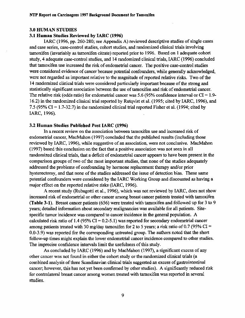
Table 3-1. Summary of Randomized Clinical Trial of Adjuvant Use of Tamoxifen (not reviewed by IARC, 1996): Endometrial Cancers in Patients Treated for Breast Cancer
Patients from the GROCTA trials and additional Italian clinic outpatients
656 tamoxifen (median age 59) and220 tamoxifen + chemotherapy (median age 55)
410 no treatment (median age 60) and 410 chemotherapy only (median age 49)
The number of observed cases of breast cancer was compared to the number of expected cases to give the Standardized Incidence Ratio and 95% confidence intervals (CI). The expected numbers of cases was derived from cancer incidence rates in the Lombardy Cancer Registry in 1983-1987 specified for age, sex, and cancer site.
30mgld
2to5yr
median40mo (tamoxifen only) to91 mo (tamoxifen + chemotherapy)
Risk ratio for endometrial cancer: 1.4 (95% Cl = 0.2-5.1)
The percent of premenopausal women was similar for the tamoxifen (22.7"/o) and no-treatment groups (22.9%). The authors noted that the short follow-up times might explain the lower endometrial cancer incidence compared to other studies.
Rubagotti et al. (1996)
10
NTP Report on Carcinogens 1997 Background Document for Tamoxifen
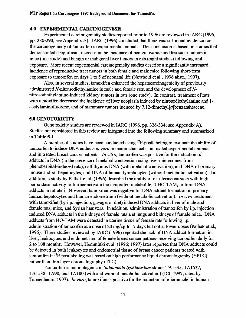
4.0 EXPERIMENTAL CARCINOGENESIS Experimental carcinogenicity studies reported prior to 1996 are reviewed in IARC (1996,
pp. 280-290, see Appendix A). IARC (1996) concluded that there was sufficient evidence for the carcinogenicity of tamoxifen in experimental animals. This conclusion is based on studies that demonstrated a significant increase in the incidence of benign ovarian and testicular tumors in mice (one study) and benign or malignant liver tumors in rats (eight studies) following oral exposure. More recent experimental carcinogenicity studies describe a significantly increased incidence of reproductive tract tumors in both female and male mice following short-term exposure to tamoxifen on days 1 to 5 of neonatal life (Newbold et al., 1996 abstr., 1997).
Also, in several studies, tamoxifen enhanced the hepatocarcinogenicity of previously administered N-nitrosodiethylamine in male and female rats, and the development ofNnitrosodiethylamine-induced kidney tumors in rats (one study). In contrast, treatment of rats with tamoxifen decreased the incidence of liver neoplasia induced by nitrosodiethylamine and 1acetylaminofluorene, and of mammary tumors induced by 7,12-dimethyl[a]benzanthracene.
5.0 GENOTOXICITY Genotoxicity studies are reviewed in IARC (1996, pp. 326-334; see Appendix A).
Studies not considered in this review are integrated into the following summary and summarized in Table 5-1.
A number of studies have been conducted using 32P-postlabeling to evaluate the ability of tamoxifen to induce DNA adducts in vitro in mammalian cells, in treated experimental animals, and in treated breast cancer patients. In vitro, tamoxifen was positive for the induction of adducts in DNA (in the presence of metabolic activation using liver microsomes from phenobarbital-induced rats), calf thymus DNA (with metabolic activation), and DNA of primary mouse and rat hepatocytes, and DNA of human lymphocytes (without metabolic activation). In addition, a study by Pathak et al. (1996) described the ability of rat uterine extracts with high peroxidase activity to further activate the tamoxifen metabolite, 4-HO-TAM, to form DNA adducts in rat uteri. However, tamoxifen was negative for DNA adduct formation in primary human hepatocytes and human endometrium (without metabolic activation). In vivo treatment with tamoxifen (by i.p. injection, gavage, or diet) induced DNA adducts in liver of male and female rats, mice, and Syrian hamsters. In addition, administration oftamoxifen by i.p. injection induced DNA adducts in the kidneys of female rats and lungs and kidneys of female mice. DNA adducts from HO-T AM were detected in uterine tissue of female rats following i.p. administration oftamoxifen at a dose of20 mg/kg for 7 days but not at lower doses (Pathak et al., 1996). Three studies reviewed by IARC (1996) reported the lack of DNA adduct formation in liver, leukocytes, and endometrium of female breast cancer patients receiving tamoxifen daily for 2 to 108 months. However, Hemminki et al. (1996; 1997) later reported that DNA adducts could be detected in both leukocytes and endometrial tissue of breast cancer patients treated with tamoxifen if 32P-postlabeling was based on high performance liquid chromatography (HPLC) rather than thin layer chromatography (TLC).
Tamoxifen is not mutagenic in Salmonella typhimurium strains TA1535, TA1537, TA1538, TA98, and TA100 (with and without metabolic activation) (ICI, 1997; cited by Tannenbaum, 1997). In vitro, tamoxifen is positive for the induction ofmicronuclei in human
11
NTP Report on Carcinogens 1997 Background Document for Tamoxifen
lymphoblastoid P450-expressing MCL-5 cells, human lymphoblastoid cells expressing CYP2El and CYP2A4 but not CYPlAl or CYP1A2, and human breast cancer MCF-7 cells (Otto et al., 1996), all without metabolic activation; for apoptosis in human breast cancer MCF-7 cells (Otto et al., 1996), for chromosome aberrations in P450-expressing MCL-5 cells (Styles et al., 1997); and for aneuploidy and morphological transformation in Syrian hamster embryo cells. The positive response in Syrian hamster cells was replicated by Tsutsui et al. (1997).
Tamoxifen was negative in vitro for the induction of unscheduled DNA synthesis (UDS) in primary rat hepatocytes, for sister chromatid exchanges (SCE) in human lymphocytes, with and without metabolic activation (Wilson et al., 1995), for micronuclei in human AHH-1 cells (without metabolic activation), and for chromosome aberrations in Syrian hamster embryo cells (Tsutsui et al., 1997).
In vivo, tamoxifen was positive for the induction of lacl gene mutations (primarily G to T transversions) in the livers of female Big Blue® transgenic rats (Davies et al., 1997), for chromosomal aberrations and micronuclei in Swiss albino mouse bone marrow (Vijayalaxmi and Rai, 1996), and for chromosomal aberrations and aneuploidy in female Sprague-Dawley rat hepatocytes (Sargent et al., 1996).
12
---
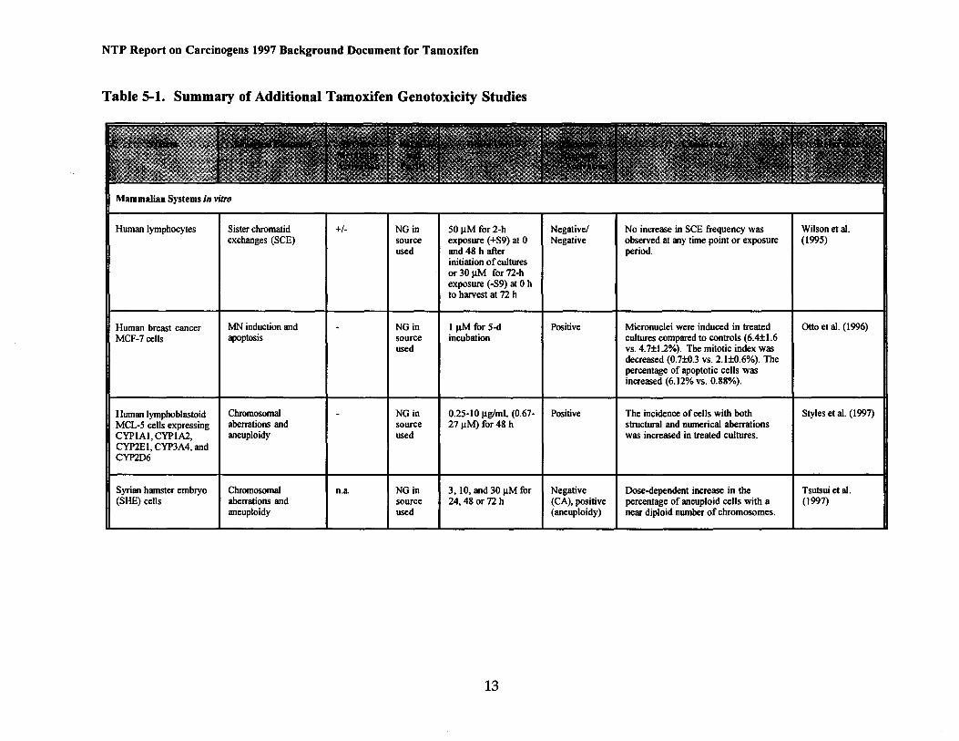
Mammalian Systems in vitro
Human lymphocytes Sister chromatid No increase in SCE frequency was Wilson et al. exchanges (SCE)
NGin Negative/SO J.lM for2-h source exposure (+S9) at 0 Negative observed at any time point or exposure (1995) used and 48 h after period.
initiation of cultures or 30 J.lM for 72-h exposure (-S9) at 0 h to harvest at 72 h
I Micronuclei were induced in treated I Otto et al. (1996)
MCF-7 cells NGin Positive1 J.lM for 5-d Human breast cancer I MN ind~ction and -
apoptosJS source incubation cultures compared to controls (6.4±1.6 used vs. 4.7±1.2%). The mitotic index was
decreased (0.7±0.3 vs. 2.1±0.6%). The percentage of apoptotic cells was increased (6.12% vs. 0.88%).
I
NGin 0.25-10 J.lg/mL (0.67- Positive The incidence of cells with both I Styles et al. (1997) MCL-5 cells expressing Human lymphoblastoid IChromosomal -
aberrations and source structural and numerical aberrations CYP1A1, CYP1A2,
27 J.lM} for 48 h aneuploidy was increased in treated cultures.
CYP2E1, CYP3A4, and CYP2D6
used
Syrian hamster embryo I Chromosomal In.a INGin I3, 10, and 30 J.lM for INegative IDose-dependent increase in the I Tsutsui et al. (SHE) cells aberrations and source 24,48 or72 h (CA), positive percentage of aneuploid cells with a (1997)
aneuploidy used (aneuploidy) near diploid number of chromosomes.
NTP Report on Carcinogens 1997 Background Document for Tamoxifen
Table 5-1. Summary of Additional Tamoxifen Genotoxicity Studies
13
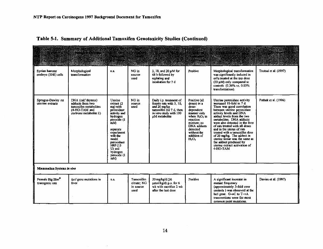
Positive Morphological transformation I Tsutsui et at. (1997) embryo (SHE) cells transformation source 48 h followed by Syrian hamster IMorphological I n.a I NGin I3, 10, and 20 J1M for
was significantly induced in used replating and cells treated at the top dose
incubation for 7 d (20 J1M} only compared to controls (0.36% vs. 0.03% transformation).
Uterine NGin Daily i.p. treatment of Positive (all Uterine peroxidase activity IPathak et at. (1996) uterine extracts adducts from two Sprague-Dawley rat I DNA (calf thymus)
extract (2 source female rats with 5, 10, doses) in a increased 10-fold in 7 d. tamoxifen metabolites There was good correlation (4-HO-TAM and
mg)with used and20mglkg doseperoxidase tamoxifen for 7 d, then dependent between uterine peroxidase
cis/trans metabolite E) manner only activity levels and DNA hydrogen activity and in vitro study with 100
adduct levels from the two peroxide (1
J1M metabolite when H202 in reaction metabolites. DNA adducts mixture; no were also detected in the liver DNA adducts
mM) of rats treated with all doses
separate detected and in the uterus of rats experiment without the treated with a tamoxifen dose with the addition of of20 mglkg. The adduct in model uterine tissue was the same as peroxidase
H202 the adduct produced by
HRP(IS uterine extract activation of U) and 4-HO-TAM hydrogen peroxide (1 mM)
Mammalian Systems in vivo
-Female Big Blue«> A significant increase in I Davies et al. (1997) transgenic rats
20 mglkg/d (36 Positive'lac! gene mutations in I n.a. I Tamoxifen liver citrate; NG mutant frequency
in source Jlmollkgld) p.o. for 6
(approximately 3-fold over used
wk with sacrifice 2 wk after the last dose controls ) was observed at the
lacl gene. G-+C to T -+A transversions were the most
NTP Report on Carcinogens 1997 Background Document for Tamoxifen
Table 5-l. Summary of Additional Tamoxifen Genotoxicity Studies (Continued)
14
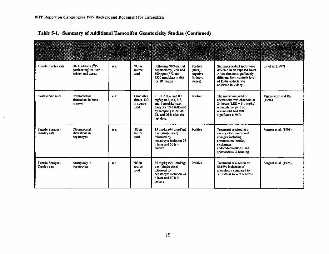
Female Fischer rats I DNA adducts e1P- In.a INGinpostlabeling) in liver, source kidney, and uterus used
I Swiss albino mice I Chromosomal n.a Tamoxifen
aberrations in bone citrate, NG marrow in source
used
Female Sprague- Chromosomal n.a. NGin Dawley rats aberrations in source
hepatocyes used
Female Sprague- Aneuploidy in n.a NGin 35 mglkg (94J1mollkg) Positive Dawley rats hepatocytes source p.o. (single dose)
used followed by hepatocyte isolation 24 h later and 50 h in culture
IFollowing 700/o partial hepatectomy, 250 and 500 ppm (672 and 1350 j.lmollkg) in diet for 18 months
0.1, 0.2, 0.4, and 0.8 mglkg (0.2, 0.4, 0.7, and 1 J.lmOI/kg) p.o. daily for 10 d followed by sampling at 24, 48, 72, and 96 h after the last dose.
35 mglkg (94 J.lmollkg) p.o. (single dose) followed by hepatocyte isolation 24 h later and 50 h in culture
Positive (liver), negative (kidney, uterus)
Positive
Positive
Six major adduct spots were I u et al. (1997) detected in all exposed livers. A low (but not significantly different from control) level of DNA adducts was observed in kidney.
The maximum yield of I Vijayalaxmi and Rai aberrations was observed at (1996) 24 hours (LED = 0.1 mglkg) although the yield of aberrations was still significant at 96 h.
Treatment resulted in a I Sargent et al. (1996) variety of chromosomal changes including chromosome breaks, exchanges, endoreduplications, and spontaneous G-banding.
Treatment resulted in an I Sargent et al. (1996) 85±7% incidence of aneuploidy compared to 3.0±3% in solvent controls.
NTP Report on Carcinogens 1997 Background Document for Tamoxifen
Table 5-l. Summary of Additional Tamoxifen Genotoxicity Studies (Continued)
15
NTP Report on Carcinogens 1997 Background Document for Tamoxifen
Table 5-l. Summary of Additional Tamoxifen Genotoxicity Studies (Continued)
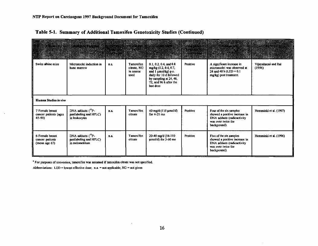
0.1, 0.2, 0.4, and 0.8 Positive bone marrow citrate, NG
Swiss albino mice I Micronuclei induction in I I Tamoxifenn.a. mglkg (0.2, 0.4, 0.7,
in source and 1 J.llllollkg) p.o. used daily for 10 d followed
by sampling at 24, 48, 72, and 96 h after the last dose
I I I I Human Studies in vivo
7 Female breast DNA adducts e2P- In.a. ITamoxifen I40 mgld (110 J.11110lld) IPositive cancer patients (ages postlabeling and HPLC) citrate for 4-21 mo 45-90) in leukocytes
Tamoxifen 20-40 mgld (54-110 Positive cancer patients postlabeling and HPLC) 6 Female breast 2 n.aIDNA adducts eP
citrate ~mol/d) for 3-60 mo (mean age 67) in endometrium
A significant increase in micronuclei was observed at 24 and 48 h (LED= 0.1 mglkg) post treatment.
IFour of the six samples showed a positive increase in DNA adducts (radioactivity was over twice the background).
Five ofthe six samples showed a positive increase in DNA adducts (radioactivity was over twice the background).
I Vijayalaxmi and Rai (1996)
I Hemminki et al. (1997)
I Hemminki et al. (1996)
• For purposes of conversion, tamoxifen was assumed iftamoxifen citrate was not specified.
Abbreviations: LED = lowest effective dose; n.a. = not applicable; NG = not given
16
NTP Report on Carcinogens 1997 Background Document for Tamoxifen
6.0 ABSORPTION, DISTRIBUTION, METABOLISM, AND EXCRETION 6.1 Absorption, Distribution, and Excretion
In humans, tamoxifen is absorbed after oral administration and is readily bound (> 99%) by plasma proteins (Lien et al., 1989; cited by !ARC, 1996). Patients with breast cancer showed steady-state mean plasma concentrations of 186-214 ng/mL after administration of40 mg/day for 2 months (Me Vie et al., 1986; cited by IARC, 1996). Male volunteers showed peak plasma concentrations of42 ng/mL tamoxifen and 12 ng/mL N-desmethyltamoxifen, following administration of a single 20-mg dose (Adam et al., 1980; cited by IARC, 1996). Steady-state concentrations oftamoxifen and N-desmethyltamoxifen were reached after 3 to 4 weeks of40 mg/day administration (Me Vie et al., 1986; cited by IARC, 1996) and after 4 to 8 weeks of 20 mg/day administration (Lien et al., 1995; cited by !ARC, 1996).
The distribution half-life (initial t112) oftamoxifen is 7 to 14 hours (Adam et al., 1980;
Me Vie et al., 1986; both cited by IARC, 1996). The apparent volume of distribution in humans is 50 to 60 L/kg (Lien et al., 1989; cited by !ARC, 1996), suggesting extensive tissue binding (Lien et al.,1991; cited by IARC, 1996).
The plasma elimination half-life was 10 hours during the first day in male volunteers given 40 mg tamoxifen (Guelen et al., 1987; cited by IARC, 1996). However, significant levels of tamoxifen and N-desmethyltamoxifen were present after 34 hours, indicative ofa lengthening of half-life with increasing study duration or the existence ofbiphasic half-lives. One study suggests biphasic pharmacokinetics with a terminal elimination phase ofabout 7 days (Fromson et al., 1973a; cited by IARC, 1996). Tamoxifen and metabolites are excreted as glucoronides and other conjugates (Furr and Jordan, 1984; cited by IARC, 1996).
6.2 Metabolites Metabolites were identified in urine and plasma of breast cancer patients (Poon et al.,
1993, 1995; cited by IARC, 1996). N-Desmethyltamoxifen, tamoxifenN-oxide, and six other metabolites were detected in plasma, while glucuronides of four hydroxylated metabolites (4hydroxytamoxifen, 4-hydroxy-N-desmethyltamoxifen, dihydroxytamoxifen, and possibly a hydroxy-N-desmethyltamoxifen) were found in urine. In biopsy and autopsy tissue samples (liver, lung, pancreas, brain, adipose), tamoxifen metabolites (N-desmethyl-, N-didesmethyl-, 4hydroxy-, and 4-hydroxy-N-desmethyl) were 10- to 60-fold higher than in serum, especially in liver and lung (Lien et al., 1991; cited by IARC, 1996).
Tamoxifen can be metabolized in vitro by microsomal cytochrome P450 and flavin monooxygenase pathways to metabolites that irreversibly bind to microsomal proteins (Mani and Kupfer, 1991; cited by IARC, 1996). In a human microsomal preparation, CYP3A4 and CYP2B6 were identified as active in tamoxifen catalysis to metabolites that bind to protein (White et al., 1993; cited by Smith and White, 1995). In human liver homogenate and a human hepatic G2 cell line treated with a mixture of tamoxifen and its deuterated analogs, several metabolites were detected, including a-hydroxytamoxifen, 4-hydroxytamoxifen, Ndesmethyltamoxifen, and tamoxifen N-oxide (Poon et al., 1995; cited by IARC, 1996). In vitro studies show that tamoxifen-protein binding is greater in liver microsomes of rat (3.8-fold) and mice (17-fold) than ofhumans (White et al., 1993; cited by Smith and White, 1995).
17
NTP Report on Carcinogens 1997 Background Document for Tamoxifen
Postulated metabolic pathways oftamoxifen are presented schematically by IARC (1996, page 293).
6.3 Structure-Activity Relationships Toremifene is structurally similar to tamoxifen; toremifene has a chlorine instead of
hydrogen atom in the ethyl group (Kuramochi, 1996). Both compounds have anti-estrogenic effects as demonstrated by their ability to compete with estrogen; they also have similar effects on estrogen-dependent breast cancer cell lines in vitro and in vivo, and have the same binding constant. However, tamoxifen is a heptocarcinogen in the rat while toremifene has not been demonstrated to induce rat liver tumors. Furthermore, in rat liver, DNA adducts are readily detected after treatment with tamoxifen, whereas toremifene has been associated with the induction of very few DNA adducts (Hard et al., 1993; cited by Kuramochi, 1996).
A study that examined the relationship between DNA-adduct forming ability and physicochemical properties of the two analogs strongly suggests that the stability of the carbocation intermediate arising from tamoxifen is greater than that for the carbocation intermediate arising from toremifene (Kuramochi, 1996). Two other tamoxifen derivatives, 4iodotamoxifen and droxifene, which demonstrate no DNA-adduct-forming ability, are also expected to have less stable carbocation intermediates than tamoxifen.
7.0 MECHANISMS 7.1 Genotoxicity
A possible mechanism by which tamoxifen is carcinogenic is via the formation ofDNA adducts induced by one or more genotoxic metabolite(s) (see section 5). Genotoxic metabolite(s) may be formed by the oxidation oftamoxifen to a DNA-reactive carbocation (Potter et al., 1994; cited by Kuramochi, 1996), or by the metabolism oftamoxifen to a DNA-reactive hydroxylamine intermediate (Cunningham et. al, 1996). Epoxide metabolites that are potentially genotoxic were produced in rat, mouse, and human liver microsomal preparations (Lim et al., 1994; cited by Smith and White, 1995). Genotoxic metabolites, as judged by the induction of micronuclei in a human cell line, are produced by human cytochrome P450s (White et al., 1992; cited by Smith and White, 1995).
In support of this mode of action, a number of studies have demonstrated the ability of tamoxifen to induce DNA adducts in vitro in cultured mammalian cells (either after metabolic activation or using metabolically competent cells), and in vivo in multiple tissues of rats, mice, and Syrian hamsters. In breast cancer patients treated with tamoxifen, several investigators have reported the lack of DNA adduct formation in liver, leukocytes, and endometrium. However, Hemminki et al. (1996; 1997) using a modified 32P-postlabeling technique, based on HPLC rather than TLC, reported the detection of DNA adducts in both leukocytes and endometrial tissue of breast cancer patients treated with tamoxifen.
Other studies support a causal relationship between in vivo genotoxicity and tumor response. Fifty percent of 24 hepatocarcinomas sampled from tamoxifen-treated female rats contained mutations in exons 5 to 9 of the p53 gene (Vancutsem et al., 1994; cited by IARC,
18
NTP Report on Carcinogens 1997 Background Document for Tamoxifen
1996). Nine of the 13 mutations detected involved A ~G transitions in codon 231, while the
other four mutations involved a silent C ~T transition in codon 294.
7.2 Tamoxifen-Estrogen Receptor Interactions As reviewed by IARC (1996, pp. 334-336; see Appendix A) and.more recently by Gallo
and Kaufman (1997), tamoxifen is an estrogen antagonist and/or agonist by binding directly to the estrogen receptor. In breast tissue, tamoxifen exerts antiestrogenic activity by binding with high affinity to the estrogen receptor, preventing normal estrogen-induced transcriptional activity (Pasqualini et al., 1987; cited by IARC, 1996). In other tissues, such as bone, uterus, and liver, tamoxifen acts as a partial agonist, thereby inducing typical estrogen-mediated alterations in gene expression and on cell growth and differentiation (Love et al., 1992b, Jordan and Prestwich, 1977; both cited by IARC, 1996). These tissue-specific effects may be involved in the ability of tamoxifen to decrease or increase cancer risk, respectively.
8.0 REFERENCES
Adam, H. K., J. S. Patterson, and J. V. Kemp. 1980. Studies on the metabolism and pharmacokinetics oftamoxifen in normal volunteers. Cancer Treatm. Rep. 64:761-764. (Cited by IARC, 1996)
Budavari, S., Ed. 1996. The Merck Index. 12th ed. Merck & Co, Inc., Whitehall, NJ.
Cook, L. S., N. S. Weiss, S.M. Schwartz, E. White, B. McKnight, D. E. Moore, and J. R. Daling. 1995. Population-based study oftamoxifen therapy and subsequent ovarian, endometrial, and breast cancers. J. Natl. Cancer lnst. 87:1359-1364. (Cited by IARC, 1996)
Cunningham, A., G. Klopman, and H. S. Rosenkranz. 1996. A study of the structural basis of the carcinogenicity of tamoxifen, toremifene and their metabolites. Mutat. Res. 349:85-94.
Curtis, R. E., J.D. Boice, Jr., D. A. Shriner, B. F. Hankey, and J. F. Fraumeni, Jr. 1996. Second cancers after adjuvant tamoxifen therapy for breast cancer. Briefcommunication. J. Natl. Cancer Inst. 88(12):832-834 (Cited by IARC, 1996 as "in press").
Davies, R., V.I. C. Oreffo, E. A. Martin, M. F. W. Festing, I. N.H. White, L. L. Smith, and J. A. Styles. 1997. Tamoxifen causes gene mutations in the livers of lambda//acl transgenic rats. Cancer Res. 57:1288-1293.
Diogenes. 1997. DIOGENES FDA regulatory updates. DIALOG database file number 158. Last updated July 1997. Record numbers 903779, 907425, 908845, 911393, 912242, 40139294013931.
Fisher, B., J. P. Costantino, C. K. Redmond, E. R. Fisher, D. L. Wickerham, and W. M. Cronin. 1994. Endometrial cancer in tamoxifen-treated breast cancer patients: Findings from the National
19
NTP Report on Carcinogens 1997 Background Document for Tamoxifen
Surgical Adjuvant Breast and Bowel Project (NSABP) B-14. J. Natl Cancer Inst. 86:527-537. (Cited by IARC, 1996)
Fromson, J. M., S. Pearson, and S. Bramah. 1973a. The metabolism oftamoxifen (ICI 46,474). Part II: In female patients. Xenobiotica 3:711-714. (Cited by IARC, 1996)
Furr, B. J. A. and V. C. Jordan. 1984. The pharmacology and clinical uses oftamoxifen. Pharmacol. Ther. 25:127-205. (Cited by IARC, 1996)
Gallo, M.A., and D. Kaufman. 1997. Antagonistic and agonistic effects oftamoxifen: Significance in human cancer. Semin. Oncol. 24:S1-71 to Sl-80.
Gennaro, A. R., Ed. 1995. Remington: The Science and Practice ofPharmacy. 19th ed., vol. II. Mack Publishing Co., Easton, PA, p. 1094. (Cited by IARC, 1996) Guelen, P. J. M., D. Stevenson, R. J. Briggs, and D. de Vos. 1987. The bioavailability of Tamoplex (tamoxifen). Part 2. A single dose cross-over study in healthy male volunteers. Methods Find. Exp. Clin. Pharmacol. 9:685-690. (Cited by IARC, 1996)
Hard, G. C., M. J. Iantropoulos, K. Jordan, L. Radi, 0. P. Kaltenberger, A. R. Imondi, and G. M. Williams. 1993. Major differences in the hepatocarcinogenicity and DNA adduct forming ability between toremifene and tamoxifen in female Crl:CD(BR) rats. Cancer Res. 53:4534-4541. (Cited by Kuramochi, 1996)
Hardell, L. 1988b. Pelvic irradiation and tamoxifen as risk factors for carcinoma of corpus uteri [letter]. Lancet ii:1432. (Cited by IARC, 1996)
Hemminki, K., H. Rajaniemi, B. Lindahl, and B. Moberger. 1996. Tamoxifen-induced DNA adducts in endometrial samples from breast cancer patients. Cancer Res. 56:4374-4377.
Hemminki, K., H. Rajaniemi, M. Koskinen, and J. Hansson. 1997. Tamoxifen-induced DNA adducts in leucocytes of breast cancer patients. Carcinogenesis (London) 18:9-13.
HSDB (Hazardous Substances Data Bank). 1997. Online database produced by the National Library of Medicine. Tamoxifen profile last updated May 9, 1997.
IARC (International Agency for Research on Cancer). 1979. Estrogens and progestins in relation to human cancer. IARC Monogr. Eval. Carcinog. Risks Chern. Hum. 21(Sex hormones [II]): 83129.
IARC (International Agency for Research on Cancer). 1996. Tamoxifen. IARC Monogr. Eval. Carcinog. Risks Chern. Hum. 66(Some Pharmaceutical Drugs):253-365.
20
NTP Report on Carcinogens 1997 Background Document for Tamoxifen
ICI (Imperial Chemical Industries). 1997. Bacterial mutagenicity study using selected strains of Salmonella typhimurium. Study no. TMV/227 3/HG/009756. (Cited by Tannenbaum, 1997)
Jordan, V. C., and G. Prestwich. 1977. Binding of [3H]tamoxifen in rat uterine cytosols: A comparison of swinging bucket and vertical tube rotor sucrose density gradient analysis. Mol. Cell Endocrinol. 8:179-188. (Cited by IARC, 1996)
Kuramochi, H. 1996. Conformational studies and electronic structures of tamoxifen and toremifene and their allylic carbocations proposed as reactive intermediates leading to DNA adduct formation. J. Med. Chern. 39:2877-2886.
Lewis, R. J. 1992. Sax's Dangerous Properties oflndustrial Materials. 8th ed., vol. Ill. Van Nostrand Reinhold, New York.
Li, D., Y. Dragan, V. C. Jordan, M. Wang, and H. C. Pitot. 1997. Effects of chronic administration oftamoxifen and toremifene on DNA adducts in rat liver, kidney, and uterus. Cancer Res. 57:1438-1441.
Lien, E. A., E. Solheim, Q. A. Lea, S. Lundgren, S. Kvinnsland, and P.M. Ueland. 1989. Distribution of 4-hydroxy-N-desmethyltamoxifen and other tamoxifen metabolites in human biological fluids during tamoxifen treatment. Cancer Res. 49:2175-2183. (Cited by IARC, 1996)
Lien, E. A., E. Solheim, and P. M. Ueland. 1991. Distribution of tamoxifen and its metabolites in rat and human tissues during steady-state treatment. Cancer Res. 51:4837-4844. (Cited by IARC, 1996)
Lien, E. A., G. Anker, and P.M. Ueland. 1995. Pharmacokinetics oftamoxifen in premenopausal and postmenopausal women with breast cancer. J. Steroid Biochem. Mol. Biol. 55:229-231. (Cited by IARC, 1996)
Lim, C. K., Z. Yuan, J. H. Lamb, I. N.H. White, F. DeMatteis, and L. L. Smith. 1994. A comparative study of tamoxifen metabolism in female rat, mouse and human liver microsomes. Carcinogenesis (London) 15:589-593. (Cited by Smith and White, 1995)
Love, R. R., R. B. Mazess, H. S. Barden, S. Epstein, P. A. Newcombe, V. C. Jordan, P. P. Carbome, and D. L. DeMets. 1992b. Effect oftamoxifen on bone mineral density in postmenopausal women with breast cancer. N. Engl. J. Med. 326:852-856. (Cited by IARC, 1996)
Mack, T. 1995. Tamoxifen ruling in California [letter]. Science 270:1102-1103.
MacMahon, B. 1997. Overview of studies on endometrial cancer and other types of cancer in humans: Perspectives ofan epidemiologist. Semin. Oncol. 24: S 1-S 139
21
NTP Report on Carcinogens 1997 Background Document for Tamoxifen
Magriples, U., F. Naftolin, P. E. Schwartz, and M. 1. Carcangiu. 1993. High-grade endometrial carcinoma in tamoxifen-treated breast cancer patients. J. Clin. Oncol. 11:485-490. (Cited by IARC, 1996)
Mani, C., and D. Kupfer. 1991. Cytochrome P-450-mediated activation and irreversible binding of the antiestrogen tamoxifen to proteins in rat and human liver: Possible involvement of flavincontaining mono-oxygenases in tamoxifen activation. Cancer Res. 51:6052-6058. (Cited by IARC, 1996)
MantyHi, E. T. E., S. H. Karlsson, and L. S. Nieminen. 1996. Induction ofendometrial cancer by tamoxifen in the rat. In: Hormonal Carcinogenesis. II. Proceedings of the Second International Symposium. Li, J. J., S. A. Li, J.-C. Gustafsson, S. Nandi, and L.l. Sekely, Eds. Springer Verlag, New York, pp. 442-445. (Cited by IARC, 1996)
McVie, J. G., G. P. C. Simonetti, D. Stevenson, R. J. Briggs, P. J. M. Guelen, and D. de Vos. 1986. The bioavailability of Tamoplex (tamoxifen). Part 1. A pilot study. Methods Find. Exp. Clin. Pharmacol. 8:505-512. (Cited by IARC, 1996)
Medical Economics. 1996. PDR®: Physicians' Desk Reference. 50th ed. Medical Economics Data
Products Co., Montvale, NJ, pp. 2842-2844. (Cited by IARC, 1996)
Newbold, R. R, B. C. Bullock, W. N. Jefferson, and E. Padilla-Burgos. 1996. Effects of developmental exposure of mice to tamoxifen. Proc. Am. Assoc. Cancer Res. 37:109 (Abstract no. 753).
Newbold, R. R., W. N. Jefferson, E. Padilla-Burgos, and B. C. Bullock. 1997. Uterine carcinoma in mice treated neonatally with tamoxifen. Carcinogenesis (London) 18(12):2293-2298.
NTP (National Toxicology Program). 1998. Report on Carcinogens, 8th ed., National Toxicology Program, U.S. Department ofHealth and Human Services, National Institute ofEnvironmental Health Sciences, Research Triangle Park, NC.
Otto, A.M., R. Paddenberg, S. Schubert, and H. G. Mannherz. 1996. Cell-cycle arrest, micronucleus formation, and cell death in growth inhibition of MCF-7 breast cancer cells by tamoxifen and cisplatin. J. Cancer Res. Clin. Oncol. 122:603-612.
Pasqualini, J. R, C. Sumida, N. A. Giambiagi, and B. L. Nguyen. 1987. The complexity of antioestrogen responses. J. Steroid. Biochem. 27:883-889. (Cited by IARC, 1996)
Pathak, D. N., K. Pongracz, and W. J. Bodell. 1996. Activation of4-hydroxytamoxifen and the tamoxifen derivative metabolite E by uterine peroxidase to form DNA adducts: Comparison with DNA adducts formed in the uterus of Sprague-Dawley rats treated with tamoxifen. Carcinogenesis 17:1785-1790.
22
NTP Report on Carcinogens 1997 Background Document for Tamoxifen
PDR® 1995. Physicians' Desk Reference® 49th ed. Medical Economics Data Products. Montvale, NJ, pp. 2757-2759.
Poon, G. K., Y. C. Chui, R. McCague, P. E. L~mning, R. Feng, M.G. Rowlands, and M. Jarman. 1993. Analysis of Phase I and Phase II metabolites oftamoxifen in breast cancer patients. Drug Metab. Dispos. 21:1119-1124. (Cited by IARC, 1996)
Poon, G. K., B. Walter, P. E. Lenning, M. N. Horton, and R. McCague. 1995. Identification of tamoxifen metabolites in human HEP G2 cell line, human liver homogenate, and patients on longterm therapy for breast cancer. Drug Metab. Dispos. 23:377-382. (Cited by IARC, 1996)
Potter, G. A., R. McCague, and M. Jarman. 1994. A mechanistic hypothesis for DNA adduct formation by tamoxifen following hepatic oxidative metabolism. Carcinogenesis (London) 15:439442. (Cited by Kuramochi, 1996)
Rubagotti, A., A. Perrotta, C. Casella, and F. Boccardo. 1996. Risk of new primaries after chemotherapy and/or tamoxifen treatment for early breast cancer. Ann. Oncol. 7:239-244.
Rutqvist, L. E., H. Johansson, T. Signomklao, U. Johansson, T. Pomander, and N. Wilking, for the Stockholm Breast Cancer Study Group. 1995. Adjuvant tamoxifen therapy for early stage breast cancer and second primary malignancies. J. Natl. Cancer Inst. 87:645-651. (Cited by IARC, 1996)
Sargent, L. M., Y. P. Dragon, C. Sattler, N. Bahnub, G. Sattler, P. Martin, A. Cisneros, J. Mann, S. Thorgeirsson, V. C. Jordan, and H. C. Pitot. 1996. Induction of hepatic aneuploidy in vivo by tamoxifen, toremifene and idoxifene in female Sprague-Dawley rats. Carcinogenesis (London) 17:1051-1056.
Sasco, A.J., G. Chaplain, E. Amoros, and S. Saez. 1996. Endometrial cancer following breast cancer: Effect of tamoxifen and castration by radiotherapy. Epidemiology 7:9-13.
Smith, L. L., and I. N.H. White. 1995. Chemoprevention of breast cancer by tamoxifen: Risks and opportunities. Toxicol. Lett. 82/83:181-186.
Strum, K., Ed. 1996. Tamoxifen citrate. In: Chemcyclopedia 97: The Manual of Commercially Available Chemicals, vol. 15, American Chemical Society, Washington, DC, p. 161.
Styles, J. A., A. Davies, R. Davies, I. N. White, and L. L. Smith. 1997. Clastogenic and aneugenic effects of tamoxifen and some of its analogues in hepatocytes from dosed rats and in human lymphoblastoid cells transfected with human P450 cDNAs (MCL-5 cells). Carcinogenesis (London) 18:303-13.
23
NTP Report on Carcinogens 1997 Background Document for Tamoxifen
Tannenbaum, S. R. 1997. Comparative metabolism oftamoxifen and DNA adduct formation and in vitro studies on genotoxicity. Semin. Oncol. 24(Suppl1):S1-81 to S1-86.
Tsutsui, T., S. Taguchi, Y. Tanaka, and J. C. Barrett. 1997. 17(3-Estradiol, diethylstilbestrol, tamoxifen, toremifene and ICI 164, 384, induce morphological transformation and aneuploidy in cultured Syrian hamster embryo cells. Int. J. Cancer 70:188-193.
Vancutsem, P.M., P. Lazarus, and G. M. Williams. 1994. Frequent and specific mutations of the ratp53 gene in hepatocarcinomas induced by tamoxifen. Cancer Res. 54:3864-3867. (Cited by IARC, 1996)
van Leeuwen, F. E., J. Benraadt, J. W. W. Coebergh, L.A. L. M. Kiemeney, C. H. F. Gimbrere, R. Otter, L. J. Schouten, R. A.M. Damhuis, M. Bontenbal, F. W. Diepenhorst, A. W. van den Belt-Dusebout, and H. van Tinteren. 1994. Risk of endometrial cancer after tamoxifen treatment ofbreast cancer. Lancet 343:448-452. (Cited by IARC, 1996)
Vijayalaxmi, K. K., and S. P. Rai. 1996. Studies on the genotoxicity oftamoxifen citrate in mouse bone marrow cells. Mutat. Res. 368:109-114.
White, I. N.H., F. DeMatteis, A. Davies, L. L. Smith, C. Crofton-Sleigh, S. Venitt, A. Hewer, and D. H. Phillips. 1992. Genotoxic potential oftamoxifen and analogues in female Fischer F344/N rats, DBA/2 and C57B 1/6 mice and in human MCL-5 cells. Carcinogenesis (London) 13:2197-2203. (Cited by Smith and White, 1995)
White, I. N. H., A. Davies, L. L. Smith, S. Dawson, and F. DeMatteis. 1993. Induction of CYP2B1 and 3A1, and associated monooxygenase activities by tamoxifen and certain analogues in the livers of female rats and mice. Biochem. Pharmacol. 45:21-30. (Cited by Smith and White, 1995)
Wilson, A. S., M.D. Tingle, M.D. Kelly, and B. K. Park. 1995. Evaluation of the generation of genotoxic and cytotoxic metabolites ofbenzo[a]pyrene, aflatoxin B1, naphthalene and tamoxifen using human liver microsomes and human lymphocytes. Hum. Exp. Toxicol. 14:507-515.
24

APPENDIX A
Excerpts from the IARC Monograph on the Evaluation of the Carcinogenic Risks of Chemicals to Humans
Volume 66 (Some Pharmaceutical Drugs) Tamoxifen, pp. 253-365, 1996

APPENDIXB
Description of Online Searches for Tamoxifen
NTP Report on Carcinogens 1997 Background Document for Tamoxifen
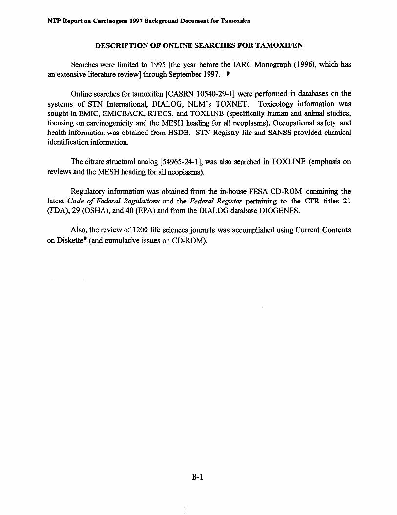
DESCRIPTION OF ONLINE SEARCHES FOR TAMOXIFEN
Searches were limited to 1995 [the year before the IARC Monograph (1996), which has an extensive literature review] through September 1997. '
Online searches for tamoxifen [CASRN 10540-29-1] were performed in databases on the systems of STN International, DIALOG, NLM's TOXNET. Toxicology information was sought in EMIC, EMICBACK, RTECS, and TOXLINE (specifically human and animal studies, focusing on carcinogenicity and the MESH heading for all neoplasms). Occupational safety and health information was obtained from HSDB. STN Registry file and SANSS provided chemical identification information.
The citrate structural analog [54965-24-1], was also searched in TOXLINE (emphasis on reviews and the MESH heading for all neoplasms).
Regulatory information was obtained from the in-house FESA CD-ROM containing the latest Code of Federal Regulations and the Federal Register pertaining to the CFR titles 21 (FDA), 29 (OSHA), and 40 (EPA) and from the DIALOG database DIOGENES.
Also, the review of 1200 life sciences journals was accomplished using Current Contents on Diskette® (and cumulative issues on CD-ROM).
B-1

APPENDIXC
Report on Carcinogens (RoC), 9th Edition Review Summary
NTP Report on Carcinogens 1997 Background Document for Tamoxifen
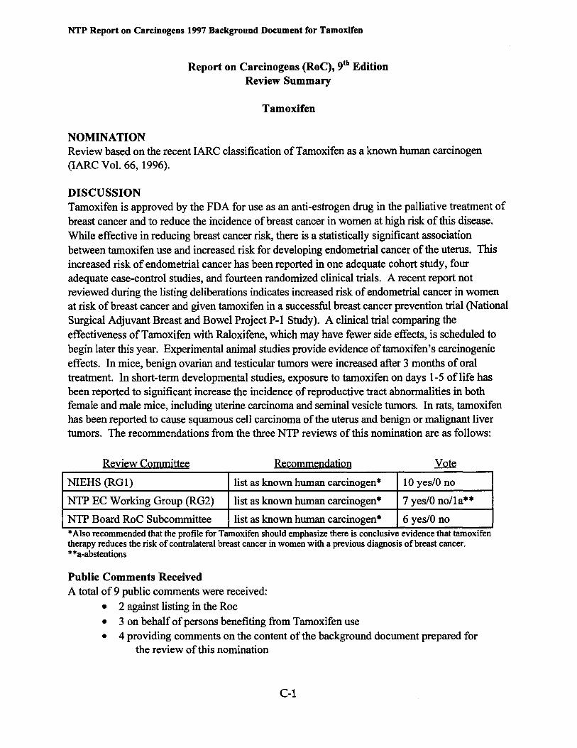
Report on Carcinogens (RoC), 9th Edition Review Summary
Tamoxifen
NOMINATION Review based on the recent IARC classification ofTamoxifen as a known human carcinogen (IARC Vol. 66, 1996).
DISCUSSION Tamoxifen is approved by the FDA for use as an anti-estrogen drug in the palliative treatment of breast cancer and to reduce the incidence ofbreast cancer in women at high risk of this disease. While effective in reducing breast cancer risk, there is a statistically significant association between tamoxifen use and increased risk for developing endometrial cancer of the uterus. This increased risk of endometrial cancer has been reported in one adequate cohort study, four adequate case-control studies, and fourteen randomized clinical trials. A recent report not reviewed during the listing deliberations indicates increased risk ofendometrial cancer in women at risk of breast cancer and given tamoxifen in a successful breast cancer prevention trial (National Surgical Adjuvant Breast and Bowel Project P-1 Study). A clinical trial comparing the effectiveness ofTamoxifen with Raloxifene, which may have fewer side effects, is scheduled to begin later this year. Experimental animal studies provide evidence oftamoxifen's carcinogenic effects. In mice, benign ovarian and testicular tumors were increased after 3 months oforal treatment. In short-term developmental studies, exposure to tamoxifen on days 1-5 of life has been reported to significant increase the incidence of reproductive tract abnormalities in both female and male mice, including uterine carcinoma and seminal vesicle tumors. In rats, tamoxifen has been reported to cause squamous cell carcinoma of the uterus and benign or malignant liver tumors. The recommendations from the three NTP reviews of this nomination are as follows:
Review Committee Recommendation Vote
NIEHS (RG1) list as known human carcinogen* 10 yes/0 no
NTP EC Working Group (RG2) list as known human carcinogen* 7 yes/0 nolla**
NTP Board RoC Subcommittee list as known human carcinogen* 6 yes/0 no *Also recommended that the profile for Tamoxtfen should emphasiZe there ts conclusive evidence that tamoxtfen therapy reduces the risk ofcontralateral breast cancer in women with a previous diagnosis ofbreast cancer. **a-abstentions
Public Comments Received A total of 9 public comments were received:
• 2 against listing in the Roc • 3 on behalfofpersons benefiting from Tamoxifen use • 4 providing comments on the content of the background document prepared for
the review of this nomination
C-1


![NTP REPORT ON CARCINOGENS BACKGROUND ......NTP Report on Carcinogens 1996 Background Document for Azacitidine 1.0 INTRODUCTION Azacitidine [320-67-2] 1.1 Chemical Identification Azacitidine](https://static.fdocuments.in/doc/165x107/5f389e46814f462fcd08ae75/ntp-report-on-carcinogens-background-ntp-report-on-carcinogens-1996-background.jpg)


