nternational Society for the Study of the Lumbar Spine
184
International Society for the Study of the Lumbar Spine 40 TH Annual Meeting SCOTTSDALE, AZ, USA May 13-17, 2013 Abstract Proceedings Institute for Clinical Sciences Sahlgrenska Academy PO Box 426 SE-405 30 Gothenburg • Sweden www.issls.org 40th Annual Meeting May 13-17, 2013 ISSLS 2013 - SCOTTSDALE
Transcript of nternational Society for the Study of the Lumbar Spine
International Society for the
Study of the Lumbar Spine
40TH Annual MeetingSCOTTSDALE, AZ, USA
May 13-17, 2013
Abstract Proceedings
Institute for Clinical Sciences
Sahlgrenska Academy
PO Box 426
SE-405 30 Gothenburg • Swedenwww.issls.org
40th Annual MeetingMay 13-17, 2013
ISSLS 2013 - SCOTTSDALE

GENERAL POSTERS
98
GP1 SUITABILITY EVALUATION OF RESTORING THE INTERVERTEBRAL DISC MECHANICS IN TREATING OSTEOPROTIC VERTEBRAL FRACTURE USING KYPHOPLASTY WITH PMMA OR CALCIUM-P BASED CEMENT Haoju Lo, Hung-Ming Chen
The Orthopaedic Department, Ren-Ai
Branch, Taipei City Hospital, Taiwan
INTRODUCTION: The biomechanical tests
about the filler materials paid little atten-
tion to vertebral endplate or intervertebral
disc in treating compression fracture. Im-
pact of cement leakage into disks on the
development of adjacent fractures was not-
ed. There is only a limited understanding
how the load shift of the intervertebral disc
after a kyphoplasty with different cement.
The intervertebral disc should be showed
altered disc pressure profile.
MATERIALS AND METHODS: 3D finite-
element osteoporotic model of L1–L3 were
developed. The influence of augmentation
level as well as bipedicular filling with
PMMA or CaP based cement was investi-
gated. The risk factors (anterior wall defi-
ciency and superior endplate cleft) were
also taken into consider as variables. Last, a
leakage of cement was also simulated.
RESULTS: The stresses and strains in the
vertebrae close to an augmentation were
increased, and change their distribution
with different risk factors. In PMMA groups,
higher stresses and strains were noted over
endplate and disc when risk factors exist.
The pressure in a bulge of the augmented
endplate and disc were increased to almost
20% of its value before the augmentation,
resulting in a stiffening of the intervertebral
disc. In CaP cement, in leakage model, seem
to show more suitable in mechanics if leak-
age happened.
DISCUSSION: With an intact endplate, the
two augmented cements shows no signifi-
cantly difference in restoring of strength
and stiffness. With a damaged endplate,
overgrowth stiffness located superior end-
plate by using PMMA maybe show an im-
pact factor for further adjacent deformity.
The change of pressure in disc with a dam-
aged endplate was supposed to develop
abnormal intradiscal pressure in flexion also
been shown to increase loading of the adja-
cent anterior vertebral cortex. Our study
indicated a compression fracture treated
with kyphoplasty by PMMA or CaP based
cement must be evaluated according the
damaged structures.
GP2 ASSESSMENT OF PAIN BEHAVIOR IN A RAT MODEL OF INTERVERTEBRAL DISK INJURY USING THE CATWALK GAIT ANALYSIS SYS-TEM Masayuki Miyagi, MD, PhD *1,2, Tetsuhiro
Ishikawa, MD, PhD *1, Hiroto Kamoda, MD,
PhD *1, Miyako Suzuki, MD*1, Yoshihiro
Sakuma, MD*1, Sumihisa Orita, MD, PhD*1,
Yasuhiro Oikawa, MD*1, Yasuchika Aoki,
MD, PhD*3, Tomoaki Toyone, MD, PhD*4,
Kazuhisa Takahashi, MD, PhD*1, Gen Inoue,
MD, PhD*1, Seiji Ohtori, MD, PhD*1
*1 Department of Orthopaedic Surgery,
Graduate School of Medicine, Chiba Univer-
sity,
*2 Alan Edwards Center for Research on
Pain, Faculty of Dentistry, McGill University
*3 Department of Orthopaedic Surgery,
Toho University Sakura Medical Center
*4 Department of Orthopaedic Surgery,
Teikyo University Chiba Medical Center
INTRODUCTION: There are few reports ex-
amining low back pain behavior in animal
models. The CatWalk is a computer-assisted
gait analysis system that provides an auto-
mated way to assess gait function and pain-
related alterations of this behavior. The
purpose of current study is to investigate
pain behavior in a rat model of IVD injury
using the CatWalk gait analysis system.
METHODS: In the IVD injury group, L5/6
disks were injured with a 24-gauge needle.
GENERAL POSTERS
99
Simultaneously, the neurotracer Fluoro-
Gold (FG) was injected into the L5/6 IVDs. In
the sham group, FG was injected into the
L5/6 IVDs only. Animals in an additional
control group received no operation. One,
2, 3, and 4 weeks after surgery, the gait of
rats in the three groups was investigated
using the CatWalk system. One, 2, and 4
weeks after surgery, in IVD injury and sham
groups, dorsal root ganglia (DRGs) from the
L1 to L6 levels were resected. DRGs were
immunostained for calcitonin gene-related
peptide (CGRP).
RESULTS: In the IVD injury group, the mean
stands of hind paws and the mean duty cy-
cle of front paws at some time points were
significantly higher compared with the sham
group. Furthermore, the mean stride length
of the front and hind paws and the mean
swing speed of the front and hind paws at
some time points were significantly shorter
compared with the sham group. The pro-
portion of CGRP-immunoreactive, FG-
labeled neurons among all FG-labeled DRG
neurons in the IVD injury group was signifi-
cantly higher than the corresponding pro-
portion in the sham group.
DISCUSSION: These results suggest that IVD
injury produced significant changes in rat
gait, including longer stance phases and
shorter strides. In the future, we may be
able to apply the CatWalk system to the
evaluation of behavior associated with pain
in rat and mouse models of low back pain.
GP3 HYPERTROPHY OF THE LIGAMENT FLAVUM IN DEGENERATIVE LUMBAR STENOSIS AS-SOCIATED WITH THE INCREASED EXPRES-SION OF FRACTALKINE (CX3CL1)/CX3CR1 CHEMOKINE In-Soo Oh, MD1, Dong-Whan Sur, MD1, Kee-
Yong Ha, MD2.
Department of Orthopaedic Surgery, In-
cheon St. Mary's Hospital1, Seoul St. Mary's
Hospital2, College of Medicine, The Catholic
University of Korea, Seoul, Korea
INTRODUCTION: Fractalkine (CX3CL1) and
its receptor (CX3CR1) comprise a chemokine
system involved in leukocyte recruitment
and adhesion in chronic inflammatory dis-
ease, but its role in spinal degenerative dis-
eases is unknown. The purpose of this study
is to investigate
1) The role of CX3CL1/CX3CR1 chemokine
on hypertrophy of the ligamentum flavum
in lumbar spinal stenosis compared with
that of non-degenerative spinal condition.
2) Correlation between expression of
CX3CL1/CX3CR1 chemokine and thickness of
ligamentum flavum.
METHODS: The m RNA concentrations of
CX3CL1/CX3CR1 chemokine were analyzed
in the surgically obtained ligamentum
flavum specimens from lumbar spinal ste-
nosis (LSS) (n =10) and non-degenerative
spinal condition (NDS) (n =11) by real-time
PCR. The localization of CX3CL1/CX3CR1
chemokine within the ligament flavum was
determined using immunohistochemical
study. Plasma levels of soluble FKN (sFKN)
and CX3CR1 were measured by enzyme-
linked immunosorbent assay (ELISA), re-
spectively. The thickness of the ligament
flavum was measured with axial T1-
weighted magnetic resonance imaging.
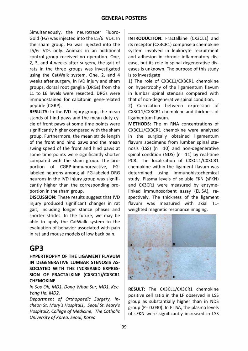
RESULT: The CX3CL1/CX3CR1 chemokine
positive cell ratio in the LF observed in LSS
group as substantially higher than in NDS
group (P= 0.030). In ELISA, the plasma levels
of sFKN were significantly increased in LSS

GENERAL POSTERS
100
patients compared with patients in the oth-
er groups (P = 0.006). There was greater
CX3CL1/CX3CR1 expression in LSS patients
as quantified by RT-PCR (P = 0.004, 0.01).
Thickness of LF in patients was significantly
correlated with serum CX3CL1 level (r =
0.48, P < 0.01) and with RNA Expression of
CX3CL1/CX3CCX3CR1 (r = 0.62, P < 0.01)(r =
0.64, P < 0.01)
DISCUSSION: This study identified for the
first time that increases in Fractalkine and
CX3CR1-expresexpressing monocytes, T
lymphocytes, and NK cells are significantly
related to LF hypertrophy, which may pro-
vide new conceptual and therapeutic ap-
proaches for treating spinal stenosis.
GP4 COMPARATIVE STUDY OF LUMBAR INTERVERTEBRAL DISC BIOMECHANICAL CHARACTERISTICS WITH OR WITHOUT HYDROGEL INJECTION AFTER NUCLEUS DISCECTOMY Gao Manman, Zhou Zhiyu, Deng Wenbin,
Dai Xuejun, Zou Xuenong
Department of Spine Surgery/Orthopedic
Research Institute The First Affiliated
hospital of Sun Yat-sen University
INTRODUCTION: Injecting hydrogels as sub-
stitute of nucleus pulposus after nucleus
discectomy partially restores the geometric
structure of lumbar intervertebral discs.
This study investigated both the static and
dynamic mechanical properties of lumbar
intervertebral discs with or without hydro-
gels injection after nucleus discectomy.
METHODS: The lumbar intervertebral discs
of three miniature pigs were randomized to
normal group (with respectively intact in-
tervertebral discs), mimic group (with hy-
drogel injection after discectomy) and con-
trol group (without hydrogel injection after
discectomy). All of the specimens were sub-
jected to static and dynamic compression
tests sequentially, then stress/strain curves
in the static test and storage modulus, loss
modulus and phase angle in the dynamic
test were worked out respectively with help
of software.
RESULTS: In the static compression test,
mimic group have a similar stress/strain
curve with normal group, whereas control
group shows obvious difference. In the dy-
namic test, results of the storage modulus,
loss modulus and phase angle showed
simlar tendency that the maximum mean
values (297.2~362.5KPa, 15.7~24.0KPa,
2.4~4.6°, respectively) were in control group
and the minimum (187.2~226.2KPa,
10.1~14.8KPa, 2.0~3.5°, respectively) in
normal group. And the mean values in mim-
ic group were 242.8~290.5KPa,
12.4~18.2KPa, 2.3~4.1°, respectively. It
should be noted that values of the storage
modulus were elevated with increasing fre-
quency, while values of the loss modulus
and phase angle were reduced with
inceasing frequency.
DISCUSSION: Injecting hydrogels after nu-
cleus discectomy can not only better re-
serve the normal lumbar geometric struc-
ture but also improve the biomechanical
properties compared with simple discecto-
my.
GP5 SPINO-PELVIC-RHYTHM IN NORMAL SUB-JECTS AND HIGH-CLASS ATHLETES WHO OVERCAME CHRONIC LOW BACK PAIN Kiyotaka Hasebe1,2, Koichi Sairyo3, Yasushi
Hada1, Akira Dezawa3, Yu Okubo4, Koji
Kaneoka2, and Yoshio Nakamura2
1Department of Rehabilitation, Teikyo Uni-
versity Mizonokuchi Hospital, Kawasaki Ja-
pan 2Faculty of Sports Science, Waseda Uni-
versity, Japan 3Department of Orthopedic
Surgery, Teikyo University Mizonokuchi
Hospital, Kawasaki, Japan 4Faculty of
Health and Medicalcare, Saitama Medical
University, Japan
PURPOSE: Spino-Pelvic-Rhythm (SPR) is re-
ported to be a good parameter to under-

GENERAL POSTERS
101
stand the spine kinematics. We evaluated
SPR in normal subjects and high-class ath-
letes who overcame chronic low back pain
(LBP).
METHODS: Eighteen male without LBP
(mean: 36 yrs) and three high-class athletes
(mean: 37 yrs) who overcame LBP partici-
pated. They have a history of LBP at least 2
years before, and presently, they have no
LBP. First, we measured the finger-to-floor
distance (FFD) at the upright posture, and
the value was indicated as 100%. Using a
spinal mouse, spinal alignment was meas-
ured at the following 4 points, i.e., (1) up-
right posture meaning 100% of FFD, (2) for-
ward bend at 50%, (3) 25% and (4) 0% of
FFD meaning finger contacting the floor.
The changes of the angle of thoracic and
lumbar spine as well as the pelvis were cal-
culated.
RESULTS: In the normal subjects, from point
1 to 2 (phase I) spino-pelvic angle moved
104 degree entirely. In this phase, lumbar
spine moved mainly. The lumbar/ pelvic (LP)
ratio was 4.0. In the second phase (point 2
to 3), it changed only 16 degree and the
ratio was 1.0. In phase III (point 3 to 4), ma-
jority of the entire motion was found in the
pelvis, and the ratio was 0.4. All three high-
class athletes including Olympic medalist of
throwing, professional baseball player and
ex-world champion of mixed martial arts,
showed totally different SPR. In phase I,
pelvis moved greater than normal subjects,
and the LP ratio was 0.5, 1.2 and 1.3. In the
second phase, lumbar spine mainly moved
and, the LP ratio was 3.2, 4.0 and 3.2.
DISCUSSION AND CONCLUSION: As the
normal SPR, the lumbar spine moved mainly
at the initial phase. At the final stage, pelvis
moves mainly. Three athletes showed simi-
lar abnormal rhythm. They tried to move
pelvis more and to reduce the lumbar mo-
tion at the initial phase. This may be due to
the defense mechanism against chronic
back pain and/or stabilization effects of the
spine by strenuous core muscle exercise.
GP6
NOVEL TWO-COMPONENT INTERBODY FUSION DEVICE IMPROVES BONE-IMPLANT INTERFACE WITHOUT COMPROMISING STABILITY: A CADAVER STUDY Hans-Joachim Wilke1, David Volkheimer1,
Bruce Robie2, Finn Bjarke Christensen2,3
1University of Ulm, Institute of Orthopaedic
Research and Biomechanics, Ulm, Germany
2FBC Device ApS, Risskov, Denmark 3Aarhus
University Hospital, Orthopaedc Research
Laboratory, Aarhus, Denmark
INTRODUCTION: A novel two-piece articu-
lating ALIF device allows limited motions of
the treated vertebral level for lordotic ad-
justments until a supplementary pedicle
screw system is rigidly fixed. A potential
benefit is the reduction of bone-implant
relative motions with a positive impact on
fusion time and sagittal balance. The aim of
the study is to compare the rigidity and im-
plant alignment of the two-piece ALIF fusion
device with a one-piece ALIF in a 360° set-
ting.
METHODS: Seven lumbosacral (L3-S1) hu-
man cadaver specimens were tested (age
50-60) in a universal spine tester. The flexi-
bility of the intact specimen, the specimens
instrumented with a two-component ALIF
(Statur® -L, FBC Device ApS, Denmark) and a
one-piece ALIF (PezoTM –A, Ulrich, Germa-
ny), both supplemented with a pedicle
screw system (tangoRSTM, Ulrich, Germany)
was tested using pure moments of ±7.5 Nm
in three principal motion directions. For
assessment of the bone-implant interface,
fluoroscopic images were captured during
motion. Three-dimensional vertebral mo-
tion was measured using an optical motion
capturing system (Vicon Mx, Vicon, UK).
Paired t-tests were performed to determine
statistical significance at a p< 0.05 level.
RESULTS: No statistically significant differ-
ences in rigidity were found between the
one and two-piece ALIF in the 360° setup
(e.g., 3.5° ± 1.9° for the one-piece and 4.2°

GENERAL POSTERS
102
±1.8°for the two-piece ALIF in flex-
ion/extension), while both configurations
significantly reduced the range of motion
compared to the intact condition (p< 0.05).
Significantly less motion at the implant-
endplate interface was found for the two-
piece device (1.0° ± 0.6°) in comparison to
the one-piece ALIF (4.2° ± 1.7°) in flex-
ion/extension.
DISCUSSION: The two-piece ALIF significant-
ly reduced the relative motion at the bone-
implant interface without compromising
stability. Theoretically, this can reduce the
risk of implant subsidence, improve fusion
and establish better sagittal balance.
GP7 BIOMECHANICAL CHARACTERIZATION OF A NOVEL LOW-STIFFNESS POSTERIOR SPINAL IMPLANT UNDER ANTERIOR SHEAR LOAD-ING IN A DEGENERATIVE SPINAL MODEL (1,2) Angela D. Melnyk, MASc, (2,3) Jason D.
Chak, MEng, (4) Vaneet Singh, MS, (1,5)
Adrienne Kelly, MD, (1,2,3) Peter A. Cripton,
PhD, (1,2,5) Charles G. Fisher, MD, (1,2,5)
Marcel F. Dvorak, MD, (1,2,3) Thomas R.
Oxland, PhD
1 Department of Orthopaedics, University of
British Columbia, Vancouver, BC, Canada, 2
International Collaboration on Repair Dis-
coveries (ICORD), University of British Co-
lumbia, Vancouver, BC, Canada, 3 Depart-
ment of Mechanical Engineering, University
of British Columbia, Vancouver, BC, Canada,
4 Medtronic Inc., Memphis, TN, USA, 5 Divi-
sion of Spine, University of British Columbia,
Vancouver, BC, Canada
INTRODUCTION: Dynamic implants have
been developed to address potential adja-
cent level effects due to rigid instrumenta-
tion. Prior to clinical use, implants should be
rigorously tested ex vivo. The objective of
our study was to determine how implant
type and specimen condition affects implant
load-sharing and specimen translation un-
der an applied anterior shear force in a nov-
el biomechanical model.
METHODS: A previously validated human
cadaveric model of degenerative
spondylolisthesis for testing implants in
shear was used. Lumbar FSUs (N=15) were
tested under a static 300 N axial compres-
sion force and a cyclic anterior shear force
(5-250 N). Translation was tracked with a
motion capture system. Four implants
(Medtronic, Inc.) were instrumented with
strain gauges to determine the sheer force
supported by the implant: Implant A Titani-
um, Implant B Oblong PEEK, Implant C
Round PEEK, and Implant D BalanCTM (PEEK
rod with a C-shape section and silicone in-
sert). Each implant was affixed to each spec-
imen, and they were tested in 3 conditions
(intact, facet gap, disc lesion), with the lat-
ter 2 simulating a degenerative spine.
RESULTS: Specimen condition and implant
type affected load-sharing and anterior
translation (p<0.0001). Load-sharing in-
creased across all specimen conditions and
decreased across the first 3 implants (Fig.
mean ± SD). Implants D and B supported
similar shear forces for all specimen condi-
tions (p>0.2). Translation tended to increase
with the first three implants. However, im-
plants D and B allowed similar translations
for all specimen conditions (p>0.3).
DISCUSSION: We tested a novel dynamic
implant (Implant D) and compared the re-
sults to those for three implants ranging
from high- to low-stiffness (implants A, B,

GENERAL POSTERS
103
and C). Implant D behaved similarly to im-
plant B in both load-sharing and translation
despite having a different design and stiff-
ness. Complex implant design and speci-
men-implant interaction necessitate pre-
clinical testing of novel implants.
GP8 EFFECTS OF SEQUENTIAL UNILATERAL FACETECTOMY ON CERVICAL SPINAL STABILITY Mageswaran P., Tolhurst S., Colbrunn R.,
Bonner T., Techy F., McLain R.
1 Spine Research Lab, Dept. of Center for
Spine Health, Cleveland Clinic, Cleveland,
OH, USA; 2 Illinois Bone and Joint Institute,
Dept. of Orthopedic Surgery, University of
Illinois, Chicago, IL, USA
INTRODUCTION: Classical biomechanical
studies have concluded that instability of
the cervical spine occurs after a complete
laminectomy and resection of more than
50% of the bilateral capsule or facet joints
at one cervical level. No study has ever
evaluated the effects of unilateral sequen-
tial facet resection without laminectomy.
This study is the first to assess spinal stabil-
ity after unilateral sequential facet resec-
tion.
METHODS: Human cadaveric C2-T1 speci-
mens (n = 7) were tested intact, and then
underwent a sequential right unilateral
facetectomy at the C6 – C7 level. The width
of the facet joint was measured and se-
quential resection was made as a percent-
age of the facet width (25 %, 50 %, 75 % and
100 %). The following loading conditions
were applied using a robotic spine testing
system: Applied moments (2.0 Nm) with
head weight load of 40N were used to simu-
late flexion-extension (FE), lateral bending
(LB), and axial rotation (AR). Vertebral mo-
tion was measured using an optoelectronic
system. Mean relative range of motion
(ROM) was compared among groups using
repeated measures analysis of variance at a
significance level of 0.05.
RESULTS: For FE, no statistically significant
change in motion was observed following
sequential unilateral facet resection com-
pared to intact state. In LB, only the com-
plete unilateral facet resection (100%) re-
sulted in a significant increase in motion of
8% (p = 0.005). In AR, there was a significant
increase in motion following 75% (18.5%, p
= 0.03) and 100% (33.8%, p < 0.001) facet
resection, respectively.
CONCLUSION: Unilateral facet resection
does not significantly increase FE motion.
However, significantly greater cervical spine
mobility in LB and AR occurs following uni-
lateral facet resection of 75% or more. We
conclude that unilateral facet resections are
not as destabilizing as are bilateral
facetectomies with laminectomy. Unilateral
facet resection less than 75% did not gener-
ate instability in any plane of motion.
GP9 DOES HEAD WEIGHT AFFECT CERVICAL SPINE STABILITY? AN IN VITRO ROBOTIC BIOMECHANICAL STUDY Mageswaran P., Tolhurst S., Colbrunn R.,
Bonner T., Techy F., McLain R.
1 Spine Research Lab, Dept. of Center for
Spine Health, Cleveland Clinic, Cleveland,
OH, USA; 2 Illinois Bone and Joint Institute,
Dept. of Orthopedic Surgery, University of
Illinois, Chicago, IL, USA
INTRODUCTION: Many biomechanical stud-
ies on cervical spine stability with extremely
strong clinical relevance were performed
without accounting for the head weight. A
biomechanical study of a constant vertical
load (head weight loading) while simulating
flexion-extension (FE), lateral bending (LB)
and axial rotation (AR) may improve the in
vivo applicability of biomechanical test re-
sults. This study hopes to provide insight
into the effect of head weight on cervical
spine motion and stability.
METHODS: Flexibility tests were conducted

GENERAL POSTERS
104
on seven human cadaveric specimens, C2–
T1, using a robotic spine testing system. The
system was used to test two conditions:
intact and destabilized (Complete Unilateral
C6 – C7 Facetectomy). Continuous applied
moment, ± 2 Nm was used to simulate FE,
LB and AR with and without a constant head
weight of 40N. The head weight was applied
in the direction of gravity. The test was re-
peated again following complete unilateral
facetectomy. Mean overall range of motion
(ROM) of C2 – T1 was compared among
groups using repeated measures analysis of
variance at a significance level of 0.05.
RESULTS: We found a significant reduction
in motion with the application of a head
weight (12.1% decrease, p = 0.02) in AR for
the intact condition. A similar reduction was
observed in the destabilized condition
(16.1% decrease, p = 0.05). There was a sig-
nificant decrease in LB motion when head
weight load was applied in the intact state
(12.4%, p = 0.01), with a larger reduction in
motion for the destabilized condition
(21.6%, p < 0.001). Intact FE motion was not
significantly affected by the addition of
head weight (p = 0.66). However, FE motion
decreased significantly with head weight in
the destabilized state (6.7% decrease, p =
0.01).
CONCLUSION: Application of a constant
head weight load with flexibility testing re-
duces the overall ROM of the cervical spine.
The magnitude of the effect is larger follow-
ing complete unilateral facetectomy.
GP10 DO PEDICLE SCREWS THAT HAVE BREACHED THE PEDICLE WALL PROVIDE STRONGER PURCHASE AS COMPARED TO SMALLER PEDICLE SCREWS PLACED INTRA CORTEX? 1) Kosaku Higashino, MD, PhD, 1) Yuichiro
Goda, MD, 2) Suzuki Daisuke, PhD, 3) Ta-
kuma Kobayashi, MD, 1) Tetsuya Matsuura,
MD, PhD, 2) Mineko Fujimiya, MD, PhD, 4)
William C. Hutton, DSc, 1) Natsuo Yasui,
MD, PhD
1) Department of Orthopaedics, University
of Tokushima, Tokushima, Japan, 2) De-
partment of Anatomy, Sapporo Medical
University School of Medicine, Sap-poro,
Japan 3) Department of Orthopaedics, Sap-
poro Medical University School of Medi-cine,
Sapporo, Hokkaido, Japan 4) Department of
Orthopaedic Surgery, Emory University
School of Medicine, Atlanta, Georgia, USA
INTRODUCTION: The pedicle diameters at
the thoracolumbar levels in elderly patients
and the pedicle diameters at the thoracic
levels in scoliosis patients are sometimes
very small. The question arises: Do pedicle
screws that are inserted and breach the
pedicle wall provide stronger purchase as
compared to pedicle screws of smaller di-
ameter that are placed intra cortex in the
pedicle. The purpose of our study was to
answer this question using cadaveric thora-
columbar vertebrae.
METHODS: Twelve thoracolumbar verte-
brae (T6-L2) were harvested from eight
male fresh cadaveric spines (mean age 85.3
years). Pedicle screws were inserted in each
of the pedicles. On one pedicle the screw to
be inserted was chosen so as to be oversize
in the pedicle, and on the other pedicle the
screw to be inserted was chosen so as to fit
the pedicle intra cortex. The pedicle diame-
ter “a” and the screw diameter “b” were
measured, and the breached percentage of
oversized screw “(b-a) x 100/a” was calcu-
lated (Figure 1). Biomechanical testing: An
adaptor was attached to the protruding end
of each pedicle screw and then, using a ma-
terials testing machine (Shimadzu Corpora-
tion, Kyoto Japan), each pedicle was tested
as follows: 1) toggling was applied in a
cephalocaudad direction for 500 cycles at
0.3 Hz at ±50 N; 2) the screws were then
pulled out by applying a tensile force down
the long axis of the screw: displacement
rate of 20.0 cm/min.
RESULTS: The mean pullout strength for the

GENERAL POSTERS
105
oversized pedicle screws was 684.9±352.5 N
and for the intra cortex pedicle screws was
640.5±301.1 N; the difference was not sig-
nificance (p<0.05). There was significant
positive correlation between extent of
breach and pullout force. In other words the
oversized pedicle screws within 12.4%
might be stronger than intra cortex pedicle
screws (Figure 1).
DISCUSSION: Pedicle screws that breach the
pedicle wall do not provide stronger pur-
chase as compared to smaller pedicle
screws that are placed intra cortex?
GP11 BIOMECHANICAL EFFECT OF THE PRO-GRESSION OF DISC DEGENERATION AT L5/S1 ON ADJACENT DISCS IN A LUMBAR SPINE Raghu N Natarajan, Gunnar BJ Andersson
Department of Orthopedic Surgery, Rush
University Medical Center, Chicago, IL, USA
INTRODUCTION: When lumbar disc degen-
eration occurs at one level, whether treated
or not, degeneration frequently develops at
mobile segments above or below the de-
generated segments referred to adjacent
segment disc disease (ASDD). Aim of the
current study is to understand how progres-
sive degeneration at L5/S1 affects the bio-
mechanics of the adjacent segments.
METHODS: A refined poro-elastic non-linear
3D finite element model of a lumbar spine
and hybrid method of analyses were used to
determine the biomechanical effects of sin-
gle level disc degeneration on the adjacent
segments. Four different grades of disc de-
generation (Thompsons Grades 2 to 5) were
modeled at L5/S1 level and their compara-
tive effect on adjacent disc biomechanical
characteristics were studied under all the
three physiological moment loading condi-
tions.
RESULTS: At adjacent L4/5 segment: Grade
5 disc at L5/S1 produced least increase in
motions of 3% under flexion and largest
under extension (125%), while under tor-
sion and lateral bending it produced 18%
increase in motions. When the increase in
motions in all the three principal directions
were combined, grade 4 and grade 5 at
L5/S1 produced an increase nearly twice
and seven times respectively as those pro-
duced when L5/S1 had grade 3 disc. As the
degenerative grade at L5/S1 increased from
2 to 5, total facet forces increased from 77
N to 162 N under extension and 145 N to
230 N under torsion. Annular stresses also
increased with a sharp increase in stress
value as the L5/S1 disc grade changed from
4 to 5.
CONCLUSIONS: Largest increase in motion,
facet forces as well annular stresses at the
adjacent discs in the lumbar spine was seen
with grade 5 disc at L5/S1. Grade 3 and 4
did not produce substantial increase in ad-
jacent disc motions as well as annular
stresses leading to the conclusion that sin-
gle level disc degeneration induces ASDD
only when the deceased disc is degenerated
to the highest grade.
GP12 INVESTIGATION OF EQUIVALENT STRESS OF ADJACENT VERTEBRAE AFTER BALLOON KYPHOPLASTY BY A CT-BASED FINITE ELEMENT METHOD Nahoko Iwakura1, Yusuke Matsuura2,
Masahiro Shiba1, Keiji Wada1, Yasuaki
Murata1, Yoshiharu Kato1
1.Department of Orthopaedic Surgery,

GENERAL POSTERS
106
Tokyo Women's Medical University, Tokyo,
Japan. 2.Department of Orthopaedic
Surgery, Graduate School of Medicine, Chiba
University, Chiba, Japan
INTRODUCTION: Balloon kyphoplasty (BKP)
is a minimally invasive surgical procedure
for treatment of painful vertebral compres-
sion fractures. It has numerous benefits
including a simple procedure, early pain
control and height restoration of the col-
lapsed vertebral body. Consequently, the
number of treatments using this procedure
has risen significantly. However, complica-
tions such as new vertebral fractures have
been reported. The aim of this study was to
evaluate equivalent stress of adjacent ver-
tebrae after BKP using the finite element
(FE) method.
METHODS: Six patients were enrolled in this
study. The mean age was 77.0 years and the
duration of follow-up was 4.8 months. All
patients underwent CT within 1 week of BKP
treatment. Three-dimensional, four-
functional unit, FE models were constructed
from the CT data using MECHANICAL FIND-
ER software (cement model). In addition, a
cancellous bone model was developed. The
model was a cement model where the ce-
ment data was replaced with the cancellous
data of adjacent vertebrae. The equivalent
stress of cement and cancellous bone, and
the front half of the adjacent vertebral body
of both groups, were analyzed and com-
pared.
RESULTS: The mean equivalent stress of
cement was significantly larger compared
with cancellous bone (1.8±0.7 and 0.6±0.4)
(p < 0.05). The mean equivalent stress of
adjacent vertebrae of the cement model
and cancellous bone model was 2.9±1.1,
and 3.1±1.0, respectively. There was no sig-
nificant difference.
DISCUSSION: Cement was stiffer than
cancellous bone, so replacing cement data
with cancellous data decreased the stiffness
of the vertebrae. However, there was no
change in equivalent stress of adjacent ver-
tebrae. This result suggests that the inci-
dence of adjacent fractures after BKP has no
relationship to stiffness of the treated ver-
tebra.
GP13 HISTOMORPHOMETRIC AND RADIO-GRAPHIC CHANGES AFTER WALLIS IM-PLANTATION IN RATS M. Melloh (1), T. Barz (2), J. Lange (3), L.P.
Staub (4), H.R. Merk (5), I. Klöting (6), N.
Follak (7)
(1) Western Australian Institute for Medical
Research (WAIMR), University of Western
Australia, Nedlands, Australia (2)
Department of Orthopaedic Surgery,
Asklepios Klinikum Uckermark, Schwedt/
Oder, Germany (3) Department of Trauma
and Reconstructive Surgery, University of
Greifswald, Greifswald, Germany (4)
NHMRC Clinical Trials Centre, University of
Sydney, Sydney, Australia (5) Department of
Orthopaedic Surgery, University of
Greifswald, Greifswald, Germany (6)
Department of Laboratory Animal Science,
Medical Faculty, University of Greifswald,
Germany (7) Orthopaedic Clinic, Pfeiffersche
Stiftungen, Magdeburg, Germany
INTRODUCTION: Clinical effectiveness of
the PEEK-non-fusion spine implant WallisTM
is well documented. However, there is a
lack of evidence on the long-term behavior
of this implant on bone, in particular its in-
fluence on structural changes of bone ele-
ments of the lumbar spine. The aim of this
study was to investigate histomorphometric
and radiographic changes in the rat model
after WallisTM implantation.
METHODS: Twenty-four male rats aged
eleven weeks underwent surgery for im-
plantation of WallisTM implants or for a
sham procedure in three groups of eight
animals each each: 1) implantation at level
L4-5; 2) implantation at level L5-6 and 3)
sham surgery. Eleven weeks postoperatively

GENERAL POSTERS
107
resorption at the implant-bone interface
was measured via X-ray, bone mineral den-
sity of vertebral bodies was analyzed using
osteodensitometry, and bone mineral con-
tent as well as resorption of the spinous
processes were examined by
histomorphometry.
RESULTS: Resorption of the spinous pro-
cesses at the site of the interspinous im-
plant was found in all treated segments.
There was no significant difference in either
bone density of vertebral bodies or
histomorphometric structure of the spinous
processes between adjacent vertebral bod-
ies, between treated and untreated seg-
ments and between groups.
DISCUSSION: These findings indicate that
resorption of spinous processes, as a result
of implant loosening, inhibit the targeted
load redistribution through the PEEK-non-
fusion interspinous device in the lumbar
spinal segment of the rat. This leads to re-
duced long-term stability of the implant in
the animal model. These results suggest
that PEEK-non-fusion interspinous devices
like the WallisTM implants may have time-
limited effects and should only be used for
specified indications.
GP14 BIOMECHANICAL ANALYSIS OF THE THO-RACIC SPINE FOLLOWING DECOMPRESSIVE PROCEDURES Healy AT, Lubelski D,Mageswaran P, Mroz T
1 Spine Research Lab, Dept. of Center for
Spine Health, Cleveland Clinic, Cleveland,
OH, USA;
INTRODUCTION: This study evaluated the
effect of the rib cage on thoracic spine sta-
bility following sequential decompressive
surgeries and instrumented fusion.
METHODS: Human cadaveric spines with
intact rib cages, C7-L1 (n = 9). An industrial
robot was used to apply a ± 5 Nm moment
applied along the spine to simulate flexion-
extension (FE), lateral bending (LB) and axi-
al-rotation (AR). The specimens were first
tested in their intact state, and then tested
after each of the following sequential surgi-
cal decompressive procedures at T4 – 5
consisting of 1) laminectomy; 2) unilateral
facetectomy; 3) unilateral
costotransversectomy and subsequently 4)
instrumented fusion from T3 - T7. Range of
Motion (ROM) between T1 - T12 and T3 - T7
were measured for each specimen using an
optoelectronic motion system. Statistical
analysis between and within group compar-
isons was done using repeated measures
analysis of variance (p < 0.05 was consid-
ered statistically significant).
RESULTS: We found that in all three planes
of motion, the sequential decompressive
procedures caused no statistically signifi-
cant change in motion between T3 - T7 and
T1 - T12 when compared to intact. In com-
paring between intact and fusion, our study
found that fusion reduced intrinsic motion
between T3 - T7 by 85.4% (p = 0.0003),
92.8% (p = 0.0046) and 91% (p = 0.0004) for
axial rotation, flexion-extension and lateral
bending respectively. We also found that
ROM between T1 - T12 also decreased un-
der fusion by 22% (p = 0.0013), 21% (p =
0.0020) and 28.2% (p = 0.0004) for axial
rotation, flexion-extension and lateral bend-
ing compared to intact.
CONCLUSION: Thoracic spine stability was
not significantly affected by sequential de-
compression procedures in thoracic seg-
ments at the level of the true ribs in all
three planes of motion. Placement of poste-
rior instrumented fusion increased study
segment rigidity at intrinsic levels and also
reduced overall ROM of the thoracic spine.
GENERAL POSTERS
108
GP15 3D-CT SIMULATION STUDY OF THE ILIAC SCREW FOR THE FIRM FIXATION Bungo Otsuki, PhD; Shunsuke Fujibayashi,
PhD; Mitsuru Takemoto, PhD; Masanori
Izeki, M.D.; Syuichi Matsuda, PhD
Department of Orthopaedic Surgery,
Graduate School of Medicine, Kyoto
University, Kyoto, Japan
INTRODUCTION: Distal fixation in thoracol-
umbar spine surgery is crucial. Iliac screw
technique has been reported with good
clinical results; however, several studies
showed high rates of iliac screw loosening.
In the osteoporotic bone, adequate pur-
chase with a screw will be acquired only by
the contact to the cortex, and the use of
longer and larger-diameter screw has been
recommended. The shape of the ilium is
complex, and the ideal direction or size of
the screw has not been fully clarified. In this
study, the contact between iliac screw and
the cortex of the ilium was analyzed by us-
ing 3D-CT simulation.
METHODS: Twelve adult pelvic CT data
were analyzed using originally written soft-
ware. Screw was placed from the posterior
superior iliac spine (PSIS) to the anterior
inferior iliac spine. In this study, we as-
sumed that the insertion point at PSIS was
fixed, and the screw tip could move freely
under the condition that whole screw was
located inside the cancellous bone of the
ilium. First, maximum screw diameter was
decided in each screw length (50-120 mm)
by changing the direction of the screw
three-dimensionally. Second, we investigat-
ed whether or not screw with a maximum
size can move in the ilium, and calculated
the area where the tip of the screw can
move (moving area).
RESULTS AND DISCUSSION: The moving
area became small as length of the screw
became long in all cases. However, moving
area did not disappear except one case
when screw length is thought to be long
enough (100mm) in clinical use. Further, the
moving area was vertically long linear shape
in remaining eleven cases, which indicate
that the screw tip can move to the cranial-
to-caudal direction. These results show that
the single iliac screw is not enough for firm
fixation even if the screw diameter and di-
rection are appropriate especially in the
case of osteoporosis. Dual iliac screw will
resolve this problem when two screws with
enough diameters are set at the cranial and
caudal end respectively.
GP16 IS THERE AN ASSOCIATION BETWEEN AB-DOMINAL MUSCLES AND DEGENERATIVE SPONDYLOLISTHESIS? Tcherveniakov P, Fraser R, Freeman BJC,
Jones CF
University of Adelaide, Adelaide, Australia
Adelaide Centre for Spinal Research, SA
Pathology, Adelaide, Australia
INTRODUCTION: The pathogenesis of de-
generative spondylolisthesis is not well un-
derstood, with many etiological factors
identified. The aims of this study were to
investigate the contributions of abdominal
muscle and aponeurosis morphology to L4-5
and L5-S1 vertebral slip and to devise mod-
els for the prediction of vertebral slip.
METHODS: Axial abdomino-pelvic comput-
ed tomography scans from 200 subjects
were examined. Those with spondylolysis
were excluded (n=14), and spondylolisthesis
was expressed as a continuous measure in
the remaining subjects. Muscle parameters
(abdominal and paraspinal muscle area and
density, aponeurosis width) and bony pa-
rameters (vertebral slip, lumbar index, disc
index, facet joint angle) were measured for
each vertebral or intervertebral level. Mul-
tiple linear regression analyses were per-
formed to form six hypothesis-driven and
predictive models for percent vertebral slip.
RESULTS: Increasing lateral abdominal mus-
cle (LAM) area (p=0.01) and decreasing rec-
GENERAL POSTERS
109
tus abdominis muscle (RAM) area (p=0.02)
were significant predictors of vertebral slip
at the L5-S1 level. Measures of aponeurosis
width did not contribute to L5-S1 vertebral
slip. Neither muscle morphology or
aponeurosis width parameters were signifi-
cant predictors of slip at the L4-5 level.
More sagittal facet joint orientation and de-
creasing lumbar index were also significant
predictors of vertebral slip at both levels in
all models.
DISCUSSION: In addition to previously iden-
tified osseous factors such as facet angle
and lumbar index, this retrospective imag-
ing study shows that abdominal muscle area
may be associated with vertebral slip at the
L5-S1 level. The reduction in RAM area may
represent increased musculoaponeurotic
laxity, which predisposes to vertebral slip by
lowering intra-abdominal pressure. The as-
sociation with increased LAM area may be
the result of a compensatory response to
vertebral slip, which increases intra-
abdominal pressure to brace an unstable
spine.
GP17 MUSCLE INJURY IN RATS INDUCES UP-REGULATION OF INFLAMMATORY CYTO-KINES IN MUSCLE AND PAIN-RELATED NEUROPEPTIDES IN DORSAL ROOT GAN-GLIA INNERVATING MUSCLE. -THE PATHO-LOGICAL-MECHANISM OF CONTINUOUS MUSCLE PAIN- Sakuma, Yoshihiro1; Miyagi, Masayuki1;
Ohtori, Seiji1; Inoue, Gen2; Yamauchi,
Kazuyo1; Orita, Sumihisa1; Kamoda,
Hiroto1; Ishikawa, Tetsuhiro1; Arai, Gen1;
Suzuki, Miyako1; Oikawa, Yasuhiro1; Kubo-
ta, Go1; Inage, Kazuhide1; Sainoh, Takeshi1;
Sato, Jun1; Nakata, Yukio1; Takahashi,
Kazuhisa1
Dept. of Orthopaedic Surgery, Graduate
School of Medicine, Chiba University, Japan1
Dept. of Orthopaedic Surgery, Kitasato Uni-
versity, School of Medicine2
INTRODUCTION: Invasion of back muscle
after spinal surgery often causes continuous
pain, yet the pathological-mechanisms be-
hind continuous muscle pain remain un-
clear. The aim of the current study was to
compare in rats the behavior of the sensory
nervous system, and to investigate histolog-
ical changes and inflammatory cytokines in
injured muscle. METHODS: In this study, we
used the right gastrocnemius contusion
model via a drop-mass technique because a
back muscle injury model would be difficult
for evaluating pain behavior. Exp.1: Pain
behavior was measured using CatWalk at
twelve hours, 1, 2, 3 days, and 1, 2, 3 weeks
after muscle contusion. the bilateral gas-
trocnemius was resected. Samples were H-E
stained and TNF-α, IL-6, and NGF levels
were quantified by ELISA. Exp.2: To detect
DRG neurons innervating the gastrocnemi-
us, fluorogold (FG) was applied to the sur-
faces of bilateral gastrocnemius 1 week be-
fore muscle contusion. DRGs from L4 to L6
levels were resected at 1, 2, and 3 weeks
after contusion, then immunostained for
CGRP (pain-related neuropeptide). The ratio
of CGRP and FG double-labeled DRG neu-
rons among all FG-labeled neurons was cal-
culated.
RESULTS: Pain behavior: The swing speed of
right hind paw was significantly lower than
that of left hind paw through 1 day. Histolo-
gy: At 3 weeks, accumulation of granulated
tissue and myofibrillogenesis for repaired
tissue was observed. Inflammatory cyto-
kines: Up-regulation of TNF-α, IL-6 and NGF
levels in the right side of right side com-
pared to the left side was observed up to 2
days but became less marked subsequently.
DRG: The expression of CGRP was signifi-
cantly higher in the right side than in the
left side until two weeks. DISCUSSION: In
this model, pain response and increase in
inflammatory cytokines recovered immedi-
ately, but pain-related neuropeptides re-
mained up-regulated for 2 weeks. These
differences may explain the pathological-

GENERAL POSTERS
110
mechanism of continuous muscle pain.
GP18 CARBON NANOTUBE-REINFORCED CALCI-UM PHOSPHATE CEMENT FOR DRUG DE-LIVERY IN MULTIPLE MYELOMA BONE DIS-EASE TREATMENT Boren Lin 1,3, Huan Zhou 2, Sarit Bhaduri
2,4, Douglas Leaman 1, Vijay Goel 3, Anand
Agarwal 3
1. Department of Biological Sciences, the
University of Toledo, 2. Department of
Mechanical, Industrial and Manufacturing
Engineering, the University of Toledo, 3.
Departments of Boengineering and
Orthopaedic Surgery, the University of
Toledo, 4. Department of Surgery
(Dentistry), the University of Toledo
INTRODUCTION: Current approaches in
treating multiple myeloma fracture or lesion
focus on developing osteoconductive frac-
ture stabilizing fillers capable of carrying
therapeutic agents to promote bone regen-
eration, which cannot be achieved by con-
ventional PMMA methods.
METHODS: We have developed a biocom-
patible carbon nanotubes (CNT) reinforced
calcium phosphate-based cement (CPC) that
exhibits desirable mechanical and handling
properties. This novel composite was capa-
ble in carrying an inhibitor against NF-kB
activated osteoclast differentiation. NF-kB
inhibitor MG132 was incorporated into the
CPC or CPC/CNT during the setting process,
and the cement was submerged in culture
medium for 24 hours. This MG132-
containing medium was harvested and add-
ed to ACHN indicator cells or preosteoclast
RAW 267.4. NF-kB activation in ACHN cells
was detected by real-time RT-PCR analysis
of NF-kB-induced IL-8 after 4 hours of TNFa
treatment, and cell viability was assessed by
crystal violet staining after 12 hours. Inhibi-
tion of RAW 264.7 differentiation was inves-
tigated after 5-day TNFa treatment by stain-
ing for TRAP positive osteoclast-like cells.
RESULTS: Media from CPC/MG132 and
CPC/CNT/MG132 both showed potent in-
hibitory effect on TNF-induced NF-kB-
mediated IL-8 gene expression. After 5-day
TNFa treatment, with MG132 present in the
medium, the number of TRAP positive cells
decreased significantly.
DISCUSSION: The results have demonstrat-
ed that our cement was effectively able to
carry MG132. Inhibition of osteoclast differ-
entiation by the CPC/CNT/MG132 medium
in a 5-day experiment confirmed the sus-
tained release of the drug from this combi-
nation. Together with its salient weight
bearing and bioactive properties, our data
suggest that the CPC/CNT is a promising
filler material for bone augmentation in
multiple myeloma related vertebral lesion
or fracture.
GP19 BIOMECHANICAL CONTRIBUTION OF TRANSVERSE CONNECTORS IN THE SETTING OF A THORACIC PEDICLE SUBTRACTION OSTEOTOMY Robert W. Tracey, MD; Haines Paik, MD;
Ronald A. Lehman, Jr., MD; Daniel G. Kang,
MD; John P. Cody, MD; Mario J. Cardoso,
MD; Anton E. Dmitriev, PhD
Walter Reed National Military Medical
Center, Bethesda, MD
INTRODUCTION: Little data is available to
guide longitudinal construct planning after a
pedicle subtraction osteotomy (PSO) in the
thoracic spine. Previous studies have sug-
gested transverse connectors (TC) may en-
hance torsional rigidity. However, the bio-
mechanical effect of augmentation with one
or two TC after PSO has not been previously
evaluated.
METHODS: 7 fresh-frozen human cadaveric
thoracic spines (T3-T11) were prepared and
intact range of motion (ROM) testing was
performed with non-destructive loading (±6
Nm) in a six-degree-of-freedom spine simu-
lator. The specimens were instrumented

GENERAL POSTERS
111
from T4-T10 with bilateral 5.5-mm pedicle
screws and 5.5-mm contoured rods, and a
PSO performed at T7. ROM was subse-
quently analyzed in the unaugmented con-
struct, with 1 TC (T8-T9) and then 2 TC (T5-
T6 and T9-T10). ROM was analyzed in axial
rotation, flexion-extension, and lateral
bending loading planes over T4-T10 and at
the PSO level (T6-T8).
RESULTS: After PSO and instrumentation
with a thoracic pedicle screw-rod construct,
T4-T10 ROM was significantly reduced in all
planes of motion from the intact condition
(p<0.05). Augmentation with either 1 or 2
TC did not significantly increase construct
stability in flexion-extension and lateral
bending compared to the unaugmented
construct (p>0.05). In contrast, during axial
rotation, T4-T10 ROM was reduced by 43%
following addition of 2 TC (p<0.05), and was
also reduced by 26% following 1 TC
(p>0.05), but did not reach statistical signifi-
cance. Focal segmental stability (T6-T8) at
the PSO level had similar improvement in
axial rotation stability following the addition
of transverse connectors, with a 48% de-
crease in axial rotation after 2 TC (p<0.05),
and addition of 1 TC decreased axial ROM
by 29%, but again did not reach statistical
significance (p>0.05).
DISCUSSION: Two TC significantly improved
torsional rigidity but no differences in stabil-
ity for all planes of motion were observed
with the use of one TC.
GP20 THE BIOMECHANICAL CONSEQUENCES OF ROD REDUCTION FOLLOWING THORACIC PONTE OSTEOTOMY AND LUMBAR FACETECTOMY Robert W. Tracey, MD; Daniel G. Kang, MD;
Adam J. Bevevino, MD; Ronald A. Lehman,
Jr., MD; John P. Cody, MD; Rachel E. Gaume,
BS; J. Paul Happel, BS; Melvin D. Helgeson,
MD; Anton E. Dmitriev, PhD
Walter Reed National Military Medical
Center, Bethesda, MD
INTRODUCTION: When a residual mismatch
occurs between the rod and pedicle screw
head, a rod persuasion device has been
found to have deleterious consequences on
pedicle screw pull-out strength (POS) in the
thoracic spine. We investigated the ability
of complete facet osteotomies in the tho-
racic and lumbar spine to counteract the
detrimental effect on POS caused by the rod
reduction technique.
METHODS: Fifteen (n=15) thoracic and nine
(n=9) lumbar three-level, fresh-frozen hu-
man cadaveric specimens were prepared.
Thoracic Ponte osteotomies and lumbar
facetectomies were performed and instru-
mented. The right side rod was intentional-
ly contoured with a 5 mm residual gap and
was then reduced using a rod-reduction
device. On the left side (paired control), a
rod with no residual rod-screw mismatch
was placed. To simulate screw depth ad-
justment as an alternative to rod reduction,
a screw from the bottom level of each
three-segment specimen was backed out
one complete revolution. Inline biomechan-
ical testing was performed and pullout
strength (POS) measured in Newtons (N).
RESULTS: After rod reduction, pedicle
screws had significantly decreased POS
compared to the control group (thoracic:
40% decrease; 419±426 N versus 708±462
N, p=0.002 and lumbar: 36% decrease;
961±352N versus 613±563N, normalized
p=0.048 ), and remained statistically signifi-
cant after adjusting for BMD. Eleven (73%)
thoracic and two (22%) lumbar pedicle
screws had visible pull-out/failure during
the reduction attempt. In both the thoracic
and lumbar specimens, no significant differ-
ence was detected in POS between the
backed-out screw and paired control
(824±402N versus 790±364N, p=0.41, tho-
racic; 790±390N versus 635±374N, p=0.16,
lumbar).
DISCUSSION: Despite facetectomies to im-
prove flexibility of the spine, the rod reduc-

GENERAL POSTERS
112
tion device still significantly decreased pedi-
cle screw pullout strength; typically result-
ing in outright failure of the screw-bone
interface.
GP21 TAPPING INSERTIONAL TORQUE PREDICTS BETTER PEDICLE SCREW FIXATION AND OPTIMAL SCREW SIZE SELECTION Robert W. Tracey, MD; Melvin D. Helgeson,
MD; Daniel G. Kang, MD; Ronald A. Lehman,
Jr., MD; John P. Cody, MD; Anton E.
Dmitriev, PhD
Walter Reed National Military Medical
Center, Bethesda, MD
INTRODUCTION: Several studies have eval-
uated pedicle screw insertional torque (IT)
and its direct correlation with pullout
strength. The objective of this study is to
investigate tapping insertional torque and
its ability to predict pedicle screw pullout
strength and optimal screw size.
METHODS: 20 osteoporotic (mean BMD
0.60±0.07 g/cm2) human cadaveric thoracic
vertebral levels were used. 5 specimens
were used for a pilot study, as no estab-
lished values for optimal tapping IT existed.
Optimal tap size was selected as the tap
diameter 1 mm smaller than the optimal
screw size. During optimal tap size insertion,
all peak tapping IT values were found to be
between 2 and 3 in-lbs. Thus, the threshold
tapping IT value for optimal pedicle screw
and tap size was determined to be 2.5 in-lbs
and a comparison tapping IT value of 1.5 in-
lbs was selected. Next, 15 specimens were
instrumented using a paired comparison
between the two threshold tapping IT val-
ues (Group 1: 1.5 in-lbs; Group 2: 2.5 in-lbs).
Pedicle screws were in-line tested and load
to failure was measured in Newtons(N).
RESULTS: The pedicle screw pullout
strength was significantly increased (23%) in
2 (877.9±235.2 N) compared to 1
(712.3±223.1 N, p=0.017). The peak tapping
IT was significantly increased (50%) in 2
(3.23±0.65 in-lbs) compared to 1 (2.15±0.56
in-lbs, p=0.0005). The peak screw IT was
also significantly increased (19%) in 2
(8.99±2.27 in-lbs) compared to 1 (7.52±2.96
in-lbs, p=0.02). An increased rate of optimal
pedicle screw size selection was found in 2
with 9 of 15 (60%) pedicle screws compared
to 1 with 4 of 15 (26.7%) pedicle screws
within 1 mm of the measured pedicle width.
A moderate correlation existed for tapping
IT with both screw IT (r=0.54) and pedicle
screw POS (r=0.55).
DISCUSSION: Tapping IT directly correlates
with pedicle screw IT, pedicle screw pullout
strength, and optimal pedicle screw size.
Tapping IT provides a reliable method to
intra-operatively judge fixation strength.
GP22 A NOVEL LOAD SHARING LUMBAR INTERBODY FUSION DEVICE (INTERPLATE®) PROVIDES BETTER STABILITY COMPARED TO THE PEDICLE SCREW SYSTEM. Palepu V+1, Kodigudla M+1, Aakash A+1,
Dhanvin D+1 , Goel VK+1, Moran J+2
+1Engineering Center for Orthopaedic Re-
search Excellence (E-CORE), Departments of
Bioengineering and Orthopaedic Surgery,
University of Toledo, Toledo, OH 43606, +2
RSB Spine LLC, Cleveland, OH 44114
INTRODUCTION: Fusion with rigid instru-
mentation resolves issues related to
pseudoarthrosis. However, excessive rigidity
may lead to stress shielding and graft
resorption. To mitigate this, dynamic de-
signs have been evaluated in the past, but
they had varied clinical outcomes. An opti-
mal load sharing must exist between unre-
strained motion and rigid fixation. A novel
integrated plate-cage system was devel-
oped that has dynamic features to enable
load sharing with the graft. The purpose of
this study is to compare the stability of this
system to the standard pedicle screw sys-
tem (PSS+C) .

GENERAL POSTERS
113
METHODS: Six ligamentous FSUs (3 L23 & 3
L45) were used. Pure moments were ap-
plied in steps up to 10 Nm to simulate phys-
iological rotations in all planes. The proce-
dure was repeated with a follower load of
400N in flexion and extension. Motion was
recorded using the Optotrak System. After
testing intact, anterior discectomy was per-
formed and cage of appropriate size was
inserted close to posterior wall and
InterPlate (IP+C) was implanted and tested
for ROM (Fig1a-c). The IP+C was removed
and PSS+C was implanted without changing
the cage position and tested again. A statis-
tical paired-t analysis was performed for
significant differences between the two
groups.
RESULTS: Without preload, IP+C and PSS+C
significantly (P<0.05) reduced ROM in all
rotations compared to intact. With preload,
both systems significantly reduced the mo-
tion in extension compared to intact. In flex-
ion, only PSS+C significantly reduced the
motion (Fig 1d). The increase in ROM for
IP+C compared to PSS+C was significant in
bending and flexion. The increase in ROM
for IP+C with preload in both extension and
flexion was significant when compared to
PSS+C.
DISCUSSION: IP+C provided significant seg-
ment stability compared to intact which is
essential to promote the fusion process.
However, it is more flexible compared to
PSS+C. This aspect may enhance the load
sharing with the graft compared to pedicle
screw rigid fixation.
GP23 INITIAL RESULTS OF A FIVE-YEAR IN VIVO LONGITUDINAL STUDY ON LUMBAR SEG-MENTAL AXIAL MOTION AND LOW BACK PAIN Oshita Y, Espinoza Orías AA, Andersson GBJ,
An HS, Inoue N.
Department of Orthopaedic Surgery, Rush
University Medical Center, Chicago, Illinois
60612
INTRODUCTION: Spinal instability, mani-
fested usually in torsion, is related to low
back pain pathogenesis and disc degenera-
tion. Axial torsion is also known to be the
motion that is most susceptible to changes
brought on by disc degeneration (DD). This
five-year longitudinal study aims to evaluate
these in a cohort of controls and low-back
pain subjects.
MATERIALS: Supine and 50° rotated-torso
images of the lumbar spine of 83 volunteers
(aged 22-59) were acquired at the beginning
and after five years in this IRB-approved
study. For the follow-up images, 23 subjects
were available. The CT data was converted
to point cloud 3D models to analyze the
segmental axial rotation angular ranges of
motion. T2-weighted MRIs were also ac-
quired to grade the discs. Differences be-
tween genders and symptoms were sought
with unpaired t-tests. ANOVA was used to
find significant associations between age
and spinal level. Data is presented as
mean±SEM. Significance was set at p<0.05.
GENERAL POSTERS
114
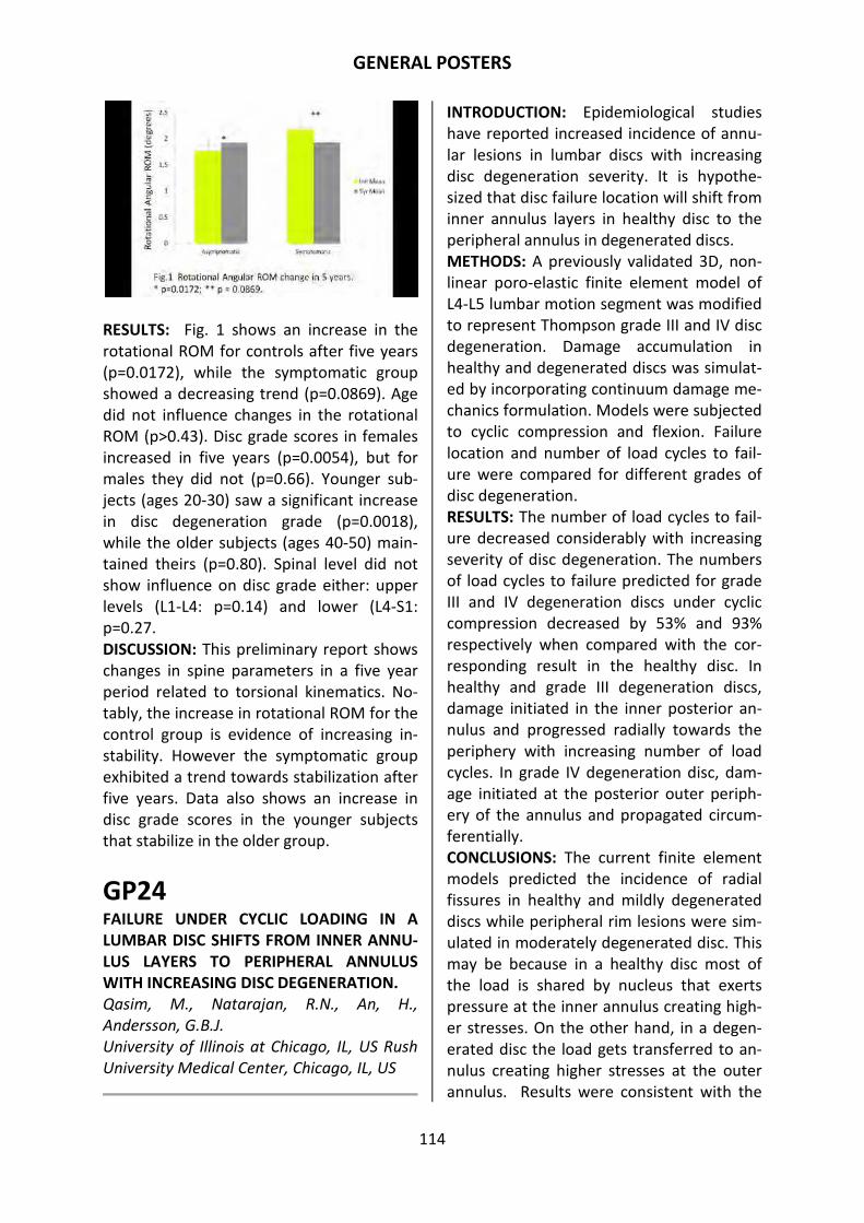
RESULTS: Fig. 1 shows an increase in the
rotational ROM for controls after five years
(p=0.0172), while the symptomatic group
showed a decreasing trend (p=0.0869). Age
did not influence changes in the rotational
ROM (p>0.43). Disc grade scores in females
increased in five years (p=0.0054), but for
males they did not (p=0.66). Younger sub-
jects (ages 20-30) saw a significant increase
in disc degeneration grade (p=0.0018),
while the older subjects (ages 40-50) main-
tained theirs (p=0.80). Spinal level did not
show influence on disc grade either: upper
levels (L1-L4: p=0.14) and lower (L4-S1:
p=0.27.
DISCUSSION: This preliminary report shows
changes in spine parameters in a five year
period related to torsional kinematics. No-
tably, the increase in rotational ROM for the
control group is evidence of increasing in-
stability. However the symptomatic group
exhibited a trend towards stabilization after
five years. Data also shows an increase in
disc grade scores in the younger subjects
that stabilize in the older group.
GP24 FAILURE UNDER CYCLIC LOADING IN A LUMBAR DISC SHIFTS FROM INNER ANNU-LUS LAYERS TO PERIPHERAL ANNULUS WITH INCREASING DISC DEGENERATION. Qasim, M., Natarajan, R.N., An, H.,
Andersson, G.B.J.
University of Illinois at Chicago, IL, US Rush
University Medical Center, Chicago, IL, US
INTRODUCTION: Epidemiological studies
have reported increased incidence of annu-
lar lesions in lumbar discs with increasing
disc degeneration severity. It is hypothe-
sized that disc failure location will shift from
inner annulus layers in healthy disc to the
peripheral annulus in degenerated discs.
METHODS: A previously validated 3D, non-
linear poro-elastic finite element model of
L4-L5 lumbar motion segment was modified
to represent Thompson grade III and IV disc
degeneration. Damage accumulation in
healthy and degenerated discs was simulat-
ed by incorporating continuum damage me-
chanics formulation. Models were subjected
to cyclic compression and flexion. Failure
location and number of load cycles to fail-
ure were compared for different grades of
disc degeneration.
RESULTS: The number of load cycles to fail-
ure decreased considerably with increasing
severity of disc degeneration. The numbers
of load cycles to failure predicted for grade
III and IV degeneration discs under cyclic
compression decreased by 53% and 93%
respectively when compared with the cor-
responding result in the healthy disc. In
healthy and grade III degeneration discs,
damage initiated in the inner posterior an-
nulus and progressed radially towards the
periphery with increasing number of load
cycles. In grade IV degeneration disc, dam-
age initiated at the posterior outer periph-
ery of the annulus and propagated circum-
ferentially.
CONCLUSIONS: The current finite element
models predicted the incidence of radial
fissures in healthy and mildly degenerated
discs while peripheral rim lesions were sim-
ulated in moderately degenerated disc. This
may be because in a healthy disc most of
the load is shared by nucleus that exerts
pressure at the inner annulus creating high-
er stresses. On the other hand, in a degen-
erated disc the load gets transferred to an-
nulus creating higher stresses at the outer
annulus. Results were consistent with the

GENERAL POSTERS
115
experimental and clinical observations in
terms of the region of failure.
GP25 IN VIVO CHARACTERIZATION OF THE 3D LUMBAR FORAMEN GEOMETRY Senoo I, Espinoza Orías AA, Park DK,
Andersson GBJ, An HS, Inoue N
Department of Orthopaedic Surgery, Rush
University Medical Center, Chicago, Illinois
60612
INTRODUCTION: There are many spinal
treatment options that require accurate
knowledge of the lumbar foramen bounda-
ry. Alas, its definition is elusive due to the
persistent use of planar methods to deter-
mine the dimensions of a true 3D contour.
This study aims to introduce innovative 3D
CT-based analysis methods for the lumbar
foramen.
METHODS: This IRB-approved study ob-
tained CT scans in supine position of 59
healthy volunteers (31M/28F) that were
used to create the 3D lumbar vertebral sur-
face models. A floating spherical coordinate
system was moved along the nerve root
path inside each foramen using an algo-
rithm that calculated the distance from this
center to the closest foramen elements
(facet joints, disc, pedicles). Its outcomes
were: foramen height (FH, distance be-
tween any two points furthest apart from
each other) and the foramen width (FW,
distance between any two points closest
together). These two parameters were cor-
related with the vertebral posterior wall
height, segmental lordotic angle, disc height
and MR disc grade (Pfirrmann’s). ANOVA
and Pearson’s correlation were used for
statistical analyses. Significance level was
set at p<0.05.
RESULTS: Both FH and FW decreased with
age. FH and FW in the lower lumbar levels
were significantly smaller than in the upper
levels. In L5/S, there was moderate or weak
negative correlation between FH and ages,
and moderate or weak positive correlation
between FH versus disc height, posterior
wall height and disc grade. There was al-
most no significance between FH and SLA
(except L1/2), and height (except L1/2). Of
note, there was a moderate negative corre-
lation between FW and age. There were no
differences between FW and disc height or
segmental lordotic angle.
DISCUSSION: The present study provided
3D foramen geometries of an asymptomatic
cohort measured in vivo. Although data is
limited to bony geometry, it could be used
as base line data for diagnosis of foraminal
stenosis and planning of treatment modali-
ty.
GP26 CHANGES IN NUCLEUS PULPOSUS MRI SIGNAL INHOMOGENEITY SHOW DISC DEGENERATION PROGRESS Yang S-H; Espinoza Orías AA; Pan C-C; Senoo
I; Andersson GBJ; An HS; Inoue N
Department of Orthopaedic Surgery, Rush
University Medical Center, Chicago, Illinois
60612
INTRODUCTION: MRI can monitor clinically
the quality of the intervertebral disc
through changes in the Nucleus Pulposus’
(NP) signal intensity. We propose that the
location of the NP centroid weighted by
signal intensity could be a parameter to
define the signal homogeneity of a region of
interest in MR data.
METHODS: T2-weighted sequences from a
1.5T MR unit provided mid-sagittal cuts
from 65 asymptomatic volunteers
(31M/34F, 22-59 y.o.) used in this IRB-
approved study. Three spine surgeons grad-
ed 288 lumbar IVDs with a clearly identifia-
ble NP boundary (Grade I: n=47, Grade II:
n=173, Grade III: n=68). A custom written
script was used to define the boundary,
geometric and weighted centroids of each
NP. The weighted centroid was located in-
side the region of the contour that had

GENERAL POSTERS
116
highest homogeneous MR NP signal, but did
not necessarily coincide with the geometric
centroid. The distances between them in
the plane of the sagittal MR slice were cal-
culated and compared with ANOVA. Statis-
tical significance set at p<0.05.
RESULTS: In 85.8% of NPs, the weighted
center was located posterior to the geomet-
ric center. The distance on the longitudinal
axis in Grade II discs was significantly larger
than that in Grade I discs (p=0.035, Fig.1).
The distance on the axis perpendicular to
the longitudinal axis in Grade III discs was
significantly larger than that in Grade I and
Grade II discs (p<0.01).
DISCUSSION: In the majority of NPs, the
weighted centers were located posterior to
the geometric centers. Grade II inter-
centroid distances were larger than in
Grade I discs. This may suggest progressive
signal intensity decrease in the anterior part
of NP during early stages of disc degenera-
tion. With advancing degeneration, the de-
crease of signal intensity reached the poste-
rior part of the disc and the weighted cen-
ters moved toward the geometric centers.
This study showed the feasibility of the in-
ter-centroid distance as a NP homogeneity
indicator.
GP27 THE EFFECT OF DIFFERENCE IN HAM-STRINGS FLEXIBILITY ON LUMBO-PELVIC RHYTHM
Matsunaga,N.*1 Okubo,Y.*2
Kaneoka,K.*3
*1 Graduate School of Sport Sciences,
Waseda University, *2 School of physical
Therapy, Faculty of Health and Medical
Care, Saitama Medical University, *3
Faculty of Sport Sciences, Waseda University
INTRODUCTION: The purpose of this study
was to investigate effect of difference in
hamstrings flexibility on lumbo-pelvic
rhythm.
METHODS: Thirteen young rhythmic gym-
nasts (RG group) and 10 healthy women
(control group) participated in this study.
After the markers were attached to the par-
ticular points, the sagittal movements of
lumbar spine and pelvis during forward
bending were collected by a digital camera
at a frequency of 30 Hz. Lumbar angle (LA)
and pelvic tilting angle (PTA) were defined
as the angle of Th12-L3 to L3-S1, and the
angle of the horizontal to anterior superior
iliac spine-posterior superior iliac spine
lines, respectively. The lumbar to hip ratio
(L/H ratio) was calculated from LA and PTA.
To normalize, we divided total time of for-
ward bending into every 20%. Moreover,
finger floor distance (FFD) was measured to
investigate hamstrings flexibility. Group
differences were assessed using independ-
ent t-tests for each movement variables.
RESULTS: The FFD of RG group and control
group were -23.4±2.6 cm and -14.0±8.1 cm,
that show a significant difference between
the groups (p<0.001). In RG group, the av-
erage of LA and PTA during total forward
bending were 28.5±10.6°and 93.3±9.7°,
respectively. Additionally, in control, the
values were 43.3±8.7°and 65.2±9.2°, re-
spectively. The above comparisons of LA
and PTA between two groups shows signifi-

GENERAL POSTERS
117
cant differences (p=0.002, p<0.001). The
L/H ratio during total forward bending is
summarized in Figure 1
There were significant differences between
the groups in 0-20%, 20-40% and 40-60% of
forward bending (p=0.001, p=0.015).
DISCUSSION: In RG group, L/H ratio was
small. It suggests that flexible hamstrings
which causes large pelvis anteversion has
lumbar spine less flexed.
Conclusion: RG group performed forward
bending by large anteversion of pelvis and
small flexion of lumbar spine. Flexible ham-
strings decrease the load of spine during
forward bending.
GP28 EFFECT OF MENTAL STRESS ON LOW BACK LOAD WHILE LIFTING AN OBJECT Junji KATSUHIRA, Ph.D.1, Ko MATSUDAIRA,
M.D, Ph.D.2, Kazuyuki IWAKIRI, Ph.D.3,
Hitoshi MARUYAMA, Ph.D.1
1Department of Health Science, Interna-
tional University of Health and Welfare,
2600-1 Kitakanemaru, Otawara, Tochigi
324-8501, Japan, 2Clinical Research Center
for Occupational Musculoskeletal Disorders,
Kanto Rosai Hospital, 1-1 Kizukisumiyoshi-
cho, Nakahara-ku, Kawasaki, Kanagawa
211-8510, Japan, 3National Institute of Oc-
cupational Safety and Health, Nagao 6-21-1,
Tama-ku, Kawasaki, Kanagawa 214-8585,
Japan
INTRODUCTION: In addition to ergonomic
factors such as frequent lifting, work-
related psychosocial factors are significant
in the onset of disabling back pain. Although
the ergonomic effects on low back load
while lifting have been investigated and are
widely accepted in the workplace, few stud-
ies have investigated the effect of psycho-
social factors on low back load while lifting.
The aim of this study was to determine the
effect of mental processing on low back
load during lifting.
METHODS: Thirteen healthy subjects lifted
a box from the ground in four different lift-
ing tasks in randomized order: (1) squat
posture, with the back and hips flexed and
knees extended; (2) stoop posture, with the
back and hips extended and knees flexed;
(3) squat posture with mental processing
using arithmetic tasks; and (4) stoop pos-
ture with mental processing using arithme-
tic tasks. A 3D motion analysis system and
four force plates were used to record kine-
matic and kinetic data. Dynamic tri-axial low
back joint moments and low back compres-
sion force were calculated as index parame-
ters of low back load under these experi-
mental conditions.
RESULTS: Mental processing significantly
increased peak low back compression force
and low back extension moment, but not
lateral flexion or rotation moment, while
lifting in both lifting postures. Also, mental
processing decreased forward pelvic tilt in
the stoop posture and increased trunk
bending angles in the squat posture.
DISCUSSION: Mental stress during lifting
tasks appears to affect both trunk and pelvis
angles in the sagittal plane for squat and
stoop postures, resulting in increased low
back load. The present findings might help
to explain the effect of not only the ergo-
nomic demands of lifting tasks but also the
psychosocial factors responsible for the on-
set of disabling back pain.

GENERAL POSTERS
118
GP29 BIOMECHANICAL ANALYSIS OF FUSION SEGMENT RIGIDITY UPON STRESS AT BOTH THE FUSION AND ADJACENT SEGMENTS - A COMPARISON BETWEEN UNILATERAL AND BILATERAL PEDICLE SCREW FIXATION Ho-Joong Kim, MDa, Kyoung-Tak Kang,
MSb, Joon-Hee Park, MDc, Jin S. Yeom,
MDa, Bong-Soon Chang, MDc, Choon-Ki Lee,
MDc
a Spine Center and Department of
Orthopaedic Surgery, Seoul National
University College of Medicine and Seoul
National University Bundang Hospital b
Department of Mechanical Engineering,
Yonsei University, c Department of
Anesthesiology and Pain Medicine,
Kangdong Sacred Heart Hospital, Hallym
University Medical College, d Department of
Orthopaedic Surgery, Seoul National
University College of Medicine and Seoul
National University Hospital
INTRODUCTION: The biomehcanical stabil-
ity of unilateral pedicle screw fixation relat-
ed to the extent of decompression remains
unknown. The purpose of this study was to
investigate the effects of unilateral pedicle
screw fixation on the fusion segment and
the superior adjacent segment after one
segment lumbar fusion using validated fi-
nite element (FE) models.
METHODS: Four L3-4 fusion models were
simulated according to the extent of de-
compression and the method of pedicle
screws fixation in L3-4 lumbar fusion. These
models included hemi-laminectomy with
bilateral pedicle screw fixation in the L3-4
segment (BF-HL model), total laminectomy
with bilateral pedicle screw fixation (BF-TL
model), hemi-laminectomy with unilateral
pedicle screw fixation (UF-HL model), and
total laminectomy with unilateral pedicle
screw fixation (UF-TL model). In each sce-
nario, intradiscal pressures, annulus stress,
and range of motion at the L2-3 and L3-4
segments were analyzed under flexion, ex-
tension, lateral bending, and torsional mo-
ments.
RESULTS: Under four moments, the unilat-
eral fixation leads to a reduction in the ROM
increase at the adjacent segment after fu-
sion, compared to bilateral fixation. At the
fusion segment, larger motions were noted
at the fusion segment in the UF-HL and UF-
TL models under four moments, compared
with the bilateral fixation models. The UF-TL
model demonstrated a vulnerability to tor-
sional moments at the fusion segment, with
an increase in remaining motion at the fu-
sion segment. Four models showed in-
creased maximal von Mises stress on the AF
at the adjacent segments under the four
moments, and unilateral fixation models led
to a prominent reduction in increased AF
stress at the adjacent segment under the
four moments
DISCUSSION: The use of unilateral pedicle
screw fixation is only warranted in limited
decompression procedures such as hemi-
laminectomies. However, reduced stiffness
in fusion segment would have a beneficial
effect on stress of adjacent segment.

GENERAL POSTERS
119
GP30 IN VIVO RESPONSE TO A PARASPINOUS TENSION BAND DEVICE WITH TITANIUM COIL. AN OVINE IMPLANTATION STUDY E J Ehrhart, DVM, PhD, Dipl ACVP [1], Louie
Fielding, MS [2], Amy Lyons Santoni, MS [3],
Brandon G Santoni, PhD [3,4], Christian M
Puttlitz, PhD [3], Dana R Ruehlman, DVM
[1], Howard B Seim III, DVM, Dipl ACVS [1] A
Simon Turner, BVSc, MS, Dipl ACVS, DVSc [1]
[1] College of Veterinary Medicine and
Biomedical Science, Colorado State
University, Fort Collins, CO [2] Simpirica
Spine, San Carlos, CA [3] Orthopaedic
Bioengineering Research Laboratory,
Department of Mechanical Engineering,
Colorado State University, Fort Collins, CO
[4] Foundation for Orthopaedic Research
and Education, Tampa, FL
INTRODUCTION: Posterior lumbar dynamic
stabilization devices have been proposed as
an alternative to fusion. Dynamic compo-
nents and fixation elements may elicit a
biological response, affecting patient health
or implant performance.
METHODS: Eight sheep were implanted
with two paraspinous tension bands (PTB;
L1/2, L4/5) under IACUC approval. Each PTB
comprised two titanium coil springs secured
to the spinous processes (SPs) with
UHMWPE straps. Four sheep were sacri-
ficed each at 3 and 6 months. PTB compo-
nents, local and distant tissues were exam-
ined macroscopically, and then evaluated
histologically for qualitative changes and
graded for inflammatory response, device
findings and bone remodelling with a scor-
ing system based on ISO standards. One coil
from each PTB underwent tensile testing to
evaluate the functional impact of adherent
tissue.
RESULTS: All animals were healthy until
euthanasia. Gross examination revealed
fibrous encapsulation of the PTB contiguous
with midline ligamentous structures. Histo-
pathology with nondecalcified sections
identified fibrovascular tissue had infiltrated
the coils. Minimal inflammation was associ-
ated with the coils. The strap was associat-
ed with slightly higher yet mild inflammato-
ry scores. Bone remodeling indicated adap-
tation to the PTB, trending toward bone
formation by the SPs. No reaction in distant
tissues or device debris was found; only
minor fraying of the cut end of the straps.
Coil stiffness increased approximately 33%
at 3 mo and 57% at 6 mo. Through the first
50% of elongation, stiffness was largely un-
affected, with most of the increase beyond
50% elongation.
DISCUSSION: These results demonstrate
feasibility of the PTB as an alternative to
spinal fusion. Inflammation was mild, with
negligible inflammation associated with the
coils and the response to the straps compa-
rable to that reported for sutures. The coils
remained functional, with increased stiff-
ness beyond 50% elongation as load was
transferred to adherent tissue.
GENERAL POSTERS
120
GP31 INSTABILITY OF POSTERIOR VERTEBRAL WALL CAUSES CANAL ENCROACHMENT IN THE ELDERLY WITH VERTEBRAL PSEUDOARTHROSIS Tetsuo Hayashi, Takeshi Maeda, Eiji Mori,
Takayoshi Ueta, Itaru Yugue, Osamu
Kawano, Tsuneaki Takao, Hiroaki Sakai,
Muneaki Masuda, Yuichiro Morishita,
Keiichiro Shiba
Department of Orthopedic Surgery, Japan
Labour Health and Welfare Organization,
Spinal Injuries Center, Fukuoka
INTRODUCTION: The purpose of this study
was to elucidate effects of loading associat-
ed with SCE in patients with insufficient
bone union following vertebral fractures in
the elderly using computed tomography
(CT) myelography in two different positions.
METHODS: Seventeen patients (mean age,
77.4 years) with various degrees of neuro-
logical deficit due to insufficient bone union
at both vertebral body and posterior verte-
bral wall were prospectively examined in
this study. CT myelography was performed
in both semi-sitting and supine positions.
Kyphotic angle, rate of dural compression,
ratio of occupation by bony fragments and
posterior vertebral body height ratio were
measured and compared between posi-
tions.
RESULTS: Ratio of occupation by bony
fragments was significantly higher in the
semi-sitting position (47.9%) than in the
supine position (33.9%, p<0.001). Similarly,
posterior vertebral body height ratio was
significantly lower in the semi-sitting posi-
tion (67.8%) than in the supine position
(76.3%), indicating a significant loss of ver-
tebral height in the semi-sitting position
(p<0.001). Rate of dural compression was
likewise significantly higher in the semi-
sitting position (48.6%) than in the supine
position (33.3%; p<0.001). Change in ratio
of occupation by bony fragments, change in
posterior vertebral body height ratio and
angular instability between positions were
13.9%, 8.5% and 13°, respectively. A signifi-
cant correlation was identified between
change in ratio of occupation by bony frag-
ments and change in posterior vertebral
body height ratio (p=0.001).
CONCLUSIONS: Our study demonstrated
that collapse of the non-united posterior
vertebral wall and intracanal protrusion of
vertebral fragments would occur simulta-
neously with axial loading, causing SCE. CT
taken in semi-sitting position appears quite
useful to evaluate the amount of SCE by an
unstable posterior wall.
GP32 DEVELOPMENT OF A NOVEL PERCUTANE-OUS GUIDE WIRE (S-WIRE™) FOR PERCU-TANEOUS PEDICLE SCREW INSERTION –ITS EFFICACY AND SAFETY– Ishii K.1, Ishihara S3., Kaneko Y.4, Iwanami
A.1, Hikata T.1, Hosogane N.1, Watanabe
K.2, Nakamura M.1, Toyama Y.1,
Matsumoto M.1
1Dept. of Orthopaedic Surgery, 2Advanced
Therapy for spine and spinal cord diseases,
School of Medicine, Keio University, Tokyo,
Japan 3Dept. of Orthopedic Surgery, Nerima
General Hospital, Tokyo, Japan 4Dept. of
Orthopedic Surgery, Kawasaki Municipal
Kawasaki Hospital, Kanagawa, Japan
INTRODUCTION: In minimally invasive spi-
nal fusion surgery such as MIS-TLIF, the
placement of percutaneous pedicle screw
(PPS) is necessary, but several intraopera-
tive complications have been reported. One
of the serious complications is the great
vessel and bowel injuries due to the anteri-
or migration and penetration of the guide
wire through the anterior cortex of the ver-
tebral body. We have established a novel
percutaneous guide wire (S-wire™) in MIS-
TLIF procedure. Our aim was to investigate
the biomechanical characteristics of S-
wire™ in cadaveric spines by comparing
conventional wire, and evaluate the efficacy

GENERAL POSTERS
121
and safety of S-wire™ in the clinical trial.
METHODS: S-wire™ consists of a hollow and
braided wire which extends at the one side
of the tips. Fresh cadaveric lumbar spines
from L1 to L5 (N=3) were used for biome-
chanical comparison between S-wire™ and
a conventional wire. The push-out (1 cm
migration) and penetration forces were
measured in two wires.
RESULTS: A braided part of S-wire™ was
moderately bent or unraveled when the
axial force was applied, resulting in the in-
creased resistance against anterior migra-
tion. The mean push-out force in S-wires™
and conventional wires were 15.5±1.9N and
5.7±0.8N (P<0.0001), respectively. The
mean penetration forces in S-wires™ and
conventional wires were 69.1±4.2N and
37.1± 4.8N (P<0.0005), respectively. In
the clinical trial of a total number of 212 S-
wires™, neither penetration of the anterior
wall nor breakage of wire was observed.
Interestingly, 181 of 212 S-wires™ (85%)
revealed increase of pull-out force after the
placement.
DISCUSSION: S-wire™ could prevent the
anterior migration and the penetration of
the anterior wall of the vertebral body and
it is safe and useful device for not only MIS-
TLIF but balloon kyphoplasty.
GP33 FINITE ELEMENT ANALYSIS FOR COMPARI-SON OF SUPRASPINOUS OSTEOTOMIES TECHNIQUE WITH CONVENTIONAL LAMI-NECTOMY Ho-Joong Kim, MDa, Kyoung-Tak Kang,
MSb, Choon-Ki Lee, MDc, Bong-Soon Chang,
MDc, Jin S. Yeom, MDa
a Spine Center and Department of Ortho-
paedic Surgery, Seoul National University
College of Medicine and Seoul National Uni-
versity Bundang Hospital b Department of
Mechanical Engineering, Yonsei University c
Department of Orthopaedic Surgery, Seoul
National University College of Medicine and
Seoul National University Hospital
INTRODUCTION: It is unknown whether
osteotomies at the base of the spinous pro-
cesses might increase the risk of spinal in-
stability despite preservation of the supra-
spinous and inter-spinous ligaments. There-
fore, the aim of this study was to evaluate
and compare the biomechanical behavior of
the lumbar spine after posterior decom-
pression with spinous process osteotomies
(SPO) technique or conventional laminec-
tomy (CL) technique using finite element
model.
METHODS: Three validated lumbar FE mod-
els (L2-5) which represented intact spine
and two decompression models using SPO
and CL technique were developed. Decom-
pression procedures were simulated at the
L3-4 segment in the SPO and CL mod-
els.Under 400 N pre-compressive loading,
7.5 Nm moments simulated flexion, exten-
sion, lateral bending, and torsion were im-
posed. In each model, the ranges of motion,
the maximal von Mises stress of the annulus
fibrosus, and the intradiscal pressures at the
index segment (L3-4) and adjacent seg-
ments (L2-3 and L4-5) were analyzed. Facet
contact forces were also compared among
three models under the extension and tor-
sion moments.
RESULTS: Compared to the intact model,
the CL and SPO models had increased range
of motion and annulus stress at both the
index segment (L3-4) and the adjacent seg-
ments under flexion and torsion. The SPO
model demonstrated a reduced range
ofmotion and annulus stress than the CL
model. The both of CL and SPO models had
the increase of the facet contact force at
the L3-4 segment under the torsion mo-
ment, compared to that of intact model. In
contrast, under the extension moment,
three models demonstrated the similar fac-
et contact force even at the L3-4 model.
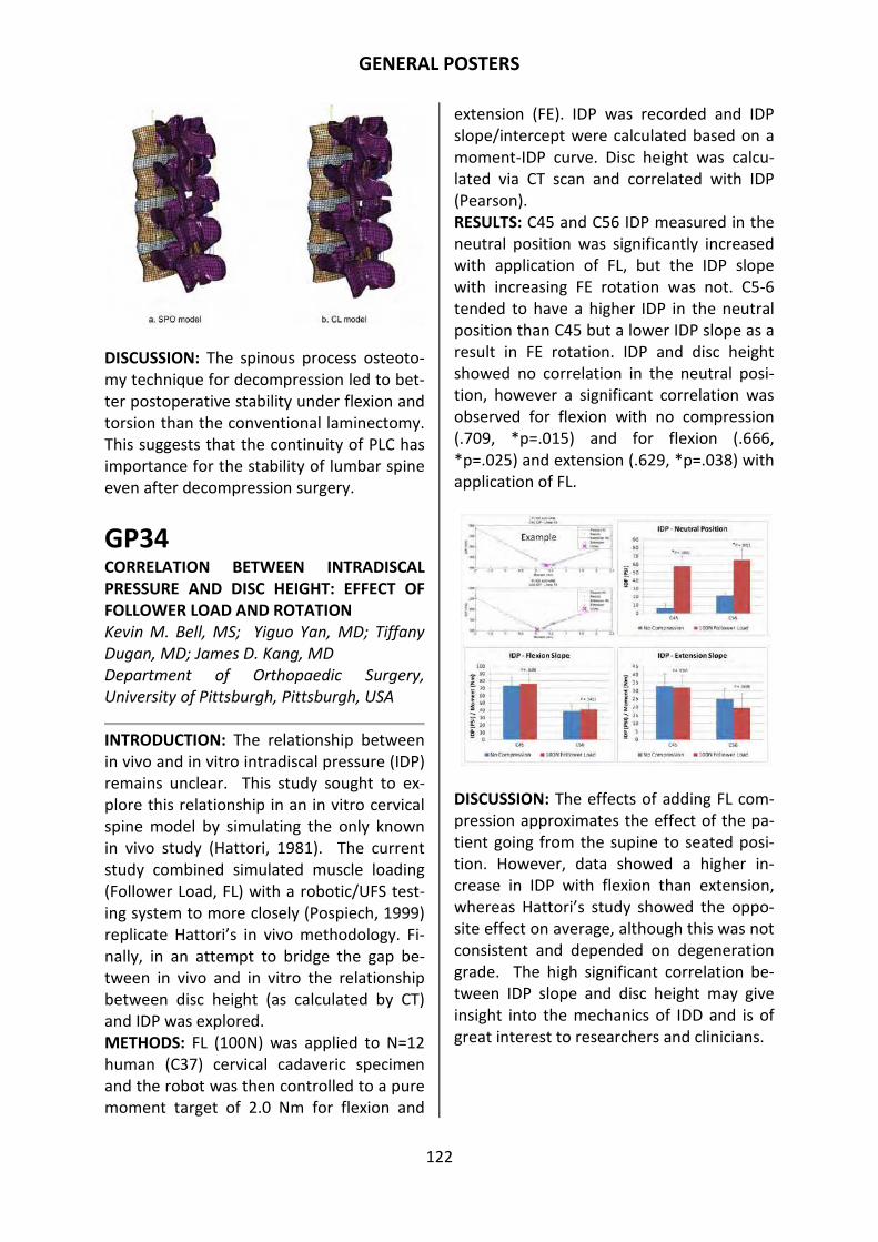
GENERAL POSTERS
122
DISCUSSION: The spinous process osteoto-
my technique for decompression led to bet-
ter postoperative stability under flexion and
torsion than the conventional laminectomy.
This suggests that the continuity of PLC has
importance for the stability of lumbar spine
even after decompression surgery.
GP34 CORRELATION BETWEEN INTRADISCAL PRESSURE AND DISC HEIGHT: EFFECT OF FOLLOWER LOAD AND ROTATION Kevin M. Bell, MS; Yiguo Yan, MD; Tiffany
Dugan, MD; James D. Kang, MD
Department of Orthopaedic Surgery,
University of Pittsburgh, Pittsburgh, USA
INTRODUCTION: The relationship between
in vivo and in vitro intradiscal pressure (IDP)
remains unclear. This study sought to ex-
plore this relationship in an in vitro cervical
spine model by simulating the only known
in vivo study (Hattori, 1981). The current
study combined simulated muscle loading
(Follower Load, FL) with a robotic/UFS test-
ing system to more closely (Pospiech, 1999)
replicate Hattori’s in vivo methodology. Fi-
nally, in an attempt to bridge the gap be-
tween in vivo and in vitro the relationship
between disc height (as calculated by CT)
and IDP was explored.
METHODS: FL (100N) was applied to N=12
human (C37) cervical cadaveric specimen
and the robot was then controlled to a pure
moment target of 2.0 Nm for flexion and
extension (FE). IDP was recorded and IDP
slope/intercept were calculated based on a
moment-IDP curve. Disc height was calcu-
lated via CT scan and correlated with IDP
(Pearson).
RESULTS: C45 and C56 IDP measured in the
neutral position was significantly increased
with application of FL, but the IDP slope
with increasing FE rotation was not. C5-6
tended to have a higher IDP in the neutral
position than C45 but a lower IDP slope as a
result in FE rotation. IDP and disc height
showed no correlation in the neutral posi-
tion, however a significant correlation was
observed for flexion with no compression
(.709, *p=.015) and for flexion (.666,
*p=.025) and extension (.629, *p=.038) with
application of FL.
DISCUSSION: The effects of adding FL com-
pression approximates the effect of the pa-
tient going from the supine to seated posi-
tion. However, data showed a higher in-
crease in IDP with flexion than extension,
whereas Hattori’s study showed the oppo-
site effect on average, although this was not
consistent and depended on degeneration
grade. The high significant correlation be-
tween IDP slope and disc height may give
insight into the mechanics of IDD and is of
great interest to researchers and clinicians.

GENERAL POSTERS
123
GP35 IN VIVO MRI MEASUREMENT OF SPINAL CORD DISPLACEMENT IN THE THORACOL-UMBAR REGION: PART 2 - COMPARISON BETWEEN UNILATERAL AND BILATERAL STRAIGHT LEG RAISE TESTS. Marinko Rade 1, Mervi Könönen 2, Ritva
Vanninen 2, Michael Shacklock 3, Markku
Kankaanpää 4, Olavi Airaksinen 1
1. Department of Physical and Rehabilita-
tion Medicine, Kuopio University Hospital,
Kuopio, Finland 2. Department of Radiology,
Kuopio University Hospital, Kuopio, Finland.
3. Neurodynamic Solutions, Adelaide, Aus-
tralia 4. Department of Physical and Reha-
bilitation Medicine, Tampere University
Hospital, Tampere, Finland
INTRODUCTION: Following on from our
previous study (Rade et al 2012) we further
measured spinal cord displacement during
the passive bilateral SLR in asymptomatic
subjects and compared these data with
those of the unilateral. The corollary is then
to develop a better understanding of the
local mechanical responses and possible
clinical implications.
METHODS: Sixteen asymptomatic male vol-
unteers were scanned with 1.5T magnetic
resonance scanner (Siemens Avanto, Erlan-
gen, Germany) using T2 weighted turbo spin
echo fat saturation sequence (TR 3880ms,
TE 80ms, 10 slices, slice thickness 3mm, gap
0.3mm, FOV 400mm2, pixel size
1.3mm*0.9mm). Coronal slices were aligned
with spinal cord.
Displacement of the conus relative to the
adjacent vertebral body during bilateral SLR
was quantified and compared with data on
the right SLR, left SLR and the neutral (ana-
tomic) position. Each movement was per-
formed twice in order to provide intraclass
correlation coefficients (ICC). Four practi-
tioners performed the maneuvers in a ran-
dom sequence in order to avoid possible
series effects.
RESULTS: Compared to the position of the
conus in the neutral (anatomic) position,
the medullar cone displaced caudally in the
spinal canal by 5.2 ± 1.5 mm with bilateral
SLR (p=0.000). This displacement was also
statistically significant when compared with
the right SLR (p=0.000) and left SLR
(p=0.000). The ICCs were as follows: bilat-
eral SLR = 0.994, right SLR = 0.997, left SLR=
0.996, unilateral SLR (right and left SLR
compared) = 0.997. Number of required
subjects to get statistically significant results
(p<0.05) is 3 for Bilateral SLR.
CONCLUSIONS: The results showed that
the distal displacement of the medullar
cone was significantly greater (almost dou-
ble) with the bilateral compared to the uni-
lateral SLR. We hypothesize that this great-
er movement may be because more force
was transmitted to the cord through a larg-
er number nerve roots with the bilateral
than unilateral SLR.
GP36 THREE DIMENSIONAL ASSESSMENT OF LUMBAR FACET JOINT TROPISM IN ASYMPTOMATIC AND LOW BACK PAIN SUBJECTS Huh J-S, Espinoza Orías AA, Otsuka Y, Simon
P, An HS, Andersson GBJ, Inoue N
Department of Orthopaedic Surgery, Rush
University Medical Center, Chicago, Illinois
60612
INTRODUCTION: Facet joint (FJ) orientation
and tropism are important parameters to
analyze FJ pathogenic contributions to vari-
ous types of spinal diseases. However, lim-
ited data on orientation data and lack of a
proper tropism definition make this topic
controversial. This study aims to describe
relationships between lumbar FJ 3D orienta-
tion, tropism, age, gender, level, and pres-
ence of chronic low back pain (LBP) and to
evaluate possible cutoff values to define
tropism.
METHODS: IRB-approved in vivo 3D orienta-
tion measurements of lumbar FJs were per-

GENERAL POSTERS
124
formed on 92 volunteers (58 asymptomat-
ic/34 chronic LBP subjects) using CT-based
3D facet joint surface models. Each facet
joint surface was represented by its normal
vector, which was subsequently plotted on
a unit sphere. Differences in symmetry be-
tween left and right orientation vectors
(tropism angle) were calculated between
each unit vector and its corresponding mir-
ror image. Angle differences were evaluated
with unpaired t tests. Gender, age and level
effects were analyzed by a one-way ANOVA
with Fischer post hoc test, and significance
set at p<0.05. Cutoff values attempted were
the mean and one standard deviation of
angle difference, 5 and 10 degrees, and
were analyzed by a chi-square test.
RESULTS: The orientations of both superior
and inferior facets changed gradually from
sagittal to coronal orientation from L1 to L5.
More coronal orientation of L1 superior
facets and more horizontal orientation of L5
inferior facets revealed transitional features
of spine. The mean tropism angle was 7.65°.
The mean angle difference of symptomatic
group was significantly larger than that of
asymptomatic group. The cutoff values over
mean angle difference showed significantly
more tropism in symptomatic group than
asymptomatic group, the others did not.
CONCLUSION: Our results of more tropism
in symptomatic group based on a three-
dimensional orientation model supports the
hypothesis of causative role of tropism to
LBP.
GP37 MODIC CHANGES FOLLOW BONE REMOD-ELING PRINCIPLES Agarwal A; Kaul V; Goel VK; Agarwal AK
Engineering Center for Orthopaedic Re-
search Excellence (E-CORE), Departments of
Bioengineering and Orthopaedic Surgery,
University of Toledo, Toledo, OH 43606, Cor-
responding author: [email protected]
INTRODUCTION: Etiology of Modic changes
is not well understood.1 We hypothesize
that Modic changes are a direct conse-
quence of changes in biomechanical envi-
ronment due to disc herniation and thus
obey Wolff’s law of bone remodeling.
METHODS: Strain energy density theory of
adaptive remodeling was implemented in
our experimentally validated 3D finite ele-
ment model of lumbar spine (L3-L5). The
algorithm was executed for flexion (10Nm),
extension (10Nm), and compression (400N)
after posterolateral disc herniation at L4-L5
IVD (nucleus pulposus-NP volume removed
- 2cm3; annulus fibrosus (AF) incised -
44mm2).
RESULTS: In L5, the average apparent densi-
ties decreased by 55% beneath the herniat-
ed region due to no load transmission and
hence decrease in strain energy density
(SED) of that region, Figure 1.
Average apparent density increased by
101%, in the regions beneath the unaffect-
ed NP due to increase in SED. Likewise, ap-
parent density of L4 showed an increase of
129% in the region above the unaffected
NP.
DISCUSSION: Decreases in the density
around herniated region are similar to loss
of trabecular bone and bone marrow deple-
tion observed clinically as Modic changes.1
Our data concur with previous studies
which state that Modic changes may be due
to changes in biomechanical environment of
the surrounding bone.2, 3 Bone resorption
occurred near the affected areas in the ver-
tebra below the herniated IVD, with in-
creases in bone densities in other areas,
both in agreement with literature.2
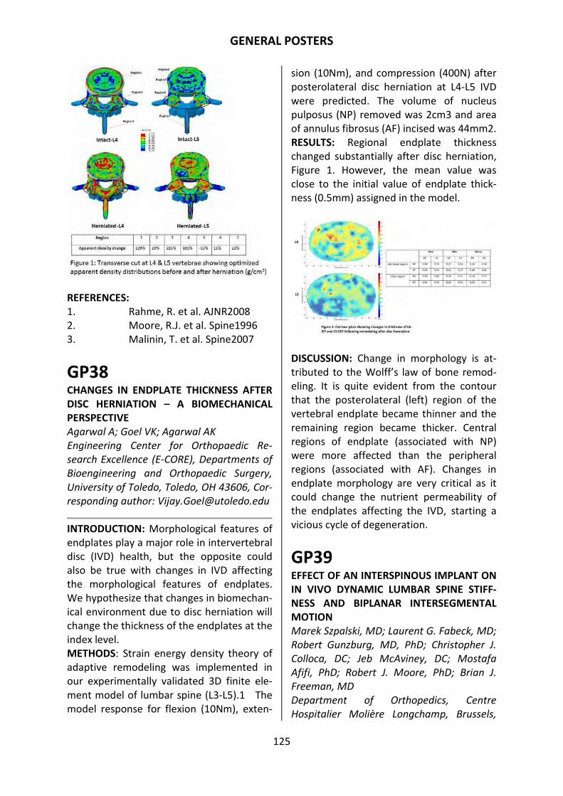
GENERAL POSTERS
125
REFERENCES: 1. Rahme, R. et al. AJNR2008
2. Moore, R.J. et al. Spine1996
3. Malinin, T. et al. Spine2007
GP38 CHANGES IN ENDPLATE THICKNESS AFTER DISC HERNIATION – A BIOMECHANICAL PERSPECTIVE Agarwal A; Goel VK; Agarwal AK
Engineering Center for Orthopaedic Re-
search Excellence (E-CORE), Departments of
Bioengineering and Orthopaedic Surgery,
University of Toledo, Toledo, OH 43606, Cor-
responding author: [email protected]
INTRODUCTION: Morphological features of
endplates play a major role in intervertebral
disc (IVD) health, but the opposite could
also be true with changes in IVD affecting
the morphological features of endplates.
We hypothesize that changes in biomechan-
ical environment due to disc herniation will
change the thickness of the endplates at the
index level.
METHODS: Strain energy density theory of
adaptive remodeling was implemented in
our experimentally validated 3D finite ele-
ment model of lumbar spine (L3-L5).1 The
model response for flexion (10Nm), exten-
sion (10Nm), and compression (400N) after
posterolateral disc herniation at L4-L5 IVD
were predicted. The volume of nucleus
pulposus (NP) removed was 2cm3 and area
of annulus fibrosus (AF) incised was 44mm2.
RESULTS: Regional endplate thickness
changed substantially after disc herniation,
Figure 1. However, the mean value was
close to the initial value of endplate thick-
ness (0.5mm) assigned in the model.
DISCUSSION: Change in morphology is at-
tributed to the Wolff’s law of bone remod-
eling. It is quite evident from the contour
that the posterolateral (left) region of the
vertebral endplate became thinner and the
remaining region became thicker. Central
regions of endplate (associated with NP)
were more affected than the peripheral
regions (associated with AF). Changes in
endplate morphology are very critical as it
could change the nutrient permeability of
the endplates affecting the IVD, starting a
vicious cycle of degeneration.
GP39 EFFECT OF AN INTERSPINOUS IMPLANT ON IN VIVO DYNAMIC LUMBAR SPINE STIFF-NESS AND BIPLANAR INTERSEGMENTAL MOTION Marek Szpalski, MD; Laurent G. Fabeck, MD;
Robert Gunzburg, MD, PhD; Christopher J.
Colloca, DC; Jeb McAviney, DC; Mostafa
Afifi, PhD; Robert J. Moore, PhD; Brian J.
Freeman, MD
Department of Orthopedics, Centre
Hospitalier Molière Longchamp, Brussels,

GENERAL POSTERS
126
Belgium; Departments of Orthopaedics and
Traumatology, Université Libre de Bruxelles,
Brussels, Belgium; Department of
Orthopaedic Surgery, Edith Cavell Clinic,
Brussels, Belgium; PhD Kinesiology Program,
School of Nutrition and Health Promotion,
Arizona State University, Tempe, Arizona,
U.S.A; Sydney Scoliosis Clinic, Sydney,
Australia; Faculty of Kinesiology, University
of Calgary, Alberta, Canada; Adelaide
Centre for Spinal Research, Adelaide,
Australia; School of Medicine, Faculty of
Health Sciences, University of Adelaide,
Australia
INTRODUCTION: This study examined the
effect of an interspinous implant on lumbar
spine stiffness and intersegmental accelera-
tions in the dorsoventral (DV) and axial (AX)
planes in an in vivo ovine model.
METHODS: Dynamic lumbar spine stiffness
and intersegmental accelerations were
quantified in eight anesthetized adolescent
Merino wethers 68.4 kg (s.d.=4.5 kg). Two,
6, and 12 Hz oscillatory loads were random-
ly applied to the L3 spinous process in the
prone and oblique positions using a previ-
ously validated technique. In each position,
measurements were obtained with the
lumbar spine intact; and following insertion
of an InSwing® interspinous device at L3-L4
secured by a tension band. DV stiffness
(N/mm) was calculated over 6 trials during
10 seconds of testing at each loading fre-
quency in each position. L3-L4 accelera-
tions were derived from tri-axial accelerom-
eters mounted to steel pins rigidly affixed to
the L3 and L4 spinous processes. Stiffness
and accelerations for each position and
condition were compared using one-way
ANOVA with repeated measures and post-
hoc analyses.
RESULTS: Mean DV stiffness ranged from
6.73 to 28.67 N/mm intact compared to
12.39 to 40.32 N/mm for the interspinous
implant condition. Significant increases in
DV lumbar spine stiffness were observed for
6 Hz (p=0.002) and 12 Hz (p=0.011) testing.
Significant decreases in DV accelerations at
L3-L4 were also found for 12 Hz testing
(p=0.024). In the AX plane, significant 2- to
6-fold reductions in intersegmental acceler-
ations were found for 2 Hz testing (p=0.026,
p=0.001) in animals with an interspinous
implant.
DISCUSSION: This experimental model pro-
vides novel evidence that an interspinous
device with tension band provides increased
segmental and intersegmental lumbar sta-
bility in the DV and AX planes and further
confirms the frequency dependence upon
stiffness testing. These results may be use-
ful when considering non-fusion devices for
patients with symptomatic lumbar instabil-
ity.
GP40 T2 VALUES OF HUMAN LUMBAR DISCS: TEMPLATE-BASED SEGMENTATION AND VARIATIONS WITH AGE, SEX AND LEVEL WC Bae, +T Yamaguchi, R Pichika, ME Lenz,
T Fujiwara, GM Bydder, K Mausda
University of California, San Diego, San
Diego, CA, USA. + Doshisha University,
Kyoto, Japan
INTRODUCTION: Quantitative MRI is useful
for evaluation of disc degeneration. Howev-
er, segmentation of NP and AF can be diffi-
cult, when the signal intensity from the NP
is low and indistinct from the AF. Aim of this
study was to describe a template-based
technique to segment NP indirectly, and to
report age- and level-dependent changes in
T2 of human discs.
METHODS: Lumbar spines (L1 to L5) from
cadavers (n=84, 58±10 yrs, 18F, 66M) were
obtained from tissue bank and imaged at
3T. Spin echo T2 mapping was performed:
mid-sagittal plane, FOV=16 to 20 cm, slice=3
mm, TR=2000 ms, TE=10 to 90 ms (8 TEs).
Discs (n=334) were Pfirrmann-graded
(1=normal, 5=degenerate) and T2 values
determined. Using grade 1 discs (n=11),
GENERAL POSTERS
127
template of AF boundary and NP region of
interest (A) was created, and applied to
other discs using affine registration (B) to
match target AF. Using this method, T2 val-
ues of NP and AF of each disc was deter-
mined.
RESULTS: Grade 1 discs readily segmented
into NP and AF due to bimodal distribution
of T2 values (A), unlike higher grade discs.
T2 values of the NP varied with age
(p<0.001) but not with sex (p=0.4) or level
(p=0.5). T2 values of the NP decreased at a
rate of -1.1 ms/yr (intercept 136 ms), a sig-
nificant correlation (p<0.001, r2=0.22). This
was stronger than that between Pfirrmann
grade and age (r2=0.10). T2 values of the AF
did not vary with age (p=0.9), sex (p=0.54)
or level (p=0.5).
DISCUSSION: A template-based method of
NP segmentation was developed and used
to assess cadaveric discs. The novel tech-
nique is useful when other methods of di-
rectly segmenting NP fail due to low image
contrast, as is often the case for degenerat-
ing discs. Additional studies to compare
accuracy of this method to reference stand-
ards would be useful. Our findings of non-
effect of gender and significant decrease in
T2 of the NP with aging agree with past
studies. Advances in knowledge include
histogram of T2 values in discs, rate of
change of T2 values with age.
GP41 SHEAR STIFFNESS OF BABOON INTERVER-TEBRAL DISC ASSESSED BY MR ELASTOGRAPHY Ephraim I Ben-Abraham, Jun Chen, Ph.D.,
Richard L Ehman, M.D.
Mayo Clinic, Rochester, Minnesota, United
States
INTRODUCTION: There are many tech-
niques to characterize disc degeneration
such as x-ray and MRI methods. However,
no techniques exist to directly assess the
material properties of the intervertebral
disc (IVD) within the intact spine. Magnetic
resonance elastography (MRE) is a sensitive,
phase contrast-based imaging technique for
non-invasively mapping the mechanical
properties of tissues. The objective of this
study is to determine if MRE is capable of
detecting shear wave propagation and gen-
erating a shear stiffness map for an axial
cross-section of the IVD.
METHODS: A fresh frozen lumbar spine
segment (L3-L4) was tested with posterior
elements removed to increase flexibility.
Shear vibration at 1,000 Hz was applied to
the upper vertebral body while the lower
vertebral body was fixed using a custom-
built testing fixture. The disc specimen was
imaged with a spin echo-based MRE se-
quence in a standard 1.5T GE full-body MRI
scanner with a 5-in diameter receive RF coil
(TE/TR = 50/290 ms).
RESULTS: Propagating waves were seen in
an axial cross-section of the IVD in all three
motion-encoding directions. A representa-
tive filtered wave image showing the shear
wave displacement pattern is shown (Fig.
1A). Distinct wave patterns can be seen in
the annulus and nucleus pulposus, with the
nucleus showing a much shorter wave-
length (“softer”). A shear stiffness map was
generated from the MRE data in the IVD
(Fig. 1B). Although the wavelength was too
long in the annular region to reliably ap-
proximate stiffness, the nucleus showed an

GENERAL POSTERS
128
average shear stiffness of 79 ± 15 kPa.
DISCUSSION: These results suggest MRE is
capable of detecting shear wave propaga-
tion in the IVD, creating a stiffness map of
the IVD, and can differentiate the nucleus
and annulus regions. Based on these results,
MRE could provide a valuable tool to study
the mechanical properties of healthy and
degenerated discs, with the potential for
investigating conservative treatments for
patients with low back pain.
GP42 EX-VIVO RESPONSE OF SPINAL CARTILAGI-NOUS TISSUES TO FLEXION-EXTENSION OF RABBIT FUNCTIONAL SPINAL UNIT Hartman RA, Bell KM, Yurube T, Vo NV,
Kang JD, Sowa GA
University of Pittsburgh, Department of
Physical Medicine & Rehabilitation; De-
partment of Bioengineering; Department of
Orthopaedic Surgery, Pittsburgh, PA, USA
INTRODUCTION: Intervertebral disc (IVD)
degeneration is characterized by degrada-
tion of disc extracellular matrix and is fre-
quently coupled with facet cartilage (FC)
degradation. Mechanical loading plays an
important role in degeneration, yet flexion-
extension (FE) has not been investigated
and simultaneous cellular responses to me-
chanical loading in IVD and FC remain un-
known. The goal of this study was to eluci-
date the influence of repeated FE simulta-
neously in IVD and FC using viable function-
al spinal units (FSU) loaded ex vivo.
MATERIALS AND METHODS: Freshly har-
vested rabbit L4-5 FSUs are mounted within
a chamber that interfaces with a serial-
linkage robot capable of six-axis motion.
Media (10% FBS) is pumped through the
chamber (1.25 ml/min), and temperature
and gas concentrations are controlled to 37
°C and 5/5% CO2/O2. FSUs are subjected to
three cycles of FE using adaptive displace-
ment control to determine the pure mo-
ment path. FSUs are rotated through (1)
large amplitudes—moment-angle curves
enter the linear region and (2) small ampli-
tudes—moment-angle curves remain within
the toe region. Following three cycles of
preconditioning, FE kinematics are repeated
for 1 h at 0.33 °/s. Unloaded FSUs (L2-3)
serve as controls. Outcome measures in-
clude relative mRNA expression of catabolic
(MMP-3 and ADAMTS-5), inflammatory
(COX-2), and anabolic (Agg) genes alongside
changes in mechanical outcomes—load re-
laxation and hysteresis.
RESULTS: Large amplitudes relative to small
ones showed relative mRNA expression
changes that were pro-inflammatory in NP
and FC, catabolic in FC, and anti-anabolic in
NP. Hysteresis changed over loading cycles
in the large amplitude but not the small
amplitude FSUs.
DISCUSSION: Preliminary data suggest larg-
er amplitudes of FE, which reflect physiolog-
ic motion extremes, promote inflammation
and matrix degradation in the NP and FC.
GP43 Empty
GP44 STATIC AND DYNAMIC COMPRESSIVE CHARACTERIZATION OF LUMBAR INTER-VERTEBRAL DISCS OF THE ADULT NEW ZEALAND WHITE RABBIT FOLLOWING IN VIVO DEGENERATION OR EX VIVO ANNU-LAR PUNCTURE †Taborek, Alex P; †Chen, Albert C;
†*Yamaguchi, Tomonori; †Nguyen,
Quynnhoa T; *Inoue, Nozomu; †Sah, Robert
L; †Masuda, Koichi.

GENERAL POSTERS
129
†University of California-San Diego, La Jolla,
CA, *Doshisha University, Kyoto, Japan
INTRODUCTION: In animal models, interver-
tebral disc (IVD) degeneration, induced in
vivo by anular puncture, involves mechani-
cal, biochemical, geometric, and histologic
changes. The effect of anular puncture on
biomechanical properties depends on de-
fect dimensions, but has not been studied in
rabbits. The objectives of this study were to
investigate the biomechanical consequenc-
es of defect size made with 18-27G needles
during ex vivo anular puncture and of de-
generation induced in vivo by 18G anular
puncture (equilibrium modulus and dynamic
stiffness of lumbar IVDs) of the rabbit.
METHODS: Adult rabbits, 9-15 months, 4.5-
5kg were used. Ex vivo study: Rabbits (n=10)
were sacrificed and 40 bone-disc-bone units
(BDB) were obtained (L1/2-L4/5). IVD thick-
ness was determined (µCT and 3D models)
and anular puncture was performed (27, 22,
18G). In vivo study: Rabbits (n=8) were sub-
jected to anular puncture (18G; L2/3 and
L4/5) to induce IVD degeneration. After 1
month, 32 BDB segments were obtained.
Biomechanics: BDB samples were tested in
unconfined compression between porous
platens (10 and 15% offset), allowed to
stress-relax to equilibrium, then subjected
to dynamic oscillations of 0.5% amplitude at
1 Hz. Equilibrium modulus and dynamic
stiffness were determined.
RESULTS: Ex vivo puncture with an 18G
needle, but not smaller needles, caused a
decrease in dynamic IVD compressive prop-
erties without affecting IVD thickness
(p<0.05). In contrast, in vivo puncture led
to decreases in both static and dynamic
compressive properties (p<0.05) and a re-
duction in IVD thickness (p<0.01). (Fig.1A, B)
DISCUSSION: Anular puncture with an 18G
needle had distinct effects on IVD ex vivo
and in vivo, indicating that softening is due
to both direct mechanical perturbation and
response to injury in vivo. Using both static
and dynamic biomechanical tests may give
greater insight into the functional changes
induced by anular injury.
GP45 IN VIVO THREE-DIMENSIONAL LUMBAR KINEMATICS: DIFFERENCES BETWEEN STATIC AND DYNAMIC MEASUREMENTS William Anderst, Ameet Aiyangar, Liying
Zheng, James Kang, Xudong Zhang
University of Pittsburgh Department of Or-
thopaedic Surgery
INTRODUCTION: It is unclear how well static
radiographic measurements represent lum-
bar kinematics during dynamic movement.
The purpose of this study was to compare in
vivo lumbar kinematics when assessed un-
der static and dynamic conditions.
METHODS: Three healthy subjects (25
±1yrs) consented to participate in this IRB-
approved study. Three-dimensional lumbar
kinematics were determined using a biplane
X-ray system and a volumetric bone-model-
based tracking process that has been vali-
dated in vivo to have sub-millimeter accura-
cy. Each subject performed a two-handed
straight-legged lifting task with three
weights (10, 20, 30 lbs.) while biplane radi-
ographs were collected at 30 images per
second. Biplane radiographs were also col-
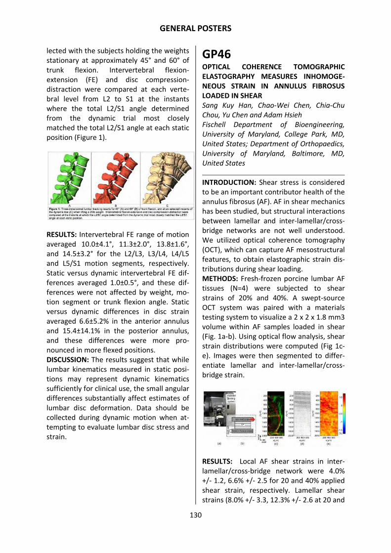
GENERAL POSTERS
130
lected with the subjects holding the weights
stationary at approximately 45° and 60° of
trunk flexion. Intervertebral flexion-
extension (FE) and disc compression-
distraction were compared at each verte-
bral level from L2 to S1 at the instants
where the total L2/S1 angle determined
from the dynamic trial most closely
matched the total L2/S1 angle at each static
position (Figure 1).
RESULTS: Intervertebral FE range of motion
averaged 10.0±4.1°, 11.3±2.0°, 13.8±1.6°,
and 14.5±3.2° for the L2/L3, L3/L4, L4/L5
and L5/S1 motion segments, respectively.
Static versus dynamic intervertebral FE dif-
ferences averaged 1.0±0.5°, and these dif-
ferences were not affected by weight, mo-
tion segment or trunk flexion angle. Static
versus dynamic differences in disc strain
averaged 6.6±5.2% in the anterior annulus
and 15.4±14.1% in the posterior annulus,
and these differences were more pro-
nounced in more flexed positions.
DISCUSSION: The results suggest that while
lumbar kinematics measured in static posi-
tions may represent dynamic kinematics
sufficiently for clinical use, the small angular
differences substantially affect estimates of
lumbar disc deformation. Data should be
collected during dynamic motion when at-
tempting to evaluate lumbar disc stress and
strain.
GP46 OPTICAL COHERENCE TOMOGRAPHIC ELASTOGRAPHY MEASURES INHOMOGE-NEOUS STRAIN IN ANNULUS FIBROSUS LOADED IN SHEAR Sang Kuy Han, Chao-Wei Chen, Chia-Chu
Chou, Yu Chen and Adam Hsieh
Fischell Department of Bioengineering,
University of Maryland, College Park, MD,
United States; Department of Orthopaedics,
University of Maryland, Baltimore, MD,
United States
INTRODUCTION: Shear stress is considered
to be an important contributor health of the
annulus fibrosus (AF). AF in shear mechanics
has been studied, but structural interactions
between lamellar and inter-lamellar/cross-
bridge networks are not well understood.
We utilized optical coherence tomography
(OCT), which can capture AF mesostructural
features, to obtain elastographic strain dis-
tributions during shear loading.
METHODS: Fresh-frozen porcine lumbar AF
tissues (N=4) were subjected to shear
strains of 20% and 40%. A swept-source
OCT system was paired with a materials
testing system to visualize a 2 x 2 x 1.8 mm3
volume within AF samples loaded in shear
(Fig. 1a-b). Using optical flow analysis, shear
strain distributions were computed (Fig 1c-
e). Images were then segmented to differ-
entiate lamellar and inter-lamellar/cross-
bridge strain.
RESULTS: Local AF shear strains in inter-
lamellar/cross-bridge network were 4.0%
+/- 1.2, 6.6% +/- 2.5 for 20 and 40% applied
shear strain, respectively. Lamellar shear
strains (8.0% +/- 3.3, 12.3% +/- 2.6 at 20 and

GENERAL POSTERS
131
40% respectively) were significantly higher
than those measured in the inter-
lamellar/cross-bridge structures.
DISCUSSION: To our knowledge, this is the
first study to quantify strain distributions
between the lamellar and inter-
lamellar/cross-bridge compartments of the
AF. This was achievable due to the
mesoscale imaging capabilities of OCT.
Based on our results, it can be concluded
that the two compartments behave distinct-
ly during AF shear. Their differential me-
chanics might play important roles in tissue
damage and cell regulation. Figure 1. (a)
Custom-designed shear testing system with
OCT scanner. (b) Schematic illustration of
tissue loading. (c) OCT images of mesoscale
structures before (red) and after (green)
applying shear load. L=lamellar.
IL=Interlamellar. CB=Cross-bridge. (d) Dis-
placement vectors (arrows) obtained from
optical flow analyses. (e) Strain map for the
corresponding area shown in (d).
GP47 DIRECTED VERTEBROPLASTIC CEMENT IN-JECTION AT LYTIC METASTATIC LESION RE-STORES STRENGTH WITH MINIMUM CE-MENT VOLUME +1Eeric Truumees, +2Ata M. Kiapour,
+2Vijay K. Goel, +2Constantine K.
Demetropoulos
+1Seton Spine and Scoliosis Center, Austin,
TX; +2Engineering Center for Orthopaedic
Research Excellence (ECORE), Departments
of Orthopaedics and Bioengineering, Univer-
sity of Toledo, Toledo, OH
INTRODUCTION: Metastatic disease in the
thoracolumbar spine represents a large and
growing problem, which can lead to pain,
pathologic fracture and neurologic com-
promise. Over the last two decades, the
utilization of PMMA for percutaneous ver-
tebral stabilization has increased. This study
was conducted to examine and compare the
biomechanical effectiveness of the standard
(anterior PMMA augmentation from cranial
to caudal endplate) and directed
vertebroplasty (VP).
METHODS: 32 specimens were harvested
from 6 cadavers (T5-S1, age 74±14, BMD
0.7±0.2). Each consisted of one full vertebra
and 2 adjacent hemi-vertebrae (physiologic
load transmission/sharing). Lytic lesions
with peripedicular cortical disruption were
created and filled with adipose tissue to
simulate tumor bulk. Specimens were ran-
domly distributed between 3 groups: lesion
alone (control), standard VP, and directed
peripedicular augmentation using a target-
ed system into the lytic lesion. Specimens
then underwent unconstrained compres-
sion using a bilateral cable system passing
through the approximate center of rotation
of the vertebral body.
RESULTS: Height reduction was significantly
higher in the anterior body (p=0.003). Mean
height loss was least for the group with di-
rected VP (Table 1). The use of directed VP
demonstrated the least increase in kypho-
sis. Average injected cement volume for the
directed VP was 49% less than the standard
VP (p<0.0005). The injected cement to ver-
tebral body volume ratio was lower for di-
rected than for standard VP. Data demon-
strated that cement augmentation signifi-
cantly increased normalized failure stress in
lumbar specimens (p=0.04).
DISCUSSION: Data suggest that an optimum
threshold cement injection volume may
exist, at which vertebral body strength is
improved with minimum cement volume.
Findings suggest that fixation by directed VP
can achieve similar augmentation to stand-
ard VP with an anterior fill, while requiring
roughly half the PMMA cement injection
volume.

GENERAL POSTERS
132
GP48
GENOME-WIDE PAIN-, NERVE- AND NEUROTROPHIN-RELATED GENE EXPRES-SION IN THE DEGENERATING HUMAN AN-NULUS Helen E. Gruber, Gretchen L. Hoelscher, Jane
A. Ingram, Edward N. Hanley
Department of Orthopaedic Surgery, Caroli-
nas Medical Center, Charlotte, NC USA
INRODUCTION: In spite of its high clinical
relevance, the relationship between disc
degeneration and low back pain is still poor-
ly understood. Genome-wide gene expres-
sion studies utilizing ontology searches pro-
vide an efficient methodology for identifica-
tion of clinically relevant genes.
METHODS: Ontology searches were used in
a genome-wide analysis of pain-, nerve-,
and neurotrophin-related gene expression
patterns in specimens of human disc tissue.
Control, non-herniated clinical and herniat-
ed clinical specimens of human annulus
tissue was studied following IRB approval.
Analyses were performed on more generat-
ed (Thompson grade IV and V) discs vs. less
degenerated discs (grades I-III), on surgically
operated discs vs. control discs, and on her-
niated vs. control discs; immunohistochem-
istry was also performed.
RESULTS: Analyses of more degenerated vs.
less degenerated discs identified significant
upregulation of well-recognized pain-
related genes (bradykinin receptor B1, calci-
tonin gene-related peptide and catechol-0-
methyltransferase). Nerve growth factor
was significantly upregulated in surgical vs.
control and in herniated vs. control discs.
All three analyses also found significant
changes in numerous proinflammatory cy-
tokine- and chemokine-related genes.
Nerve, neurotrophin and pain-ontology
searches identified many signal-
ing/functional genes with known disc im-
portance. Immunohistochemistry con-
firmed the presence of calcitonin gene-
related peptide, catechol-0-methyltrans-
ferase and bradykinin receptor B1 at the
protein level.
DISCUSSION: Since the degenerating hu-
man disc is primarily an avascular tissue site
into which disc cells have contributed high
levels of proinflammatory cytokines, these
substances are not cleared from the tissue
and remain there over time. We hypothe-
size that as nerves grow into the human
annulus, they encounter a proinflammatory
cytokine-rich milieu which may sensitize
nociceptors and exacerbate pain produc-
tion.
GP49 MATRIX METALLOPROTEINASE-12, MAC-ROPHAGE METALLOELASTASE, IS CONSTI-TUTIVELY EXPRESSED AND PRODUCED IN THE HUMAN INTERVERTEBRAL DISC IN VI-VO Helen E. Gruber, Gretchen L. Hoelscher, Jane
A. Ingram, Natalia Zinchenko , Synthia
Bethea and Edward N. Hanley, Jr.
Department of Orthopaedic Surgery, Caroli-
nas Medical Center, Charlotte, N.C. USA
INTRODUCTION: Matrix metalloproteinase-
12 (MMP-12), also known as macrophage
metalloelastase, degrades elastin and other
extracellular matrix (ECM) components in-
cluding type IV collagen, fibronectin,
laminin, gelatin, vitronectin, chondroitin
sulfates and fibrinogen. To date, no study
has assessed the presence or gene expres-
sion of MMP-12 in the intervertebral disc.
The objective of this study was to determine
if MMP-12 is present in the degenerating
human annulus utilizing immunohistochem-
istry and microarray analyses.
METHODS: IRB approval was obtained for
this study. Since MMP-12 also has known
relationships to cytokine and chemokines,
we examined human intervertebral disc
tissue expression of MMP-12 using microar-
ray analysis in discs from 23 subjects;
immunohistochemical localization studies
were performed in 18 specimens.

GENERAL POSTERS
133
RESULTS: Molecular studies showed that
MMP-12 exhibited a 1.45 fold significant
down regulation in more degenerated discs
compared to healthier discs (p = 0.006).
Immunohistochemistry showed localization
of MMP-12 in disc cells, but not disc extra-
cellular matrix (ECM).
DISCUSSION: MMP-12 is involved with
elastin degradation; the elastic extension
property of the intervertebral disc is an im-
portant biologic function. From cartilage
literature we know that MMP-12 can be
mediated by local cytokines via a number of
pathways. Here we have shown novel doc-
umentation of the expression and produc-
tion of MMP-12 by cells of the human disc.
Since MMP-12 has been implicated in the
breakdown of numerous ECM components,
including elastin, in other cells and tissues,
and since MMP-12 has additional roles in-
volving proinflammatory cytokines in other
tissues, we look forward to future research
which will better elucidate the role of MMP-
12 in disc health and disease.
GP50 FEASIBILITY OF NP CELL ACTIVITY UPREGULATION BY HTERT GENE TRANS-FECTION Di-Ke RUAN, MD; Hong-Kui XIN, MD; Chao
ZHANG, MD; Deli WANG; Zhiyuan SHI, MD;
Tao GU, MD;Chao-Feng WANG; Jianhong
WU,MD; Yan ZHANG, MD; Qin HE, MD
Department of Orthopedic Surgery The Navy
General Hospital,Beijing, P R. China
OBJECTIVE: To investigate the NP cells activ-
ity and phenotype preservation after
rAAV2-hTERT transfection.
METHODS: Nucleus pulposus cells were
transfected with rAAV2-hTERT at multiplici-
ties of infection of 1×10⁵ genomes per cell.
The expression of hTERT mRNA and protein
were assessed by RT-PCR and Western-blot
at 5,10,15,30 and 60 days post-transfection;
Real-time PCR and Elisa were used to test
proteoglycan and type Ⅱcollagen secreted
by nucleus pulposus cells, comparison anal-
ysis were performed between the test
group and the control.
RESULTS: NP cells could be effectively trans-
fected by rAAV2, positive hTERT mRNA and
protein expression could be detected at
5,10,15,30 and 60days post transfection. No
positive hTERT mRNA and protein expres-
sion was detected at the control group. The
hTERT gene expression reached a peak at
10days post transfection and dropped slow-
ly with increasing culturing time. RAAV2-
hTERT transfection effectively upregulated
the proteoglycan and type Ⅱcollagen ex-
pression which was significantly higher than
the control. As to the expression of type Ⅰ
collagen, however, no significant difference
was detected between the test and control.
CONCLUSION: RAAV2-hTERT could effec-
tively transfect NP cells and upregulate the
proliferative capacity and extracellular ma-
trix expression of NP cells cultured in vitro.
This hTERT activated NP cell has the poten-
tial to be used in the biological therapy for
degenerative disc disease.
GP51
INJECTION OF BUPIVACAINE INTO DISC SPACE TO DETECT PAINFUL NONUNION AFTER ALIF SURGERY IN PATIENTS WITH DISCOGENIC LOW BACK PAIN. Kimura S, Ohtori S, Inoue G, Orita S,
Yamauchi K, Eguchi Y, Aoki Y, Ishikawa T,
Miyagi M, Kamoda H, Suzuki M, Sakuma Y,
Kubota G, Oikawa Y, Inage K, Sainoh T, Sato
J, Toyone T, Takahashi K.
Dept. Orthop. Surg., Chiba University, Chiba,
Japan
INTRODUCTION: Bupivacaine is commonly
used for the treatment of back pain and the
diagnosis of its origin. Nonunion is some-
times observed after spinal fusion surgery;
however, whether the nonunion causes
pain is controversial. In the current study,
we aimed to detect painful nonunion by
injecting bupivacaine into the disc space of

GENERAL POSTERS
134
patients with nonunion after ALIF surgery
for discogenic low back pain.
METHODS: From 52 patients with low back
pain, we selected 42 who showed disc de-
generation at only one level (L4–L5 or L5–
S1) on magnetic resonance imaging and
were diagnosed by pain provocation on dis-
cography and pain relief by discoblock (the
injection of bupivacaine). They underwent
ALIF surgery. If the patients showed low
back pain and nonunion 2 years after sur-
gery, we injected bupivacaine into the non-
union disc space. Patients showing pain re-
lief after injection of bupivacaine under-
went additional posterior fixation using
pedicle screws. These patients were fol-
lowed up 2 years after the revision surgery.
RESULTS: Of the 42 patient subjects, 7
showed nonunion. Four of these did not
show low back pain; however, 3 showed
moderate or severe low back pain. These 3
patients showed pain reduction after injec-
tion of bupivacaine into their nonunion disc
space and underwent additional posterior
fixation. They showed bony union and pain
relief 2 years after the revision surgery.
DISCUSSION: Injection of bupivacaine into
the nonunion disc space after ALIF surgery
for discogenic low back pain is useful for
diagnosis of the origin of pain.
GP52 MOLECULAR INTERACTIONS BETWEEN HUMAN CARTILAGINOUS ENDPLATES AND NUCLEUS PULPOSUS CELLS: A PRELIMI-NARY INVESTIGATION Cornelia Neidlinger-Wilke1, Antje Mietsch1,
Fabio Galbusera1, Claus Carstens2,
Christoph Brochhausen3, Aron Lazary4,
Marco Brayda-Bruno5, Anita Ignatius1,
Hans-Joachim Wilke1
1Institute of Orthopaedic Research and
Biomechanics, Centre of Musculoskeletal
Research, University of Ulm, 89081 Ulm,
2Galenus-Klinik Stuttgart, 3REPAIR-lab,
Institute of Pathology, University of Mainz,
Germany, 4National Center for Spinal
Disorders, Budapest, Hungary, 5Istituto
Ortopedico Galeazzi, Milano, Italy
INTRODUCTION: Impaired endplate perme-
ability due to degeneration and calcification
is considered to be a key contributor to in-
tervertebral disc (IVD) degeneration. An up-
regulation of metalloproteinases and in-
flammatory cytokines has been observed in
degenerated IVDs. We hypothesize that the
cartilaginous endplate (CEP) contributes to
the regulation of disc matrix degradation via
molecular interactions with the disc tissue.
METHODS: To identify these possible mo-
lecular interactions we analyzed in a bioas-
say conditioned media of cartilaginous end-
plates (CEP-CM) from disc degeneration
patients (n=4) with regard to their influence
on matrix turnover and inflammatory fac-
tors on nucleus pulposus cells (in alginate
beads) of the same patient. The CEP-CM
was further analyzed by protein array for
inflammatory cytokines. Further CEP sam-
ples underwent histological (n=15), ultra
structural (n=8) and cell biological analysis
(n=5) to determine alterations of tissue
structure and differentiation potential of
CEP cells.
RESULTS: NP-cells exposed to their donor-
corresponding CEP-CM exhibited dose-
dependent up-regulation of inflammation-
factors (IL 1β, IL-6, IL-8 – up to 400-fold),
matrix metalloproteinases (MMP3, MMP13
– up to 1000-fold) and a slight decrease in
matrix protein expression. Pro-
inflammatory cytokines were identified in
the CEP-CM. The occurrence of apoptotic
cells and degraded matrix fragments as well
as the differentiation capacity of CEP-
derived cells varied strongly between do-
nors.
DISCUSSION: Our results indicate interac-
tions between the CEP and the NP tissue via
molecular factors that may up-regulate ma-
trix degrading enzymes and inflammatory
cytokines and thereby influence the patho-
physiology of disc degeneration. Ongoing

GENERAL POSTERS
135
investigations are necessary to further iden-
tify the regulative role of potential molecu-
lar factors that are responsible for these
degenerative alterations.
ACKNOWLEDGEMENT: Funded in part by
FP7-EU-Project Genodisc (HEALTH-F2-2008-
201626)
GP53 SENESCENT DISC CELLS EXHIBIT SEVERELY PERTURBED MATRIX HOMEOSTASIS Ngo, Kevin; Nasto, Luigi; Sowa, Gwendolyn;
Kang, James; Vo, Nam
Department of Orthopaedic Surgery,
University of Pittsburgh
INTRODUCTION: Age-related disc matrix
proteoglycan (PG) loss is primarily a cell-
driven process due to a combination of re-
duced PG synthesis and increased PG
proteolytic breakdown. However, the disc
cell population(s) with perturbed PG home-
ostasis has not been identified. The link be-
tween aging and senescent cells was noted
in human discs, but it is still not known if
senescent disc cells differ in matrix homeo-
stasis from their non-senescent counter-
parts. Hence the goal of this study is to de-
termine PG homeostatic characteristics of
senescent disc cells.
METHODS: Culture of human nucleus
pulposus (hNP) cells isolated from surgical
specimens were induced to enter senes-
cence by treating with 0.5mM H2O2 for 2
hours followed by culturing the cells for 4
days in fresh media (F-12, 10% FBS, 1% PS);
the same procedure was repeated twice to
induce complete senescence. Cellular se-
nescence (SA β-gal), PG synthesis (35S-
sulfate incorporation), mRNA expression
(qRT-PCR) and protein levels (ELISA) of se-
lected catabolic factors in the culture condi-
tioned media were determined.
RESULTS: Most (>90%) of the hNP cells
were stained positive for SA β-gal following
H2O2 treatment, confirming induction of
senescence. H2O2–induced senescent hNP
cells incorporated 6.3 + 0.6 pmoles 35S-
sulfate/g DNA, about a three-fold lower
than that seen in untreated non-senescent
hNP cells. Compared to non-senescent hNP
cells, senescent hNP cells greatly up-
regulated mRNA expression of MMP-1
(27X), MMP-3 (12X), IL-6 (6X) and IL-8 (9X),
the protein products of which were also
found in dramatically elevated levels in cul-
ture conditioned media.
DISCUSSION: Strong oxidant-induced disc
senescent cells exhibited greatly perturbed
matrix homeostasis, showing decreased
capacity for new PG synthesis as well as
enhanced expression and secretion of key
inflammatory cytokines and matrix
metalloproteinases, the latter may have
profound catabolic effects on neighboring
cells and the extracellular matrix.
GP54 PROLYL HYDROXYLASE 3 (PHD3) MODULATES CATABOLIC EFFECTS OF TUMOR NECROSIS FACTOR ALPHA ON CELLS OF THE NUCLEUS PULPOSUS THROUGH CO-ACTIVATION OF NUCLEAR FACTOR KAPPA B/P65 SIGNALING Fujita, Nobuyuki (1,2); Gogate, Shilpa (1);
Chiba, Kazuhiro (3); Matsumoto, Morio (4);
Toyama, Yoshiaki (4), Shapiro, Irving (1);
Risbud, Makarand (1)
1 Department of Orthopaedic Surgery and
Graduate Program in Cell and
Developmental Biology, Thomas Jefferson
University, Philadelphia, USA 2 Department
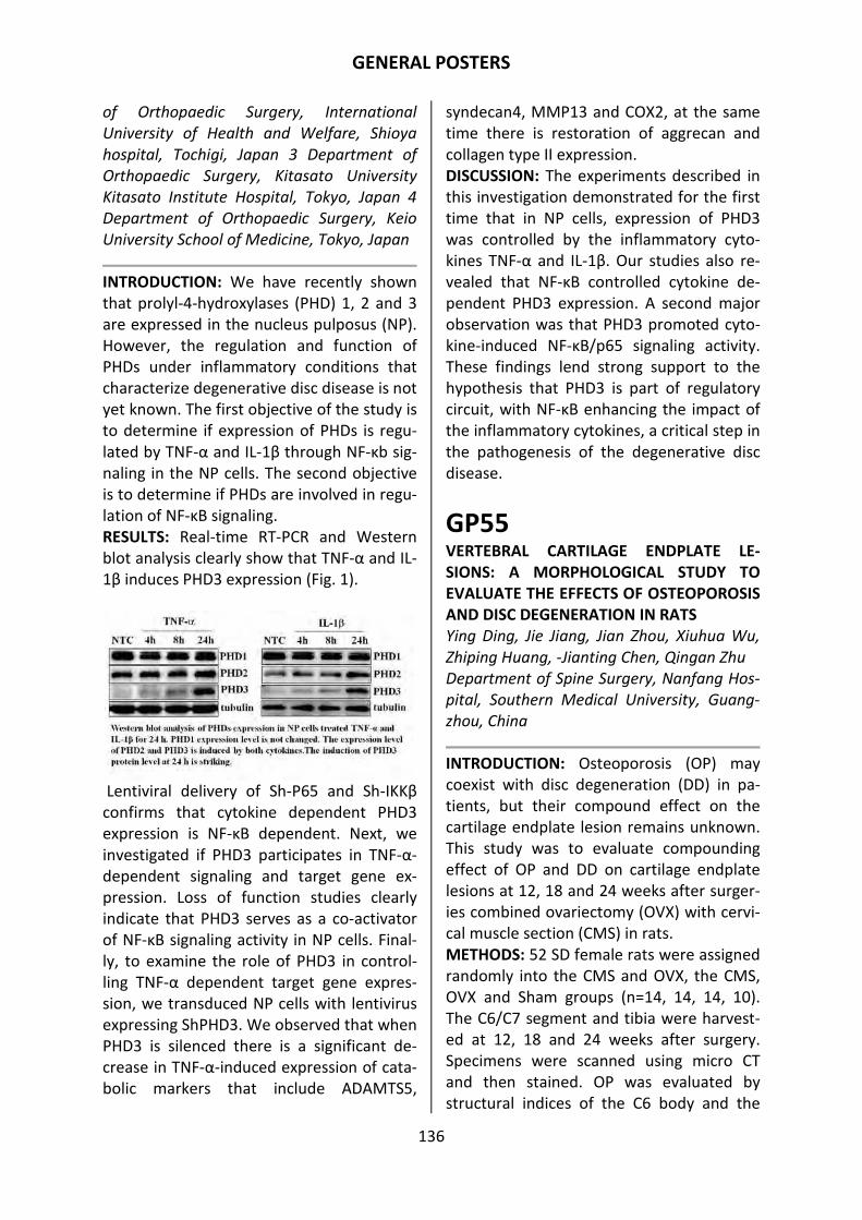
GENERAL POSTERS
136
of Orthopaedic Surgery, International
University of Health and Welfare, Shioya
hospital, Tochigi, Japan 3 Department of
Orthopaedic Surgery, Kitasato University
Kitasato Institute Hospital, Tokyo, Japan 4
Department of Orthopaedic Surgery, Keio
University School of Medicine, Tokyo, Japan
INTRODUCTION: We have recently shown
that prolyl-4-hydroxylases (PHD) 1, 2 and 3
are expressed in the nucleus pulposus (NP).
However, the regulation and function of
PHDs under inflammatory conditions that
characterize degenerative disc disease is not
yet known. The first objective of the study is
to determine if expression of PHDs is regu-
lated by TNF-α and IL-1β through NF-κb sig-
naling in the NP cells. The second objective
is to determine if PHDs are involved in regu-
lation of NF-κB signaling. RESULTS: Real-time RT-PCR and Western
blot analysis clearly show that TNF-α and IL-
1β induces PHD3 expression (Fig. 1).
Lentiviral delivery of Sh-P65 and Sh-IKKβ confirms that cytokine dependent PHD3
expression is NF-κB dependent. Next, we investigated if PHD3 participates in TNF-α-
dependent signaling and target gene ex-
pression. Loss of function studies clearly
indicate that PHD3 serves as a co-activator
of NF-κB signaling activity in NP cells. Final-
ly, to examine the role of PHD3 in control-
ling TNF-α dependent target gene expres-
sion, we transduced NP cells with lentivirus
expressing ShPHD3. We observed that when
PHD3 is silenced there is a significant de-
crease in TNF-α-induced expression of cata-
bolic markers that include ADAMTS5,
syndecan4, MMP13 and COX2, at the same
time there is restoration of aggrecan and
collagen type II expression.
DISCUSSION: The experiments described in
this investigation demonstrated for the first
time that in NP cells, expression of PHD3
was controlled by the inflammatory cyto-
kines TNF-α and IL-1β. Our studies also re-
vealed that NF-κB controlled cytokine de-
pendent PHD3 expression. A second major
observation was that PHD3 promoted cyto-
kine-induced NF-κB/p65 signaling activity. These findings lend strong support to the
hypothesis that PHD3 is part of regulatory
circuit, with NF-κB enhancing the impact of the inflammatory cytokines, a critical step in
the pathogenesis of the degenerative disc
disease.
GP55 VERTEBRAL CARTILAGE ENDPLATE LE-SIONS: A MORPHOLOGICAL STUDY TO EVALUATE THE EFFECTS OF OSTEOPOROSIS AND DISC DEGENERATION IN RATS Ying Ding, Jie Jiang, Jian Zhou, Xiuhua Wu,
Zhiping Huang, -Jianting Chen, Qingan Zhu
Department of Spine Surgery, Nanfang Hos-
pital, Southern Medical University, Guang-
zhou, China
INTRODUCTION: Osteoporosis (OP) may
coexist with disc degeneration (DD) in pa-
tients, but their compound effect on the
cartilage endplate lesion remains unknown.
This study was to evaluate compounding
effect of OP and DD on cartilage endplate
lesions at 12, 18 and 24 weeks after surger-
ies combined ovariectomy (OVX) with cervi-
cal muscle section (CMS) in rats.
METHODS: 52 SD female rats were assigned
randomly into the CMS and OVX, the CMS,
OVX and Sham groups (n=14, 14, 14, 10).
The C6/C7 segment and tibia were harvest-
ed at 12, 18 and 24 weeks after surgery.
Specimens were scanned using micro CT
and then stained. OP was evaluated by
structural indices of the C6 body and the

GENERAL POSTERS
137
tibial metaphyseal region. After the lesion
was identified on axial CT images, ratio of
the lesion area to the whole endplate area
was calculated. Intervertebral disc height
(IDH) was measured, and DD was graded for
each sample.
RESULTS: OP occurred at 12 weeks after
OVX as structural indices was dropped sig-
nificantly, and became severe afterwards.
Maximum lesion was on the ventral carti-
lage endplates at 12 weeks after CMS sur-
gery, and was severe in the compound
group (28%, P<0.05) and the caudal end-
plate (P>0.05). The lesion was shrunk and
calcified in situ at 18 and 24 week. The CMS
surgery resulted in a significant decrease of
IDH, while the OVX added surgery led to IDH
decreasing significantly at 24 weeks
(P=0.043). DD scores were higher following
CMS or OVX surgery. DD score correlated to
the lesion at 12, 18 and 24 weeks (P<0.01),
and IDH at 18 and 24 weeks (P<0.01) re-
spectively.
DISCUSSION: Our results suggested that
disc degeneration induced cartilage end-
plate lesion while osteoporosis appeared to
deteriorate the lesion more to the degener-
ating discs. The lesion may be related to
cartilage endplate wearing at the early
stage and calcification in situ later. The le-
sion and IDH were associated with the DD
levels. The results imply that osteoporosis
may accelerate the disc degeneration at
specific time.
GP56 EXPRESSION OF THE TISSUE RENIN-ANGIOTENSIN SYSTEM IN RAT AND BOVINE INTERVERTEBRAL DISCS Koji Akeda, Ryo Morimoto, Ryu Iida, Koichiro
Murata, Akihiro Sudo
Department of Orthopaedic Surgery, Mie
University Graduate School of Medicine
INTRODUCTION: The renin-angiotensin sys-
tem (RAS) plays a vital role in regulating the
physiology of the cardiovascular system.
Recent studies have shown that locally pro-
duced angiotensin II (Ang II), the “tissue
renin-angiotensin system (tRAS), contribut-
ed to inflammation and fibrosis in many
organs. We have previously reported that
tRAS was expressed by rat intervertebral
disc (IVD) cells. In this study, we compared
(1) the expression of tRAS by bovine (a
chondrodystrophoid animal) and rat IVD
cells, and (2) examined the biological role of
tRAS by rat IVD cells using Ang II peptide.
METHODS: tRAS expression by rat and bo-
vine IVD cells was examined using real-time
polymerase chain reaction (PCR) (rat) and
immunohistochemistry (rat and bovine). Rat
annulus fibrosus (AF) cells in monolayer
culture were used to examine the biological
role of tRAS in vitro. The effect of Ang II
peptide on extracellular matrix metabolism
was assessed by real-time PCR.
RESULTS: mRNA of tRAS components, in-
cluding angiotensin converting enzyme (Ang
II), Ang II receptor type 1, Ang II receptor
type 2, and Cathepsin D (a renin-like en-
zyme), were clearly confirmed by real-time
PCR analysis. In AF and nucleus pulposus
(NP) cells from rat and bovine in monolayer
culture, immunohistochemical analysis
showed that the expression of each tRAS
component was clearly observed. In rat IVD
tissues, immunoreactivity to each antibody
for tRAS components was also observed.
Proliferation of rat AF cells was mildly
stimulated by Ang II peptide. Ang II peptide
also had a minor stimulatory effect on the

GENERAL POSTERS
138
expression of extracellular matrix compo-
nents, growth factors and catabolic pro-
teins.
DISCUSSION: Our results demonstrate that
the tRAS components necessary to activate
tRAS have been found in normal rat and
bovine IVD cells. To elucidate the associa-
tion between tRAS and the process of IVD
degeneration, the expression and function
of tRAS in the human degenerated IVD
should be examined in a future study.
GP57 SIGNATURE MIRNA EXPRESSION PROFILE OF SPINAL FUSION USING EQUINE BONE PROTEIN EXTRACT, RHBMP-2 AND AUTOGRAFT DURING THE PROCESS OF ANTERIOR LUMBAR INTERBODY FUSION Zhou Zhiyu, Dafu Chen, Dai Xuejun,
Manman Gao, Zou Xuenong
Department of Spine Surgery/Orthopedic
Research Institute The First Affiliated
hospital of Sun Yat-sen University
INTRODUCTION: Using microarray technol-
ogy and data analysis, we examined the
gene-expression profiles after implantation of equine boneprotein extract, rhBMP-2 and
autograft and found the different molecular
mechanisms underlying the diverse patterns
of histological ossification. However, the mechanisms of the epigenetic adjustment
have not yet been delineated in detail.
METHODS: Eighteen pigs underwent a 3-
level ALIF procedure at L3-4, L4-5, and L5-6
in the present study using equine bone pro-
tein extract, rhBMP-2 and autograft, respec-
tively. The fused vertebral bodies of 6 ani-
mals at each time-point were observed for
2, 4, or 8 weeks post-operatively. The pool-
ing RNA from each tissue sample analyzed
by CapitalBio Corporation for miRNA micro-
array and Porcine Genome Array
(Affymetrix) experiments. Hierarchical clus-
tering with average linkage was carried out
by Cluster 3.0. The differentially expressed
genes and Micro-RNAs were performed
conjoint analysis by Gene Ontology (GO)
terms and Kyoto Encyclopedia of Genes and
Genomes (KEGG) pathways based on the
human accession id, using MAS platform.
RESULTS: Forty eight Micro-RNAs were
highlighted in comparing among equine
boneprotein extract, rhBMP-2 and autograft
at different time points, which regulated
over 100 genes in bone regeneration. Ac-
companying with the down-regulations of
miR-129, 140, 133, etc, the genes related to
inflammatory were up-regulation, ALIF us-
ing equine bone protein extract in particu-
lar. The genes related to bone formation
were increased following the decreased of
miR-19, 29, 218, etc.
DISCUSSION: The rhBMP-2 recruits progeni-
tor cells, proliferationand differentiation
and then leads to preferable membranous
ossification and bone remodeling. Con-
versely, equine bone protein extract results
in endochondral ossification via
upregulation of cartilage-related genes.
MicroRNAs participate the bone regenera-
tion after the transplantation of equine
bone protein extract, rhBMP-2 and
autograft.
GP58
CANINE NOTOCHORDAL CELL-SECRETED FACTORS PROTECT MURINE AND HUMAN NUCLEUS PULPOSUS CELLS FROM APOPTOSIS BY INHIBITION OF ACTIVATED CASPASES -9, AND -3/7 A. Mehrkens 1, 2, R. Hilario 1, S. Kim 1, M. Z.
Karim 1, M. G. Fehlings 1, M. W. Erwin 1
1 Toronto Western Research Institute,
University of Toronto, Toronto, Canada 2
Department of Orthopaedic Surgery - Spine
Unit, Basel University Hospital, Basel,
Switzerland
INTRODUCTION: Effective therapies that
may stop or reverse disc degeneration re-
main elusive. A minimally invasive method
through which nucleus pulposus cell viabil-
ity could be achieved would revolutionize

GENERAL POSTERS
139
the treatment of degenerative disc disease.
We have investigated if non
chondrodystrophic (NCD) canine disc (IVD)-
derived notochordal cell conditioned medi-
um (NCCM) and chondrodystrophic (CD)
canine IVD-derived conditioned medium
(CDCM) are able to protect murine and hu-
man NP cells from apoptosis.
METHODS: We developed NCCM and CDCM
from hypoxic culture of freshly isolated NPs
from NCD and CD canines respectively. We
obtained murine NP cells from 9 different
C57BL/6 mice and human NP cells from 4
patients who underwent surgery for discec-
tomy. Cells were cultured with aDMEM/F-
12 (control media), NCCM or CDCM under
hypoxic conditions (3.5% O2) and treated
with IL-1ß+FasL or Etoposide. All media
were supplemented with 2% fetal bovine
serum. We then determined the expression
of specific apoptotic pathways in the murine
and human NP cells by recording caspase-8,
and -9 and -3/7 activity.
RESULTS: In murine NP cells, NCCM inhibits
IL-1ß+FasL- and Etoposide- mediated apop-
tosis via suppression of activated caspase-9
and caspase-3/7, CDCM demonstrated an
inhibitory effect on IL-1ß+FasL mediated
apoptosis via caspase-3/7 (Fig1A).
In human NP cells, NCCM inhibits
Etoposide- mediated apoptosis via suppres-
sion of activated caspase-8, caspase-9 and
mainly caspase-3/7. CDCM demonstrated
an inhibitory effect on Etoposide- mediated
apoptosis via suppression of activated
caspase-8, caspase-9 and mainly caspase-
3/7, though not as effective as NCCM
(Fig1B).
DISCUSSION: Soluble factors secreted by
the NCD IVD NP strongly protect murine and
human NP cells from induced apoptosis via
suppression of activated Caspase -9 and -3/-
7. A better understanding and harnessing of
the restorative powers of the notochordal
cell could lead to novel cellular and molecu-
lar strategies for treatment of DDD
GP59 ABILITY OF T2 MAPPING AND T1 RHO MAPPING TO EVALUATE LUMBAR INTERVERTEBRAL DISC DEGENERATION AND GLYCOSAMINOGLYCAN CONTENT OF THE NUCLEUS PULPOSUS Oikawa, Yasuhiro1; Watanabe, Atsuya2;
Kamoda, Hiroto1; Ishikawa, Tetsuhiro1;
Miyagi, Masayuki1; Sakuma Yoshihiro1;
Inoue, Gen1; Yamauchi, Kazuyo1; Orita
Sumihisa1; Suzuki, Miyako1; Kubota, Go1;
Inage, Kazuhide1; Sainoh, Takeshi1; Sato,
Jun1; Ohzone, Humio3; Obata, Takayuki4;
Okubo, Toshiyuki3; Toyone, Tomoaki3;
Ohtori, Seiji1; Takahashi, Kazuhisa1
1: Department of Orthopaedic Surgery,
Graduate School of Medicine, Chiba Univer-
sity. 2: Department of Orthopaedic Surgery,
Teikyou University Chiba Medical Center,
Japan. 3: Department of Orthopaedic Radi-
ology, Teikyou University Chiba Medical
Center,Japna. 4: National Institute of Radio-
logical Sciences, Japan.
GENERAL POSTERS
140
INTRODUCTION: T2 mapping and T1ρ map-
ping by MRI are non-invasive quantification
tools to measure intervertebral disc (IVD)
degeneration. MRI T2 relaxation time is a
quantitative parameter that is sensitive to
change in collagen and water content, and
T1ρ relaxation time reflects water and sul-
fated glycosaminoglycan (GAG) content in
the nucleus pulposus of IVDs. The purpose
of this study was: (1) to evaluate degenera-
tive IVDs quantitatively with T2 mapping
and T1ρ mapping, (2) to analyze water and GAG content histologically, and (3) to in-
spect the efficacy of a quantitative imaging
of the degenerated IVDs.
METHODS: Eight-week-old New Zealand
White rabbits were used in this study. IVDs
were punctured 10, 5, 1 or 0 (control) times
with an 18-gauge needle at each level. MRI
was performed 4 weeks and 8 weeks after
injury and water and GAG content of the
nucleus pulposus were calculated.
RESULTS: GAG content in the nucleus de-
creased in all the puncture groups com-
pared with the control group. Conversely,
there was no strong correlation of GAG con-
tent and the number of punctures or dura-
tion after injury. Water content was lower
in the puncture groups than in the control
group, and lower in the 8 weeks group than
the 4 week group. T1ρ mapping showed stronger correlation of GAG and water con-
tent than T2 mapping. GAG and water con-
tent decrease with the progression of IVD
degeneration in the puncture groups.
DISCUSSION AND CONCLUSIONS: Previous
studies reported that the degeneration of
IVDs correlates with the number of punc-
tures. This study shows that GAG and water
content tended to decrease in early stage of
IVD degeneration and that T1ρ mapping evaluated the degenerating IVDs more ac-
curately than T2 mapping. GAG and water
content were lower in IVDs punctured once
than in the control. Therefore, T2 relaxation
and T1ρ times can detect early stages of IVD degeneration. Quantitative MRI has the
potential to detect degeneration and evalu-
ate repair and regeneration.
GP60 THE MECHANISM OF THE INFLAMMATORY RESPONSES IN THE INTERVERTEBRAL DISC HERNIATION EXPLORED BY CO-CULTURE MODEL IN VITRO Yamamoto Junya1, Maeno Koichiro1, Taka-
da Toru1, Kakutani Kenichiro1, Yurube Ta-
kashi1, Zhang Zhongying1, Hirata Hiroaki1,
Kurakawa Takuto1, Shingo Miyazaki1, Sakai
Daisuke2, Mochida Joji2, Doita Minoru3,
Kurosaka Masahiro1, Nishida Kotaro1
1. Dept of Orthopaedics, Kobe University,
Kobe, Japan. 2. Dept of Orthopaedics, Tokai
University, Kanagawa, Japan. 3. Dept of
Orthopaedics, Takatsuki General Hospital,
Takatsuki, Japan.
INTRODUCTION: We have clarified that Fas
ligand (FasL) is expressed on human NP cells
and that the interaction between NP cells
and macrophages causes the inflammatory
responses which are considered to cause
pains in the intervertebral disc herniation. A
Disintegrin And Metalloproteinase
(ADAM)10 is reported to regulate FasL cell
surface expression. Therefore, we assumed
that FasL and ADAM10 participate the in-
flammatory responses in the intervertebral
disc herniation. To validate this assumption,
We explored the function of FasL and
ADAM10 using co-culture model in vitro.
METHODS: FasL over-expression and
ADAM10 knock-down were induced by
transferring FasL plasmid and ADAM10
shRNA plasmid respectively into human NP
cell line using the electroporation method.
Human NP cell line and macrophage (GM-
CSF) cell line were co-cultured on the differ-
ent surfaces of microporous membrane.
After 12- and 24-hour co-culture, each cell
was collected respectively. The mRNA-
quantification of pro-inflammatory cyto-
kines (IL-1β, TNF-α, and IL-6) and ADAM10

GENERAL POSTERS
141
were estimated with the real-time RT-PCR.
And the qualitative analysis of proteins of
those was detected by the Western Blot-
ting.
RESULTS: The production of all pro-
inflammatory cytokines and ADAM10 in-
creased significantly in the case of co-
culture, differing from the cases of inde-
pendent culture respectively. This tendency
was more remarkable in the case of co-
culture with FasL over-expression. On the
other hand, ADAM10 knock-down induced
to decrease both the expression of FasL and
the production of pro-inflammatory cyto-
kines.
DISCUSSION: It has been reported that the
reverse signaling in the TNF family contrib-
utes to the production of pro-inflammatory
cytokines. In our results, it seems reasona-
ble to conclude that FasL has great possibil-
ity to regulate the production of pro-
inflammatory cytokines and that ADAM10
regulates the FasL reverse signaling and
participates the inflammatory responses in
the intervertebral disc herniation.
GP61 CATABOLIC EFFECTS OF ENDOTHELIAL CELL-DERIVED MICROPARTICLES ON IN-TERVERTEBRAL DISC CELLS Pedro Henrique Isoldi Pohl, Hong Joo Moon,
Thomas Lozito, Thais Cuperman, Kevin Ngo,
Takashi Yurube, Gwendolyn Sowa, Rocky
Tuan, James D Kang, Nam Vo
Department of Orthopaedic Surgery,
University of Pittsburgh Medical Center, PA,
USA., The Ferguson Lab for Orthopaedic and
Spine Research, Center for Cellular and
Molecular Engineering
INTRODUCTION: Neo-vascularization, a
prominent feature of injured and degener-
ated discs, requires the invasion of endothe-
lial cells, the primary producer of
microparticles (MPs). MPs, also known as
microvesicles or exosomes, are 0.1 to 1 μm particles shed from cell plasma membrane
blebs in response to stimulus or stress. MPs
consist of membrane and cytoplasmic sub-
components along with a number of pro-
teins and extracellular RNA characteristics
of their parental cells. Emerging evidence
suggest that MPs are biologic information
messengers with important roles in both in
pathological and non-pathological process-
es. Because the interaction between endo-
thelial MPs and disc cells are likely to occur
during neovascularization, the objective of
this study is to evaluate the influence of the
endothelial MPs on disc cells metabolism.
METHODS: Endothelial microparticles
(EMPs) were isolated as described (J Cell
Physiol. 2012 227:534-49) from the culture
media of a microvascular endothelial cell
line grown on monolayer culture for two
weeks. 15 μl of 14mg/ml EMPs (total pro-
tein) were added to each well of the 6-well
plate of human annulus fibrous (hAF) cell
cultures, and the cultures were incubated
for 3 days. Gene expression of MMP-1,
MMP-2, MMP-3, MMP-13, and aggrecan
were determined by qRTPCR.
RESULTS: Increased gene expression of
MMP-1, MMP-2, MMP-3 and MMP-13 were
observed in EMP-treated hAF cells com-
pared to untreated hAF cells. In contrast,
aggrecan gene expression decreased in
EMP-treated hAF cells compared to un-
treated hAF cells.
DISCUSSION: Endothelial cell microparticles
promote catabolic metabolism in disc cells.
If confirmed in vivo, this could represent a
new mechanism by which disc extracellular
matrix is degraded during the degenerative
process.
GP62 THE NOTOCHORDAL CELL VIABILITY AF-FECTED BY DYNAMIC MECHANICAL LOAD-ING VIA ALPHA5/BETA1 INTEGRINS IN RAT IVDS Kurakawa T. 1, Kakutani K. 1, Morita Y. 2,,
Takada T. 1, Maeno K. 1, Yurube T. 1,
Yamamoto J. 1, Hirata H. 1, Miyazaki S. 1,

GENERAL POSTERS
142
Doita M. 3, Kurosaka M. 1, Nishida K. 1
1. Dept of Orthopaedic Surgery, Kobe
University Graduate School of Medicine,
Kobe, Japan., 2. Dept of Biomedical
Engineering, Doshisha University, Kyoto,
Japan., 3. Dept of Orthopaedic Surgery,
Takatsuki General Hospital, Osaka, Japan.
INTRODUCTION: It is essential to clarify the
notochordal cell (NC) viability to elucidate
intervertebral disc (IVD) degeneration, but it
remains unclear. The α5β1 integrin is a transmembrane protein that mediates bio-
logic interactions between cells and extra-
cellular matrix. The aim of this study was to
assess the NC viability affected by dynamic
mechanical loading via α5β1 integrins in rat IVDs.
METHODS: Consecutive two en-bloc coc-
cygeal IVDs were dissected from 24 male SD
rats. Discs were assigned to following condi-
tions and cultured for 6 days using an organ
culture system, Group T; with an inhibitory
peptide treatment against α5β1 integrin (GRGDSP), Group L; with mechanical loading
(1.3MPa, 1Hz at initial state), Group TL;
both with treatment and loading, Group C;
without any treatment or loading. Cell via-
bility analysis was examined using calcein
green staining. Histomorphology was as-
sessed by Masuda’s degeneration scale.
Immunopositivity of a NC marker (galectin-
3) and α5/β1 integrins were evaluated by the percentage of positive cells. mRNA ex-
pressions of aggrecan, collagen type-1/-2,
MMP-3/-13, and α5/β1 integrins were cal-
culated by real-time RT-PCR.
RESULTS: Cell viability of Group L (65.9%)
and Group TL (77.9%) was decreased than
Group C (91.6%) in the nucleus pulposus
(NP) (P<0.05). Degenerative changes were
mainly seen in the NP. Histological grade of
Group TL was lower than Group L.
Immunopositivity of galectin-3 was de-
creased in Group L (3.6%) and TL (30.1%)
compared to Group C (81.7%) (P<0.05). The
% positive cells of α5/β1 integrins were sig-
nificantly decreased with NC disappearance.
mRNA expressions of aggrecan and collagen
type 2 were up-regulated in Group TL com-
pared to Group L (P<0.05), whereas signifi-
cant up-regulation of MMPs and α5/β1 integrins in Group L was not seen in Group
TL.
DISCUSSION: Our results suggested that the
loss of NCs due to dynamic mechanical load-
ing was affected by α5β1 integrin expres-
sions in rat IVDs.
GP63 A QUANTITATIVE PROTEOMIC ANALYSIS OF THE NON-CHONDRODYSTROPHIC AND CHONDRODYSTROPHIC CANINE NUCLEUS PULPOSUS L. DeSouza 1, M. Z. Karim 2, O. Masui 1, S.
Maedler 1, A. Mehrkens 3, S. Kim 2, R.
Hilario 2, W. M. Erwin 4, 5,*
1Chemistry, York University, 2Genetics ,
3Orthoapedic surgery, 4Orthoapedic and
neurological surgery, 5Genetics and Devel-
opment, University Health Network, Toronto
Western Hospital, Toronto, Canada
INTRODUCTION: The morphology of the
intervertebral disc (IVD) nucleus pulposus
(NP) varies dramatically between the
notochordal cell-rich non-
chondrodystrophic (NCD) and the
notochordal cell poor chondrodystrophic
(CD) canine as does their respective suscep-
tibility to degenerative disc disease (DDD).
Here we used iTRAQ quantitative proteomic
analysis to determine the differential pro-
tein/peptide ‘fingerprint’ of the NCD and CD
canine NP.
METHODS: NPs obtained from the IVDs of 8
(4 NCD and 4 CD) dogs were mechanically
homogenized/centrifuged and identical ali-
quots of soluble proteins from the superna-
tant were then analyzed using 4-plex iTRAQ
analysis. Proteins/peptides separated on a
nano-LC column and the eluting peptides
were analyzed a minimum of two times
each on either a QStar Elite or the 5600 Tri-

GENERAL POSTERS
143
ple TOF instrument and the subsequent MS
data were then analyzed using ProteinPilot
software. Proteins demonstrating consistent
differential expression in either the NCD or
CD samples greater than two-fold relative to
the reference pool were selected for further
investigation.
RESULTS: Dramatic differences in the ex-
pression of a number of important extracel-
lular matrix-related proteins within the NPs
of these two canine sub-species were de-
tected. Significant differences in protein
expression were: Fibronectin, decorin,
biglycan, cartilage intermediate layer pro-
tein (CILP), cartilage oligomeric protein
(COMP), fibromodulin, biglycan, and iso-
form B of proteoglycan-4 (lubricin) all of
which were dramatically under expressed in
the NCD canine NP as compared to the CD
canine NP.
CONCLUSIONS: We have determined that
the small leucine rich proteoglycans (SLRPs)
or interstitial proteoglycans such as decorin,
biglycan as well as fibromodulin were far
more abundant in the CD NP homogenate
ascompared to the NCD canine. Degenera-
tive changes underway in the CD as com-
pared to the NCD NP animal may make this
an ideal biological entity with which to
study the biology of DDD.
GP64 LUMBAR INTERVERTEBRAL DISC INJURY IN RATS INDUCES UPREGULATION OF TRPV1 EXPRESSION IN THE SENSORY NERVE IN-NERVATING THE DISCS Yusuke Shibata1), Miyako Suzuki2), Yoshihiro
Sakuma2), Yasuhiro Oikawa2), Masayuki
Miyagi2), Tetsuhiro Ishikawa2), Kazuhide
Inage2) , Go Kubota2), Takeshi Sainoh2), Jun
Sato2), Sumihisa Orita2), Kazuyo Yamauchi2),
Hiroto Kamoda2), Seiji Ohtori2), Gen Inoue3),
Kazuhisa Takahashi2
1) School of Medicine, Chiba University 2)
Department of Orthopaedic surgery, Chiba
University 3)Department of Orthopaedic
surgery, Kitasato University
INTRODUCTION: Low back pain (LBP), which
is caused by the sensory nerve innervating a
degenerated intervertebral disc(IVD), can be
induced by several inflammatory peptides.
However, there is little clinical information
on the effects of some inflammatory
peptide inhibitors on LBP. Recent studies
have shown that the capsaicin receptor
TRPV1 responds to acidity, pH fluctuation,
and temperature and is related to
inflammation and pain. We aimed to
determine the therapeutic significance of
TRPV1 in cases of LBP by
immunohistochemical evaluation of its
levels in the sensory nerve innervating
injured rat IVDs.
METHODS: 8w-old female rats were used in
this study. In the disc-injury group, L5/6
discs were injured 10 times with a 24G
needle; simultaneously, the neurotracer
Fluoro-gold(FG) was injected into the L5/6
IVD (injured group,N=5;sham group,N=5).
The L5/6 IVD and the dorsal root
ganglion(DRGs) from L1 to L6 were resected
at 7days after surgery. The expression levels
of TRPV1 and induction levels of the
calcitonin gene-related peptide(CGRP) in the
DRGs were evaluated using immunohisto-
chemistry. Then, the rate of TRPV1-
immunoreactive(IR) DRG cells among FG-
labeled DRG cells and the rate of CGRP-IR
DRG cells among FG-labeled DRG cells in
both groups were compared statistically.
RESULTS: The rate of TRPV1-IR cells was
significantly higher in the injured group
(42.5%) than in the sham group (29.5%)
(p<0.05). Similarly, the rate of CGRP-IR cells
was higher in the injured group (45.0%)
than in the sham group (20.1%) (p<0.05).
And, the rate of TRPV1-IR cells and CGRP-IR
cells were practically equal in both groups.
DISCUSSION: Disc injury in rats increased
the levels of TRPV1 and CGRP in DRGs. It
was reported degenerative IVDs condition
related to inflammatory mediators such as
acidity and pH fluctuation. These results

GENERAL POSTERS
144
suggest inflammatory mediators induce
TRPV1 expression in degenerative IVDs.
Thus, TRPV1 may serve as a new target for
the treatment of degenerative discogenic
LBP.
GP65
CERVICAL INTERVERTEBRAL DISC INJURY PRODUCES UP-REGULATION OF TRPV1 AND NEUROPEPTIDES IN SENSORY DISC-INNERVATING NERVES IN RATS Takahito Arai1), Miyako Suzuki2), Yoshihiro
Sakuma2), Yasuhiro Oikawa2), Tetsuhiro Ishi-
kawa2), Masayuki Miyagi2), Hiroto
Kamoda2), Kazuhide Inage2), Go Kubo-
ta2), Takeshi Sainoh2), Jun Sato2), Seiji
Ohtori2), Gen Inoue3),Kazuyo Yamau-
chi2), Sumihisa Orita2),Kazuhisa Takahashi2
1) School of Medicine, Chiba University
2)Dept of Orthopaedic surgery, Chiba
University 3) Dept of Orthopaedic surgery,
Kitasato University
INTRODUCTION: Cervical intervertebral
discs induce obscure neck and interscapular
pain. However, the detailed mechanism of
discogenic cervical pain is unknown. It was
recently reported that TRPV1-a capsaicin
receptor-that senses acid, pH fluctuation,
and temperature change in human
intervertebral disc(IVD), is related to
inflammatory pain. The goal of this study
was to evaluate by immunohisto-
chemistry(IHC), the up-regulation of TRPV1
calcitonin gene-related peptide(CGRP) as
the inflammatory neurotransmitter inner-
vated sensory nerve in rat cervical
intervertebral injured disc.
METHODS: We used 8w-old male SDrats. In
the disc-injured group, C5/6 discs were
injured 10 times with a 24G needle;
simultaneously, the neurotracer Fluoro-
gold(FG) was injected into the C5/6
IVD(disc-injured groupN=5,sham groupN=5).
Dorsal root ganglion(DRGs) from the C1 to
C8 levels were resected 7days after surgery.
Using IHC, we evaluated the DRGs with
TRPV1 and CGRP. Subsequently, the rate of
TRPV1-immunoreactive(IR) DRG cells among
the FG-labeled DRG cells and the rate of
CGRP-IR DRG cells among the FG-labeled
DRG cells were compared in each groups.
RESULTS: FG-labeled cells from C5/6IVD
were bilaterally distributed from C2 to C8
DRG. The rate of TRPV1-IR cells among the
FG-labeled cells was significantly higher in
the disc-injured group(55.3%) than in the
sham group(23.2%)(p<0.05). Similarly, the
rate of CGRP-IR cells among the FG-labeled
cells was significantly higher in the disc-
injured group (59.4%) than in the sham
group(31.0%)(p<0.05).
DISCUSSION: These results showed that the
innervated sensory nerves from C5/6IVD
were distributed from DRGs C2 to C8 and
might induce discogenic cervical pain over a
wide area. Furthermore, disc injury in rats
increased TRPV1 and neuropeptides in
DRGs. These results suggest that these
inflammatory mediators from degenerative
IVDs relate to the mechanism of
degenerated discogenic cervical pain, and
TRPV1 may play the role of a new target of
one’s therapy
GP66 LONGITUDINAL EVALUATION OF SCHMORL NODES, MODIC CHANGES AND RELATED LOW BACK PAIN Ryuhei Kawamura, Hideki Murakami,
Takafumi Horii, Akira Izumi, Yoshitaka
Konari, Toshio Wada
Dept. of Orthop. Surg, Towadahigashi Hos-
pital
INTRODUCTION: The association of MRI
findings such as Schmorl nodes (SNs) and
Modic changes (MCs) with low back pain is
somewhat controversial. Furthermore the
association between SNs and MCs is not
well known. Therefore we investigated the
association among SN, MCs and low back
pain.
MATERIALS AND METHODS: SNs and MCs

GENERAL POSTERS
145
on lumbar MRI were observed in total 135
patients between 2004-2005. Another MRI
was taken on those same patients again
more than 24 months later. We retrospec-
tively reviewed their medical records and
MRI to have low back pain and changes in
size of SNs and type of MCs at the initial
examination and final follow-up.
RESULTS: At the initial examination, SNs
were observed in 335 vertebrae out of total
675 vertebrae. In these vertebrae, MCs type
I were observed in 74 vertebrae (25.1%),
type II were observed in 35 vertebrae
(11.9%), type III were 11 vertebrae (3.7%).
At the final follow-up, vertebrae increasing
size of SNs were observed in 69 vertebrae
(37.7%), and those decreasing size of SNs
were observed in 71 vertebrae (38.8%). In
the 69 vertebrae increasing size of SNs,
nonexistent MCs were 37 (53.6%), MCs type
I were 27 (39.1%), MCs type II were 27
(39.1%) and no MCs type III at the initial
examination. In the 71 vertebrae decreasing
size of SNs, nonexistent MCs were 23
(32.4%), MCs type I were 31 (43.7%), MCs
type II were 12 (16.9%) and MCs type III
were 5 (7.0%) at the initial examination. Ten
patients were diagnosed that their low back
pain were caused from SNs. All of their SNs
were observed at the upper lumbar and
associated with MCs type I.
DISCUSSION: It was revealed that longitudi-
nal changes in size of SNs and type of MCs
in lumbar spine were not uncommon. Alt-
hough association among SNs, MCs and low
back pain was still unclear, all patients who
were diagnosed as SNs related low back
pain had MCs type I. These MCs type I might
be reflected bony edematous of vertebral
body caused by SNs.
GP67 DISCOGENIC DIFFERENTIATION OF HUMAN BONE MARROW MESENCHYMAL STEM CELL USING ADENOVIRAL-HYPOXIA INDUC-IBLE FRACTOR-1Α KANG, YOUNG MI *,+; KIM, HO JUNG 2; LEE,
BYUNG HO +; PARK, JIN-O +; LEE, HWAN MO
+; MOON, SEONG HWAN*,+
*BK21 Medical science Graduated School,
College of Medicine, Yonsei University,
Seoul, Korea +Department of Orthopaedic
Surgery, College of Medicine, Yonsei
University, Seoul, Korea
INTRODUCTION: Recently, various ap-
proaches to biological repair and regenera-
tion of the intervetebral disc cell function
are under investigation, including gene
therapy, growth factor injection, cell thera-
py and cell-based tissue engineering. In this
study, we investigate mesenchymal stem
cells (MSC) isolated from human bone mar-
row can be induced into IVD cells using var-
ious oxygen concentration and adenovirus-
hypoxic inducible factor 1 alpha construct
(Ad-HIF-1a).
MATERIAL AND METHODS: The bone mar-
row MSCs (BMSCs) were obtained from pa-
tients’ iliac crest during surgery for lumbar
spinal stenosis. BMSCs were cultured in se-
quential change in oxygen tension and with
transduction of Ad-HIF-1a. Controls were
cultures with normal oxygen tension and
Ad-GFP. RT-PCR was performed to detect
expressions of aggrecan, type II collagen,
HIF-1α and GLUT-1 at the mRNA, western
blot was used for proteoglycan synthesis.
RESULTS: BMSCs with Ad-HIF-1showed
increased expression of aggrecan, GLUT-1
and type II collagen mRNA at 24 and 48
hours in normoxic condition compared to
control. Then same cultures switched to
hypoxic condition (6%) and cultured for 48
hour more. BMSCs withAd-HIF-1, initially
under normoxic(48hr) then hypoxic condi-
tion(48hr) demonstrated more enhanced
expression of aggrecan, GLUT-1 and type II

GENERAL POSTERS
146
collagen mRNA at 96 hours compared to
cultures with normoxic condition continu-
ously. In proteolgylcan synthesis, BMSCs
with Ad-HIF-1and sequential change in
oxygen tension (normoxic then hypoxic)
exhibited increased synthesis of proteogly-
can compared to controls.
DISCUSSION AND CONCLUSION: Making
cellular environment simulating hypoxic
condition by transducingAd-HIF-1renders
more discogenic phenotypical expression.
Moreover BMSCs with Ad-HIF-1and initial
normoxic then hypoxic condition provides
optimal phenotypical expression and matrix
synthesis. Hence meticulous modification of
oxygen related gene and oxygen tension in
BMSCs might open new arena for disc engi-
neering.
GP68 A NOVEL PHARMACOLOGICAL ACTION OF PROSTAGLANDIN E1 FOR INTERVERTEBRAL DISC DEGENERATION AND INNERVATION. Yasunobu Sawaji, Wuqikun Alimasi, Kenji
Endo, Hidekazu Suzuki, Hidetoshi Tanaka
and Kengo Yamamoto
Department of Orthopedic Surgery, Tokyo
Medical University
INTRODUCTION: Degeneration of interver-
tebral disc (IVD) and following innervation
are considered to be one of the mecha-
nisms of developing chronic low back pain
(LBP). Since the extracellular matrix in IVD
mainly consists of type I and II collagen, ma-
trix metalloproteinase (MMP)-1 is thought
to play an important role for degeneration
of the tissue. Nerve growth factor (NGF)
expressed by IVD is known to stimulate pe-
ripheral nerves to penetrate into the de-
generated tissue. Our previous work
demonstrated that PGE2 inhibits MMP-1
and NGF expression in a negative feedback
fashion in human IVD cells stimulated with
interleukin (IL)-1. These observations
prompted us to examine whether PGE1,
which is clinically used in Japan, also has
similar actions.
METHODS: Isolated cells from human IVD
were stimulated with IL-1 with or without
PGE1 or PGE2 in the presence or absence of
a selective COX-2 inhibitor (NS398). The
expressions of MMP-1 and NGF were quan-
tified by realtime-PCR. NGF production was
measured by ELISA. MMP-1 production was
determined by Western blotting.
RESULTS: IL-1 strongly induced both NGF
and MMP-1 expressions in human IVD cells.
These expressions were enhanced when the
cells were stimulated in the presence of
NS398 at the concentrations at which PGE2
release was substantially inhibited. The
over-inductions of MMP-1 and NGF were
reversed when PGE1 as well as PGE2 was
supplemented to the culture.
DISCUSSION: Our previous findings revealed
that PGE2 has suppressive roles for both
MMP-1 and NGF expression, thereby inhibi-
tion of COX-2 activity may have limited effi-
cacy since it cancels the suppressive action
of PGE2. Although PGE1 is widely used and
reliefs LBP, its pharmacological action has
been unknown except for peripheral vasodi-
lation. Our findings that PGE1 inhibited
MMP-1 and NGF expressions suggest that
PGE1 could prevent IVD from degeneration
and innervation. This would be a novel
pharmacological action of PGE1 for the
treatment of patients with LBP.
GP69 ELEVATED LEVELS OF INFLAMMATORY MARKERS FOLLOWING EXPOSURE TO REPETITIVE LIFTING AND MENTAL LOAD BY SUBJECTS WITH DIFFERENT PERSONALITY TRAITS Riley E. Splittstoesser, William S. Marras,
Thomas M. Best
The Ohio State University Biodynamics La-
boratories, The Ohio State University
Biodynamics Laboratories, The Ohio State
University Department of Family Medicine
INTRODUCTION: Recent research indicates

GENERAL POSTERS
147
an inflammatory response to occupational
loading of lumbar spine tissues. Mental
stress has been shown to alter inflammato-
ry responses and slow wound healing. In-
flammation is the basic process the body
uses to respond to cellular injury or infec-
tion so the presence and modification of an
inflammatory response to occupational
tasks is of concern as it may impact the de-
velopment of occupationally related low
badck pain.
METHODS: Two groups of 10 subjects
grouped by preference for sensor or intuitor
personality traits performed two, two hour
bouts of repetitive 6.8kg lifting combined
with a high or low mental workload task
separated by one week. Spinal loading was
assessed using an EMG-assisted subject-
spedcific biomechanical model and immune
markers (WBC counts, Creatine Kinase (CK),
Substance P (SP), TNFα, IL-1, IL-6, IL-8, IL-10,
cortisol) were collected before and 0, 2 and
20 hours after exposure.
RESULTS: CK, WBC percentages, TNFα, IL-6
and IL-8 changed over time. SP levels and
WBC percentages differed between sen-
sor/intuitor preferences. Cortisol levels dif-
fered between mental load conditions. A
significant interaction was found between
sensor/intuitor preference and mental load
for CK (figure) and SP levels.
DISCUSSION: The results suggest a well-
regulated inflammatory response follows
moderate occupational lifting exposure.
Inflammatory markers had not returned to
baseline 20 hours after exposure – corre-
sponding to when a worker would return to
work. This creates potential for repeated
inflammatory exposure before previous
inflammation has resolved. Further, the
inflammatory response appears to be influ-
ence by mental load, subject personality
and a mismatch between personality pref-
erences and job task demands. The pres-
ence and modification of this finammatory
injury process has implications for workers
who regularly perform repetitive lifting.
GP70 TNF-ALPHA INDUCED WNT SIGNAL IN INTERVERTEBRAL DISC CELLS. Katsuya Yokoyama, Akihiko Hiyama,
Fumiyuki Arai, Tadasi Nukaga, Daisuke
Sakai, Joji Mochida
Department of Orthopaedic Surgery,
Surgical Science Tokai University School of
Medicine
INTRODUCTION: Low back pain has been
associated with intervertebral disc (IVD)
degeneration. We previously reported that
Wnt/β-catenin (hereafter called Wnt) signal
plays an essential role in the control of cell
proliferation and cell senescence in IVD
cells. (Hiyama et al. Arthritis and Rheum
2010). However, little is known about how
upstream stimuli regulate Wnt signal in IVD
cells. Elevated levels of pro-inflammatory
cytokines have been reported to be present
in degenerate discs including tumor necro-
sis factor alpha (TNF-alpha). Therefore, the
major objective of the investigation in this
study was to examine whether TNF-alpha
regulates the Wnt signal by IVD cells.
METHODS: Nucleus pulposus (NP) cells
were isolated from 11-week-old Sprague-
Dawley (SD) rat lumber discs (n=20). The
expression of beta catenin messenger RNA
(mRNA) and protein in rat NP cells after the
treatment of TNF-alpha was assessed by
using several transfection experiments, real-
time PCR, western blot and immunofluores-

GENERAL POSTERS
148
cence analyses.
RESULTS: Rat NP cells exhibited increased
beta-catenin mRNA (1.5-fold) and protein
upon stimulation with TNF-alpha compared
to control cells. In addition, T-cell-specific
transcription factor (TCF) promoter activity
(TOPflash) was found to increase the follow-
ing stimulation with TNF-alpha (10ng
/ml).We analyzed the role of TNF-alpha in
the regulation of aggrecan and col2. We
assessed the effects of TNF-alpha on
aggrecan and col2 reporter activity. The
cells were cultured for 48 hours after trans-
fection, and the reporter activity was meas-
ured thereafter. The transfected cells were
treated with or without a TNF-alpha, and
the reporter activity was measured. The
aggrecan and col2 reporter activity was
suppressed in a TNF-alpha treatment.
DISCUSSION: The present study demon-
strates for the first time that in nucleus
pulposus, TNF-alpha regulate Wnt signal,
which plays a key role in pathogenesis of
degenerative disc disease by promoting
aggrecan and col2 degradation.
GP71 THE EFFECTS OF NOTOCHORDAL CELLS AND CHONDROCYTE-LIKE CELLS ON THE MORPHOLOGY OF SPINAL NERVE TISSUE: AN EXPERIMENTAL STUDY IN RATS Larsson K (1), Örndal C (2), Nordborg C (2),
Sasaki N (3), Runesson E (1), Brisby H (1),
Rydevik B(1)
1 Department of Orthopaedics, Institute of
Clinical Sciences, 2 Department of
Pathology, Institute of Biomedicine,
University of Gothenburg, Gothenburg,
Sweden. 3 Department of Orthopaedic
Surgery, Fukushima Medical University,
Fukushima, Japan
INTRODUCTION: Lumbar disc herniation
and sciatica are common conditions in
which interactions between intervertebral
discs (IVD) and neural tissue are considered
to be involved in the underlying pathophys-
iology. The IVD cells, notochordal cells (NC)
and chondrocyte-like cells (CHC) can, ac-
cording to previous studies, affect nerve
tissue, but the detailed mechanisms of such
effects are incompletely known. The aim of
the present study was to investigate the
morphology of spinal nerve tissue exposed
to the two disc cell populations and sub-
jected to mechanical displacement and nu-
cleus pulposus (NP) application.
METHODS: A total of 61 Sprague Dawley
rats were used in this study. L4 spinal
nerves were exposed to NC (25,000 cells),
CHC (25,000 cells), NP (3 mg), mechanical
displacement + application of NP or F12
diluent. Sham and naïve rats were used as
control animals. Seven days after surgery
the L4 spinal nerve with its DRG were har-
vested and prepared for blinded
neuropathological analysis using light mi-
croscopy.
RESULTS: The rats subjected to displace-
ment + application of NP of the spinal nerve
showed more extensive nerve tissue dam-
age as compared to all the groups without
mechanical deformation of the spinal nerve.
DISCUSSION: Light microscopic examination
was performed to evaluate the possible
influence of the deposition of two relevant
cell types derived from IVD as well as NP,
F12 diluent and mechanical displacement,
on the morphology of spinal nerve tissue.
The results indicate that application of NC,
CHC or NP, per se, did not structurally affect
neural tissue compared to spinal nerves
which had been mechanically displaced and
subjected to application of NP. These ob-
servations support the concept that spinal
nerve involvement in association with disc
herniation is based on a combination of
mechanical spinal nerve deformation and
biological effects mediated via nucleus
pulposus.
GENERAL POSTERS
149
GP72 SIMILAR PATTERNS OF CELLS MIGRATING FROM NICHES IN THE INTERVERTEBRAL DISC REGION AND IN THE KNEE JOINT RE-GION DETECTED BY IN SITU LABELLING AN EXPERIMENTAL STUDY IN THE NEW ZEALAND WHITE RABBIT *Henriksson HB,**Lindahl A,**Skioldebrand
E,***Tängemo C,****Mattsson J and
*Brisby H
*Inst. of Clinical Sciences, Dept. of Ortho-
paedics, Sahlgrenska University Hospital,
Gothenburg University, Sweden **Inst. of
Biomedicine Dept. of Clinical Chemistry and
Transfusion Medicine, Sahlgrenska Universi-
ty Hospital, Gothenburg, Sweden*** Centre
for Cellular Imaging, The Sahlgrenska Acad-
emy, University of Gothenburg, Sweden
****Inst. of Biomedicine Dept. of microbiol-
ogy and immunology, Sahlgrenska Universi-
ty Hospital, Gothenburg, Sweden
INTRODUCTION: Regeneration of the inter-
vertebral disc (IVD) and articular cartilage
(AC) is sparsely described. Recently, poten-
tial stem cell niches were described lateral
to the epiphyseal plate in different mammal
IVDs (rat, pig, rabbit,) and in knee joints,
(KJ)(rabbit,) and cellular migration in the
IVD region reported. The aim of the present
study was to examine the direction of cellu-
lar migration from niches by in situ labeling
toward/ within the IVD and compare to AC
of the KJ.
METHODS: 12 rabbits were used in this in
situ labelling study. Two different types of
cell tracers; 10 µl iron-nanoparticles
(Endorem® ,BraunMedical, Germany) or the
fluorochrome compound (CDFA, Invitrogen,
USA) were injected under anaesthesia, us-
ing image intensifier into niche areas of
IVDs(two lumbar levels) and KJ(proximal
tibia). Negative controls were naive IVDs
and tibia from the same animals. Animals
were sacrificed after 2-3 and 5-6 weeks.
Labelled cells were visualized by the ferric-
iron-staining-method (Preussian-blue-
reaction) and by confocal imaging (Laser-
MAI-TAIDeepSee microscope).
RESULTS: Fe+ cells with elongated migrato-
ry phenotype were detected within annulus
fibrosus (AF) in all injected animals (all time
points)(>1200 µm from the injection site).
Most Fe+ cells were observed in outer AF, in
a few animals Fe+ cells were seen in middle
part of AF. In the KJ, Fe+ cells were detected
in 56% of the animals in the AC. Results
were supported by confocal imaging anal-
yses.
DISCUSSION: Detection of labeled cells in
cartilaginous tissues, after in situ labeling of
local cells in niche areas, indicates that cells
migrate from niches toward/into cartilagi-
nous tissues. The similar pattern of cellular
migration within IVD and KJ areas suggest
similar mechanisms of growth/regeneration
in different types of cartilaginous tissues.
Knowledge of cellular migration patterns
and potential possibilities to stimulate these
cells is of interest from clinical tissue engi-
neering perspective.
GP73 DO INTERVERTEBRAL DISCS DEGENERATE BEFORE OR AFTER HERNIATION? Polly Lama, Christine LeMaitre*, Laura
Spooner, J Ben St Joseph, Patricia Dolan, Ian
J Harding**, Michael A Adams
Centre for Comparative and Clinical
Anatomy, University of Bristol, Bristol U.K,
*Sheffield Hallam University, Sheffield, U.K.,
** Frenchay Hospital, Bristol, U.K.
INTRODUCTION: Herniated disc tissue re-
moved at surgery usually appears degener-
ated, and MRI often reveals degeneration in
adjacent discs and vertebrae. This has fos-
tered the belief (which has medicolegal sig-
nificance) that a disc must degenerate be-
fore it can herniate. We hypothesise that
degenerative changes in herniated disc tis-
sue differ from those found in discs that
have degenerated in-situ, and are con-
sistent with being consequences rather than

GENERAL POSTERS
150
causes of herniation.
METHODS: 21 surgically-removed herniated
(extruded) discs were compared with 11
age-matched discs that had reached a simi-
lar Pfirrman grade of degeneration without
herniating. Histology was used to grade de-
generative features separately in three tis-
sue types: nucleus, inner annulus, and outer
annulus. Immunofluorescence, using laser-
scanning confocal microscopy and 30µm
frozen sections, was used to quantify
nerves, blood vessels, and cells positive for
matrix-degrading enzymes (MMPs). Mann-
Whitney U tests were used to compare
‘herniated’ and ‘degenerated’ tissues.
RESULTS: Herniated tissues had significant-
ly greater fissuring, greater proteoglycan
loss, greater infiltration by inflammatory
cells, greater neovascularisation as indicat-
ed by CD31, greater innervation as indicated
by PGP 9.5 and Substance P, and greater
expression of MMPs 1 and 3. Differences
were most marked in the outer annulus,
less in the inner annulus, and not significant
in the nucleus.
DISCUSSION: Herniated annulus tissue is
disrupted and therefore able to swell, lose
proteoglycans and allow invasion by in-
flammatory cells, blood vessels and nerves.
These events could explain most observed
differences between ‘herniated’ and ‘de-
generated’ annulus. Results for nucleus tis-
sues suggest that disc herniation (in our
patients) did not occur because of acceler-
ated nucleus degeneration. Results there-
fore support our hypothesis, and warn
against assuming that degenerative changes
must precede (or cause) disc herniation.
GP74 IN VIVO RETENTION AND BIOACTIVITY OF IL-1RA MICROSPHERES IN THE RAT CAUDAL INTERVERTEBRAL DISC Deborah J. Gorth, John T. Martin, Dawn M.
Elliott, George R. Dodge, Neil R. Malhotra,
Robert L. Mauck, Lachlan J. Smith
Department of Orthoapedic Surgery,
University of Pennsylvania
INTRODUCTION: Inflammation plays a cen-
tral role in the progression of disc degenera-
tion. We recently demonstrated in vitro that
IL-1ra delivered from poly(lactic co-glycolic
acid) (PLGA) microspheres could attenuate
the degradative effects of IL-1β for up to 20 days. The objectives of this in vivo study
were: 1) to investigate whether PLGA mi-
crospheres could be retained in the NP, and
2) to test whether IL-1ra delivered from
those microspheres could effectively inhibit
IL-1β induced glycosaminoglycan loss using a rat caudal disc model.
METHODS: PLGA microspheres were fabri-
cated using the water-oil-water double
emulsion technique. For retention studies,
microspheres were labeled with coumarin-6
to facilitate post-mortem localization, and
injected into the caudal discs of 6 rats, eu-
thanized at 1, 7, 14, 28 and 56 days. Micro-
spheres were located using fluorescent mi-
croscopy. To see if microspheres could ef-
fectively inhibit IL-1β in vivo, 4 discs for each of 4 rats were allocated to: intact; sa-
line; IL-1β; or IL-1β+IL-1ra microspheres.
Rats were sacrificed at 7 days and NP GAG
content was measured.
RESULTS: Release kinetics demonstrated
that these microspheres could deliver a sus-
tained dose of IL-1ra for up to 35 days. Fluo-
rescently labeled microspheres were visible
in the disc at all time points except 56 days.
The GAG content of discs injected with IL-1β alone was significantly lower than that of
the intact controls and the microsphere
treatment group. For discs injected with
both IL-1β and IL-1ra microspheres, GAG
content was not different from intact con-
trols.
DISCUSSION: Local sustained delivery of
therapeutics for disc degeneration is im-
portant as the avascular nature of the disc
limits the efficacy of systemic delivery, and
repeated injections are impractical and may
induce damage. In this study we demon-

GENERAL POSTERS
151
strated that IL-1ra delivered from micro-
spheres prevents IL-1β induced GAG loss in vivo, and that microspheres are retained in
the disc space for 4 weeks.
GP75 M1 MACROPHAGE EXISTS IN THE NON-HERNIATED DEGENERATIVE DISC. T. Takada, K. Maeno, K. Kakutani, T. Yurube,
J. Yamamoto, H. Hirata, T. Kurakawa, S.
Miyazaki, M. Kurosaka, K. Nishida
Department of Orthopedic Surgery Kobe
University Graduate School of Medicine
INTRODUCTION: In herniated disc disease,
intervertebral disc and macrophages inter-
action play important role not only disc ab-
sorption but also inflammatory reaction and
pain. In non-herniated degenerative disc,
CD68-positive cells exist. Macrophage is
classified M1 macrophage (inflammation
inducing) and M2 macrophage (anti- in-
flammatory). But CD68-positive cell’s deri-
vation (chondrocyte phenotype change or
infiltrating from extra disc) and class in non-
herniated degenerative disc are not still
clarified. In this study, we clarified the exist-
ence of macrophages and their classifica-
tion.
MATERIALS AND METHODS: 33 non-
herniated degenerative disc specimen slices
were stained with anti CD68, CD14 and
iNOS monoclonal antibody using
immunohistochemical methods. The CD68
positive macrophages were counted in 10
consecutive high-power (×400) fields, and
expressed as cells per high-power field. We
compared correlation between Pfirrman
MRI grading scale and CD68-positive cells
per high-power field using Mann-Whitney’s
U test. Under CD14 (M1 and M2 commonly
observed antigen) and iNOS (M1 specific
antigen) multistaining procedure, we evalu-
ated M1/M2 ratio in each samples.
RESULTS: Among 33 specimens, 32 speci-
mens showed CD68 positive cells. CD68 pos-
itive cell were frequently observed in the
cleft of nucleus pulposus, and pore space of
lamella complex and cleft in annulus
fibrosus. There was no correlation between
CD68 positive cells in high-power field and
Pfirrman MRI grading scale. M1/M2 ratio
was 95.5/4.5.
DISCUSSION: Non-herniated degenerative
disc consists of macrophages which express
CD68 and CD14. Almost of these macro-
phages are M1 macrophages. These mac-
rophages are thought to be derived from
extra disc. M1 macrophages induce inflam-
matory reaction and foreign body elimina-
tion. M1 macrophages in non-herniated disc
can play important role in the etiology of
disc degeneration and discogenic pain.
GP76 PATHOGENESIS OF OSSIFICATION OF LIGAMENTUM FLAVUM Naresh-Babu J, Neelima Govada
Mallika Spine Centre and Osmania Medical
College, Hyderabad, INDIA
INTRODUCTION: Ossification of ligamentum
flavum (OLF) is a rare disorder. Present
study aims at histopathological analysis of
OLF with special emphasis on the altera-
tions in the elastin fibers, collagen, matrix,
cellularity, chodrocyte proliferation and
osteoid metaplasia.
METHODS: The study involves
histopathologic examination of surgically
harvested ligamentum flavum (LF) speci-
mens from 10 OLF, five with degenerative
spinal stenosis and five normal LF as con-
trols. The specimens were studied with
Hematoxylin and Eosin (H&E), Verhoff’s van
Gieson (VvG), Toluidine blue (TB), Masons
trichrome (MT) and Alcian Blue (AB). All
specimens were analysed qualitatively for
1.elastic fiber orientation, integrity and loss,
2) Collagen bundles 3) Fibrosis 4) Chondroid
metaplasia 5) Chondrocyte proliferation and
6) Osteoid metaplasia. The quantitative
analysis was done using SCION image analy-
sis software.

GENERAL POSTERS
152
RESULTS: All ten OLF specimens showed
extensive loss of elastic fibers associated
with cartilage matrix formation. There were
many areas of irregularly oriented elastic
fibers, areas of ossification and in between
there was a transitional area of cartilage
matrix with chondrocytes progressing to-
wards ossification.
Elastic fiber occupying ratio was 60.96%,
40.25% and 22.66%; no of Chondrocytes per
mm2 was 37.56, 93.35 and 117.68; area of
chondroid matrix (%) was 11.34, 32.37 and
73.56 and the area of osteoid matrix (%)
was 0, 9.37 and 33.43 respectively for nor-
mal controls, degenerative group and OLF
group. The areas of cartilage matrix were
more on the dorsal side compared to the
dural side of LF. In contrast, cartilage matrix
osteogenesis was more in dural than dorsal
surface of LF.
CONCLUSIONS: In OLF, loss of elastic fibers
was associated with proliferation of colla-
gen fibers, fibrosis, chondrogenesis and
osteogenesis. The pathological process initi-
ates in the dorsal surface with chondroid
metaplasia gradually elevating the mature
cartilage matrix towards the dural surface
GP77 A NOVEL NUCLEOTOMY MODEL WITH INTACT ANULUS FIBROSUS TO TEST INTERVERTEBRAL DISC REGENERATION STRATEGIES Vadalà G. 1, Russo F. 1, Patappa G. 2,
Peroglio M. 2, Grad S. 2, Stadelmann V. 2,
Alini M. 2, Denaro V. 1
1 University Campus Bio-Medico, Rome,
Italy 2 AO Research Institute, Davos,
Switzerland
INTRODUCTION: New cells/hydrogel based
treatments for intervertebral disc (IVD) re-
generation need to be tested on animal
models before clinical translation. Ovine IVD
represents a good model but doesn’t allow
the injection of a significant volume into
intact IVD. The objective of the study was to
compare different methods to create a cavi-
ty into ovine nucleus pulposus (NP) by en-
zymatic digestion (E), mechanical discecto-
my (M) and a combination of both (E+M), as
a model to study IVD regeneration strate-
gies with intact anulus fibrosus (AF).
METHODS: Ovine lumbar functional spinal
units (FSU) were used. The transpedicular
approach via the endplate route (2mm tun-
nel) was performed to access the NP with
AF intact. FSUs were treated through M
(Arthroscopic shaver), E (Tryp-
sin/Collagenase) and E+M. The cavity was
macro- and micro-scopically evaluated. The
degradation of GAG (gel chromatography)
around the cavity (inner AF) was assessed.
The cavity volume was quantified through
µCT after injection of Agarose gel/Contrast
agent.
RESULTS: The cavity has been successfully
created using all methods. The M group
showed high reproducibility, low GAG deg-
radation and no endplate thinning com-
pared to other groups. The histology analy-
sis demonstrated NP matrix degradation in
E groups while the proteoglycan content
was still homogenous in the M. The per-
centage of the cavity volume normalized to
the total IVD volume was 5.2% ± 1.6 in E, 5%
±1.4 in E+M and 4,2% ± 0.1in M.
DISCUSSION: M represents the best method
to create a reproducible and less destruc-
tive cavity in the NP. Indeed, E-based meth-
ods perform better in terms of cavity vol-
ume but the GAG of the surrounding tissue
may be affected. While a lesion of the end-
plate might lead to further IVD degenera-
tion, this approach is minimal invasive

GENERAL POSTERS
153
(2mm) and can be easily sealed using bone
cylinder, cements or scaffolds. The biome-
chanical characterization and in vivo evalua-
tion are on going.
GP78 THE TRANSPEDICULAR APPROACH AS NEW ROUTE FOR INTERVERTEBAL DISC REGENERATION Vadalà G. 1, Russo F. 1, Patappa G. 2,
Schiuma D. 2, Peroglio M. 2, Grad S. 2,
Benneker L. 3, Alini M. 2, Denaro V. 1
1 University Campus Bio-Medico, Rome,
Italy, 2 AO Research Institute, Davos, Swit-
zerland, 3 Bern University Hospital, Bern,
Switzerland.
INTRODUCTION: Present delivery ap-
proaches of therapeutic agents (growth
factors/cells/hydrogels) within the interver-
tebral disc (IVD) need to be through injec-
tion, via the anulus fibrosus (AF). However,
it has been demonstrated that even a small
needle puncture of the AF leads to further
degeneration and disc herniation [1]. The
aim of the study is to describe a novel
transpedicular approach to deliver thera-
peutic agents into IVDs of ovine and human
cadaveric spinal segments.
METHODS : Lumbar ovine and human func-
tional spinal units (FSUs) were used. Under
fluoroscopy, a 2 mm K-wire has been intro-
duced in the caudal vertebra of FSU. The K-
wire was inserted through the pedicle and
the inferior endplate to the nucleus
pulposus (NP). µCT (HR-pQCT) was per-
formed to assess the right position of the K-
wire in the pedicle. Discography and
nucleotomy have been performed using
barium sulfate (BS) through a 16G cannula
and arthroscopic shaver (2mm), respective-
ly. Nucleoplasty was performed delivering
agarose gel/BS and evaluated through µCT.
The bonny tunnel was sealed with a porous
polyurethane scaffold.
RESULTS: Fluoroscopy and µCT images
showed that the NP could be approached
through the pedicle and the endplate both
in ovine and human spine (Fig-A/B), without
affecting the spinal canal and the neural
foramina (Fig-C). The BS was delivered into
the IVD (Fig-D). Nucleotomy was successful-
ly performed and the cavity filled with the
gel. NP was removed from the disc by the
transpedicular approach and filled with the
gel (Fig-E).
DISCUSSION: The transpedicular approach
represents a new route to the NP for IVD
regeneration, whilst maintaining the AF
intact. The injection of BS into the NP,
demonstrated the feasibility of a drug/cell
therapy to treat early disc degeneration
(IDD) through this approach. The tunnel
might be also used to perform nucleoplasty
in moderate or severe stages of IDD allow-
ing the delivery of a hydrogel/cells con-
struct.
[1] Carragee, Spine 2009

GENERAL POSTERS
154
GP79 THE ROLE OF ADVANCED GLYCATION END-PRODUCTS IN THE DEGENERATION OF THE INTERVERTEBRAL DISC +Tang, SY(1); Morse, LJ(2); Chen, L(2);
Sharan, AD(3)
1 Orthopaedic Surgery, Washington
University in St Louis, St Louis MO, 2
Orthopaedic Surgery, Univ Calif San
Francisco, San Francisco, CA, 3 Orthopaedic
Surgery, Albert Einstein College of Medicine,
Bronx, NY
INTRODUCTION: Intervertebral Disc (IVD)
degeneration occurs with cellular & matrix
changes that result in altered mechanical &
cellular behavior. Advanced glycation
endproducts (AGEs), the result of sugar-
protein binding IVD tissue during
degenation, but its role remains unknown.
AGEs may play a significant role in IVD de-
generation by modifying tissue material
behavior & inducing inflammation in IVD
cells.
METHODS: Cadaver lumbar discs (11 do-
nors, 56 discs) of varying Thompson grades
were split into the annulus (AF) & nucleus
(NP) tissues and then divided into 3 groups:
control; elevated AGEs (in vitro induction);
& AGEs-breaker group. Each sample was
microindentated to quantify the elastic &
viscoelastic behavior, & then assayed for
proteoglycans (PG), AGEs, collagen, & wa-
ter. To determine the effects of AGEs on
IVD cell inflammation cascade, primary AF &
NP cells were extracted from the bovine
caudal discs, cultured to confluency, & then
exposed to either (BSA)-AGEs, BSA, or con-
trol media for 3 days. The gene expression
of RAGE, IL-1, IL-6, NFκB, & TNFα were ex-
amined by PCR.
RESULTS: NP exhibited reduced PG, water
content, & AGEs content in degeneration,
while the AF had reduced PG & water con-
tent, but significant increases in collagen &
AGEs. AF stiffened with degeneration, while
the NP declined in viscoelasticity. The high
AGEs discs exhibited mechanical changes
consistent with degeneration, & treating
these discs with AGE-breakers reverted
them to a healthy state. Both NP & AF cells
exhibited increases in RAGE and inflamma-
tory cytokine expression with dose-
dependently with AGEs.
DISCUSSION: AGEs accounted for a signifi-
cant decline in NP viscoelasticity & AF stiff-
ening. Removal of AGEs restored the me-
chanical properties in these discs. Primary
IVD cells increase inflammatory cytokines
when exposed to AGEs. Taken together,
these results demonstrate the prominent
role that AGEs plays in IVD degeneration
and a possible therapy for IVD degenera-
tion.

GENERAL POSTERS
155
GP80 EFFECT OF THE NON-PROTEIN BIOACTIVE COMPOUND, NEUROTROPIN® ON INTERLEUKIN-1Β-INDUCED EXPRESSION LEVELS OF CYTOKINES, MATRIX ENZYMES AND PAIN MARKERS IN HUMAN INTERVERTEBRAL DISC EXPLANT CULTURES *Pichika, Rajeswari; *§Yamaguchi, Tomon-
ori; *Fujiwara, Tatsuhiko; †Bae, Won C,
*Lenz, Mary E; δNaiki, Mitsuru; *Masuda, Koichi
*Orthopaedic Surgery, †Radiology, Universi-
ty of California at San Diego, CA, United
States. §Biomidical Engineering, Doshisha
University, Kyoto, Japan. δNippon Zoki
Pharm, Osaka, Japan.
INTRODUCTION: Disc degeneration increas-
es innervation, causing low back pain. Ani-
mal model studies showed that pain corre-
lates with increased nerve growth factor
(NGF) expression. Disc injury initiates
proinflammatory cytokine production re-
sulting in hyperalgesia. Cytokines that are
mediators of pain (IL-1β/tumor necrosis factor-α (TNF-α)) are localized in degener-
ated discs. Treatment of chronic pain with
NSAIDs, Opioids and anti-depressants has a
number of side effects. Neurotropin® (NTP),
a non-protein extract from inflamed skin of
rabbits inoculated with vaccinia virus, is
used to treat pain. NTP, shown to suppress
TNF-α and cyclooxygenase-2 (COX-2) ex-
pression, stimulates proteoglycan synthesis
by human and bovine intervertebral disc
(IVD) cells. The purpose of this study was to
determine if NTP suppresses the expression
of cytokines, matrix enzymes and pain
markers induced by IL-1β in human IVD ex-
plant cultures.
METHODS: Anulus fibrosus (AF) and nucleus
pulposus (NP), dissected from human ca-
daveric IVDs (grades 2-3; n=3, age: 53.3±8),
were cultured as explants in triplicate in
DMEM/F12+0.1%ITS for 24 hours then
treated: 1)Control 2)IL-1β (5ng/ml) 3) IL-
1β+NTP (10, 100 and 200mNU/ml, Nippon
Zoki Pharma, Japan) for 48hrs. Expression
levels of cytokines (IL-1β, IL-6, TNF-α), en-
zymes (TS4, TS5, MMP3) and pain markers
(NGF, VEGF and COX-2) were analyzed.
RESULTS: NTP addition to IL-1β-stimulated
cultures significantly down-regulated ex-
pression of IL-1β, TNF-α, IL-6, COX-2, TS4
and TS5 in a dose-dependent manner (Fig-
ure). For MMP3, NGF and VEGF, higher dos-
es of NTP (100and200mNU/ml) suppressed
mRNA expression.
DISCUSSION: NTP significantly suppressed
the IL-1β-induced expression of cytokines,
MMP3, TS4, TS5, NGF and COX2 by NP and
AF explant cultures, suggesting that NTP
may be anti-inflammatory and have benefi-
cial effects on pain and IVD homeostasis.
Further studies may prove the usefulness of
intradiscal injections of NTP as a valid ther-
apeutic approach.

GENERAL POSTERS
156
GP81 CLONIDINE, AN ALPHA-2 ADRENERGIC AG-ONIST DECREASES CYTOKINE, MATRIX DEGRADATION AND ASSOCIATED PAIN MARKER EXPRESSION IN HUMAN INTER-VERTEBRAL DISC EXPLANT CULTURES *Pichika, Rajeswari; *§Yamaguchi,
Tomonori; *Fujiwara, Tatsuhiko; *Lenz,
Mary E; †Bae, Won C; ‡Penta, Kalyani;
‡Binette, Francois; ‡McKay, William F;
*Masuda, Koichi.
*Orthopaedic Surgery, University of
California-San Diego, CA, United States.
§Biomedical Engineering, Doshisha
University, Kyoto, Japan. †Radiology,
University of California-San Diego, La Jolla,
CA, United States. ‡Medtronic Spinal and
Biologics, TN, United States.
INTRODUCTION: Treatment of chronic low
back pain (LBP) in disc degenerative disease
is often limited to pain alleviation and/or
invasive surgical intervention. Opioids pre-
scribed to relieve pain are associated with
undesirable side effects. Clonidine has been
used as a spinal analgesic in humans to treat
chronic LBP. Clonidine provides neurological
blockade in patients with LBP by a mecha-
nism resulting from axonal membrane stabi-
lization, an α2/α1-agonist effect on neu-
rons, or a combination of these two effects.
The aims of the present study were to de-
termine the direct effects of clonidine on
disc tissues, specifically in suppressing the
expression of cytokines, matrix-degrading
enzymes, and pain markers (cyclooxygen-
ase-2, nerve growth factor) by human inter-
vertebral disc (IVD) explant cultures.
METHODS: Anulus fibrosus (AF) and nucleus
pulposus (NP) tissues dissected from human
cadaveric IVDs (n=5, age 64.2+10) were cul-
tured as explant cultures for 24h
(DMEM/F12+ITS) then treated: 1)control 2)
IL-1β (10ng/ml) 3)clonidine hydrochloride (10, 100, 1000umol/ml) 4)clonidine+IL-1 for
48hrs. Nitrite (NO) and PGE2 media levels
and mRNA expression of cytokines (IL-1β, IL-
6, TNF-α), enzymes (ADAMTS4/5, MMP3)
and pain markers (NGF and PTGS2) were
measured.
RESULTS: IL-1β significantly increased total NO and PGE2 release and the mRNA expres-
sion levels of cytokines, matrix enzymes and
pain markers (p<0.05). Clonidine dose-
dependently decreased the IL-1-induced
release of nitrate and PGE2 into the media
and decreased expression levels of all
markers tested (p<0.05).
DISCUSSION: We demonstrated the sup-
pressive effects of clonidine on NO and
PGE2 production and mRNA expression of
various cytokines, matrix-degrading en-
zymes and pain markers under IL-1 stimu-
lated conditions in human IVD tissues. Our
data suggest that clonidine is not simply an
α2/α1-agonist, but may be involved in mul-
tiple mechanisms in the pain generation
associated with degenerative disc disease.

GENERAL POSTERS
157
GP82 ENDPLATE ROUGHNESS IN HUMAN LUMBAR SPINES: VARIATIONS WITH AGE, LEVEL AND REGION WC Bae, J He, I Shieh, +T Yamaguchi, +N
Inoue, K Masuda
University of California, San Diego, San
Diego, CA, USA. + Doshisha University,
Kyoto, Japan.
INTRODUCTION: The cartilaginous end-
plates (CEP) form interfaces between the
intervertebral disc (IVD) and vertebral body.
Calcification of the CEP, which increases
surface roughness and clogs open pores,
may hinder solute transport and lead to disc
degeneration. The goal of the study was to
determine variations in micro-CT measure
of endplate roughness with age, level, and
region.
METHODS: Nine cadaveric lumbar spines
were obtained and divided into two groups
based on age: <60 yr (51±3 yr, 3M, 2F) and
>60 yr (70±6 yr, M). Cylindrical disc-bone
cores (n=173) were harvested from anteri-
or, central, posterior and lateral regions of
L2 inferior (L2i), L3 superior (L3s), L4i, and
L5s. Cores were imaged using micro CT at 9
µm. Using Matlab, images were thresholded
and coordinates of endplate surface were
determined. Baseline contour was then sub-
tracted to determine local surface rough-
ness. Effects of age, level, and region on
surface roughness were determined using
3-way ANOVA with posthoc test.
RESULTS: Endplate roughness varied with
age group (p<0.05) and region (p<0.001),
but not with level (p=0.3). Roughness was
higher (A) in aged samples (73±41 µm) than
younger samples (59±35 µm). Level-wise,
roughness varied from 56 to 71 µm (B), but
no differences were found. Region-wise,
lateral region had the lowest roughness
(49±30 µm), followed by anterior (49±30
µm), central (80±39 µm), and posterior
(91±36 µm) regions (C). Both the lateral and
anterior regions had significantly lower
(each p<0.05) roughness than the central or
the posterior regions.
DISCUSSION: Higher roughness in aged
samples is similar to previously reported
results in human and animal endplates, and
may contribute to disc degeneration in aged
population. Regional variation in roughness
was independent of aging and may reflect
intrinsic structure. This study is limited sinc
roughness is an indirect measure of calcifi-
cation, and it is unclear if increased rough-
ness values reflects altered transport.
GP83 INTERVERTEBRAL DISC REPAIR USING NEONATAL HUMAN DERMAL FIBROBLASTS IN THE RABBIT MODEL *Chee, Ana; *Shi, Peng; ΔCha, Thomas; †Kao, Ting-Hsien; #Yang, Shu-Hua; *Zhang,
Yejia; *An, Howard S.
*Dept of Orthopaedics, Rush University
Medical Center, Chicago, IL, United States;
ΔMassachusetts General Hospital, Boston, MA, United States; †Taichung Veterans
General Hospital, Taichung, Taiwan,
Republic of China; # National Taiwan
University Hospital and National Taiwan
University College of Medicine, Taipei,
Taiwan, Republic of China.
INTRODUCTION: Transplanting cells to re-
populate the intervertebral disc (IVD) and to
provide growth signals to resident cells may
be effective in reversing disc degeneration.
Human dermal fibroblasts from the pa-
tient’s skin tissue or from a donor tissue
may be a promising cell source. The purpose
of these studies is to determine the effects
of neonatal human dermal fibroblast cell
(nHDF) therapy on the progression of IVD

GENERAL POSTERS
158
degeneration.
METHOD: New Zealand white rabbits (n=16)
received an annular puncture using an 18-
guage needle to induced disc degeneration.
nHDFs (Invitrogen) were labeled with infra-
red dye. Four weeks after injury, rabbit IVDs
were treated with nHDFs (n=8), or saline
(n=8). At two (n=2 per group) and eight
weeks post-treatment (n=6 per group), an-
imals were sacrificed. X-ray, T2 weighted
magnetic resonance imaging (MRI) and in-
frared images were obtained. IVDs were
isolated for histological, biochemical and
RNA analyses.
RESULTS: The infrared images showed that
nHDFs were retained in the IVD at both 2
and 8 weeks after transplantation. There
was a 10% increase in the disc height index
after 8 weeks of treatment with nHDFs
when compared to 4 weeks after injury
(p≤0.05) while there was no significant dif-ference in the saline group. In the RNA
analysis at 8 weeks after treatment, the
collagen type II/collagen type I ratio, a
marker for disc repair, was 1.63 in the IVDs
treated with nHDFs and 1.0 in those treated
with saline. There was no significant differ-
ence in the biochemical measures, MRI indi-
ces and histological scores between the two
groups. However, NP cellularity in the histo-
logical grading in the nHDF treated discs
were higher than the saline treated discs
(p≤0.05). DISCUSSION: After 8 weeks of treatment,
nHDFs were retained in the IVD, there was
some disc height recovery, NP cellularity
increased, and the ratio of Collagen type II
and I gene expression was favorable sug-
gesting that nHDF is a promising cell thera-
py option for disc degeneration.
GP84 INTERVERTEBRAL DISC CELLS PRODUCE INTERLEUKIN-8 (IL-8) IN RESPONSE TO INFLAMMATORY STIMULI *†ΔYejia Zhang, M.D., Ph.D.,*Ana Chee,
PhD, *Peng Shi, DDS, PhD, ‡Dessislava
Markova, ‡D. Greg Anderson, MD,
†Gabriella Cs-Szabo, PhD, #Sherrill L.
Adams, PhD, *Howard S. An, M.D.
*Department of Orthopedic Surgery,
†Biochemistry, and ΔPhysical Medicine and Rehabilitation, Rush University Medical Cen-
ter, Chicago, IL; #Department of Biochemis-
try, School of Dentistry, University of Penn-
sylvania, Philadelphia, PA; ‡Department of
Orthopedic Surgery, Thomas Jefferson Uni-
versity, Philadelphia, PA.
INTRODUCTION: The mechanism of
discogenic back pain is not well understood.
A better understanding of the underlying
pathophysiology for discogenic axial back
pain is needed to facilitate the development
of targeted therapies for this prevalent dis-
ease. We hypothesize that certain cyto-
kines, specifically interleukin (IL)-8, in inter-
vertebral disc (IVD) tissues from patients
with discogenic back pain differ from those
in non-painful disc tissues and that IL-8 can
be expressed by IVD cells.
METHODS: IVD tissues from patients who
underwent surgery for discogenic pain con-
firmed with discography (painful discs) or
spinal deformity reconstruction (control
discs) were collected and paired according
to MRI grade. Using cytokine array analysis,
cytokine levels in the painful and control
discs were compared. To determine if cyto-
kines can be expressed by IVD cells, cells
were isolated from surgical and donor nu-
cleus pulposus (NP) and annulus fibrosus
(AF) tissues and stimulated with IL-1β (10 ng/mL) or TNF-α (10 ng/mL) for 24 hours. IL-
8 gene expression was analyzed using real-
time PCR and IL-8 protein levels in the su-
pernatant were analyzed using enzyme-
linked immunosorbent assays.
RESULTS: Among the 42 cytokines studied,
there was a trend for IL-8 levels to be ele-
vated in patients with discogenic back pain
(p= 0.19). IL-8 gene expression was
upregulated in both NP and AF cells that
were stimulated with either IL-1β or TNF-α.

GENERAL POSTERS
159
High levels of IL-8 protein were detected in
the supernatant of cells after stimulation.
DISCUSSION: We have shown, for the first
time, that IL-8 is present in painful IVDs,
thus providing a physiological basis for pain
generation.We have shown that cultured
IVD cells produce massive amounts of IL-8 in
response to inflammatory stimuli (e.g., TNF-
α and IL-1β).
GP85 BIGLYCAN DOWN-REGULATES TNF-ALPHA-INDUCED PROTEASE AND CYTOKINE GENE EXPRESSION IN HUMAN INTERVERTEBRAL DISC CELLS ‡Samuel Buchanan, BS, ‡David Gerard, MS,
§‡†Yejia Zhang, MD, PhD, §Ana Chee, PhD,
§Howard S. An, MD., §Gunnar Andersson
and ‡§Gabriella Cs-Szabo, PhD
Departments of ‡Biochemistry §Orthopedic
Surgery and †Physical Medicine and Reha-
bilitation, Rush University Medical Center,
Chicago, IL.
INTRODUCTION: Inflammatory mediators
like TNF-alpha (TNF) can trigger release of
cytokines and enzymes to the matrix. Blood
vessels and nerves can grow into compro-
mised tissue leading to pain. We found that
the concentration of biglycan (BGN) is sig-
nificantly increased in IVDs of the elderly
who clinically rarely experience discogenic
back pain. Our goal was to determine if BGN
could counteract the effect of TNF and play
a protective role in IVD degeneration and
pain generation.
METHODS: Annulus fibrosus (AF) and nu-
cleus pulposus (NP) cells from 8 deceased
human donors were treated with TNF (1
and 10 ng/ml) in the absence or presence of
2.2 μg/ml BGN. NF-κB signaling was deter-
mined by Western blots; gene expression of
matrix metalloproteinases (MMP)-1, MMP-
13, interleukin (IL)-8, nerve growth factor
(NGF), and GAPDH by real-time PCR, and
TNF receptor density by immunohistochem-
istry. One-way ANOVA with Tukey post-hoc
analysis was performed.
RESULTS: NP cells were more sensitive to
TNF than AF cells (p<0.005). NP cells had a
higher density of TNF receptor (TNFR) than
AF cells. TNF treatment resulted in signifi-
cantly elevated levels of pNF-κB compared to the control (p<0.01), which was down-
regulated by BGN. TNF significantly up-
regulated the expression of MMP-1, MMP-
13, NGF and IL-8 (p<0.05); TNF+BGN treat-
ment resulted in the down-regulation of
these mRNA species compared to TNF alone
(p< 0.05).
DISCUSSION: The difference in sensitivity of
NP and AF cells to TNF is partly due to dif-
ferences in cell-surface TNFR. TNF can
upregulate NGF expression, promoting
nerve ingrowth, and of IL-8, which may
stimulate invading nerve endings. BGN can
counteract the effect of TNF-alpha by reduc-
ing expression of MMPs, NGF and cytokines
thus inhibiting pain generation. We propose
that BGN protects against TNF, leading to
reduction of matrix degeneration and inhi-
bition of pain generation through a poten-
tial mechanism of stimulating the Erk path-
way or reducing TNFR on the cell surface
GP86 THE SAFETY AND EFFICACY OF MESENCHYMAL STEM CELLS FOR PREVEN-TION OR REGENERATION OF INTERVERTE-BRAL DISC DEGENERATION: A SYSTEMATIC REVIEW (1) Rita Lok-hay Yim, (1) Juliana Lee, (2)
Patrick Vavken, (1) Dino Samartzis
(1) Department of Orthopaedics and Trau-
matology, The University of Hong Kong,
Pokfulam, Hong Kong, SAR, China (2) De-
partment of Orthopaedic Surgery, Harvard
Medical School, Boston, USA
INTRODUCTION: Mesenchymal stem cells
(MSCs) have been used to halt the progres-
sion or regenerate the disc with hopes to
prevent or treat discogenic back pain. How-
ever, the safety and efficacy of the use of
GENERAL POSTERS
160
MSCs for such treatment in animal and hu-
man models at short and long term assess-
ment (i.e. greater than 48 weeks) have not
been systematically addressed. This study
addressed a systematic review of compara-
tive controlled studies addressing the use of
MSCs to that of no treatment/saline for the
treatment of disc degeneration.
METHODS: Online databases were exten-
sively searched. Controlled trials in animal
models and humans were eligible for inclu-
sion. Trial design, MSC characteristics, injec-
tion method, disc assessment, outcome
intervals, and complication events were
assessed. Validity of each study was as-
sessed addressing trial design. Two individ-
uals independently addressed the afore-
mentioned.
RESULTS: Twenty-two animal studies were
included. No human comparative controlled
trials were reported. All three types of MSCs
(i.e. derived from bone marrow, synovial
and adipose tissue) showed successful inhi-
bition of disc degeneration progression.
From three included studies, bone marrow
derived MSC showed superior quality of disc
repair when compared to other treatments,
including TGF-β1, NP bilaminar co-culture
and axial distraction regimen. However,
osteophyte development was reported in
two studies as potential complication of
MSC transplantation.
CONCLUSIONS: Based on animal models,
the current evidence suggests that in the
short-term MSC transplantation is safe and
effective in halting disc degeneration; how-
ever, additional and larger studies are
needed to assess the long-term regenera-
tive effects and potential complications.
Inconsistency in methodological design and
outcome parameters prevent any robust
conclusions. In addition, randomized con-
trolled trials in humans are needed to as-
sess the safety and efficacy of such therapy.
GP87 EFFECTS OF IL-1BETA ON AXONAL OUTGROWTH FROM ADULT RAT LUMBAR DORSAL ROOT GANGLIA USING A NOVEL 3D CULTURE SYSTEM Hyunchul Kim 1, Sameer B. Shah 2, Adam H.
Hsieh 1,3
1 Fichell Department of Bioengineering,
University of Maryland, College Park , USA 2
Departments of Orthopaedic Surgery &
Bioengineering, University of California, San
Diego, USA 3 Department of Orthopaedics,
University of Maryland, Baltimore, USA
INTRODUCTION: Increased innervation of
the intervertebral disc (IVD) by neurons
originating from dorsal root ganglia (DRG)
has been implicated in low back pain. As a
first step toward improving our understand-
ing of interactions between disc cells and
the DRG in the inflammatory environment
of a degenerate disc, we developed a novel
experimental system to measure axonal
outgrowth of adult rat lumbar DRG neurons
into collagen hydrogels, and examined the
effects of IL-1β treatment. METHODS: Lumbar DRGs were harvested
from adult Sprague-Dawley rats, bisected to
expose cell bodies, and seeded onto type I
collagen gels prepared in 24-well Transwell
inserts. DRGs were then cultured with 0, 1,
and 10 ng/ml of IL-1β (n=3 per group) in NGF-supplemented media. After 7 days,
collagen constructs with DRGs were fixed
and whole mount staining for neurofilament
was performed. Simple Neurite Tracer (Fi-
ji/ImageJ) was used to measure neurite
lengths from confocal image stacks. Lengths
of the ten longest neurites per DRG were
compared among groups.

GENERAL POSTERS
161
RESULTS: Robust neurite outgrowth into
collagen gels was observed in all groups.
Treatment by IL-1β significantly decreased (p<0.05) neurite outgrowth by 39%
(1ng/mL) and 38% (10ng/mL) compared
with the NGF-only group. However, there
was no significant effect of IL-1β concentra-
tion.
DISCUSSION: This study utilized a novel 3D
culture system to investigate factors gov-
erning neuronal ingrowth. Advantages of
such a system include the use of physiologi-
cally-relevant adult DRGs, maintenance of
native DRG microenvironment for cell bod-
ies, and the presence of appropriate bio-
physical cues for neurons invading a com-
pliant collagen matrix. We found that adult
rat lumbar DRGs responded well, with NGF
promoting neurite outgrowth over separate
NGF-free treatments. Similar to previous
studies, IL-1β significantly decreased the lengths of growing axons. We are currently
using this system to investigate the role of
disc cell interaction in these processes.
GP88 NOVEL INSIGHTS INTO THE HALLMARKS OF HUMAN NUCLEUS PULPOSUS CELLS WITH PARTICULAR REFERENCE TO CELL VIABILITY, PHAGOCYTIC POTENTIAL AND LONG PROCESS FORMATION Yu-Fei Chen1, Yong-Zhao Zhang1, Hai-Qiang
Wang1, Zhi-Heng Liu1, Wei-Lin Zhang1,
Zhong-Yuan Wan1, Zhen Sun1, Shu Zhu1,
Dino Samartzis2, Chun-Mei Wang1, Zhuo-
Jing Luo1
1 Department of Orthopaedics, Xijing
Hospital, Fourth Military Medical University,
Xi’an, China; 2 Department of Orthopaedics
and Traumatology, University of 9 Hong
Kong, Pokfulam, Hong Kong, SAR China
INTRODUCTIONS: As a main cellular com-
ponent within the disc, nucleus pulposus
(NP) cells play important roles in disc physi-
ology. However, little is known on the bio-
logic hallmarks of human NP cells. There-
fore, the present study aimed to address
the features of human NP cells.
METHODS: Human NP samples were col-
lected from normal donators, patients with
scoliosis and disc degeneration as normal,
disease control and degenerative NP, re-
spectively. The NP samples were studied
using transmission electron microscopy and
TUNEL assay. Pre-digested NP samples were
studied using flow cytometry with
PI/Annexin V staining.
RESULTS: Both control and degenerative
human NP consisted of mainly viable cells
with a variety of morphology. Both necrosis
and apoptosis were noted in human NP as
forms of cell death with increased apoptosis
in degenerative NP, which was further con-
firmed by the TUNEL assay. Phagocytic NP
cells had the hallmarks of both stationary
macrophages with lysosomes and NP cells
with the endoplasmic reticulum. Annulus
fibrosus cells have similar morphologic
characteristics with NP cells in terms of cell
nest, phagocytosis and intracellular organs.
Moreover, NP cells with long processes ex-
isted in degenerative and scoliotic NP rather
than normal NP. When cultured in glucose-
free medium, NP cells developed long and
thin processes.
DISCUSSION: Human degenerative NP con-
sists of primarily viable cells. We present
direct and in vivo evidence that both human
annulus fibrosus and NP cells have phago-
cytic potential. Moreover, NP cells with long
processes exist in both scoliotic and degen-
erative NP with lack of glucose as one of the
possible underlying mechanisms.

GENERAL POSTERS
162
GP89 EVALUATION OF NON-SPECIFIC LOW BACK PAIN USING A DETAILED VISUAL ANALOGUE SCALE FOR PATIENTS IN MOTION, STANDING AND SITTING: CHARACTERIZING NON-SPECIFIC LOW BACK PAIN IN ELDERLY PATIENTS Yasuchika Aoki1, Shiro Sugiura2, Koichi
Nakagawa1, Arata Nakajima1, Hiroshi
Takahashi1, Seiji Ohtori3, Kazuhisa
Takahashi3, Satoru Nishikawa2
1Department of Orthopaedic Surgery, Toho
University Sakura Medical Center,
2Nishikawa Orthopedics Clinic,
3Department of Orthopaedic Surgery, Grad-
uate School of Medicine, Chiba University
INTRODUCTION: This study aimed to evalu-
ate low back pain using a detailed visual
analog scale (VAS) scoring system. Because
of our clinical impression that elderly pa-
tients have low back pain while in motion
and standing, but less pain when sitting, we
investigated characteristics of non-specific
low back pain (NSLBP) in elderly patients. METHODS: One hundred eighty-nine pa-
tients with NSLBP were included. The pa-
tients were divided into an elderly group
(≥65 years-old; n=56) and a young group
(<65 years-old, n=133). Low back pain was
evaluated by (i) a traditional VAS scoring
system (0-10 cm), (ii) the Oswestry Disability
Index (ODI), and (iii) a new detailed VAS
scoring system in which pain is inde-
pendently evaluated in three different pos-
tural situations (in motion, standing, and
sitting).
RESULTS: Mean scores for the traditional
VAS and ODI for low back pain were 4.8 and
24.6 in the elderly group and 5.0 and 23.2 in
the young group with no significant differ-
ence found between the groups. Mean
scores for the detailed VAS for low back
pain (motion, standing, and sitting) were
(3.8, 3.7, and 2.8) in the elderly group, (4.4,
3.8, and 4.2) in the young group. The results
showed no significant differences between
the two groups in low back pain while in
motion and standing. However, the elderly
group showed significantly lower VAS score
for low back pain while sitting compared to
the young group. DISCUSSION: In this study, differences in
characteristics of NSLBP between elderly
and young patients were successfully de-
tected by a detailed VAS scoring system.
The detailed VAS revealed that elderly pa-
tients with NSLBP have significantly less
pain than the young group when sitting.
This minor modification of the traditional
VAS may be useful for characterizing and
evaluating low back pain.
GP90 LUMBAR SCOLIOSIS IN RHEUMATOID ARTHRITIS -EPIDEMIOLOGIC RESEARCH WITH A DXA COHORT- Makino, Takahiro; Kaito, Takashi; Fujiwara,
Hiroyasu; Ishii, Takahiro; Yonenobu, Kazuo
Department of Orthopaedic Surgery,
National Hospital Organization Osaka
Minami Medical Center
INTRODUCTION: The prevalence of lumbar
scoliosis in the normal adult population has
been reported, but that in patients with
rheumatoid arthritis (RA) remains unclear.
The aim of this study was to identify the
prevalence of and risk factors for lumbar
scoliosis in patients with RA using lumbar
images obtained from dual-energy X-ray
absorptiometry (DXA).
METHODS: Subjects comprised 241 patients
with RA who underwent annual DXA. Cobb
angles of the lumbar spine were measured
by lumbar anteroposterior DXA and the
prevalence of lumbar scoliosis (curvature
≥10°) was calculated. Correlations between lumbar scoliosis and potential risk factors
(age, sex, duration of RA, T-score of lumbar
spine and hip, and severity of hand deformi-
ty) were analyzed.
RESULTS AND DISCUSSION: The prevalence
of lumbar scoliosis in RA patients was

GENERAL POSTERS
163
32.0%. Mean Cobb angle was 7.1 ± 5.5º
among all subjects, compared to 13.6 ± 4.4º
(range, 10-32º) among subjects with scolio-
sis. Subjects with scoliosis were significantly
older (67.8 years) than those without (61.6
years, p<0.0001). T-score of the hip was
significantly smaller in subjects with scolio-
sis (-1.79) than in those without (-1.26,
p=0.0005). Multivariate logistic regression
analysis revealed age as the sole risk factor
for lumbar scoliotic changes in RA patients
(odds ratio, 1.069; 95% confidence interval,
1.032-1.108; p=0.0002). The prevalence of
lumbar scoliosis in RA patients was about
three- or four-times higher than
prevalences from previous reports in DXA
cohorts irrespective of RA. Increased age
represented an independent risk factor for
lumbar scoliosis in patients with RA.
GP91 COMPARISON OF IMAGE QUALITY AND RADIATION EXPOSURE FROM C-ARM FLUOROSCOPES WHEN USED FOR IMAGING THE SPINE 1Mark L Prasarn MD, 2Ellen Coyne MS,
2Michael Schreck MD, 2Glenn R Rechtine
MD
1University of Texas, 2University of
Rochester
INTRODUCTION: There are no previous
published studies comparing mobile C-arm
machines commonly used in clinical practice
for imaging the spine. We sought to com-
pare the fluoroscopic images produced by 4
different fluoroscopes for image quality and
radiation exposure when used for imaging
the spine.
METHODS: Anterior-posterior and lateral
images of the cervical, thoracic, and lumbar
spine were taken of a cadaver placed supine
on a radiolucent table. The fluoroscopy
units used for the study included: 1. GE OEC
9900 Elite (2010 model), 2. Philips BV
Pulsera (2009 model), 3. Philips BV Pulsera
(2010 model), and 4. Siemens Arcadis
Avantic (2010 model). The images were
then downloaded, placed into a randomizer
program, and evaluated by a group of spine
surgeons and neuroradiologists inde-
pendently. The reviewers, who were blind-
ed to the fluoroscope the images were
from, ranked them from best to worst using
a numeric system. In addition, the images
were rated according to a quality scale from
1 to 5, with a 1 representing the best image
quality. The radiation exposure level for the
fluoroscopy units was also compared and
was based on energy emission.
RESULTS: According to the mean values for
rank the following order of best to worst
was observed: 1. GE OEC> 2. Philips 2010 >
3. Philips 2009 > 4. Siemans. The exact same
order was found when examining the image
quality ratings. When comparing the radia-
tion exposure level difference it was ob-
served that the OEC was the lowest, and
there was a minimum 30% decrease in en-
ergy emission from the OEC versus the oth-
er C-arms studied.
DISCUSSION: This is the first time the spine
image quality and radiation exposure of
commonly used C-arm machines has been
compared. The OEC was ranked the best,
produced the best quality images, and had
the least amount of radiation.
“Glenn Rechtine, M.D., Associate Chief of
Staff, Department of Veterans Affairs,
Bay Pines VA Healthcare System, Bay Pines,
Florida.” Adjunct Professor, Departments
of Surgery and Orthopaedic Sur-
gery, University of South Florida and De-
partment of Orthopaedic Surgery, University
of Rochester.“This material is the result of
work supported with resources and the use
of facilities at the Bay Pines VA Healthcare
System.”
“The contents of this paper do not repre-
sent the views of the Department of Veter-
ans Affairs or the United States Govern-
ment.”

GENERAL POSTERS
164
GP92 STIR MRI AND SPECT BONE SCAN IN THE EVALUATION OF PAINFUL OSTEOPOROTIC VERTEBRAL FRACTURES L H Lee, T B Beckingsale, A Manoj-Thomas, P
L Sanderson
Dept of Spinal Orthopaedic Surgery, Royal
Victoria Infirmary, Newcastle upon Tyne, UK
INTRODUCTION: STIR MRI is the currently
preferred invstigation of choice in assessing
suitability of vertebral body fractures for
vertebroplasty. The Short T1 Inversion Re-
covery (STIR) offers high sensitivity for the
presence of oedema. Bone scan was tradi-
tionally used in assessing suitability of oste-
oporotic fractures for veretebroplasty and
has been shown to have a 93% positive pre-
dictive value (1). No such figures exist for
STIR MRI. Our aim was to assess the rela-
tionship of the STIR MRI and Spect bone
scan in these fractures.
METHOD: A prospective analysis of 36 con-
secutive patients with painful osteoporotic
thoracolumbar fractures. Each patient had a
MRI and a radionuclide spect bone scan.
The MRI's and bone scans were assessed for
high signal and increased uptake respective-
ly in the vertebral body by an independent
radiologist.
RESULTS: 24 female and 12 males with 53
thoracolumbar fractures were assessed.
There were 33 thoracic fractures and 20
lumbar fractures. The mean age was 65
years (range 20-91). Bone scan showed in-
creased radionuclide uptake in 41 fractures.
Of these, only 24 (58.5%) demonstrated
high signal change in the STIR images. In the
12 normal bone scans, 10 (83.3%) also
showed no STIR high signal. Only 2 (16.7%)
of the normal bone scans showed a high
STIR sequence signal. STIR sequence images
were normal in 27 fractures. Of these 17
(63%) had increased uptake on bone scan.
When there was a high signal on STIR se-
quences, 24/26 (92.3%) of bone scans
showed increased uptake.
DISCUSSION: Bone scan was more sensitive
than STIR in detecting changes associated
with painful osteoporotic fractures. It is not
clear to us why STIR MRI has superceded
bone scan in the assessment of these frac-
tures. We are not aware of any literature
that suggests that MRI has a better positive
predictive value than a bone scan. By just
relying on MRI 40% of fractures may be de-
nied treatment.
1) Maynard AS et al. AJNR 2000 21(10)
1807-12.
GP93 DEVELOPMENT OF A COMPUTER-BASED CLINICAL DECISION SUPPORT TOOL FOR SELECTING APPROPRIATE REHABILITATION INTERVENTIONS FOR INJURED WORKERS Douglas P. Gross, PhD 1,2, Jing Zhang, MSc
Student 3, Ivan Steenstra, Ph.D. 4, Susan
Barnsley, MSc. 2, Calvin Haws, BSc. O.T. 2,
Tyler Amell, Ph.D 5, Greg McIntosh, MSc. 6,
Juliette Cooper, Ph.D. 7, Osmar Zaiane,
Ph.D. 3
1 ) Department of Physical Therapy, Univer-
sity of Alberta, Canada , 2) Workers’ Com-
pensation Board-Alberta Millard Health,
Edmonton, Canada 3) Department of Com-
puting Science, University of Alberta 4) In-
stitute for Work and Health, Toronto, Cana-
da 5) Centric Health, Calgary, Canada 6) CBI
Health, Toronto, Canada 7) University of
Manitoba, Winnipeg, Manitoba, Canada
INTRODUCTION: Selecting the interventions
that will lead to successful outcomes re-
mains a challenging task. The purpose of
our project was to develop a classification
algorithm and accompanying computer-
based clinical decision support tool to help
categorize injured workers toward optimal
rehabilitation interventions based on
unique worker characteristics.
METHODS: Population-based historical co-
hort design. Data were extracted from a
Canadian provincial workers’ compensation
database on all claimants undergoing work

GENERAL POSTERS
165
assessment between December 2009 and
January 2011. Data were available on: 1)
numerous personal, clinical, occupational,
and social variables; 2) type of rehabilitation
undertaken; and 3) outcomes following re-
habilitation (receiving time loss benefits or
undergoing repeat programs). Machine
learning, concerned with the design of algo-
rithms to discriminate between classes
based on empirical data, was the founda-
tion of our approach to build a classification
system with multiple independent and de-
pendent variables.
RESULTS: The population included 8,611
unique claimants. Subjects were predomi-
nantly employed (85%) males (64%) with
diagnoses of sprain/strain (44%). Baseline
clinician classification accuracy was high
(ROC = 0.86) for selecting programs that
lead to successful RTW. Classification per-
formance for machine learning techniques
outperformed the clinician baseline (ROC =
0.94). The final classifiers were multifactori-
al and included the variables: injury dura-
tion, occupation, job attachment status,
work status, modified work availability, pain
intensity rating, self-rated occupational dis-
ability, and 9 items from the SF-36 Health
Survey.
DISCUSSIONS: The use of machine learning
classification techniques appears to have
resulted in classification performance better
than clinician decision-making. The final
algorithm has been integrated into a com-
puter-based clinical decision support tool
that requires additional validation in a clini-
cal sample.
GP94 DOES PERFORMANCE-BASED FUNCTIONAL CAPACITY EVALUATION ENHANCE RETURN TO WORK ASSESSMENT? Gross DP1,2, Asante AK2, Miciak M1, Battié
MC1, Carroll LJ4, Sun A2, Welch J2, Mikalsky
M2, Huellstrung R2, Niemeläinen R2
1 Department of Physical Therapy,
University of Alberta, Edmonton, Canada
2 Workers' Compensation Board of Alberta,
Edmonton, Canada
3 Asante Rehab Consulting, Calgary, Canada
4 School of Public Health, University of
Alberta, Edmonton, Canada
INTRODUCTION: Functional capacity evalu-
ations (FCE) are commonly used to identify
work abilities and to inform decisions about
return-to-work (RTW). FCE is time-
consuming and expensive compared with
self-report functional assessment. We stud-
ied whether use of FCE leads to better re-
turn-to-work outcomes.
METHODS: We conducted a cluster ran-
domized controlled trial at a rehabilitation
facility. Data were collected on all claimants
undergoing RTW assessment at the facility
since November 2011. Twenty-six clinicians
at the facility are trained and experienced in
performing the WorkWell FCE protocol.
These clinicians were randomized into two
groups, with 14 selected to conduct a struc-
tured functional interview instead of FCE.
Outcomes included recommendation fol-
lowing assessment (RTW or other) and func-
tional work ability level determined (seden-
tary, light, medium, heavy or very heavy
level).
RESULTS: Therapist groups were compara-
ble in terms of discipline, sex, and years of
experience. To date, 339 claimants have
been assessed of whom 163 were tested
with FCE and 176 were tested with struc-
tured interview. Claimants were predomi-
nantly job attached (79%) males (65%) with
sub-acute or chronic musculoskeletal condi-
tions. Groups were similar with the excep-
tion of a higher proportion of males in the
interview group (70% versus 60%). No sta-
tistically significant difference was observed
between groups in assessment outcome
(52% RTW in interview group versus 47% for
FCE, p=0.38). However, a statistically signifi-
cant difference was observed on functional
work ability levels (58% sed/light for the
interview group versus 41% for FCE,
GENERAL POSTERS
166
p<0.001).
CONCLUSIONS: Performance testing ap-
pears to lead to higher functional work abil-
ity levels, but no difference in RTW recom-
mendations. Whether determinations using
the two methods lead to different RTW out-
comes remains to be determined. Data col-
lection is ongoing and claimants will be con-
tacted over the following year to determine
RTW status.
GP95
PRESCRIPTION OPIOIDS FOR BACK PAIN AND USE OF MEDICATIONS FOR ERECTILE DYSFUNCTION Richard A. Deyo MD, MPH, David H.M.
Smith, RPh, PhD, Eric S. Johnson, PhD, Carrie
J. Tillotson MPH, Marilee Donovan RN, PhD,
, Xiuhai Yang MS, Amanda Petrik MS, Ben-
jamin J. Morasco PhD, Steven K. Dobscha,
MD
Oregon Health and Science University; Kai-
ser Permanente Center for Health Research;
and the Portland VA Medical Center, Port-
land, OR
INTRODUCTION: Men with chronic pain
may experience erectile dysfunction related
to depression, smoking, age, or opioid-
related hypogonadism. The prevalence of
this problem in back pain populations and
the relative importance of several risk fac-
tors are unknown. We sought to examine
associations between use of medication for
erectile dysfunction or testosterone re-
placement and use of opioid therapy, pa-
tient age, depression, and smoking status
METHODS: This was a cross-sectional analy-
sis of electronic pharmacy and medical rec-
ords for men with back pain in a large group
model HMO during 2004. Relevant prescrip-
tions were considered for six months before
and after the index visit.
RESULTS: There were 11,327 men with a
diagnosis of back pain. Men who received
medications for erectile dysfunction or tes-
tosterone replacement (n = 909) were sig-
nificantly older than those who did not, and
had greater comorbidity, depression, smok-
ing, and use of sedative-hypnotic medica-
tions. In logistic regressions, long-term opi-
oid use was associated with greater use of
medications for erectile dysfunction or tes-
tosterone replacement, compared to pa-
tients with no opioid use (OR 1.45, 95% CI
1.12, 1.87, p<0.01). Age, comorbidity, de-
pression, and use of sedative-hypnotics
were also independently associated with
use of medications for erectile dysfunction
or testosterone replacement. Patients pre-
scribed daily opioid doses of 120 mg mor-
phine-equivalent or more had greater use of
medication for erectile dysfunction or tes-
tosterone replacement than patients with-
out opioid use (OR 1.58, 95% CI 1.03, 2.43),
even with adjustment for duration of opioid
therapy.
DISCUSSION: Opioid dose and duration, as
well as age, comorbidity, depression, and
use of sedative-hypnotics were associated
with evidence of erectile dysfunction. These
findings may be important in the process of
decision-making for long-term opioid use.
GP96 NOCTURNAL PAIN AND/OR PAIN IN RECUMBENCY OF CAUDA EQUINA TUMOR Masahiko Watanabe, Daisuke Sakai ,
Toshihiro Nagai, Akihiko Hiyama, Masato
Sato, Joji Mochida
Dept of Orthopaedic Surgery, Surgi-cal Sci-
ence, Tokai University School of Medi-cine
INTRODUCTION: Nocturnal pain relieved by
walking and/or pain in recumbency (n-pain)
is a specific clinical feature of a cauda
equina tumor. However, the mechanism
responsible for the pain generation is un-
known. Previous reports postulated that n-
pain is caused by stretching of the cauda
equina with a change in body posture,
whereas another report suggested that in-
creasing pressure of CSF exerts downward
GENERAL POSTERS
167
pressure on the nerve roots. To identify the
mechanism responsible for n-pain, we re-
viewed the patients with a cauda equina
tumor.
METHODS: Our series included 35 patients
(24 men, 11 women, mean age 53.1 years).
We classified the patients into 2 groups: 10
patients who experienced n-pain and 25
patients who had no history of n-pain. Data
on the duration of symptoms, maximum
occupation ratio in the subarachnoid space
on MRI, and CSF blockage were investigated
retrospectively. Multivariate logistic analysis
was performed to identify the factors corre-
lated with pain, including CSF blockage and
occupation ratio > 70%.
RESULTS: Age did not differ significantly
between the 2 groups, but the proportion
of women was significantly higher in the
pain group (p < 0.05). The mean duration
from the initial symptoms to surgery was
4.0 months in the pain group, which was
significantly shorter than the 15.5 months in
the nonpain group (p < 0.01). The mean
occupation ratio was 75.3% in the pain
group, which was significantly higher than
the 55.7% in the nonpain group (p < 0.01).
Eight of 10 patients in the pain group and 8
of 25 in the nonpain group showed blockage
of CSF (p < 0.05). The occupation ratio re-
mained significant, even after adjusting for
blockage of CSF.
DISCUSSION: All patients in the pain group
exhibited a mobile tumor. It is difficult to
understand how an unfixed tumor can
stretch the cauda equina during a change in
body posture. We postulate that a mobile
and large tumor causes n-pain by obstruct-
ing CSF by acting in a ball–valve manner to
increase the CSF pressure.
GP97
PREVALENCE AND CHARACTERISTICS OF DISK LEAKS Conor O'Neill, M.D.
University of California San Francisco, De-
partment of Orthopedics, San Francisco, CA
INTRODUCTION: A variety of biologics and
pharmaceuticals are under investigation for
treatment of discogenic pain. Most of these
treatments are administered by intra-discal
injection. Leakage of these substances out
of the disc could lead to two undesirable
outcomes- a toxic effect on neural struc-
tures in the epidural space and insufficient
dose delivered to the disc. The aim of this
study was to quantify the risk of disk leak-
age.
MATERIALS AND METHODS: The records of
all patients undergoing discography in a
specialty spine practice over a five year pe-
riod were reviewed. Each discogram was
done under pressure monitoring and the
pressure and volume that leaks (if present)
occurred at were recorded.
RESULTS: A total of 1759 discs were inject-
ed. The prevalence of discs that leaked was
6.9%. The mean volume that a disc leaked
at was 2.01 cc and the mean pressure 32
psi.
DISCUSSION: Discography is the only test
that can determine whether a disc will leak
on injection. The downside of a discogram is
that disc puncture can initiate/worsen disc
degeneration, which may interfere with the
success of subsequent intra-discal treat-
ments. These data demonstrate 1) the
prevalence of leaky discs in patients under-
going discography for investigation of spinal
pain is low (6.9%) and 2) among discs that
do leak the average volume that can be in-
jected is relatively high (2.01 cc). This data
may allow discography to be eliminated
prior to intra-discal treatments, depending
on the pharmacology/toxicity of the inject-
ed substance.
GENERAL POSTERS
168
GP98 INTERSPINOUS LIGAMENT LIDOCAINE AND STEROID INJECTIONS FOR THE MANAGE-MENT OF BAASTRUP’S DISEASE Norimoto M, Ohtori S, Inoue G, Orita S,
Yamauchi K, Eguchi Y, Aoki Y, Ishikawa T,
Miyagi M, Kamoda H, Suzuki M, Sakuma Y,
Kubota G, Oikawa Y, Inage K, Sainoh T, Sato
J, Toyone T, Takahashi K.
Dept. Orthop. Surg., Chiba University, Chiba,
Japan
INTRODUCTION: Baastrup’s disease is asso-
ciated with axial low back pain. Baastrup’s
disease has been more recently described
as the “kissing spinous processes” disease.
Several authors have reported methods for
the diagnosis and treatment of the disease.
However, there has been only one report of
patients receiving interspinous ligament
injections of agents for the treatment of
Baastrup’s disease.
MATERIALS AND METHODS: Seventeen
patients showed severe low back pain be-
tween spinous processes at L3–L4 or L4–L5.
X-ray imaging, CT, and MRI revealed kissing
spinous processes, consolidation of spinous
process, or inflammation of an interspinous
ligament. Pain relief after lidocaine and
dexamethasone administration into
interspinous ligament as therapy for low
back pain was examined and followed up.
RESULTS: Low back pain scores significantly
improved immediately after injection of the
agents into interspinous ligaments. At final
follow-up (1.4 year), low back pain scores
significantly improved compared with be-
fore treatment.
CONCLUSION: Findings from the current
study indicate that lidocaine and dexame-
thasone administration into interspinous
ligament in patients diagnosed with
Baastrup’s disease is effective for manage-
ment of the pain associated with this dis-
ease.
GP99 PAINVISION APPARATUS IS EFFECTIVE FOR ASSESSING LOW BACK PAIN. Ohtori S (1), Kawaguchi H (2), Takebayashi T
(3), Inoue G (1), Orita S (1), Yamauchi K (1),
Eguchi Y (1), Aoki Y (1), Ishikawa T (1),
Miyagi M (1), Kamoda H (1), Suzuki M (1),
Sakuma Y (1), Kubota G (1), Oikawa Y (1),
Inage K (1), Sainoh T (1), Sato J (1), Toyone T
(1), Takahashi K (1). Konno S (4).
1, Dept. Orthop. Surg., Chiba Univ., Chiba,
Japan. 2, Dept. Orthop. Surg., Tokyo Univ.,
Tokyo, Japan. 3, Dept. Orthop. Surg.,
Sapporo Med. Univ., Sapporo, Japan. 4,
Dept. Orthop. Surg., Fukushima Med. Univ.,
Fukushima, Japan.
INTRODUCTION: Tools developed to assess
low back pain and its effect on quality of life
are mostly dependent on subjective evalua-
tion by patients in self-reported question-
naires. It is therefore difficult to compare
scores between patients at different times.
A newly developed device, PainVision PS-
2100 (Nipro, Osaka, Japan), has been used
to assess the perception of pain in a quanti-
tative manner. In the current study, we
aimed to evaluate the efficacy of PainVision
for assessing low back pain.
METHODS: We assessed 89 patients with
low back pain. The numeric rating scale
(NRS) score, McGill Pain Questionnaire
(MPQ) score, and degree of pain calculated
using PainVision were measured twice at 4-
week intervals in each patient. An electrode
was patched on the forearm surface of the
patients, and both the current for percep-
tion threshold and current at which the pa-
tient reported the same intensity as their
low back pain were measured. The degree
of pain was calculated automatically (de-
gree of pain = 100 x (current producing pain
comparable with low back pain ‒ current at
perception threshold/current at perception
threshold)). Correlations between NRS and
MPQ scores and the degree of pain were
determined using Spearman’s rank correla-
GENERAL POSTERS
169
tion test.
RESULTS: There was a strong correlation
between the NRS and MPQ scores at each
time point (rs = 0.60, P < 0.0001). The de-
gree of pain also showed moderate correla-
tion with NRS and MPQ scores at each time
point (rs = 0.40, P < 0.03). Change in the
degree of pain over 4 weeks showed a
moderate correlation with changes in NRS
and in MPQ scores (rs = 0.40, P < 0.01).
DISCUSSION: PainVision is as useful to as-
sess low back pain as self-reported ques-
tionnaires.
GP100 THE EFFICACY OF SPINAL NERVE ROOT INFILTRATION FOR LUMBAR SPINAL CANAL STENOSIS WITH UNILATERAL RADICULAR SYMPTOMS Tanno, Takaaki 1; Ataka, Hiromi 1;
Miyashita, Tomohiro 2
1 Matsudo Orthopaedic Hospital, Spine
Center, Chiba, Japan; 2 Matsudo City
Hospital, Spine Center, Chiba, Japan
OBJECTIVE: To investigate the efficacy of
spinal nerve root infiltration (NRI) for uni-
lateral radicular symptoms caused by lum-
bar spinal canal stenosis (LCS).
METHODS: Seventy-five consecutive cases
of LCS with unilateral radicular symptoms
treated by NRI were reviewed. Fifty-one
patients had LCS without degenerative
spondylolisthesis at the stenotic level (CS
group) and 24 had LCS with
spondylolisthesis (DS group). NRI was re-
peated upon patient request and number of
treatments noted. The efficacy of NRI was
investigated for at least 3 months after final
NRI. The efficacy of NRI was classified into
three grades as follows: excellent, pain scale
0–2 (10 being the degree of symptoms be-
fore NRI) or no need for medication; good,
pain scale 3–6 or less need for medication;
and poor, pain scale 7–10 or continuing
need for medication. Furthermore, the effi-
cacy of NRI in the DS and CS groups was
compared, and the relationship between
the efficacy and the degree of slipping also
evaluated.
RESULTS: In all 75 cases, the mean number
of NRI treatments was 2.8 (range 1 to 6) and
there was no relationship between treat-
ment frequency and efficacy. For total cas-
es, efficacy was: excellent, 40 cases (53%);
good, 21 (28%); and poor, 14 (19%), 11 of
which required surgical treatment. For the
CS group efficacy was: excellent, 29 cases
(57%); good, 17 (33%), and poor, 5 (10%).
For the DS group efficacy was: excellent, 11
cases (46%); good, 4 (17%); and poor, 9
(37%). The rate of poor outcome in the DS
group was significantly higher than in the CS
group (P < 0.05) and all 9 cases of poor out-
come showed more than 15% slip or disc
ROM by more than 10°.
DISCUSSION: NRI was extremely effective
for radicular symptoms caused by LCS,
compatible with findings of previously pub-
lished studies. However, efficacy in the DS
group was significantly low and dependent
on the degree of slipping. Therefore, the
current study helps us to provide useful
informed consent for patients requiring NRI.
GP101 THE TREATMENT OF NOCTURNAL LEG CRAMPS BY MEDIAL BRANCH BLOCKADE OF THE DEEP PERONEAL NERVE FOLLOWING LUMBAR SURGERY. Imura T, Inoue G, Nakazawa T, Ueno M, Saito
W, Adachi K, Uchida K, Takahira N, Takaso
M;
Department of Orthopaedic Surgery, Kitasato
University, School of Medicine Kanagawa,
Japan
INTRODUCTION: Patients with lumbar dis-
ease sometimes complain of leg cramps,
even after successful lumbar spinal surgery.
Effectiveness of physiological or pharmaco-
logical treatment for leg cramps has been
reported. However, the use of peripheral
nerve blocks has not been recognized as an

GENERAL POSTERS
170
effective treatment. So, we evaluated the
effectiveness of the block as a treatment for
nocturnal leg cramps sustained after lumbar
surgery.
METHODS: Forty-three postoperative pa-
tients underwent blockade of the medial
branch of the deep peroneal nerve with 5.0
ml of lidocaine (1% solution without epi-
nephrine), and we investigated the frequen-
cy and degree of relief every 2 weeks until 6
months after the blockade. Correlation of the
effectiveness and another factors including
patients’ profile were also evaluated.
RESULTS: Compared with baseline before the
block, the frequency of nocturnal leg cramps
was reduced to less than a quarter in 60% of
the patients, and reduced to less than half in
77%. The reduction of frequency of cramps
was sustained for 16.8±9.1 weeks in patients
who had their nocturnal cramps reduced to
less than half (n=32). The subjective severity
of each cramp was reduced in 64% of pa-
tients. Patients with bilateral leg cramps
demonstrated less effectiveness with the
blockade. No adverse side effects were ob-
served during the study in all patients.
DISCUSSION: Our findings demonstrate that
blockade of the medial nerve branch of the
peroneal nerve is an effective treatment for
nocturnal leg cramps sustained after lumbar
spinal surgery, which is easy to perform with
low risk, and long lasting.
GP102 ASYMMETRIC MODIC TYPE 1 CHANGE AND ITS INTRADISCAL TREATMENT AMONG HIGH-CLASS ATHLETES WITH LOW BACK PAIN. Yasuhiro Kitagawa, Koichi Sairyo, Akira
Dezawa.
Department of Orthopedic Surgery, Teikyo
University Mizonokuchi Hospital, Kawasaki,
Japan
INTRODUCTION: Modic type 1 is reported
to be related to the backache and it may be
related to the micro-instability of the lum-
bar segment. In the literature, there are
many reports on low back pain and Modic
type 1 change. However, there are only few
reports of the Modic type 1 among athletes.
The purpose of this paper is to review 8
cases showing Modic type 1 among high-
class athletes.
METHODS: MRI was taken for athletes hav-
ing chronic backache. T1-wt, T2-wt and
STIR images were taken to understand the
type of Modic changes. The dynamic radio-
graphs were also taken to understand the
instability. Intradiscal steroid injection was
conducted to treat their low back pain due
to the inflammation. Location of Modic
change, clinical outcome and time course of
Modic type was evaluated.
RESULTS: Total 8 athletes were diagnosed
as Modic type 1 for their pain cause. All
cases showed asymmetrical presentation.
For 2 wrestlers, the changes are only in the
anterior aspect. For 4 right-handed golfers,
the change is seen at the right lateral corner
of the endplate. For 1 baseball player, the
site is at the right anterior corner. For re-
maining 1 hammer thrower, it is at the right
corner. Any of them did not have the lum-
bar instability on the dynamic radiographs.
All but one received intradiscal therapy at
least once, and the chronic backache disap-
peared. After the treatment, with decreas-
ing their low back pain the type was
changed to type 2 or type 3, suggesting fat-
ty marrow or sclerosis.
DISCUSSION AND CONCLUSION: Among
high-class athletes, Modic change would
appear without spinal instability and the
change is appeared asymmetrically at the
performance-specific location.

GENERAL POSTERS
171
GP103 MULTI-DETECTOR CT-BASED ANALYSIS OF PELVIC INCIDENCE OF PATIENTS WITH LUMBAR SPONDYLOLYSIS Yoichiro Takata, Toshinori Sakai, Kosaku
Higashino, Shinsuke Kato, Koichi Sairyo
Dept. of Orthopedic surgery, The university
of Tokushima
INTRODUCTION: Lumbar spondylolysis is a
stress fracture of pars interarticularis and
hyperlordosis of lumbar spine is thought to
be a risk factor of spondylolysis or
spondylolisthesis. Pelvic incidence (PI) is an
anatomical factor that is not influenced by
individual position. Higher PI is associated
with higher lumbar lordosis (LL).
MATERIALS AND METHODS: We analyzed
115 subjects (78 men and 37 women) with
lumbar spondylolysis and 170 age- and sex-
matched control subjects (110 men and 60
women) without spondylolysis. PI was
measured from multi-detector computed
tomographic (CT) images using 3-
dimentional reconstruction. Originally modi-
fied Japanese Orthopedic Association (JOA)
score, which focused on symptoms and an
extent of difficulty in activity of daily living,
was obtained from each control subject.
RESULTS: PI was significantly higher in sub-
jects with spondylolysis (51.5° ± 8.7°) com-
pared with the control subjects (47. 6° ±
9.2°) (p < 0.01). In the male subjects, signifi-
cantly higher PI was obtained in subjects
with spondylolysis (p < 0.01). In the female
subjects, there was no significant difference
between subjects with and without
spondylolysis. PI of control subjects did not
correlate with modified JOA score (r = -0.18,
p < 0.05)
DISCUSSION: These findings suggest that
higher PI contributes to the biomechanical
mechanisms of the development of lumbar
spondylolysis, especially in male subjects.
Based on the gender difference of the pelvic
morphology, female subjects with
spondylolysis may have other factors. Alt-
hough correlation between JOA score and PI
was not found, subjects with greater PI
tended to show lower JOA score.
GP104 MULTIMODAL ASSESSMENTS OF EFFICACY OF PHYSICAL THERAPY FOR PATIENTS WITH NEUROGENIC CLAUDICATION DUE TO LUMBAR SPINAL STENOSIS Minetama, Masakazu*;Miyake, Takahiro*;
Moriki, Takashi*; Sakon, Nana*; Kinoshita,
Yasumasa*; Kadosaka, Yasunori*;
Matsuoka, Toshiko*; Nakao, Shinichi*;
Fukui, Daisuke†; and Kawakami, Mamoru*
*Spine Care Center, Wakayama Medical
University Kihoku hospital.
†Department of Orthopaedic Surgery,
Wakayama Rosai hospital.
INTRODUCTION: It has been reported that
preoperative depression status affects the
postoperative outcome significantly in pa-
tients with lumbar spinal stenosis (LSS)
treated with surgery. However, responders
of physical therapy for patients with LSS are
still unknown. The purposes of this prospec-
tive study were to examine the efficacy of
physical therapy using multimodal assess-
ments and to evaluate which patients with
LSS were responders or not.
METHODS: Patients presenting with symp-
toms of neurogenic claudication caused by
LSS (confirmed by MRI or CT) were enrolled.
Patients were treated with manual therapy,
stretching and strengthening exercises, and
body-weight-supported treadmill walking
once a week for 6 weeks. Clinical outcomes
were measured using the Zurich Claudica-
tion Questionnaire (ZCQ); a visual analog
scale of low back pain, leg pain, and leg
numbness; the Japanese Orthopaedic Asso-
ciation Back Pain Evaluation Questionnaire;
the Pain Catastrophizing Scale; the Pain
Anxiety Symptoms Scale; the Hospital Anxi-
ety and Depression Scale; the Self-Rating
Questionnaire for Depression (SRQ-D); and
a Biodex isokinetic dynamometer. Accord-

GENERAL POSTERS
172
ing to Stucki’s criteria for the satisfaction
scores of the ZCQ subscales, patients were
divided into the effective group (Group I)
and the ineffective group (Group II). The
characteristics of those patients with LSS
who can obtain satisfactory results with our
physical therapy programs were clarified.
RESULTS: Groups I and II had 22 and 12 pa-
tients, respectively. There were no differ-
ences in the demographic data and MRI
findings between the two groups. At base-
line, however, there was a significant differ-
ence in the SRQ-D (Group I, 7.1 points vs.
Group II, 10.8 points).
DISCUSSION: Depressive symptoms inter-
fere with the ability of patients to obtain an
optimal physical therapy outcome. Our re-
sults suggest that assessments of depres-
sion are needed to improve the clinical out-
comes of physical therapy for patients with
LSS.
GP105 NEUROPROTECTIVE EFFECT OF HYPO-THERMIA FOLLOWING SPINAL CORD INJU-RY IN RATS- COMPARATIVE STUDY BE-TWEEN EPIDURAL AND SYSTEMIC HYPO-THERMIA-. Ha, Kee-Yong, Kim, Young-Hoon
Department of Orthopedic Surgery, Seoul St.
Mary’s Hospital, College of Medicine, The
Catholic University of Korea
INTRODUCTION: Experimental moderate
epidural hypothermia (MEH) or moderate
systemic hypothermia (MSH) has been at-
tempted for neuroprotection after ischemic
or traumatic spinal cord injury (SCI). How-
ever, there are no comparative study on
neuroprotective effect of MEH and MSH
following SCI. If hypothermia is to be con-
sidered as one modality for treating SCI,
further studies on the advantages and dis-
advantages of hypothermia will be manda-
tory. This study was designed to compare
neuroprotective effect of hypothermia be-
tween MEH and MSH following SCI.
METHODS: A spinal cord contusion was
produced in all 32 rats, and these rats were
randomly divided into 4 groups, 8 rats each
group; (1) the control group (spinal cord
contusion only), (2) the methylprednisolone
(MP) group, (3) the MEH group (28℃ for 48
hours), (4) the MSH group (32℃ for 48
hours). The functional recovery was as-
sessed using BBB scale and anti-apoptotic
and anti-inflammatory effect were assessed.
RESULTS: The BBB scales in both the hypo-
thermia groups were significantly higher
than that of the control group at 6 weeks.
The numbers of TUNEL-positive cells and
OX-42 positive cells were significantly lower
in both the MEH and the MSH groups com-
pared to that of the control group. The p38
MAPK expression of the treated groups was
significantly lower than that of the control
group. The expression of caspase-8 and
caspase-9 significantly decreased in the
treated groups compared with that of the
control group. However, in terms of
caspase-3, only the MSH group has shown
to be significantly lower than that of the
control group.
CONCLUSION: This study presented that
both systemic and epidural hypothermia
demonstrated neuroprotective effects fol-
lowing spinal cord injury. Systemic hypo-
thermia showed more neuroprotective ef-
fect by anti-apoptotic and anti-
inflammatory effects.
GENERAL POSTERS
173
GP106 RISK FACTORS AFFECTING PROGRESSIVE COLLAPSE OF ACUTE OSTEOPOROTIC SPINAL FRACTURE. Ha, Kee-Yong, Kim, Young-Hoon
Department of Orthopedic Surgery, Seoul St.
Mary's Hospital, College of Medicine, The
Catholic University of Korea.
INTRODUCTION: This study was designed to
investigate the clinical and radiological risk
factors related to progressive collapse of
acute osteoporotic spinal fractures (OSF).
METHODS: In total, 100 patients with acute
OSF were prospectively enrolled at a single
institute. Five pathological fractures were
excluded. Twelve patients dropped out of
the study because of conversion to surgical
treatment during follow-up. Eight patients
were excluded as follow-up losses. Thus, 75
patients were analyzed. Clinical data and
radiological data were recorded and ana-
lyzed. As a definition of progressive OSF,
height loss ≥ 15%, kyphotic angle ≥ 10° and the occurrence of an intravertebral cleft
sign at the 6 month follow-up compared to
the initial values were adopted. Correlation
analysis and multiple logistic regression
analyses were performed to elucidate the
related clinical or radiological factors for
progressive OSF.
RESULTS: The occurrence of intravertebral
cleft was not related to any significant dif-
ferences in the clinical results, and was only
related to the fracture level in the regres-
sion analysis. A ≥ 15% increase in height loss and a ≥ 10° increase in kyphotic angle were related to worse clinical results. Mid-portion
type fractures and involvement of the pos-
terior wall were significant risk factors with
relatively high odds ratios for progressive
OSF under these criteria.
CONCLUSION: During the 6-month follow-
up of conservative treatment for acute OSF,
mid-portion type fracture, involvement of
the posterior wall and the thoracolumbar
area were predictive factors with relatively
high ORs for progressive collapse. These
findings should be considered when treating
patients with acute OSF. And more atten-
tion should be paid to patients with OSF and
these risk factors during conservative man-
agement.
GP107 CLASSIFICATION FOR FORAMINAL STENO-SIS ON A SAGITTAL MRI Yasuaki Murata, M.D., Ph.D., Kohichi
Kanaya, M.D., Ph.D., Hiroyoshi Wada, M.D.,
Ph.D., Keiji Wada, M.D., Ph.D., Masahiro
Shiba, M.D., Satoshi Hatta, M.D., Ken Kato,
M.D., Yoshiharu Kato, M.D., Ph.D..
Dept. of Orthop. Surg., Tokyo Women's
Medical University
INTRODUCTION: Foraminal stenosis of the
lumbar spine can cause lower limb pain.
However, it requires endeavor to diagnose
foraminal stenosis on MRI, because the
compression to the spinal nerve is some-
times invisible on 2-dimensional images.
The purpose of the present study was to
classify the shapes of foramen on the sagit-
tal MRI, and to apply the classification for
the diagnosis.
METHODS: Patients who underwent lumbar
spine surgery were investigated by electro-
myography, nerve-conduction studies, neu-
rological findings, and spinal nerve infiltra-
tion to detect foraminal stenosis (n = 203).
The shapes of the 2030 foramen were clas-
sified into 4 groups; the foraminal space
below the caudal edge of the vertebra is
remained (type 1); the space is totally occu-
pied with disc below the caudal edge of the
vertebra (type 2); the space is occupied with
disc above the cranial edge of the disc (type
3); and the space is filled with the disc (type
4). The relation between the types and
foraminal stenosis was studied.
RESULTS: On the sagittal MRI, 1927 fora-
men were classified into type 1, 58 into type
2, 40 into type 3, and 5 into type 4. Patients
who were diagnosed as foraminal stenosis
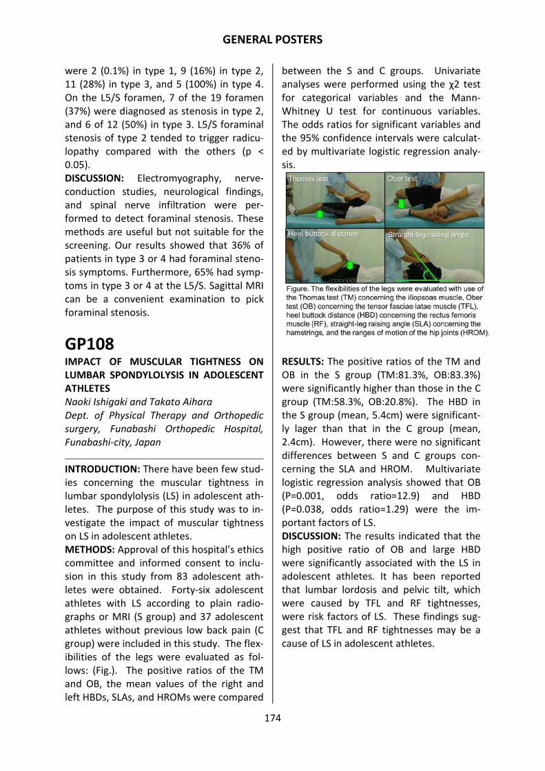
GENERAL POSTERS
174
were 2 (0.1%) in type 1, 9 (16%) in type 2,
11 (28%) in type 3, and 5 (100%) in type 4.
On the L5/S foramen, 7 of the 19 foramen
(37%) were diagnosed as stenosis in type 2,
and 6 of 12 (50%) in type 3. L5/S foraminal
stenosis of type 2 tended to trigger radicu-
lopathy compared with the others (p <
0.05).
DISCUSSION: Electromyography, nerve-
conduction studies, neurological findings,
and spinal nerve infiltration were per-
formed to detect foraminal stenosis. These
methods are useful but not suitable for the
screening. Our results showed that 36% of
patients in type 3 or 4 had foraminal steno-
sis symptoms. Furthermore, 65% had symp-
toms in type 3 or 4 at the L5/S. Sagittal MRI
can be a convenient examination to pick
foraminal stenosis.
GP108 IMPACT OF MUSCULAR TIGHTNESS ON LUMBAR SPONDYLOLYSIS IN ADOLESCENT ATHLETES Naoki Ishigaki and Takato Aihara
Dept. of Physical Therapy and Orthopedic
surgery, Funabashi Orthopedic Hospital,
Funabashi-city, Japan
INTRODUCTION: There have been few stud-
ies concerning the muscular tightness in
lumbar spondylolysis (LS) in adolescent ath-
letes. The purpose of this study was to in-
vestigate the impact of muscular tightness
on LS in adolescent athletes.
METHODS: Approval of this hospital’s ethics
committee and informed consent to inclu-
sion in this study from 83 adolescent ath-
letes were obtained. Forty-six adolescent
athletes with LS according to plain radio-
graphs or MRI (S group) and 37 adolescent
athletes without previous low back pain (C
group) were included in this study. The flex-
ibilities of the legs were evaluated as fol-
lows: (Fig.). The positive ratios of the TM
and OB, the mean values of the right and
left HBDs, SLAs, and HROMs were compared
between the S and C groups. Univariate
analyses were performed using the χ2 test for categorical variables and the Mann-
Whitney U test for continuous variables.
The odds ratios for significant variables and
the 95% confidence intervals were calculat-
ed by multivariate logistic regression analy-
sis.
RESULTS: The positive ratios of the TM and
OB in the S group (TM:81.3%, OB:83.3%)
were significantly higher than those in the C
group (TM:58.3%, OB:20.8%). The HBD in
the S group (mean, 5.4cm) were significant-
ly lager than that in the C group (mean,
2.4cm). However, there were no significant
differences between S and C groups con-
cerning the SLA and HROM. Multivariate
logistic regression analysis showed that OB
(P=0.001, odds ratio=12.9) and HBD
(P=0.038, odds ratio=1.29) were the im-
portant factors of LS.
DISCUSSION: The results indicated that the
high positive ratio of OB and large HBD
were significantly associated with the LS in
adolescent athletes. It has been reported
that lumbar lordosis and pelvic tilt, which
were caused by TFL and RF tightnesses,
were risk factors of LS. These findings sug-
gest that TFL and RF tightnesses may be a
cause of LS in adolescent athletes.
GENERAL POSTERS
175
GP109 A STUDY ON PATHOGENESIS OF LUMBAR SPONDYLOLYSIS USING MRI FINDINGS Sakai T.1, Goda Y. 1, Takata. 1, Higashino K.
1, Katoh S. 1, Sairyo K. 2
1Dept. Orthopaedic Surgery, Tokushima
University, Tokushima, Japan 2Dept. Ortho-
paedic Surgery, Teikyo University, Kawasaki,
Japan
INTRODUCTION/AIM: Spondylolysis is
thought as a stress fracture of the pars
interarticularis (pars). However, there are
still a lot of unknown factors affecting on its
pathogenesis. In this study, to elucidate it,
we evaluated MRI in patients with very ear-
ly- and early-stage spondylolysis and ana-
lyzed the localization of the signal changes.
METHODS: A total of 28 patients (5 women
and 25 men) with very early- or early-stage
lumbar spondylolysis were included in this
study. Mean age was 14.1 (10 to 17) years.
We analyzed the localizations of MRI high
signal change (HSC) around pedicle adjacent
to the pars using both sagittal and axial
views of T2- and STIR-weighted images. We
divided the localizations of HSC into the
following three zones (Zone1: pedicle-
interarticuaris, Zone 2: interarticularis-
spinous process, Zone 3: extraosseous).
RESULTS AND DISCUSSION: HSCs were de-
tected bilaterally in 18 vertebrae and 13
vertebrae. A total of 49 HSCs were ana-
lyzed. Thirty-one HSCs (63.3%) were found
in only Zone 1, five HSCs (10.2%) were
found in Zone 1 & 2, and seven HSCs
(14.3%) were found in Zone 1 & 3. Six HSCs
(12.2%) were found in Zone 1 & 2& 3.
Basically, MRI bone marrow changes on
stress fracture of long bone shows signal
changes in both proximal and distal sites
across the fracture site, and sometimes in
extraosseous region. In this study, more
than half HSCs (63.3%) were localized in
only one side across the fracture site (pars).
This result suggests that lumbar
spondylolysis is not a common stress frac-
ture. Also, a total of 13 HSCs (26.5%) includ-
ing extraosseous region were found. In the
past, Sairyo, et al. reported the
extraosseous HSC and it causes radiculopa-
thy, indicating hematoma in the vicinity of
the fracture site. From the results of this
study, spondylolysis is thought as an un-
common stress fracture.
GP110 PAINVISION APPARATUS IS EFFECTIVE FOR ASSESSING LEG PAIN AND NUMBNESS ORIGINATED FROM LUMBAR DISEASE. Furusawa S (1), Hashikawa T (1), Terakado
A (1), Orita S (2), Takahashi K (2), Ohtori S
(2)
1, Kitachiba Orthop. Surg. Mihama Clin.,
Chiba, Japan. 2, Dept. Orthop. Surg., Chiba
Univ., Chiba, Japan.
INTRODUCTION: Tools developed to assess
low back / leg pain and its effect on quality
of life are mostly dependent on subjective
evaluation by patients in self-reported ques-
tionnaires. It is therefore difficult to com-
pare scores between patients at different
times. A newly developed device, PainVision
PS-2100 (Nipro, Osaka, Japan), has been
used to assess the perception of pain and
numbness in a quantitative manner. In the
current study, we aimed to evaluate the
efficacy of PainVision for assessing leg pain
and numbness originated from lumbar dis-
ease.
METHODS: We assessed 39 patients with
leg pain and numbness originated from
lumbar spinal stenosis. The numeric rating
scale (NRS) score, Roland-Morris Disability
Questionnarie (RDQ) score, and degree of
pain and numbness calculated using
PainVision were measured twice at 4-week
intervals in each patient. An electrode was
patched on the forearm surface of the pa-
tients, and both the current for perception
threshold and current at which the patient
reported the same intensity as their leg pain
and numbnsess were measured. The degree

GENERAL POSTERS
176
of pain and numbness was calculated auto-
matically (degree of pain and numbness =
100 x (current producing pain and numb-
ness comparable with leg pain and numb-
ness ‒ current at perception thresh-
old/current at perception threshold)). Cor-
relations between NRS and RDQ scores and
the degree of pain and numbness were de-
termined using Spearman’s rank correlation
test.
RESULTS: There was a strong correlation
between the NRS and RDQ scores at each
time point (rs = 0.40, P < 0.05). The degree
of pain and numbness also showed moder-
ate correlation with NRS and RDQ scores at
each time point (rs = 0.40, P < 0.05). Change
in the degree of pain and numbness over 4
weeks showed a moderate correlation with
changes in NRS and in RDQ scores (rs = 0.50,
P < 0.01).
DISCUSSION: PainVision is as useful to as-
sess pain and numbness originated from
lumbar spinal stenosis as self-reported
questionnaires.
GP111 LACK OF VALUE IN SCREENING FOR FEAR OF MOVEMENT VIA THE TAMPA SCALE FOR KINESIOPHOBIA Greg McIntosh MSc, Chris Gregg MHealSc,
Hamilton Hall MD, Chris Hoffman MD, Tom
Carter BSc PT
CBI Health Group Research Department,
Toronto, Canada; The Back Institute, Wel-
lington, New Zealand
INTRODUCTION: The purpose of this study
was to assess the role of baseline screening
for fear of movement and pain-related fear
- Tampa Scale for Kinesiophobia (TSK) - in a
cohort of low back pain (LBP) patients com-
pleting active exercise-based treatment.
METHODS: This was a prospective study of
LBP cases (n=416) treated at four spine care
rehabilitation clinics in New Zealand be-
tween January 2008 and October 2012. In
addition to baseline data on pain, function,
sociodemographics, all patients completed
a Tampa Scale for Kinesiophobia (TSK). All
patients had mechanical LBP with no neu-
rology, as determined by the Saskatchewan
Spine Pathway triage methodology.
RESULTS: The mean age of the cohort was
39.9 years (SD=11.3, range=18-69) with
63.7% males. The median symptom dura-
tion was 173 days (29% acute, 71% chronic).
The average overall TSK score was 40.8
(SD=9.03, range 6-70). There were no base-
line statistically significant differences in TSK
scores based on dominant pain location,
medication use, constancy, work status,
presence of sleeping difficulties, symptom
duration, smoking status, legal consultation
or pain classification. Males had significantly
higher scores than females (41.9 v 38.6)
(p=0.001). There was no correlation be-
tween TSK scores and age or baseline func-
tion. For clinical outcomes, there was no
correlation between TSK scores and number
of days in treatment, change in numerical
pain rating or functional improvement.
There were no statistically significant differ-
ences in TSK scores based on global pain
rating or return to work status.
DISCUSSION: The presence of pain-related
fear, as measured by the Tampa Scale for
Kinesiophobia (TSK) was not associated with
higher disability, poorer function or more
pain in this sample. In addition, baseline
characteristics were similar regardless of
TSK score. The lack of differences by TSK
score revealed that screening for fear of
movement was not worth the effort and
provided little clinical benefit.

GENERAL POSTERS
177
GP112 EFFICACY OF DIFFUSION-WEIGHTED IMAG-ING (DWI) BASED ON MRI TO EVALUATE HEMATOLOGICAL MALIGNANCIES IN LUM-BAR SPINE. Masaomi Yamashita, MD, PhD*, Kazuhiko
Kikuchi+, Koki Abe, MD*, Keishi Yamashita,
MD*, Akiyoshi Yamaoka, MD*, Seiji Ohtori,
MD, PhD**, Kazuhisa Takahashi, MD,
PhD**
*Department of Orthopedic Surgery,
Funabashi Central Hospital, Chiba, Japan
+Department of Radiology, Funabashi Cen-
tral Hospital **Department of Orthopedic
Surgery, Graduate School of Medicine, Chiba
University
INTRODUCTION: Patients with hematologi-
cal malignancies such as multiple myeloma,
lymphoma, and leukemia, undergo the re-
placement of spinal hemopoietic bone mar-
row. Plain radiography and conventional
MRI (C-MRI), including T1- and T2-weighted
imaging, have been used to estimate patho-
logic lesion of low back pain (LBP). However,
diagnosis of hematological malignancies is
difficult in use of C-MRI. Diffusion-weighted
imaging (DWI) based on MRI can provide a
non-invasive method that reveals the mi-
crostructure of tissues on the basis of its
ability to monitor the random movements
of water molecules. DWI has been widely
used clinically in the evaluation of malig-
nancy for diagnosis of diseases. The aim of
this study is to evaluate the potential of
DWI in hematological malignancies in lum-
bar spine.
METHODS: 1437 patients with LBP form July
1 .2009 to June 30. 2012 were examined
and underwent plain radiography, C-MRI,
DWI-MRI of the lumbar spine, and blood
examination. We measured the apparent
diffusion coefficient (ADC) of bone marrow
in vertebral bodies using 1.5-T MRI, and
compared ADC in bone marrow with final
diagnosis of lumbar spine.
RESULT: In 1437 patients, diagnosis were
lumbar spondylosis (70%), fracture (15%),
lumbar disc herniation (LDH 6%), hemato-
logical malignancies (3.4%, multiple myelo-
ma 1.9%, malignant lymphoma 1.3%, leu-
kemia 0.2%), metastatic spinal tumor (3%),
and infectious spondylitis (2.6%). Fracture,
metastatic spinal tumor, and infection could
be diagnosed by C-MRI. However, hemato-
logical malignancies were not diagnosed by
C-MRI. The mean value of ADC in bone mar-
row in hematological malignancies (Mean:
1.86×10⁻⁴ mm2/s) was significantly higher
than those of spondylosis and LDH (Mean:
1.31×10⁻⁴ mm2/s) (p<0.05).
DISCUSSION: In LBP patients, 3.4% patients
were hematological malignancies in current
study. Only C-MRI did not clarify hematolog-
ical malignancies, however, DWI may have
the potential of diagnostic capacity in he-
matological malignancies.
GP113 PHARMACOLOGICAL ANXIOLYSIS WITH PROMETHAZINE AS AN ADJUNCTIVE THERAPY FOR ACUTE LOW BACK PAIN IN THE ADULT EMERGENCY DEPARTMENT Eyal Behrbalk, The Spine Unit - Queen's
Medical Centre, Nottingham, United
Kindoms. Ofir Uri, Royal National
Orthopaedic Hospital NHS Trust Brockley
Hill, Stanmore, Middlesex. Bronek
Maximilian Boszczyk, The Spine Unit-
Queen’s Medical Centre, Nottingham,
United Kindoms Ruth M Parks,University of
Nottingham, Medical School, Queen's
Medical Centre.United Kindoms Pinchas
Halpern, The Tel-Aviv Sourasky Medical
Centre, Israel
INTRODUCTION: Low back pain (LBP) is a
common and disabling problem presenting
to the emergency department (ED); analge-
sia is an important step in immediate man-
agement. It has been proven that non-
pharmacological methods of anxiety relief
can be beneficial. A prospective randomised
double-blind study carried out to determine

GENERAL POSTERS
178
whether combination treatment of pro-
methazine and morphine was superior to
morphine alone, for severe acute LBP.
METHODS: All patients admitted to the ED
with simple acute LBP were candidates for
the study. Patients included met the follow-
ing criteria: LBP< 3weeks duration, pain
>65mm on a 100mm visual analogue scale,
age between 18-65, American Society of
Anesthesiologists (ASA) grade 1 or 2, systol-
ic blood pressure >90 mmHg, no known
hypersensitivity to the medications, able to
provide informed consent. Pregnant wom-
en, LBP after traumatic event, history of
malignancy, neurological deficiencies or
atypical pain were excluded. 59 patients
concluded the study and assessed for pain
and anxiety reduction as well as satisfaction
of treatment received.
RESULTS: Neither treatment regime proved
superior in the relief of pain. patients re-
ceiving promethazine and morphine, were
more likely to require a longer duration of
stay in the ED (p=0.0427). They were also
more likely to experience adverse events;
however, these findings were not statistical-
ly significant. Patients satisfaction and am-
bulation status were similar for both
groups.
DISCUSSION: We do not recommend rou-
tine administration of promethazine for
anxiety and pain reduction in the treatment
of acute LBP in the ED. Non-
pharmacological methods of anxiety relief
may still be helpful.
GP114 A VALIDATION STUDY OF A SYMPTOM SCALE FOR LUMBAR SPINAL STENOSIS Miho Sekiguchi 1), Takafumi Wakita 2), Koji
Otani 1), Yoshihiro Onishi 3), Shunichi
Fukuhara 4), Shin-ichi Kikuchi 1), Shin-ichi
Konno 1)
1) Department of Orthopaedic Surgery,
Fukushima Medical University School of
Medicine; 2) Department of Sociology,
Kansai University; 3) Institute for Health
Outcomes & Process Evaluation Research
(iHope International) 4) Department of
Epidemiology and HealthCare Research,
Graduate School of Medicine and Public
Health, Kyoto University;
INTRODUCTION: Lumbar spinal stenosis
(LSS) has various symptoms and they
change with activities of daily living and
posture. We have developed and validated
a self-administered symptom scale for LSS
(Spine 37, 2012). The aim of this study was
to a) verify a criterion validity using a lum-
bar extension test and a gait-loading test as
objective indexes, and b) responsiveness of
the LSS symptom scale.
METHODS: One hundred and sixty seven
patients with a diagnosis of LSS completed
questionnaires including the LSS symptoms
scale before and after a medical therapy. A
lumbar extension test and a gait-loading
test were performed to evaluate standing
and walking capacities. Subjects were divid-
ed into three groups according to these
tests, low; less than mean-1/2 standard de-
viation (SD), middle; between low and high
groups, high; greater than mean+1/2SD. The
responsiveness to medication effects was
analyzed with an interval of 8 weeks. ANO-
VA was used to test differences in the LSS
symptom scale scores among groups.
RESULTS: In the lumbar extension test, a
mean standing capacity was 164.6±109 s.
The LSS symptom scale score was signifi-
cantly higher in accordance with a shorter
standing time (p<0.01). In the gait-loading
test, the mean walking distance was
213.0±154m. The LSS scale scores in the low
group was higher than that of the middle
and high groups (p<0.01). The changes of
LSS scale scores were higher in accordance
with improvement of symptoms by the two
tests, both before and after medication.
DISCUSSION: The results suggested that the
LSS symptom scale is a reliable and valid
instrument with good responsiveness to
non-surgical therapeutics. This scale may be

GENERAL POSTERS
179
useful to evaluate patient’s progress of
symptoms, to establish standards for indica-
tion of treatment, and to assess treatment
effectiveness.
GP115 EFFICACY OF ZOLEDRONIC ACID FOR CHRONIC LOW BACK PAIN ASSOCIATED WITH MODIC CHANGES IN MRI: A RANDOMIZED CONTROLLED TRIAL Koivisto K, Kyllönen E, Haapea M, Niinimäki
J, Sundqvist K, Pehkonen T, Seitsalo S,
Paldanius P, Tervonen O, Karppinen J
Department of Physical and Rehabilitation
Medicine, Oulu University Hospital, Oulu,
Finland
INTRODUCTION: Modic changes (MC) are
associated with low back pain (LBP) but no
effective treatments are available. The aim
of this randomized, placebo-controlled,
double-blinded trial was to evaluate the
efficacy of Zoledronic acid (ZA) for the
chronic LBP of patients with MC in magnetic
resonance imaging (MRI).
METHODS: Inclusion criteria were LBP for at
least three months, an LBP intensity of ≥ 6/10 on 10-cm VAS or an Oswestry Disabil-
ity Index (ODI) of ≥ 30%, and MC in MRI. Exclusion criteria included e.g. renal im-
pairment, and willingness to retire early.
Patients were randomized into single intra-
venous infusion of ZA 5 mg in 100 ml saline
(n=20), or saline (n=20) groups. The primary
outcome was LBP intensity and secondary
outcomes disability (ODI), health-related
quality of life (RAND-36), lumbar flexibility,
sick leaves and pain medication use. The
treatment differences at 1 and 12 months
were analysed using ANCOVA with adjust-
ments for age and gender.
RESULTS: At baseline, the treatment groups
had no significant differences. Both the
treatment difference in intensity of LBP
(1.3, 95% CI -0.2 to 2.8; p=0.097) and pro-
portion of patients with 20% improvement
(ZA 55% vs. placebo 25%; p=0.105) tended
to favour ZA at 1 month. The improvements
in ODI were of similar magnitude. At base-
line, the treatment groups reported no dif-
ferences in non-steroidal anti-inflammatory
drug use, whereas at 12 months, only 20%
of patients in the ZA group used these drugs
compared to 60% in the placebo group
(p=0.022). No significant differences were
observed in RAND-36 or sick leaves. Flu-like
symptoms emerged in 90% of patients in
the ZA group, compared to 15% in the pla-
cebo group. Only one serious adverse ef-
fect, sinusitis requiring hospitalization, oc-
curred in the ZA group.
DISCUSSION: The tendency for improve-
ment was greater in the ZA group. Although
the results are encouraging, larger studies
are required to prove the efficacy of ZA.
(ClinicalTrial.gov identifier NCT01330238)
GP116 MINIMAL CLINICAL IMPORTANT IM-PROVEMENT (MCII) AND PATIENT AC-CEPTABLE SYMPTOM STATE (PAAS) VAL-UES OF THE DALLAS PAIN QUESTIONNAIRE (DPQ) IN PATIENTS WITH LOW BACK PAIN M Marty (1) S. Genevay (2) ; D. S.
Courvoisier (3); V. Foltz (4); G. Mahieu (5); C.
Desmoulin (6); A. Gierasimowicz Fontana
(7); M. Norberg (8); P. de Goumoëns (8); C.
Cedraschi (9); S. Rozenberg (4); Section
Rachis de la Société Française de
Rhumatologie
(1) Service de Rhumatologie, C.H.U. Henri
Mondor, Créteil France ; (1) Rhumatologie,
Hôpitaux Universitaires de Genève, Genève,
Suisse; (3) Epidémiologie Clinique, Hôpitaux
Universitaire de Genève, Genève, Suisse; (4)
Service de Rhumatologie, C.H.U Pitié-
Salpêtrière, Paris, France; (5) Unité du Dos,
C.H. de Dinant, Dinant, Belgique; (6) Bat B;
Sart-Tilman, Isepk, Liège, Belgique; (7)
Clinique de Médecine Physique et
Réadaptation, CHU Brugmann, Bruxelles,
Belgique; (8) Appareil Locomoteur, CHUV,
Lausanne, Suisse; (9) Réhabilitation
Médicale, Hôpitaux Universitaire de Genève,

GENERAL POSTERS
180
Genève, Suisse.
INTRODUCTION: The DPQ has demonstrat-
ed its validity in patients with chronic low
back pain (CLBP). Norms for clinically signifi-
cant differences are missing. This work
aimed to estimate Minimal Clinical Im-
portant Improvement (MCII) and Patient
Acceptable Symptom State (PASS) values of
DPQ.
METHOD: 168 patients with no specific LBP
lasting for at least 4 weeks were evaluated
at baseline and 6 months later after a non
standardized treatment. Question for MCII
addressed patient-reported response to
treatment at FU on a 5-point Likert scale,
and PASS a yes/no answer concerning satis-
faction with present state. MCII was com-
puted as the difference in mean DPQ scores
between patients reporting treatment as
effective vs. patients reporting treatment as
not effective, and PASS was computed as
the 3rd quantile of the DPQ score among
patients who report being satisfied with
their present state.
RESULTS: Mean (SD) intensity of back pain
(0-10 VAS) and Roland Morris Disability
Questionnaire scores were 5.5 (2.0) and
12.9 (5.0) at baseline, 3.7 (2.6) and 7.5 (6.5)
at FU, respectively.
DPQ component Baseline M6 MCII PASS (0-100) Daily activity 60.5 40.6 22.2 29.4
Work leisure 57.8 37.2 23.1 23.1
Social interest 34.1 24.5 2.0 20.0
Anxiety
depression 42.5 29.4 9.5 21.3
Total 48.7 32.4 14.2 23.9
The PASS total score threshold (23.9) cor-
rectly classified 84.1% of the patients who
declared being unsatisfied with their pre-
sent state.
DISCUSSION: These values give information
of paramount importance for clinicians to
interpret change over time of the DPQ
measure. Authors should be encouraged to
report the MCII and PASS in RCT and cohort
studies to help clinicians interpreting clinical
results.
G117 QUANTITATIVE EVALUATION OF INJURED NERVE WITH DIFFUSION TENSOR IMAGING Oikawa, Yasuhiro1; Eguchi, Yawara1;
Ohtori, Seiji1; Inoue, Gen3; Yamauchi,
Kazuyo1; Orita, Sumihisa1; Kamoda,
Hiroto1; Ishikawa, Tetsuhiro1; Miyagi, Ma-
sayuki1; Suzuki, Miyako1; Sakuma, Yoshihi-
ro1; Kubota, Go1; Inage, Kazuhide1; Sainoh,
Takeshi1; Sato, Jun1; Watanabe, Atsuya2;
Takahashi, Kazuhisa1
1 Department of Orthopaedic Surgery,
Graduate School of Medicine, Chiba Univer-
sity, Japan 2 Department of Orthopaedic
Surgery, Teikyou University Chiba Medical
Center, Japan 3 Department of Orthopaedic
Surgery, Kitasato University, School of Medi-
cine, Japan
INTRODUCTION: Conventional imaging mo-
dalities are useful for the morphological
evaluation of spinal nerves, but not for the
quantitative evaluation of nerve damage.
Diffusion tensor imaging (DTI) is a possible
new diagnostic tool for lumbar spinal de-
generative disease. Using DTI, a lower frac-
tional anisotropy (FA) value is observed in
symptomatic nerves than in asymptomatic
nerves. The purpose of this study was to
evaluate the efficacy of DTI in the quantita-
tive evaluation of injured nerves.
METHODS: Twelve patients with single-
sided radiculopathy who had undergone DTI
using 3.0T magnetic resonance imaging
(MRI) and surgical treatment were included.
The FA values of lumbar nerve roots were
calculated from FA maps in both injured and
intact nerves. Minimum and mean FA values
were investigated, and the differences be-
tween injured and intact nerves were also
investigated. Neurological severity was as-
sessed using the Japanese Orthopaedic As-
sociation (JOA) and visual analog scale (VAS)
pain scores.

GENERAL POSTERS
181
RESULTS: In injured nerves, e.g., those
compressed by disc herniation, lower FA
values were found at the injured portion.
The mean FA of injured nerves was 0.220;
the mean difference between injured and
intact nerves was 0.097. The mean and min-
imum FA values were not directly correlated
with JOA and VAS scores; however, pre- and
postoperative VAS scores were higher in
patients with larger differences in intact and
injured FA values.
CONCLUSIONS: Lower FA values were ob-
tained at the compressed portion in injured
nerves, and the difference between the FA
values of the intact and injured nerves
tended to correlate with neurological sever-
ity. This means that although it is difficult to
evaluate an injury using only its FA value, it
may be possible to utilize the difference
between the injured nerve and an intact
nerve. DTI is a possible new diagnostic tool
for degenerated lumbar disease as it is ca-
pable of both detecting lesions and quanti-
tatively evaluating nerve damage.
GP118 DEGENERATIVE CHANGES OF FACET JOINTS IN ADULTS WITH LUMBAR SPONDYLOLYSIS Goda Y. 1, Sakai T. 1, Harada T. 2, Takao S.
2, Harada M. 2, Takata Y. 1, Higashino K. 1,
Katoh S. 1, Yasui N. 1, Sairyo K. 3
1Department of Orthopaedic surgery,
Tokushima University, Tokushima, Japan
2Department of Radiology, Tokushima
University, Tokushima, Japan 3Department
of Orthopaedic Surgery, Teikyo University,
Kawasaki, Japan
INTRODUCTION: Lumbar spondylolysis
(spondylolysis) is a bony defect of pars
interarticularis. Once bony discontinuity of
bilateral laminae occurred, it is called a “
floating lamina.” In such a situation, biome-
chanical stresses on adjacent facet joints
are thought to be decreased. In this study,
we evaluated multi-detector CT to analyze
degenerative changes of facet joints with
spondylolysis and compared with those with
no spondylolysis.
METHODS: We have previously reported
the incidence of lumbar spondylolysis in the
general population in japan based on
multidetector computed tomography scans
from 2000 subjects (Sakai T, et al. Spine,
2009). Among those, 107 subjects (37
women and 70 men) with L5 single-level
spondylolysis were evaluated as
spondylolysis group (Sp (+)). Mean age was
64.1(22 to 88) years. The gender- and age-
matched control group (Sp (-)) was chosen
randomly from the other subjects with no
spondylolysis. Four changes (narrowing,
sclerosis, osteophyte, bone cyst) of degen-
erative changes of facet joints (L4/5, L5/S)
adjacent to the pars defects.
RESULTS AND DISCUSSION: In a total of 214
L4/5 facet joints, the following results were
found (Sp (+): Sp (-)): narrowing (210: 187),
sclerosis (200: 137), osteophyte (116: 82),
and bone cyst (52:3). In a total of 214 L5/S
facet, narrowing (210: 181), sclerosis (194:
145), osteophyte (65: 40), and bone cyst
(16: 2) were found (Figure 1). Totally, de-
generative changes of facet joints with lum-
bar spondylolysis were more severe than
those of control, statistical significantly
(p<0.05). Unfortunately, the reasons of this
result were not clarified in this study. But,
there are many candidate reasons of the
results in this study. Particularly, alignment,
slip grade, morphological changes as facet
tropism, facet orientation, should be inves-
tigated in the next step.
GP119 PROTEOMIC ANALYSIS OF LIGAMENTUM FLAVUM FROM LUMBAR SPINAL CANAL STENOSIS Goda Y. 1, Sato N. 1, Taniguchi T. 2, Takata
Y. 1, Kosaka H. 1, Sakai T. 1, Higashino K. 1,
Sairyo K. 3, Katoh S. 1, Taniguchi H. 2, Yasui
N. 1
1Department of Orthopaedic surgery,
Tokushima University, Tokushima, Japan

GENERAL POSTERS
182
2Division of Disease Proteomics, Institute for
Enzyme Research, Tokushima University,
Tokushima, Japan 3Department of
Orthopaedic Surgery, Teikyo University,
Kawasaki, Japan
INTRODUCTION/AIM: Lumbar spinal canal
stenosis (LCS) is a common lumbar spinal
disorder in aged population. Hypertrophy
of ligamentum flavum is one of main factors
of LCS but its pathomechanism has not been
clarified yet. In many diseases, proteomic
analysis has been applied to understanding
of their pathogenesis. Although proteomic
analysis of ligamentum flavum was difficult
due to the intrinsic insolubility of the tissue,
Sato et al. succeeded in solubilization of
ligamentum flavum. The purpose of this
study is to analyze proteomic changes in
components of the hypertrophic
ligamentum flavum in the LCS patients.
METHODS: All samples of the ligamentum
flavum were obtained from patients with
LSC or lumbar disc herniation (LDH) at the
time of operation, and divided them into
the two groups. LCS group includes 3 male
patients at the age of 80 (A), 74 (B), and 59
(C) years. LDH group includes 17 year-old
male (D) and 22 (E), 30 (F) year-old female
patients. All samples were axially cut into 1-
μm slices with cryotome. They were treat-
ed with CNBr under N2 gas and digested
with various enzymes. LC/MS/MS analysis
has been performed. Analysis on protein
database were performed using the Mascot
software.
RESULTS AND DISCUSSION: In the LCS
group, the contents of elastin were (A)14%,
(B)38%, (C)50%, and the contents of colla-
gen were (A)77%, (B)56%, (C)40%. In the
LDH group, the contents of elastin were
(D)57%, (E)66%, (F)57%, and the contents of
collagen were (D)35%, (E)18%, (F)33%. The
contents of elastin in LCS group were lower
than those in LDH group and the contents of
collagen in LCS group were higher than
those in LDH group. Various types of colla-
gens were identified (Figure 1). Since elas-
tin contributes to the elasticity (softness) of
the tissue, the decrease of elastin in
ligamentum flavum with hypertrophy af-
fects not only the thickness of the tissue,
but also the softness of that. These results
may explain, at least in part, the cause of
the LCS.
GP120 SPINO-PELVIC SAGITTAL ALIGNMENT IN ELDERLY PATIENTS WITH ISTHMIC SPONDYLOLISTHESIS Hidekazu Suzuki*, Kenji Endo*, Hirosuke
Nishimura*, Hidetoshi,Tanaka*, Shinsuke
Sakai**, Kengo Yamamoto*
* Department of Orthopedic Surgery, Tokyo
Medical University ** Department of
Orthopedic Surgery, Tokyo Medical
Unversity Ibaraki Medical Center
INTRODUCTION: The risk for progression of
isthmic spondylolisthesis appears to be
greatest during the adolescent growth spurt
and subsequently decreased to the mini-
mum after adolescence. However, some of
the patients have an onset of radiculopathy
at an old age. Spinal sagittal alignment plays
an important role in the development of
spondylolysis and spondylolisthesis. How-
ever, there are few studies about spinal
sagittal alignment of elderly patients with
isthmic spondylolisthesis (IS). The purpose
of this study is to evaluate spino-pelvic
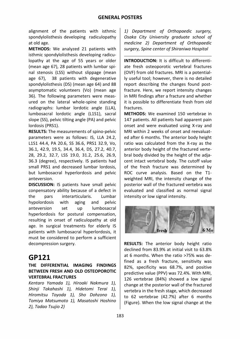
GENERAL POSTERS
183
alignment of the patients with isthmic
spondylolisthesis developing radiculopathy
at old age.
METHODS: We analyzed 21 patients with
isthmic spondylolisthesis developing radicu-
lopathy at the age of 55 years or older
(mean age 67), 28 patients with lumbar spi-
nal stenosis (LSS) without slippage (mean
age 67), 38 patients with degenerative
spondylolisthesis (DS) (mean age 64) and 88
asymptomatic volunteers (Vo) (mean age
36). The following parameters were meas-
ured on the lateral whole-spine standing
radiographs: lumbar lordotic angle (LLA),
lumbosacral lordotic angle (L1S1), sacral
slope (SS), pelvic tilting angle (PA) and pelvic
lordosis (PRS1).
RESULTS: The measurements of spino-pelvic
parameters were as follows: IS, LLA 24.2,
L1S1 44.4, PA 20.6, SS 36.6, PRS1 32.9, Vo,
36.1, 42.9, 19.5, 34.4, 36.4, DS, 27.2, 40.7,
28, 29.2, 32.7, LSS 19.0, 31.2, 25.6, 26.9,
36.3 (degree), respectively. IS patients had
small PRS1 and decreased lumbar lordosis,
but lumbosacral hyperlordosis and pelvic
anteversion.
DISCUSSION: IS patients have small pelvic
conpensatory ability because of a defect in
the pars interarticularis. Lumbar
hypolordosis with aging and pelvic
anteversion set up lumbosacral
hyperlordosis for postural compensation,
resulting in onset of radiculopathy at old
age. In surgical treatments for elderly IS
patients with lumbosacral hyperlordosis, it
must be considered to perform a sufficient
decompression surgery.
GP121 THE DIFFERENTIAL IMAGING FINDINGS BETWEEN FRESH AND OLD OSTEOPOROTIC VERTEBRAL FRACTURES Kentaro Yamada 1), Hiroaki Nakmura 1),
Shinji Takahashi 1), Hidetomi Terai 1),
Hiromitsu Toyoda 1), Sho Dohzono 1),
Tomiya Matsumoto 1), Masatoshi Hoshino
2), Tadao Tsujio 2)
1) Department of Orthopaedic surgery,
Osaka City University graduate school of
medicine 2) Department of Orthopaedic
surgery, Spine center of Shiraniwa Hospital
INTRODUCTION: It is difficult to differenti-
ate fresh osteoporotic vertebral fractures
(OVF) from old fractures. MRI is a potential-
ly useful tool; however, there is no detailed
report describing the changes found post-
fracture. Here, we report intensity changes
in MRI findings after a fracture and whether
it is possible to differentiate fresh from old
fractures.
METHODS: We examined 150 vertebrae in
147 patients. All patients had apparent pain
onset and were evaluated using X-ray and
MRI within 2 weeks of onset and reevaluat-
ed after 6 months. The anterior body height
ratio was calculated from the X-ray as the
anterior body height of the fractured verte-
bral body divided by the height of the adja-
cent intact vertebral body. The cutoff value
of the fresh fracture was determined by
ROC curve analysis. Based on the T1-
weighted MRI, the intensity change of the
posterior wall of the fractured vertebra was
evaluated and classified as normal signal
intensity or low signal intensity.
RESULTS: The anterior body height ratio
declined from 83.9% at initial visit to 63.8%
at 6 months. When the ratio >75% was de-
fined as a fresh fracture, sensitivity was
82%, specificity was 68.7%, and positive
predictive value (PPV) was 72.4%. With MRI,
126 vertebrae (84%) showed a low signal
change at the posterior wall of the fractured
vertebra in the fresh stage, which decreased
to 62 vertebrae (42.7%) after 6 months
(Figure). When the low signal change at the
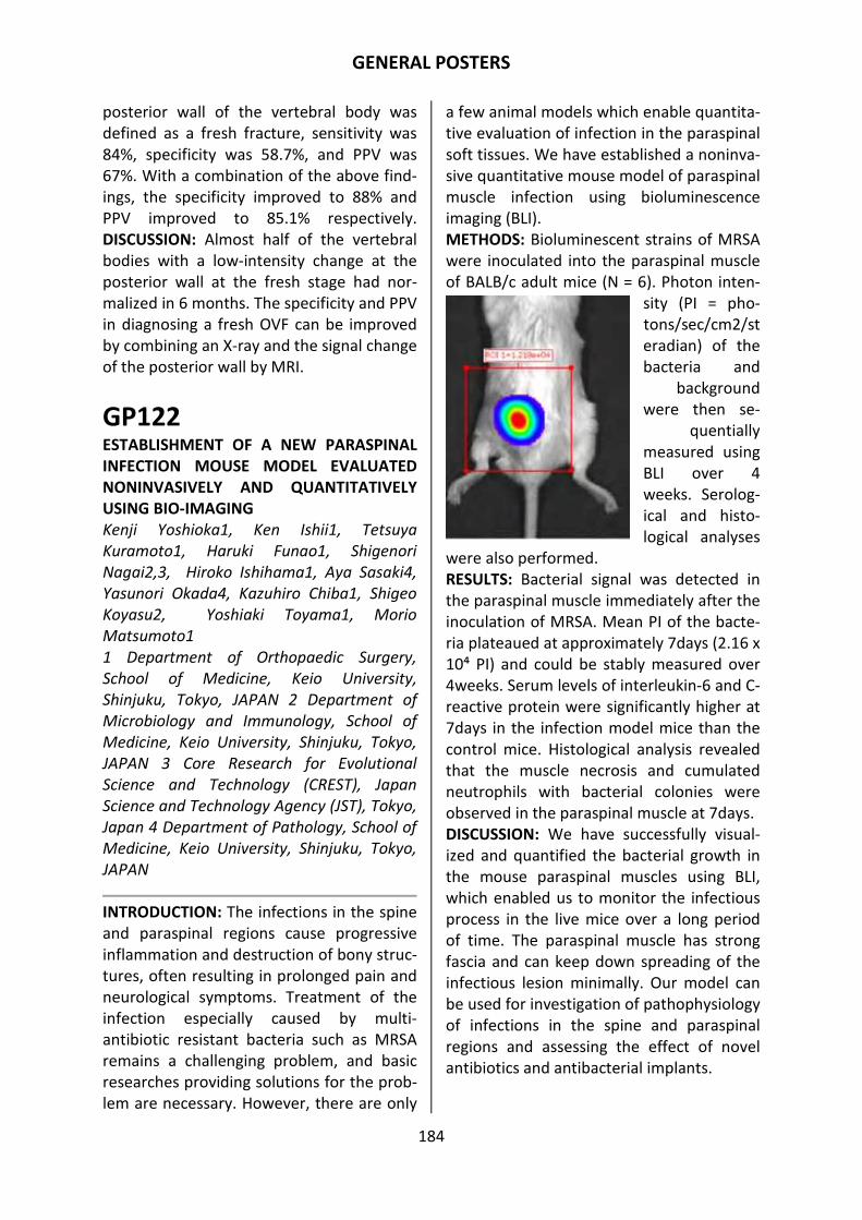
GENERAL POSTERS
184
posterior wall of the vertebral body was
defined as a fresh fracture, sensitivity was
84%, specificity was 58.7%, and PPV was
67%. With a combination of the above find-
ings, the specificity improved to 88% and
PPV improved to 85.1% respectively.
DISCUSSION: Almost half of the vertebral
bodies with a low-intensity change at the
posterior wall at the fresh stage had nor-
malized in 6 months. The specificity and PPV
in diagnosing a fresh OVF can be improved
by combining an X-ray and the signal change
of the posterior wall by MRI.
GP122 ESTABLISHMENT OF A NEW PARASPINAL INFECTION MOUSE MODEL EVALUATED NONINVASIVELY AND QUANTITATIVELY USING BIO-IMAGING Kenji Yoshioka1, Ken Ishii1, Tetsuya
Kuramoto1, Haruki Funao1, Shigenori
Nagai2,3, Hiroko Ishihama1, Aya Sasaki4,
Yasunori Okada4, Kazuhiro Chiba1, Shigeo
Koyasu2, Yoshiaki Toyama1, Morio
Matsumoto1
1 Department of Orthopaedic Surgery,
School of Medicine, Keio University,
Shinjuku, Tokyo, JAPAN 2 Department of
Microbiology and Immunology, School of
Medicine, Keio University, Shinjuku, Tokyo,
JAPAN 3 Core Research for Evolutional
Science and Technology (CREST), Japan
Science and Technology Agency (JST), Tokyo,
Japan 4 Department of Pathology, School of
Medicine, Keio University, Shinjuku, Tokyo,
JAPAN
INTRODUCTION: The infections in the spine
and paraspinal regions cause progressive
inflammation and destruction of bony struc-
tures, often resulting in prolonged pain and
neurological symptoms. Treatment of the
infection especially caused by multi-
antibiotic resistant bacteria such as MRSA
remains a challenging problem, and basic
researches providing solutions for the prob-
lem are necessary. However, there are only
a few animal models which enable quantita-
tive evaluation of infection in the paraspinal
soft tissues. We have established a noninva-
sive quantitative mouse model of paraspinal
muscle infection using bioluminescence
imaging (BLI).
METHODS: Bioluminescent strains of MRSA
were inoculated into the paraspinal muscle
of BALB/c adult mice (N = 6). Photon inten-
sity (PI = pho-
tons/sec/cm2/st
eradian) of the
bacteria and
background
were then se-
quentially
measured using
BLI over 4
weeks. Serolog-
ical and histo-
logical analyses
were also performed.
RESULTS: Bacterial signal was detected in
the paraspinal muscle immediately after the
inoculation of MRSA. Mean PI of the bacte-
ria plateaued at approximately 7days (2.16 x
10⁴ PI) and could be stably measured over 4weeks. Serum levels of interleukin-6 and C-
reactive protein were significantly higher at
7days in the infection model mice than the
control mice. Histological analysis revealed
that the muscle necrosis and cumulated
neutrophils with bacterial colonies were
observed in the paraspinal muscle at 7days.
DISCUSSION: We have successfully visual-
ized and quantified the bacterial growth in
the mouse paraspinal muscles using BLI,
which enabled us to monitor the infectious
process in the live mice over a long period
of time. The paraspinal muscle has strong
fascia and can keep down spreading of the
infectious lesion minimally. Our model can
be used for investigation of pathophysiology
of infections in the spine and paraspinal
regions and assessing the effect of novel
antibiotics and antibacterial implants.

GENERAL POSTERS
185
GP123 FACET ORIENTATION IN PATIENTS WITH LUMBAR DEGENERATIVE SPONDYLOLISTHESIS Miyamoto H, Hidekazu Suzuki*, Kenji Endo*,
Wuqikun Alimasi*, Hirosuke Nishimura*,
Hidetoshi,Tanaka*, Shinsuke Sakai**, Kengo
Yamamoto*
* Department of Orthopedic Surgery, Tokyo
Medical University ** Department of
Orthopedic Surgery, Tokyo Medical
Unversity Ibaraki Medical Center
INTRODUCTION: Several reports have indi-
cated a correlation between lumbar degen-
erative spondylolisthesis (DS) and increased
sagittal orientation of the facet joints.
Sagittalization of lumbar facet joints is a
major cause of DS, however, the majority of
studies of lumbar facets were analyzed in
the axial plane. We analyzed the spinal
morphological character by examining both
the axial and sagittal views in DS patients
and compared these parameters to patients
with lumbar spinal stenosis (LSS, without
slippage) and low back pain (LBP, without
slippage nor neurological symptoms).
METHODS: We analyzed 119 patients divid-
ed into 3 groups based on the clinical and
imaging findings: 42 patients with DS, 39
with LSS, and 38 with LBP. The following
parameters were measured from L2-3 to L5-
S1 levels: axial facet angle (AFA) in an axial
orientation, oblique facet angle (OFA) and
lamina angle (LA) in sagittal orientations.
RESULTS: The AFA of the DS group was sig-
nificantly smaller than that of the LSS and
LBP groups at the slippage level. However,
the OFA of the DS and LSS groups had no
significant difference at any of the levels.
The AFA and OFA in the DS and LSS groups
were smaller than those in the LBP group at
all levels. From the L2 to L4 levels, the LA of
the DS and LSS groups had a tendency to be
larger than LBP group. At the L5 level, how-
ever, the LA was similar among all groups.
DISCUSSION: Patients with DS had more
sagittally oriented facet joints in the axial
plane than the two other groups at the slip-
page level. However, facet orientation was
not horizontal in the DS group compared
with the LCS group on the oblique and lat-
eral planes. The sagittal orientation of facet
joints is more likely due to a secondary re-
modeling of the joint orientation rather
than a pre-existing morphologic feature.
GP124 EMPIRIC ANTIBIOTIC THERAPY FOR PYROGENIC SPONDYLITIS Sei Terayama, Takashi Yamazaki, Keiji
Hayakawa, Hiroshi Nakamura
Department of Orthopaedic Surgery,
Japanese Red Cross Musashino Hospital
INTRODUCTION: It is common practice to
use first-generation cephalosporins (CEZ)
empirically for pyogenic spondylitis before
infectious organisms are isolated. Infectious
organisms causing pyrogenic spondylitis are
commonly staphylococci and streptococci.
The objective of this study is to review in-
fectious organisms causing pyogenic spon-
dylitis and to clarify what kind of antibiotics
we should use empirically.
METHODS: We retrospectively reviewed
cases of pyogenic spondylitis in 117 patients
who were treated in our hospital from 1997
to 2011, including 75 men and 42 women
with a mean age of 66.5 years (range 32 to
88 years).
RESULTS AND DISCUSSION: Conditions in-
cluding diabetes mellitus, cancer, steroid
use, hemodialysis, liver cirrhosis, and others
compromised 62 patients (53%). Organisms

GENERAL POSTERS
186
were isolated from 70 patients (42 com-
promised and 28 uncompromised patients),
included Staphylococcus aureus, Methicillin-
resistant Staphylococcus aureus (MRSA),
streptococci, Staphylococcus epidermidis,
and Pseudomonas aeruginosa and others.
Organisms in 22 of 70 cases (31%) had no
sensitivity for CEZ. In uncompromised hosts,
6 of 28 cases(21%) had no sensitibity for
CEZ. On the othrer hand, in compromised
hosts, 16 of 42 cases(38%) had no sensitivity
for CEZ, including MRSA in 10 cases, Pseu-
domonas aeruginosa in 3 cases, and others
in the remaining 3 cases. 6 cases required
broad spectrum antibiotics and 10 needed
anti-MRSA drugs. Antibiotic-resistant organ-
isms were more commonly isolated from
compromised patients (Figure).
In patients with negative cultures, CEZ was
used empirically in 16 cases in uncompro-
mised patients and broad spectrum antibi-
otics were needed in 1 case (6%). CEZ was
used empirically in 8 cases in compromised
patients and broad spectrum antibiotics
were needed in 2 cases (25%).
This study suggests that broad spectrum
antibiotics and anti-MRSA drugs should be
used empirically for pyogenic spondylitis in
compromised patients before isolating
causative organism
GP125 THORACOLUMBAR VERTEBRAL ENDPLATE LESIONS IN YOUNG GYMNASTS Urushibara Makoto, Aihara Takato,
Hatakeyama Kenji
Funabashi orthopedic hospital
INTRODUCTION: An increased frequency of
radiologic abnormalities in the thoracolum-
bar spine has been reported among young
gymnasts. The purpose of this study is to
evaluate the incidence of thoracolumbar
vertebral endplate lesions in young gym-
nasts.
METHODS: We examined the thoracolum-
bar spine of 126 young gymnasts aged 6-19
years (mean age, 15.4years) and 103 control
subjects (23 baseball, 18 soccer, 11 athlet-
ics, 8 basketball, 8 volleyball, 8 tennis, and
27 other sports players) aged 10–18 years
(mean age, 14.9 years). All subjects suffered
from low back pain. We investigated the
incidence of radiological abnormalities of
vertebral endplate lesions in them. We di-
vided the involved site into two regions, the
Thoracolumbar Junction of the Spine “TL
group” defined as T11 to L2 and the Lower
Lumbar region “LL group” defined as L3 to
S1. The incidences of these abnormalities
were then compared between the TL group
and LL group. The incidences of these ab-
normalities were then compared between
the gymnasts and control subjects, and be-
tween the TL group and LL group.
RESULTS: Vertebral endplate lesions were
seen in 38 lesions of 26 (20.1%) gymnasts
and 2 lesions of 2 (1.9%) control subjects.
They occurred significantly more often in
the gymnasts than in the control subjects.
The locations of the lesions were more fre-
quent in the TL group (26 lesions) than in
the LL group (12 lesions) among the gym-
nasts. No difference was found in the loca-
tion of the lesions in the control subjects.
DISCUSSION: The young gymnasts demon-
strated a higher rate of vertebral endplate
lesions and occurred more often in the

GENERAL POSTERS
187
thoracolumbar
junction. There is a
possibility that deep
flexion of the trunk
under high velocity
exerts great pres-
sure on the spine,
especially on the
anterior part of the
vertebrae of the
thoracolumbar
junction.
(Figure) 18y.o male
gymnast. Lateral
lumbar radiography sho-wed deformation of
vertebral endplate of L1 and kyphosis at the
same level.
GP126 DIAGNOSIS OF INFECTIVE ENDOCARDITIS WITH PYOGENIC SPONDYLITIS 1Masatugu Tukamoto, 1Tadatsugu
Morimoto, 1Tomohito Yoshihara, 1Hajime
Kugisaki, 2Itaru Furuichi, 1Motoki Sonohata,
1Masaaki Mawatari
1. Department of Orthopedic Surgery,
Faculty of Medicine, Saga University, Saga,
Japan. 2. Department of Orthopedic
Surgery, Ureshino Medical Center, Saga,
Japan
INTRODUCTION: It is known that the patho-
genesis of pyogenic spondylitis (PS) is main-
ly hematogenous and that infective endo-
carditis (IE) can cause persistent bactere-
mia. In recently a few reports has been rec-
ognized that IE may be the source of PS
more frequently than has been believed.
However, the incidence of IE in PS patients,
and the outcome of these patients are still
uncertain. The purpose of the present study
was to investigate the incidence, the diag-
nostic methods, and the outcome for IE in
patients with PS.
METHODS: We retrospectively reviewed 61
cases of PS diagnosed at our institution
from 2004 to 2012. There were 33 males
and 28 females, with a mean age of 72
years old (range, 32 to 91 years old). The
following data were recorded the causative
organism, complications, echocardiographic
studies, and outcome.
RESULTS: The causative organism was iso-
lated in 45 patients (73.7%). MSSA was pre-
sent in 14 cases, MRSA in 12 cases, and
E.coli in 4 cases. 26 patients (42.6%) had
complications, such as IE, meningitis, pseu-
domembranous colitis, and drug rashes.
Blood cultures were performed for 88.5% of
patients, and the positive detection rate
was 61.1%. Transthoracic echocardiography
was performed in 42.6% of all patients. IE
was diagnosed in 4 patients (6.6%). All of
their blood cultures were positive. Cardiac
surgery was required for 3 patients, and
refractory bacteremia occurred in 1 patient.
DISCUSSION: Some studies reported the
prevalence of IE in patients with PS to range
from 9.3% to 43.3%. In this study, the
prevalence of IE was 6.6%, which was lower
than in other studies. The reasons might be
a lack of recognition of IE at our institution.
Echocardiography was performed for only
43% of patients, so the search for IE may
have been insufficient. In patients with PS,
the possibility of complications of IE should
be kept in mind, and patients should be
evaluated by echocardiography and blood
cultures.
GP127 AGONISTIC SWIMMING IN ADOLESCENCE: RISK FACTOR FOR SCOLIOSIS AND LOW BACK PAIN? RESULTS OF A CROSS-SECTIONAL STUDY Fabio Zaina*, Barbara Bonzanini°, Barbara
Colli°, Stefano Negrini#
*ISICO, Milan, Italy – °Università Cattolica
del Sacro Cuore, Milan, Italy. # Brescia Uni-
versity, Italy
INTRODUCTION: Swimming is widely prac-
ticed by adolescent in many countries, and

GENERAL POSTERS
188
has been considered for a long time a safe
sport and even a scoliosis treatment. Re-
cently this opinion has changed, but data
are still scant.
AIM: Verify prevalence of postural changes,
spinal deformities and LBP in adolescent
agonistic swimmers compared to normal
controls.
METHODS: 112 adolescents (62 girls) com-
pared to 217 scholars (106 girls) of the same
age range (12.5 and 12.1 respectively). We
proposed a validated questionnaire to col-
lect data on LBP. We collected a series of
already validated measurements:
• plumbline distances from kyphosis apex
of the C7 and L3 vertebrae
• ATR (Angle of Trunk Rotation) according
to Bunnell.
According to previous studies, we consid-
ered these normal references:
• ATR: 5°
• C7: 15-55 mm
• L3: 30-60 mm
Statistics: ANOVA;2 test; Kruskall Wallis
test.
RESULTS: LBP: Compared to control, female
swimmers had a higher prevalence (69% vs
53%, p<0.05), and 49% of them had to re-
duce their recreational activity compared to
29% of control (p<0.05). 30% of female
swimmers had a medical visit for back pain
vs 21%. In the male group the differences
were not significant.
We found more spinal deformities in
swimmers:
• ATR: 5.3°±2.7 (female swimmers) vs
4.5°±1.9, p<0.05.
• ATR >5°: Males (swimmers vs school)
58% vs 44%, p<0.05; females 56% vs
51%, p<0.05
• C7: Males (swimmers vs school)
41.4±12.6 vs 33.6±11.0 p<0.05.
Females 36.0±16.7 vs 27.7±12.3.
• L3 > 60: 16% vs 8% (female swimmers
vs schoolgirls).
DISCUSSION: The correlation between
swimming and LBP, scoliosis and
hyperkyphosis is quite new. Swimming is
generally considered a complete sport and a
treatment option but this is contradicted by
our data. No causal effect can be deter-
mined with this study design, but a correla-
tion exists.
CONCLUSION: Swimmers showed more LBP,
scoliosis and hyperkyphosis than controls.
These findings should change completely
the perception of swimming.
GP128 IS SPINO PELVIC ALIGNMENT ASSOCIATED WITH OSTEONECROSIS AFTER THORACOL-UMBAR VERTEBRAL COMPRESSION FRAC-TURE? Takashi Onishi, Masahiro Kanayama,
Tomoyuki Hashimoto, Keiichi Shigenobu,
Fumihiro Oha, Akira Iwata, Masaru Tanaka
Spine Center, Hakodate Central General
Hospital
INTRODUCTION: This study aims to examine
which spino-pelvic parameters are associat-
ed with vertebral osteonecrosis after osteo-
porotic vertebral compression fracture
(VCF).
METHODS: Forty six patients who under-
went conservative treatment for a single-
level thoracolumbar VCF (T10 to L3) were
reviewed retrospectively. Pelvic incidence
(PI), anterior deviation of C7 plumb line
(C7PL) to the sacrum, distance of C7PL to
the fractured vertebra, kyphotic wedge an-
gle and mid-vertebral body height of frac-
tured vertebra were measured on initial
standing whole spine radiograph. As an out-
come measure, presence or absence of ver-
tebral osteonecrosis was evaluated
radiographically at the final follow-up.
Mean follow-up period was 18 months.
RESULTS: Multiple logistic regression analy-
sis demonstrated that mid-vertebral body
height of fractured vertebra (p=0.04, odds
ratio=1.263) and distance of C7PL to the
fractured vertebra (p=0.08, odds ra-
tio=1.031) were associated with osteone-

GENERAL POSTERS
189
crosis after VCF. Relative risk for osteone-
crosis increased 1.6 times by 3 mm, 2.5
times by 5 mm, 8.2 times by 10 mm loss of
mid-vertebral body height of fractured ver-
tebra, and 2.4 times by 3cm, 4.3 times by
5cm and 19.5 times by 10cm anterior devia-
tion of C7PL to the fracture site. Anterior
deviation of C7PL to the sacrum (p=0.27),
pelvic incidence (p=0.49), vertebral wedge
angle (p=0.50) were not significant risk fac-
tors.
DISCUSSION: It was suggested that the larg-
er anterior deviation of gravity center, the
larger flexion moment to the fractured site,
which led to the impairment of bone heal-
ing.
GP129 EVALUATION ON THE DEGENERATION OF FACET JOINT IN LUMBAR SPINE USING T2 MAPPING Daisuke Yamabe, Murakami Hideki, Yoshida
Satoshi, Takayuki Kikuchi, Ken Yama-zaki,
Tadashi Shimamura
Department of Orthopaedic Surgery, School
of Medicine,Iwate Medical University
INTRODUCTION: Degenerative changes of
lumbar facet joints seem to be associated
with low back pain. The radiological evalua-
tion of degenerative change of cartilage has
been based on plain radiography, CT and
MRI. In recent years, T2 mapping is one
promising candidate for clinical quantitative
imaging of cartilage in knee joint. Therefore
we assessed that T2 mapping of lumbar
facet joints yield a same feasibility.
MATERIALS AND METHODS: Twenty pa-
tients with grade I degenerative
spondylolisthesis (9 men and 11 women;
mean age 69.2 years) were examined by
axial T2 mapping. The facet joint at the level
of spondylolisthesis (L2-3; 1, L3-4; 5, L4-5;
11 patients), and adjacent cranial and cau-
dal level were sliced and the regions of in-
terest (ROIs) were selected manually. T2
values were compared between facet joints
at the level of spondylolisthesis and those at
the adjacent levels for each patient. Statisti-
cal analyses were performed with SPSS ver-
sion 20.
RESULTS: Mean T2 values of facet joint at
the level of spondylolisthesis was
88.7±9.9msec on the right and
89.3±8.5msec on the left. Mean T2 values of
facet joint at the cranial adjacent level
was76.3±7.4msec on the right and
77.7±9.0msec on the left. There was signifi-
cant difference. Mean T2 values of facet
joint at the caudal adjacent level was
81.3±10.5msec on the right and
82.5±11.2msec on the left. There was also
significant difference.
DISCUSSION: It has been reported that T2
mapping reflects water content and colla-
gen structure and indicates early degenera-
tion of cartilage in such as large joint. In this
study, T2 values of facet joint at the level of
spondylolisthesis were increased as com-
pared with those at the adjacent levels with
significant difference. This result suggest
that T2 mapping has an efficacy for evalua-
tion of cartilage degeneration in small joint
and particularly helpful to diagnose an early
stage of osteoarthritis in facet joint.
GP130 CHARACTERISTICS OF LOW BACK PAIN IN ADOLESCENT PATIENTS WITH EARLY-STAGE SPONDYLOLYSIS EVALUATED BY A DE-TAILED VISUAL ANALOG SCALE Shirou Sugiura1, Satoru Nishikawa1, Tetsuo
Shiga1, Takeshi Toyooka1, Kazumi Otsuki1,
Emi Aikawa1, Kazuhisa Kitou1, Yuka
Takata1, Toru Ishizaki1, Yasuchika Ao-
ki2
1Nishikawa Orthopaedic Clinic 2Department
of Orthopaedic Surgery, Toho University
Sakura Medical Center
INTRODUCTION: Early-stage spondylolysis
(ESS) is one of the common causes of acute
low back pain (LBP) in adolescent patients.
Although accurate diagnosis of ESS is im-
GENERAL POSTERS
190
portant to provide appropriate treatment,
ESS is difficult to differentiate, using pain
characteristics, from other low back disor-
ders without the use of magnetic resonance
imaging (MRI). The purpose of this study
was to elucidate the characteristics of LBP in
patients with ESS.
METHODS: Adolescent patients (n=79) with
acute LBP were included (<1 month after
onset). All patients were evaluated by plain
radiography and MRI; patients who showed
obvious pathological finding other than ESS
(e.g., disc herniation, infection, etc.) were
excluded. LBP was evaluated by a traditional
VAS scoring system, the Oswestry Disability
Index (ODI), and a detailed VAS scoring sys-
tem in which pain is independently evaluat-
ed in 3 different postural situations (in mo-
tion, standing, and sitting).
RESULTS: Of 79 patients, 42 had ESS (ESS
group: mean age: 14.4 years-old; 34 male/8
females) and 37 had no pathological find-
ings that explained the LBP origin (non-
specific LBP group (NS-LBP): mean age: 14.6
years-old; 21 males/16 females). ODI and
traditional VAS scores were 16.5, 4.9 in the
ESS group, and 26.3, 6.4 in the NS-LBP
group, respectively. Both scores were signif-
icantly higher in the NS-LBP group. The re-
sults of the detailed VAS revealed that the
ESS group showed significantly greater pain
intensity while in motion, than in standing
and sitting (4.2, 2.0, and 1.9, respectively),
whereas the NS-LBP group showed similar
pain intensity in the three situations (5.4,
3.9, and 5.1, respectively).
DISCUSSION: The present study revealed
LBP characteristics capable of distinguishing
ESS from the LBP of other low back disor-
ders. Because early diagnosis is essential for
the treatment of spondylolysis, MRI exami-
nation is recommended for those patients
who have severe pain in motion, but less
pain while standing and sitting.
GP131 DOES THE RESPONSE TO PHYSICAL THERAPY FOR CHRONIC NON-SPECIFIC LOW BACK PAIN VARY ACCORDING TO AGE? Shirou Sugiura1, Satoru Nishikawa1, Kazumi
Otsuki1, Takeshi Toyooka1, Keita Nakamu-
ra1, Hiroyuki Obara1, Tetsuo Shiga1, Takato
Oyama1, Chikako Sakaida1, Yasuchika Ao-
ki2
1Nishikawa Orthopaedic Clinic 2Department
of Orthopaedic Surgery, Toho University
Sakura Medical Center
INTRODUCTION: There are few studies de-
scribing differences in response to physical
therapy for non-specific chronic low back
pain (CLBP) between elderly and adult pa-
tients. This study aimed to examine the re-
sponse to physical therapy in elderly and
younger adult patients with CLBP, and to
elucidate differences in response between
these two populations.
METHODS: Patients (n=47) with CLBP (>3
months) without lower-extremity symptoms
were divided into the elderly group (≥65 yrs-old; n=18; mean age: 73.8 yrs; 8
males/10 females) and the younger group
(<65 yrs-old; n=29; mean age: 45.2 yrs; 13
males/16 females) and received physical
therapy for at least 3 months. Evaluation of
the patients’ low back pain (visual analog
scale: VAS), disability (Oswestry Disability
Index: ODI), and quadriceps muscle strength
(Weight Bearing Index: WBI) were per-
formed before treatment and at 1-, and 3-
months follow-up.
RESULTS: VAS scores before treatment and
at 1-, and 3-months follow-up were 4.0, 3.0,
2.7 in the elderly group, and 4.6, 3.3, 2.1 in
the younger group, respectively. At 1-month
follow-up, the elderly group did not show a
significant decrease in VAS, while the
younger group showed a significant de-
crease. At 3-months follow-up, the elderly
group showed a non-significant tendency
(p=0.087) towards less pain compared with

GENERAL POSTERS
191
before treatment, and the younger group
showed a further significant decrease in VAS
compared with the 1-month follow-up. The
elderly group did not show significant im-
provement in ODI and WBI even at 3-
months follow-up, while the younger group
showed significant improvement at the 1-
month follow-up.
DISCUSSIONS: This study showed that el-
derly patients with CLBP require a longer
time to respond to physical therapy than
younger patients. In elderly patients, alt-
hough physical therapy for 3 months did not
significantly improve disability and muscle
strength, low back pain tended to improve,
although the response was less and slower
than in younger patients.
GP132 T2 MAPPING EVALUATION OF LUMBAR FACET JOINTS BEFORE AND AFTER SPINAL MOTIONS Takayuki kikuchi, Hideki Murakami, Satoshi
Yoshida, Daisuke Yamabe, Ken Yamazaki,
Tadashi Simamura
Department of Orthopaedic Surgery, School
of Medicine, Iwate Medical University,
Iwate, Japan
INTRODUCTION: Joint cartilage has ability
of releasing and taking water that compose
extracellular matrix by mechanical stress
such as compression and shear force. In
recent years, T2 mapping is one promising
candidate for clinical quantitative imaging
of cartilage, because T2 mapping reflects
water contents and collagen structure. The
purpose of this study is to reveal the chang-
es of water contents in the cartilage of lum-
bar facet joint before and after lumbar spi-
nal motions using T2 mapping.
METHODS: Twenty healthy volunteers (10
men and 10 women; mean age 19.3 years)
were examined by axial T2 mapping. The
facet joint at the center of intervertebral
level of L1-2, L2-3, L3-4, L4-5 and L5-S1 were
sliced and the regions of interest (ROIs)
were selected manually. T2 values were
compared between each facet joints before
certain lumbar spinal motions (flexion, ex-
tension and rotation) and those immediate-
ly after those motions.
RESULTS: In men, mean T2 values of facet
joints before and after lumbar motion were
57.1±6.2 and 49.0±2.2msec at the L1-2,
59.1±4.3 and 49.8±4.4msec at the L2-3,
59.7±5.6 and 51.1±6.5msec at the L3-4,
62.8±6.1 and 50.9±5.3msec at the L4-5, and
57.2±7.9 and 49.1±6.6msec at the L5-S1. In
women, mean T2 values of facet joints be-
fore and after lumbar motion were
54.3±11.8 and 42.8±5.7msec at the L1-2,
59.3±11.3 and 47.4±6.5msec at the L2-3,
61.4±10.0 and 53.8±4.8msec at the L3-4,
62.5±7.0 and 50.2±6.8msec at the L4-5, and
62.9±6.3 and 52.6±6.1msec at the L5-S1. At
the all levels, there were significant differ-
ences (p<0.05) between T2 values before
and after lumbar motion.
DISCUSSION: T2 values of facet joints after
lumbar motion was significant lower than
those before lumbar motion at the all levels.
These results suggest that water contents in
cartilage of lumbar facet joints increased
after lumbar motion. It is possible that ex-
tracellular matrix enhanced taking of water
inward to oppose compression and shear
force during lumbar motion.
GP133 CORONAL ALIGNMENT OF THE LUMBAR SPINE AND PELVIS IN PATIENTS WITH AN ANKYLOSED HIP YOSHIHARA T, Morimoto T, Kugisaki H,
Tsukamoto M, Shiraki M, Kitajima M,
Sonohata M, Mawatari M
Department of Orthopaedic Surgery, Faculty
of Medicine Saga University, Saga city,
Japan,
INTRODUCTION: A fixed deformity at the
hip joint may cause pelvic obliquity, with a
secondary lumbar scoliosis. However, there
have been few reports on the influence of

GENERAL POSTERS
192
the fixed position at the hip on the coronal
alignment of the spine. The present investi-
gation examined the coronal alignment of
the spine and pelvis in patients with an
ankylosed hip (AS)
METHODS: The subjects included 19 male
and 32 female patients (45-80 years of age)
treated by THA due to unilateral AS after
spontaneous (n = 24) or surgical (n =28)
fusion of the hip joint. Patients with bilat-
eral AS and a previous history of THA or
BHA or spine surgery were excluded from
the study. Regarding coronal spinopelvic
alignment, the following parameters were
measured: the lumbar scoliosis (LS; L1-L5
Cobb angle), pelvic tilt (PT) and ankylosed
hip angle (AHA). The PT and AHA were de-
fined as the angle between the inter-
teardrop line and the horizontal line, and
the long axis of the femur of the AH side.
The variable was calculated a formula with
the abduction side regarded as a positive
angle and adduction side as a negative an-
gle. For each parameter, correlations be-
tween parameters were evaluated by a re-
gression analysis. P< 0.05 was considered to
be significant.
RESULTS: The average angles of the LS, PT
and AHA were 7.8°, 7.2°, and 12°, respec-
tively. The correlation coefficients between
the AHA and LS, AHA and PT, and PT and LS
were 0.65, 0.55, and 0.72, respectively
(P<0.01).
DISCUSSION: Our study provides evidence
to suggest that, in patients with AH, the
abduction position is positively correlated
with the downward PT and the convexity of
the LS toward the AH side. On the other
hand, the adduction position positively cor-
related with the results on the opposite
side. These results are in good agreement
with the clinical impression that an inade-
quate position in patients with AH could
cause compensatory pelvic tilt and lumbar
scoliosis in order to maintain proper body
balance.
GP134 EARLY STAGE MRI FAT SUPPRESSION IMAGE PREDICT THE PROGNOSIS OF ACUTE OSTEOPOROTIC VERTEBRAL COMPRESSION FRACTURE TREATED BY HARD-CORSET TREATMENT Nobuyuki Suzuki, Muneyoshi Fukuoka, Jun
Mizutani, Seiji Otsuka, Takanobu Otsuka
Department of Orthopaedic Surgery,
Nagoya City University Graduate School of
Medical Sciences
INTRODUCTION: The vertebral body frac-
ture is the most frequent type of osteopo-
rotic vertebral fractures (OVFs). We report-
ed more than 4/5 patients were suffering
long-lasting pain over the year. From the
view of the fracture treatment, that is too
high rate of unsuccessfulness. The purpose
of this study was to identify the pattern of
the MR image that can predict a risk for
delayed union or sever collapse of OVFs.
METHODS: All the patients over 40 years
who visited our department and were diag-
nosed having fresh OVF were enrolled in
this study. Plain X-ray, CT and MR images
were underwent within one week from the
first visit and also at 3weeks and 3months.
The treatment was in all cases conservative
using hard corset. The occurrence of pseu-
do-arthritis and degree of collapse were
evaluated by using CT at three months.
RESULTS: A total of 20 patients were en-
rolled in this study. Mean age was 79.4 .
Fracture level was 3 for T12, 7 for L1, 3 for
L2, 2 for L5, and 1 for T8, T9, T11, L3, and
L4. All of the cases showed the tendency
that the area of low intensity in the fat sup-
pression image collapsed. Six of them col-
lapsed more than half of the vertebral
height and became pseudo-arthritis. Four of
those six had low intensity in fat suppres-
sion image more than half of the vertebral
area, and two of those had spot low intensi-
ty in fat suppression image almost all verte-
bral area. Two collapsed less than half of
the vertebra, but became pseudo-arthritis,

GENERAL POSTERS
193
which showed low intensity line in fat sup-
pression.
DISCUSSION: This study showed the low
intensity area of the fat suppression will
collapse. So the low area of the fat suppres-
sion image can predict how much the ver-
tebra will collapse afterwards. Furthermore
if more than half of the vertebral area is
low, the vertebra will be lead to pseudo-
arthritis. So we can apply the vertebro- or
kyphoplasy in early stage to such patients
who have large fat suppression low area to
prevent pseudo-arthritis or further collapse.
GP135 EPIDEMIOLOGY OF LUMBAR SPINAL STENOSIS -MULTICENTER COHORT STUDY- Hara N 1), Takeshita K 1), Takebayashi T 2),
Ida K 2), Yamashita T 2), Sato K 3), Nagata K
3)
1Dept. of Orthopaedic Surgery, University of
Tokyo, Tokyo, Japan, 2 Dept. of Orthopaedic
Surgery, Sapporo Medical University School
of Medicine, Sapporo, Japan , 3 Dept. of
Orthopedic Surgery, Kurume University,
Kurume, Japan
INTRODUCTION: Epidemiological evidence
of lumbar spinal stenosis (LSS) is not enough
because its definition has not been clearly
established. The objective of this prospec-
tive multicenter cohort study was to deter-
mine the degree of severity and define the
disease state of LSS using common outcome
measures.
METHODS: Patients who visited the hospi-
tals located in 3 regions in Japan who were
diagnosed with LSS were prospectively en-
rolled in this study. LSS was defined accord-
ing to the North American Spine Society
guidelines. Patients who had already been
treated for LSS were excluded. A total of
238 patients (127 males) with a mean age of
71.5y were enrolled. Assessments were
made at baseline and 1-year follow-up
based on an LSS diagnostic support tool, the
degree of stenosis from MRI, EuroQol
(EQ5D), Zurich Claudication Questionnaire
(ZCQ), Hospital Anxiety and Depression
Scale (HADS), and Pain DETECT (a diagnostic
tool for neuropathic pain).
RESULTS: At 1 year, 170 (72%) patients
were followed-up. 37 patients had opera-
tive treatment (22: decompression alone,
15: decompression and fusion) and 133 had
non-operative treatment. Patients treated
with surgery had severe symptoms and
lower ADL scores than non-operative group.
Both operative treatment and non-
operative treatment improved EQ5D, ZCQ
Symptom and Function score, but operative
cohort significantly improved in all these
measurements. The improvement of HADS
was not statistically different between two
groups. The total score for PainDETECT was
significantly improved in operative cohort.
Patients who had operative treatment had
higher ZCQ satisfaction score than non-
operative group.
DISCUSSION: It would appear from this
study that both operative treatment and
non-operative treatment improved the
symptoms and the QOL in patients with LSS
at 1-year follow-up. However, operative
treatment significantly improved the symp-
toms and the QOL, and patients had higher
satisfaction for operative treatment.
GP136 THE SPINAL STENOSIS PEDOMETER AND NUTRITION LIFESTYLE INTERVENTION (SSPANLI) PILOT Tomkins-Lane CC, Lafave LZ, Parnell J,
Rempel J, Rogerson M, Wilson P, Hu R,
Moriartey S.
Mount Royal University
INTRODUCTION: Owing to age and mobility
limitations, people with lumbar spinal ste-
nosis (LSS) are at risk for sedentary behavior
and obesity. Obesity is one of the most im-
portant determinants of quality of life
(QOL), and is linked to inferior outcomes of
LSS surgery. Therefore, weight management

GENERAL POSTERS
194
in LSS is critical. BMI is the strongest predic-
tor of function in LSS, suggesting that
weight loss may promote physical activity
and provide a unique treatment option for
increasing function. Currently there is a lack
of research on weight loss in LSS. We pro-
pose a lifestyle modification approach of
physical activity and nutrition education,
delivered through an online platform.
METHODS: The e-health platform was de-
veloped. We recruited 10 overweight or
obese individuals with LSS. Baseline and
follow-up testing included dual energy x-ray
absorptiometry, blood draw, 3-day food
record, 7-day accelerometry, questionnaire,
and walking test. Intervention: During Week
1, participants received a pedometer, a per-
sonalized consultation with a Dietitian, and
an Exercise Physiologist. For 12 weeks par-
ticipants logged on to the website to access
personal step goals, walking maps, nutrition
videos, and a discussion board. Follow-up,
including structured interviews, occurred at
Week 13.
RESULTS: Nine participants had a mean age
of 67.5±6.7 yrs (60% female). A significant
decrease was observed in fat mass, and
symptom severity (Swiss Symptom Scale)
(p<0.05). Non-significant improvements
were observed for waist circumference,
pain, ODI, and obesity bio-markers. 70% lost
weight, 50% increased walking capacity, and
60% increased QOL. The mean increase in
steps was 15%.
DISCUSSION: The SSPANLI intervention was
shown to be feasible, attractive to partici-
pants, and effective. This intervention pro-
vides people with LSS the opportunity to
participate in their own health manage-
ment, potentially improving access to care,
and outcomes of surgery. Efficacy is being
assessed in a randomized trial.
GP137 STAPHYLOCOCCUS DECOLONIZATION IN ELECTIVE SPINE SURGERY IS EFFECTIVE
Chen, Antonia F.; Farber, Nicholas J.;
Zammerilla, Lauren L.; Nowicki, Andrea L.;
Kang, James D.; Lee, Joon Y.; Donaldson,
William F.
University of Pittsburgh Medical Center
INTRODUCTION: Surgical site infections
(SSIs) may be prevented by screening and
decolonizing Staphylococcus (MSSA/MRSA)
in elective spine patients.
METHODS: A prospective study was con-
ducted from 4/2012-10/2012 evaluating
135 consecutive elective spine patients.
Eleven patients cancelled, and 12 swabs
were not repeated on the day of surgery,
leaving 112 patients (26 primary cervical, 59
primary lumbar, 3 revision cervical, and 24
revision lumbar). Patients were screened for
nasal MRSA/MSSA colonization 2-6 weeks
prior to surgery. Those who were positive
underwent decolonization using intranasal
mupirocin twice a day and chlorhexidine
body wash daily 5 days before surgery. Pa-
tients were reswabbed on the day of sur-
gery to determine if the decolonization pro-
tocol was effective. Statistical analysis was
performed using chi-squared and McNemar
tests.
RESULTS: For pre-operative nasal swabs, 27
patients (23.9%) were positive for MSSA
colonization and 2 patients (1.8%) were
positive for MRSA colonization. On the day
of surgery, 4 patients (3.5%) were positive
for MSSA and 0 patients were positive for
MRSA. The reduction in MSSA colonization
was significant (p<0.001), and there were
no patients with MRSA on the day of sur-
gery. Patients undergoing primary (p=0.06),
lumbar (p=0.30) and no fusion (p=0.05) sur-
gery were more likely to be positive for
MSSA prior to surgery, with no difference
between groups for MRSA. On the day of
surgery, all 4 patients positive for MSSA
were primary lumbar patients, with 2 fusion

GENERAL POSTERS
195
and 2 laminectomy cases. During the study
period, 2 primary lumbar patients devel-
oped SSIs (MRSA and Streptococcus), both
of which were successfully treated with I&D
and IV antibiotics. Neither patient was posi-
tive on either MRSA/MSSA screening prior
to or on the day of surgery.
DISCUSSION: Our study demonstrates that
MRSA/MSSA decolonization in elective
spine cases is effective. Ongoing studies are
evaluating whether screening will reduce
postoperative infections.
GP138 A NEW PREDICTIVE INDEX FOR BACK MUSCLE DEGENERATION AND SAGITTAL ALIGNMENT ASSOCIATED WITH AGING
1 Kazushi Takayama, 1 Teruo Kita, 2 Fumiaki
Kanematsu, 2 Toshiya Yasunami, 1 Shigehiro
Ikeda, 1 Yoshinori Kyo, 1 Hideki Sakanaka, 1
Yoshiki Yamano
1 Dept. of Orthopaedic Surgery, Seikeikai
Hospital, Osaka, Japan; 2 Dept. of
Orthopaedic Surgery, Saiseikai Nakatsu
Hospital, Osaka, Japan
INTRODUCTION: It is widely accepted that
maintaining both lumbar lordosis and
paraspinal muscle function is important in
the prevention of low back pain. Our study
focused on the groove between the left and
right lumbar paraspinal muscles to obtain a
simple index for evaluating back muscle
degeneration. We have already reported
that the cross-sectional area (CSA) of a
paraspinal muscle tends to decrease with
age. Our new index, referred to as the T-
back value, strongly correlated with CSA.
The aim of this study was to determine the
rate of occurrence of T-back values ≦0 and
to evaluate the imaging features in patients
showing these T-back values.
METHODS: A total of 704 patients who un-
derwent an MRI of the lumbar spine at our
hospital during 2010 were included in this
study. Sagittal T2-weighted MRI was used to
measure lumbar lordosis (L1-S1 angle). Axial
T2-weighted MRI was used to measure CSA
and fat infiltration of the paraspinal muscle
at the intervertebral disc level from L1 to L5.
To quantify the depth of the groove be-
tween the paraspinal muscles, our own im-
age indicator, the T-back value, equal to the
length of the bulge of the muscle to the
attachment of the spinous process, was also
measured. We then determined the rate of
occurrence of T-back values ≦0 with age
and evaluated the imaging features of
them.
RESULTS: Of the 704 patients, 45 male
(13%) and 80 female (22%) had T-back val-
ues ≦0, and their mean age was 68 years.
The occurrence of T-back values ≦0 in-
creased markedly in individuals over 60
years of age. Their mean lumbar lordosis
was 19.5 degrees. CSA of the paraspinal
muscle decreased and fat infiltration in-
creased to a greater extent in elderly pa-
tients than in patients with good sagittal
balance. Patients with T-back value ≦0 in-
cluded young patients who exhibited de-
creased lumbar lordosis without degenera-
tive changes and elderly patients with de-
generative changes such as disc narrowing,
vertebral fractures, and focal kyphosis.
GENERAL POSTERS
196
GP139 EFFECT OF WORKSITE EXERCISE ON BACK AND CORE MUSCULAR ENDURANCE IN FIREFIGHTERS
John M. Mayer; William S. Quillen; Simon
Dagenais
School of Physical Therapy & Rehabilitation
Sciences, Morsani College of Medicine,
University of South Florida
INTRODUCTION: Achieving an optimal level
of physical fitness is important for workers,
such as firefighters, who are exposed to
physically-demanding environments and at
risk for low back pain. The purpose of this
study was to: 1) assess the efficacy of a
worksite exercise program on the develop-
ment of back and core muscular endurance
in firefighters; and 2) explore barriers and
facilitators to participation in the exercise
program.
METHODS: An RCT was conducted with ca-
reer firefighters (n = 96) from a municipal
fire department. Participants were random-
ized by fire station to exercise (n = 54) (su-
pervised work-site core stability and pro-
gressive resistance exercise for the back
extensors, 2X/week, 24 weeks) or control (n
= 42). All participants continued their usual
fitness routines. Back and core muscular
endurance was assessed by standard fitness
tests before and after the intervention. Fo-
cus groups were held with participants, us-
ing open-ended questions to identify barri-
ers and facilitators to participation in the
exercise program.
RESULTS: At 24 weeks, adjusted isometric
back and core muscular endurance times
were 12% (p = 0.021) and 21% (p < 0.001)
greater, respectively, for the exercise group
compared with control. Related side effects
were generally usual responses to exercise,
minor, self-limiting, and did not disrupt job
performance. Key facilitators to exercise
were group cohesiveness, competitiveness,
interest in exercise, financial incentives, and
perceived health benefits. Key barriers were
lack of self-motivation, lack of support from
colleagues, and inadequate time to exer-
cise.
DISCUSSION: A supervised worksite exercise
training program is safe and effective in
improving back and core muscular endur-
ance in firefighters. Future exercise pro-
grams in research or implementation set-
tings aimed at preventing low back pain in
firefighters should address barriers and fa-
cilitators to improve participation and out-
comes.
GP140 RELATIONSHIPS BETWEEN LOW BACK PAIN-RELATED CEREBRAL SUBSTRATES AND SURROUNDING NEURAL NETWORKS, AND PSYCHOPHYSICAL CHARACTERIZA-TION IN CHRONIC LOW BACK PAIN PA-TIENTS. - A FUNCTIONAL MAGNETIC RESO-NANCE IMAGING STUDY - Yohei Matsuo1, Jiro Kurata2, Katsuhiro
Yoshida1, Miho Sekiguchi1, Takuya
Nikaido1, Shin-ichi Kikuchi1, Shin-ichi
Konno1
1. Department of Orthopaedic Surgery,
Fukushima Medical University School of
Medicine 2. Department of Anesthesiology,
Tokyo Medical & Dental University
BACKGROUND: Functional magnetic reso-
nance image (fMRI) is used to investigate
brain activation in patients with chronic
pain. It is reported that there are brain are-
as, which presents deactivation in common
by various tasks or sensory and cognitive
activities in recent years. This area is called
default mode network (DMN).
The aim of the present study was to investi-
gate the relationships between LBP-related
cerebral substrates included DMN, and psy-
chophysical characterization in chronic low
back pain (cLBP) patients.
METHODS: Nine cLBP patients and 8
healthy subjects (HS) were enrolled in this
study. Three 30-second blocks of pressure
GENERAL POSTERS
197
stimulus were applied with 30 seconds of
intervening rest conditions during the imag-
ing (3.0 Tesla MRI scanner). Short-form
McGill Pain Questionnaire (MPQ) Japanese
version was used to evaluated sensory and
affect of pain. Blood oxygenation level-
dependent (BOLD) signal was analysed using
BrainVoyager QX○R software. The correla-
tion with the BOLD signal of the area ob-
tained by subtraction analysis and MPQ
score was examined.
RESULTS: In the HS group, the right insular
cortex and prefrontal cortex (PFC), the left
anterior cingulate cortex (ACC) and posteri-
or cingulate cortex were activated by a
stimulus, while the bilateral DMN were de-
activated. In the cLBP group, the bilateral
DMN and left supplemental motor area
were deactivated. The BOLD amplitude of
the right DMN deactivation in the cLBP
group was smaller than in the HS group. In
the subtraction analysis, at the PFC and
ACC, the BOLD amplitude was activated in
the HS group, while deactivated in the cLBP
group. The correlation coefficient of the
%BOLD signal and MPQ scores were
R2=0.53 and R2=0.32, respectively, at the
PFC and ACC.
CONCLUSION: The results suggested that
right DMN in the cLBP patients occur dys-
function. In addition, hypofunction of the
PFC and ACC which are both considered as
the cerebral terminal of the descending pain
inhibitory system might be induced in the
cLBP patien
GP141 RELATION BETWEEN THE LOW BACK PAIN AND ROTATION RANGE OF MOTION OF THE HIP JOINT IN THE FEMALE PROFES-SIONAL GOLFER
Tomofumi Ezure and Takato Aihara
Dept. of Physical Therapy and Orthopedic
Surgery, Funabashi Orthopedic Hospital,
Funabashi-city, Japan
INTRODUCTION: There has been reported
that the low back pain (LBP) in the American
male professional golfer is caused by the
restricted internal rotation of the left hip
joint. To the best of our knowledge, there
have been no studies concerning the female
professional golfer (FPG). The purpose of
this study was to determine the relation
between the LBP and rotation range of mo-
tion of the hip joint in the FPG.
METHODS: Thirty-two right-handed FPGs in
our country (mean age, 31.6 years, mean
height, 162.5 cm, and mean weight, 62.1
kg), who participated in our periodical phys-
ical check-up, were examined. Twenty of
them had previous or present LBP (LBP
group: LG) and 12 of them had been no LBP
(NLBP group: NG). We measured the inter-
nal rotation angles (IRA) and external rota-
tion angles (ERA) of the bilateral hip joints
which were 90°flexed. All angles were
compared between LG and NG, and be-
tween the right and left sides by Mann-
Whitney U test. A P value <0.05 was con-
sidered statistically significant.
RESULTS: The left IRA was significantly
smaller in LG than in NG (Fig.).
There were no significant differences be-
tween LG and NG concerning the right IRA,
the left ERA, and the right ERA. The left IRA
was significantly smaller than the right IRA
in LG (Fig.). However, there were no signifi-
cant differences between the right and left
sides in regard to the IRA in NG, the ERA in
LG, and the ERA in NG.

GENERAL POSTERS
198
DISCUSSION: From the results of this study,
only the left IRA was restricted in right-
handed FPG who had previous or present
LBP. We think that the restricted internal
rotation of the left hip joint prevents the
weight shift to the finish position, then the
FPG compensates those by the lumbar ex-
tension which can lead to LBP. These find-
ings suggest that it is important to pay at-
tention to the IRA in FPG and the flexibility
of the hip rotation is called for the preven-
tion of LBP.
GP142 THE ASSOCIATION OF BENIGN JOINT HYPERMOBILITY WITH SPINAL INSTABILITY AND ITS CLINICAL IMPLICATION IN ACTIVE YOUNG MAN Ho-Joong Kim, MD*, Jin S. Yeom, MD*,
Dong-Bong Lee, MD*, Joon-Hee Park, MD++,
Kyoung-Tak Kang, MS+, Bong-Soon Chang,
MD**, Choon-Ki Lee, MD**
* Spine Center and Department of
Orthopaedic Surgery, Seoul National
University College of Medicine and Seoul
National University Bundang Hospital, 166
Gumiro, Bundang-gu, Sungnam, 463-707,
Republic of Korea, + Department of
Mechanical Engineering, Yonsei University,
134 Shinchon-dong, Seodaemun-gu, Seoul,
Republic of Korea, ++ Kangdong Sacred
Heart Hospital, Hallym University Medical
College, ** Department of Orthopaedic
Surgery, Seoul National University College of
Medicine and Seoul National University
Hospital, 101 Daehangno, Jongno-gu, Seoul,
110-744, Republic of Korea
INTRODUCTION: In spite of the similarity in
definition between JHM and spinal instabil-
ity, there have been few studies clarifying
the association between two entities. Fur-
thermore, the clinical significance of the
spinal instability in the young men with JHM
still remains unknown. Therefore, the pur-
pose of this study was to firstly, to evaluate
spinal intervertebral mobility in patients
with joint hypermobility (JHM) and matched
controls without JHM, and secondly, to in-
vestigate the influence of JHM on back pain,
disability, and general health status in the
young man with a retrospective case-
control analysis of prospectively collected
data.
METHODS: The JHM group included 32 sub-
jects who had Beighton scale of 4 or more
according to nine-degree Beighton scale.
The age-matched 32 young men without
JHM were selectively included in the control
group. In both groups, Oswestry Disability
Index (ODI), Visual Analog Pain Scale (VAS)
for back pain, and Short Form-36 (SF-36)
was assessed. Radiological sagittal plane
motions for each segment and whole lum-
bar spine were calculated as the difference
between the Cobb angle measurements in
the flexion and extension plain radiographs.
RESULTS: The JHM group showed signifi-
cantly larger range of motion both at each
matched segment and at whole lumbar
spine (L1-S1) than the control group. The
JHM group had significant increased VAS for
back pain and ODI score, compared to con-
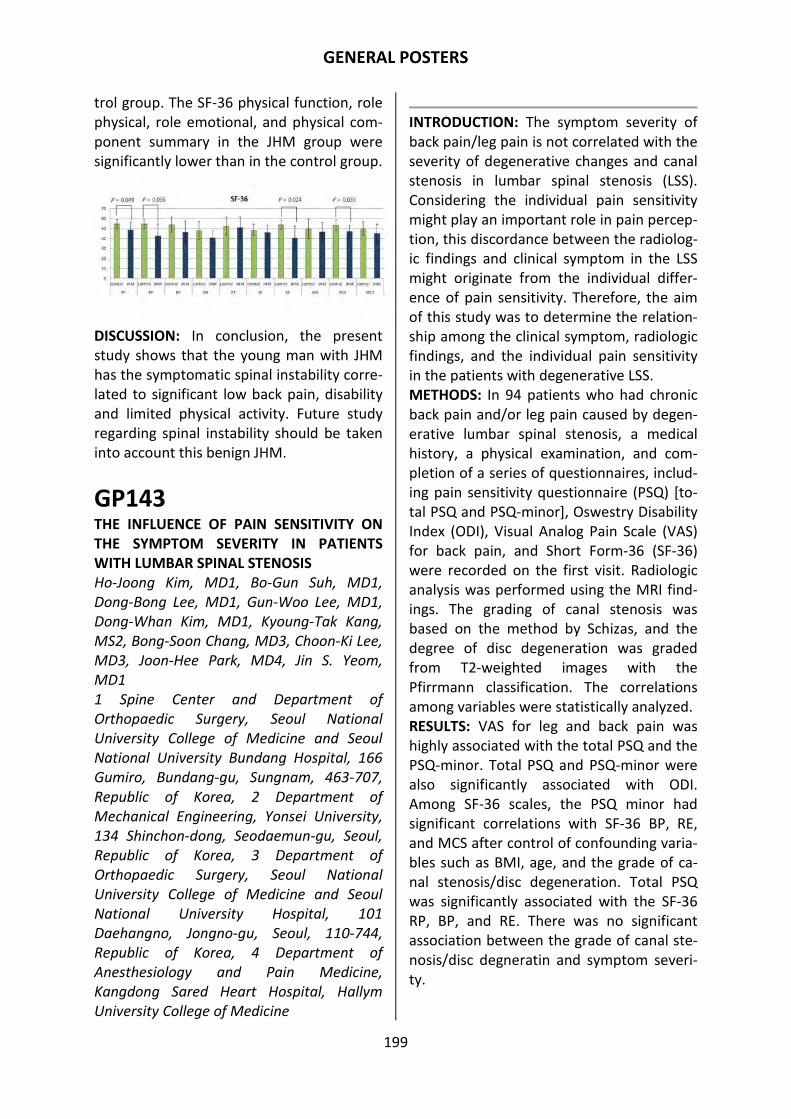
GENERAL POSTERS
199
trol group. The SF-36 physical function, role
physical, role emotional, and physical com-
ponent summary in the JHM group were
significantly lower than in the control group.
DISCUSSION: In conclusion, the present
study shows that the young man with JHM
has the symptomatic spinal instability corre-
lated to significant low back pain, disability
and limited physical activity. Future study
regarding spinal instability should be taken
into account this benign JHM.
GP143 THE INFLUENCE OF PAIN SENSITIVITY ON THE SYMPTOM SEVERITY IN PATIENTS WITH LUMBAR SPINAL STENOSIS
Ho-Joong Kim, MD1, Bo-Gun Suh, MD1,
Dong-Bong Lee, MD1, Gun-Woo Lee, MD1,
Dong-Whan Kim, MD1, Kyoung-Tak Kang,
MS2, Bong-Soon Chang, MD3, Choon-Ki Lee,
MD3, Joon-Hee Park, MD4, Jin S. Yeom,
MD1
1 Spine Center and Department of
Orthopaedic Surgery, Seoul National
University College of Medicine and Seoul
National University Bundang Hospital, 166
Gumiro, Bundang-gu, Sungnam, 463-707,
Republic of Korea, 2 Department of
Mechanical Engineering, Yonsei University,
134 Shinchon-dong, Seodaemun-gu, Seoul,
Republic of Korea, 3 Department of
Orthopaedic Surgery, Seoul National
University College of Medicine and Seoul
National University Hospital, 101
Daehangno, Jongno-gu, Seoul, 110-744,
Republic of Korea, 4 Department of
Anesthesiology and Pain Medicine,
Kangdong Sared Heart Hospital, Hallym
University College of Medicine
INTRODUCTION: The symptom severity of
back pain/leg pain is not correlated with the
severity of degenerative changes and canal
stenosis in lumbar spinal stenosis (LSS).
Considering the individual pain sensitivity
might play an important role in pain percep-
tion, this discordance between the radiolog-
ic findings and clinical symptom in the LSS
might originate from the individual differ-
ence of pain sensitivity. Therefore, the aim
of this study was to determine the relation-
ship among the clinical symptom, radiologic
findings, and the individual pain sensitivity
in the patients with degenerative LSS.
METHODS: In 94 patients who had chronic
back pain and/or leg pain caused by degen-
erative lumbar spinal stenosis, a medical
history, a physical examination, and com-
pletion of a series of questionnaires, includ-
ing pain sensitivity questionnaire (PSQ) [to-
tal PSQ and PSQ-minor], Oswestry Disability
Index (ODI), Visual Analog Pain Scale (VAS)
for back pain, and Short Form-36 (SF-36)
were recorded on the first visit. Radiologic
analysis was performed using the MRI find-
ings. The grading of canal stenosis was
based on the method by Schizas, and the
degree of disc degeneration was graded
from T2-weighted images with the
Pfirrmann classification. The correlations
among variables were statistically analyzed.
RESULTS: VAS for leg and back pain was
highly associated with the total PSQ and the
PSQ-minor. Total PSQ and PSQ-minor were
also significantly associated with ODI.
Among SF-36 scales, the PSQ minor had
significant correlations with SF-36 BP, RE,
and MCS after control of confounding varia-
bles such as BMI, age, and the grade of ca-
nal stenosis/disc degeneration. Total PSQ
was significantly associated with the SF-36
RP, BP, and RE. There was no significant
association between the grade of canal ste-
nosis/disc degneratin and symptom severi-
ty.
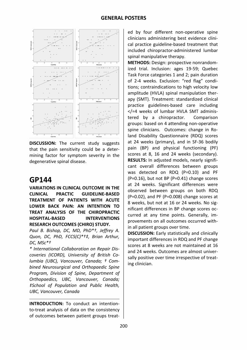
GENERAL POSTERS
200
DISCUSSION: The current study suggests
that the pain sensitivity could be a deter-
mining factor for symptom severity in the
degenerative spinal disease.
GP144 VARIATIONS IN CLINICAL OUTCOME IN THE CLINICAL PRACTIC GUIDELINE-BASED TREATMENT OF PATIENTS WITH ACUTE LOWER BACK PAIN: AN INTENTION TO TREAT ANALYSIS OF THE CHIROPRACTIC HOSPITAL-BASED INTERVENTIONS RESEARCH OUTCOMES (CHIRO) STUDY. Paul B. Bishop, DC, MD, PhD*†, Jeffrey A.
Quon, DC, PhD, FCCS(C)*†‡, Brian Arthur,
DC, MSc*†
* International Collaboration on Repair Dis-
coveries (ICORD), University of British Co-
lumbia (UBC), Vancouver, Canada; † Com-
bined Neurosurgical and Orthopaedic Spine
Program, Division of Spine, Department of
Orthopaedics, UBC, Vancouver, Canada;
‡School of Population and Public Health,
UBC, Vancouver, Canada
INTRODUCTION: To conduct an intention-
to-treat analysis of data on the consistency
of outcomes between patient groups treat-
ed by four different non-operative spine
clinicians administering best evidence clini-
cal practice guideline-based treatment that
included chiropractor-administered lumbar
spinal manipulative therapy.
METHODS: Design: prospective nonrandom-
ized trial. Inclusion: ages 19-59; Quebec
Task Force categories 1 and 2; pain duration
of 2-4 weeks. Exclusion: “red flag” condi-
tions; contraindications to high velocity low
amplitude (HVLA) spinal manipulation ther-
apy (SMT). Treatment: standardized clinical
practice guidelines-based care including
</=4 weeks of lumbar HVLA SMT adminis-
tered by a chiropractor. Comparison
groups: based on 4 attending non-operative
spine clinicians. Outcomes: change in Ro-
land Disability Questionnaire (RDQ) scores
at 24 weeks (primary), and in SF-36 bodily
pain (BP) and physical functioning (PF)
scores at 8, 16 and 24 weeks (secondary).
RESULTS: In adjusted models, nearly signifi-
cant overall differences between groups
was detected on RDQ (P=0.10) and PF
(P=0.16), but not BP (P=0.41) change scores
at 24 weeks. Significant differences were
observed between groups on both RDQ
(P=0.02), and PF (P=0.008) change scores at
8 weeks, but not at 16 or 24 weeks. No sig-
nificant differences in BP change scores oc-
curred at any time points. Generally, im-
provements on all outcomes occurred with-
in all patient groups over time.
DISCUSSION: Early statistically and clinically
important differences in RDQ and PF change
scores at 8 weeks are not maintained at 16
and 24 weeks. Outcomes are almost univer-
sally positive over time irrespective of treat-
ing clinician.
GENERAL POSTERS
201
GP145 FACET JOINT TROPISM AND DEGENERA-TIVE SPONDYLOLISTHESIS – A STUDY FROM THE AOSAP RESEARCH COLLABORATION 1 Samartzis D, 2 Rajasekaran S, 3 Kawaguchi
Y, 4 Acharya S, 5 Kawakami M, 6 Satoh S, 7
Chen S, 8 Park CK, 9 Lee CS, 10 Foocharoen T, 11 Nagashima H, 12 Kuh S, 13 Zheng Z, 14 Con-
dor R, 15 Ito M, 16 Iwasaki M, 17 Jeong JH, 1
Luk KDK, 18 Prijambodo B, 19 Rege A, 20 Jahng
TA, 21 Luo Z, 22 Tassanawipas W, 23 Acharya
N, 24 Pokharel R, 25 Shen Y, 26 Ito T, 27
Zhang Z, 28 Aithala J, 29 Kumar GV, 30 Jabir
RA, 31 Basu S, 32 Li B, 33 Moudgil V, 1 Sham P, 34 Williams R
(1) Department of Orthopaedics and Trau-
matology, University of Hong Kong, Hong
Kong, SAR China (2) Department of Ortho-
paedics, Ganga Hospital, Coimbatore, India
(3) Department of Orthopaedic Surgery,
University of Toyama, Toyama, Japan(4)
Department of Orthopedics, Sir Gangaram
Hospital, New Delhi, India (5) Department of
Orthopaedic Surgery, Wakayama Medical
University, Kihoku Hospital, Ito-gun, Japan
(6) Department of Spine Surgery, Eniwa
Hospital, Hokkaido Japan (7) Orthopaedic
Department, Chang Gung Memorial Hospi-
tal, Taoyuan, Taiwan
(8) Department of Neurosurgery, Seoul St.
Mary's Hospital, Catholic University of Ko-
rea, Seoul, South Korea (9) Department of
Orthopedic Surgery, Samsung Medical Cen-
ter, Sungkyunkwan University School of
Medicine, Seoul, South Korea (10) Depart-
ment of Orthopaedic Surgery, Khonkaen
Regional Hospital, Khonkean, Thailand (11)
Department of Orthopedic Surgery, Faculty
of Medicine, Tottori University, Yonago, Ja-
pan (12) Department of Neurosurgery,
Gangnam Severance Hospital, Seoul, South
Korea (13) Department of Spine Surgery,
The First Hospital Affiliated of Zhongshan
University, Guangzhou, China (14) Depart-
ment of Orthopedics, Cebu Orthopaedic In-
stitute, Cebu, Philippines (15) Department of
Advanced Medicine for Spine and Spinal
Cord Disorders, Hokkaido University Gradu-
ate School of Medicine, Sapporo, Japan (16)
Department of Orthopaedic Surgery, Osaka
University Graduate School of Medicine,
Suita, Japan (17) Department of Neurosur-
gery, College of Medicine, Hangang Sacred
Heart Hospital, Hallym University, Seoul,
South Korea (18) Department of Orthopae-
dic and Traumatology, Faculty of Medicine
Airlargga University, Dr Soetomo Teaching
Hospital, Surabaya, Indonesia (19) Depart-
ment of Orthopaedics, Deenanath
Mangeshkar Hospital, Jehangir Hospital,
Pune, India (20) Department of Neurosur-
gery, Seoul National University Bundang
Hospital, Seongnam, South Korea (21) Spine
Service, Department of Orthopaedic Sur-
gery, Xijing Hospital, the Fourth Military
Medical University, Xi'an, China (22) De-
partment of Orthopedics,
Phramongkuthklao Army Hospital, Bangkok,
Thailand (23) Dwaraka Institute of Spine
Care, Bellary, India (24) Department of Or-
thopedics & Trauma Surgery, Spine Unit,
Tribhuvan University, Teaching Hospital,
Kathmandu, Nepal (25) Department of Spine
Surgery, The Third Hospital of Hebei Medical
University of China, Shijiazhuang, China (26)
Department of Orthopaedic Surgery, Niigata
City General Hospital, Niigata, Japan (27)
Department of Orthopaedic Surgery, Beijing
361 Hospital (Aviation General Hospital),
Beijing, China (28)Department of Orthope-
dics, Kasturba Medical College, Manipal
University, Mani, Udupi, India (29) Depart-
ment of Neurosurgery, Fortis Hospital, Kol-
kata, India (30) Orthopaedic and Trauma-
tology Department, University of Indonesia /
RS Ciptomangunkusumo, Jakarta, Indonesia
(31) Neurosciences Division, Park Clinic, Kol-
kata, India (32) Department of Spine Sur-
gery, The Third Hospital of Hebei Medical
University of China, Shijiazhuang, China (33)
Department of Orthopedic, Punjab Institute
of Medical Sciences Jalandhar, Jalandhar,
India (34) Department of Orthopaedics,
Princess Alexandra Hospital, Brisbane, Aus-

GENERAL POSTERS
202
tralia
INTRODUCTION: Orientation of lumbar fac-
et joints (FJ) may be associated with the
pathogenesis of degenerative
spondylolisthesis (dSpl). However, the role
of FJ tropism (i.e. asymmetry between facet
joint orientations at the same level) in dSpl
is undetermined. This study addresses the
role of FJ tropism in relation to dSpl.
METHODS: A multi-national, cross-sectional
study was performed in 34 institutions in
the Asia Pacific region. Lateral standing ra-
diographs and axial MRIs and/or CT scans
were obtained for subjects with single level
lumbar dSpl. Imaging was analyzed to assess
lumbar levels with L4-5 dSpl (Group A) or
without dSpl (Group B). FJ tropism was de-
fined as a ≥ 7 degree angulation difference and also assessed on ROC curve analysis to
identify critical values. Subject de-
mographics were also noted.
RESULTS: The study included 351 patients
(36.9% males, 63.1% females) with a mean
age of 61.8 years. There were 267 patients
(76.1%) in Group A and 84 individuals
(23.9%) in Group B (control). A significant
difference was noted in FJ angulations be-
tween Group A (mean right: 57.5 degrees;
left: 55.4 degrees) and Group B (mean right:
48.4 degrees; left: 46.5 degrees) (p<0.001).
Based on FJ tropism of 7 degrees, there was
no statistically significant difference be-
tween Groups. ROC analysis identified FJ
angulation difference of 15 degrees or
greater associated with dSpl. Based on age
and FJ angulation-adjusted multivariate
analysis, FJ tropism with a critical value of
≥15 degrees noted an odds ratio of 2.43 (95% CI: 1.20 - 4.91; p=0.014) associated
with dSpl. Slippage was noted with in-
creased FJ tropism, but the effects could not
be discerned.
DISCUSSION: Greater sagittal FJ orientation
was associated with dSpl, as was joint tro-
pism. A critical value of 15 degrees FJ angle
difference produced a two-fold increased
likelihood of dSpl. Our study broadens the
understanding of FJ morphology and its role
in degenerative sagittal plane instability.
GP146 HIP FLEXION CONTRACTURE AND OUT-COME OF CORRECTIVE SURGERY FOR ADULT SPINAL DEFORMITY WITH SAGITTAL IMBALANCE
Atsunori Yuasa, PT, Masahiro Kanayama,
MD, Tomoyuki Hashimoto, MD, Keiichi
Shigenobu, MD, Fumihro Oha, MD, Akira
Iwata, MD, Takashi Onishi, MD, Masaru
Tanaka, MD
Spine Center, Hakodate Central General
Hospital, Hon-cho 33-2, Hakodate 040-8585,
Japan
INTRODUCTION: Hip flexion contracture
might be a potential risk for fixation failure
and loss of correction in surgical treatment
of sagittal imbalance. This study aimed to
evaluate preoperative hip flexion contrac-
ture and to investigate the effect of periop-
erative physical therapy on the outcomes of
corrective surgery sagittal imbalance.
METHODS: Fourteen patients who under-
went corrective surgery for adult spinal de-
formity with sagittal imbalance were in-
cluded in this study. The mean age was 68
years (54-78 years). Surgical procedures
were posterior or transforaminal lumbar
interbody fusion (PLIF/TLIF) in eight pa-
tients, pedicle subtraction osteotomy (PSO)
in five patients, and posterior vertebral col-
umn resection (PVCR) in one patient. Hip
range of motion (ROM) was measured pre-
operatively in all patients. Improvement of
ROM was also assessed in patients with
physical therapy intervention for hip con-
tracture. “Remaining sagittal imbalance”
was defined as more than 10 cm deviation
of C7 plumb line even after surgery.
RESULTS: Ten patients achieved improve-
ment of sagittal balance (successful correc-
tion group), but sagittal imbalance re-
mained in four patients (poor correction
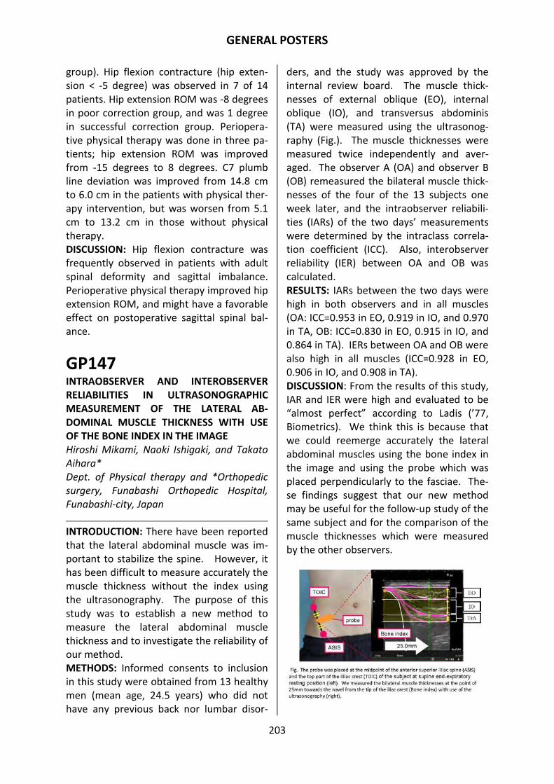
GENERAL POSTERS
203
group). Hip flexion contracture (hip exten-
sion < -5 degree) was observed in 7 of 14
patients. Hip extension ROM was -8 degrees
in poor correction group, and was 1 degree
in successful correction group. Periopera-
tive physical therapy was done in three pa-
tients; hip extension ROM was improved
from -15 degrees to 8 degrees. C7 plumb
line deviation was improved from 14.8 cm
to 6.0 cm in the patients with physical ther-
apy intervention, but was worsen from 5.1
cm to 13.2 cm in those without physical
therapy.
DISCUSSION: Hip flexion contracture was
frequently observed in patients with adult
spinal deformity and sagittal imbalance.
Perioperative physical therapy improved hip
extension ROM, and might have a favorable
effect on postoperative sagittal spinal bal-
ance.
GP147 INTRAOBSERVER AND INTEROBSERVER RELIABILITIES IN ULTRASONOGRAPHIC MEASUREMENT OF THE LATERAL AB-DOMINAL MUSCLE THICKNESS WITH USE OF THE BONE INDEX IN THE IMAGE
Hiroshi Mikami, Naoki Ishigaki, and Takato
Aihara*
Dept. of Physical therapy and *Orthopedic
surgery, Funabashi Orthopedic Hospital,
Funabashi-city, Japan
INTRODUCTION: There have been reported
that the lateral abdominal muscle was im-
portant to stabilize the spine. However, it
has been difficult to measure accurately the
muscle thickness without the index using
the ultrasonography. The purpose of this
study was to establish a new method to
measure the lateral abdominal muscle
thickness and to investigate the reliability of
our method.
METHODS: Informed consents to inclusion
in this study were obtained from 13 healthy
men (mean age, 24.5 years) who did not
have any previous back nor lumbar disor-
ders, and the study was approved by the
internal review board. The muscle thick-
nesses of external oblique (EO), internal
oblique (IO), and transversus abdominis
(TA) were measured using the ultrasonog-
raphy (Fig.). The muscle thicknesses were
measured twice independently and aver-
aged. The observer A (OA) and observer B
(OB) remeasured the bilateral muscle thick-
nesses of the four of the 13 subjects one
week later, and the intraobserver reliabili-
ties (IARs) of the two days’ measurements
were determined by the intraclass correla-
tion coefficient (ICC). Also, interobserver
reliability (IER) between OA and OB was
calculated.
RESULTS: IARs between the two days were
high in both observers and in all muscles
(OA: ICC=0.953 in EO, 0.919 in IO, and 0.970
in TA, OB: ICC=0.830 in EO, 0.915 in IO, and
0.864 in TA). IERs between OA and OB were
also high in all muscles (ICC=0.928 in EO,
0.906 in IO, and 0.908 in TA).
DISCUSSION: From the results of this study,
IAR and IER were high and evaluated to be
“almost perfect” according to Ladis (’77,
Biometrics). We think this is because that
we could reemerge accurately the lateral
abdominal muscles using the bone index in
the image and using the probe which was
placed perpendicularly to the fasciae. The-
se findings suggest that our new method
may be useful for the follow-up study of the
same subject and for the comparison of the
muscle thicknesses which were measured
by the other observers.

GENERAL POSTERS
204
GP148 THE EFFICACY OF LP-IDOPE AS A NEAR-INFRARED PROBE IN THE MANAGEMENT OF PAIN CAUSED BY SPINAL METASTASIS
Kazuyo Yamauchi1), Kazuhide Inage1),
Miyako Suzuki1), Yoshihiro Sakuma1), Yasuhi-
ro Oikawa1), Tetsuhiro Ishikawa1), Masayuki
Miyagi1), Hiroto Kamoda1), Go Kubota1),
Takeshi Saino1), Jun Sato1), Sumihisa Orita1),
Kazuhisa Takahishi1), Yutaka Tamura2), Seiji
Ohtori1)
1) Department of Orthopaedic surgery,
Chiba University 2)Department of
Bioinformatics, Chiba University
INTRODUCTION: The cause of and
mechanism underlying pain due to spinal
metastasis, despite severe and refractory
therapy, is unclear. No standard therapy has
yet been established for the management of
pain due to spinal metastatic tumors. In a
recent study, we have reported that
liposomally formulated 1,2-dioleoyl-sn-
glycero-3-phosphoethanolamine(LP-iDOPE)
along with indocyanine green as a near-
infrared(NIR) probe accumulates near fragile
vascular tissue. LP-iDOPE accumulates near
various tumors and inhibits tumor growth
after NIR irradiation. However, to our
knowledge, no studies have reported
whether it plays a role in relieving pain
caused by spinal metastasis. The aim of this
study was to determine the efficacy of LP-
iDOPE as an NIR probe for alleviating pain
due to spinal metastatic tumors.
METHODS: Rat breast tumor cell-line cluster
transplanted at the L6 vertebral body was
used as the spinal metastasis model. Seven
days after surgery, the L6 vertebra was
intravenously injected with LP-iDOPE and
subjected to NIR irradiation. Then, in-vivo
fluorescence imaging of the L6 was
performed after 24, 48, and 168 h. Further,
the bilateral dorsal root gangliaon(DRGs) at
the L1–L6 levels were resected and
immunohistochemically evaluated using
calcitonin gene-related peptide(CGRP)
induction with TRPV1 as a capsaicin
receptor. The percentages of TRPV1-
immunoreactive (IR) DRG cells and CGRP-IR
DRG cells were statistically compared
between the groups.
RESULTS: Tumor growth was inhibited in the
region showing spinal metastasis after NIR
irradiation. The percentage of CGRP- and
TRPV1-IR DRG cells was lower in the NIR-
irradiated group than in the non-NIR-
irradiated.
DISCUSSION: Spinal metastasis is associated
with acid and pH fluctuation. We found that
the levels of TRPV1, a sensor of acid and pH
fluctuation, and CGRP, a nociceptive
neurotransmitter, decrease after NIR
irradiation. Therefore, LP-iDOPE, as an NIR
probe, is effective in managing the pain
caused by spinal metastasis.
GP149 HEALTH, SOCIAL AND ECONOMIC CONSE-QUENCES OF NECK INJURIES: A CON-TROLLED NATIONAL STUDY EVALUATING SOCIETAL EFFECTS ON PATIENTS AND THEIR PARTNERS
Tom Bendix (1), Poul Jennum (2), Jakob
Kjellberg (3), Rikke Ibsen (4).
(1) Center for Rheumathology and spinal
Diseases, University of Copenhagen,
Glostrup Hospital, Denmark; (2) Danish
Center for Sleep Medicine, Department of
Clinical Neurophysiology, Center for Healthy
Aging, Faculty of Health Sciences, University
of Copenhagen, Glostrup Hospital, Copen-
hagen, Denmark; (3) Danish Institute for
Health Services Research, Copenhagen,
Denmark; (4) itracks, Klosterport 4E, 4, Aar-
hus, Denmark.
INTRODUCTION: Despite neck injuries
cause significant socio-economic burdens,
there is insufficient information about the
time course, as well as the effect on their
partners. The aim was to estimate health-
care costs of neck injuries, except fractures,
in patients and their partners, both follow-

GENERAL POSTERS
205
ing and preceeding the injury.
METHODS: In a case-control study, using
records from the Danish National Patient
Registry 1998-2009, all patients with a diag-
nosis of neck injury and their partners were
identified and compared with randomly
chosen controls matched for age, gender,
geographic area and civil status. Direct costs
included frequency of primary and hospital
sector contacts, procedures and medica-
tion. Indirect costs included the effect on
labour supply. Social transfer payments
were included to illustrate the effect on
national accounts.
RESULTS: The register contributed 94,224
patients, and 372,341 matched controls
were identified. Approximately 47.5% were
married or cohabiting in both groups. Neck
injury patients had significantly higher rates
of health-related contacts, medication use
and higher socio-economic costs than con-
trols. To a lesser extent, they also had lower
employment rates, and those employed
generally had lower incomes. Furthermore,
the patients had already presented negative
social- and health-related status up to 11
years before the first diagnosis, which be-
came more pronounced for those with the
highest costs. The health effects on costs
were present regardless of age group and
gender, and it was also seen for the pa-
tients’ partners.
DISCUSSION/CONCLUSION: Neck injuries
are associated with major socio-economic
consequences for patients, their partners
and society. However, the increased ex-
penses during subsequent years cannot be
explained by the injury alone, because these
patients already had elevated expenses pri-
or to the injury. This indicates some selec-
tion of increased vulnerability for both pa-
tients and their partners.
GP150 CT CLASSIFICATION OF VACUUM PHENOM-ENON FOR THE LUMBAR INTERVERTEBRAL DISC
K. Nishida, K. Maeno, K. Kakutani, T. Taka-
da, J. Yamamoto, H Hirata, T. Kurakawa, S.
Miyazaki, T Yurube, *T. Iguchi, M. Kurosaka
Dept. of Orthopaedic Surgery, Kobe Univer-
sity Graduate School of Medicine, Kobe, Ja-
pan * Dept. of Orthopaedic Surgery, Hyogo
Rehabilitation Center Hospital, Kobe, Japan
INTRODUCTION: Vacuum phenomenon in
the intervertebral disc has been well-
recognized phenomena in disc degeneration
process. However, the clinical significance
as well as the comprehensive analysis has
not been assessed. Our objective was to
clarify characteristics of vacuum phenome-
non in lumbar disc.
METHODS: The study involved 120 lumbar
discs from 24 patients (mean 70.4 years)
with existence of vacuum phenomenon de-
tected by reconstructed sagittal CT images.
Vacuum phenomenon was classified into 5
grades: Grade 1, dot- like change in the nu-
cleus pulposus (NP); Grade2, linear cleft in
the NP; Grade 3, massive defect in the NP;
Grade 4, massive defect from the NP
through annulus fibrosus (AF); and Grade 5,
linear cleft through the disc. Disc height was
measured by CT. Segmental instability was
evaluated by flexion/extension radiographs.
Correlation of vacuum phenomenon grades
with MRI Pfirrmann degeneration grades
and Modic change type was investigated.
RESULTS: Vacuum phenomenon was de-
tected in 49% of 120 discs (L1/2, 3%; L2/3,
6%; L3/4, 10%; L4/5, 17%; and L5/S, 13%),
which were classified into Grade 1, 6%;
Grade 2, 8%; Grade 3, 8%; Grade 4, 14%;
and Grade 5, 13%. Disc height was signifi-
cantly decreased in Grade 5. In comparison
between vacuum phenomenon grades and
Pfirrmann grades, while no disc with
Pfirrmann Grade 1-2 was observed, and
77% of discs with Grade 4 vacuum were

GENERAL POSTERS
206
identified as Pfirrmann Grade 4, and 100%
of discs with Grade 5 vacuum were classi-
fied as Pfirrmann Grade 5. Type 2 Modic
change was observed in 73% of vacuum
Grade 5 discs. In functional radiographs,
10% of segments with vacuum Grade 3 disc
and 18% of segments with vacuum Grade 4
disc showed translation over 3mm.
DISCUSSION: Our grading system for vacu-
um phenomenon facilitates classifying discs
with advanced degeneration in MRI. Even in
Pfirrmann Grade 4-5 discs, vacuum Grade 3-
4 discs with massive defect from NP
through AF may have the potential to in-
duce segmental instability.
GP151 TRUE POSITIVE CONDUCTION TESTS OCCUR RARELY IN CLINICAL PRACTICE Greg McIntosh MSc, Hamilton Hall MD, Tom
Carter BSc PT
CBI Health Group Research Department,
Toronto, Canada
INTRODUCTION: Conduction tests are a
necessary component of the low back pain
(LBP) physical examination. This study com-
pares conduction test findings with location
of dominant pain to establish false positives
from true positives.
METHODS: This was a prospective observa-
tional cohort study of low back pain (LBP)
cases (n=2196) treated non-operatively at
40 spine care rehabilitation clinics across
four provinces between January 2008 and
June 2010. All patients had mechanical LBP
as determined by the Saskatchewan Spine
Pathway triage methodology.
Level test #positive
(back)
#positive
(leg)
total
L4 knee reflex 65 5 70
L4-5 ankle dorsiflexion 58 14 72
L5 trendelenberg 14 0 14
L5 ext. hallicus longus 60 9 69
S1 ankle reflex 82 5 87
S1 plantar flexion 57 12 69
S1 gluteus maximus 14 4 18
RESULTS: The Table displays seven conduc-
tion tests evaluated in this study and the
frequency of occurrence based on location
of dominant pain. There was a statistically
significant difference between conduction
tests that were positive among those with
back pain vs those with leg pain (p<0.05).
DISCUSSION: Positive tests in those without
leg dominant pain or with the reproduction
of LBP only are highly unlikely to be associ-
ated with the current clinical problem. They
may represent a pre-existing neurological
deficit or, more frequently an error in exam-
ination. True positive conduction tests oc-
cur only in those with leg dominant pain;
thus, the ‘positive back’ column is consid-
ered false positives while the ‘positive leg’
column are the true positives. Clinicians
need to cross reference test results with
location of dominant pain to ensure accu-
rate diagnoses and avoid false positives.
GP152 ANALYSIS OF THE S1 SACRAL FORAMINAL LOCATION USING A THREE-DIMENSIONAL CT RENDERING TECHNIQUE
Yusuke Sato , Kotani Toshiaki,Tetsuharu
Nemoto,Tsutomu Akazawa,Tsuyoshi Sa-
kuma, Mitsuhiro Kitamura1, kazuhisa
Takahashi2, Shohei Minami1
1. Dept. of Orthopaedic Surgery, Seirei Sa-
kura Citizen Hospital, Chiba, Japan, 2. Dept.
of Orthopaedic Surgery, Chiba University,
Chiba, Japan
INTRODUCTION: Nerve root blocks (NRBs)
have been widely performed under fluoro-
scopic guidance. Because of the lack of
three-dimensional (3D) anatomical infor-
mation, an S1 NRB is more difficult than a
lumbar NRB. The purpose of the present
study was to investigate the location of the
S1 foramen using a 3D computed tomo-
graphic (3D CT) rendering technique, ray-
sum projection.
METHODS: We retrospectively assessed 60

GENERAL POSTERS
207
patients (30 female, 30 male; age 22‒84
years, average 54.4 years) with lumbar de-
generative disease. Spiral CT was performed
using a multi-detector CT scanner in the
axial plane. Images were networked to im-
aging software Synapse Vincent
V3.0.0003(Fujifilm Medical, Japan) for 3D
imaging. On axial CT imaging, we marked
the center of the posterior sacral foramina
(PS1). The marked point (PS1) was displayed
by 3D CT with ray-sum projection as a sub-
stitute for fluoroscopic radiography. Ray-
sum projection is a 3D rendering technique
in which the attenuation value of each voxel
is summed along a projection line. A ray-
sum image resembles a conventional radio-
graph. The center of the L5 pedicle (PL5)
was also marked on a ray-sum image. The
distances between PS1 and PL5 were meas-
ured in the vertical and lateral directions,
respectively.
RESULTS: PS1 was located 39.3 ± 7.0 mm
caudally and 3.5 ± 3.7 mm laterally from
PL5.
DISCUSSION: The present findings demon-
strate the excellent feasibility of the 3D-CT
rendering technique for assessing the 3D
anatomy of the S1 foramen. Quantitative
analysis indicates that a fluoroscope should
be aimed 39.3 ± 7.0 mm caudal and 3.5 ±
3.7 mm lateral from the center of the L5
pedicle to perform an S1 NRB under fluoro-
scopic guidance. The present findings may
be useful when performing an S1 NRB.
GP153 CLINICAL EVALUATION OF PREGABALIN FOR LUMBAR CANAL STENOSIS PATIENTS WITH PERIPHERAL NEUROPATHIC PAIN
Sumihisa Orita, Gen Inoue, Kazuyo
Yamauchi, Hiroto Kamoda, Masayuki
Miyagi, Tetsuhiro Ishikawa, Miyako Suzuki,
Yoshihiro Sakuma, Go Kubota, Yasuhiro
Oikawa, Kazuhide Inage, Takeshi Sainoh, Jun
Sato, Yukio Nakata, Kazuhisa Takahashi,
Seiji Ohtori
Department of Orthopaedic Surgery, School
of Medicine, Chiba University, Chiba, Japan
INTRODUCTION: Lumbar spinal stenosis
(LSS) patients may experience neuropathic
pain (NeP) that is refractory to existing an-
algesics. Pregabalin (PGB), a novel analgesic
developed for NeP management, suppress-
es the secretion of pain-related mediators
in the synapses by binding to α2δ subunits of Ca2+ channels. PGB has already been
reported to be effective for post-
therapeutic neuralgia and diabetic neuropa-
thy; however, its efficacy in LSS patients
with NeP has not yet been evaluated. The
present study aimed to investigate whether
PGB has an effect on NeP management in
LSS patients.
MATERIALS AND METHODS: The subjects
were LSS patients with NeP identified using
a screening tool, Pain DETECT. Individuals
who experienced leg pain(Visual Analog
Scale [VAS] score > 30) and intermittent
claudication(<300 m) refractory to NSAIDs
for more than a month were included. They
were administered PGB at a dose of 25
mg/d, which was gradually increased to 150
mg/d for 6 wk according to their pain inten-
sities. VAS scores and Japan Orhthopaedic
Association Back Pain Evaluation Question-
naire (JOABPEQ) responses were assessed
before and 1 mo after the PGB treatment
was initiated. JOABPEQ is a novel, validated
method for evaluating the degree of low
back pain (LBP) based on the Roland-Morris
Disability Questionnaires and Short Form 36
containing the following 5 subscores: pain-
related disorders (PD), gait disturbance
(GD), lumbar spine dysfunction (LD), social
life disturbance (SD), and psychological dis-
orders (PD).
RESULTS: Fifty-seven LSS patients were as-
sessed (ave. 71.0 yo). The use of PGB signifi-
cantly decreased their VAS scores (P < 0.01).
With respect to JOABPEQ, PD(P < 0.01),
LD(P = 0.031), GD(P = 0.028), and PD(P =
0.014) showed significant improvement
after PGB treatment. Complications such as
dizziness or nausea were not observed dur-

GENERAL POSTERS
208
ing the study.
CONCLUSION: PGB can alleviate the pain
and improve LBP-related factors such as PD,
LD, GD, and PD in LSS patients with NeP
symptoms.
GP154 EPIDEMIOLOGY, DETERMINANTS, AND FUNCTIONAL STATUS OF SCIATICA: A LARGE-SCALE POPULATION-BASED STUDY IN CHINESE (1) Dino Samartzis, (2) Jaro Karppinen, (1)
Keith DK Luk, (1) Kenneth MC Cheung
(1)Department of Orthopaedics and
Traumtology, University of Hong Kong,
Pokfulam, Hong Kong, SAR China (2) De-
partment of Physical and Rehabilitation
Medicine, University of Oulu, Finland
INTRODUCTION: The prevalence, risk fac-
tors, and outcomes of sciatica have been
sporadically reported in the literature,
largely patient-based and mainly related to
Caucasians. The following study addressed
the epidemiology, determinants, and func-
tional status of sciatica within a Chinese
population.
METHODS: A cross-sectional population-
based study of 2,597 Southern Chinese was
performed assessing sagittal T2-weighted
lumbar MRIs for the presence of radio-
graphic findings from L1-S1 (e.g. HIZ, Modic
changes, Schmorl's nodes). A summated
disc bulge/extrusion (DBE) score was ob-
tained of all levels. Self-reported sciatica
during the past year was assessed. Subject
demographics, environmental/lifestyle fac-
tors, and functional status (i.e. SF-36, ODI)
were also assessed.
RESULTS: There were 60% females and 40%
males (mean age: 42 years). The overall
prevalence of sciatica was 39.4%, which
increased with progression of each age cat-
egory (p=0.005). An elevated DBE score was
found in subjects with reported sciatica
(mean: 0.80) compared to those without
(mean: 0.63) (p<0.001). DBE was most prev-
alent at L5-S1 (31.3%) and L4-L5 (24.3%).
Based on an adjusted multivariate model,
DBE (OR: 1.36; 95% CI: 1.15-1.60), females
(OR: 1.38; 95% CI: 1.17-1.64), obesity (OR:
1.68; 95% CI: 1.25-2.24), and moder-
ate/heavy workload (OR:1.74; 95% CI: 1.11-
2.74) were associated with sciatica. Subjects
with sciatica reported worse SF-36 and ODI
functional outcomes (p<0.05).
DISCUSSION: This is one of the largest
population-based studies to assess de-
mographics, environmental/lifestyle, func-
tional, and imaging factors in relation to the
development of self-reported sciatica. DBE,
females, obesity, and moderate/heavy
workload were significant determinants
associated with sciatica. Decreased func-
tional status was also more prevalent in
sciatica subjects.
GP155 CHANGES IN OBJECTIVELY MEASURED WALKING CAPACITY OVER A 2 YEAR PERI-OD IN INDIVIDUALS WITH LUMBAR SPINAL STENOSIS
Christy C. Tomkins-Lane,* Michele C. Battie
**
* Mount Royal University, Calgary, Canada
** University of Alberta, Edmonton, Canada
INTRODUCTION: Walking capacity is recog-
nized as a key outcome in people with lum-
bar spinal stenosis (LSS). The majority of
studies investigating walking in LSS have
used inaccurate self-report tools. Little is
known about long term changes in objec-
tively measured walking.
OBJECTIVE: Assess changes in objectively
measured walking capacity over 2 years in
people with LSS. Methods: Of 49 subjects
enrolled in a study of walking in LSS, we
were able to follow 26. All subjects had LSS
diagnosed by a spine surgeon, confirmed on
imaging. At baseline and 2 yrs subjects
completed a Self-Paced Walking Test, and a
questionnaire. At follow-up they indicated
treatments accessed over the 2 yr period.

GENERAL POSTERS
209
For analysis subjects were divided into sur-
gical, non-surgical treatment, and no treat-
ment groups.
RESULTS: The 26 subjects had a mean base-
line age of 68.4(9.2) and 65% were female;
11 had surgery over the 2 year period, while
8 had some form of non-surgical treatment
and 7 had no treatment of any kind. The
only significant difference between groups
for baseline walking was between the sur-
gery and no treatment groups. Over the 2
yrs, the surgery group had a non-significant
increase in capacity from 871m(867) to
1402(1009), the non-surgical treatment
group had a significant decrease from
1868m(907) to 960(783), and subjects who
had no treatment showed no change
(1463m(839) to 1301(974)).
DISCUSSION: Individuals who elected to
have surgery had lower baseline walking
capacity compared to those who had no
treatment. A significant decrease in walking
was observed for the 8 subjects who had
non- surgical intervention, yet despite a
similar baseline capacity, the 7 subjects who
had no treatment showed relatively stable
walking capacity over 2 years. Given the
observational nature of this study, we can
make limited conclusions regarding compar-
ison of treatments. However, these results
may aid us in better understanding the na-
ture of walking limitation in LSS.
GP156 OPIOID SENSITIVITY AFFECTS POSTOPERA-TIVE OUTCOME IN LUMBAR SPINE SUR-GERY
Kazuhide Inage. Sumihisa Orita. Kazuyo
Yamauchi. Miyako Suzuki. Yoshihiro Sa-
kuma. Go Kubota. Yasuhiro Oikawa. Takeshi
Sainoh. Jun Sato. Kazuhisa Takahashi. Seiji
Ohtori.
Dept. of Orthopaedic Surgery, Graduate
School of Medicine, Chiba University
INTRODUCTION: Opioid sensitivity altered
by the A118G polymorphism in the opioid
receptor Mu 1 gene is reported to affect
postoperative outcome. However, it is un-
clear whether this is true for lumbar spine
surgery. The purpose of this study was to
prospectively evaluate whether preopera-
tive opioid sensitivity alters postoperative
outcome in patients undergoing lumbar
spine surgery.
METHODS: Subjects included 22 patients
suffering from low back pain (LBP) and radi-
ated leg pain who were treated with opioids
(fentanyl or tramadol) at our hospital from
September 2009 to August 2012. Cases in-
cluded 4 lumber disc herniation and 18
lumbar spinal stenosis patients who under-
went posterior lumbar discectomy and pos-
terior lumbar decompression accompanied
by instrumented posterolateral fusion, re-
spectively. We evaluated their LBP and leg
pain improvement rate on the visual analog
scale (VAS) in response to opioids before
and after surgery. Patients were divided
into two groups according to their response
to presurgical opioid administration: higher
improvement rate on VAS (>50%; HR group)
and lower improvement rate (<50%; LR
group). Symptoms are indicated by a sub-
script “LBP” or “LEG” (leg pain).
RESULTS: For LBP, the HRLBP group showed
58.6% postsurgical improvement, while the
LRLBP group experienced 28.3% improve-
ment; this difference was statistically signif-
icant (P<0.05). For leg pain, the HRLEG
group showed 64.3% postsurgical improve-
ment, while the LRLBP group experienced
32.7% improvement; this difference was not
statistically significant.
DISCUSSION: Higher opioid sensitivity was
indicative of significantly higher postsurgical
improvement in LBP, while no such associa-
tion was observed for leg pain. The results
of the present study suggest that the inten-
sity of LBP derived from lumbar disorders
can be associated with altered opioid sensi-
tivity. This implies that preoperative opioid
sensitivity can be a potential predictive fac-

GENERAL POSTERS
210
tor for postsurgical outcome.
GP157 CHARACTERISTICS OF PAIN-RELATED SEN-SORY INNERVATION ON OSTEOPOROTIC LUMBAR VERTEBRAE WITH OVARIECTOMIZED RATS. Miyako Suzuki*1, Sumihisa Orita*1, Gen
Inoue*2, Masayuki Miyagi*1, Tetsuhiro Ishi-
kawa*1, Hiroto Kamoda*1, Yawara
Eguchi*1, Gen Arai*1 ,Kazuyo Yamauchi*1,
Yoshihiro Sakuma*1, Go Kubota*1, Yasuhiro
Oikawa*1, Kazuhide Inage*1, Takeshi
Sainoh*1, Kazuhisa Takahashi*1, Seiji
Ohtori*1
*1 Department of Orthopaedic Surgery,
Graduate School of Medicine, Chiba
University, Chiba, Japan *2 Department of
Orthopaedic Surgery, Kitasato University
School of Medicine
INTRODUCTION: Osteoporotic patients
without fractures sometimes experience
chronic low back pain of unknown origin.
We have previously reported that the ex-
pression of calcitonin gene-related peptide
(CGRP), a marker of inflammatory pain, is
increased in dorsal root ganglia (DRG) neu-
rons innervating lumbar vertebrae of osteo-
porotic rats. However there is few evicence
what is happening in the osteoporotic spinal
dorsal horn where microglia is activated in a
state of chronic neuropathic pain. The pre-
sent study aimed to examine the character-
istics of pain-related sensory inervation in
rat osteoporotic lumbar vertebrae in their
DRG and spinal dorsal horn.
MATERIALS AND METHODS: As an osteopo-
rosis group(OVX group), we used 21-weeks-
old female rats ovariectomized at 5 weeks
(n=20). As the control group, sham-
operated rats without ovariectomy were
used (n=20). Fluoro-Gold (FG), retrograde
neurotracer, was applied into the myeloid
of L3 to label DRG neuronal cells innervating
the vertebra. Four weeks after the FG-
labeling, the animals were killed to resect
bilateral L1 to L6 DRGs and spinal cord. The
CGRP expression in FG-labeled DRG neu-
rons, Iba-1 (a marker of microglia) expres-
sion in spinal dorsal horn were statistically
examined between the two groups using
immunofluorescence.
RESULTS: The proportions of FG-labeled
CGRP immunoreactive (-ir) DRG neurons in
the OVX group were significantly elevated
compared with the control (p < 0.05). Fur-
thermore, the proportion of Iba-1-ir neu-
rons in the dorsal horn of spinal cord were
also significantly increased in the OVX group
(p <0.05).
DISCUSSION: The result of the present
study suggested that osteoporotic state
might increase CGRP expression in DRG
neurons innervating L3 vertebrae and mi-
croglial activation in spinal dorsal horn, im-
plying that the central nervous system may
be activated as well as peripheral in osteo-
porosis rats. That should be lead to clarify
the pathology of osteoporotic pain.
GP158 SEVERE SCOLIOSIS DURING ADULTHOOD: IS SURGERY THE ONLY POSSIBLE TREAT-MENT?
Fabio Zaina*, Alessandra Negrini*, Silvana
Parzini*, Michele Romano*, Salvatore
Atanasio*, Stefano Negrini+
*ISICO, Milan,Italy; +Don Gnocchi Founda-
tion, Brescia University, Italy
INTRODUCTION: Surgery is generally con-
sidered the only approach for worsening
asult scoliosis. Nevertheless there are pa-
tients who cannot undergo surgery for poor
general health condition or who are willing
not to be fused. An evidence based treat-
ment for these patients is necessary. In our
facility we treat adult scoliosis patients with
specidic exercise to avoid/stop progression.
The aim of this study is to report our results
in a cohort of patients
METHODS: Population: Thirty one patients
(3 males) of 38 ± 11 years and 55 ± 14° Cobb

GENERAL POSTERS
211
scoliosis, treated for 3 (range 1–18) years
because of progression subjectively per-
ceived (17 patients), or objectively docu-
mented (14 patients: subgroup A, previous
observation of 10 years, range 1–27), have
been included. 6 patients (sub-group B)
were observed also after stopping treat-
ment for 6 (3–10) years. Patients were
treated according with a scoliosis specific
exercise protocol (SEAS).
RESULTS: Exercises caused a statistically
significant decrease of 3.6 ± 5° of scoliosis (-
3.2 ± 4.3° per year): 1 patient progressed,
45% improved; in sub-group A results were
identical, after a previous worsening of 9.7
± 6.8° (+2.1 ± 4.3° per year); in sub-group B
stopping exercises caused a progression of
8.3 ± 3.8° (+1.4 ± 0.5° per year). The best
results were observed in patients exercising
since, even if some patients continued to
decrease their curve during the years.
CONCLUSION: SEAS exercises revert the
progression of adult scoliosis, and a pro-
spective study is already under way. The
different results according to length of
treatment could be due to a plateau of cor-
rection or to an increase of quality of the
protocol applied (SEAS changes continuous-
ly according to new nowledge in the litera-
ture). These results question the immediate
need for surgery when facing progression of
deformity in adulthood.
GP159 A STUDY ON DIFFUSION WEIGHTED IMAG-ING FOR LUMBAR DISEASES ~ITS USEFUL-NESS AND LIMIT ~
Hatakeyama Kenji, Ato Shogo, Aihara
Takato, Urusibara Makoto
Funabashi Orthopedic Hospital
INTRODUCTION: Conventional MRI is very
useful for lumbar diseases, but it is often
not sensitive for intra- and extra foraminal
lesions. Since Jun.2011, we are trying to
picturize the nerve root lesions by using
diffusion weighted imaging (DWI), which
can be examined within a short time, in ad-
dition to conventional MRI. The purpose of
this study was to evaluate the usefulness
and the limit of DWI for lumbar diseases.
METHODS: One-hundred fifty two consecu-
tive patients, who were performed MRI and
DWI (1.5-T, b-value 400, imaging time
86seconds) in Jan 2012 for lumbar diseases
were included. One spine surgeon and one
radiological technician interpreted DWI and
recorded the abnormality findings of nerve
root as follows; swelling, stoppage and de-
viation of the runway of nerve root. In addi-
tion, the high intensity of tissues around
nerve was recorded. When both viewers’
findings coincided, they were defined to be
DWI positive. Neurological symptoms and
diagnosis were checked by a medical record
and MRI, so that its sensitivity and specifici-
ty were evaluated.
RESULTS: One-hundred forty four patients
were able to be interpreted (84 men and 60
women). Mean age was 56 (range: 14 to
84). There were 56 lumbar disc herniation
patients (lateral type 5 patients), spinal ca-
nal stenosis 25, spondylolisthesis 20, com-
pression fracture 13, foraminal stenosis 5,
spondylolysis 5, and normal findings were
20 patients. 100 patients had unilateral or
bilateral sciatica. DWI positive were 33 pa-
tients. The sensitivity of DWI positive to
sciatica was 36% and specificity was 20%,
therefore it was not useful. However, as to
lateral disc herniation and foraminal lesions,
sensitivity was 86% and specificity was 80%.
In addition, 2 spondylolysis and 2 metastatic
tumors were detected.
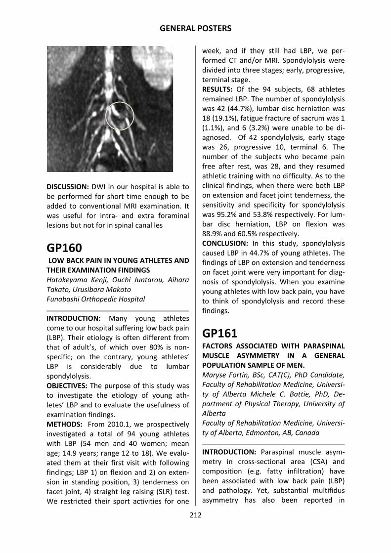
GENERAL POSTERS
212
DISCUSSION: DWI in our hospital is able to
be performed for short time enough to be
added to conventional MRI examination. It
was useful for intra- and extra foraminal
lesions but not for in spinal canal les
GP160 LOW BACK PAIN IN YOUNG ATHLETES AND THEIR EXAMINATION FINDINGS
Hatakeyama Kenji, Ouchi Juntarou, Aihara
Takato, Urusibara Makoto
Funabashi Orthopedic Hospital
INTRODUCTION: Many young athletes
come to our hospital suffering low back pain
(LBP). Their etiology is often different from
that of adult’s, of which over 80% is non-
specific; on the contrary, young athletes’
LBP is considerably due to lumbar
spondylolysis.
OBJECTIVES: The purpose of this study was
to investigate the etiology of young ath-
letes’ LBP and to evaluate the usefulness of
examination findings.
METHODS: From 2010.1, we prospectively
investigated a total of 94 young athletes
with LBP (54 men and 40 women; mean
age; 14.9 years; range 12 to 18). We evalu-
ated them at their first visit with following
findings; LBP 1) on flexion and 2) on exten-
sion in standing position, 3) tenderness on
facet joint, 4) straight leg raising (SLR) test.
We restricted their sport activities for one
week, and if they still had LBP, we per-
formed CT and/or MRI. Spondylolysis were
divided into three stages; early, progressive,
terminal stage.
RESULTS: Of the 94 subjects, 68 athletes
remained LBP. The number of spondylolysis
was 42 (44.7%), lumbar disc herniation was
18 (19.1%), fatigue fracture of sacrum was 1
(1.1%), and 6 (3.2%) were unable to be di-
agnosed. Of 42 spondylolysis, early stage
was 26, progressive 10, terminal 6. The
number of the subjects who became pain
free after rest, was 28, and they resumed
athletic training with no difficulty. As to the
clinical findings, when there were both LBP
on extension and facet joint tenderness, the
sensitivity and specificity for spondylolysis
was 95.2% and 53.8% respectively. For lum-
bar disc herniation, LBP on flexion was
88.9% and 60.5% respectively.
CONCLUSION: In this study, spondylolysis
caused LBP in 44.7% of young athletes. The
findings of LBP on extension and tenderness
on facet joint were very important for diag-
nosis of spondylolysis. When you examine
young athletes with low back pain, you have
to think of spondylolysis and record these
findings.
GP161 FACTORS ASSOCIATED WITH PARASPINAL MUSCLE ASYMMETRY IN A GENERAL POPULATION SAMPLE OF MEN. Maryse Fortin, BSc, CAT(C), PhD Candidate,
Faculty of Rehabilitation Medicine, Universi-
ty of Alberta Michele C. Battie, PhD, De-
partment of Physical Therapy, University of
Alberta
Faculty of Rehabilitation Medicine, Universi-
ty of Alberta, Edmonton, AB, Canada
INTRODUCTION: Paraspinal muscle asym-
metry in cross-sectional area (CSA) and
composition (e.g. fatty infiltration) have
been associated with low back pain (LBP)
and pathology. Yet, substantial multifidus
asymmetry has also been reported in
GENERAL POSTERS
213
asymptomatic men and little is known
about other factors influencing asymmetry.
The goal of this study was to identify a
range of behavioral, environmental and
constitutional factors associated with
paraspinal muscle asymmetry.
METHODS: A cross-sectional study of a gen-
eral population sample of 202 adult male
twins was conducted. Data were collected
through a structured interview, physical
examination and MRI. Multifidus and erec-
tor spinae CSA (muscle size), and the ratio
of fat-free CSA to total CSA (muscle compo-
sition) measurements were obtained from
T2-weighted axial images at L3-L4 and L5-S1
spinal levels.
RESULTS: In multivariable analyses, greater
asymmetry in multifidus CSA (size) at L3-4
was associated with less occupational phys-
ical demands and disc narrowing. No factors
studied were associated with multifidus CSA
asymmetry at L5-S1. For erector spinae,
greater age and disc narrowing were associ-
ated with CSA asymmetry at L3-4 and sports
activity, disc narrowing and familial aggre-
gation at L5-S1. In multivariable analyses of
asymmetry in muscle composition, familial
aggregation was associated with side to side
differences in multifidus and erector spinae
at both levels measured, explaining 9-17%
of the variance. Sports activity and pain se-
verity also entered the model for erector
spinae asymmetry at L5-S1, and disability
and disc narrowing for multifidus at L5-S1.
CONCLUSION: Few of the factors investigat-
ed were associated with paraspinal muscle
asymmetry, and associations were generally
modest, explaining little of the variance in
paraspinal muscle asymmetry. With the
exception of familial aggregation, and disc
narrowing to a lesser degree, associated
factors were not consistent across muscles
or spinal levels.
GP162 THE USA FUNGAL MENINGITIS OUTBREAK AFTER EPIDURAL STEROID INJECTIONS: THOUGHTS ON EPIDEMIOLOGY AND FU-TURE PATIENT SAFETY
David A Wong MD, MSc, FRCS(C)
Co-Chairman NASS Patient Safety Commit-
tee
INTRODUCTION: The September 2012 out-
break of fungal meningitis in the United
States following epidural steroid injections
(ESI) has raised a number of issues concern-
ing the epidemiology of the infection and
future patient safety.
METHODS: The Patient Safety Committee of
the North American Spine Society (NASS)
has liased with the US Centers for Disease
Control (CDC) through the outbreak. CDC
and various state department of health
websites were monitored to follow the in-
vestigation. Literature on complications of
ESI's was reviewed.
RESULTS: Recent CDC data indicates 461
cases of meningitis from approximately
14,000 injections/exposures. This translates
to an infection rate of approximately 3%.
There have been 32 deaths. The CDC has
suggested that the epidemiology is inocula-
tion of the epidural space from an ESI, com-
promise of the immune system from the
systemic steroid effect and the fungus even-
tually "boring" through the dura to infect
the CSF causing meningitis.
DISCUSSION: Questions arise whether there
is an alternate explanation to the epidemi-
ology suggested by the CDC. A literature
review indicates that the accepted rate of
inadvertent intrathecal injection is about 3%
. This is similar to the rate of meningitis in-
fection after ESI with tainted methylpredni-
solone. Thus, one explanation for the epi-
demiology of the meningitis would be that
we are seeing infections develop as a result
of the expected incidence of transdural in-
jection with ESI's. Other technical issues
include whether there is a higher rate of

GENERAL POSTERS
214
infections using a sharper Quincke needle
versus a duller Tuohy, if use of fluroscopy
allows more precise needle placement,
whether multiple ESI's are a risk factor to
infection and whether a transforaminal ESI
has a lower rate of infection compared to
translaminar. These questions will hopefully
be clarified as the investigation proceeds.
The answers will assist in deciding on the
safest injection techniques for our patients
into the future.
GP163 REDUCTION OF SURGICAL REVISION RATES AND ECONOMIC ANALYSIS OF USING AN INTRAOPERATIVE CT-BASED NAVIGATION SYSTEM FOR PEDICLE SCREW PLACEMENT Scott D. Hodges, DO; Jason C. Eck, DO, MS;
Dannette Newton, MS
Center for Sports Medicine and Orthopae-
dics, Chattanooga, TN
INTRODUCTION: Incidence of pedicle screw
breech varies based on anatomic location,
body habitus, surgeon experience, spinal
deformity, and surgical technique. Pedicle
breeches have been reported to occur in up
to 40% of screws. The purpose of the cur-
rent study was to compare the rates of re-
turning to the operating room for revision
of pedicle screw placement when using C-
arm versus O-arm assessment of pedicle
screws.
METHODS: A retrospective study was per-
formed to determine the rates of returning
to the operating room for revision of a
breeched pedicle screw when using C-arm
versus O-arm visualization. An economic
analysis was also performed based on esti-
mated cost of return to the operating room
for revision of the pedicle screws.
RESULTS: There were 4 (1%) of 386 control
cases that required a return to the operat-
ing room for pedicle screw revision for a
breeched pedicle screw not identified with
intraoperative C-arm fluoroscopy. In the
study group none of the 331 patients were
returned to the operating room when using
the O-arm to assess the placement of pedi-
cle screws. Based on the 1% rate of return-
ing to the operating room for pedicle screw
revision in the control group, the annual
rate of cases nationwide requiring pedicle
screw revision would be approximately
2300 with a cost of approximately
$40,595,000.
DISCUSSION: These results suggest that the
use of intraoperative CT can reduce the
need for return to the operating for revision
of a breeched pedicle screw. This can po-
tentially lead to a major cost savings.
GP164 ROD BREAKAGE AFTER LONG CONSTRUCT FUSION IN SPINAL DEFORMITY: CLINICAL AND RADIOGRAPHIC RISK FACTORS
Akazawa, Tsutomu; Kotani, Toshiaki;
Sakuma, Tsuyoshi; Nemoto, Tetsuharu;
Sato, Yusuke; Kitamura, Mitsuhiro; Minami,
Shohei; Ohtori, Seiji; Takahashi, Kazuhisa.
Seirei Sakura Citizen Hospital, Dept.
Orthopedic Surgery, Sakura, Japan; Chiba
University, Graduate School of Medicine,
Dept. Orthopedic Surgery, Chiba, Japan.
INTRODUCTION: The aim of this study was
to analyze clinical and radiographic risk fac-
tors of rod breakage after long construct
fusion in spinal deformity.
METHODS: The survey subjects were 155
cases who were diagnosed with spinal de-
formity and underwent correction and fu-
sion surgery with long construct instrumen-
tation (>3 levels, average 10.3 levels) be-
tween July 2004 and June 2010. The sub-
jects comprised 32 males and 123 females
with a mean age of 19.0 (range 8-78) years.
The mean follow-up period was 46.1
months (range: 24-94 months). All patients
were examined for rod breakage in x-rays at
a follow-up visit. Univariate analysis and
logistic regression analysis were performed.
RESULTS: Rod breakage occurred in 8 of 155
cases (5.2%). The mean period from surgery

GENERAL POSTERS
215
to rod breakage was 18.1 months (range 2-
37). Six patients had breakage near the
fused lower end and two patients had it at
the thoracolumbar junction. The clinical
symptoms were pain in 2 patients and a
breaking sound of a crack in 2 patients.
There was no symptom in 4 patients.
Univariate analysis revealed that non-
ambulatory status, preoperative kyphosis,
small diameter rods, multiple surgeries, and
use of iliac screws were significant risk fac-
tors in rod breakage. Sex, obesity, severity
of preoperative scoliosis, and rod material
were not significant risk factors. Logistic
regression analysis revealed that the follow-
ing were risk factors for rod breakage: use
of iliac screws (odds ratio: 81.9, 95% confi-
dence interval: 7.2-935.0, p<0.001) and
small diameter rods (diameters of less than
6 mm) (odds ratio: 16.3, 95% confidence
interval: 1.7-152.6, p=0.015).
DISCUSSION: The incidence of rod breakage
after long construct fusion in spinal deform-
ity was 5.2%. Iliac screw fixation and small
diameter rod were risk factors for rod
breakage.
GP165 GOOD FUSION RATE AND CLINICAL RE-SULTS OF LESS INVASIVE FACET FUSION WITH SEXTANT PEDICLE SCREW SYSTEM FOR DEGENERATIVE LUMBAR SPONDYLOLISTHESIS
Tomohiro Miyashita *1, Hiromi Ataka *2,
Takaaki Tanno *2
*1: Spine Center, Matsudo City Hospital,
Matsudo, Japan *2: Spine Center, Matsudo
Orthopaedic Hospital, Matsudo, Japan
INTRODUCTION: After a long clinical and
radiological follow-up study of
posterolateral fusion (PLF) for degenerative
lumbar spondylolisthesis (DLS), we recog-
nized that facet fusion (FF) alone would be
sufficient for spinal fusion. We devised a
new, simple, less invasive FF using SEXTANT
pedicle screw system for DLS. In this study,
we assessed fusion rate and clinical results
of FF.
METHODS: Forty-eight patients who un-
derwent FF for single-level DLS were retro-
spectively reviewed at a minimum one year
follow-up. They consisted of 27 women and
21 men with an average age of 68.4 years.
In our surgical method, we added 5cm skin
incision, bilateral laminar fenestration and
FF with autologous bone harvested from
spinous process. Then, pedicle screws were
inserted bilaterally by minimum access
technique.
We evaluated facet fusion rate by comput-
ed tomography (CT), range of motion (ROM)
at the fused level on a flexion-extension
lateral X-ray preoperatively and at the final
follow-up, and therapeutic effectiveness of
FF using Japanese Orthopaedic Association
Back Pain Evaluation Questionnaire
(JOABPEQ).
RESULTS: The facet fusion rate was 83.3% of
patients. ROM at fused level was less than
two degrees at the final follow-up in all the
patients whose facets were sufficiently
fused on CT. In eight patients whose facet
fusion wasn’t sufficient, the average ROM
significantly decreased to 3.8 degrees,
whereas it was 13.4 degrees preoperatively.
The percentage of the patients who showed
therapeutic effectiveness in JOABPEQ were
92.7% in Walking ability and 75.0% in Low
back pain.
DISCUSSION: Park et al reported FF alone is
an effective surgical option for DLS. Howev-
er, in their method, a bone graft was har-
vested from the iliac crest and conventional
pedicle screw system was used. We devised
less invasive FF using local bone graft and
SEXTANT system. The fusion rate and the
clinical results of FF compared well with
those of conventional PLF. Thus, FF is a use-
ful technique for DLS.

GENERAL POSTERS
216
GP166 INTEROBSERVER RELIABILITY OF CT SCANS TO ASSESS RADIOGRAPHIC FUSION CRITE-RIA WITH A NOVEL TITANIUM INTERBODY DEVICE Paul J. Slosar MD; Jay Kaiser MD; Luis Mar-
rero MD; Damon Sacco MD
SpineCare Medical Group/ San Francisco
Spine Institute
INTRODUCTION: The accuracy of CT scans
of titanium implants to assess lumbar
interbody fusion has been questioned in the
past. Published reports have focused on
older cage technologies. Radiologists report
difficulty assessing fusion bone due to arti-
fact and small graft windows. A new titani-
um interbody implant has been developed
with a large footprint and wide aperture for
graft. Purpose: To determine the
interobserver reliability of CT scans to as-
sess radiographic fusion variables with a
new titanium interbody device.
METHODS: Patients underwent ALIF at L3-
S1 as part of a separate clinical outcomes
study. All patients received the same im-
plant type (Endoskeleton TA®; Titan Spine.).
Each cage was packed with 2 sponges of
InFuse® (Medtronic) plus 1-3 cc of HA/ TPC
(MasterGraft®). CT scans with reconstructed
images were performed randomly at 6, 9, or
12 months. Two independent radiologists
reviewed the scans and were blinded to the
other’s interpretation. Interobserver relia-
bility was calculated using the kappa statis-
tic.
RESULTS: 33 patients/ 56 spinal fusion seg-
ments were analyzed. 17 had CT at 6
months, 9 at 9 months, 7 at 12 months. No
differences in results were noted between
time points. Kappa coefficient was calculat-
ed separately for each variable. The kappa
for the overall study was 0.88 (p<.001)
demonstrating agreement between the 2
radiologists in 345 of the 392 data points
reviewed. The agreement for fusion for-
mation was 0.77 (p<.001). Other criteria are
in Table. All measurements reached statisti-
cal significance.
DISCUSSION: Clinicians have been reluctant
to rely on CT scans with titanium devices
due to concerns that the interpretation of
the images may be inaccurate. This device
demonstrated minimal artifact, minimal
subsidence, and trabecular bone was easily
identified through the implant in the majori-
ty of cases. Our study confirms radiographic
fusion criteria can be assessed with high
interobserver reliability with this new tita-
nium implant.
GP167 MICROENDOSCOPIC DISCECTOMY FOR LUMBAR DISC HERNIATION: DOES THE INSTABILITY AFFECT THE LOW BACK PAIN?
Takato Aihara, Kenji Hatakeyama, Makoto
Urushibara, and Juntaro Ouchi
Dept. of Orthopedic Surgery, Funabashi
Orthopedic Hospital, Funabashi-city, Japan
INTRODUCTION: This study was undertaken
to determine the influence of instability on
low back pain (LBP) after microendoscopic
discectomy (MED) for lumbar disc herni-
ation (LDH).
METHODS: MED was used to treat LDH in
42 patients, and the intensity of their leg
pain, leg numbness, and LBP measured on a
visual analogue scale (VAS) was recorded
before and after MED. The degree of im-
provement (DOI) was calculated by the fol-
lowing formula: preoperative score - post-
operative score, and the patients were clas-
sified into two groups based on the DOI in
LBP concerning VAS: a group whose DOI
was more than twenty (M group) and a
group whose DOI was less than twenty (L

GENERAL POSTERS
217
group). Clinical outcomes were evaluated
using the Japanese Orthopaedic Association
Back Pain Evaluation Questionnaire
(JOABPEQ). Lateral radiographs in maximal
flexion and extension, and in neutral posi-
tion were obtained before and after MED to
measure the intervertebral angle and hori-
zontal displacement (translation) in flexion
and extension, and to measure the disc
height of the affected intervertebral space.
Using MRI, disc degeneration was also
graded. All measurements and DOIs were
statistically compared by unpaired t test
between the M and L groups.
RESULTS: The DOIs in LBP concerning VAS
and JOABPEQ were significantly greater in
the M group (n=19) than in the L group
(n=23). There were no statistically signifi-
cant differences between the two groups in
DOI in leg pain or leg numbness concerning
VAS, nor in DOIs in the other four functional
scores concerning JOABPEQ. Preoperative
and postoperative translation in flexion and
extension in the L group had slipped signifi-
cantly posteriorly in comparison with the M
group (Fig.)
DISCUSSION: From the results of this study,
the large posterior translation in flexion and
extension could lead to poor improvement
in LBP after MED. These findings suggest
that posterior translation in flexion and ex-
tension may be a cause of LBP in LDH.
GP168 INFECTION RATE AND COMPLICATIONS FOLLOWING ASSOCIATED WITH EPIDURAL STEROID PASTE USED IN POSTERIOR LUMBAR SURGERY - A CASE-CONTROLLED REVIEW Eva U Asomugha, M.D., Robert F McLain,
M.D.,
Cleveland Clinic Center for Spine Health, and
Cleveland Clinic Department of Orthopaedic
Surgery
INTRO: The use of various epidural agents
to reduce postoperative pain and inflamma-
tion following lumbar surgery is common.
These agents are typically administered as
steroid and/or analgesic laced paste, spong-
es, or foams. Studies have shown the effica-
cy in reducing pain and narcotic usage
acutely with minimal adverse effects. How-
ever, Kramer found a nearly 8-fold increase
in surgical site infections (SSI) after laminec-
tomy when a morphine-prednisolone nerve
paste was used. We tested the hypothesis
that a steroid containing epidural pain
paste, applied after laminotomy, does not
increase risk of acute SSI.
METHODS: A case-controlled retrospective
review of patients treated by lumbar de-
compression, either with or without analge-
sic steroid paste, was done. Demographic
characteristics including age, gender, BMI,
smoking and alcohol status, and type of
operation were analyzed.
Acute SSI were defined as any infection
within the first six weeks postoperatively.

GENERAL POSTERS
218
Late infections were not encountered. SSI
was coded as superficial, deep-suprafascial,
or deep-subfascial.
RESULTS: We analyzed 62 consecutive pa-
tients with no paste, and 61 patients receiv-
ing pain paste. Male patients represented
57. 4% in the paste group and 66.1% in the
no paste group (p=0.32). The average age
of patients in the two groups was 49.8 and
46.1 respectively (p=0.15). There were four
suspected or documented infections, (6.7%)
in the paste group, and one (1.67% [0.03%,
8.7%]) in the no paste group (p=0.21). One
additional patient in the paste group had a
slow healing wound. There were no wound
complications in the no paste group.
DISCUSSION: Our data does not demon-
strate a statistical difference in the rates of
infection with the use of an epidural steroid
paste, but the observed three-fold increase
in the rate is concerning. This trend neces-
sitates a larger review, currently ongoing,
which will allow better statistical certainty.
For the moment we use no steroids in our
paste.
GP169
EPIDEMIOLOGICAL TRENDS IN THE USE OF BONE MORPHOGENIC PROTEIN IN SPINAL FUSIONS FROM 2002-2009
Steven Fineberg MD*, Matthew Oglesby
BA*, Alpesh Patel MD^, Miguel Pelton BS*,
Kern Singh MD*
*Rush University Medical Center, Chicago, IL
^Loyola University Medical Center, May-
wood, IL
INTRODUCTION: The use of bone
morphogenic proteins (BMP) as an adjunct
to spinal fusion has increased since first
approved for use in anterior lumbar
interbody fusions in 2002. The purpose of
this study was to analyze a population-
based database to describe national trends
of BMP use in terms of incidence, de-
mographics, costs and mortality.
METHODS: Data from the Nationwide Inpa-
tient Sample database was obtained from
2002-2009. Patients undergoing anterior or
posterior cervical fusions (ACF/PCF), anteri-
or or posterior lumbar fusions (ALF/PLF), or
thoracic fusions were identified and BMP
use was examined by year. Demographics,
co-morbidities, costs (adjusted for inflation),
and mortality were assessed. Statistical
analysis was performed using Pearson’s
correlation to assess for trends with a p-
value <0.01 to denote significance.
RESULTS: An estimated 482,141 procedures
utilized BMP from 2002-2009 in the United
States (Table 1). The number of procedures
per year that utilized BMP significantly in-
creased from 1,734 in 2002 to 106,168 in
2009 (p<0.0005, R=0.9). ALF had the highest
rates of BMP use, increasing to 53.9% of all
ALFs in 2007 and then decreasing to 50.2%
in 2009 (Figure 1). PLFs accounted for the
majority of cases that used BMP with
290,237 cases from 2002-2009. There was a
significant trend of older patients with in-
creasing co-morbidities receiving BMP dur-
ing this time period. Hospital costs signifi-
cantly increased an average of $5,875 from
2002-2009 (Table 2). Mortality did not
change with utilization of BMP from 2002-

GENERAL POSTERS
219
2009.
DISCUSSION: Our study demonstrates that
the use of BMP has dramatically increased
from 2002-2009. Off-label use of BMP ac-
counts for the vast majority of BMP utiliza-
tion. The increase in costs from 2002-2009
is likely multi-factorial; older patients with
more co-morbidities undergoing surgery as
well as increasing use of BMP are likely to
affect costs. Further studies are needed to
identify long-term outcomes of BMP use in
spinal fusions
GP170
EPIDEMIOLOGICAL TRENDS IN KYPHOPLASTY PROCEDURES BETWEEN 2004-2009
Steven Fineberg MD*, Miguel Pelton BS*,
Alpesh Patel MD^, Matthew Oglesby BA*,
Kern Singh MD*
*Rush University Medical Center, Chicago, IL
^Loyola University Medical Center, May-
wood, IL
INTRODUCTION: Kyphoplasty was intro-
duced as a minimally invasive procedure to
treat vertebral compression fractures with
deformity. The purpose of this study was to
analyze a population-based database to
describe national trends of kyphoplasty
procedures in the United States.
METHODS: Data from the Nationwide Inpa-
tient Sample database was obtained from
2004-2009. Patients undergoing
kyphoplasty for the diagnosis of pathologic
fractures of the thoracolumbar spine were
identified. Demographics (gender, age, and
race), Charlson Co-morbidity Index (CCI),
costs (adjusted for inflation), and mortality
were assessed. Statistical analysis was per-
formed using Pearson's correlation to iden-
tify trends. Logistic regression was per-
formed to identify predictors of mortality.
RESULTS: The total number of patients un-
dergoing kyphoplasty increased from
44,459 in 2004-2005 to 85,530 in 2006-2007
and then decreased to 76,583 in 2008-2009.
The mean pre-operative CCI increased from
4.59 in 2004 to 5.06 in 2009
(p<0.0005)(Table 1).
Mean in-hospital costs significantly in-
creased from $12,815 in 2004 to $16,853 in
2009 (p=0.006). Mortality did not change
across this time period (p=0.32). Predictors
of mortality in patients undergoing
kyphoplasty were metastatic cancer, flu-
id/electrolyte disorders, congestive heart
failure, pulmonary circulation disorders,
renal failure, and weight loss.
DISCUSSION: Our study demonstrates that
kyphoplasty procedures have increased in
incidence from 2004-2009. We identified a
trend of older patients with increasing co-
morbidities undergoing kyphoplasty. Mor-
tality has not changed despite the increas-
ing co-morbidity of patients. This patient
population also incurred increasing hospital-
ization days and costs which may be due to
increasing chronic illnesses. Patients with
mortality risk factors indicative of chronic
illness should be carefully followed for
complications in the peri-operative period
after kyphoplasty procedures.
GP171 TRENDS IN COMPLICATIONS AFTER LUM-BAR SPINE SURGERY FROM 2002-2009
Miguel Pelton BS*, Steven Fineberg MD*,
Alpesh Patel MD^, Matthew Oglesby BA*,
Kern Singh MD*
*Rush University Medical Center, Chicago, IL
^Loyola University Medical Center, May-
wood, IL
GENERAL POSTERS
220
INTRODUCTION: The frequency of compli-
cations after lumbar decompressions (LD)
and lumbar fusions (LF) varies widely in the
reported literature. The purpose of this
study was to analyze a population-based
database to describe national complication
trends of these two procedures.
METHODS: Data from the Nationwide Inpa-
tient Sample database was obtained from
2002-2009. Patients undergoing LD or LF for
degenerative etiologies were identified.
Eight categories of complications were
tabulated using ICD-9 codes including pul-
monary embolism (PE), deep vein throm-
bosis (DVT), infection, cardiac complica-
tions, hematomas, CSF leaks, post-operative
shock, and neurologic complications.
RESULTS: A total of 578,457 LDs and LFs
were identified from 2002-2009 (Table 1).
Both surgical groups demonstrated signifi-
cant trends of increasing ages, co-
morbidities, and costs during this time peri-
od. LFs had higher complication rates at all
time points compared to the LD group.
There were significant trends in the LD
group of increasing overall complications:
specifically increasing rates of DVTs, infec-
tions, hematomas (p<0.01). Increasing rates
of post-operative shock from 2002-2009
was the only statistically significant trend in
the LF group (p=0.005), while increasing
rates of DVTs approached significance
(p=0.02).
CONCLUSION: Our findings demonstrate
that from 2002-2009, patients undergoing
lumbar procedures had increasing trends of
age and co-morbidities reflecting increases
in surgical challenges and complexity. These
changes in patient demographics may ex-
plain the increasing complication rates after
LDs. Both groups demonstrated a trend of
increasing costs, which may be explained by
increasing ages and co-morbidities of pa-
tients as well as the increasing complication
rates. The complication rate in the LF group
was approximately double that of the LD
group for all years. Despite a trend of in-
creasing risk factors during this 8-year peri-
od, mortality did not change.
GP172
INCIDENCE AND MORTALITY OF THROM-BOEMBOLIC EVENTS AFTER LUMBAR SPINE SURGERY
Steven Fineberg MD*, Matthew Oglesby
BA*, Alpesh Patel MD^, Miguel Pelton BS*,
Kern Singh MD*
*Rush University Medical Center, Chicago, IL
^Loyola University Medical Center, May-
wood, IL
INTRODUCTION: Pulmonary embolism (PE)
and deep vein thrombosis (DVT) are poten-
tial complications of orthopaedic proce-
dures. Incidences of these complications are
not well characterized after lumbar spine
surgery. A population-based database was
analyzed to identify incidence, risk factors,
and mortalities associated with lumbar de-
compression (LD) and lumbar fusion (LF).
METHODS: Data from the Nationwide Inpa-
tient Sample database was obtained from
2002-2009. Patients undergoing LD or LF for
degenerative etiologies were identified.
Acute PE and DVT incidences and mortality
rates were calculated. Co-morbidities were
calculated using a modified Charlson Co-
morbidity Index. Statistical analysis using a

GENERAL POSTERS
221
p-value of <0.0005 was used to denote sta-
tistical significance. Logistic regression was
used to identify independent predictors of
thromboembolic events.
RESULTS: A total 578,457 LDs and LFs were
identified from 2002-2009. DVT incidences
were 2.4 and 4.3 per 1,000 cases in the LD
and LF groups respectively (Table 1). PE in-
cidences were 1.0 and 2.6 per 1,000 cases in
the LD and LF groups (Table 2).
LF patients with thromboembolic events
were younger, had less co-morbidity, and
incurred higher costs than LD patients. Sta-
tistically significant predictors of DVT were
pulmonary circulation disorders, coagulopa-
thy, fluid/electrolyte disorders, anemia,
obesity, teaching hospital status, and larger
hospitals. Predictors for the development of
a PE were pulmonary circulation disorders,
fluid/electrolyte disorders, anemia, African-
American ethnicity and teaching hospitals
status.
DISCUSSION: Patients undergoing LD or LF
are at inherent risk of thromboembolic
events. DVT and PE are more common after
LF procedures. Pre-operative pulmonary
circulation disorders, fluid/electrolyte dis-
orders, deficiency anemia, and teaching
hospital status were significant risk factors
for both DVT and PE. Preventive measures
in patients at risk may decrease the inci-
dence of thromboembolic events.
GP173 EPIDEMIOLOGICAL TRENDS IN LUMBAR SPINE SURGERY BETWEEN 2002-2009
Steven Fineberg MD*, Matthew Oglesby
BA*, Alpesh Patel MD^, Miguel Pelton BS*,
Kern Singh MD*
*Rush University Medical Center, Chicago, IL
^Loyola University Medical Center, May-
wood, IL
INTRODUCTION: Lumbar decompressions
(LD) and lumbar fusions (LF) are routinely
performed for lumbar degenerative pathol-
ogy. The purpose of this study was to ana-
lyze a population-based database to de-
scribe national trends of these procedures
in terms of incidence, demographics, co-
morbidities, cost and mortality. METHODS: Data from the Nationwide Inpa-
tient Sample database was obtained from
2002-2009. Patients undergoing LD (i.e.
laminectomy +/- discectomy) or LF for de-
generative etiologies were identified. Co-
morbidities were calculated using a modi-
fied Charlson Co-morbidity Index (CCI). De-
mographics, costs (adjusted for inflation),
and mortality was also assessed. Statistical
analysis was performed using Pearson’s
correlation to assess trends. A p-value of
p<0.01 was used to denote significance.
RESULTS: The percentage of the population
undergoing lumbar procedures did not
change from 2002-2009 (p=0.789)(Table 1-
adjusted for U.S. Census reported popula-
tion). Patients undergoing LD decreased
33% while LF surgeries increased 50%
throughout this time period (Figure 1).
The mean age of patients increased by 4.0
years (p<0.0005). The mean pre-operative
CCI increased by 0.5 points (p<0.0005). This
co-morbidity increase was due to an in-
creasing trend towards patients with colla-
gen vascular disease, anemia, coagulopathy,
diabetes, and hypertension. Hospital costs
GENERAL POSTERS
222
increased by $8,296 over the 8-year period
(p<0.0005). In-hospital mortality did not
change significantly for any cohort.
DISCUSSION: Our study demonstrates that
the total number of lumbar procedures has
not changed over the 8-year period. There
was an overall trend towards more lumbar
fusions and fewer decompressions per-
formed. Mortality did not change despite an
aging population with greater medical co-
morbidities. There was a trend towards in-
creasing hospital costs despite decreasing
hospitalizations in the lumbar fusion group
which may be due to increased utilization of
more expensive implants and biologics.
GP174
RISK FACTORS FOR HEMATOMAS AND NEUROLOGIC COMPLICATIONS AFTER LUMBAR SPINE SURGERY
Matthew Oglesby BA*, Miguel Pelton BS*,
Alpesh Patel MD^, Steven Fineberg MD*,
Kern Singh MD*
*Rush University Medical Center, Chicago, IL
^Loyola University Medical Center, May-
wood, IL
INTRODUCTION: Post-operative hemato-
mas and neurologic complications are po-
tentially catastrophic events that can occur
after spine procedures. A population-based
database was analyzed to identify inci-
dence, mortality, and risk factors associated
with these complications after lumbar de-
compression (LD) and lumbar fusion (LF). METHODS: Data from the Nationwide Inpa-
tient Sample database was obtained from
2002-2009. Patients undergoing LD or LF for
degenerative etiologies were identified.
Incidences of hematomas, neurologic com-
plications, and mortalities were calculated.
Co-morbidities were calculated using a
modified Charlson Co-morbidity Index. Sta-
tistical analysis was performed using p-
value <0.0005 to denote significance. Lo-
gistic regression was used to identify inde-
pendent predictors of hematoma and neu-
rologic complications.
RESULTS: A total of 578,457 lumbar proce-
dures were identified in the database from
2002-2009. Incidences of hematomas were
6.3 and 14.1 per 1,000 cases for LD and LF
groups, respectively (p<0.0005). Incidences
of neurologic complications were 7.6 and
7.3 for LD and LF groups (p=0.152). In both
surgical groups, patients who suffered from
both complications were older with more
co-morbidities (p<0.0005). Independent
predictors of hematomas were emergent
admissions, Native-American ethnicity, pre-
existing coagulopathy, paralysis, and flu-
id/electrolyte disorders. Predictors for neu-
rologic complications were emergent ad-
mission, female gender, pre-operative pa-
ralysis, obesity, and teaching hospital sta-
tus.
DISCUSSION: Our results identified that
older patients with more co-morbidity are
at increased risks for hematomas and neu-
rologic complications. Our findings suggest

GENERAL POSTERS
223
patients undergoing LF have greater hema-
toma risk, whereas LD patients are more
prone to neurologic complications. Patients
undergoing LD or LF procedures with pre-
operative paralysis or obesity should be
counseled regarding these risks and warrant
careful post-operative monitoring.
GP175 INCIDENCE AND MORTALITY OF SURGICAL SITE INFECTIONS AFTER LUMBAR SPINE SURGERY
Matthew Oglesby BA*, Miguel Pelton BS*,
Alpesh Patel MD^, Steven Fineberg MD*,
Kern Singh MD*
*Rush University Medical Center, Chicago, IL
^Loyola University Medical Center, May-
wood, IL
INTRODUCTION: Surgical site infections
(SSI) are a common complication after or-
thopaedic procedures. The incidence of SSI
in lumbar spine surgery has not been re-
ported in large patient populations. A popu-
lation-based database was analyzed to iden-
tify incidence, mortality, and costs of SSIs
associated with lumbar decompressions
(LD) and lumbar fusions (LF).
METHODS: Data from the Nationwide Inpa-
tient Sample database was obtained from
2002-2009. Patients undergoing LD or LF for
degenerative etiologies were included. Cas-
es of SSI were identified and associated
mortality was assessed. Co-morbidities
were calculated using a modified Charlson
Co-morbidity Index. Statistical analysis was
performed a p-value of <0.0005 to denote
significance. Logistic regression was used to
identify independent predictors of SSI.
RESULTS: A total 578,457 lumbar proce-
dures were identified from 2002-2009 (Ta-
ble 1). Incidences of SSI were 7.4 and 8.4
(per 1000 cases) for the LD and LF groups
respectively. Patients with SSI had increased
co-morbidity scores, hospitalizations, and
costs (p<0.0005). SSI's were associated with
increased in-hospital mortality of 18.9 and
16.4 (per 1000 cases) for the LD and LF
groups (p<0.0005). An increased rate of
SSIs, increased hospitalizations and costs
was observed in LF patients over LD patients
(p<0.0005). The most significant predictors
of SSI were anemia, drug abuse, liver dis-
ease, fluid/electrolyte disorders, and weight
loss.
DISCUSSION: Our study demonstrates a
national incidence of 0.8% for SSI in patients
undergoing lumbar spine surgery. SSI's in
both surgical groups resulted in increased
hospitalizations, costs, and mortality. The
incidence of SSI is significantly higher after
LF compared to LD. We conclude that pa-
tients undergoing lumbar surgery with spe-
cific co-morbidities are at an increased risk
for SSI. Perioperative antibiotic protocols
and pre-operative risk factors identified to
decrease the incidence of SSI in patients at
risk for infection
GP176
OUTCOMES OF LUMBAR TOTAL DISC ARTHROPLASTY REVISIONS AND ANTERIOR LUMBAR INTERBODY FUSION REVISIONS FROM 2002-2009
Matthew Oglesby BA*, Steven Fineberg
MD*, Alpesh Patel MD^, Miguel Pelton BS*,
Kern Singh MD*
*Rush University Medical Center, Chicago, IL
^Loyola University Medical Center, May-
wood, IL

GENERAL POSTERS
224
INTRODUCTION: Lumbar total disc re-
placements (TDR) and anterior lumbar
interbody fusions (ALIF) are both proce-
dures for the treatment of lower back pain.
Revisions of these procedures are less
common
and little is known about their outcomes.
This study aims to identify the epidemiolo-
gy, complication rates and cost differences
based upon revisions of these two proce-
dures. To characterize these differences on
a national level, a population-based data-
base was analyzed with regards to patient
demographics, complications, mortality and
costs.
METHODS: Data from the Nationwide Inpa-
tient Sample database was obtained from
2002-2009. Patients undergoing revision for
either lumbar TDR or for ALIF were identi-
fied. Patient demographics, co-morbidities,
major complications, hospitalization days,
and costs were assessed. SPSS v.20 was
used to calculate statistical significance us-
ing χ2 tests for categorical data and Inde-
pendent-Samples T tests for continuous
data. A p-value of 0.001 was used to denote
statistical significance.
RESULTS: There were 2,899 ALIF
revisionsand 390 lumbar TDR revisions rec-
orded in the NIS database from 2002-2009.
There were no significant differences be-
tween groups when comparing average age,
gender, and co-morbidity scores. Costs were
significantly greater in ALIF revisions com-
pared to TDR revisions (p<0.0005). There
were statistically significant longer hospital-
izations (p=0.001) and greater costs
(p<0.0005) in ALIF revisions compared to
lumbar TDR revisions. There were no signifi-
cant differences of complications between
groups.
DISCUSSION: Our study demonstrated a
larger amount of ALIF revisions than revi-
sion lumbar TDRs from 2002-2009. Patient’s
age, gender, and co-morbidities were not
significantly different between groups. Pa-
tients undergoing revision ALIF had longer
hospitalizations and greater costs compared
to the TDR revision cohort. In the acute set-
ting, there appears to be minimal differ-
ences in complications between these two
procedures.
GP177 OUTCOMES OF LUMBAR TOTAL DISC RE-PLACEMENT AND ANTERIOR LUMBAR INTERBODY FUSION FROM 2002-2009
Matthew Oglesby BA*, Steven Fineberg MD*,
Alpesh Patel MD^, Miguel Pelton BS*, Kern
Singh MD*
*Rush University Medical Center, Chicago, IL
^Loyola University Medical Center, Maywood,
IL
INTRODUCTION: Favorable outcomes have
been reported using anterior lumbar
interbody fusion (ALIF) and total disc re-
placement (TDR) for lumbar disc degenera-
tion. Epidemiology, complication rates and
cost differences based upon these surgical
approaches are not well known. To character-
ize these differences on a national level, a
population-based database was analyzed with
regards to patient demographics, complica-
tions, mortality and costs.

GENERAL POSTERS
225
METHODS: Data from the Nationwide Inpa-
tient Sample (NIS) of the Healthcare Cost and
Utilization Project was obtained for each year
from 2002-2009. Patients undergoing lumbar
TDR and primary ALIF were identified.
Patientdemographics, co-morbidities, hospi-
talization days, costs, major complications,
and mortalities were assessed. SPSS v.20 was
used to calculate statistical significance using
the χ2 test for categorical data and Student's T-test for continuous data. A p-value of 0.001
was used to denote statistical significance.
RESULTS: There were 51,778 ALIFs and 2,255
lumbar TDRs identified from 2002-2009 in the
United States. Patients undergoing ALIF were
significantly older with more co-morbidities
(p<0.0005). Females underwent significantly
more ALIFs, whereas males underwent more
TDRs (p<0.0005). Hospitalization duration and
costs were significantly increased in the ALIF
group (p<0.0005). ALIFs demonstrated statis-
tically significant greater complications in-
cluding infections, hematomas, and DVTs.
DISCUSSION: Our study demonstrates that
ALIFs were performed much more frequently
than lumbar TDRs in the United States from
2002-2009. The data demonstrates that pa-
tients undergoing ALIF are more susceptible
to developing complications including infec-
tions, hematomas, and DVTs. We suggest that
older age and increased co-morbidities of the
patient population undergoing ALIFs may ex-
plain this discrepancy in complication rates as
there is a similar approach to both proce-
dures.
GP178 OUTCOMES OF LUMBAR SPINE SURGERY IN
TEACHING AND NON-TEACHING HOSPITALS
Miguel Pelton BS*, Steven Fineberg MD*,
Alpesh Patel MD^, Matthew Oglesby BA*,
Kern Singh MD*
*Rush University Medical Center, Chicago, IL
^Loyola University Medical Center, May-
wood, IL
INTRODUCTION: Lumbar decompression
(LD), and Lumbar fusion (LF) are commonly
performed procedures in both academic
and private settings. Perception biases exist
on outcomes based upon the hospital envi-
ronment. A population-based database was
analyzed to characterize differences regard-
ing teaching-hospital status on the national
level.
METHODS: Data from the Nationwide Inpa-
tient Sample database was obtained from
2002-2009. Patients undergoing LD (i.e.
laminectomy and discectomy), or LF for de-
generative etiologies were identified and
separated into cohorts (Teaching and Non-
teaching hospitals). Demographics, hospital-
ization days, costs, complications, and mor-
tality were assessed for both groups. Statis-
tical analysis was performed using a p-value
of <0.0005 to denote significance. Logistic
regression was used to assess independent

GENERAL POSTERS
226
predictors of mortality.
RESULTS: A total of 578,457 lumbar proce-
dures were identified from 2002-2009 (Ta-
ble 1). Patients treated in teaching hospitals
were younger, more likely to have African-
American ethnicity, and Medicaid or private
insurance (P<0.0005). The incidence of pro-
cedure-related complications was higher at
teaching hospitals across both surgical
groups. DVTs, pulmonary embolisms, surgi-
cal site infections, and cardiac complications
were increased in LF patients in the teach-
ing hospital cohort. Mortality did not signifi-
cantly differ between teaching and non-
teaching hospitals. Regression analysis re-
vealed that significant predictors of mortali-
ty were male gender, congestive heart fail-
ure, coagulopathy, and neurologic disor-
ders, and weight loss.
DISCUSSION: Patients treated in teaching
hospitals for lumbar spine surgery were
younger, had longer hospitalizations, but
equivalent costs and mortality compared to
patients treated in non-teaching hospitals.
Incidences of several complications were
identified as higher in teaching hospitals.
Our analysis confirmed that teaching-
hospital status is not a significant predictor
of mortality.
GP179
RISK FACTORS FOR URINARY COMPLICA-TIONS AFTER LUMBAR SPINE SURGERY
Steven Fineberg MD*, Matthew Oglesby
BA*, Alpesh Patel MD^, Miguel Pelton BS*,
Kern Singh MD*
*Rush University Medical Center, Chicago, IL
^Loyola University Medical Center, May-
wood, IL
INTRODUCTION: Complications of the uri-
nary-renal system are common after ortho-
paedic procedures. In order to characterize
the national burden of urinary complica-
tions, a population-based database was
analyzed to identify incidence, mortality,
and costs associated with lumbar decom-
pression (LD) and lumbar fusion (LF).
METHODS: Data from the Nationwide Inpa-
tient Sample database was obtained from
2002-2009. Patients undergoing LD or LF for
degenerative conditions were included. Uri-
nary complications were identified as uri-
nary tract infections (UTI) and renal compli-
cations (e.g. anuria/oliguria, acute renal
failure). Co-morbidities were assessed using
a modified Charlson Co-morbidity Index
(CCI). Mortality associated with urinary
complications was also assessed. Statistical
analysis was performed using a p-value of
<0.0005 to denote significance.
RESULTS: A total 578,457 lumbar proce-
dures were identified from 2002-2009. The
incidence of UTIs (per 1,000 cases) was 14.8
and 24.6 for the LD and LF groups respec-
tively (p<0.0005)(Table 1). The incidence of
renal complications was 9.9 and 9.7 for the

GENERAL POSTERS
227
LD and LF groups (p<0.0005) (Table 2).
Patients who developed either urinary
complication were significantly older with
more co-morbidities (p<0.0005). Women
were significantly more likely to have a UTI,
while men were more likely to have a renal
complication (p<0.0005). Hospital stays,
costs, and mortality were significantly
greater in patients with urinary complica-
tions.
DISCUSSION: Our study identified that old-
er, female patients with greater co-
morbidities are at significantly increased
risk of developing UTIs, whereas male pa-
tients are at increased risk for renal compli-
cations. Urinary complications in both surgi-
cal groups increased hospitalizations, costs,
and mortality. Early removal of Foley cathe-
ters and careful monitoring of fluid status in
patients at risk may help decrease the rate
of urinary complications after lumbar spine
surgery.
GP180
EFFECTIVENESS OF DECOMPRESSION ALONE WITH PROPER MANAGEMENT OF EPIDURAL MEMBRANE AND PERIRADICULAR SHEATH FOR LUMBAR SPINAL STENOSIS AND ITS LIMITATION
Miyauchi, Akira; Sumida, Tadayoshi;
Manabe, Hideki; Kobayashi, Kenji; Mikami,
Yukio
Hiroshima City Asa Hospital, Orthopaedic
Department, Hiroshima, Japan
INTRODUCTION: Decompression alone un-
der microscopy or endoscopy has been de-
veloping into a reliable option for lumbar
spinal stenosis (LSS) with degenerative
spondylolisthesis (DS). Proper management
of epidural membrane (EM) and
periradicular sheath (PS) has been reported
to be useful for successful decompression.
The purpose of this prospective study was
to report how to successfully perform de-
compression alone for LSS, its outcomes
and limitation.
METHODS: With a more than 2 year-follow
up period, 122 patients with LSS who un-
derwent decompression alone at the L4-5
under microscopy were evaluated. Patients
were classified into three groups: Group A
as a control without DS had < 10% slip, B
had 10 to 20% slip and C had ≥ 20% slip. The remarkable EM and PS (Fig.): obstructing
dural tube expansion and compressing the
nerve root were recorded and detached.
The clinical outcomes were compared
among the 3 groups with JOA score.
RESULTS: The age at surgery averaged 68.5
years; 60 males and 62 females. Among the
3 groups: A (n = 63), B (n = 39) and C (n =
20), there was no difference in the age at
surgery, symptom duration, follow-up peri-
od or preoperative JOA score (p = 0.29,
0.98, 0.21 and 0.98, respectively). The % slip
increased significantly in Group B and C (p <
0.01). The remarkable EM and PS were ob-
served in 7 and in 35 patients, respectively;
the latter was more frequently in Group C

GENERAL POSTERS
228
than in A (p < 0.05). However, the JOA score
of each group increased significantly (p <
0.01) without any significant difference in
the recovery rate (p = 0.66). Eight patients
(6.6%) had secondary surgery due to steno-
sis: 2 at the same level (one each in A and B)
and 6 at the different levels.
DISCUSSION: Despite slip progression after
surgery, decompression alone is effective
for LSS patients with DS. However, re-
stenosis due to degeneration at the decom-
pressed level requires re-operation in a tiny
number of patients, which is the limitation
of decompression alone.
GP181 SURGICAL RESULTS OF PAINLESS FOOT DROP CAUSED BY DEGENERATIVE LUMBAR DISEASES Hiroyuki Aono MD, PhD, Yukitaka
Nagamoto MD, PhD, Hidekazu Tobimatsu
MD, Motoki Iwasaki MD, PhD
Department of Orthopedic Surgery, Osaka
National Hospital
PURPOSE: Foot drop can be seen by several
etiologies and claudication caused by foot
drop is known as steppage gait. Foot drop
caused by lumbar degenerative disease of-
ten involves radiating leg pain. However, we
have patients who has foot drop without leg
pain. In these cases, it is questionable if we
have to have the operation to expect neuro-
logic improvement. We retrospectively
studied recoveries after surgeries for pain-
less drop foot caused by degenerative lum-
bar diseases. We also studied the factors
which influence on recovery from drop foot.
METHODS: This study consisted of 19 pa-
tients presented foot drop without leg pain
preoperatively who underwent lumbar
spine surgery for degenerative lumbar dis-
eases. Foot drop was defined as motor
weakness of tibialis anterior (TA) by MMT
less than 3 of 5. Mean age at surgery was 51
years. Mean follow-up period was 2.2 years.
Mean duration of palsy before surgery was
130 days. Surgical outcome was evaluated
“excellent” when muscular strength of TA
recovered to 4 or 5, “good” when it reached
to 3 or 3+, “fair” when it recovered but
within less than 3, and “poor” when there
was no improvement in palsy of TA. The
preoperative factors we studied were diag-
nosis, number of involved segment, age,
severity of palsy and duration of palsy.
RESULTS: Surgical result was “excellent” in 8
patients, “good” in 4, “fair” in 3 and “poor”
in 4. 63% of all cases showed excellent to
good results after surgery. Looking at the
results by dividing each factor, there was
significant difference in duration of palsy.
But the other factors have no significant
difference.
DISCUSSION: There are a few reports of
foot drop by lumbar degenerative diseases
including our previous report. However, no
report can be found which focused on pain-
less foot drop. In current study, 63% of the
patients had recovered from foot drop. As
this surgery only aims recovery of palsy, we
have to be careful with surgical intervention
for patients with long duration period.
GP182 2-YEAR PROSPECTIVE COHORT STUDY FOR THE CHANGE OF BONE MINERAL DENSITY FOLLOWING LUMBAR SPINE SURGERY
Y.Kawaguchi, M.Nakano, T.Yasuda, S.Seki,
T.Hori, K.Suzuki, T.Kimura
Department of Orthopaedic Surgery, Univer-
sity of Toyama, Toyama, Japan
INTRODUCTION: Inactive patients may have
a low bone mineral density (BMD) due to
immobilization. Successful lumbar spine
surgery improves the ADL. Thus, it might
provide an increase in BMD. The purpose of
this study was to determine whether or not
lumbar spine surgery increases BMD of the
body, and if in fact lumbar surgery is related
to an increase of postoperative BMD, this
study was further designed to analyze the
factors involved in the BMD increase.
GENERAL POSTERS
229
MATERIALS AND METHODS: Forty-seven
patients, aged more than 60 years old who
had lumbar surgery in the period from Jan.
to Dec. 2009, were participated. BMD of the
vertebral body and the femoral neck was
measured by DEXA preoperatively, at 1 year
and 2 years after surgery. The surgical re-
sults were evaluated by JOA score. Walking
ability was evaluated using the Nurick scale.
RESULTS: The average preoperative BMD
gradually decreased postoperatively. De-
spite the age-related loss of BMD, 11 pa-
tients had increased BMD of the lumbar
spine at 2 years postoperatively. There were
also 9 patients who showed increased BMD
of the femoral neck. The % decrease of BMD
was lower in excellent and moderate groups
compared to that in poor group evaluated
by the postoperative Nurick scale. Accord-
ing to the analysis of repeated measures
ANOVA, there were significant differences
in the % change of BMD among the groups
by postoperative Nurick scale. The % de-
crease of BMD in excellent and moderate
groups was significantly less compared to
that in poor group.
CONCLUSIONS: Postoperative BMD was
affected by postoperative walking ability.
The postoperative Nurick scale was the
most significant factor regarding the %
change of both BMDs. These results indicate
that postoperative activity, especially walk-
ing ability, might directly effect on the
change of postoperative BMD. Successful
lumbar spine surgery might increase BMD of
the femoral neck. Thus, it might be useful
for the prevention of hip fracture.
GP183 ANALYSIS OF LIFE PROGNOSTIC FACTORS IN HEMODIALYSIS PATIENTS AFTER LUM-BAR SPINE SURGERY
Hanaoka E
Orthopedic Department, Chiba Social Insur-
ance Hospital, Chiba, Japan
INTRODUCTION: The purpose of this study
was to perform a retrospective analysis of a
series of varied lumbar spine surgeries on
hemodialysis patients determine and to risk
factors for life prognostic factors after lum-
bar spinal surgery.
METHODS: Between 4/2003 and 7/2012, 30
lumbar spinal surgeries for hemodialysis
patients were performed at our institution.
Perioperative (life prognostic) risk factors
were compared between the death group
and the survival group. This study was a
follow-up study using medical records and
telephone. The mortality rate was calculat-
ed using the Kaplan-Meier method. For
each factor, the comparison between the
two groups was used Student's t-test.
RESULT: The 15 patients were operated on
the herniotomy & the laminectomy, and 15
patients were operated on the anterior or
the posterior spinal instrumentation. Of
these patients, already 13 patients (43.3%)
died after the surgery. The perioperative
complication ratio was 40%. Survival rate
after the surgery was 89.6% one year later
and 77.4% five years later. Perioperative
risk factors, which were compared in the
death group and survival group, almost
have no significant difference. But in the
total protein and the albumin, the survival
group data were higher than the death
group data (p<0.05).
DISCUSSION: In spinal surgery, it has been
reported that many complications on he-
modialysis patients happen to compared
with healthy patients. After the start of he-
modialysis, 1-year survival rate was 87.7%

GENERAL POSTERS
230
and the 5-year survival rate was 60.3% in
the year 2010 in Japan. By comparison, the
results of this study were good. Ejection
fraction, Ca × P and intact PTH data, which
is known to relation to survive on the he-
modialysis patients, were no significant dif-
ference in survival. But total protein and
albumin were involved in the survival of
dialysis patients after lumbar spinal sugary.
So perioperative dialysis management was
important to lumbar spinal surgery patients,
especially nutritional management.
GP184 COMPARISON OF CORTICAL BONE TRAJECTORY METHOD TLIF AND WILTSE APPROACH TLIF AIMING AT LESS INVASIVENESS
Yuzawa Yohei, Morito Toshiyuki
Spine Center Tokyo-West Tokushukai
Hospital, Tokyo Japan
INTRODUCTION: Cortical bone trajectory
(CBT) is a method of placing a pedicle screw
using a more medial-to-lateral path than the
conventional one. The trajectory follows a
caudocephalad path sagittally and a lateral-
ly directed path in the transverse plane. As
the entry point is on the medial side of the
facet joint, it likely decreases the need for
muscle stripping during exposure and the
possibility of damaging the medial branch
nerve of the dorsal rami. The CBT method is
assessed aiming at less invasiveness com-
pared with the Wiltse approach.
METHODS: Twenty-one single level TLIF
patients (CBT group (C) : 11, Wiltse group
(W) : 10) were examined retrospectively.
The survey items included operation time,
intraoperative blood loss, VAS score of the
low back on the first postoperative day,
length of hospital stay and CPK, CRP and
WBC of the first, third and seventh postop-
erative day.
RESULTS AND DISCUSSION: The mean op-
eration time (C: 127, W: 163 min) and the
mean intraoperative blood loss (C: 164, W:
377 g) were significantly less in the CBT
group. The VAS score of the low back on the
first postoperative day (C: 48.6/100, W:
56.8/100) tended to be less in the CBT
group but was not significant. There was no
difference in the length of hospital stay (C:
11.1, W: 10.0 days). The CPK decreased sig-
nificantly in the CBT group of every postop-
erative day. There were no differences in
CRP and WBC.
The operation time and the blood loss were
less and CPK decreased in the CBT group, so
the CBT method is probably less invasive
than the Wiltse approach. The operation
time was shorter than the Wiltse approach,
which indicates there is no learning curve in
the CBT method whereas the learning curve
is one of the issues to solve in order to per-
form MIS procedures. Some of the merits of
MIS procedures especially in percutaneous
screwing are less muscle injury and dener-
vation. The CBT method will probably in-
volve less muscle injury and denervation
and there is no learning curve.
GP185 MINIMALLY INVASIVE PERCUTANEOUS ENDOSCOPIC LUMBAR DISCECTOMY FOR ATHLETES Atsuhisa Yamada, Koichi Sairyo, Akira
Dezawa.
Department of Orthopedic Surgery, Teikyo
University Mizonokuchi Hospital
INTRODUCTION: Percutaneous endoscopic
discectomy (PED) was first reported in 2002
and showed good clinical outcome for nor-
mal subjects. However, there are few re-
ports for athletes. Since the PED is minimal-
ly invasive for the back muscles, the tech-
nique must be suitable for athletes. The
purpose of this report is to understand the
effectiveness of PED for athletes.
METHODS: Ten male athletes with herniat-
ed nucleus pulposus underwent PED surgery
under local anesthesia. Magnetic resonance
imaging (MRI) revealed HNP (herniated nu-

GENERAL POSTERS
231
cleus pulposus) in the lumbar spine in all
cases. The affected level was L2–L3 (n=1),
L3–L4 (n=1), and L4–L5 (n=8). One patient
had far-lateral disc herniation at L4–L5, and
the remaining nine patients showed
intracanal type HNP. Operation time, blood
loss, and surgery-related complications
were recorded. Time to return to play (RTP),
RTP rate, and recurrence were also evaluat-
ed. Changes in visual analog scale (VAS)
scores for low back pain and leg pain before
and after surgery were reviewed.
RESULTS: Herniated fragments were suc-
cessfully removed endoscopically with local
anesthesia. Mean operation time was 53.5
min, blood loss during operation was negli-
gible, and no surgery-related complications
such as dural tear, nerve root injury, hema-
toma, or surgical site infection were ob-
served. Subjects returned to their sport 6 to
8 weeks after surgery. The mean VAS score
(out of 10) for low back and leg pain was 0.5
and 0, respectively at the final follow-up,
compared with 6.5 and 4.5 before surgery.
Of the patients, 9 (90%) had complete RTP,
and one (10%) showed recurrence.
CONCLUSION: The minimal invasiveness
and good clinical outcome of PED favor it as
a gold standard for disc surgery in athletes
with HNPs.
GP186 COMPARISON OF POSTERIOR FIXATION WITH OR WITHOUT VERTEBRAL REDUC-TION FOR THORACOLUMBAR BURST FRAC-TURE Nagai T, Watanabe M, Sakai D, Hiyama A,
Sato M, Mochida J
Department of Orthopaedic Surgery Tokai
University School of Medicine
INTRODUCTION: In order to evaluate the
efficacy of transpedicular reduction with
anterior column reconstruction without
posterior fusion for thoracolumbar burst
fracture, we compared the surgical outcome
of patients with AO classification type A or B
thoracolumbar burst fracture treated by
posterior fixation with or without vertebral
reduction.
METHOD: Among thirty-three patients, 12
patients with type A and 11 patients with
type B fractures were treated with
posterolateral fusion (L). Five patients with
type A and 5 patients with type B fractures
were treated with transpedicular reduction
using the Schanz system combined with
vertebroplasty by hydroxyapatite blocks (S).
Operative time, blood loss, radiographic
analysis including vertebral wedge deformi-
ty and segmental kyphosis were evaluated
at 1-year follow-up.
RESULT: All cases achieved fusion. Opera-
tive time was shorter and less bleeding was
found in cases treated by S (p<0.05). In type
A fractures, differences were not seen in
wedge deformity (L: pre-op 36.9% post op
25.6%, S: pre-op 30.8% post op 22.1%) and
kyphosis (L: 6.3° S: 6.5°). However, in type B
fractures, wedge deformity was significantly
reduced and maintained in cases treated
with S (L: pre-op 43.4% post op 36.5%, S:
pre-op 52.3% post op 24.1%)(p<0.05). In-
terestingly, in type B fractures treated with
S tended to increase kyphosis 1-year post
operation (L: 7.0° S: 10.3°).
DISCUSSION: Result showed that thoracol-
umbar burst fractures treated with Schanz
system offer less operative time and blood
loss compared to PLF. For wedge deformity
and segmental kyphosis, type A fractures
showed no differences between proce-
dures. However, in type B fractures with
posterior ligament complex disruption,
wedge deformity was superiorly reduced
with Schanz system, but segmental kyphosis
was increased. Results of the current study
suggest that type B thoracolumbar burst
fractures, surgical treatment with Schanz
system assisted with posterior bone graft
may be ideal.

GENERAL POSTERS
232
GP187 CLINICAL OUTCOMES AND COMPLICA-TIONS OF MINIMALLY INVASIVE POSTERI-OR LUMBAR INTERBODY FUSION: COM-PARISON OF MINIMALLY INVASIVE AND TRADITIONAL OPEN SURGERY. Kentaro Yamane, Masato Tanaka, Kazuo
Nakanishi, Yoshihisa Sugimoto, Haruo
Misawa, Tomoyuki Takigawa, Yasuyuki
Shiozaki, Teturo Mazaki, Syuhei Osaki
Department of Orthopaedic Surgery,
Okayama University
INTRODUCTION: Minimally invasive poste-
rior lumbar interbody fusion (MIS-PLIF) has
rapidly been spreading because of lower
tissue damage. This procedure can deliver
similar clinical outcomes when compared to
a traditional approach. There has been also
reported some complications by using MIS
instruments. The purpose of this study was
to investigate the clinical outcomes and
complications of MIS-PLIF.
METHODS: A retrospective study was per-
formed with 144 cases treated for more
than one level fusion between March 2007
and February 2012. 74 cases received MIS-
PLIF (group M), while another 70 cases un-
derwent through a traditional open ap-
proach (group T). The clinical outcomes
were assessed with following question-
naires: Oswestry Disability Index (ODI), the
Short Form of the Medical Outcome Study
(SF-36), and pain scales in 63 cases who
answered the questions. In all 114 cases, we
investigated complications associated with
both spinal fusion surgeries. In particular,
the accuracy of pedicle screw placement
was evaluated by using computed tomogra-
phy scan.
RESULTS: There was no significant differ-
ence in ODI, SF-36 and pain scales between
the two groups. In group M, there were 2
cases that had reoperations because of se-
vere frank penetration of pedicle screws, 2
fractures of transverse process and 2 cage
retropulsions. In group T, 2 reoperations
because of penetration of pedicle screws, 2
cage retropulsions and 1 reoperation be-
cause of pseudoarthrosis. The overall rate
of screw perforation rate was 12% in group
M, and 6% in group T.
DISCUSSION: We retrospectively compared
114 cases who underwent instrumented
PLIF using either MIS systems or open tradi-
tional approach. The results indicate that
MIS-PLIF could achieve similar clinical re-
sults as conventional procedures, and on
the other hand this is technically demanding
because of the higher screw misplacement
rate.
GP188 SELECTIVE DECOMPRESSION AND LONG SEGMENTAL FUSION OF DEGENERATIVE LUMBAR SCOLIOSIS Meng Xianglong, Hai Yong.
Orthopedic Department, Chaoyang Hospital,
Capital Medical University, Beijing, China
INTRODUCTION: To analyse the surgical
results of selective decompression and long
segmental fusion for degenerative lumbar
scoliosis (DLS).
METHODS: 19 patients with DLS were
treated surgically. 7 males and 12 females
were included, aged from 56 to 78 years
old. The selective decompression and long
segmental fusion with pedicle screws were
employed and fusion levels exceed scoliosis
curves. The follow-up time was 1.5 to 3.5
years. We evaluated preoperative and post-
operative cobb angle, lumbar lordotic an-
gles, Visual Analog Pain Scale (VAS) and
Oswestry Disability Index (ODI), and statisti-
cal difference was analysed.
RESULTS: Mean number of fusion range
from 5 to 9 vertebral bodies. Preoperative
VAS score was 4 to 9, averaged 6.3±2.1, and
final follow-up VAS score was 1 to 7 points,
averaged 3.3±2.3. Preoperative ODI score
was 21 to 43 points, averaged
32.5±13.9,and final follow up ODI score
was 5 to 33, averaged 18.2±13.5. The statis-

GENERAL POSTERS
233
tical difference between preoperative and
follow-up VAS score was significant, and
statistical difference was also found be-
tween preoperative ODI scores and final
follow-up scores. Preoperative cobb angle
of lumbar spine was between 26° and 43°,
averaged 31.2°±5.6°. Final follow up cobb
angle of lumbar spine was between 7°and
22°, averaged 10.1°±6.9°. Lumbar spine
lordotic angle was between 9°and 36°, av-
eraged 19.1±10.2°, and postoperative lum-
bar spine lordotic angle was between
21°and 48°, averaged 31.4±11.6°. The signif-
icant difference was found between pre-
operative and follow-up cobb angles. Early
complication occurred to 47.4 percent of
patients, including urinary infection, ileus et
al.
DISCUSSION: Selective decompression and
long segmental fusion is an effective way to
treat degenerative lumbar scoliosis with
main symptom of lower back pain and low-
er limbs discomforts, large coronal cobb
angle, coronal or sagittal imbalance.
GP189 CORRELATION OF PELVIC PARAMETERS WITH PATIENT REPORTED CLINICAL OUTCOMES—A FOLLOW-UP STUDY OF THE MRC SPINE STABILISATION TRIAL Steven McGillion, Jing Yu, Anne Mannion,
Jeremy Fairbank
Nuffield Orthopaedic Centre, Oxford
University Hospitals NHS Trust, Oxford, UK
INTRODUCTION: It is claimed there is a pre-
dictive relation between pelvic parameters
(Pelvic Incidence (PI), Sacral Slope (SS) and
Pelvic Tilt (PT)) and patient reported clinical
outcomes in spinal fusion.
METHODS: We calculated PI, SS and PT on a
recent radiograph in a cohort from the MRC
Spine Stabilisation Trial previously random-
ised to either operative management or
intensive rehabilitation for chronic low back
pain. 10-year follow up data imaging are
available for 131 patients. This was related
to Oswestry Disability Index (ODI), EQ-5D
and Visual Analogue Scores (Back and Leg).
Results of first 39 patients are included. 26
patients had operative intervention and 13
had non-operative rehabilitation. In the
operative group we found no strong corre-
lation between any of the angles and out-
come scores (r ranged from 0.002 to 0.05).
In the non-operative group there was evi-
dence of a stronger correlation between
some of the variables but this did not reach
statistical significance. Pelvic Tilt had a posi-
tive correlation with ODI (r = 0.2) and a neg-
ative correlation with EQ-5D (r = 0.08). Sa-
cral slope had a negative correlation with
ODI (r = 0.17) and a positive correlation with
EQ-5D (r = 0.05). Pelvic Incidence did not
have any correlation to outcome scores in
the non-operative group.
DISCUSSION: The preliminary results in this
study appear to support the limited evi-
dence available on the correlation of pelvic
parameters with patient reported outcome
measures. Lazennec reported that those
with a larger pelvic tilt were more likely to
experience residual back pain following sur-
gery. Lafage had similar findings in a more
heterogeneous group and no differentiation
was made between operative and non-
operative cases. Our results have shown
that a larger degree of pelvic tilt has a less
favourable outcome. Pelvic incidence and
sacral slope do not appear to be strong indi-
cators of patient reported outcomes.
GP190 TREATMENT OF NEUROPATHIC PAIN BY SENSORY NERVE STIMULATION OF THE LUMBAR EXITING NERVE DRG USING A PERCUTANEOUS TRANSFORAMINAL AP-PROACH Anthony Yeung, MD Laura Tyler Perryman,
MS, MBA
Desert Institute for Spine Care Stimwave
Technologies, Scottsdale Arizona
INTRODUCTION: Epidural Spinal column

GENERAL POSTERS
234
stimulation (SCS) is used to salvage FBSS.
This study investigated the advantages of
direct nerve placement of a wirelessly pow-
ered Sensory Nerve Stimulator (SNS) placed
transforaminally by the DRG of the painful
spinal segment.
METHOD: Five FBSS patients with back and
leg pain were implanted with a SNS lead
containing four electrodes. A 14 gauge
Tuohy needle guided the lead
percutaneously to the axilla of the exiting
and traversing nerve by the DRG. After func-
tional verification of pain relief stimulation,
the needle is removed and the lead’s tubing
is anchored subdermally. The SNS lead is
not anchored. Patients continue the stimu-
lation at home for up to 30 days before
explantation. Anterior-posterior and lateral
X-rays were used every three days to moni-
tor lead migration.
RESULTS: All five patients reported success-
ful stimulation as defined by at least 50%
reduction of VAS and 50% paresthesia cov-
erage of the pain distribution area. Baseline
VAS score averaged 8.5 to 2.3 by the end of
the trial period. Lead migration ranged from
1.1 mm to 5.4 mm after 30 days.
DISCUSSION: Placement of a SNS directly to
the DRG is closer to the pain source com-
pared to traditional epidural SCS. Patients
with bilateral pain that received only one
SNS lead on one side of the dorsal column
only reported one-side coverage of their
total pain area. This suggests that optimal
treatment and complete coverage may re-
quire multi-level or bilateral stimulator
placements. The patient with dual lead
placement had close to 100% pain relief
coverage. Although lead migration was ob-
served in all patients, the SNS electrode
coverage area overcompensated for any
migration effects.
Wirelessly powered SNS enables direct
transforaminal implantation of the lead
closer to the pain generator in FBSS pa-
tients. Wireless SNS is a practical and simpli-
fied therapy that may provide a viable, min-
imally invasive treatment option for chronic
low back and/or leg pain from FBSS.
GP191
THE MINIMALLY INVASIVE TRANSFORAMINAL ENDOSCOPIC AP-PROACH IS EFFECTIVE FOR THE TREAT-MENT OF FBSS DUE TO RECURRENT HNP AND LATERAL STENOSIS Anthony T. Yeung, M.D.
Desert Institute for Spine Care
INTRODUCTION: Treatment of Failed back
surgery syndrome due to recurrent herni-
ation and foraminal stenosis can be as suc-
cessful as the index procedure, but revision
surgery often incorporates more extensive
decompression necessitating fusion.
METHODS: 40 consecutive patients with
FBSS from recurrent HNP and lateral steno-
sis elected to undergo percutaneous
transforaminal endoscopic discectomy and
foraminoplasty. Outcome data at each visit
included MacNab, VAS and ODI. All patients
requested the non-fusion procedure first,
even if fusion is indicated in the face of
Isthmic or grade I spondylolisthesis. All pro-
cedures were performed at an ambulatory
surgical center under local anesthesia. The
average follow up time was, average 40
months, Minimal 12 months.
RESULTS: Average pre-operative VAS im-
proved from 7.2 to 4.0, and ODI 48% to
31%. Endoscopic foraminoplasty was per-
formed to either decompress the bony fo-
ramen for foraminal stenosis, and allow for
endoscopic visual confirmation of the de-
compressed traversing and exiting nerve.
While temporary dysesthesia occurred in 4
patients in the early post-operative period,
all were happy with to avoid “open” de-
compression or fusion surgery.
DISCUSSION: The transforaminal endoscop-
ic approach is effective for FBSS due to re-
current HNP and lateral stenosis. Failed in-
dex surgery may involve failure to recognize
GENERAL POSTERS
235
pathoanatomy in the axilla of the foramen
housing the traversing and the exiting
nerve, including the DRG. The
transforaminal endoscopic approach effec-
tively decompresses the foramen and does
not further destabilize the spine. It avoids
going through the previous surgical site.
Residual axial back pain may be improved
further with dorsal endoscopic rhizotomy
performed dorsally or from the foramen.
Foraminal endoscopic decompression is a
MIS technique that does not “burn bridges”
for a more conventional approach. Endo-
scopic foraminal decompression will at least
add to the surgical armamentarium of FBSS.
GP192 A COMPARATIVE OUTCOME EVALUATION OF LUMBAR TRANSFORAMINAL ENDO-SCOPIC DISCECTOMY VERSUS MICRO-LUMBAR DISCECTOMY FOR LUMBAR DISC HERNIATION Anthony T Yeung , M.D. Presenting author,
Christopher Yeung, M.D, Justin Field, M.D.
Desert Institute for Spine Care, Phoenix,
Arizone
INTRODUCTION: The outcome of MIS tech-
niques for lumbar disc herniation treated by
transforaminal endoscopic discectomy
(SED) versus micro-lumbar discectomy
were compared.
METHOD: 250 consecutive patients who
underwent transforaminal “selective endo-
scopic discectomy” (SED) and 50 consec u-
tive patients who underwent micro-lumbar
discectomy (MLD) were compared using
(VAS) and (ODI). The procedure MLD vs
SEDwas a shared patient/surgeon decision. Data was collected and recorded at each
office visit and at final follow up before dis-
charge from care. All procedures were per-
formed in a spine ambulatory surgical cen-
ter owned by the practice. Average follow
up was, minimum 12, average 38 months.
RESULTS: 50 Cases of MLD: L4-5=15, L5-
S1=35. Average VAS=6.5-1.7, Average ODI
44%-30%. Complications=1 seroma,
1durotomy. Patients receiving MLD was for
extruded, migrated, or sequestered HNP
believed better for MLD and not appropri-
ate for SED. Patients in this spine practice, known nationally for SED, usually chose SEDover MLD when given a both choices by the operating surgeon. SED included extruded, migrated, sequestered HNP at all
levels felt possible by the operating sur-
geon. The SED group numbered 250, with
309 total levels. Average VAS was 6.6-2.5
and ODI was 46%-32%. 37 patients (15%)
developed temporary dysesthesia in the 2
week post-operative period. Improvement
in VAS and ODI was comparable to MLD.
Patient satisfaction in the endoscopic group
was high in spite of dysesthesia, usually oc-
curring and predicted when furcal nerves
were identified and occasionally sacrificed.
DISCUSSION: In spite of dysesthesia not
usually experienced by MLD patients, pa-
tient satisfaction remained high, as the pa-
tient’s ultimate results were similar. The
more difficult extruded herniations where
access was not possible or limited due to
anatomic considerations were encouraged
to chose MLD.
GP193 ENDOSCOPICALLY GUIDED DORSAL RHIZOTOMY IS MORE EFFECTIVE THAN PULSED RADIOFREQUENCY LESIONING FOR NON-DISCOGENIC AXIAL BACK PAIN. Anthony T. Yeung, MD Yinggang Zheng, MD
Desert Institute for Spine Care Phoenix, Ari-
zona
INTRODUCTION: Pulsed Radiofrequency
lesioning of the medial branch, dorsal ra-
mus, a standard technique to treat facet
pain, is compared to an endoscopic visually
guided technique.
METHOD: A prospective non randomized
study of 50 initial patients assessed the effi-
cacy of endoscopic rhizotomy. Patients with
lumbar spondylosis and facet arthrosis who

GENERAL POSTERS
236
had at least 50% pain relief by medial
branch blocks met the inclusion criteria. The
initial 50 patient study expanded to over
400 patients by May 2012. The surgical
technique was guided by cadaver dissec-
tions that revealed variable locations of the
media branch, thus the need for visualized
rhizotomy.
RESULTS: At one year follow-up, VAS im-
proved 6.2-2.5, and ODI 48-28. All patients
had VAS improvement equal or greater than
injection. The results remained constant
with additional surgical cases. Approximate-
ly 10 percent returned at one year follow-up
with mild recurrence of their axial back
pain. Rhizotomy of the upper lumbar facets
provided additional relief in selected pa-
tients.
DISCUSSION: The cadaver studies demon-
strated considerable variability in the loca-
tion of the medial and lateral branches. Var-
iability was most common cephalad to L3-4.
The dorsal ramus can be visualized ventral
to the intertransverse ligament. Neuromas
of the dorsal ramus have been identified
endoscopically, and may also contribute to
axial back pain. In the upper lumbar spine,
we were not able to find the medial branch
to the facet consistently. The nerve to the
facet joint did not cross the transverse pro-
cess. Nerve Ablation at these two levels
may require lesioning of the dorsal ramus or
targeting the innervation on the facet wall
or capsule.
Discussion: Endoscopically guided facet
rhizotomy provides more consistent abla-
tion of the medial and lateral branches of
the lumbar dorsal ramus than
radiographically guided pulsed radiofre-
quency. The variations in the location of
facet innervation dictate a need for visually
guided MIS procedure.
GP194 CLINICAL AND RADIOLOGICAL OUTCOMES OF MINI-TLIF VIA PARASPINAL APPROACH USING CAGE FILLED WITH MORSELIZED LOCAL EXCISED BONE AS AUTOGRAFT AND ADDITIONAL ALLOGRAFT: MINIMUM 2-YEAR FOLLOW-UP Won Noh*, Dong-Yun Kim†, Sang Ki Chung†,
Joon Ki Hong‡, Sei-Yoon Kim†, Deok-Joo
Rhee†, Sung-Woo Lee§, Youn-Woong Lim†,
Ho Seok Jeong†, and Hee-Cheol Cho†
*Department of Orthopedic Surgery,
Uijeongbu Chuk Spine Hospital, Gyeonggi,
Korea; †Department of Neurosurgery, Seoul
Chuk Spine Hospital, Seoul, Korea;
‡Department of Neurosurgery, Uijeongbu
Chuk Spine Hospital, Gyeonggi, Korea; and
§Department of Orthopedic Surgery, Seoul
Chuk Spine Hospital, Seoul, Korea
INTRODUCTION: Paraspinal Mini-TLIF has
become a favored surgical treatment for the
management of lumbar degenerative dis-
eases. Autologous iliac bone is more suita-
ble for biologic bone healing ability alt-
hough there may be some questions such as
donor site morbidity. The purpose of this
study was to evaluate the clinical and radio-
logical outcomes of paraspinal Mini-TLIF
using cage filled with morselized local ex-
cised bone.
METHODS: Retrospectively, 46 patients
who underwent paraspinal Mini-TLIF using
cage filled with local excised bone graft and
additional allograft at single hospital were
evaluated with minimum 2-year follow-up.
Clinical outcomes were assessed with pre-
operative and postoperative Oswestry Disa-
bility Index (ODI) and visual analogue scale
(VAS) for leg and low back pain. Radiological
fusion was evaluated using dynamic plain
radiographs and computed tomography (CT)
scan with coronal and sagittal reconstruc-
tions.
RESULTS: Of the 46 patients, there were 37
females and 9 males, with a mean age of
62.22 years (range, 44-80). The mean fol-

GENERAL POSTERS
237
low-up period was 32.6 months (range, 26-
39). The mean preoperative and last follow-
up ODI, leg pain VAS and back pain VAS
were 28.1/7.9, 7.7/1.6 and 5.2/1.6, respec-
tively. Average improvement of ODI, leg
pain VAS and back pain VAS were 20.1, 6.1
and 3.6 at last follow-up after surgery, re-
spectively (p<0.001). Excellent or good clini-
cal improvement was seen in 93.5% (43/46)
of the patients. Solid fusion was demon-
strated on plain X-ray and reconstructed CT
scan in 89.1% (41/46) of the patients at last
follow-up. Reconstructed sagittal and coro-
nal CT scan at last follow-up showed a con-
tinuity of the trabecular bone traversing the
inferior and superior endplates both inside
and around the cages (Figure 1).
DISCUSSION: In conclusion, paraspinal Mini-
TLIF using cage filled with morselized local
excised bone can provide satisfactory clini-
cal outcomes and high fusion rates without
harvesting autologous iliac bone graft.
GP195 THE COMPRESSION FRACTURE WITH THE VACUUM CLEFT SHOW MORE INTRAVERTEBRAL INSTABILITY AND SEVERE SYMPTOMS COMPARED WITH THE SIMPLE OSTEOPENIC COMPRESSION FRACTURE Yongchan Kim‡, Won Noh*, Hee-Cheol
Cho†, Youn-Woong Lim†, Ho Seok Jeong†,
Deok-Joo Rhee†, Sei-Yoon Kim†, Joon Ki
Hong‡, Sang Ki Chung†, and Dong-Yun Kim†
‡Department of Neurosurgery, Uijeongbu
Chuk Spine Hospital, Uijeongbu, Korea;
*Department of Orthopedic Surgery,
Uijeongbu Chuk Spine Hospital, Uijeongbu,
Korea; and †Department of Neurosurgery,
Seoul Chuk Spine Hospital, Seoul, Korea
INTRODUCTION: While the vertebral frac-
tures have been the focus of extensive stud-
ies, little is known about the difference be-
tween the compression fracture with vacu-
um cleft and without that. We hypothesized
that the compression fracture with the vac-
uum cleft showed more intra-vertebral in-
stability and severe symptoms, while
vertebroplasty for that would have more
favorable clinical outcomes compared with
the simple osteopenic compression frac-
ture.
METHODS: Eighty-three patients who un-
derwent vertebroplasty for the treatment of
vertebral compression fracture were partic-
ipated. Patients were divided into two
groups; the vacuum group with the intra-
vertebral vacuum cleft (IVC) (n=45, mean
age: 74.2 [61-87]) and the non-vacuum
group without IVC (n=38, mean age: 72.5
[53-86]). The intra-vertebral instability was
identified by analyzing the kyphotic angle
and the body height of 6 different dynamic
plain radiographs: the combinations of (1)
neutral, extensional, and flexional, (2) su-
pine and standing. Visual Analogue Scale
(VAS) and Oswestry Disability Index (ODI)
for pain were measured before surgery and
at 1-month follow-up.
RESULTS: The range of the change of single
vertebra kyphotic angle was higher in the
vacuum than the non-vacuum group
(p<0.05). The mean percentage of the ver-
tebral body height (standing flexional to
standing neutral) was significantly lower in
the vacuum group (88.5 ± 13.4) than the
non-vacuum group (95.2 ± 9.0) (p=0.038). At
1-month follow-up, the mean improvement
of VAS and ODI were significantly higher in
vacuum group compared to those of non-
vacuum group (p<0.05).

GENERAL POSTERS
238
DISCUSSION: The compression fracture with
the vacuum cleft show more intra-vertebral
instability and severe symptoms compared
with the simple osteopenic compression
fracture. After the vertebroplasty, there was
a marked improvement in the vacuum
group. Therefore, the compression fracture
with the vacuum cleft should be treated
with vertebroplasty earlier and actively.
GP196 A PROSPECTIVE STUDY OF A UNIQUE TITANIUM INTERBODY FUSION IMPLANT; CLINICAL AND RADIOGRAPHIC OUTCOMES AT 1 AND 2 YEAR FOLLOW UP Paul J. Slosar MD, Adam Cabalo MD, James
B. Reynolds MD
SpineCare Medical Group/ San Francisco
Spine Institute
INTRODUCTION: Acid etched titanium is
used extensively in dental implants. Pub-
lished research found acid etched Ti induces
better osteoblast differentiation and BMP
than PEEK or smooth Ti. Purpose: assess the
outcomes of patients having ALIF with an
acid etched Ti implant.
METHODS: Prospective consecutive en-
rollment between 2008-2010. Surgical
technique: ALIF interbody cage (Titan
Spine) + Infuse (Medtronic) 3 mg/ cage.
Pedicle screw fixation/ decompression at
the discretion of the surgeon. Visual Numer-
ical Scale (VNS) and Oswestry Disability In-
dex (ODI) scores were collected 0, 6, 12, and
24 months. Fusion assessed by independ-
ent radiologist review of X-rays and CT
scans.
RESULTS: 77 patients; 42 male and 35 fe-
male. Ave age 46 years (range 23-67). Diag-
nosis: DDD n= 52 (67%); Spondylolisthesis
n=19(25%); Nonunion n=6 (8%). 48 (63%)
had ALIF + decompression + pedicle screw
fixation, 24 (31%) had ALIF + percutaneous
pedicle screw fixation, 5 patients (6%) had
stand-alone ALIF. VNS and ODI scores im-
proved significantly at 6, 12, and 24 months
(Table). Clinical outcomes at 6 months were
statistically unchanged at 12 and 24
months. Radiographic analysis revealed 94
% fusion rate between 6-12 months and no
appreciable subsidence or lysis. 1 case of
retrograde ejaculation (2.3%) resolved by 3
months.
DISCUSSION: This study confirms the acid
etched Ti cage with BMP is safe and effec-
tive in achieving excellent outcomes in lum-
bar disc degeneration. Meaningful Clinical
Improvements (ODI >15 and VNS >3) were
noted in the majority of cases. Clinical out-
comes did not change significantly after the
6 mo data point. We postulate this is due
to rapid bone-implant incorporation and
lack of subsidence. Further study (without
BMP) is underway to determine if a less
expensive biologic can be used without sac-
rificing clinical results.
GP197 UNI- AND BI-LATERAL INSTRUMENTED POSTEROLATERAL FUSION OF THE LUMBAR SPINE WITH LOCAL BONE GRAFTING: A PROSPECTIVE STUDY WITH A 2-YEAR FOLLOW-UP. Ohtori S, Koshi T, Suzuki M, Inoue G, Orita S,
Yamauchi K, Eguchi Y, Aoki Y, Ishikawa T,
Miyagi M, Kamoda H, Suzuki M, Sakuma Y,
Kubota G, Oikawa Y, Inage K, Sainoh T, Sato
J, Toyone T, Yamagata M, Takahashi K.
Dept. Orthop. Surg., Chiba University, Chiba,

GENERAL POSTERS
239
Japan
INTRODUCTION: The iliac crest bone graft
technique for lumbar posterolateral fusion
surgery is widely used; however, donor site
problems such as pain and sensory disturb-
ance have been reported. Local bone has
been used for bilateral multi-segment fu-
sion surgery; however, outcomes have been
poor because of insufficient amounts of
local bone used. The current study evaluat-
ed unilateral and bilateral posterolateral
fusion at 3 levels using a local bone graft.
MATERIALS AND METHODS: Forty-two pa-
tients diagnosed with degenerated
spondylolisthesis at 3 levels were divided
into 2 groups. All underwent decompression
and bilateral instrumented posterolateral
fusion. However, a unilateral local bone
graft was used in 22 patients and bilateral
local bone graft was used in 20 patients.
The amount of bone grafting, proportion of
patients with bone union, duration of bone
union, visual analog scale (VAS) score, Japa-
nese Orthopedic Association (JOA) score,
and Oswestry Disability Index (ODI) were
evaluated before and 2 years after surgery.
RESULTS: VAS score, JOA score, and ODI
were not significantly different between the
2 groups before and after surgery (P > 0.05).
The amount of local bone graft used for
each segment was significantly less in the
bilateral group (P < 0.05). The rates of bone
union and instability were 86% and 9% re-
spectively in the unilateral group, but signif-
icantly poorer at 60% and 34% in the bilat-
eral group.
DISCUSSION AND CONCLUSION: If multi-
segment fusion (3 level fusions) is per-
formed, bilateral local bone grafting results
in a poor rate of bone union because of an
insufficiency of local bone. Unilateral bone
grafting is recommended because better
rates of bone union and stability are
achieved.
GP198 REDUCING BLOOD LOSS USING A BIPOLAR SEALER IN LUMBAR POSTEROLATERAL FUSION: A RANDOMIZED CONTROLLED TRIAL Fukui D., Kawakami M., Nakao S., Ando M.,
Sasaki S., Morishita S., Matsuoka T.
Spine Care Center, Wakayama Medical
University Kihoku Hospital Department of
Orthopaedic Surgery, Wakayama Rosai
Hospital
INTRODUCTION: Although a bipolar sealer,
which provides hemostasis at lower tem-
peratures than conventional electrocautery,
was an effective coagulation alternative for
total hip and knee arthroplasties, it is still
unknown if this devise reduces blood loss
and tissue damage in lumbar posterolateral
fusion (PLF). The purpose of this study was
to analyze the efficacy of this devise in af-
fecting blood loss during exposure of the
lumbar spine for PLF and postoperative
pain.
METHODS: A randomized controlled trail
was conducted. Thirty-five patients, who
underwent PLF, were prospectively enrolled
between October 2011 and August 2012.
Eighteen patients were randomized to the
bipolar sealer (BS) group, and 17 patients
were assigned to a standard electrocautery
(control) group. Demographic data, the
number of fusion levels, time and blood loss
during exposure of posterior bony elements
including the transverse process for PLF,
visual analog scale (VAS) of postoperative
pain and the period from the surgery to
hospital discharge were compared. Data
were analyzed and P < 0.05 was considered
significant.
RESULTS: There were no differences in de-
mographic data, the number of the fusion
level, time and blood loss during the expo-
sure between two groups. Time and blood
loss during the exposure one per level were
significantly lower in the BS group than
those in the control group (exposure time:

GENERAL POSTERS
240
15 vs 24 min, blood loss: 37 vs 72 ml) There
was a tendency towards lower VAS at post-
operative 1 week and shorter hospitaliza-
tion from the surgery in the BS group than
the control group (VAS: 26 vs 47 mm, hospi-
talization: 15 vs 26 days).
DISCUSSION: We found that a bipolar sealer
is effective in PLF to reduce time and blood
loss during the exposure for the PLF. A ten-
dency toward less postoperative pain and
shorter hospitalization might result from a
less invasiveness against soft tissue. Collec-
tively, these results suggest that the bipolar
sealer is an effective coagulation alternative
for PLF.
GP199 MIDTERM RESULTS OF THE YELLOW LIGA-MENT FLOATING METHOD FOR LUMBAR SPINAL CANAL STENOSIS Takashi Yamazaki, Sei Terayama, Keiji
Hayakawa, Hiroshi Nakamura
Dept. Orthopedic Surgery, Musashino Red
Cross Hospital, Japan
INTRODUCTION: We found our yellow liga-
ment floating method (YFLM) useful for
decompression of lumbar spinal canal ste-
nosis without disc herniation because of
less incidental dural tears, less postopera-
tive hematoma disorders, and good short-
term results (J. Spine. Res 2010). However,
midterm results have not yet been report-
ed.
METHODS: The Japanese Orthopedic Asso-
ciation score (JOAS); Oswestry disability
index (ODI); numerical rating scale (NRS) for
low back pain and other variables; satisfac-
tion rate; and the appearance of radiologi-
cal instability were measured in patients
followed for more than three years.
RESULTS: We evaluated 55 patients (aver-
age age 71.2 years) from 77 consecutive
cases treated with the YLFM. We excluded 5
patients because they had no preoperative
record, 6 patients with other disease that
made evaluation difficult, and 12 patients
without follow-up. The average follow-up
was 50 months (range 36-75 mo). There was
significant improvement in average JOAS
(16.1 to 22.6), in ODI (39.3 to 20.1), and NRS
scores for low back pain (3.7 to 2.1), leg
pain (5.3 to 1.6), and leg numbness (6.4 to
3.8). Satisfaction rate was 91%. Radiological
instability (including more than 3 mm slip-
page and >3° wedge disc, as defined by
Ohtani) appeared in 30 of 76 spinal seg-
ments (39.4%).
DISCUSSION AND CONCLUSIONS: Because
the deep layer of the yellow ligament and
epidural fat are preserved in the YLFM, and
the dura is rarely seen during surgery (fig-
ure), this technique is relatively safe for
nerve tissue. The midterm results are com-
parable with our previous results using con-
ventional methods (JOAS improvement
from 14.6 to 22.9). Weinstein et al. reported
an ODI improvement from 43.2 to 22.7 and
68.2% satisfaction rate (NEJM 2008). Ohtani
reported an appearance of instability of
34% in wide fenestration. Thus, the mid-
term results of the YLFM are comparable
with results using conventional methods.
We recommend the YLFM as more useful
because of its safety.

GENERAL POSTERS
241
GP200 VERTEBRAL ENDPLATE CYST AS A PREDIC-TOR OF NONUNION AFTER LUMBAR INTERBODY FUSION: TITANIUM ALLOY CAGE VS PEEK CAGE Shunsuke Fujibayashi, Mitsuru Takemoto,
Masanori Izeki, Bungo Otsuki, Shuich
Matsuda
Department of Orthopaedic Surgery, Gradu-
ate School of Medicine, Kyoto University
INTRODUCTION: We reported the clinical
significance of vertebral endplate cyst for-
mation as an early predictor of nonunion
after lumbar interbody fusion (LIF) at the
annual meeting of ISSLS in 2011. One of the
limitations of that study was the lack of data
on the effects of different cage materials.
PEEK has gained significant popularity as the
biomaterial of choice for interbody fusion
because of its radiolucency and low elastic
modulus, similar to that of cortical bone. A
prospective comparative study of the titani-
um alloy (Ti) cage and the PEEK cage in LIF
was performed to clarify the different ef-
fects of the two materials.
METHODS: Nineteen patients treated with
22 PEEK cages were compared with 76 pa-
tients treated with 93 Ti cages. The relation-
ship between vertebral endplate cyst for-
mation and bony union was assessed with
CT preoperatively, three months postopera-
tively, and one year postoperatively. Cases
of the de novo appearance of endplate cyst
or a progression in cyst size after surgery
were defined as cyst positive. Cases with no
development of an endplate cyst or a re-
gression in cyst size were defined as cyst
negative.
RESULTS: In the PEEK group at three
months, the patients were cyst positive at
three levels and cyst negative at 19 levels.
The union rate was 77.3% after one year.
The sensitivity of cyst positivity/negativity at
three months to subsequent bony union at
one year was 60%, the specificity was 100%,
the positive predictive value was 100%, and
the negative predictive value was 89.4%.
The results for the PEEK group were almost
identical to those of the Ti group.
DISCUSSION: Our radiological findings con-
cerning bony union and vertebral endplate
cysts were almost identical in the Ti and
PEEK groups. Regardless of the material
used, vertebral endplate cyst formation in
the early period after surgery was con-
firmed as a significant predictor of nonun-
ion. We also found no advantage in using
the PEEK cage rather than the Ti cage for
LIF.
GP201 DISCOGRAPHY FOLLOWED BY DISCOBLOCK ENSURES SUCCESSFUL SURGERY FOR DISCOGENIC LOW BACK PAIN. Tomoyuki Ozawa1, Tomoaki Toyone1, Seiji
Ohtori2, Kunimasa Inada3 , Ryutaro
Shiboi1, Kazuhisa Takahashi2
1 Teikyo University Chiba Medical Center. 2
Department of Orthopedic Surgery, Gradu-
ate School of Medicine, Chiba University, 3
Katuira orthopedic clinic, Chiba, Japan.
INTRODUCTION: Discogenic low back pain
(LBP) is usually diagnosed by magnetic res-
onance imaging and discography. However,
the reliability of discography is still contro-
versial. Recently, the usefulness of
discoblock for diagnosis was reported. The
purpose of the current study was to evalu-
ate the results of surgery for discogenic LBP
which was diagnosed by discography and
discoblock.
METHODS: This study was based on a pro-
spective cohort of 14 consecutive patients
with a chief complaint of low back pain who
responded to the diagnostic procedures;
discography (1.5 mL of contrast medium)
and discoblock (intradisc injection of 0.75
mL of 1% lidocaine.) for intervertebral disc
to confirm pain provocation and reduction
were done. All patients underwent one level
fusion using cages which was performed at
L4/5 in 6 patients and at L5/S in 8 patients.
GENERAL POSTERS
242
Clinical and radiologic findings were exam-
ined preoperatively and 1-year after sur-
gery.
RESULTS: Low back pain improved from the
mean visual analogue scale score of 48mm
(25-73) preoperatively to 18mm (5-25)
postoperatively (P<0.05). The mean Roland
and Morris Disability index also improved
from 12.5 preoperatively to 2.1 postopera-
tively (P<0.05). Radiologically, the mean
angle of lumbar lordosis (L1-S1) improved
from 37.9o (28.7-56.3o) preoperatively to
40.3o (28.2-54.7o) postoperatively, and the
mean lordotic angle of the fused segment
changed from 14.0o (9-25o) to 15.7o (0-
22o), respectively.
Based on patient satisfaction, excellent and
good results were obtained in all patients.
DISCUSSION AND CONCLUSIONS: Inflam-
matory cytokines are expressed in the
pathological human intervertebral disc. Ad-
ditional discoblock following discography
should ensure the diagnosis of discogenic
pain, leads to success of the surgery.
GP202 RETROPERITONEAL OBLIQUE WINDOW TO THE L2-S1 INTERVERTEBRAL DISCS IN THE LATERAL POSITION: AN ANATOMIC STUDY Timothy T Davis, MD-1, Richard A Hynes,
MD FACS-2, Scott W Spann, MD-3, Michael
MacMillan, MD-4, Brian Kwon, MD-5, John
Liu, MD-6, Frank Acosta, MD-6, Thomas E
Drochner, MS-7, Daniel A. Fung, MD-1
1. The Center for Spine and Joint Restora-
tion, Santa Monica, CA, 2. The B.A.C.K Cen-
ter, Melbourne, FL, 3. Westlake Orthopae-
dics and Spine, Austin, TX, 4. University of
Florida Orthopaedics and Rehabilitation,
Gainesville, FL, 5. Tufts Universiy School of
Medicine, Boston, MA, 6. Cedars-Sinai Med-
ical Center, Los Angeles, CA, 7. Medtronic
Spine & Biologics, Memphis, TN
INTRODUCTION: Access to the interverte-
bral discs from L2-S1 in one surgical position
can be challenging. The lateral transpsoas
minimally invasive surgical (MIS) approach
is preferred by many surgeons. The direct
lateral transpsoas approach does however
pose potential risk to neural structures of
the lumbar plexus as they course through
the psoas. The lumbar plexus and overlap of
the iliac crest cause access to the L4-5 disc
to be more complex. The L5-S1 level has
not been a viable option from a direct lat-
eral perspective.
METHODS: Twenty fresh frozen cadaveric
specimens (11 female, 9 male; 31-103 yrs;
BMI 15.0-39.7) with peritoneal contents
intact were dissected while in a right lateral
decubitus position. An oblique anatomic
window to access the L2-S1 discs was exam-
ined. Measurements were taken in a static
state and then with mild retraction of the
psoas. The access window was defined at
L2-3, L3-4, and L4-5 as the left lateral border
of the aorta (or iliac artery) and the anterior
medial border of the psoas. The L5-S1 win-
dow of access was defined transversely
from the mid-sagittal line of the inferior
endplate of L5 to the medial border of the
left common iliac vessel and vertically to the
first vascular structure that crosses midline.
RESULTS: The mean static/retracted access
window was as follows; L2-3 =
17.8mm/24.6mm, L3-4 = 18.7mm/26.3mm,
L4-5 = 14.5mm/23.8mm. The L5-S1 disc
space means were 15.1mm between mid-
line and left common iliac vessel, and
24.6mm from the first midline vessel to the
inferior endplate of L5.
DISCUSSION: The MIS oblique window al-
lows access to the L2-5 discs as well as L5-S1
while keeping the patient in a lateral decu-
bitus position. Minimal psoas retraction
without significant tendon disruption al-
lowed for a generous corridor to the disc
space.This study supports the potential of
an MIS oblique retroperitoneal approach to
the L2-S1 discs.

GENERAL POSTERS
243
GP203 PSYCHIATRIC DISORDERS AND MAJOR SPINE SURGERY: EPIDEMIOLOGY AND PERIOPERATIVE OUTCOMES Mariano E. Menendez, Valentin Neuhaus,
Arjan G. J. Bot, David Ring, Thomas D. Cha
Orthopaedic Spine Service, Massachusetts
General Hospital, Boston (MA), U.S.A.
INTRODUCTION: Psychiatric comorbidity is
a known risk factor for impaired health-
related quality of life and poor long-term
outcomes following spine surgery, yet little
is known about its impact in the periopera-
tive spine surgery setting. The purpose of
this study was to evaluate the influence of
preoperative depression, anxiety, schizo-
phrenia or dementia on in hospital (1) ad-
verse events, (2) mortality and (3) non rou-
tine discharge in patients undergoing major
spine surgery.
METHODS: Using the National Hospital
Discharge Survey database for the years
1990 through 2007, all patients undergoing
either spinal fusion or laminectomy were
identified and separated into groups with
and without psychiatric disorders. Multivar-
iable binary logistic regression analysis was
performed for each of the outcome varia-
bles.
RESULTS: Between 1990 and 2007, an es-
timated 5,382,343 spinal fusions and lami-
nectomies were performed. The prevalence
of diagnosed depression, anxiety and schiz-
ophrenia among the study population in-
creased significantly over time. Depression,
anxiety, schizophrenia and dementia were
associated with higher rates of non routine
discharge. Depression, schizophrenia and
dementia were associated with higher rates
of adverse events. Dementia was the only
psychiatric disorder associated with a higher
risk of in hospital mortality.
DISCUSSION: Patients with a psychiatric
comorbidity undergoing major spine surgery
are at increased risk for perioperative ad-
verse events and post hospitalization care.
Pre-surgical psychological screening of spine
surgery candidates might ultimately lead to
the enhancement of perioperative out-
comes in this growing segment of the US
population.
GP204
CLINICAL OUTCOMES OF SPINOUS PRO-CESS-SPLITTING LAMINECTOMY FOR LUM-BAR SPINAL CANAL STENOSIS. Satoshi Komatsubara, Nobuo Arima, Sei
Shibuya, Shiro Oka, Tetsuji Yamamoto
DEPARTMENT OF ORTHOPAEDIC SURGERY,
FACULTY OF MEDICINE, KAGAWA UNIVER-
SITY
INTRODUCTION: Spinous process splitting
lumbar laminectomy (SPL) for the treatment
of one or two levels of lumbar spinal canal
stenosis has been reported to be less inva-
sive that conventional laminectomy (CON).
However, no studies have evaluated the
invasiveness of SPL in multilevel decom-
pression. The aim of this study is to evaluate
the invasiveness of SPL and CON.
METHODS: This study comprised 102 pa-
tients (male, 57; female, 45; mean age, 73
years) with lumbar spinal canal stenosis
treated by lumbar laminectomy retrospec-
tively. CON was performed from January
1995 to December 2004, and SPL was per-
formed from January 2005. Intra-operative
time, intra-operative blood loss and Japa-
nese Orthopaedic Association (JOA) score
were evaluated. The serum level of C-
reactive protein (CRP), white blood cell
(WBC) count and creatine phosphokinase
(CPK) were evaluated preoperatively and on
postoperative day (POD) 1, 7 and 14. Pa-
tients were grouped according to whether
decompression was performed at one, two,
three or four levels.
RESULTS AND DISCUSSION: For each num-
ber of decompression levels, no differences
were observed between groups for the in-
tra-operative blood loss, JOA score, WBC
count and CRP levels. SPL intra-operative

GENERAL POSTERS
244
time was shorter than that with CON when
one or two levels of decompression were
performed. The level of CPK on POD 1 was
lower after SPL than after CON when one
level of decompression was performed, but
when two or more levels of decompression
were performed, the level of CPK on POD 1
was not significantly different between
groups. In conclusion the SPL approach for
lumbar laminectomy is less invasive for one
or two levels of decompression. However, in
multilevel decompression, the extent of
invasion is not significantly different than
with the CON approach.
GP205 EFFECTIVENESS OF MIS-TLIF AS REVISION SURGERY
Kaneko Y.1, Ishii K.2, Ishihara S.4, Hikata
T.2, Iwanami A.2, Hosogane N.2, Watanabe
K. 3, Nakamura M.2, Koyanagi T.1,Toyama
Y.2, Matsumoto M.2
1Dept. of Orthopedic Surgery, Kawasaki
Municipal Kawasaki Hospital, Kanagawa,
Japan; 2Dept. of Orthopedic Surgery, School
of Medicine, Keio University, Tokyo, Japan;
3Dept. of Advanced Therapy for Spine and
Spinal Cord Disorders, Keio University,
Tokyo, Japan; 4Dept. of Orthopedic Surgery,
Nerima General Hospital, Tokyo, Japan;
INTRODUCTION: To evaluate short- to mid-
dle-term clinical and radiographic outcomes
of minimally invasive transforaminal lumbar
interbody fusion (MIS-TLIF) and convention-
al open TLIF as revision surgery for patients
who previously had undergone discectomy
or decompression surgery.
METHODS: A total of 33 patients (19 males,
14 females) who underwent 1-level MIS-
TLIF (MIS: n=17, mean 65.4±11.7 years) or
conventional open TLIF (Open: n=16, mean
53.6±14.0 years) as revision surgery were
retrospectively reviewed. Follow-up period
averaged 18.3±12.6 months. Previous sur-
geries performed were discectomy (n=13),
laminectomy (n=12), and fenestration (n=8),
respectively. MIS-TLIFs were performed by
one spine surgeon and open TLIFs by three
experienced spine surgeons. Operation
time, blood loss, complications, ODI, and
VAS for low back pain and leg pain at pre-
operative and final follow-up period were
assessed.
RESULTS: Significant differences were found
in operating time (MIS/Open:
127±38/186±57 min, p=0.002), blood loss
(135±131/362±240ml, p=0.004), final ODI
(14.6±3.8/23.6±4.4, p=0.01), and final VAS
for low back pain (23.5±15.9/38.8±14.9,
p=0.006). Significant improvements were
obtained in ODI (MIS: 53.3±15.6 to
14.6±3.8, p<0.001; Open: 45.5±15.2. to
23.6±4.4, p=0.02) and VAS for leg pain (MIS:
68.8±15.6 to 17.9±7.7, p<0.001; Open:
74.1±15.3 to 22.8±14.0, p<0.001). Im-
provement of VAS for low back pain in MIS
was significant (63.6±19.4 to 23.5±15.9,
p<0.001), while in Open not significant
(45.0±19.7 to 38.8±14.9, p=0.35). Complica-
tions in MIS group included transient leg
pain due to epidural hematoma in two pa-
tients. In Open group, there were two cases
of deep wound infection. Dural tear oc-
curred in two cases each in MIS and Open
groups.
DISCUSSION: MIS-TLIF can yield better im-
provement in low back pain than conven-
tional TLIF in revised patients with less sur-
gical invasiveness, although it is associated
with several complications including dural
tear and epidural hematoma.
GP206 Empty

GENERAL POSTERS
245
GP207 CORRELATIONS BETWEEN SELF-REPORT, LABORATORY, AND ACCELEROMETY MEASURES 6-MONTHS FOLLOWING DE-COMPRESSION SURGERY FOR LUMBAR SPINAL STENOSIS Matthew Smuck1, Matthew P. Buman2,
Agnes Martinez-Ith1, William L. Haskell3,
Ming-Chih J. Kao4
1 Stanford University, Department of Ortho-
paedics, Redwood City CA, USA, 2 Arizona
State University, Department of Exercise
and Wellness, Phoenix AZ, USA, 3 Stanford
University, Department of Medicine, Palo
Alto CA, USA, 4 Stanford University, De-
partment of Anesthesiology, Redwood City
CA, USA
INTRODUCTION: Accelerometers record
free-living physical activity and recently
gained the attention of spine researchers.
An objective and quantifiable outcomes tool
has obvious advantages, yet the role of
acceleromety in spine research remains
undefined. The aim of this study was to
evaluate, in patients undergoing lumbar
stenosis surgery, correlations between in-
terval changes in accelerometry and those
in laboratory and self-reported outcomes.
METHODS: A prospective observational
study of 16 patients scheduled for lumbar
stenosis surgery. Baseline and 6mo post-op
evaluations included accelerometry for 7
consecutive days (Actigraph GT3x+), self-
reported measures (ODI, NCOS, SF-36 and
SSS), and laboratory evaluation with a self-
paced walking test (SPWT) and Short Physi-
cal Performance Battery (SPBB).
Accelerometry data was computed as daily
count thresholds and activity bouts. Spear-
man Correlations were used to investigate
the relationships between interval changes
in accelerometry and those in laboratory
and self-reported measures.
RESULTS: Overall, stronger correlations
were observed between accelerometry and
self-reported outcomes than laboratory
measures. Strong correlations were ob-
served between moderate/vigorous range
activity and the SF-36 physical component
score (r=0.5 to 0.7) and the SSS physical
function subscale (r=0.6 to 0.7). Moderate
correlations were observed for
light/moderate range activity and the NCOS
(r=0.4 to 0.6) and the ODI (r=0.4 to 0.5).
Light range activity demonstrated moderate
correlations to the SPWT (r=0.4 to 0.6) and
strong correlations to the balance portion of
the SPPB (r=0.6 to 0.8). Overall, bout-based
accelerometry measures appeared more
sensitive to change than daily count thresh-
olds.
DISCUSSION: In patients with lumbar steno-
sis, post-surgical improvements in free-
living physical activity, especially activity
bout lengths, are more sensitive to change
linked to self-reported measures than la-
boratory assessments of function.
GP208 RISK FACTORS FOR ADJACENT SEGMENT DISEASE AFTER POSTERIOR LUMBAR INTERBODY FUSION AND EFFICACY OF SIMULTANEOUS DECOMPRESSION SUR-GERY FOR SYMPTOMATIC ADJACENT SEG-MENT DISEASE. Tomohiro Hikata, Akio Iwanami, Naobumi
Hosogane, Kota Watanabe, Ken Ishii, Ma-
saya Nakamura, Michihiro Kamata, Yoshiaki
Toyama and Morio Matsumoto.
Department of Orthopaedic Surgery, School
of Medicine, Keio university.
INTRODUCTION: Posterior Lumbar
Interbody Fusion (PLIF) increases mechani-
cal stress and can cause degenerative
changes at the adjacent segment. However,
the precise causes of ASD after PLIF are not
known, and it is unclear whether simulta-
neous decompression surgery for sympto-
matic ASD is effective. The aim of this study
is to investigate, radiographically and symp-
tomatically, the risk factors for adjacent
segment disease (ASD) in the lumbar spine

GENERAL POSTERS
246
following L4/5 PLIF and to examine whether
decompression surgery for the adjacent
segment (L3/4) reduces the occurrence of
symptomatic ASD.
METHODS: Fifty-four patients who under-
went L4/5 PLIF for L4 degenerative
spondylolisthesis and could be followed for
at least 2 years were included. Of these, 37
were treated simultaneously with decom-
pression surgery at L3/4. We measured ra-
diographic changes and assessed symptoms
from the cranial adjacent segment.
RESULTS: Thirty-one patients (57.4%) met
radiological criteria for ASD. The length of
follow-up (p=0.004) and simultaneous de-
compression surgery at L3/4 (p=0.009) were
statistically significant factors for radiologic
diagnosis of ASD. Seven patients (13.0%)
had symptomatic ASD: 6 in the decompres-
sion group (16.2%) and one in the PLIF-only
group (5.9%). Simultaneous decompression
surgery did not reduce the incidence of
symptomatic ASD (p=0.256). Local lordosis
at the fused segment (p=0.005) and the
sagittal angle of the facet joint at L3/4
(p=0.024) were statistically significant pre-
dictors of symptomatic ASD, which was ac-
companied by postoperative anterior
listhesis above the fused segment (S group,
8.4 ± 8.0%; NS group: -0.7 ± 5.0%, P=0.024).
DISUCUSSION: Patients whose facet joint at
the adjacent segment had a more sagittal
orientation had postoperative anterior
listhesis, which caused symptomatic ASD.
Simultaneous decompression surgery with-
out fusion at adjacent level was not effec-
tive for these patients, but rather there was
a possibility that it induced symptomatic
ASD
GP209 A PROSPECTIVE RANDOMIZED CON-TROLLED STUDY COMPARING SUPINE PO-SITION AND LATERAL POSITION FOR POST-OPERATIVE POSITIONING; WHICH POSI-TION IS COMFORTABLE AFTER SURGERY FOR LUMBAR DEGENERATIVE DISEASE?
Makoto Suzuki, Miki Hayakawa, Koichi
Nakagawa, Arata Nakajima, Masato
Sonobe, Yoshifumi Shibata, Hiroshi
Takahashi, Shinji Taniguchi, Manabu
Yamada, Yasuchika Aoki
Department of Orthopaedic Surgery, Toho
University Sakura Medical Center
INTRODUCTION: Patients are usually placed
in a supine position after lumbar surgery;
however, a considerable number of patients
are unable to maintain that position. To
date, there have been no studies comparing
postoperative pain and discomfort between
the two positions. A prospective random-
ized study was conducted to determine
which position is more comfortable for pa-
tients.
METHODS: Patients with lumbar degenera-
tive disease (n=45) treated by decompres-
sion surgery (n=23) or fusion surgery (n=22;
<2 levels) were randomly assigned to re-
ceive either supine (n=24) or lateral (n=21)
postoperative positions. Patients were
asked to maintain that postoperative posi-
tion until a day after surgery; those unable
to do so were allowed to change their posi-
tion. A day after surgery, postoperative back
pain and discomfort were evaluated using a
visual analogue scale (VAS; 0-100 mm).
Rates of patients able to maintain their po-
sition were also evaluated.
RESULTS: VAS scores for postoperative back
pain (supine: 64.4; lateral: 54.7) showed no
significant difference. VAS scores for the
supine group did reflect a non-significant
trend to experience more severe discomfort
(74.1), compared to the lateral group (63.7,
p=0.08). In the supine group, significantly
fewer patients were able to maintain their
position (29.2%) than in the lateral group
(58.3%). When limited to patients that re-
ceived decompression surgery, there was
no significant difference in the rate of pa-
tients who maintained their position be-
tween the supine (45.5%: 5/11) and lateral
(75.0%: 9/12) groups. When limited to fu-

GENERAL POSTERS
247
sion surgery, there was a non-significant
trend of a difference (p=0.07) between the
supine (15.4%: 2/13) and lateral groups
(55.6%: 5/9).
DISCUSSION: A difference in patients’ post-
operative back pain and discomfort be-
tween the supine and lateral postoperative
positions has been demonstrated. Particu-
larly, patients receiving fusion surgery may
have difficulty maintaining the postopera-
tive supine position.
GP210 THE EFFECT OF PLATELET-RICH PLASMA ON INVASIVE PARASPINAL MUSCLES DURING SPINAL SURGERY Kamoda H., Ohtori S., Ishikawa T., Miyagi
M., Suzuki M., Sakuma Y., Oikawa Y.,Kubota
G., Inage K., Saino T., Orita S., Yamauchi K.,
Inoue G., Takahashi K.
Dept. of Orthopedic Surgery, Chiba Univ
INTRODUCTION: We examined the effect of
platelet-rich plasma (PRP) on spinal muscle
pain and inflammation in a rat model of a
posterior surgical approach.
METHODS: Rats were divided into three
groups: a sham group, a surgery group, and
a surgery with PRP group. In the sham
group, only an incision was made. In the
surgery group, bilateral lumbar back mus-
cles were separated and the neurotracer
FluoroGold (FG) applied between the sepa-
rated muscles. In the surgery with PRP
group, the same surgery was performed
applying PRP in the same area. One and two
weeks after treatment, a Catwalk system
was used to evaluate back pain, and back
muscles were resected and evaluated histo-
logically using hematoxylin and eosin stain-
ing. In addition, dorsal root ganglia were
resected to evaluate inflammation using
immunohistochemistry with CGRP as a
marker.
RESULTS: The platelet concentration in PRP
was 4.3 times higher than that in normal
blood. The concentration of growth factors
in PRP was more than 10 times higher than
that in normal blood. The Catwalk analysis
showed that contact pressure by the hind
paws was decreased and the duration of
paw contact was shortened in the surgery
and surgery with PRP groups compared with
the sham group (P<0.05). No significant dif-
ference was found between the surgery and
surgery with PRP groups. The proportion of
FG-labeled and CGRP-immunoreactive DRG
neurons was increased in the surgery group
compared with the sham group, and was
significantly lower in the surgery with PRP
group compared with the surgery group.
The histology showed that the numbers of
neutrophils and lymphocytes were in-
creased in the surgery group. The increase
was significantly suppressed in the surgery
with PRP group.
DISCUSSION: Local inflammation of back
muscles was suppressed by adding PRP, but
no significant difference was found in be-
havior suggesting the presence of back pain.
PRP did not significantly reduce low back
pain resulting from surgical damage.
GP211 ANATOMICAL EVALUATION OF THE LUMBAR SPINAL NERVE ROOTS WITH DIFFUSION TENSOR TRACTOGRAPHY: PRESURGICAL IMAGING FOR DIRECT LATERAL INTERBODY FUSION Oikawa, Yasuhiro1; Eguchi, Yawara1;
Ohtori, Seiji1; Inoue, Gen2; Yamauchi,
Kazuyo1; Orita, Sumihisa1; Kamoda,
Hiroto1; Ishikawa, Tetsuhiro1; Miyagi, Ma-
sayuki1; Suzuki, Miyako1; Sakuma, Yoshihi-
ro1; Kubota, Go1; Inage, Kazuhide1;
Sainoh,Takeshi1; Nakata, Yukio1; Sato,
Jun1; Watanabe, Atsuya3; Takahashi,
Kazuhisa1
1.Department of Orthopaedic Surgery,
Graduate School of Medicine Chiba
University, Japan 2.Department of
Orthopaedic Surgery, Kitasato University,
School of Medicine, Japan 3.Department of
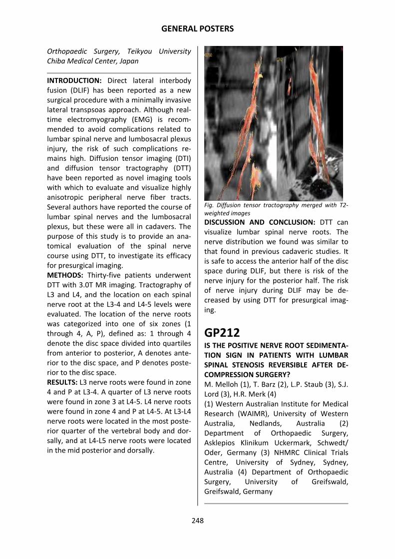
GENERAL POSTERS
248
Orthopaedic Surgery, Teikyou University
Chiba Medical Center, Japan
INTRODUCTION: Direct lateral interbody
fusion (DLIF) has been reported as a new
surgical procedure with a minimally invasive
lateral transpsoas approach. Although real-
time electromyography (EMG) is recom-
mended to avoid complications related to
lumbar spinal nerve and lumbosacral plexus
injury, the risk of such complications re-
mains high. Diffusion tensor imaging (DTI)
and diffusion tensor tractography (DTT)
have been reported as novel imaging tools
with which to evaluate and visualize highly
anisotropic peripheral nerve fiber tracts.
Several authors have reported the course of
lumbar spinal nerves and the lumbosacral
plexus, but these were all in cadavers. The
purpose of this study is to provide an ana-
tomical evaluation of the spinal nerve
course using DTT, to investigate its efficacy
for presurgical imaging.
METHODS: Thirty-five patients underwent
DTT with 3.0T MR imaging. Tractography of
L3 and L4, and the location on each spinal
nerve root at the L3-4 and L4-5 levels were
evaluated. The location of the nerve roots
was categorized into one of six zones (1
through 4, A, P), defined as: 1 through 4
denote the disc space divided into quartiles
from anterior to posterior, A denotes ante-
rior to the disc space, and P denotes poste-
rior to the disc space.
RESULTS: L3 nerve roots were found in zone
4 and P at L3-4. A quarter of L3 nerve roots
were found in zone 3 at L4-5. L4 nerve roots
were found in zone 4 and P at L4-5. At L3-L4
nerve roots were located in the most poste-
rior quarter of the vertebral body and dor-
sally, and at L4-L5 nerve roots were located
in the mid posterior and dorsally.
Fig. Diffusion tensor tractography merged with T2-
weighted images
DISCUSSION AND CONCLUSION: DTT can
visualize lumbar spinal nerve roots. The
nerve distribution we found was similar to
that found in previous cadaveric studies. It
is safe to access the anterior half of the disc
space during DLIF, but there is risk of the
nerve injury for the posterior half. The risk
of nerve injury during DLIF may be de-
creased by using DTT for presurgical imag-
ing.
GP212 IS THE POSITIVE NERVE ROOT SEDIMENTA-TION SIGN IN PATIENTS WITH LUMBAR SPINAL STENOSIS REVERSIBLE AFTER DE-COMPRESSION SURGERY? M. Melloh (1), T. Barz (2), L.P. Staub (3), S.J.
Lord (3), H.R. Merk (4)
(1) Western Australian Institute for Medical
Research (WAIMR), University of Western
Australia, Nedlands, Australia (2)
Department of Orthopaedic Surgery,
Asklepios Klinikum Uckermark, Schwedt/
Oder, Germany (3) NHMRC Clinical Trials
Centre, University of Sydney, Sydney,
Australia (4) Department of Orthopaedic
Surgery, University of Greifswald,
Greifswald, Germany

GENERAL POSTERS
249
INTRODUCTION: The Nerve Root Sedimen-
tation Sign (SedSign) has been established
as a new radiological sign in the diagnosis of
lumbar spinal stenosis (LSS). In patients
without LSS, on supine Magnetic Resonance
Imaging (MRI) lumbar nerve roots sediment
to the dorsal part of the dural sac. In pa-
tients with LSS, this nerve root sedimenta-
tion is not always seen. The absence of
nerve root sedimentation has been defined
as positive SedSign. As patients with a posi-
tive SedSign may benefit from surgical de-
compression the Sign might assist the spine
surgeon to determine the indication for
surgery. The aim of this study was to inves-
tigate if in patients with LSS the positive
SedSign will turn negative after lumbar de-
compression.
METHODS: Retrospective cohort study of
consecutive spine patients with an initial
MRI of the lumbar spine in whom a follow-
up MRI was performed after decompression
surgery. Comparison of the primary MRIs
with the follow-up MRIs in regard to the
presence of nerve root sedimentation (neg-
ative SedSign).
RESULTS: Fifty-seven of 118 LSS patients
had a positive SedSign, of these 39 had un-
dergone spine surgery. In this group, 12
patients were identified who had a post-op
MRI of the lumbar spine (7 male, 5 female,
mean age 69 yrs [range 52-83]). Eight pa-
tients demonstrated a negative SedSign, 3
patients had a positive SedSign associated
with a newly occurred stenosis in another
previously not affected segment. One pa-
tient had an arachnoiditis with a positive
SedSign but without stenosis.
DISCUSSION: Despite several limitations of
this study the reversibility of a pre-op posi-
tive SedSign was demonstrated after de-
compressing the affected level; except for a
case of arachnoiditis which could not be
differentiated from spinal stenosis due to
the parietal clumping of nerve roots. Fur-
ther research is required to investigate in a
prospective representative patient sample if
the reversibility of a positive SedSign is cor-
related with an improved clinical outcome.
GP213 INVERTED Y APPROACH TO LUMBOSACRAL REGION- ANALYSIS OF 525 CASES OVER 10 YEARS. Krishna Kumar Ramachandran Nair,
Renjitkumar J
Department of Orthopaedics, Amrita Insti-
tute of Medical Sciences, Cochin, Kera-
la,India
INTRODUCTION: Traditional approach to
lumosacral region utilizes midline incision,
which often requires long incision to get
sufficient exposure. We describe our expe-
rience with inverted Y approach to lum-
bosacral junction.
OBJECTIVE: To analyze the surgical steps,
complications and results of inverted Y inci-
sion to approach lumbosacral region in a
tertiary care referral center( Amrita Insti-
tute of Medical Sciences, Kochi) by file re-
view.
MATERIALS AND METHODS: Total of 525
patients were included in the study with the
range of age from 17 to 75 years and mean
age being 51.24 years. Mean follow-up pe-
riod was 26 months, with a range from 10
to 64 months. The surgeries included
PLF,PLIF,TLIF and Iliolumbar fixations.
RESULTS: 522 patients had primary wound
healing and 3 patients had wound break-
down. Of these 3 patients, two had previous
radiation treatment to lumbosacral region
and one patient had multiple surgeries for
spondylodiscitis earlier.
Discussion: Inverted Y Approach to
lumosacral region is a simple and safe tech-
nique with minimal wound complications.

GENERAL POSTERS
250
GP214 ADJACENT SEGMENT PATHOLOGY AFTER THORACOLUMBAR FUSION IN THORACOL-UMBAR FRACTURE: AN AGE AND SEX MATCHED-COHORT STUDY WITH LOWER LUMBAR FUSION Jeon, Chang-Hoon, Chung, Nam-Su, Park,
Jeong-Uk, Kim, Jea-Joong
Department of Orthopaedic Surgery, Ajou
University School of Medicine, Suwon, Re-
public of Korea
INTRODUCTION: Although numerous stud-
ies have reported the adjacent segment
pathology (ASP) after lumbar fusion, little
attention has been paid to the ASP after
thoracolumbar fusion. The purpose of this
study was to investigate the ASP after
thoracolumbar fusion as compared with
lower lumbar fusion.
METHODS: Forty-four patients with thora-
columbar (T10-L2) fracture treated with
posterior fusion using pedicle screw instru-
mentation that had taken MRI at the time
of index treatment and at least 2-year fol-
low-up were included. A matched cohort of
47 patients that underwent lower lumbar
(L3-S1) fusion with pedicle screw instrumen-
tation was identified. The ASPs including
disc degeneration, disc herniation, spinal
stenosis and spondylolisthesis between the
2 groups were compared with MRI.
RESULTS: There was no difference regarding
the patients’ age (P=0.066), gender
(P=0.152), and the duration of follow-up
MRI (P=0.150) between thoracolumbar fu-
sion group and lower lumbar fusion group.
The mean instrumentation level was 3.5 ±
0.9 in thoracolumbar fusion and 1.6 ± 0.7 in
lower lumbar fusion (P< 0.001). Adjacent
disc degeneration was found in 11 of thora-
columbar fusion and 14 of lower lumbar
fusion (P=0.567). Adjacent disc herniation
was found in 4 of thoracolumbar fusion and
6 of lower lumbar fusion (P=0.201). Spinal
stenosis was found in 5 of thoracolumbar
fusion and 7 of lower lumbar fusion
(P=0.427). Spondylolisthesis was found in 1
of thoracolumbar fusion and 2 of lower
lumbar fusion (P=0.328).
DISCUSSION: The occurrences of ASPs after
thoracolumbar fusion were not different
with those of lower lumbar fusion. The limi-
tation of this study was devoid of including
thoracolumbar fracture without fusion.
GP215 DO RISK FACTORS MATTER IN PROPHYLAC-TIC ANTIBIOTICS REDUCTION AFTER SPINAL SURGERY?: A PROSPECTIVE COMPARATIVE STUDY Jeon, Chang-Hoon, Chung, Nam-Su, Park,
Jeong-Uk, Kim, Jea-Joong
Department of Orthopaedic Surgery, Ajou
University School of Medicine, Suwon,
Republic of Korea
INTRODUCTION: Prophylactic antibiotics
duration after spinal surgery remains con-
troversial, especially for patients with risk
factors. We designed a prospective compar-
ative study to evaluate whether risk factors
affect the occurrence of surgical site infec-
tion (SSI) and the changes of host immune
responses depending on the duration of
prophylactic antibiotics.
MATERIALS AND METHODS: This study in-
volved 150 consecutive patients who had
undergone a spinal surgery and were ad-
ministrated first generation cephalosporin
as prophylactic antibiotics. The duration of
antibiotics administration had been altered
for each fifty consecutive patients as 5-day,
3-day, and 1day. Preoperative antibiotics
prophylaxis was commonly administrated
for the 3 groups within 1 hour prior to sur-
gical incision with the same trial antibiotics.
Intraoperative bacterial culture on the sur-
gical site was performed. The occurrences
of SSI were evaluated as described by Na-
tional Nosocomial Infection Surveillance
guideline. SSI was considered as acute if it
occurred within postoperative 1 month,
subacute for < 3 months, and chronic for > 3

GENERAL POSTERS
251
months. Serial changes of hematologic in-
flammatory markers and DIC profiles were
compared until postoperative 2 weeks. Risk
factors such as age, sex, body mass index,
estimated blood loss, diabetes mellitus,
smoking, nutritional status, baseline labora-
tory values, catheter indwelling, the dura-
tion of drain, and type of surgery were ana-
lyzed.
RESULTS: Incisional SSI was found in 2, 3,
and 3 patient with 5-day, 3-day, and 1-day
administration, respectively (P=0.887). Or-
gan/space SSI occurred in 1 patients in all
the three groups. All SSIs were acute infec-
tion. There was no difference for intraoper-
ative bacterial culture. Laboratory results
revealed that postoperative changes of he-
matologic inflammatory markers and DIC
profiles were not different among the three
groups (all P>0.05). The occurrence of SSI
was not affected by having risk factors.
DISCUSSION Prophylactic an
GP216 THE LUMBO-PELVIC SAGITTAL ALIGNMENT AND EARLY ADJACENT SEGMENT DISOR-DER AFTER PLIF IN PATIENTS WITH LUM-BAR DEGENERATIVE SPONDYLOLISTHESIS . Nishimura H, Endo K, Suzuki H, Tanaka H,
Yorifuji M, Yamamoto K
Department of Orthopedic Surgery, Tokyo
Medical University
PURPOSE: Spondylodesis is indicated for a
number of patients with lumbar degenera-
tive spondylolisthesis (SD); however, its in-
fluence on early onset adjacent segment
disorder is a concern. The purpose of this
study was to evaluate the early onset adja-
cent segment disorder (ASD) after PLIF and
lumbo-pelvic sagittal alignment.
SUBJECTS AND METHODS: Subjects were 69
patients with unstable DS who underwent
PLIF and postoperative follow-up for 3 years
or longer. Subjects were classified into 2
groups; namely, an adjacent segment disor-
der which occurred within one year after
surgery (early ASD) and an adjacent seg-
ment disorder which occurred in the later
periods (late ASD). Intervertebral height,
range of motion, slip, and sagittal alignment
were measured by preoperative XP and in-
tervertebral joint angle by CT, and then ad-
jacent vertebral narrowing, absence or
presence of instability, and an increase or
decrease of range of motion with time dur-
ing postoperative follow up were examined.
RESULTS AND DISCUSSION: Adjacent verte-
bral narrowing occurred in 15 of 69 patients
(21.0%), including ten in the early ASD and
five in late ASD, showing that more ASD
occurred within one year. In the early ASD,
L3/4 disorder occurred in three patients by
L4/5 PLIF + L3/4 fenestration, L2/3 disorder
occurred in four. Rregarding to the onset of
ASD, there were no significant difference
about the facet angle. However, sagittal
plane was affected to the onset of ASD. Ta-
ble showed the results of sagittal alignment.
In early ASD group, the trunk tilted forward
more severely, and the pelvis tilted more
forward. before the operation. In general,
DS has posterior rotated pelvis in order to
compensate for lumbar slip. However, if the
pelvic anterior rotation was severe before
surgery, the LLA should be correct enough.
Otherwise, because the postoperative
change of pelvic rotation is small, the me-
chanical stress above PLIF segment would
be increased.
GP217 VERTEBROPLASTY FOR VERTEBRAL PSEUDOARTHROSIS IN PATIENTS WITH SECONDARY OSTEOPOROSIS: COMPARA-TIVE STUDY WITH PRIMARY AND SECOND-ARY OSTEOPOROSIS Yoshikazu Shinohara, Hiromitsu Toyoda,
Hidetomi Terai, Akinobu Suzuki, Sho
Dohzono, Tomiya Matsumoto, Kentaro
Yamada, Shinji Takahashi, Kuniaki
Tsukiyama, Hiroaki Nakamura
Osaka City University Graduate School of
GENERAL POSTERS
252
Medicine
INTRODUCTION: Osteoporosis has been
classified as either primary or secondary.
Although secondary osteoporosis is less
common, it is becoming more frequently
diagnosed. The purpose of this study was to
compare the clinical outcomes of
vertebroplasty (VP) for patients with prima-
ry and secondary osteoporosis.
METHODS: In our hospital we perform VP
using calcium phosphate cement (CPC) for
the treatment of vertebral pseudoarthrosis.
We reviewed 45 patients (Pts) undergoing
49 VPs with more than 1 year follow up.
There were 7 men and 38 women with an
average age of 76.0. We divided these pa-
tients into patients with primary osteoporo-
sis (P-group) and patients with secondary
osteoporosis (S-group). Outcomes including
pain, complications, frequency of new frac-
tures and radiographic changes (anterior
vertebral height restoration) in S-group
were compared with P-group.
RESULTS: Fourteen Pts were included in S-
group. The causes of secondary osteoporo-
sis were followed. Long-term corticosteroid
therapy for the treatment of the coexisting
illness such as rheumatologic diseases was
7, Diabetes mellitus was 7, gastrectomy
patient was 2, and Liver cirrhosis was 1.
There was no significant difference between
the two groups in pre- and post- operative
JOA score and VAS. The frequency of new
fractures was not significantly different be-
tween the two groups. The 23.0% of the
anterior vertebral body height correction
was lost in S-group, but 13.5% in P-group.
Number of cases of height loss > 10% were
significantly higher in the S- group than P-
group
DISCUSSION: VP performed for vertebral
pseudoarthrosis as a result of secondary
osteoporosis is as safe and effective in re-
lieving pain as those of primary osteoporo-
sis. However, patients with secondary oste-
oporosis showed higher risk in loss of cor-
rection in the treated vertebrae.
GP218 SURGICAL DECOMPRESSION OF THE THO-RACIC SPINAL CANAL IN ACHONDROPLASIA; INDICATION AND OUTCOME CL Vleggeert-Lankamp and WC Peul
Department of Neurosurgery, Leiden Uni-
versity Medical Centre (LUMC), Leiden, the
Netherlands
INTRODUCTION: The achondroplastic spinal
canal is narrow due to short pedicles and a
small interpediculate distance. Compression
of neural structures passing through this
canal is therefore regularly encountered,
but scarcely described. Symptomatology,
radiologic evaluation, and treatment of 20
achondroplasts that underwent decompres-
sion of the thoracic medulla is described
and outcome is correlated to the size of the
spinal canal and the thoracolumbar
kyphotic angle.
METHODS: The modified JOAscore, Nurick
score, European Myelopathy Score, Cooper
myelopathy scale for lower extremities, and
Odom criteria before and after surgery were
compared. MR imaging was evaluated for
the size of the spinal canal, medullary com-
pression, and presence of myelomalacia.
The thoracolumbar kyphotic angle was
measured on fluoroscopy.
RESULTS: Symptomatology included deteri-
oration of walking pattern, pain, cramps,
spasms, and incontinence. MRIs of all pa-
tients demonstrated medullary compression
due to degenerative changes. Surgery re-
sulted in (slight) improvement on all afore-
mentioned ranking scales. Surgery at the
wrong level occurred in 15%, but no serious
complications occurred. The mean thora-
columbar kyphotic angle was 20 degrees,
and no correlation was established with
outcome after surgery. No post-operative
increase in this angle was reported. Neither

GENERAL POSTERS
253
was there a correlation between size of the
spinal canal and outcome.
DISCUSSION: Decompressive surgery of the
achondroplast thoracic medulla can be per-
formed safely if anatomical details are taken
into account. Spondylodesis did not appear
obligatory. Special attention should be paid
to the method of surgery, how to identify
the level of interest and follow up of the
thoracolumbar kyphotic angle.
GP219 DORSAL VENTRAL DORSAL THREE STAGE CORRECTION SURGERY FOR LUMBAR KYPHO-SCOLIOSIS DEFORMITY Kazuyuki Otani M.D., Ph.D., Osamu Nakai
M.D., Ph.D., Shigeo Shindo M.D., Koichi
Mizuno M.D., Ph.D.
Department of Orthopaedic Surgery,
Kudanzaka Hospital, Tokyo, Japan
INTRODUCTION: Sagittal balance restora-
tion is essential in achieving positive out-
comes in correction of adult spinal deformi-
ties. We have performed dorsal ventral dor-
sal (DVD) three-stage correction for patients
with maximum kyphosis correction re-
quired. The aim of the study was to deter-
mine the efficacy of the DVD procedure for
patients with severe lumbar kyphosis de-
formity.
METHODS: Retrospective review of 82 pa
tients (8 male and 74 female). Mean age 66
years (46-78). Mean follow-up period 3.7
years (1-12). Facetectomy at 2-3 levels per-
formed in first stage, followed by anterior
release and interbody fusion at same level;
posterior fusion with instrumentation per-
formed in final stage. Mean fused levels 5.4
(range 3-11). Clinical assessment performed
using JOA score and visual analog scale for
low back pain. Radiological measurement
performed using pre-operative, post-
operative and final films.
RESULTS: The mean pre-operative JOA
score of 16.4 changed to 24.4 at final fol-
low-up, with a recovery rate of 64.1%. The
mean pre-op VAS of 62.7mm was reduced
to 20.6mm at final follow-up. Re-operation
was performed on 4 patients (proximal
junctional kyphosis in 2 cases; disc herni-
ation at L5/S in 2 cases). The fusion rate was
89% with 8 of 9 non-union cases were those
at L5/S. Subsequent vertebral fracture was
observed in 14.6% of the patients. The pre-
op lumbar lordosis (L1-S) of 2.5˚ changed to 48.8˚ at post-op and 34.8˚ at follow-up. The
obtained lordosis at the fusion area was
48.6˚ at post-op and 35.2˚ at follow-up. The
single level obtained lordosis at DVD correc-
tion level was 17.6̊at post -op and 15.1̊ at follow-up. The pre-op sagittal vertebral axis
of 98.0mm changed to 49.2mm at follow-
up.
DISCUSSION: The DVD procedure was use-
ful in the correction of kyphosis and provid-
ed good clinical results. The remaining chal-
lenges are how to determine the fusion area
and how to avoid L5/S pseudarthrosis, sub-
sequent vertebral fracture and proximal
junctional kyphosis.

GENERAL POSTERS
254
GP220
ASSOCIATION BETWEEN VERTEBRAL ENDPLATE SIGNAL CHANGES AND LOW BACK PAIN IN PATIENTS WITH CURRENT OR PREVIOUS SYMPTOMS OF SCIATICA A. el Barzouhi1, C.L.A.M. Vleggeert-
Lankamp1, B.F. Van der Kallen2, G.J.
Lycklama à Nijeholt2, W.B. van den Hout3,
B.W. Koes4, and W.C. Peul1,5
1 Department of Neurosurgery, Leiden
University Medical Centre, Leiden, the
Netherlands 2 Department of Radiology,
Medical Centre Haaglanden, the Hague, the
Netherlands 3 Department of Medical
Decision Making, Leiden University Medical
Centre, Leiden, the Netherlands 4
Department of General Practice,
ErasmusMC, University Medical Centre,
Rotterdam, the Netherlands 5 Department
of Neurosurgery, Medical Centre
Haaglanden, the Hague, the Netherlands
BACKGROUND: Patients with current or
previous symptoms of sciatica frequently
experience low back pain (LBP). One of the
proposed causes for LBP is Vertebral End-
plate Signal Changes (VESC) visualized by
Magnetic Resonance Imaging. We investi-
gated the association between VESC and
LBP in patients with current or previous
symptoms of sciatica.
METHODS: The study population consisted
of 283 patients who were randomized to
surgery or to prolonged conservative care
with surgery if needed. Participants under-
went MRI both at baseline and after 1 year.
The presence of VESC was correlated to LBP
(defined as a visual-analogue scale of at
least 40mm on a scale of 0-100) using Fish-
er’s exact tests and logistic regression.
RESULTS: VESC were observed in 43% of the
patients at baseline. Of the patients with
VESC at baseline 40% had LBP compared to
38% of the patients with no VESC (P=0.70).
The prevalence of type 1 VESC increased
from 1% at baseline to 35% one year later in
the surgical group compared to an increase
from 3 to 11% in the conservative group.
The prevalence of type 2 VESC decreased
from 39 to 29% in the surgical group while
remaining comparable in the conservative
group. The prevalence of LBP at one year
was 12% in patients with no VESC, 16% in
patients with type 1 VESC, 11% in patients
with type 2 VESC and 3% in patients with
mixed type 1 and 2 VESC (P=0.34). Patients
who showed an increase in the amount of
VESC after one year did not significantly
report more LBP compared to patients who
did not (OR 1.21, 95% CI 0.57-2.58, P=0.61).
Undergoing surgery was associated with
progression in the amount of VESC (OR
8.56, 95% CI 4.67-15.67, P<0.001).
DISCUSSION: Surgery highly accelerates the
development of VESC. However, in this
group of patients with current or previous
sciatica no association existed between the
presence or progression of VESC and LBP.
Therefore, VESC cannot be responsible for
remaining or new LBP after surgical or con-
servative treatment for sciatica.
GP221 PREDICTION OF SURGERY FOR SCIATICA WITH MRI A. el Barzouhi1, C.L.A.M. Vleggeert-
Lankamp1, B.F. Van der Kallen2, G.J.
Lycklama à Nijeholt2, W.B. van den Hout3,
B.W. Koes4, and W.C. Peul1,5
1 Department of Neurosurgery, Leiden
University Medical Centre, Leiden, the
Netherlands 2 Department of Radiology,
Medical Centre Haaglanden, the Hague, the
Netherlands 3 Department of Medical
Decision Making, Leiden University Medical
Centre, Leiden, the Netherlands 4
Department of General Practice,
ErasmusMC, University Medical Centre,
Rotterdam, the Netherlands 5 Department
of Neurosurgery, Medical Centre
Haaglanden, the Hague, the Netherlands
BACKGROUND: A randomized trail showed

GENERAL POSTERS
255
that surgery compared to a prolonged con-
servative care strategy for patients with 6 to
12 weeks sciatica yields similar functional
recovery rate results by one year. However,
39% of the patients assigned to prolonged
conservative care underwent surgery during
the first year after randomization. Previous-
ly we reported that sequestered herniations
and the level of the disc herniation on Mag-
netic resonance imaging (MRI) were not
predictive for delayed surgery. The objec-
tive of this study was to evaluate the
preditice value of a more detailed qualita-
tive and quantitative MRI assessment for
delayed surgery.
METHODS: MRIs of the 142 patients who
were assigned to receive conservative care
were assessed, both qualitatively (by two
neuropradiologists and one neurosurgeon
who were not previously involved in earlier
published papers) and quantitatively (by an
independent investigator also not previous-
ly involved). Logistic regression model and
Receiver operating characteristic (ROC)
curve analysis were used to determine the
predictive value of qualitative and quantita-
tive MRI measurements on the occurrence
of surgery during the first year. Area under
the ROC curve (AUC) above 0.7 was consid-
ered acceptable.
RESULTS: MRI variables such as the proba-
bility of definite nerve root compression,
location and form of the disc herniation,
and baseline size of the disc herniation did
not have any predictive value for delayed
surgery. Size of the dural sac spinal canal did
predict delayed surgery (per 25 mm2 in-
crement in size odds ratio 0.80 [95% CI 0.67-
0.96] and 0.82 [95% CI 0.71-0.95] respec-
tively). Using the size of the dural sac and
spinal canal to predict delayed surgery re-
vealed an AUC of 0.62 (95% CI 0.53-0.72)
and 0.62 (95% CI 0.53-0.72) respectively.
DISCUSSION: MRI is not acceptably able in
assisting clinical decision making for surgical
or non-surgical management in patients
who suffer from sciatica for 6 to 12 weeks.
GP222 SURGICAL OUTCOME OF DROP FOOT CAUSED BY DEGENERATIVE LUMBAR DISORDERS; FOCUSED ON THE RECOVERY OF MOTOR DEFICITS AND THE IMPROVEMENT OF LOW BACK AND LOWER EXTREMITY SYMPTOMS. Hiroshi Takahashi, Yasuchika Aoki, Arata
Nakajima, Masato Sonobe, Yoshifumi
Shibata, Shinji Taniguchi, Manabu Yamada,
Teruo Furufu, Koichi Nakagawa, Kazuhisa
Takahashi
Toho University Sakura Medical Center
INTRODUCTION: Drop foot, sometimes
caused by degenerative lumbar disorders, is
a neuromuscular condition in which pa-
tients are unable to dorsiflex the ankle. It is
important to advise patients as to how
much motor function will improve after
surgery, and whether low back and lower
extremity symptoms will resolve, but there
are no reports describing surgical outcomes
such as residual pain and numbness in pa-
tients with lumbar degenerative disorders
complicated by drop foot. We conducted a
retrospective study to evaluate these out-
comes.
METHODS: Between April 2010 and Sep-
tember 2011, 20 patients (12 men, 8 wom-
en) with degenerative lumbar disorders and
drop foot underwent lumbar spine surgery.
We evaluated patient age, the causative
disorder, and the duration from symptom
onset to surgery. We measured preopera-
tive and 1 year postoperative tivialis anteri-
or (TA) strength using the manual muscle
test (MMT). Low back and lower extremity
symptoms were evaluated with a visual ana-
logue scale (VAS), and activities of daily liv-
ing (ADL) by Oswestry Disability Index (ODI).
RESULTS: Twelve patients had lumbar disc
herniation (LDH) and 8 had lumbar spinal
stenosis (LSS). TA strength was improved in
LDH patients (pre-op, post-op: 2.46±0.77,
4.23±1.09) to a significantly greater degree
than in LSS patients (1.43±0.79, 3.14±1.77).

GENERAL POSTERS
256
The age at onset was negatively correlated
with the TA strength recovery rate (p<0.01).
There was also a negative correlation be-
tween this rate and the duration from onset
to surgery (p<0.05). Following surgery, sig-
nificant improvement in low back pain, low-
er extremity pain, and numbness was ob-
served by both VAS and ODI in patients with
both unfavorable (MMT<4), and favorable
(MMT≥4) motor recovery. CONCLUSIONS: Causative disorder, age, and
duration to surgery are prognostic factors
for surgical outcome in patients with drop
foot. ADLs, low back pain, and lower ex-
tremity symptoms were improved after sur-
gery even when motor deficits remained.
GP223 VERTEBROPLASTY FOR PATIENTS WITH NEUROLOGICAL DEFICITS CAUSED BY INSUFFICIENT UNION FOLLOWING OSTEOPOROTIC VERTEBRAL FRACTURE Masatoshi Hoshino 1, Tadao Tujio 1, Minori
Kato 2, Akinobu Suzuki 3, Tomiya
Matsumoto 3, Hiromitsu Toyoda 3, Akira
Matsumura 2, Hidetomi Terai 3, Sadahiko
Konishi 2, Masahiko Seki 1, Hiroaki
Nakamura 3
1 Spine center, Shiraniwa Hospital, Nara,
Japan, 2 Department of Orthopedic Surgery,
Osaka City General Hospital, Osaka, Japan,
3 Department of Orthopedic Surgery, Osaka
City University Graduate School of Medicine,
Osaka, Japan
INTRODUCTION: We have previously re-
ported that intravertebral instability is a
significant factor in neurological deficits
following insufficient union after osteopo-
rotic vertebral fracture (OVF). On the basis
of this finding, we prospectively performed
vertebroplasty without decompression in
patients with neurological deficits. The pur-
pose of this study was to clarify the useful-
ness and limitations of vertebroplasty for
insufficient union of OVF with lower limb
neurological symptoms.
METHODS: Twenty-eight patients (7 males
and 21 females) underwent vertebroplasty
for neurological deficits caused by insuffi-
cient union following OVF. Twenty-one pa-
tients who underwent posterior decom-
pression and fusion surgery with instrumen-
tation were selected as controls (group-
PDF). Neurological improvement (modified
Frankel classification), invasiveness of sur-
gery, surgical complications, and radio-
graphic results were evaluated between the
2 groups.
RESULTS: The neurological function deter-
mined using the modified Frankel classifica-
tion were as follows: C1, 2; C2, 4; D1, 10;
D2, 1; and E, 11 preoperatively and C1, 0;
C2, 0; D1, 2; D2, 10; and E, 16 at the final
follow-up. The symptoms improved in all
patients with sensory disturbances or blad-
der and rectal disturbances. The mean op-
eration time was 120 min, mean blood loss
was 119 mL, and transfusion rate was 3.6%;
these were significantly lower than corre-
sponding values in group-PDF. Subsequent
adjacent vertebral fracture rate was 32% (9
patients). Among these 9 patients, 2 with
adjacent vertebral collapse in the early
postoperative period developed severe
thigh pain. In the kyphotic angle, there were
no significant differences between the 2
groups.
DISCUSSION: We obtained satisfactory re-
sults of vertebroplasty for insufficient union
of OVF with lower limb neurological symp-
toms. Advanced collapse of the adjacent
vertebral body in the early postoperative
period caused poor results.
GP224 INSTRUMENTATION AND EQUIPMENT RELATED INEFFICIENCY IN SPINE SURGERY: POTENTIAL FOR COST SAVINGS Zhou, Hanbing; Egge, Natalie; Lari, Hamid;
Lapinsky, Anthony; Eck, Jason; Connolly,
Patrick; DiPaola, Christian;
University of Massachusetts

GENERAL POSTERS
257
INTRODUCTION: Spine procedures repre-
sent a large portion of healthcare expendi-
ture in the orthopedics specialty. Studies
have shown that intra-operative waste has
exacerbated the high cost of surgeries. Our
objective is to identify and quantify the in-
cidence of pre- and intra-operative equip-
ment related events that generate ineffi-
ciency.
METHODS: Data was collected over a 9-
month period. A data collection instrument
was developed to record procedure related
details and instrument/equipment related
issues. Administrative records of equip-
ment issues were also obtained for the
same period. For each case with equipment
related issues, estimates of cost increase
and time delay was made. Operating cost
of OR time is based on hospital financial
data ($1061.03/hour).
RESULTS: 53% of the spine procedures
(325/613) were recorded in the database.
Equipment related issues occurred in 43.5%
of cases. Issues included missing equipment
(19.2%), improperly packaged pans (7.1%),
improperly maintained equipment (16.2%),
and other (1.0%). Unanticipated extra pans
were opened in 18.6% of the cases (total of
91 pans, range 1-5). In cases requiring extra
pans, the average cost increase was $135.6
(range $26.05 to $742.75). Comparing cas-
es with pre-operative equipment issues and
those without: the difference in the first
cases of the day is 15 minutes ($265.26, 29
vs. 14 minutes, P<0.05), the difference in
the rest of the cases is 7.8 minutes ($137.9,
48 vs. 40.2 minutes, P=0.06). Examining the
administrative record of the cases in our
study, hospital record captured 46 (14%)
equipment related issues.
DISCUSSION: Perioperative inefficiency in
spine surgery can be material or temporal.
Material inefficiency is demonstrated by the
high incidence of equipment related issues.
Indirect costs such as delay in OR start time
can be a contributor to overall hospital
waste. Our method was more capable of
capturing equipment issues than adminis-
trative process (43.5% vs. 14%).
GP225 RISK FACTORS FOR RECOLLAPSE OF THE TREATED VERTEBRAE IN PATIENTS WITH VERTEBRAL PSEUDARTHROSIS AFTER VERTEBROPLASTY Hitomitsu Toyoda, Hidetomi Terai, Shinji
Takahashi, Akinobu Suzuki, Sho Dozono,
Tomiya Matsumoto, Kentaro Yamada,
Kuniaki Tsukiyama, Yoshikazu Shinohara,
Hiroaki Nakamura
Dept. Orthopaedic Surgery, Osaka City
University Graduate School of Medicine,
Osaka, Japan
INTRODUCTION: Recollapse of the treated
vertebra after vertebroplasty (VP) is one of
the severe complications. Pre-operative
vertebral pseudoarthrosis has been report-
ed as predisposing factor. However, which
characteristics of vertebral pseudoarthrosis
increase the risk of recollapse remains un-
clear. The purpose of this study was to in-
vestigate the risk factors for this complica-
tion using uni- and multivariate regression
analysis.
METHODS: A total of 49 vertebral
pseudoarthrosis treated with VP using calci-
um phosphate cement were include in this
study. Demographic data, comorbidities and
all complications were analyzed. Morpho-
logical variables, including posteri-
or/anterior wall injury, endplate injury and
anterior vertebral height restoration (AVH)
of treated vertebrae were also analyzed. In
this study, we made a new scoring system
based on the extent of damage to the ver-
tebral wall (Vertebral wall injury severity
score. 1: only anterior wall injury, 2: anteri-
or and posterior walls injury without end-
plate injury, 3: both anterior and posterior
walls injury with upper or lower endplate
injury, 4: both anterior and posterior wall
injury with both upper and lower endplate
injuries)
GENERAL POSTERS
258
RESULTS: Recollapse was occured in 14 ver-
tebrae (28.6%). In univariate analysis, he-
moglobin level, bone metabolic marker, ASA
physical classification system (ASA), poste-
rior wall injury, lower endplate injury, ver-
tebral wall injury severity score, and AVH
were significantly associated with the inci-
dence of recollapse. In multivariate analysis,
ASA (odds ratio, 6.2; p=0.065) and vertebral
wall injury severity score (odds ratio, 6.8;
p<0.05) were significantly associated with
recollapse.
DISCUSSION: Recollapse after VP is com-
mon in patients with vertebral
pseudoarthrosis and the residual bone con-
dition (vertebral wall injury severity score)
was the most important predictive factor
for this complication than other predictive
factors such as age and bone mineral densi-
ty.
GP226 COMPARISON OF PULMONARY FUNCTION IN ADULTS YOUNGER AND OLDER THAN AGE 60 UNDERGOING SPINAL DEFORMITY SURGERY Robert W. Tracey, MD; Ronald A. Lehman,
MD; Daniel G. Kang, MD.; John P. Cody, MD;
Lawrence G. Lenke, MD; Jeremy J.
Stallbaumer, MD; Brenda A Sides, MS
Walter Reed National Military Medical
Center, Bethesda, MD Washington
University School of Medicine, Saint Louis,
MO
INTRODUCTION: The objective of this study
was to determine differences in pulmonary
function in adult patients who are either
younger (Y) or older (O) than age 60 follow-
ing spinal deformity surgery.
METHODS: 128 consecutive adult deformity
patients with idiopathic scoliosis undergoing
surgical treatment were evaluated at a sin-
gle institution with minimum 2 yr follow-up.
Prospectively collected PFTs, clinical records
and radiographs were analyzed.
RESULTS: There were 102 patients in Y
group (avg age 39.3+14.1 yrs) and 26 in O
group (avg age 63.7+2.7 yrs), with similar
F/U (Y=2.9 v O=2.6 yrs, p=0.27). There were
no differences in avg preop main thoracic
(MT) curve magnitude (Y=50.0deg,
O=54.8deg, p=0.27), however O patients
had significantly greater # of lumbar (5.9 v
4.2, p=0.00), thoracic (9.1 v 7.3, p=0.00),
and total (15.0 v 11.5, p=0.00) levels fused.
We also found O patients had significantly
lower absolute pre-op FEV1 (2.1 v
2.6L,p=0.02) and FVC (2.7 v 3.3L, p=0.05),
but no differences in %pred PFTs. This rela-
tionship remained at 2 yrs, with lower abso-
lute FEV1 (1.9 v 2.5L, p=0.00) and FVC (2.5 v
3.1L, p=0.00). A clinically significant decline
in PFTs (greater than 10% pred FEV1) oc-
curred in 8 (31%) O patients and 26 (25%) Y
patients, which was not statistically differ-
ent. (p=0.63). We also observed pre-op PFT
impairment (less than 65%pred FEV1) in 1
(4%) O patient, which significantly increased
to 6 (23%; p=0.02) postoperatively, com-
pared to Y group experiencing no change in
the number of patients (n=12, 12%) with
PFT impairment postoperatively.
DISCUSSION: Despite age related reduction
in PFTs, older patients (over age 60) had no
significant difference in %pred PFTs com-
pared to younger patients following spinal
deformity surgery and no differences in the
rate of clinically significant PFT decline
(≥10% pred FEV1). However, older patients
more frequently (23% v 12%) experience
PFT impairment (<65%pred FEV1) after spi-
nal deformity surgery.
GP227 RETURN TO SPORTS AFTER SURGERY TO CORRECT ADOLESCENT IDIOPATHIC SCOLIOSIS Robert W. Tracey, MD; Ronald A. Lehman,
MD; Daniel G. Kang, MD.; John P. Cody, MD;
Lawrence G. Lenke, MD
Walter Reed National Military Medical Cen-
ter, Bethesda, MD Washington University
School of Medicine, Saint Louis, MO

GENERAL POSTERS
259
INTRODUCTION: Participation in sports and
athletic activities by children and adoles-
cents has become an important aspect of
society, and is a frequent question following
surgery for adolescent idiopathic scoliosis.
No studies in the last decade using modern
posterior spinal implants have provided
recommendations on when surgeons’
should allow patients’ to return to athletic
activities following spinal fusions for de-
formity surgery.
METHODS: A survey was administered to
members of the Spinal Deformity Study
Group (SDSG). The survey consisted of sur-
geon demographic information, 6 clinical
case scenarios and three different construct
types (hooks, pedicle screws, and hybrid).
RESULTS: Twenty-three (23) surgeons com-
pleted the survey, with 43% being ortho-
paedic spine surgeons and 57% being pedi-
atric orthopaedic surgeons. Respondents
were all experienced, expert deformity sur-
geons, supported by 50% performing an
average of 21-50 AIS cases per year with
44% of surgeons with >20 years in practice.
Most patients were allowed to return to
both non-contact and contact sports within
3-6 months, and collision sports within 6-12
months postoperatively. We also found
pedicle screw instrumentation allows earlier
return to contact & non-contact sports. For
all construct types, approximately 20% of
respondents never allow return to collision
sports, whereas all surgeons allow eventual
return to contact & non-contact sports re-
gardless of construct type. Of all surgeons’
surveyed, there was only 1 reported cata-
strophic failure in a patient with implant
pullout after snowboarding 2 weeks post-
operatively.
DISCUSSION: Our study found that modern
posterior instrumentation allows surgeons
to recommend earlier return to sports fol-
lowing fusion for AIS, with the majority al-
lowing non-contact and contact sport at 6
months and collision sports at 12 months.
There was only one reported catastrophic
construct failure after return to sports post-
operatively.
GP228 THE RELIABILITY OF THE THORACOLUMABAR INJURY CLASSIFICA-TION AND SEVERITY SCORE AMOUNG OR-THOPEDIC SURGEONS AT DIFFERENT LEV-ELS OF TRAINING Robert W. Tracey, MD; Adam J. Bevevino,
MD; Ronald A. Lehman, Jr., MD; Daniel G.
Kang, MD; John P. Cody, MD
Walter Reed National Military Medical
Center, Bethesda, MD
INTRODUCTION: Despite the increased use
of Thoracolumbar Injury Classification and
Severity Score (TLICS), it has not yet gained
universal acceptance. Our objective was to
examine the reliability of TLICS between
orthopedic physicians from intern to staff
level surgeons.
METHODS: Ten cases of thoracolumbar
spine fractures were reviewed and scored
using TLICS by eight evaluators: intern (n=2),
junior level resident (n=3), senior level resi-
dent (n=2), spine surgeon (n=1). Each par-
ticipant evaluated the same cases on three
different occasions. Statistical analysis with
Intraclass Correlation Coefficient (ICC) was
calculated assessing the inter- and intra-
observer reliability of the TLICS classifica-
tion.
RESULTS: Interobserver reliability yielded
moderate to excellent agreement between
evaluators in all three rounds of testing.
Neurologic injury produced the highest ICC
values, ranging from 0.820 to 0.902. Frac-
ture morphology demonstrated the lowest
interobserver ICC scores with moderate
agreement of 0.449 and 0.423.
Intraobserver ICC scores improved with in-
creasing levels of training. The intern eval-
uators recorded the lowest intraobserver
ICC values, while the staff surgeon scored
the highest. Staff surgeon ICC values all

GENERAL POSTERS
260
ranked above 0.800 for excellent
intraobserver agreement. Fracture mor-
phology produced the lowest intraobserver
ICC values, ranging 0.586 to 0.683. The to-
tal severity score yielded the highest
intraobserver ICC values of 0.768 to 0.920
for interns through senior residents.
CONCLUSIONS: The use of TLICS demon-
strated moderate to excellent intra- and
inter-observer reliability among all training
levels. Senior residents and Staff demon-
strated improved ICC scores in higher train-
ing levels, however, interns and Junior Resi-
dents were able to reliably classify spinal
trauma injuries. This suggests that the
TLICS scheme is a reliable way to successful-
ly communicate thoracolumber injury in-
formation.
GP229 CLINICAL OUTCOME OF UNILATERAL DE-COMPRESSION WITHOUT FUSION FOR UNILATERAL RADICULOPATHY DUE TO LUMBAR SPINAL STENOSIS Koji Otani, Shinichi Kikuchi, Soji Yabuki,
Takuya Nikaido, Kazuyuki Watanabe, Kinshi
Kato, Shinichi Konno
Dept. of Orthopaedic Surgery, Fukushima
Medical University School of Medicine, Fu-
kushima City, Japan
INTRODUCTION: It is still controversy that
unilateral decompression without fusion for
unilateral radiculopathy due to lumbar spi-
nal stenosis (LSS) is sufficient or not. The
purpose of this study was to assess the clin-
ical outcome of unilateral decompression
without fusion for unilateral radiculopathy
due to LSS.
PATIENTS AND METHODS: From 2001 to
2008, 258 patients with LSS caused by
spondylosis (SP) or degenerative
spondylolisthesis (DS) received the initial
operation at the University hospital. Repeat
operation was excluded. At those periods,
there were no fusion and no bilateral de-
compression for symptomatic unilateral
radiculopathy. 37 patients with unilateral
radiculopathy were received unilateral de-
compression without fusion and 30 patients
were followed more than 3 years (SP: 25
pts, DS: 5 pts). Contralateral symptom of
the initial operation level and new symptom
due to the different site from the initial op-
eration level were assessed.
RESULTS: 1) Six of 30 patients (20%) showed
contralateral symptom at the same of the
initial operation level. Five of 6 patients re-
ceived the additional operation.
2) Three of 5 patients who received the ad-
ditional operation showed that the different
level from the initial operation was the re-
sponsible level for the new symptom.
3) One of 5 patients with DS showed new
symptom but did not receive the second
operation. On the other hand, five of 25
patients with SP received the additional
operation.
DISCUSSION AND CONCLUSION: From the
current study, there could not detect the
prognostic factors for the occurrence of the
contralateral symptom. So it is concluded
that unilateral decompression is the first
choice of operation procedure for unilateral
radiculopathy due to LSS, and when contra-
lateral radiculopathy occurs, the additional
operation should be considered. Preventa-
tive operation for the contralateral side
might not be necessary at the initial opera-
tion.
GP230 COMPLICATIONS FOLLOWING ANTERIOR LUMBAR SPINAL FUSION IN PATIENTS WITH A HISTORY OF PRIOR INTRA-ABDOMINAL SURGERY Kim SD, DeBlasi GJ, McGuire KJ, Berger D,
Mansfield FL, Wood KB
Massashusetts General Hospital, Boston,
MA
INTRODUCTION: With different approaches
available for lumbar interbody grafting, it is

GENERAL POSTERS
261
unclear whether a history of prior ab-
dominal/ pelvic surgery confers an in-
creased risk of approach related complica-
tions when undergoing anterior lumbar
interbody fusion via an anterior retroperi-
toneal approach.
METHODS: We present a retrospective
chart review of 104 patients who under-
went an infra-umbilical retroperitoneal ap-
proach to the anterior aspect of the lower
lumbar spine. There was a minimum 1 year
follow-up. Data collected included age,
gender, procedure performed, levels fused,
intraoperative blood loss, length of hospital
stay, duration of surgery, preoperative di-
agnosis, blood transfused, intraoperative
and peri-operative complications, and reso-
lution of any complications.
RESULTS: Out of 104 patients, 23 patients
(23%) had 24 approach-related complica-
tions, either intraoperatively (9%), during
the immediate postoperative period (5%),
or within 1 year of surgery (10%). One pa-
tient had both an intraoperative or immedi-
ate postoperative complication in addition
to a delayed complication. After control-
ling for other factors such as age, gender,
diagnostic groups, and level of procedure,
there was a statistically significant increase
in the odds of having a complication when
there was a history of abdominal or pelvic
surgery (OR 1.35 P <0 .014). 19% of pa-
tients with a history of abdominal or pelvic
surgery had a delayed complication; com-
pared to only 4% who did not have ab-
dominal or pelvic surgery in the past (p <
.024)
DISCUSSION: Anterior lumbar interbody
fusion via a midline incision and a retroperi-
toneal approach was associated with a 23%
overall rate of complication. Many of these
complications were minor and did not re-
quire intervention. However 5 (4.8%) pa-
tients did require revision procedures. Our
study suggests that a history of prior ab-
dominal or pelvic surgery puts the patients
at a significantly higher risk for approach-
related complication.
GP231 LUMBAR STABILIZATION WITH A PARASPINOUS TENSION BAND DEVICE IN PATIENTS WITH DEGENERATIVE SPONDYLOLISTHESIS AND SPINAL STENO-SIS: 24-MONTH DATA. JNA Gibson, MD, FRCS [1] C Beadle, BSc, BA
[1] L Fielding, MS [2] M Wachna, BS [2] TF
Alamin, MD [2,3]
1] The Royal Infirmary and Edinburgh
University, Edinburgh, Scotland 2] Simpirica
Spine, San Carlos, CA 3] Stanford University,
Stanford, CA
INTRODUCTION: Segmental instability and
recurrent symptoms can occur after surgical
decompression in the presence of degener-
ative spondylolisthesis (DS), so surgeons
often perform a segmental fusion. However,
fusion can result in morbidity and adjacent
segment degeneration. This study evaluated
clinical and radiographic outcomes after
implantation of a paraspinous tension band
(PTB) device designed to control lumbar
sagittal plane instability.
METHODS: Patients with DS were enrolled
in a single arm prospective study. Clinical
and radiographic evaluations were per-
formed preoperatively and at 3, 6, 12, and
24-months, and included evaluation of pain
(VAS) and function (ODI), along with anal-
yses of segmental rotation, flexion-
translation, anterolisthesis, and device sta-
tus.
RESULTS: Twenty-five patients (23f, 2m,
mean age 69) were surgically decompressed
and stabilized with the PTB at L4/5 (n=20)
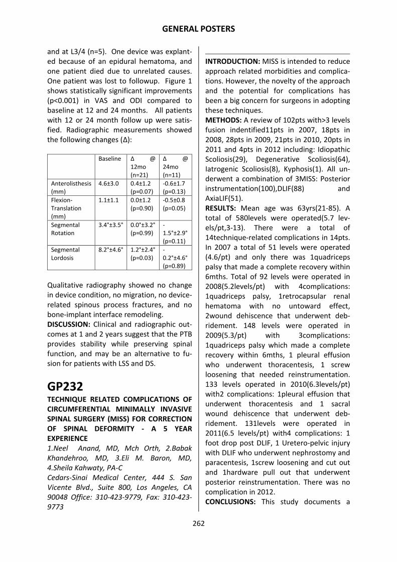
GENERAL POSTERS
262
and at L3/4 (n=5). One device was explant-
ed because of an epidural hematoma, and
one patient died due to unrelated causes.
One patient was lost to followup. Figure 1
shows statistically significant improvements
(p<0.001) in VAS and ODI compared to
baseline at 12 and 24 months. All patients
with 12 or 24 month follow up were satis-
fied. Radiographic measurements showed
the following changes (∆):
Baseline Δ @ 12mo
(n=21)
Δ @ 24mo
(n=11)
Anterolisthesis
(mm)
4.6±3.0 0.4±1.2
(p=0.07)
-0.6±1.7
(p=0.13)
Flexion-
Translation
(mm)
1.1±1.1 0.0±1.2
(p=0.90)
-0.5±0.8
(p=0.05)
Segmental
Rotation
3.4°±3.5° 0.0°±3.2°
(p=0.99)
-
1.5°±2.9°
(p=0.11)
Segmental
Lordosis
8.2°±4.6° 1.2°±2.4°
(p=0.03)
-
0.2°±4.6°
(p=0.89)
Qualitative radiography showed no change
in device condition, no migration, no device-
related spinous process fractures, and no
bone-implant interface remodeling.
DISCUSSION: Clinical and radiographic out-
comes at 1 and 2 years suggest that the PTB
provides stability while preserving spinal
function, and may be an alternative to fu-
sion for patients with LSS and DS.
GP232 TECHNIQUE RELATED COMPLICATIONS OF CIRCUMFERENTIAL MINIMALLY INVASIVE SPINAL SURGERY (MISS) FOR CORRECTION OF SPINAL DEFORMITY - A 5 YEAR EXPERIENCE 1.Neel Anand, MD, Mch Orth, 2.Babak
Khandehroo, MD, 3.Eli M. Baron, MD,
4.Sheila Kahwaty, PA-C
Cedars-Sinai Medical Center, 444 S. San
Vicente Blvd., Suite 800, Los Angeles, CA
90048 Office: 310-423-9779, Fax: 310-423-
9773
INTRODUCTION: MISS is intended to reduce
approach related morbidities and complica-
tions. However, the novelty of the approach
and the potential for complications has
been a big concern for surgeons in adopting
these techniques.
METHODS: A review of 102pts with>3 levels
fusion indentified11pts in 2007, 18pts in
2008, 28pts in 2009, 21pts in 2010, 20pts in
2011 and 4pts in 2012 including: Idiopathic
Scoliosis(29), Degenerative Scoliosis(64),
Iatrogenic Scoliosis(8), Kyphosis(1). All un-
derwent a combination of 3MISS: Posterior
instrumentation(100),DLIF(88) and
AxiaLIF(51).
RESULTS: Mean age was 63yrs(21-85). A
total of 580levels were operated(5.7 lev-
els/pt,3-13). There were a total of
14technique-related complications in 14pts.
In 2007 a total of 51 levels were operated
(4.6/pt) and only there was 1quadriceps
palsy that made a complete recovery within
6mths. Total of 92 levels were operated in
2008(5.2levels/pt) with 4complications:
1quadriceps palsy, 1retrocapsular renal
hematoma with no untoward effect,
2wound dehiscence that underwent deb-
ridement. 148 levels were operated in
2009(5.3/pt) with 3complications:
1quadriceps palsy which made a complete
recovery within 6mths, 1 pleural effusion
who underwent thoracentesis, 1 screw
loosening that needed reinstrumentation.
133 levels operated in 2010(6.3levels/pt)
with2 complications: 1pleural effusion that
underwent thoracentesis and 1 sacral
wound dehiscence that underwent deb-
ridement. 131levels were operated in
2011(6.5 levels/pt) with4 complications: 1
foot drop post DLIF, 1 Uretero-pelvic injury
with DLIF who underwent nephrostomy and
paracentesis, 1screw loosening and cut out
and 1hardware pull out that underwent
posterior reinstrumentation. There was no
complication in 2012.
CONCLUSIONS: This study documents a
GENERAL POSTERS
263
technique-related complication rate of
13.7%. Most were not life threatening and
were easily treated with proper interven-
tion. Understanding these possible compli-
cations can lead to effective adoption of
MISS.
GP233 DOES MINIMALLY INVASIVE SURGICAL (MIS) TREATMENT OF ADULT SPINAL DEFORMITIES AFFECT THE INCIDENCE OF PROXIMAL JUNCTIONAL KYPHOSIS (PJK)? 1.Neel Anand, MD, Mch Orth, 2.Babak
Khandehroo, MD, 3.Eli M. Baron, MD,
4.Sheila Kahwaty, PA-C
Cedars-Sinai Medical Center, 444 S. San
Vicente Blvd., Suite 800, Los Angeles, CA
90048, Office: 310-423-9779, Fax: 310-423-
9773
INTRODUCTION: PJK as a possible side ef-
fect of surgical treatment for spinal deform-
ities is the result of trunk self-adjustment
after surgery to achieve overall balance in
the sagittal plane.
METHODS: A review of 176pts underwent
MIS correction identified 105pts with>2
levels (age:21-84) including Degenerative
Scoliosis(73),Idiopathic Scolio-
sis(22),Iatrogenic Scoliosis(8),Kyphosis(2).
All underwent a combination of 3 MIS tech-
niques: Post. instrumentation(103), DLIF(92)
and AxiaLIF(45). PJK was defined as the sag-
ittal Cobb angle between the lower end-
plate of the uppermost instrumented verte-
bra and the upper endplate of two vertebra
supra-adjacent, which is >10o and at least
10o greater than the pre-op.
RESULTS: Mean FU was 33mths(10-59)
with>1yr in 99pts.The mean pre-op Cobb
was 27oand corrected to 11o.The pre-op
Sagittal balance was 51.9mm(-47 to160)
and corrected to 11mm(-119 to113.9). The
pre-op lumbar lordosis which was 43o,
maintained at 41.7o by the last FU. The in-
cidence of PJK was 3.8%(4 of 103pts) and
only 2pts needed revision surgery. In the
first pt, the pre-op angle changed from 8.1o
to 28.9o at 5mths post-op. This pt under-
went kyphoplasty with MIS extension of
post. instrumentation 3 levels cephalad. In
the 2ndpt the angle increased from 0.6o to
17.8o at 15mths post-op secondary to a
compression fracture and it was corrected
by kyphoplasty. In the 3rdpt, the pre-op
angle was 10o and increased to 26.5o at
24mths post-op. In the 4th patient the pre-
op angle increased from 1.7o to 13.7o, at
28mths post-op. Although theoretically this
patient was placed in PJK group, she was
asymptomatic and declined any revision.
Last FU shows all pts were asymptomatic.
CONCLUSIONS: A combination of 3 novel
MIS techniques offers excellent curve cor-
rection, maintains lumbar lordosis and ap-
propriate kyphosis at thoracolumbar junc-
tion. The incidence of PJK in our study was
3.8% that is considerably less than the 26-
46%rate quoted regarding surgery for spinal
deformity.
GP234 THE SELECTION OF THE DISTAL INSTRUMENTATION LEVEL IN LENKE TYPE 1A CURVES USING SEGMENTAL SCREW FIXATION: THE NOTTINGHAM EXPERIENCE Eyal Behrbalk Hossein Mehdian; George
Arealis, Sherief Elsayed
Centre for Spinal Studies and Surgery,
Queens Medical Centre, Nottingham
INTRODUCTION: To define the correct Low-
est Instrumented Level (LIV) in Lenke Type
1A curves, in order to prevent the adding-
on phenomenon.
METHODS: Over a 3 year period (May 2008
onwards), 35 Type 1A Lenke curves were
operated upon. The average age was 17
years (range 13-20). The average thoracic
spine curve was 45 degrees (range 40 to 95
degrees). Review of the pre- and post-
operative radiographs was undertaken in
order to determine potential risk factors for
the ‘adding-on’ phenomenon.

GENERAL POSTERS
264
RESULTS: The average correction obtained
was 76%. We noted an average operative
duration of 148 minutes (range 120 – 220)
and an average operative blood loss of 653
mls (range 510-850).
The ‘adding-on’ phenomenon was noted in
5 cases early on. There was no correlation
with the pre-operative Cobb angle, degree
of curve correction or utilising the stable
vertebra (SV) as the Lowest Instrumented
Level (LIV). Selecting the End Vertebra (EV)
as the LIV was associated with distal adding-
on in most cases. We also noted an in-
creased incidence of the adding-on phe-
nomenon when the preoperative LIV had a
greater than 10mm distance from the cen-
tre sacral vertical line (CSVL) to the mid-
point of the vertebral body.
DISCUSSION: Selecting the Stable Vertebra
minus 1 level or Neutral Vertebra plus 1
level as the LIV in Lenke Type 1A curves is
less likely to result in the adding-on phe-
nomenon. In addition, the LIV should be less
than 10mm from the CSVL.
GP235 SPINO-PELVIC SAGITTAL ALIGNMENT IN PATIENTS WITH POSTOPERATIVE PERSIS-TENT LOW BACK PAIN Makiko Yorifuji, Kenji Endo, Hirosuke
Nishimura, Hidekazu Suzuki, Hidetoshi
Tanaka, Kengo Yamamoto
Department of Orthopedic Surgery, Tokyo
Medical University
INTRODUCTION: Postoperative spino-pelvic
sagittal mal alignment causes persistent
chronic low back pain and gait disorder in-
patient with lack of lumbar lordosis, hip
extension disorder, and gravity imbalance.
The aim of this study was to analyze spino-
pelvic sagittal alignment including a pelvic
incidence (PI) and understand the relation-
ship of postoperative low back pain and
flatback deformity.
OBJECT AND METHOD: 59 patients who
passed after two years or more from the
operation and who were able to measure
spino-pelvic sagittal alignment before and
after the operation were divided to two
groups. The group of low back pain was
more than seven at VAS score (LBP group:
16 persons, average of 72.7 years old). The
group of no low back pain does not use
painkiller (N-LBP group: 43 persons, average
of 68.0 years-old). We measure lumbar
lordotic angle(LLA),pelvic tilt(PT), sa-
cral slope(SS) and PI. Then we evaluated
sagittal imbalance (SI) by using the SRS-
Schwab classification (PI-LL> 20 degrees,
SVA>95mm, PT>30 degree).
RESULT: The result of spino-pelvic sagittal
alignment of LBP group and N-LBP group
were SVA 93.8/55.3(mm), LLA26.9/32.2 (°)
PT 32.0/27.0 (°) SS 20.7/25.2 (°) PI 53.0/52.0
(°) respectively. The incidences of abnormal
SI parameters in each group were PI-LL
68.8/44.2%, SVA 43.8/11.6%, and PT
68.8/43.9%. In results, the LBP group had
significant large SVA, PI-LLA and large PT.
DISCUSSION: In recent years, PI has been
reported which peculiar to an individual,
made a large influence on a lumbar align-
ment and lumbar disease. When flatback is
evaluated, PI should be included. The cause
of postoperative low back pain is known as
multifactorial factors, however, our results
confirmed SI due to flat back and posterior
rotated pelvis was an important element.
Optimal LLA considered by PI would keep a
suitable inclination of pelvis and prevent the
forward inclination of trunk.
GP236 POSTERIOR LUMBAR INTERBODY FUSION OUTCOMES IN 17 PATIENTS WITH RHEUMATOID ARTHRITIS Shuhei Murase, Satoshi Ogihara, Toshihito
Mori
Department of Orthopaedic Surgery,
National Hospital Organization Sagamihara
National Hospital
INTRODUCTION: Compared with the cervi-

GENERAL POSTERS
265
cal spine, little attention has been paid to
lumbar spine with rheumatoid arthritis(RA).
Because of the instability and deformity of
lumbar spine accompanied by lumbar spinal
canal stenosis, we often need lumbar fusion
for RA patients. But only a few articles have
described the outcomes of lumber fusion in
RA patients. We performed posterior lum-
bar interbody fusion ( PLIF ) on seventeen
patients with RA and evaluated the effec-
tiveness.
METHODS: The subjects were 17 patients
with RA who underwent instrumented PLIF ,
1 man and 16 women, mean age 62.5 years
old (range, 47 to 75 years), and the mean
duration of RA was 22.3 years (range, 10 to
34 years). The mean duration of follow-up
was 46 months ( range, 11 to 74 months ).
As the clinical questionnaires we checked
VAS, Zurich Claudication Questionnaires (
ZCQ ), and Oswestry Disability Index ( ODI )
at the time of preoperative and final follow-
up and a student’s t-test was used to com-
pare them. We also evaluated radiographic
assessment.
RESULTS: Statistically significant improve-
ment was achieved in VAS, ZCQ, and ODI.
Plain radiographics showed adjacent seg-
ment degeneration in 5 patients ( 29.4% ),
collapse of adjacent vertebra in 3 patients (
17.6% ), migration of pedicle screw in 1 pa-
tient ( 5.9% ). Two patients required re-
operation because of severe radiculopachy
caused by adjacent segment disease
(11.8%).
DISCUSSION: Because of osteoporosis and
subluxation, we were concerned about
complications and poor results of lumbar
fusion in RA patients . But little has been
reported about that. In our cases postoper-
ative radiographic assement showed some
complications related to osteoporosis and
subluxation which included two reopera-
tion, but clinical outcomes at final follow-up
showed an overall improvement. This re-
sults suggest that PLIF for RA patients can
expect good clinical outcome and option for
surgical treatment.
GP237 PROSPECTIVE COHORT STUDY OF THE PER-FORMANCE STATUS AND ACTIVITY OF DAI-LY LIVING IN PATIENTS WITH METASTATIC SPINE TUMORS Kakutani K1. Sakai Y2. Maeno K1. Yurube
T1. Yamamoto J1. Hirata H1. Kurakawa T1.
Miyazaki S1.Takada T1. Doita M3.Kurosaka
M1. Nishida K1.
1. Department of Orthopaedic Surgery, Kobe
University Graduate School of Medicine,
Kobe, Japan 2. Division of Rehabilitaion
Medicine, Kobe University Graduate School
of Medicine, Kobe, Japan 3 Deparment of
Orthopaedic Surgery, Takatsuki General
Hospital, Osaka, Japan
INTRODUCTION: With the improvement of
prognosis of malignant tumor, the patients
with a spinal metastasis are increasing,
which severely deteriorated the ability of
life (ADL). The aim of this study was to clari-
fy longitudinal transition of the ADL in spinal
metastasis patients who received surgery or
did not.
METHODS: Consecutive 37 patients with
progressive paralysis due to metastatic spi-
nal tumors were prospectively followed. 25
patients received spine surgery (Group S);
12 patients chose conservative treatment
(Group C). At the day of surgical indication,
1 and 6 month later, final follow-up, ADL
was evaluated by the Eastern Cooperative
Oncology Group Performance Status (PS)
and Barthel index. Neurological status was
assessed by the Frankel classification.
RESULTS: 2 patients underwent laminecto-
my while 23 patients had it with instrumen-
tation. The mortality was 12 cases (48%) in
the Group S whereas 8 cases (67%) in the
Group C; mean lifetime after the day of sur-
gical indication was 202 and 211 days, re-
spectively. Mean lifetime of survival pa-
tients was 460 days in both groups. Group S
patients reduced PS with time (mean: 3.68

GENERAL POSTERS
266
at the day of surgical indication, to 1.89 at
the final follow-up,) but Group C patients
deteriorated PS (3.31 to 3.62). In the Group
S, 92% of patients improved PS but 16% re-
deteriorated at the final follow-up. Group S
patients elevated Barthel index and re-
tained it through the final follow-up (mean:
39.2→63.6→75.8→65.2 ) but Group C pa-
tients lost scores with time (50.4→33.1). 80% of Group S improved Barthel index but
12% re-deteriorated. 84% of Group S pa-
tients improved 1≥ scores for the Frankel classification but 8% re-deteriorated, while
no patient in the Group C improved Frankel
classification.
DISCUSSION: Surgery for metastatic spinal
tumors elevated and maintained PS and
Barthel index in more than 80% patients.
This study suggests that surgical treatment
can improve ADL as well as neurological
deficit in patients with metastatic spinal
GP238 PERCEPTIONS OF SCOLIOSIS SURGERY FROM RADIOGRAPHS Toshiaki Kotani1, Tsutomu Akazawa1, Tsuy-
oshi Sakuma1, Tetsuharu Nemoto1, Yusuke
Sato1, Mitsuhiro Kitamura1, Kazuhisa
Takahashi2, Shohei Minami1
1. Dept. of Orthopaedic Surgery, Seirei Sa-
kura Citizen Hospital, Chiba, Japan, 2. Dept.
of Orthopaedic Surgery, Chiba University,
Chiba, Japan
INTRODUCTION: We investigated whether
the length and density of instrumentation
for scoliosis surgery could provide a prelim-
inary determination of medical staff mem-
bers’ perceptions of pain, outcome, and
desire for surgery.
MATERIALS: Subjects (N=52) completed a
questionnaire containing 2 sets of 4 postop-
erative radiographic images with similar
curve magnitudes and patterns. The first set
included radiographs of instrumentation of
2 lengths: A. thoracic, B. upper thoracic,
thoracic, and lumbar fusion. The second
included radiographs of instrumentation of
2 densities: C. segmental pedicle screws and
D. hybrid instrumentation with wiring. We
asked 4 questions for each set: Q1. Which
procedure seems more painful? Q2. Which
procedure seems better for correction? Q3.
Which procedure seems more difficult? Q4.
Which procedure would you choose?
RESULTS: For the first set, the probability of
choice B was significantly greater than that
of choice A when staff members were asked
which images were associated with pain,
good correction and difficulty. The probabil-
ity of choice A was significantly greater than
that of choice B when they were asked
which procedure they preferred. For the
second set, the probability of choice C was
significantly greater than that of choice D
when staff members were asked which im-
ages were associated with pain, good cor-
rection and difficulty. The probability of
choice C was greater than that of choice D
(but not significant) when they were asked
which procedure they preferred.
DISCUSSION: Long fusion seemed painful
and difficult, but was associated with good
correction versus short fusion; subjects
tended to prefer short fusion. Segmental
pedicle screws seemed painful and difficult,
but were associated with good correction,
and twice as many subjects preferred seg-
mental pedicle screws to hybrid methods.
The perceptions of scoliosis patients should
be investigated. These results may impact
patient satisfaction.

GENERAL POSTERS
267
GP239 LESS INVASIVE SURGICAL INTERVENTION CAN PREVENT THE LONG-TERM DEVELOP-MENT OF LUMBAR DEGENERATIVE SCOLI-OSIS. 10-YEAR-FOLLOWUP STUDY OF CON-CAVE PLIF AT THE WEDGED DISC BELOW THE CAUDAL END VERTEBRA FOR DEGEN-ERATIVE LUMBAR SCOLIOSIS Toyone T, Ozawa T, Inada K, Shirahata T,
Watanabe A, Matsuki K, Kitahara S, Shiboi
R, Wada Y, Hasue F, Fujiyoshi T, Tanaka T,
Ohtori S, Takahashi K.
Teikyo University Chiba Medical Center,
Department of Orthopaedic Surgery,
Ichihara, Chiba, Japan.
INTRODUCTION: The objective of this study
was to assess the efficacy of concave PLIF
for lumbosacral hemi curve in treating pa-
tients with degenerative lumbar scoliosis.
METHODS: Twelve patients (53-72 years
old) were operatively treated and prospec-
tively studied. The surgical procedure was;
following decompression of the affected
nerve root, to correct the wedged disc be-
low the caudal end vertebra, which allowed
for the end vertebra to become horizontal,
as well as restoration of lumbar lordosis by
way of concave PLIF using cages filled with
local autologous bone. The follow-up peri-
od was 10 years.
RESULTS: Mean VAS (100mm) was 77 be-
fore surgery, 19 at 2 years after surgery, and
18 at the final follow-up in leg pain, 68,
27,and 30, respectively, in low back pain,
and 13, 6, and 8, respectively, in the disabil-
ity score (Rolland-Morris). The mean Cobb
angle was 24, 17, and 18 degrees, respec-
tively, in scoliosis, 16, 8, and 10 degrees,
respectively, in caudal end vertebral tilt, and
22, 28, and 26 degrees, respectively, in
lumbar lordosis. In six patients with Cobb
angle less than 30 degrees, the Cobb angle
of the unoperated lumbar or thoracolumbar
curve improved over time (fig.).
DISCUSSION: The results of this study sug-
gest that this less invasive surgical interven-
tion may prevent the long-term develop-
ment of lumbar degenerative scoliosis.
GP240 DEGENERATIVE DISC DISEASE: A CASE OF MISTAKEN IDENTITY Michele C. Battié
Faculty of Rehabilitation Medicine,
University of Alberta, Canada
INTRODUCTION: Degenerative disc disease
(DDD) is an important concept in the cur-
rent diagnosis and care of common spinal
disorders. It drives much diagnostic imaging
and increasingly underlies decisions for
spine surgery. Yet, DDD as a concept and
term remains controversial and advances in
its understanding have been modest since
use of the term appeared in the scientific
literature in 1947. The objective of this
study was to investigate the current status
of the concept of DDD through a systematic
investigation of definitions of DDD used in
recent genetic studies, where precise phe-
notype definitions are of particular im-
portance.
METHODS: To get a sense of phenotypes
currently representing DDD in genetic stud-
ies, a simple Medline search of publications
from the year 2000 to the time of this re-
view in 2012 (in the English language) using
the terms “degenerative disc disease” and
“gene*” was conducted.
RESULTS: The search yielded gene associa-
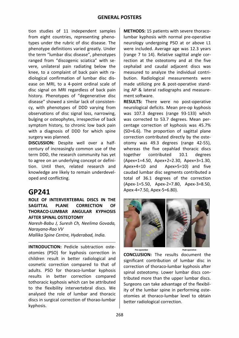
GENERAL POSTERS
268
tion studies of 11 independent samples
from eight countries, representing pheno-
types under the rubric of disc disease. The
phenotype definitions varied greatly. Under
the term “lumbar disc disease”, phenotypes
ranged from “discogenic sciatica” with se-
vere, unilateral pain radiating below the
knee, to a complaint of back pain with ra-
diological confirmation of lumbar disc dis-
ease on MRI, to a 4-point ordinal scale of
disc signal on MRI regardless of back pain
history. Phenotypes of “degenerative disc
disease” showed a similar lack of consisten-
cy, with phenotypes of DDD varying from
observations of disc signal loss, narrowing,
bulging or osteophytes, irrespective of back
symptom history, to chronic low back pain
with a diagnosis of DDD for which spine
surgery was planned.
DISCUSSION: Despite well over a half-
century of increasingly common use of the
term DDD, the research community has yet
to agree on an underlying concept or defini-
tion. Until then, related research and
knowledge are likely to remain underdevel-
oped and conflicting.
GP241 ROLE OF INTERVERTEBRAL DISCS IN THE SAGITTAL PLANE CORRECTION OF THORACO-LUMBAR ANGULAR KYPHOSIS AFTER SPINAL OSTEOTOMY Naresh-Babu J, Suresh Ch, Neelima Govada,
Narayana-Rao VV
Mallika Spine Centre, Hyderabad, India.
INTRODUCTION: Pedicle subtraction oste-
otomies (PSO) for kyphosis correction in
children result in better radiological and
cosmetic correction compared to that of
adults. PSO for thoraco-lumbar kyphosis
results in better correction compared
tothoracic kyphosis which can be attributed
to the flexibility intervertebral discs. We
analysed the role of lumbar and thoracic
discs in surgical correction of thorao-lumbar
kyphosis.
METHODS: 15 patients with severe thoraco-
lumbar kyphosis with normal pre-operative
neurology undergoing PSO at or above L1
were included. Average age was 12.3 years
(range 7 to 14). Relative sagittal angle cor-
rection at the osteotomy and at the five
cephalad and caudal adjacent discs was
measured to analyze the individual contri-
bution. Radiological measurements were
made utilizing pre & post-operative stand-
ing AP & lateral radiographs and measure-
ment software.
RESULTS: There were no post-operative
neurological deficits. Mean pre-op kyphosis
was 107.3 degrees (range 93-133) which
was corrected to 53.7 degrees. Mean per-
centage correction of kyphosis was 45.7%
(SD=6.6). The proportion of sagittal plane
correction contributed directly by the oste-
otomy was 49.3 degrees (range 42-55),
whereas the five cepahlad thoracic discs
together contributed 10.1 degrees
(Apex+1=4.50, Apex+2=2.30, Apex+3=1.30,
Apex+4=10 and Apex+5=10) and five
caudad lumbar disc segments contributed a
total of 36.1 degrees of the correction
(Apex-1=5.50, Apex-2=7.80, Apex-3=8.50,
Apex-4=7.50, Apex-5=6.80).
CONCLUSION: The results document the
significant contribution of lumbar disc in
correction of thoraco-lumbar kyphosis after
spinal osteotomy. Lower lumbar discs con-
tributed more than the upper lumbar discs.
Surgeons can take advantage of the flexibil-
ity of the lumbar spine in performing oste-
otomies at thoraco-lumbar level to obtain
better radiological correction.

GENERAL POSTERS
269
GP242 FREEDOM DISC VS. PRODISC-L FOR TREATMENT OF 1-LEVEL LUMBAR DISC DISEASE: RESULTS FROM ONE INVESTIGATIVE SITE OF THE USA FDA RANDOMIZED CLINICAL TRIAL Harvinder Bedi,MD; Michael. Kropf,MD;
L.E.A. Kanim,MA; Janice Kim,BA; Hyun
Bae,MD; Alexandre Rasouli,MD; Rick B.
Delamarter, MD
Spine Center, Cedars-Sinai Medical Center,
Los Angeles, CA
INTRODUCTION: The Freedom Artificial Disc
(FD) is studied in order to treat single-level
lumbar disc disease L3 to S1. The design of
the Freedom Artificial Disc allows potential-
ly less range of motion in flexion, extension,
axial rotation, and translation than does the
comparison ProDisc-L (PD).
OBJECTIVE: To assess and compare the self-
reported outcomes for patients treated for
1-level lumbar disc disease with the FD
compared to PD. To report the two year
results from one site participating in the
RCT.
METHODS: USA-FDA approved prospective
and randomized (1:1) clinical trial of the
Freedom® Lumbar Disc (AxioMed Spine
Corp., Cleveland, OH) versus ProDisc-L
(Synthes, West Chester, PA). Self-reported
inventories of Visual Analog Scale for back
pain (VAS back pain) and VAS leg pain,
Oswestry disability Index, physical exams,
adverse events, and range of motion (ROM)
were collected preoperatively and at 6
weeks, 3, 6, 12, 18, and 24 months annually
postoperatively.
RESULTS: For FD treated patients (n=20 1-
level), significant improvements of 80% in
VAS back pain (7.3 pre vs 1.5 24mo), 86% in
VAS leg pain (2.7 pre to 0.3 at 24mo) and
74% in Oswestry disability (59.5 pre vs. 14.2
24mo) were observed by 6 weeks and well
maintained to 24 months (p< 0.05). For PD
(n=19 1-level), significant improvements of
68% in VAS back pain (7.3 pre vs 2.5 24mo),
60% in VAS leg pain (3.9 pre to 1.8 at 24 mo)
and 63% in Oswestry disability (57.1 pre vs.
21.2 at 24mo) were observed by 6 weeks
and maintained to 24 months. There was
slight earlier treatment benefit for Freedom
by 6 wks [VAS back: 2.6 (FD) vs 4.2 (PD) at
6wks, p< 0.02; 2.3 (FD) vs 3.9 (PD) at 3
mo,p< 0.04, and 1.5 (FD) vs 3.8 (PD) at 6
mos p< 0.001; ODI: 19.3 (FD) vs 30.5 (PD) at
6 mos, p< 0.02] that disappeared at longer
follow-up (NS)
CONCLUSIONS: Significant recovery was
similarly reported by patients treated using
either the ProDisc-L or Freedom ADRs with
earlier recovery with Freedom.
GP243 SINGLE-LEVEL INSTRUMENTED POSTEROLATERAL FUSION VERSUS POSTE-RIOR LUMBAR INTERBODY FUSION FOR UNSTABLE LUMBAR SPONDYLOLISTHESIS Shuugo Kuraishi, M.D.1), Jun Takahashi,
M.D.1),Hiroki Hirabayashi, M.D.1), , Keijiro
Mukaiyama, M.D.1),Masayuki Shimizu,
M.D.1) ,Futatugi Masatoshi, M.D.1), Hi-
royuki Kato, M.D.1) ,Hisatoshi Kinoshita,
M.D.2) Yutaka Tateiwa, M.D.2)
1)Departments of Orthopaedic Surgery,
Shinshu University, School of Medicine
2)Nagano Prefectural General Rehabilitation
Center
INTRODUCTION: Surgery for lumbar
spondylolisthesis is widely performed.
However, few reports have compared out-
comes between posterolateral fusion (PLF)
and posterior lumbar interbody fusion
(PLIF). This study retrospectively compared
the clinical and radiographic outcomes of
PLF and PLIF for L4 unstable
spondylolisthesis.
METHODS: Patients with L4 unstable
spondylolisthesis with Meyerding Grade II
or more, slip of >10°or >4 mm upon maxi-
mum flexion and extension bending, and
posterior opening of >5° upon flexion bend-
ing were studied. Patients were treated by

GENERAL POSTERS
270
L4-L5 level decompression and posterior
instrumented fusion from January 2008 to
January 2010. Patients who underwent PLF
and PLIF (n = 11 per group) and were fol-
lowed-up for >2 years were studied. The
mean age and follow-up period of the PLF
group were 71.3 years and 2.8 years, re-
spectively, and those of the PLIF group were
70.0 years and 2.6 years, respectively. Radi-
ographic findings and clinical outcomes
evaluated by the Japanese Orthopaedic As-
sociation (JOA) score were compared be-
tween groups.
RESULTS AND DISCUSSION: JOA scores of
the PLF group before surgery and at final
follow-up were 11.6 ± 4.8 and 23.9 ± 3.7,
respectively; those of the PLIF group were
14.0 ± 4.8 and 21.2 ± 7.8, respectively, with
no significant difference between groups.
Correction of slip estimated from postoper-
ative slip angle, translation, and mainte-
nance of intervertebral disc height was bet-
ter in the PLIF group (P < 0.05); however,
there was no significant difference in lum-
bar lordotic angle and slip angle and transla-
tion angle upon maximum flexion and ex-
tension bending. Fusion rates of the PLIF
and PLF groups were 91.0% and 72.3%, re-
spectively, showing no significant differ-
ence. These results suggest that L4-L5 level
posterior instrumented fusion for unstable
spondylolisthesis using both PLF and PLIF
ameliorates clinical symptoms when local
stability is achieved.

INDEX
271
A Aakash A, GP22
Aalto T, O65,SP40,SP56
Abdullah KG, O35
Abe K, SP20,GP112
Acharya N, GP145
Acharya S, GP145
Acosta F, GP202
Adachi K, GP101
Adams MA, O27,SP0,SP11,
SP14,GP73
Adams SL, GP84
Afifi M, GP39
Agarwal A, GP18,GP37,GP38
Agarwal AK, GP37, GP38
Aghdasi B, SP06
Aihara T, SP36,SP58,SP108,SP125
GP141, GP147,GP159-160,GP167
Airaksinen O, O65,SP30,SP40,SP56,
GP35
Aithala J, GP145
Aiyangar A, GP45
Aizawa M, O24,SP42
Akazawa T, GP152,GP164,GP238
Akeda A, SP50
Akeda K, GP56
Akune T, O38, O40
Ala-Korpela M, O16
Alamin TF GP231
Alimasi W, GP68
Alini M, O20,GP77,GP78
Alvares L, O68,
An HS, O8,SP10,SP25,GP23-26,
GP36,GP83-85,GP93
Anand N, GP232-233
Andersson GBJ, O8, SP10,GP11,
GP23-26,GP36,GP84-85
Anderst W, GP45
Ando K, O20
Ando M, GP198
Annesley-Williams DJ, SP11,
Aoki M, SP31,SP51,SP52, SP58
Aoki Y, GP2,GP51,GP89,GP98-99,
GP130-131,GP197,GP209, GP222
Aono H, GP181
Arai F, GP70
Arai G, GP17,GP157
Arai I, SP22, SP58
Arai T, GP65
Arealis G, GP234
Arima N, GP204
Arisaka M, O61
Armijo-Olivo S, O4
Arthur B, O2,GP144
Asahara H, O26
Asante AK, GP94
Asomugha EU, GP168
Ataka H, O49,SP58,GP100,GP165
Atanasio S, GP158
Aya S, O50,GP122,GP159
Azuma S, O44
B Bae H, GP242
Bae WC, O21,O26,GP40,GP80-82
Bai M, O14
Bajaj N, O53
Baker H, O27,SP11
Balderston RA, O60
Balkovec C, SP09
Barnsley S, GP93
Baron EM, GP232-233
Barz T, O74,SP45,GP13,GP212
Basu S, GP145
Battié MC, O12,O66,GP94,GP155,
GP161,GP240
Beadle C, GP231
Becker H-J, O58
Beckingsale TB, GP92
Bedi H, GP242
Behrbalk E, GP113,GP234
Bell KM, GP34,GP42
Ben-Abraham EI, GP41
Bendix T, GP149
Benneker L, GP78
Ben-Porath Y, O63
Benzel EC, O35
Berg S, O78
Berger D GP230
Best TM GP69
Bethea S GP49
Bevevino AJ, GP20, GP228
Bhaduri S, GP18
Binette F, GP81
Bishop PB, O2,GP144
Block AR, O63
Blumenthal SL, O56, O59
Bonner T, GP8, GP9
Bonzanini B, GP127
Boonen S, O57
Boszczyk BM, GP113
Bot AGJ, GP203
Bradley W, SP48
Braun S, O68
Brayda-Bruno M, SP08,GP52
Brisby H, GP71-72
Brochhausen C, GP52
Brox JI2, O54
Buchanan S, GP85
Buller C, SP45,GP212
Buman MP, SP59,GP207
Bydder GM, GP40
Bünger CE, O36
C Cabalo A, GP196,
Callary S, SP34,
Cambridge EDJ, O30,
Cappuccino A, O59,
Carandang G, SP12
Cardoso MJ, GP19,
Carolin Miltenburger5 O57,
Carroll LJ O66,GP94
Carstens C, GP52,
Carstensen M, SP09,
Carter T SP03, GP111,GP151
Cavanaugh JM, O29,
Cedraschi C, GP116,
Cha T, GP83,GP203
Chak JD, O25,GP7
Chan D, O16,O20
Chang BS, SP23,GP29,GP33,
GP142-143
Chen AC, GP44,GP83-85
Chen AF, GP137,
Chen CW, GP46,
Chen CY, O29,
Chen D, GP57,
Chen HM GP1,
Chen J, GP41,GP55
Chen L, GP79,
Chen WJ, GP145,
Chen Y, GP46,
Chen YF, GP88,
Cheung KMC O16,GP154
Chiba K, SP38, GP54
Ching A, O77,
Cho HC, GP194-195
Chou CC, GP46,
Christensen FB, O36, GP6
Chung NS, O11,GP214-215
Chung SK, GP194-195
Cody JP, GP19-21, GP226-228
Colbrunn R, GP8-9
Colli B, GP127,
Colloca CJ, GP39,
Condor R, GP145,
Connolly P, O43,O55,GP224
Cooper J, GP93,
Coric D, O59,
Courvoisier DS, GP116,
Coyne E, O3, GP91
Cripton PA, O25,GP7
Cs-Szabo G, GP84-85
Cuperman T, GP61,
D Dagenais S GP139
Daisuke S, GP10,
Danielson E O18,

INDEX
272
Daubs MD, SP06,
Davis K, SP05, SP24
Davis TT, GP202,
DeBlasi GJ GP230
Deissroth K, O48,
Delarmarter RB O22,SP41,GP242
Demetropoulos CK GP47,
Denaro V GP77-78
Desmoulin C, GP116,
DeSouza L, GP63,
Deyo RA O7, O77,GP95
Dezawa A, GP5,GP102,GP185
Dhanvin D, GP22
Dick B, O4,
Dimmig T, O59,
Ding Y, GP55,
DiPaola C O43,O55,GP224
Dmitriev AE, GP19-21
Dobscha SK GP95,
Dodge GR, GP74,
Dohzono S, O69,GP217
Doita M, GP60,GP62,GP237
Dolan P,P14,P27,SP07,SP11,GP73
Donaldson WF GP137,
Dong Q, SP16,
Donovan M, GP95,
Dozono S, GP225,
Drochner TE, GP202,
Dugan T, GP34,
Dvorak MF, O25,GP7
E Ebata S, O71,
Eck J, O43,O55,GP163,GP224
Edidin AA, O67,
Egge N, O43,GP224
Eguchi Y, O17, SP51,SP58,GP51,
GP98-99,GP117,GP157, GP197,
GP211
Ehman RL GP41,
Ehrhart EJ, GP30,
el Barzouhi A, GP220-221
Elfering A O62,
Elliott DM, GP74,
Elsayed S GP234,
Endo K, GP68, GP216,GP235
Endo T, SP04, SP29
Enyo Y, SP04,
Erwin MW GP58,
Erwin WM GP63,
Espinoza Orías AA, O8,SP10,SP25,
GP23. GP25,GP26,GP36
Eysel P SP28,
Ezure T, GP141,
F
Fabeck LG, GP39,
Fairbank JC,O5,O42,O54, SP01,
GP189
Fan Y, SP16,
Farber NJ, GP137,
Fehlings M, GP58,
Ferguson SA, O9,
Field J GP192,
Fielding L, GP30,GP231
Fineberg S, O47, SP46,GP169-179
Fisher CG, O25,GP7
Follak N GP13,
Foltz V, GP116,
Foocharoen T, GP145,
Fortin M, GP161,
Fraser RD SP34,GP16
Fredrik Borgström1,2, O57,
Freedman BA, O28,
Freeman BJC, SP34,GP16,GP39
Fruensgaard S, O36,
Fuentes J, O4,
Fujibayashi S, GP15,GP200
Fujii T, SP37,
Fujimaki H, SP33,
Fujimiya M, GP10,
FujitaN, GP54,
Fujiwara H, GP90,
Fujiwara T,O21,SP19,GP40,GP80-81
Fujiyoshi T, O34,GP239
Fukuda H, SP22,
Fukuhara S, O37,GP114
Fukui D O23,O76,GP104,GP198
Fukumori N, O37,
Fukuoka M, GP134,
Fumiaki K, SP54,
Funabashi M, O4,
Funao H, SP42,
Fung DA GP202,
Furufu T, SP31,GP222
Furuichi I, GP126,
Furusawa S, GP110,
Försth P, O31,
G Galbusera F, SP08, GP52
Galimberti F, O35,
Gao M, GP57,
Gaume R, GP20
Geiss A, SP28,
Genevay S, GP116,
Gerard D, GP85,
Gerdhem P O78,
Ghanayem AJ, SP12
Gibons L, O12,
Gibson AC SP44,
Gibson JNA, GP231,
Gierasimowicz Fontana A, GP116,
Gilbert S, O27, SP11
Goda Y, GP10,GP109,GP118-119
Goel V, GP18,
Goel VK, GP18,GP22,GP37-38,GP47
Gogate S, GP54,
Gorth DJ, GP74,
Goumoëns P, GP116,
Grad S, O20, GP77-78
Gregg C, GP111,
Gross D O4,GP93-94
Gruber HE, GP48-49
Gu T, SP18,GP50
Gunzburg R, GP39,
Guruprakash DK, O29,
Guyer RD O56,O59,O63,SP43
H Ha JW, O32,
Ha KY GP3,GP105-106
Haapea M, GP115,
Hada Y, GP5,
Hai Y GP188,
Haig AJ, O75,
Haldin K, SP34,
Hall DJ, SP34,
Hall H, SP03, GP111,GP151
Halpern P, GP113,
Hamasaki T, O28,
Hanaoka E, SP58,GP183
Handa J, SP22,
Hanley EN GP48-49
Hansson T O13,
Happel JP, GP20,
Hara N, O44,SP37,GP135
Harada M, GP118
Harada T, GP118,
Harding IJ, SP14, GP73
Hargens AR, O26,
Haro H, O71,
Harrigan M, O43,
Harris MB, O51,
Hart R, O48,
Hartman RA, GP42,
Haruki F, GP122,
Hasebe K, GP5,
Hasegawa K, SP32,
Hasegawa T, O71,
Hashikawa T, GP110,GP128, GP146
Hashimoto T, O72,
Hashizume H, O38,O39,O40
SP04,SP29
Haskell WL, SP59,GP207
Hasue F, O34, GP239
Hatakeyama K SP36,GP125,GP159-
160,GP167
Hatta S, GP107,
Havey RM, SP12

INDEX
273
Haws C, GP93,
Hayakawa J, GP199,
Hayakawa K, GP124,
Hayakawa M, GP209,
Hayashi T, SP06,GP31
He J, O21,O26,GP82
He Q GP50,SP18
Healy AT, GP14,
Hebelka H, O13,
Helgeson MD, GP20,GP21
Henriksson HB, GP72,
Hidekazu S, GP120,GP123
Hideki M, GP129,
Hideki S, SP54,
Hidetoshi,T, GP120121,GP123
Higashino K, GP10, GP103,GP109,
GP118-119
Hikata T, O33,GP32,GP205,GP208
Hilario R, GP58,GP63
Hirabayashi H, O71,GP243
Hirata H,SP17, GP60,GP62,GP75,
GP150,GP237,
Hiratzaka S, O48,
Hirayama J, SP58,
Hiroaki K, O50,
Hiroaki N, GP121,
Hiroko I, GP122,
Hiromitsu T, GP121,
Hirosuke N, GP120,GP123
Hiroto K , O17,
Hiyama A, GP70,GP96,GP186
Hodges SD, GP163,
Hoelscher GL, GP48-49
Hoffman C, GP111,
Homma T SP32,
Hong JK, GP194-195
Hori T, GP66,GP182,
Hoshino M, O69,GP223
Hosogane N, O33,SP38,SP47,GP32,
GP205,GP208
Hsieh AH, GP46,GP87,
Hu R, GP136,
Hu S O45,
Hu Y, SP02,
Huang Z, GP55,
Huellstrung R, GP94,
Huh J-S, GP36,
Humadi AH, SP34,
Hutton WC O28,O52,GP10
Hynes RA, GP202,
I Ibsen R GP149,
Ichiji K, SP22,
Ida K,GP135,
Ignatius A, GP52,
Iguchi T GP150
Iida R, GP56,
IkedaS,GP138,
Imura T, SP33,
Inada K, O34,GP117,GP148,
GP153,GP156-157, GP197,GP201,
GP210-211,,GP239
Inage K, O1,O70,SP15,SP35, SP51,
SP58,GP17,GP51,GP59,GP64-65,
GP98-99
Ingram JA, GP48-49
Inoue G, O17, SP33,SP35,SP51,
SP58,GP2,GP17,GP51,GP59,GP64-
65,GP98-99,GP101,GP117, GP153,
GP157,GP197,GP210-211
Inoue N O8,O26,SP10,GP23, GP25,
GP26,GP36,GP44,GP82
Inuzuka K, O61,
Ishigaki N, GP108,GP147
Ishihama H, O24,SP42
Ishihara S, O33, GP32,GP205
Ishii K, O24,O33,SP32,SP38, SP42,
GP32,GP205,GP208
Ishii T, GP90,
Ishikawa T, O17, SP35,SP51,SP58,
GP2,GP17,GP51,GP59,GP64-65,
GP98-99,GP117,GP148,GP153,
GP157,GP197,GP210-211
Ishimoto Y, O38,O40
Ishizaki T, GP130,
Ito M, GP145,
Ito T, SP58,GP145
Iwanami A, O33,SP38,GP32,GP205,
GP208
Iwasaki H, SP04,SP29
Iwasaki M, GP145,GP181
Iwasaki R, SP21,
Iwata A, O72,GP128,GP146 Izeki M, GP15,GP200
Izumi A, GP66
J
Jabir RA, GP145,
Jahng TA, GP145,
Jarvik JG, O77,
Jennum P, GP149,
Jeon CH, O11,GP214-215
Jeong HS, GP194-195
Jeong JH, GP145,
Jiang J, GP55,
Johnson ES, GP95,
Johnsson R, O78, Möller A, O78,
Jones CF GP16,
Jönsson B, O73,
K
Kadosaka Y, GP104,
Kahwaty S GP232-233
Kaito T, GP90,
Kakinuma H, O24,SP42
Kakutani K, SP17,GP60,GP62,
GP75,GP150,GP237
Kallakuri S, O29,
Kamata M, GP208,
Kamoda H, SP35,SP51,SP58, GP2,
GP17,GP51,GP59,GP64-65,GP98-
99,GP117,GP148,GP153,GP157,
GP197,GP210-211
Kanaya K, GP107,
Kanayama M, O72,GP128,GP146
Kane M, O48,
Kaneko S, SP38,
Kaneko Y, O33, GP32,GP205
Kanematsu F, GP138,
Kaneoka K, GP5,GP27
Kang DG, GP19-21, GP226-228
Kang JD, O14, O19,SP16,GP34,
GP42, GP44-45,GP53,GP61,GP137
Kang KT, GP29,GP33,GP142-143
Kang YM, GP67,
Kangas AJ, O16,
Kanim L, O22,SP41
Kanim LA, SP41,
Kanim LEA, GP242,
Kankaanpää M, SP30,GP35
Kanna R, O53,
Kanna RM, O10,
Kanzaki T, SP53,
Kao MCJ SP59,GP207
Kao TH, GP83,
Karim MZ, GP58,GP63
Karppinen J, O16, GP115,GP154
Kasperk C, O68,
Kato H, GP243,
Kato K, O64,SP55,SP60,GP107,
GP229
Kato M, GP223,
Kato S, O44 GP103
Kato T, SP50,
Kato Y GP12,GP107
Katoh S, GP109,GP118-119
Katsuhira J, GP28,
Kaul V, GP37,
Kawaguchi H, GP99,
Kawaguchi Y SP52,GP145,GP182
Kawakami M, O23,O76,GP145,
GP198
Kawamura N, O44, GP66
Kawano O, GP31,
Kayama S, SP22,
Kazuhiro C GP122,
Keeney BJ, O46,
Kelly A, O25, GP7
Ken I, O50,GP122

INDEX
274
Kengo Y GP120,GP123
Kenji E, GP120,GP123
Kenji Y, O50,GP122
Kentaro Y, GP121,
Khandehroo B, GP232,
Khong PL, SP13,
Kiapour AM, GP47,
Kikuchi K, GP112,
Kikuchi N, O61,
Kikuchi S, O37, O64,SP55,GP114,
GP140,GP229
Kikuchi T, GP129,GP132
Kim DW, GP143,
Kim DY, GP194-195
Kim H, GP87,
Kim HJ, SP23GP29,GP33,GP67,
GP142-143
Kim HS, SP39,SP57
Kim HY, O14,
Kim J, GP242,
Kim JH, O28,
Kim JJ O11,GP214-215
Kim S O51,
Kim S, GP58,GP63
Kim SD, GP230,
Kim SY, GP194-195
Kim TH, SP57,
Kim YC, GP195,
Kim YH GP105-106
Kimiaki SK, SP37,
Kimura S, GP51,
Kimura T GP182,
Kinoshita H, GP243,
Kinoshita T, SP58,
Kinoshita Y, O76,GP104
Kita T, GP138,
Kitagawa Y,
Kitahara S, GP239,O34
Kitajima M, GP133,
Kitamura M, GP152,GP164,GP238
Kitou K, GP130,
Kjellberg J, GP149,
Kjellby Wendt G, O18,
Klöting I, GP13,
Kobayash T, GP10,
Kobayashi K GP180
Kodigudla M, GP22
Koes BW GP220-221
Koivisto K, GP115,
Komatsubara S, GP204,
Konari Y, GP66
Konishi S, GP223,
Konishi H O61,
Konno S O37,O64,SP22,SP55,SP60,
GP99,GP114,GP140,GP229
Kono H, SP38,SP47
Konstantinos V, O3,
Kosaka H, GP119,
Koshi T, GP197,
Kotani T, GP164,GP238
Kotowski S, SP05,SP24
Koyanagi T, GP205,
Koyasu S, O24,SP42
Kreuter W, O77,
Kropf M, GP242,
Kröger H, SP56,
Kubota G, O1,O70,SP15,SP35, SP51,
SP58,P17,BP51,GP59, GP64-65,
GP98-99,GP117, GP148, GP153,
GP156-157,GP210-211
Kugisaki H, GP126,GP133
Kuh S, GP145,
Kuittinen P, SP56,
Kumar GV, GP145,
Kumar K, GP213,
Kuniyoshi K, SP21,
Kuraishi S, GP243,
Kurakawa T, SP17GP60,GP62,
GP75,GP150,GP237
Kuramoto T, O24,SP42
Kurata J GP140
Kurosaka M, SP17,GP60,GP62,
GP75,GP150,GP237
Kurtz SM O67,
Kuy Han SK, GP46,
Kwok JWL, SP02,
Kwon B, GP202,
Kyllönen E, GP115,
Kyo Y, GP138,
Käser A, O62,
Könönen M, SP30, GP35
L
Lafave LZ, GP136,
Lama P, SP14,GP73
Landham PR, O27,SP07,SP11
Lange A, O68,
Lange J, GP13,
Lapinsky A, O55,GP224
Lari H, O43, GP224
Larsson K, GP71,
Lau E, O67,
Law T, SP13,
Lavender SA, O9,
Lazary A, GP52,
Leaman D, GP18,
Lee A, SP12
Lee BH, SP39,SP57,GP67
Lee CK, SP23, GP29,GP33,GP142-
143
Lee CS, GP145,
Lee DB, GP142-143
Lee GW, GP143,
Lee HM, SP39,SP57,GP67
Lee J, O19,GP86
Lee JYB, SP25, GP137
Lee LH, GP92,
Lee SH, SP39,SP57
Lee SW, GP194,
Lee YT, O32,
Lehman Jr. RA, GP19-21
Lehman RA, GP226-228
Lehto S, O65,SP40
Leinonen V, SP56,
LeMaitre C, GP73,
Leme A, O19,
Lemen L SP24,
Lenke LG, GP226-227
Lenz ME, O21,SP19,GP40,
GP80-81
Leung A, SP09,
Lim YW, GP194-195
Limbäck Svensson G, O18,
Lin B, GP18,
Lindahl A, GP72,
Liu J, GP202,
Liu ZH, GP88,
Lo HJ, GP1,
Lord SJ, SP45,GP212
Lotz J, O26,
Lozito T, GP61,
Lubelski D, O35,GP14
Luk KDK, O16,SP02,SP13, GP145,
GP154
Luo J, SP07,
Luo ZJ GP88,GP145
Lurie JD, O7, O74
Lycklama à Nijeholt GJ GP220-221
M
Machida M, SP38,
MacMillan M, GP202,
Maeda T, GP31,
Maedler M, GP63,
Maeno K, SP17,GP60,GP62,GP75,
GP150,GP237
Magee D, O4,
Mageswaran P, GP8, GP9,GP14
Mahieu G, GP116,
Makino T, GP90,
Malhotra NR, GP74,
Mamoru A, O50,
Mamoru K GP104,
Manabe H GP180
Manman G, GP4,
Mannion AF, O42,O54, O58,SP01,
GP189
Manoj-Thomas A, GP92,
Mansfield FL GP230
Marek R, O63,
Markova D, GP84,

INDEX
275
Marras WS, O9,SP05,GP69
Marrero L, GP166,
Martin BI, O7, O46,O77
Martin JT, GP74,
Martinez A, SP59, GP207
Marty M, GP116,
Maruyama H GP28,
Maruyama T, O44,
Marx J, SP19,
Masatoshi F, GP243,
Masatoshi H, GP121,
Massimo T, SP16,
Masuda K, O20, O21,O26,SP19,
SP20,GP44,GP80-82
Masuda M, GP31,
Masui O, GP63,
Matsu Y, GP140,
Matsuda S GP15,GP200
Matsudaira K , O61,GP28
Matsuki K, O34, GP239
Matsumoto M, O24,O33,SP38,
SP42,SP47,GP32,GP54,GP205,
GP208
Matsumoto T, O69,GP217, GP223,
GP225
Matsumura A, GP223,
Matsunaga N, GP27,
Matsuo Y, O64,
Matsuoka T O76,GP104,GP198
Matsushita O, SP33,
Matsuura M, GP12,
Matsuura T, GP10,
Matsuura Y, SP21,
Matsuyama Y O71,
Matthew P. SP59
Mattsson J, GP72,
Mauck RL, GP74,
Mausda K GP40,
Mawatari M SP26,SP27,GP126,
GP133
Mayer JM, GP139,
Mazaki T, GP187,
McAviney J, GP39,
Mcclincy M, O14,
McGill SM O30,SP09
McGillion S, GP189,
McGuire KJ GP230
McIntosh G, SP03,GP93,GP111,
GP151
McIntosh TC O60,
McKay WF, SP19,GP81
McKillop AB, O66,
McLain R GP8,GP9
McLain RF GP168,
MD, Khandehroo B, GP233,
Mehdian H, GP234,
Mehrkens A, GP58,GP63
Melloh M, O62, O74, SP45,GP13,
GP212
Melnyk AD, O25,GP7
Mendel E O9,
Menendez ME, GP203,
Meng X, GP188,
Merk HR SP45,GP13,GP212
Metzger M, O22,SP41
Michael J, SP28,
Michaëlsson K, O31,
Miciak M, O4, GP94
Mietsch A, GP52,
Mikalsky M, GP94,
Mikami H, GP147,
Mikami Y GP180
Mimura M, SP58,
Minami S GP152,GP164,GP238
Minamide A, O38,SP04,SP29
Mirza SK, O7,O46,O77
Misawa H, GP187,
Miyagi M, O17,SP35,SP51,SP58,
GP2,GP17,GP51,GP59,GP64-65,
GP98-99,GP117,GP148, GP153,
GP157,GP197,GP210-211
Miyake T, GP104,
Miyamoto E, O76,
Miyashita T O49,
SP58,GP100,GP165
Miyauchi A, GP180,
Miyazaki S, SP17, GP62,GP75,
GP150,GP237
Miyoshi K, O44, O61
Mizuno K GP219,
Mizutani J, GP134,
Mochida J O20,GP60,GP70,
GP96,GP186
Montgomery SR, SP06,
Moon HJ, GP61,
Moon SH SP39,SP57,GP67
Moore R, SP34,GP95
Moran J, GP22
Morasco BJ, GP95,
Mori E, GP31,
Mori H, SP53,
Mori T GP236,
Moriartey S GP136,
Morii J, O44,
Moriki T, GP104,
Morimoto R, GP56,
Morimoto T, SP26,SP27,GP126,
GP133
Morinaga T, SP58,
Morio M O50,GP122
Morishita S, GP198,
Morishita Y, GP31,
Morita Y, GP62,
Morse LJ, GP79,
Moses RA, O74,
Moudgil V, GP145,
Mroz TE O35,GP14
Mukaiyama K, GP243,
Munakata Y , SP22,
Murak S, O39,
Murakami H, GP66,GP132,
Murakami M, O44,
Muraki S, O38, O40
Murase S, GP236,
Murata K O15,SP50,GP56
Murata Y, SP58,GP12,GP107
N
Nagai S, O24,SP42
Nagai T, GP96,GP186
Nagamachi A, SP49,
Nagamoto Y, GP181
Nagashima H, GP145,
Nagata K, O38,O40,SP37,GP135
Naiki M SP20,GP80
Nair R, GP213,
Nakagawa K, SP31,GP89,GP209,
GP222
Nakagawa Y, SP04,SP29
Nakai O, GP219,
Nakai T, O20,
Nakajima A, SP31,GP89,GP209,
GP222
Nakamura H O69,SP06,GP124,
GP199,GP217,GP223,GP225
Nakamura K, O20,GP131
Nakamura M, O33,SP38,GP32,
GP205,GP208
Nakamura Y GP5,
Nakanishi K, GP187,
Nakano M, GP182,
Nakao S, O23, O76,SP29, GP104,
GP198
Nakata Y, GP153,GP211
Nakata Y, SP15, GP17
Nakazawa T, SP33,GP101
Narayana-Rao VV GP241,
Naresh-Babu J, GP76, GP241
Nasto L, O19,GP53,
Natarajan RN, GP11,GP24
Neelima G GP76,GP241
Negrini A, GP158,
Negrini S GP127,GP158
Neidlinger-Wilke C, SP08,GP52,
Nemoto T, GP152,GP164,GP203
Neuhaus V, GP203,
Newton DGP,163,
Ngo K, O19, SP16,GP53,GP61
Nguyen QT, GP44,
Nielsen CV, O36,
Niemeläinen R GP94,

INDEX
276
Niinimäki J, GP115,
Nikaido T, O64, SP55,SP60, GP140,
GP229
Nilsson A, O13,
Nishi H, SP04,SP29
Nishida K SP17,GP60,GP62,GP72,
GP150,GP237,
Nishikawa S GP89,GP130-131
Nishimura A, SP50,
Nishimura H, GP216,GP235
Nobukiyo M, SP52,
Noh W, GP194-195
Norberg M, GP116,
Nordborg C, GP71,
Norimoto M, GP98,
Nowicki AL, GP137,
Nukaga T, GP70,
Numazawa T, SP20,
O
O’Dowd JK , O5,
Obara H, GP131,
Obata, T, GP59,
Oestergaard LG, O36,
Ogihara S, O44,GP236
Oglesby M, O47,SP46,GP169-
GP179
Oh IS, GP3,
Oha F, O72, GP128,GP146
Ohnmeiss DD, 041,O56,O63,SP43
Ohtori S, O1,O17,O34, O70,SP15,
SP21,SP31,SP35,SP51,SP52,SP53,SP
58,GP2,GP17,GP51,GP59,GP64-
65,GP89,GP98-99,GP110,GP112,
GP117,GP148,GP153,GP156-157,
GP164,GP197,GP201,GP210-211,
GP239
Ohzone H, GP59,
Oikawa Y, O1,O17,O70, SP15,
SP35,SP51,SP58,GP2,GP17,GP51,G
P59,GP64-65,GP98-99, GP117,
GP148,GP153,GP156-157, GP197,
GP211GP210,
Oka H, O38, O40
Oka S GP204,
Okada T, SP20,
Okada Y, O24,SP42
Okamoto Y, SP58,
Okubo T, GP59,
Okubo Y, GP5,GP27
O'Neill C, GP97
Ong KL, O67,
Onishi T, O72,GP128,GP146
Onishi Y, O37,GP114
Orita S, O1, O17,O70,SP15,SP35,
SP51,SP52,SP53,SP58,GP2,GP17,GP
51,GP59,GP64-65,GP98-99, GP110,
GP117,GP148,GP153,GP156-57,
GP197,GP210-211
Osaki S GP187,
Oshita Y, GP23,
Otani K, O37, SP55,SP60,GP114,
GP219,GP229
Otoshi K, O64,
Otsuka S, GP134,
Otsuka T GP134,
Otsuka Y, GP36,
Otsuki B, GP15,GP200
Otsuki K, GP130-131
Ouchi J, SP36,GP160,GP167
Oxland TR O25,GP7
Oyama T, GP131,
Ozawa T, O17,O34,O70,SP58,
GP201,GP239
P
Paik H, GP19
Pakarinen M, O65,SP40
Paldanius P, GP115,
Palepu V, GP22
Pan CC, O8,GP26
Park CK, GP145,
Park DK, GP25,
Park JH, SP23,GP29,GP142-143
Park JO, SP57,GP67
Park JU, O11,GP214-215
Park Y, O32,
Parks RM, GP113,
Parnell J, GP136,
Parzini S, GP158,
Patappa G, GP77-78
Patel A, gp177
Patel A, O47,SP46,GP169-179
Patwardhan AG SP12
Pedtke A, O45,
Pehkonen T, GP115,
Pelton M, gp177
Pelton M, O47,SP46,GP169-179
Peng C, GP83,
Penta K, GP81,
Peroglio M, GP77-78
Perry E, O48,
Petrik A, GP95,
Pettine K, O59,
Peul WC GP218,GP220-221
Phan K, SP06,
Pichika R, O21,GP40,GO80-81
Pittet V, O58,
Pohl PHI, GP61,
Pokharel R, GP145,
Pollintine P, O27,SP07,SP11,GP22
Porchet F, O58,
Portia-Anthony M, SP13,
Prasarn ML, O3,GP91
Prijambodo B, GP145,
Puttlitz CM, GP30,
Q
Qasim M, GP24,
Quillen WS, GP139,
Quon JA, O2,GP144
R
Rade M, SP30, GP35
Rajasekaran S O10,O53,GP145
Ramsey T, SP05,
Rashiq S, O4,
Rasouli A, GP242,
Rechtine GR O3,GP91
Rege A, GP145,
Rempel J, GP136,
Renjitkumar J GP213,
Reynolds JB GP196,
Rhee DJ,GP194-195
Ring D, GP203,
Risbud M, O20,GP54
Robie B, GP6,
Robinson S, O22,
Robinson ST, SP41,
Robson-Brown K, O27,
Rogerson M, GP136,
Roh J O59,
Romano M, GP158,
Roughley P, O19,
Rozenberg S GP116,
Ruan DK, SP18,GP50
Ruehlman DR, GP30,
Runesson E, GP71,
Russo F, SP16,GP77-78
Rydevik B GP71,
S
Saari T, SP56,
Sacco D GP166,
Sah RL, O26,GP44
Sahlstrand T, O78,
Saino T, SP35,GP148,GP210-211
Sainoh T, O1, O70,SP15,SP51-52,
SP58-59,GP5,GP10,GP17,GP98-
99,GP102-103,GP117-119,GP151,
GP153,GP156-157,GP185,GP197,
GP211
Sairyo K, GP5,
Saito M, SP38,SP47
Saito W, SP33,GP101
Sakai D, O20,GP60,GP70, GP96,
P186
Sakai H, GP31,
Sakai T, GP103,GP109,GP118-119
Sakai Y, GP237,

INDEX
277
Sakaida C, GP131,
Sakanaka H, GP138,
Sakon N, GP104,
Sakuma T, GP152,GP164,GP238
Sakuma Y, O1,O17,O70,SP15,SP35,
SP51,SP58,GP2,GP17,GP51,GP59,
GP64-65,GP98-99,GP117, GP148,
GP153,GP156-157,GP197,
GP210-211
Samartzis D, O16, SP13,GP86,
GP88,GP145,GP154
Sameda H, SP58,
Sandella DE, O75,
Sandén B O31,
Sanderson PL GP92,
Santoni AL, GP30,
Santoni BG, GP30,
Sasaki A, O24, SP42
Sasaki N, GP71,
Sasaki S, GP198,
Sato J, O1,O70,SP15,SP51,GP17,
GP51,GP59,GP64-65,GP98-99,
GP117,GP148,GP153,GP156,
GP197,GP211
Sato M, GP96,GP186
Sato N, GP119,
Sato Y, GP152,GP164,GP238
Satoh S, GP145,
Satoshi Y, GP129,
Sauermann S, O68,
Sawaji Y, GP68,
Savolainen S, SP56,
Schairer W, O45,
Schiuma D, GP78,
Schreck M, GP91,
Scott TP, SP06,
Seim III HB, GP30,
Seitsalo S, GP115,
Seki M, GP223,
Seki S, GP182,
Sekiguchi M, O37,O64,GP114,
GP140
Senoo I, GP25, GP26
Shacklock M, SP30, GP35
Shah SB, GP87,
Sham P, GP145,
Shapiro I, GP54,
Shapiro S, O19,
Sharan AD GP79,
Shen Y, GP145,
Shetty AP, O10,O53
Shetty JY, O10,
Shi P, GP83-84
Shi Z, SP18,GP50
Shiba K GP31,
Shiba M, GP12,GP107
Shibata Y, SP31,GP64,GP209,
GP222
Shiboi R, O34,GP201,GP239
Shibuya S, GP204,
Shieh I, O21,GP82
Shiga T, GP130-131
Shigehiro I, SP54,
Shigenobu K, O72,GP128,GP146
Shigenori N, O50,GP122
Shigeo K, O50,GP122
Shimamura T GP129,
Shimizu M, GP243,
Shimoda H, SP32,
Shindo S, GP219,
Shingo M, GP60,
Shinji T, GP121,
Shinohara Y, SP53,GP217,GP225
Shinsuke S, GP120,GP123
Shiono Y, O24, SP42
Shiota M, SP38,
Shiozaki Y, GP187,
Shirahata T, O34,GP239
Shiraki M, SP26,GP133
Shivers J, SP43,
Sho D, GP121,
Shoji S, SP20,
Sides BA GP226,
Sidorkewicz N, O30,
Siemionow K, SP25,
Sigmundsson FG, O73,
Simamura T GP132,
Simon P, O8, SP10,GP36
Sinclair MK, SP12
Singh K O47,SP46,GP169-179
Singh V, GP7,
Sinikallio S, O65, SP40,SP56
Sipola P, SP56,
Skioldebrand E, GP72,
Slikker III W, SP25,
Slosar PJ, GP166,GP196
Smith DHM, GP95,
Smith LJ GP74,
Smuck M, SP59,GP207
Sobottke R, SP28,
Sogaard R O36,
Soininen P, O16,
Soliman A, O78,
Sonobe M, SP31,GP209,GP222
Sonohata M, SP26, SP27,GP126,
GP133
Sowa G, O14,O19,SP16,GP42,
GP53,GP61
Spann SW, GP202,
Splittstoesser R, O9,GP69
Spooner L, GP73,
Spratt KF, 06
Spruit M, O5,
St Joseph JB, GP73,
Stadelmann V, GP77,
Stallbaumer JJ, GP226,
Staub LP, O74,SP45,GP13,GP212
Stauff MP, O28,
Steenstra I, GP93,
Stefanakis M, SP07,SP14
Steiger F, O58,
Ström O, O57,
Strömqvist B O73,O78
Sudo A SP50,GP56
Sugimoto Y, GP187,
Sugiura S, GP89,GP130-131
Suh BG, GP143,
Sumida T GP180
Summers B, SP14,
Sun A, GP94,
Sun Z, GP88,
Sundqvist K, GP115,
Sung NY O32,
Sur DW, GP3,
Suresh Ch, GP241,
Suzuki A, O69,SP06,GP217,
GP223,GP226
Suzuki H, GP68,GP216,GP235
Suzuki K,GP182,
Suzuki M, O1,O17,O70,SP15,GP2,
GP17,SP35,SP51,SP58,GP51,GP59,
GP64-65,GP98-99,GP117,GP148,
GP153,GP156-157,GP197,GP209-
211,
Suzuki N, GP134,
Suzuki T, SP21,
Sychev I, SP14,
Szpalski M, GP39,
T
Taborek A, O21,GP44
Tadao T GP121,
Takada T, SP17, GP60,GP62,GP75,
GP150,GP237
Takahashi H, SP31,GP89,GP209,
GP222
Takahashi J, GP243,
Takahashi K, O1,O17,O34,O70,
O71,SP15, SP21,SP31,SP35, SP37,
SP51-53,SP58-59,GP2,GP17, GP64-
65,GP89,GP98-99,GP110, GP112,
GP117,GP148,GP152-153,GP156-
157,GP164,,GP197, GP201,GP210-
211,GP222,GP238-239,
Takahashi N, SP22,
Takahashi S, GP217,GP225
Takahashi Y SP49,
Takahira N, SP33,GP101
Takao S, GP118,
Takao T, GP31,

INDEX
278
Takaso M SP33,GP101
Takata Y, GP103,GP109,GP118-119,
GP130
Takatori R, O8,
Takayama K, SP54,GP138
Takebayashi T, SP37,GP99,GP135
Takegami M, O37,
Takemoto M, GP15,GP200
Takeshita K, SP37,GP135
Takigawa T, GP187,
Takiguchi N, O38,O40,
Tamura Y, GP148,
Tan K, O21,
Tanaka H, GP68,GP216,GP235
Tanaka M O72,GP128,GP146,
GP187
Tanaka T, O34,GP239
Tang SY, GP79,
Taniguchi H, GP119,
Taniguchi S, SP31,GP209,GP222
Taniguchi T, GP119,
Tanimoto K, O29,
Tanno T, O49, GP100,GP165
Tassanawipas WA, GP145,
Tateiwa Y GP243
Tcherveniakov P, GP16,
Techy F, GP8,GP9
Terada M, SP29,
Teraguchi M, O39,
Terai H, O69, GP217,GP223, GP225
Terakado A, SP53,GP110
Terayama S, O44,GP124,GP199
Teruo K, SP54,
Tervonen O, GP115,
Tetsuhiro GP148,
Tetsuya K, O50,GP122
Thomeé R, O18,
Tibiletti M, SP08,
Tillotson CJ, GP95,
Tobimatsu H, GP181
Toki S, SP49,
Tolhurst S, GP8, GP9
Tomiya M, GP121,
Tomkins-Lane CC O75,GP136,
GP155
Toshiaki K, GP152,
Toshiya Y, SP54,
Toshiyuki M GP184,
Tosteson ANA, O7,O77
Toyama Y, O24,O33,SP38,SP42,
SP47,GP32,GP54,GP205,GP208
Toyoda H, O69,GP217,GP223,
GP225
Toyone T, O17,O34,O70,SP51,
SP59,GP2,GP51,GP59,GP98-99,
GP197,GP201,GP239
Toyooka T, GP130-131
Tracey RW, GP19-21, GP226-228
Truumees E, GP47,
Tse JYH, SP02,
Tsuji T, SP38,
Tsujio T, O69,
Tsukamoto M, SP26,SP27,GP133
Tsukiyama K, GP217,GP225
Tsutsui S, SP04,SP29
Tuan R, GP61,
Tubaki V, O53,
Tujio T, GP223,
Tukamoto M, GP126,
Tullberg T O78,
Turner AS GP30,
Turunen V, SP56,
Tyler Perryman L GP190
Tängemo C, GP72,
U
Uchida K, SP33, GP101
Ueno M, SP33, GP101
Ueta T, GP31,
Uri O, GP113,
Urushibara M, SP36GP125, GP159,
GP160,GP167
V-W
Wachna M, GP231,
Wada H, GP107,
Wada K, GP12,GP107
Wada T, GP66
Wada Y, O17,O34GP239
Vadalà G, SP16,GP77-78
Vader J-P, O58,
Wakita T, O37,GP114
van den Hout WB GP220-221
Van der Kallen BF GP220-221
van Hooff ML, O5,O42
van Limbeek J O5,
Wan ZY, GP88,
Wang CF, SP18,GP50
Wang CM, GP88,
Wang D, O19,SP18,GP50
Wang HQ, GP88,
Wang JC SP06,
Wang T, O35,
Wang Z, O52,
Vanhanen S, O65,SP40
Vanninen R, SP30,GP35
Wardlaw D, O57,
Warren S, O4,
Watanabe A, O17, O34,GP59,
GP117,GP211,GP239
Watanabe K, O33, SP38,SP47,
SP55,SP60,GP32, GP205,GP208,
GP229
Watanabe M, GP96,GP186
Vavken P, GP86,
Welch J, GP94,
Wen TL, O25,
Wenbin D, GP4,
Whang PG SP48,
Videman T, O12,
Viinamäki H, O65,
Viinamäki H, SP40,
Wilke H-J SP08,GP6,GP52
Williams R GP145,
Wilsey JT, SP19,
Wilson P, GP136,
Virk GS, O29,
Vleggeert-Lankamp CL, GP218,
GP220-221
Vo N, O14,O19,SP16,GP42,GP53,
GP61
Wojewnik B, SP12
Volkheimer D, GP6,
Wong DA, GP162
Wood KB GP230
Voronov LI, SP12
Wu J, SP18,GP50
Wu X, GP55,
Wuqikun A GP123,
X
Xin HK, SP18,GP50
Xuejun D, GP4,GP57
Xuenong Z GP4,GP57
Y
Yabuki S, O37, SP55,SP60,GP229
Yagi M, SP38,
Yaldo J, O29,
Yamabe D, GP129,GP132
Yamada A, GP185,
Yamada H, O38,O39,O40,SP04,
SP29
Yamada K O44,GP217,GP225
Yamada M, GP209,GP222,
Yamada T, O44,
Yamagata M, SP58,GP197
Yamaguchi T, O21,O26,SP19,GP40,
GP44,GP80-82
Yamakawa KSJ, O75,
Yamamoto J, SP17,GP60, GP62,
GP75, GP150,GP237
Yamane K, GP68,GP187,GP216,
GP235
Yamano Y GP138,
Yamaoka A, GP112,
Yamashita K, GP112,
Yamashita M, SP58, GP112
Yamashita T, SP37,GP135

INDEX
279
Yamauchi K, O1, O70,SP15,SP35,
SP51,SP58,GP17,GP51,GP59,GP64-
65,GP98-99,GP17,GP117, GP148,
GP153,GP156-157,GP197,
GP210-211,
Yamazaki K, GP129,GP132
Yamazaki T, O44,GP124,GP199
Yan Y, GP34,
Yang S-H, GP26,GP83
Yang X, GP95,
Yasuda H, O69,
Yasuda T, GP182,
Yasui N GP10,GP118-119
Yasunori O, O50, GP122
Yeom JS SP23,GP29,GP33,
pGP142-143
Yeung A, GP190-193
Yim RL, GP86,
Yogoro M, SP33,
Yokoyama K, GP70,
Yonenobu K GP90,
Yoon ST, O28,O52
Yorifuji M, GP216,GP236
Yoshiaki T, O50,GP122
Yoshida K GP140
Yoshida M O38,O39,
O40,SP04,SP29
Yoshida S, GP132,
Yoshihara T, SP26,SP27,GP126,
GP133
Yoshiki Y SP54,
Yoshimura N, O38, O39,O40
Yoshinori K, SP54,
Yoshioka K, O24, SP42
Yu J, GP189,
Yuasa A, GP146,
Yugue I, GP31,
Yurube T, SP16,SP17,GP42,
GP60-62,GP75,GP150,GP237
Z
Zaina F, GP127,GP158
Zammerilla LL, GP137,
Zhang C, SP18,GP50
Zhang J, GP93,
Zhang WL, GP88,
Zhang X GP45,
Zhang Y, SP18,GP50,GP83-85
Zhang YZ, GP88,
Zhang Z, GP60,GP145
Zhao L, O22, SP41
Zhao W, O74
Zheng L, GP45,
Zheng Y GP193,
Zheng Z, GP145,
Zhiyu Z, GP4,GP57
Zhou H, O43,O55,GP18,GP224
Zhou J, GP55,
Zhu Q GP55,
Zhu S, GP88,
Zigler JE O56, SP43
Zinchenko N, GP49,
Ö
Örndal C, GP71,