NTD Section III - Filariasis and Onchocerciasis Dec2012 FINAL_0.pdf
47
Lymphatic Filariasis Page 1 Akre M Adja*, Sina Helbig, Alia Tayea, Neil Arya ** Prepared as part of an education project of the Global Health Education Consortium and collaborating partners *First author **Corresponding author
Transcript of NTD Section III - Filariasis and Onchocerciasis Dec2012 FINAL_0.pdf
Lymphatic Filariasis
Page 1
Akre M Adja*, Sina Helbig, Alia Tayea, Neil Arya**
Prepared as part of an education project of the Global Health Education Consortium and
collaborating partners
*First author **Corresponding author
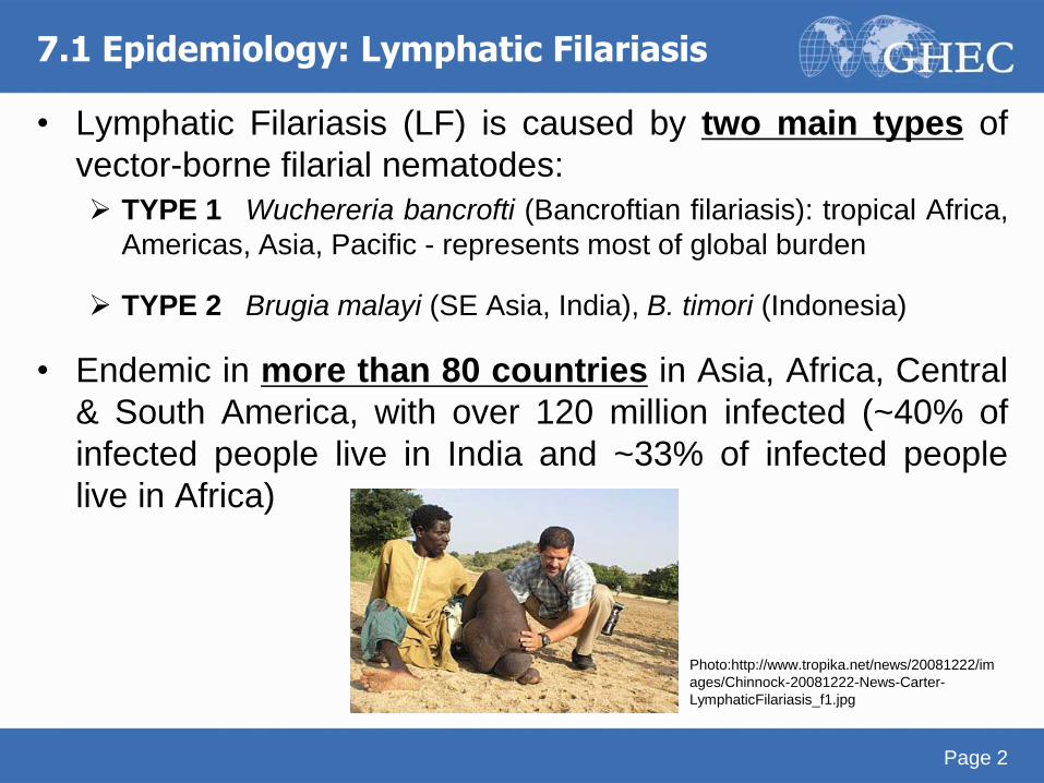
7.1 Epidemiology: Lymphatic Filariasis
• Lymphatic Filariasis (LF) is caused by two main types of
vector-borne filarial nematodes:
TYPE 1 Wuchereria bancrofti (Bancroftian filariasis): tropical Africa,
Americas, Asia, Pacific - represents most of global burden
TYPE 2 Brugia malayi (SE Asia, India), B. timori (Indonesia)
• Endemic in more than 80 countries in Asia, Africa, Central
& South America, with over 120 million infected (~40% of
infected people live in India and ~33% of infected people
live in Africa)
Page 2
Photo:http://www.tropika.net/news/20081222/im
ages/Chinnock-20081222-News-Carter-
LymphaticFilariasis_f1.jpg
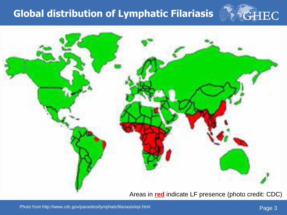
Areas in red indicate LF presence (photo credit: CDC)
Global distribution of Lymphatic Filariasis
Page 3 Photo from http://www.cdc.gov/parasites/lymphaticfilariasis/epi.html
7.1 Epidemiology: Lymphatic Filariasis
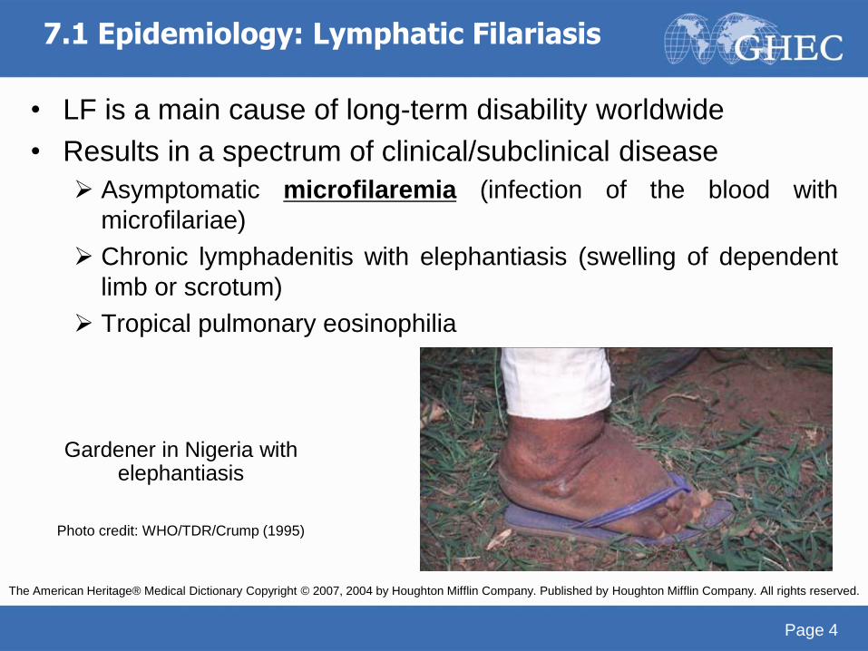
• LF is a main cause of long-term disability worldwide
• Results in a spectrum of clinical/subclinical disease
Asymptomatic microfilaremia (infection of the blood with
microfilariae)
Chronic lymphadenitis with elephantiasis (swelling of dependent
limb or scrotum)
Tropical pulmonary eosinophilia
Page 4
Gardener in Nigeria with elephantiasis
Photo credit: WHO/TDR/Crump (1995)
The American Heritage® Medical Dictionary Copyright © 2007, 2004 by Houghton Mifflin Company. Published by Houghton Mifflin Company. All rights reserved.
7.1 Epidemiology: Lymphatic Filariasis
• Even in microfilaremic patients who appear healthy,
there is some degree of suppression of the immune
system
Infected people are thus more vulnerable to other infections
such as TB
Specific effects of HIV infection on LF are largely unknown
One study showed that the replicative capacity of HIV is
significantly enhanced in peripheral blood mononuclear cells
from patients with untreated LF
Many also have evidence of impaired renal function
Page 5
The American Heritage® Medical Dictionary Copyright © 2007, 2004 by Houghton Mifflin Company. Published by Houghton Mifflin Company. All rights
reserved. -- J Infect Dis. 2000 Dec;182(6):1804-8. Epub 2000 Nov 8.
Filarial infections increase susceptibility to human immunodeficiency virus infection in peripheral blood mononuclear cells in vitro.Gopinath
R, Ostrowski M, Justement SJ, Fauci AS, Nutman TB.
Helminth Immunology Section, Laboratory of Parasitic Diseases, National Institute of Allergy and Infectious Diseases, Bethesda, MD 20892, USA.
7.2 Risk factors: Lymphatic Filariasis
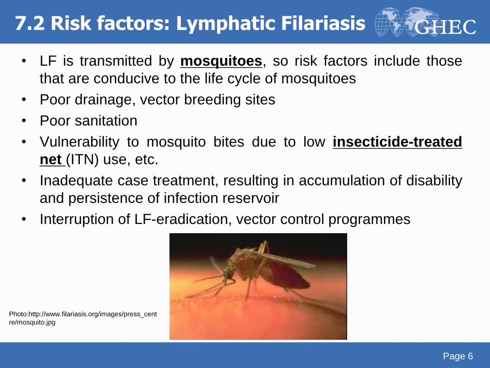
• LF is transmitted by mosquitoes, so risk factors include those
that are conducive to the life cycle of mosquitoes
• Poor drainage, vector breeding sites
• Poor sanitation
• Vulnerability to mosquito bites due to low insecticide-treated
net (ITN) use, etc.
• Inadequate case treatment, resulting in accumulation of disability
and persistence of infection reservoir
• Interruption of LF-eradication, vector control programmes
Page 6
Photo:http://www.filariasis.org/images/press_cent
re/mosquito.jpg
7.3 Biology: Lymphatic Filariasis
• Mosquito vectors Anopheles spp.: most common vector in Africa
Culex spp.: most common vector in the Americas
Aedes spp.: can transmit in Asia & Pacific regions
Mansonia spp.: can transmit in Asia & Pacific regions
• Adult worms reside in the lymphatic system of the human host Female W. bancrofti: 80-100 x 0.25 mm
Male W. bancrofti: 40 x 0.1 mm
Brugia spp are only half of this size
Page 7
7.3 Biology: Lymphatic Filariasis
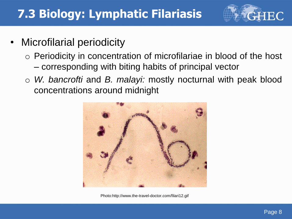
• Microfilarial periodicity
o Periodicity in concentration of microfilariae in blood of the host
– corresponding with biting habits of principal vector
o W. bancrofti and B. malayi: mostly nocturnal with peak blood
concentrations around midnight
Page 8
Photo:http://www.the-travel-doctor.com/filari12.gif
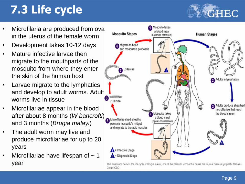
7.3 Life cycle
• Microfilaria are produced from ova
in the uterus of the female worm
• Development takes 10-12 days
• Mature infective larvae then
migrate to the mouthparts of the
mosquito from where they enter
the skin of the human host
• Larvae migrate to the lymphatics
and develop to adult worms. Adult
worms live in tissue
• Microfilariae appear in the blood
after about 8 months (W bancrofti)
and 3 months (Brugia malayi)
• The adult worm may live and
produce microfilariae for up to 20
years
• Microfilariae have lifespan of ~ 1
year
Page 9
7.4 Symptoms: Lymphatic Filariasis
• Wide range of clinical presentations
• Asymptomatic microfilaremia in individuals: o Who have not been sufficiently exposed to be infected
o With prepatent infection, adult worm infection without microfilaremia
o Who have cleared the infection
• Acute manifestations: adenolymphangitis (filarial fever)
o Characteristic of both Bancroftian and Malayan filariasis
o Episodic attacks of fever, malaise, chills (resembles malaria)
o Enlarged painful lymph nodes draining the affected part
o Duration usually 1 wk, may recur multiple times within 1 yr
o May also have filarial abscess; pus sterile or with bacteria
Page 10
• Chronic manifestations o Hydrocele
o Lymphedema
o Elephantiasis (mainly lower limbs, less commonly arms,
genitals, breast)
o Chyluria
o Tropical pulmonary eosinophilia
• Brugian filariasis o Less genital involvement
o Elephantiasis usually restricted to areas below the knee
Page 11
7.4 Symptoms: Lymphatic Filariasis
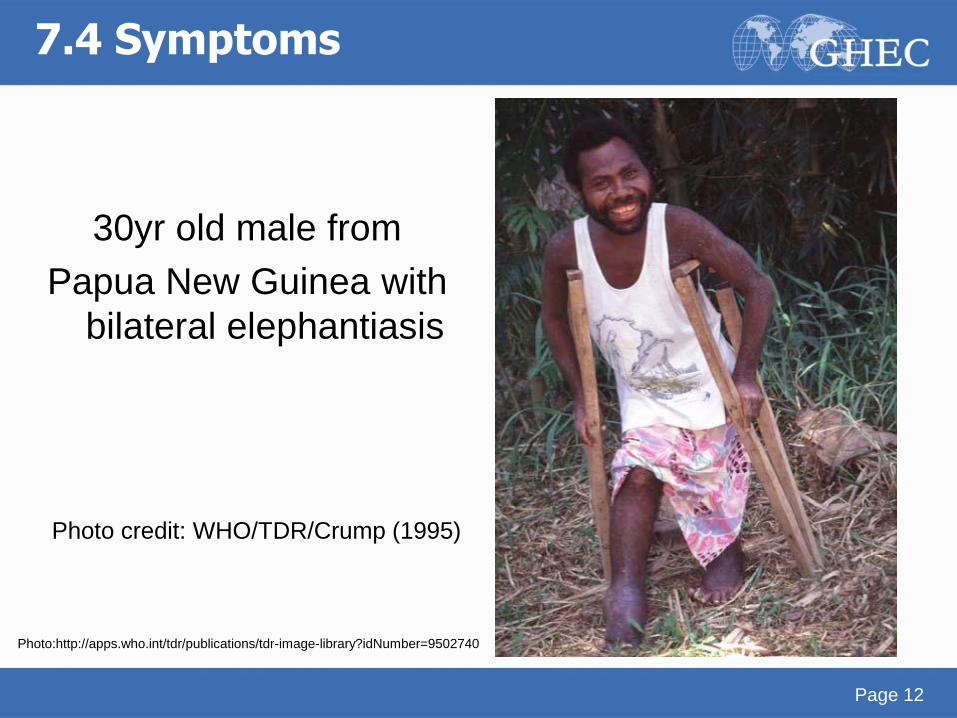
7.4 Symptoms
30yr old male from
Papua New Guinea with
bilateral elephantiasis
Page 12
Photo:http://apps.who.int/tdr/publications/tdr-image-library?idNumber=9502740
Photo credit: WHO/TDR/Crump (1995)
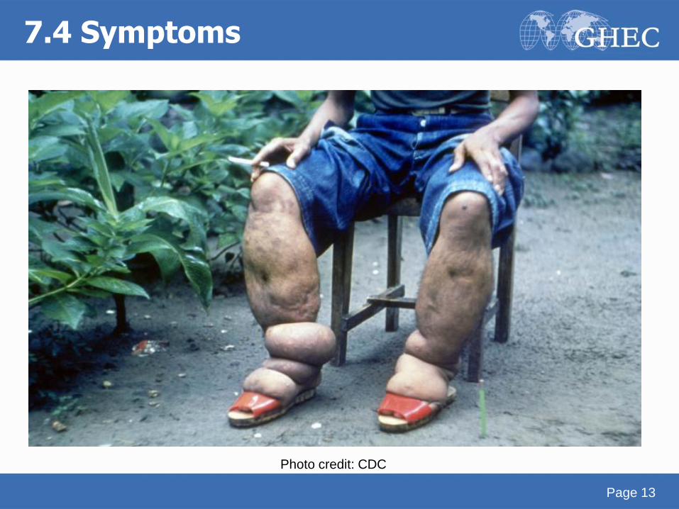
7.4 Symptoms
Photo credit: CDC
Page 13
7.5 Diagnosis
• Based on symptoms
• Parasitological o Microfilariae in patient’s blood at peak time of concentration
(at night) Measured by counting chamber
Staining techniques (Giemsa or hematoxylin and eosin)
Membrane filtration technique (Nucleopore) – high sensitivity, but costly filters
Knott concentration technique
o Adult worm usually not detectable
o Absence of microfilaremia does not exclude filarial disease, nor does microfilaremia denote it
o Microfilariae in hydrocele fluid, sometimes urine or other fluid
Page 14
• Immunological and PCR o Serology only has a role in visitors to the area
o IgG4 Antibody = marker of active infection
o Periodicity – independent
o PCR - need at least one microfilaria in blood volume
• Ultrasonography o Detection of W. bancrofti filarial worms in lymphatic
vessels of limbs and scrotum of infected males
o Less reliable in female genitals
Page 15
7.5 Diagnosis
7.6 Treatment
• Treatment consists of drugs directed against microfilariae
(microfilaricidal) and adult worms (macrofilaricidal) combined with
symptomatic treatment to relieve already caused by the disease
• Diethylcarbamazine (DEC)
o Microfilaricidal: most common agent used since >50 yrs
o Macrofilaricidal: capable to kill a proportion of adult W. bancrofti and
Brugia spp
• Ivermectin
o Microfilaricidal, not macrofilaricidal
o Since not macrofilaricidal repeated doses every 6 months or yearly
are needed
• Symptomatic
o Bandaging
o Physiotherapy
o Infection-prevention
o Treatment of secondary bacterial infection (e.g. in filarial abscess)
Page 16
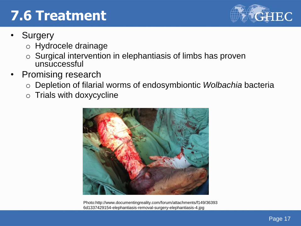
• Surgery o Hydrocele drainage
o Surgical intervention in elephantiasis of limbs has proven unsuccessful
• Promising research o Depletion of filarial worms of endosymbiontic Wolbachia bacteria
o Trials with doxycycline
Page 17
7.6 Treatment
Photo:http://www.documentingreality.com/forum/attachments/f149/36393
6d1337429154-elephantiasis-removal-surgery-elephantiasis-4.jpg
7.7 Control
• Avoiding mosquito bites
o Sleeping under an ITN
o Use of repellent/long clothing between dusk and dawn
• Awareness raising in high-risk and endemic areas
• Annual mass community treatment
o Reduces microfilarial load and diminishes transmission
o Added benefit of this as drugs used also help control other
helminthic infections!
Page 18
Onchocerciasis
River blindness
Page 19
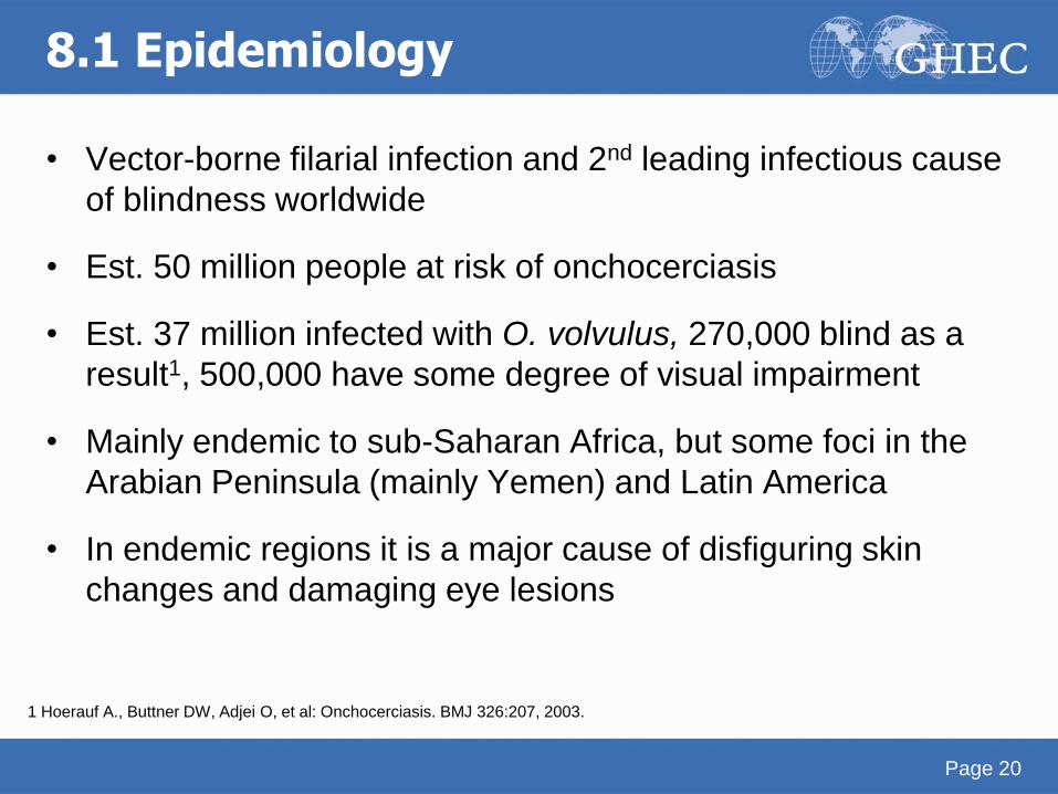
8.1 Epidemiology
• Vector-borne filarial infection and 2nd leading infectious cause
of blindness worldwide
• Est. 50 million people at risk of onchocerciasis
• Est. 37 million infected with O. volvulus, 270,000 blind as a
result1, 500,000 have some degree of visual impairment
• Mainly endemic to sub-Saharan Africa, but some foci in the
Arabian Peninsula (mainly Yemen) and Latin America
• In endemic regions it is a major cause of disfiguring skin
changes and damaging eye lesions
Page 20
1 Hoerauf A., Buttner DW, Adjei O, et al: Onchocerciasis. BMJ 326:207, 2003.
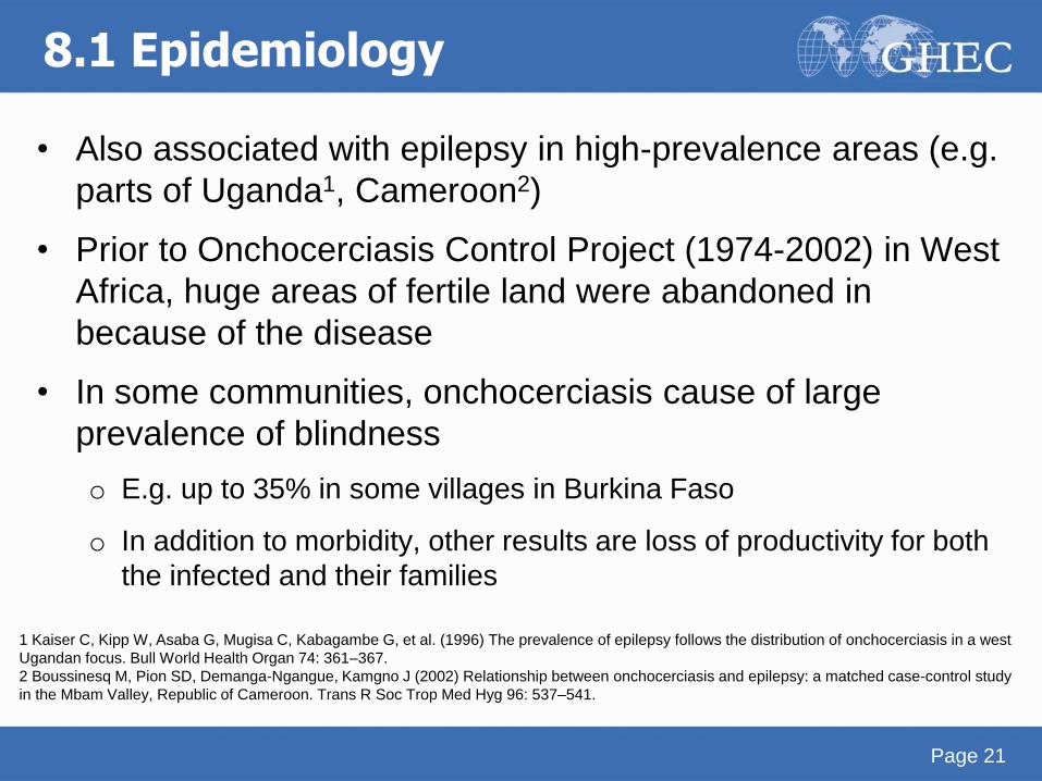
• Also associated with epilepsy in high-prevalence areas (e.g.
parts of Uganda1, Cameroon2)
• Prior to Onchocerciasis Control Project (1974-2002) in West
Africa, huge areas of fertile land were abandoned in
because of the disease
• In some communities, onchocerciasis cause of large
prevalence of blindness
o E.g. up to 35% in some villages in Burkina Faso
o In addition to morbidity, other results are loss of productivity for both
the infected and their families
Page 21
8.1 Epidemiology
1 Kaiser C, Kipp W, Asaba G, Mugisa C, Kabagambe G, et al. (1996) The prevalence of epilepsy follows the distribution of onchocerciasis in a west
Ugandan focus. Bull World Health Organ 74: 361–367.
2 Boussinesq M, Pion SD, Demanga-Ngangue, Kamgno J (2002) Relationship between onchocerciasis and epilepsy: a matched case-control study
in the Mbam Valley, Republic of Cameroon. Trans R Soc Trop Med Hyg 96: 537–541.
Page 22
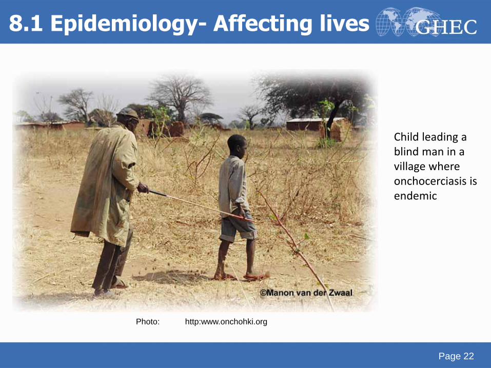
Child leading a blind man in a village where onchocerciasis is endemic
8.1 Epidemiology- Affecting lives
Photo: http:www.onchohki.org
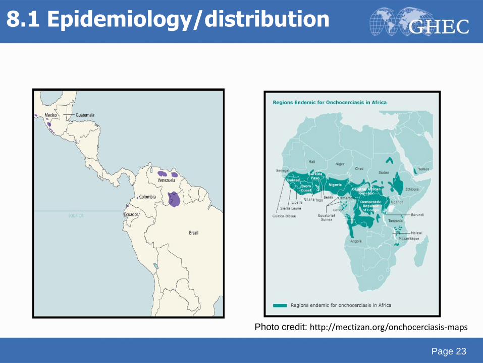
8.1 Epidemiology/distribution
Page 23
Photo credit: http://mectizan.org/onchocerciasis-maps
8.2 Risk factors
Page 24
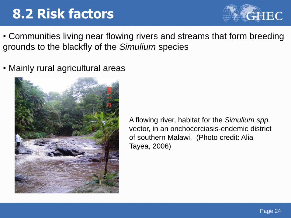
• Communities living near flowing rivers and streams that form breeding
grounds to the blackfly of the Simulium species
• Mainly rural agricultural areas
A flowing river, habitat for the Simulium spp.
vector, in an onchocerciasis-endemic district
of southern Malawi. (Photo credit: Alia
Tayea, 2006)
8.3 Biology
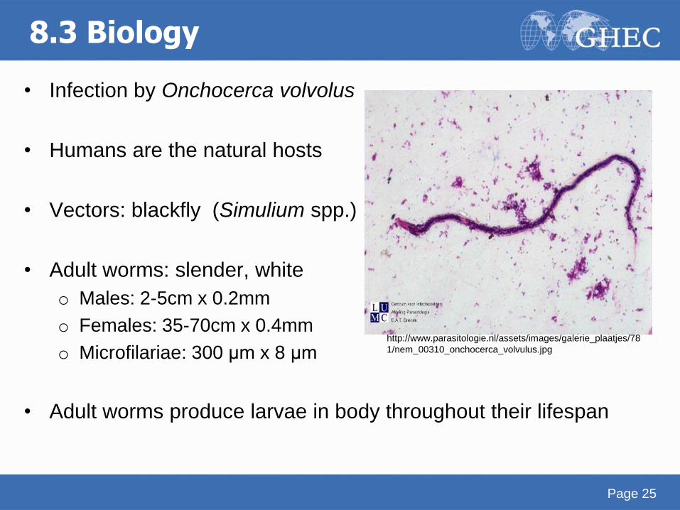
• Infection by Onchocerca volvolus
• Humans are the natural hosts
• Vectors: blackfly (Simulium spp.)
• Adult worms: slender, white
o Males: 2-5cm x 0.2mm
o Females: 35-70cm x 0.4mm
o Microfilariae: 300 μm x 8 μm
• Adult worms produce larvae in body throughout their lifespan
Page 25
http://www.parasitologie.nl/assets/images/galerie_plaatjes/78
1/nem_00310_onchocerca_volvulus.jpg
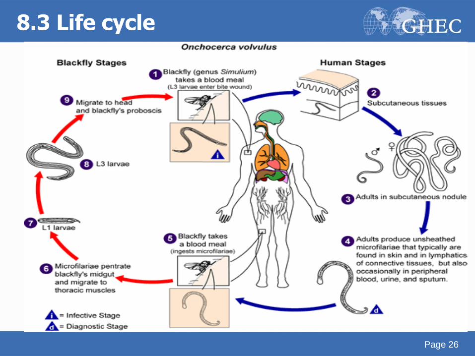
8.3 Life cycle
Page 26
8.4 Symptoms
• Main manifestations
o Skin lesions
o Nodule formation
o Severe itching due to body’s inflammatory response
o Eye lesions (late)
• Lymphedema, hydrocele, hanging groin
• Long exposure is usually required to develop symptoms –
many cases are asymptomatic
• Severity depends on intensity of infection
Page 27
8.4 Symptoms - skin
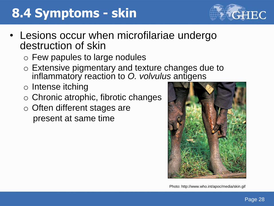
• Lesions occur when microfilariae undergo destruction of skin o Few papules to large nodules
o Extensive pigmentary and texture changes due to inflammatory reaction to O. volvulus antigens
o Intense itching
o Chronic atrophic, fibrotic changes
o Often different stages are
present at same time
Page 28
Photo: http://www.who.int/apoc/media/skin.gif
Page 29
• Early symptoms o Itching (filarial itch)
o Rash (consists of raised papules = microabscesses)
• Later symptoms o Heavy lichenification (lizard skin)
o Loss of elastic fibers in skin of the groin (hanging groin)
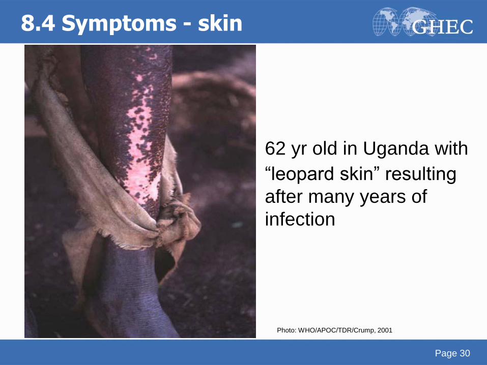
o Leopard skin (loss of pigment, degeneration of dermal
collagen, thinning of epidermis) – mainly pretibial region
o Onchocercomata = granulomas resulting from a tissue
reaction around adult worms
Early symptoms oItching (filarial itch) oRash (consists of raised papules = microabscesses)
Later symptoms oHeavy lichenification (lizard skin) oLoss of elastic fibers in skin of the groin (hanging groin) oLeopard skin (loss of pigment, degeneration of dermal collagen, thinning of epidermis) – mainly pretibial region oOnchocercomata = granulomas resulting from a tissue reaction around adult worms
8.4 Symptoms - skin
8.4 Symptoms - skin
62 yr old in Uganda with
“leopard skin” resulting
after many years of
infection
Page 30
Photo: WHO/APOC/TDR/Crump, 2001
8.4 Symptoms - eyes
• Ocular involvement as a result of microfilariae migrating through, collecting and dying in eye tissue
• Damage caused by microfilariae and inflammatory response to these, particularly dying/dead microfliariae
• May progress to blindness
• Anterior and posterior segment can be involved
Page 31
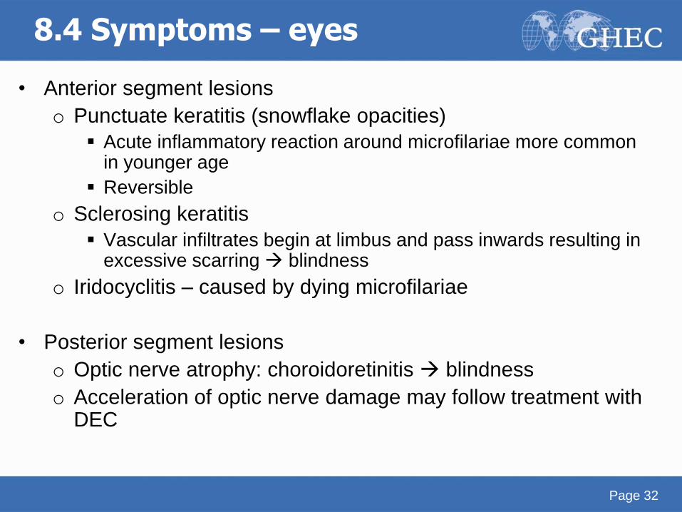
• Anterior segment lesions
o Punctuate keratitis (snowflake opacities)
Acute inflammatory reaction around microfilariae more common in younger age
Reversible
o Sclerosing keratitis
Vascular infiltrates begin at limbus and pass inwards resulting in excessive scarring blindness
o Iridocyclitis – caused by dying microfilariae
• Posterior segment lesions
o Optic nerve atrophy: choroidoretinitis blindness
o Acceleration of optic nerve damage may follow treatment with DEC
Page 32
8.4 Symptoms – eyes
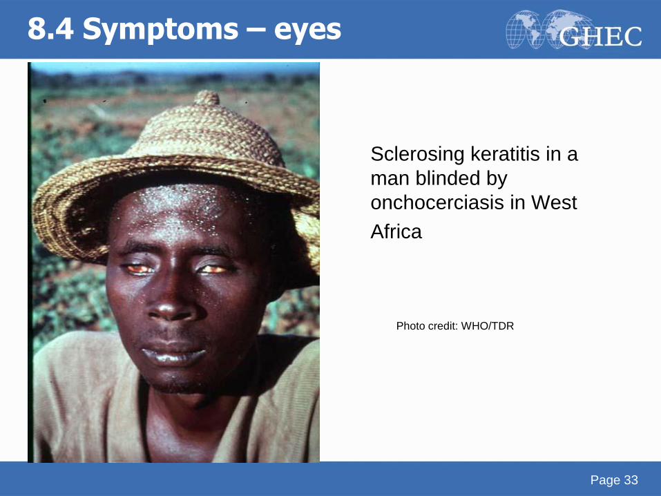
Sclerosing keratitis in a
man blinded by
onchocerciasis in West
Africa
Page 33
8.4 Symptoms – eyes
Photo credit: WHO/TDR
8.5 Diagnosis
• Clinical - skin/eye lesions, subcutaneous nodules
• Ultrasonography - detection of nodules in tissues, follow up of drug effects on worms
• Mazzotti test - administration of small dose of DEC p.o. if itching/rash develops could be diagnostic for infection
• DEC patch test (historically used but no longer recommended) o Gauze soaked in 20% DEC solution applied to skin (hip), site later
checked for inflammatory reaction
o Less risk of systemic inflammatory reaction than Mazzotti test
o Good test for re-emergence of infection
Page 34
• Parasitological diagnosis
o Demonstrating microfilariae that have emerged from
bloodless skin snips (biopsy preferred from iliac crest or
below) – immersion of snips in isotonic saline, counting of
emerging microfilariae
• Immunologic and PCR based diagnosis
o Antibody detection to crude Ag is of limited practical use
o Specific IgG4 Antibodies
o PCR – identification of worm DNA and distinction between
various strains
Page 35
8.5 Diagnosis
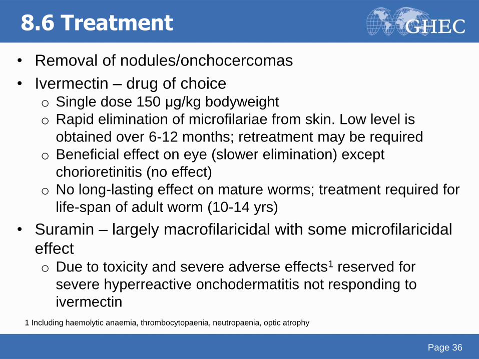
8.6 Treatment
• Removal of nodules/onchocercomas
• Ivermectin – drug of choice o Single dose 150 μg/kg bodyweight
o Rapid elimination of microfilariae from skin. Low level is
obtained over 6-12 months; retreatment may be required
o Beneficial effect on eye (slower elimination) except
chorioretinitis (no effect)
o No long-lasting effect on mature worms; treatment required for
life-span of adult worm (10-14 yrs)
• Suramin – largely macrofilaricidal with some microfilaricidal
effect o Due to toxicity and severe adverse effects1 reserved for
severe hyperreactive onchodermatitis not responding to
ivermectin
Page 36
1 Including haemolytic anaemia, thrombocytopaenia, neutropaenia, optic atrophy
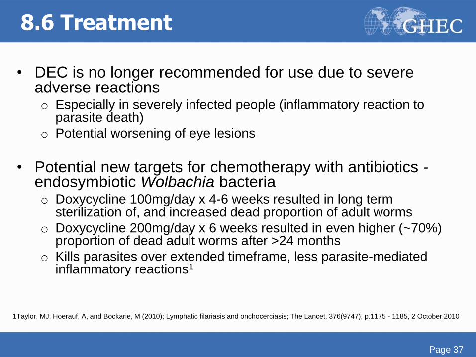
8.6 Treatment
• DEC is no longer recommended for use due to severe adverse reactions o Especially in severely infected people (inflammatory reaction to
parasite death)
o Potential worsening of eye lesions
• Potential new targets for chemotherapy with antibiotics - endosymbiotic Wolbachia bacteria o Doxycycline 100mg/day x 4-6 weeks resulted in long term
sterilization of, and increased dead proportion of adult worms
o Doxycycline 200mg/day x 6 weeks resulted in even higher (~70%) proportion of dead adult worms after >24 months
o Kills parasites over extended timeframe, less parasite-mediated inflammatory reactions1
Page 37
1Taylor, MJ, Hoerauf, A, and Bockarie, M (2010); Lymphatic filariasis and onchocerciasis; The Lancet, 376(9747), p.1175 - 1185, 2 October 2010
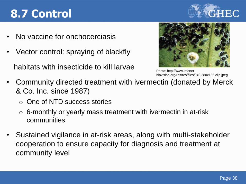
8.7 Control
• No vaccine for onchocerciasis
• Vector control: spraying of blackfly
habitats with insecticide to kill larvae
• Community directed treatment with ivermectin (donated by Merck
& Co. Inc. since 1987)
o One of NTD success stories
o 6-monthly or yearly mass treatment with ivermectin in at-risk
communities
• Sustained vigilance in at-risk areas, along with multi-stakeholder
cooperation to ensure capacity for diagnosis and treatment at
community level
Page 38
Photo: http://www.infonet-
biovision.org/res/res/files/949.280x185.clip.jpeg
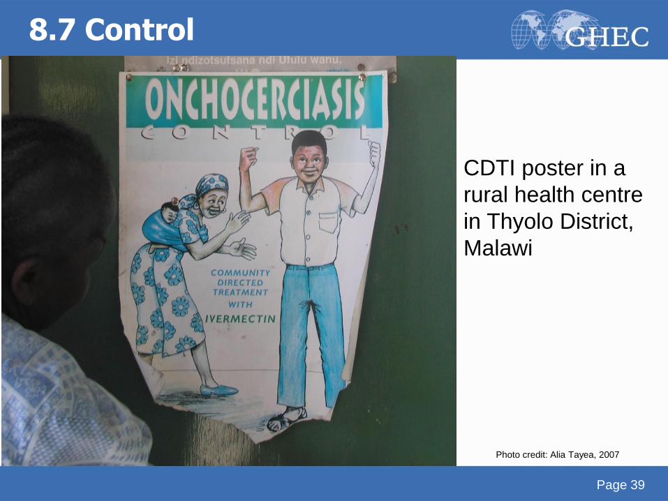
8.7 Control
Page 39
CDTI poster in a
rural health centre
in Thyolo District,
Malawi
Photo credit: Alia Tayea, 2007
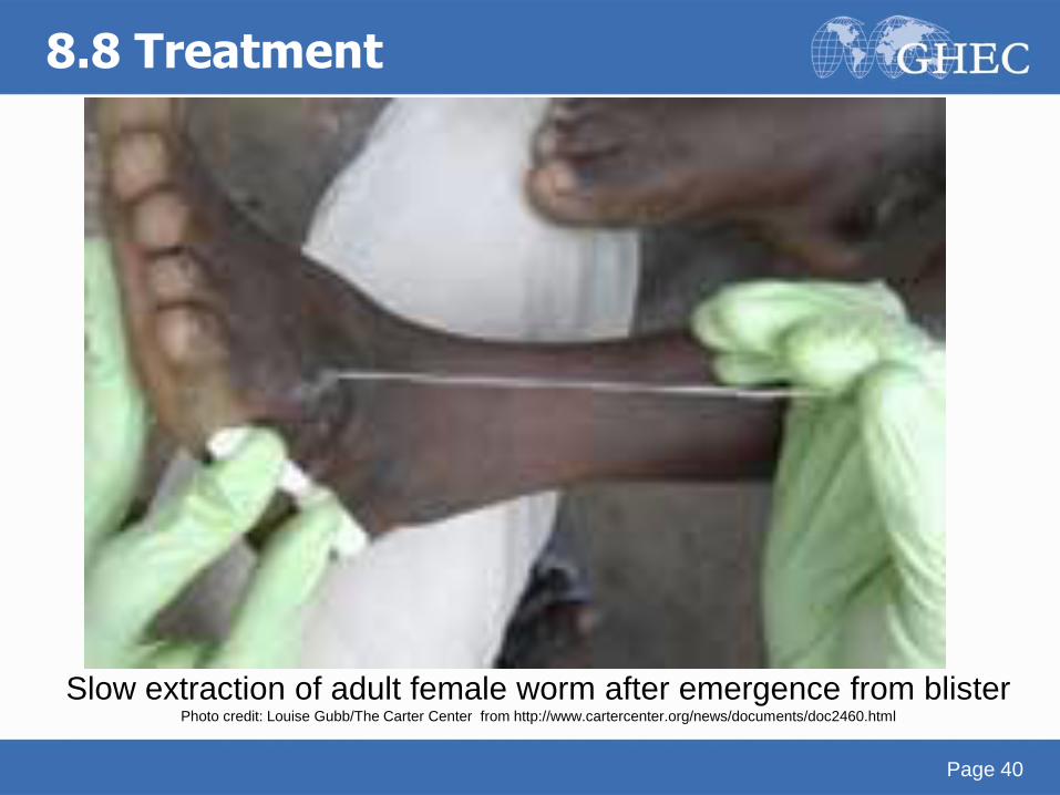
Slow extraction of adult female worm after emergence from blister Photo credit: Louise Gubb/The Carter Center from http://www.cartercenter.org/news/documents/doc2460.html
8.8 Treatment
Page 40
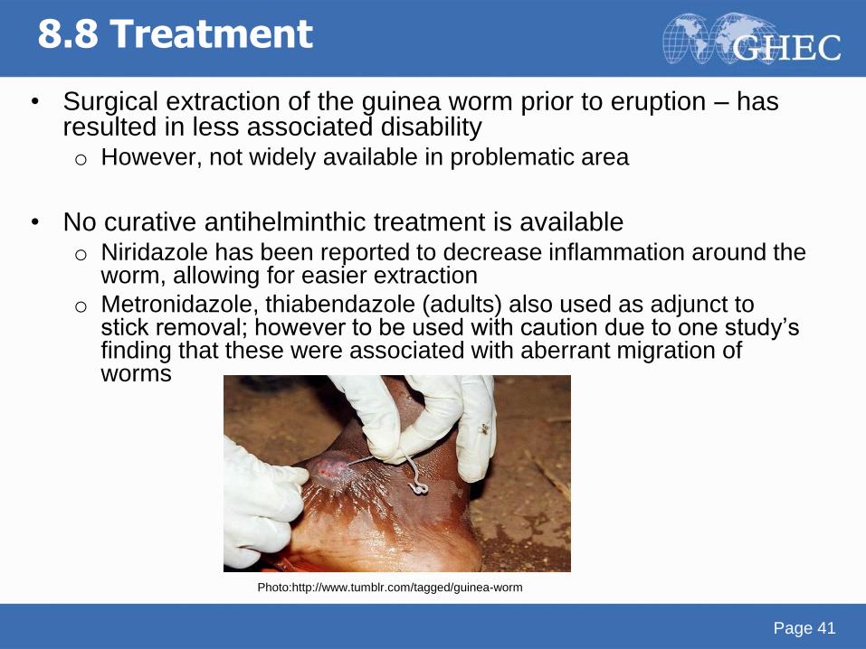
• Surgical extraction of the guinea worm prior to eruption – has resulted in less associated disability o However, not widely available in problematic area
• No curative antihelminthic treatment is available
o Niridazole has been reported to decrease inflammation around the worm, allowing for easier extraction
o Metronidazole, thiabendazole (adults) also used as adjunct to stick removal; however to be used with caution due to one study’s finding that these were associated with aberrant migration of worms
Page 41
8.8 Treatment
Photo:http://www.tumblr.com/tagged/guinea-worm
8.9 Control
• Community education on disease & transmission o Educating affected individuals not to immerse the affected
areas in water which is used for public consumption
• Promotion and provision of safe drinking water sources
• Boiling water
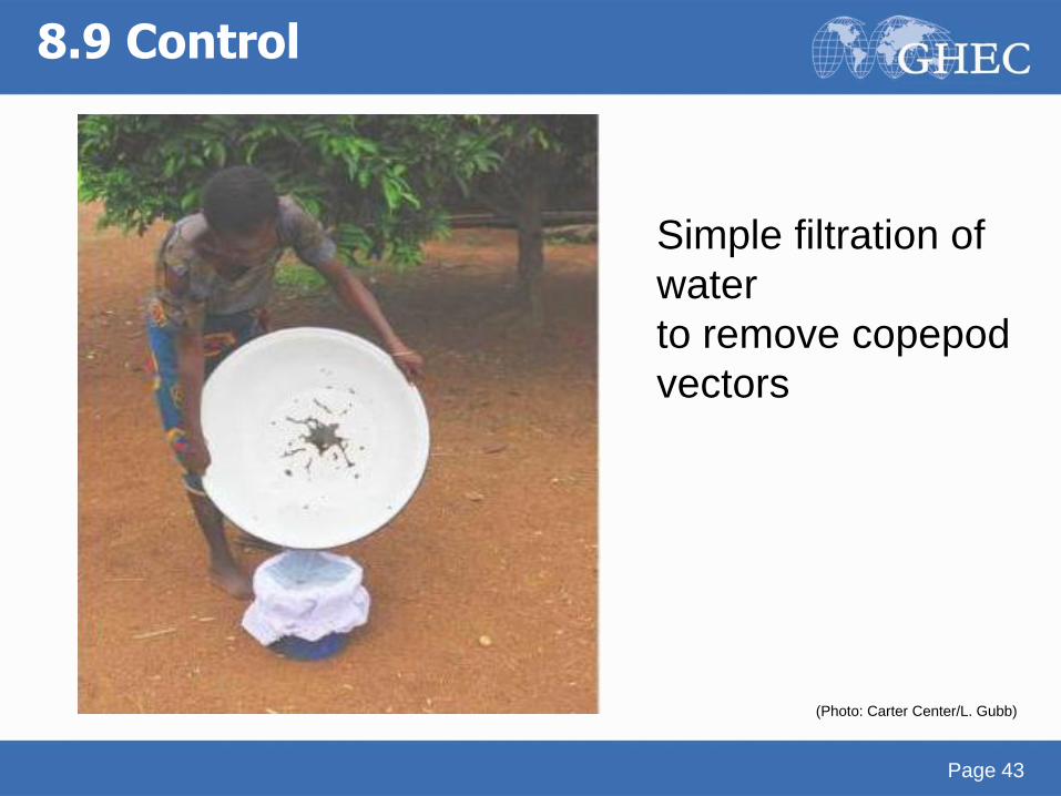
• Point-of-use filtration of drinking water to “strain”
copepods o Nylon filters, straw filters
o Low-cost methods effective, e.g. filtration through clean cloth
• Larvicide to kill copepods
Page 42
8.9 Control
Page 43
Simple filtration of
water
to remove copepod
vectors
(Photo: Carter Center/L. Gubb)
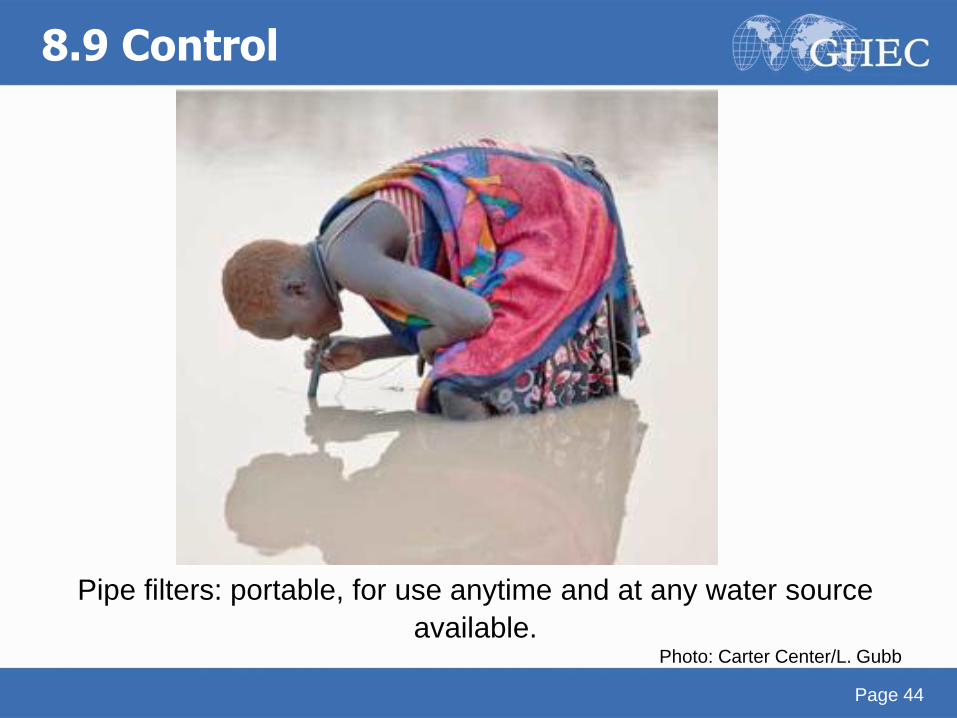
Pipe filters: portable, for use anytime and at any water source
available. Photo: Carter Center/L. Gubb
8.9 Control
Page 44

Page 45
Acknowledgments
• Thanks to Jenna Kelly, Shazeen Bandukwala and
Melissa Whaling for critical editing.
• We appreciate Tim Brewer and Jackeline Alger for
thoughtful review.
Page 45
Credits
Akre M Adja1, Sina Helbig2, Alia Tayea3, Neil Arya4
1: Institut Pierre Richet, Université de Cocody Abidjan
2: Boston University School of Medicine, Division of
Infectious Diseases, Boston, MA, USA
3: Médecins Sans Frontières
4: Western University, University of Waterloo, McMaster
University
Contact [email protected]
The Global Health Education Consortium and the Consortium of
Universities for Global Health gratefully acknowledge the support
provided for developing teaching modules from the:
Margaret Kendrick Blodgett Foundation
The Josiah Macy, Jr. Foundation
Arnold P. Gold Foundation
This work is licensed under a Creative Commons Attribution-Noncommercial-No Derivative Works 3.0 United States
License.