NOVEL MULTILAYERED MAGNETOPLASMONIC NANOPARTICLES...
125
NOVEL MULTILAYERED MAGNETOPLASMONIC NANOPARTICLES FOR THERANOSTIC APPLICATIONS By Charleson Sherard Bell Dissertation Submitted to the Faculty of the Graduate School of Vanderbilt University in partial fulfillment of the requirements for the degree of DOCTOR OF PHILOSOPHY in Biomedical Engineering December, 2015 Nashville, Tennessee Approved: Todd D. Giorgio, Ph.D. Frederick R. Haselton, Ph.D. Melissa C. Skala, Ph.D. Richard F. Haglund, Ph.D. Eric P. Skaar, Ph.D., M.P.H.
Transcript of NOVEL MULTILAYERED MAGNETOPLASMONIC NANOPARTICLES...
i
NOVEL MULTILAYERED MAGNETOPLASMONIC NANOPARTICLES FOR THERANOSTIC APPLICATIONS
By
Charleson Sherard Bell
Dissertation
Submitted to the Faculty of the
Graduate School of Vanderbilt University
in partial fulfillment of the requirements
for the degree of
DOCTOR OF PHILOSOPHY
in
Biomedical Engineering
December, 2015
Nashville, Tennessee
Approved:
Todd D. Giorgio, Ph.D.
Frederick R. Haselton, Ph.D.
Melissa C. Skala, Ph.D.
Richard F. Haglund, Ph.D.
Eric P. Skaar, Ph.D., M.P.H.

ii
To my Mom and Dad… this is for you.
iii
ACKNOWLEDGEMENTS
First, I give all thanks and glory to GOD, my father in heaven, his Son, Jesus Christ my Lord
and Savior, the Holy Spirit, a gift from God himself – without Him I am nothing and would
have never made it this far…
I would like to gratefully acknowledge the Department of Defense Concept CDMRP Award
(#W81XWH-08–1-0502), Department of Defense PRMRP Award(#W81XWH-13-1-0397)
and the Vanderbilt University intramural Discovery Grant Award (#4–48-999–9132) for
funding of my dissertation work.
My research was successfully completed thanks to the help of many individuals who assisted
me along the way. I gratefully acknowledge my Preceptor, first nanotechnology professor
and friend, Dr. Todd D. Giorgio, for the patience and guidance he gave me towards the
completion my degree. The road was long and obstacle-ridden and I am grateful for his
persistence in guiding me through my early struggles and later triumphs.
I am extremely grateful to my Dissertation Committee as they thoughtfully guided me
through the completion of my aims and my development as a researcher and problem solver.
I would like to deeply acknowledge Dr. Raquel Mejias, Sinead Miller and Jasmine Greer for
their hands-on contributions to the completion of this work. Without them, their
contributions and their support, it would have been an arduous struggle to complete this
breadth of work in the time that was allotted. Furthermore, I would like to thank Andre
iv
Stevenson, Dr. Ryan Ortega, Dr. Shann Yu, Mary Dockery, Dr. Ian McFadden, Zoe Johnson,
Andrew Cook, Sue Hyun Lee, Cheryl Lau and Kristin Engerer for their support during our
long days and nights in the laboratory and office.
I would also like to thank, Dr. Sarah Sewell Pierce, Dr. Amanda Lowery, and Dr. Chinmay
Soman for their guidance and assistance in my early graduate school endeavors. Without
their hands-on approach to training, I would not have quickly obtained the laboratory
experience and dexterity that I have today. I would like to thank Dr. James McBride, Dr. Don
Stec, Dr. Daniel Colvin, Dr. Anthony Hmelo, and Rosanne Delapp for the contribution of their
expertise, time and kind patience.
I would like to thank my immediate and extended family for their undying support and
encouragement through this process. Mom, Dad, Charreau, Maceo, Grandpa, my late
Grandma, aunts, uncles, cousins and the like – I thank you and love you all!
To my Brothers of the Nu Rho Chapter of Kappa Alpha Psi, your passionate support and our
deep Bond as a family-away-from-family helped me Achieve this goal. φνπ
Finally, thank you to all who I have not specifically named in this acknowledgement. You
know who you are and I sincerely and graciously thank you for your encouragement and
support throughout my experiences here at Vanderbilt University.
Thank you.

v
TABLE OF CONTENTS
ACKNOWLEDGEMENTS .......................................................................................................................................... iii
TABLE OF CONTENTS ................................................................................................................................................ v
LIST OF TABLES ........................................................................................................................................................ vii
LIST OF FIGURES ...................................................................................................................................................... viii
LIST OF ABBREVIATIONS ....................................................................................................................................... ix
CHAPTER 1 ..................................................................................................................................................................... 1
INTRODUCTION ....................................................................................................................................................... 1
Objectives .................................................................................................................................................................... 3
Specific Aims .............................................................................................................................................................. 4
Background and Significance ............................................................................................................................. 4
CHAPTER II .................................................................................................................................................................. 18
THE MULTISTRATA NANOPARTICLE: ........................................................................................................ 18
Introduction ............................................................................................................................................................ 21
Materials and Methods ....................................................................................................................................... 23
Results and Discussion ....................................................................................................................................... 28
Conclusions ............................................................................................................................................................. 39
Summary .................................................................................................................................................................. 39
CHAPTER III ................................................................................................................................................................ 42
SONOCHEMICAL SYNTHESIS AND TOMOGRAPHIC CHARACTERIZATION ................................ 42
Introduction ............................................................................................................................................................ 44
Methods and Materials ....................................................................................................................................... 48
Results and Discussion ....................................................................................................................................... 53
Conclusion ............................................................................................................................................................... 63
Summary .................................................................................................................................................................. 64
vi
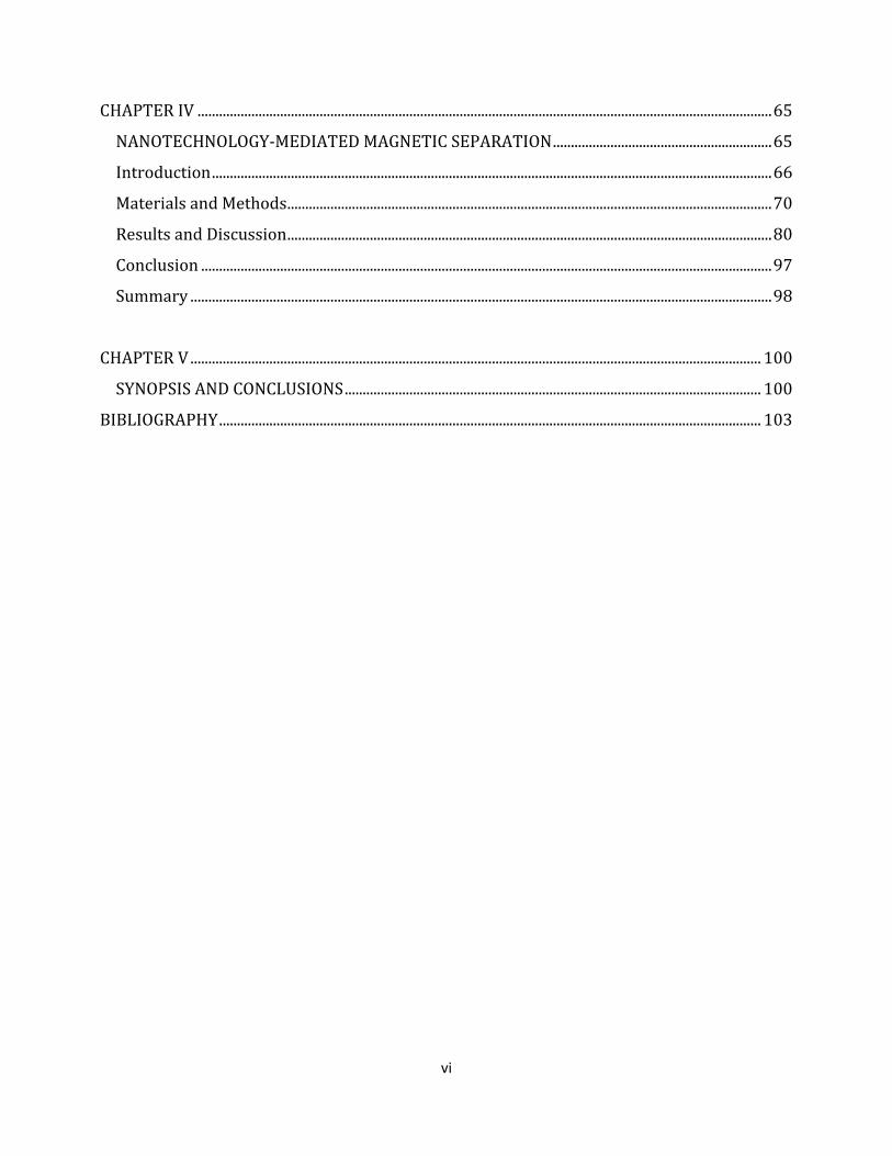
CHAPTER IV ................................................................................................................................................................ 65
NANOTECHNOLOGY-MEDIATED MAGNETIC SEPARATION ............................................................. 65
Introduction ............................................................................................................................................................ 66
Materials and Methods ....................................................................................................................................... 70
Results and Discussion ....................................................................................................................................... 80
Conclusion ............................................................................................................................................................... 97
Summary .................................................................................................................................................................. 98
CHAPTER V ............................................................................................................................................................... 100
SYNOPSIS AND CONCLUSIONS .................................................................................................................... 100
BIBLIOGRAPHY ....................................................................................................................................................... 103
vii
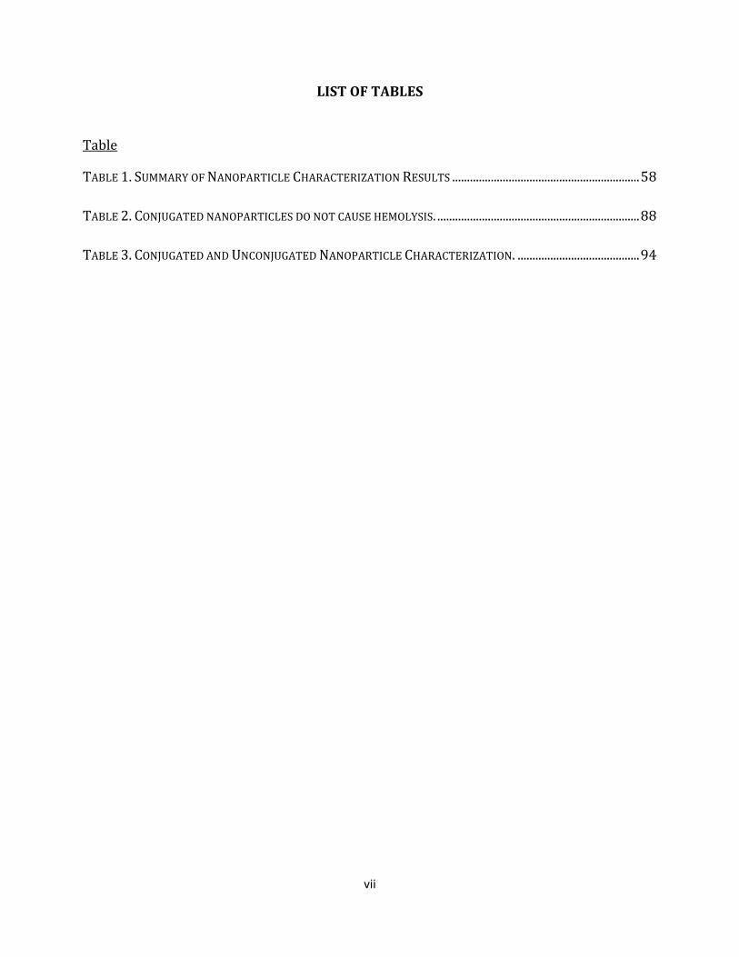
LIST OF TABLES
Table
TABLE 1. SUMMARY OF NANOPARTICLE CHARACTERIZATION RESULTS ............................................................... 58 TABLE 2. CONJUGATED NANOPARTICLES DO NOT CAUSE HEMOLYSIS. .................................................................... 88 TABLE 3. CONJUGATED AND UNCONJUGATED NANOPARTICLE CHARACTERIZATION. ......................................... 94
viii
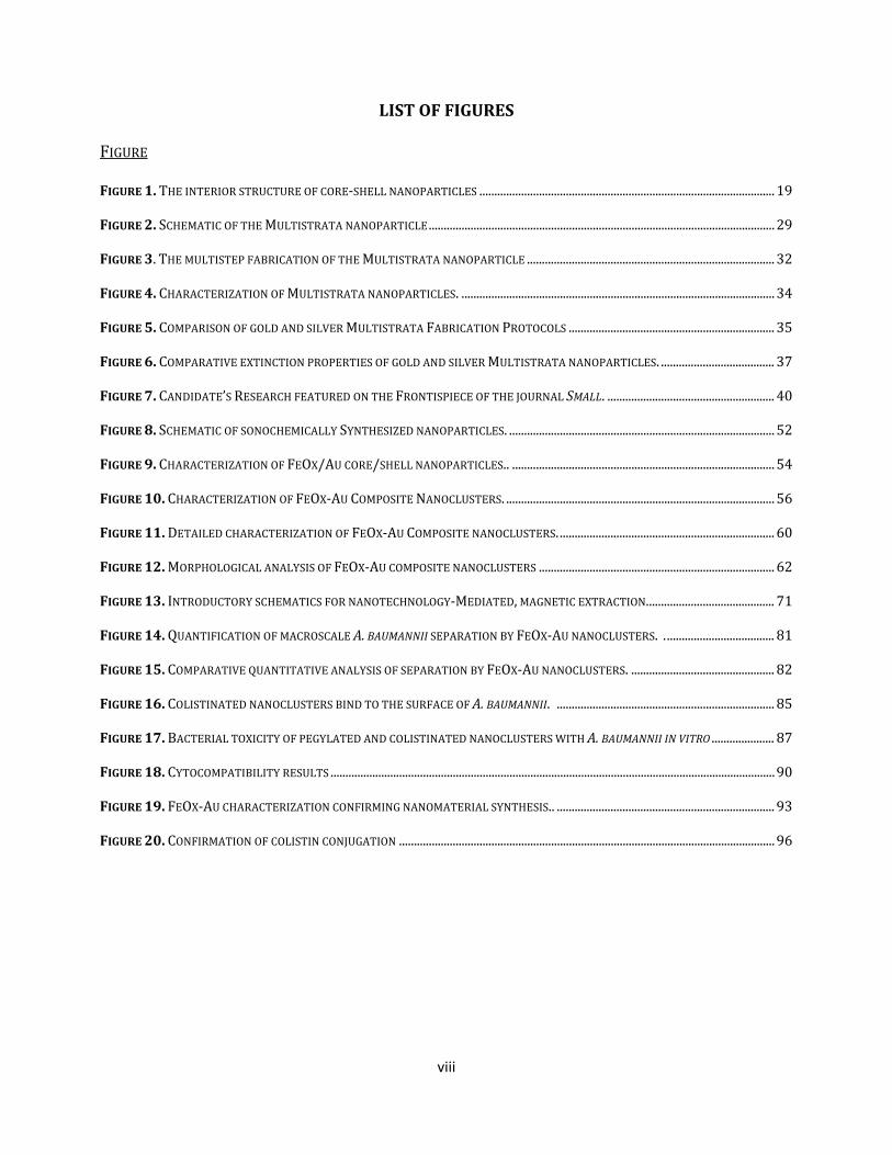
LIST OF FIGURES
FIGURE
FIGURE 1. THE INTERIOR STRUCTURE OF CORE-SHELL NANOPARTICLES ................................................................................................... 19
FIGURE 2. SCHEMATIC OF THE MULTISTRATA NANOPARTICLE .................................................................................................................... 29
FIGURE 3. THE MULTISTEP FABRICATION OF THE MULTISTRATA NANOPARTICLE ................................................................................... 32
FIGURE 4. CHARACTERIZATION OF MULTISTRATA NANOPARTICLES. ......................................................................................................... 34
FIGURE 5. COMPARISON OF GOLD AND SILVER MULTISTRATA FABRICATION PROTOCOLS ..................................................................... 35
FIGURE 6. COMPARATIVE EXTINCTION PROPERTIES OF GOLD AND SILVER MULTISTRATA NANOPARTICLES. ...................................... 37
FIGURE 7. CANDIDATE’S RESEARCH FEATURED ON THE FRONTISPIECE OF THE JOURNAL SMALL. ........................................................ 40
FIGURE 8. SCHEMATIC OF SONOCHEMICALLY SYNTHESIZED NANOPARTICLES. ......................................................................................... 52
FIGURE 9. CHARACTERIZATION OF FEOX/AU CORE/SHELL NANOPARTICLES.. ........................................................................................ 54
FIGURE 10. CHARACTERIZATION OF FEOX-AU COMPOSITE NANOCLUSTERS. .......................................................................................... 56
FIGURE 11. DETAILED CHARACTERIZATION OF FEOX-AU COMPOSITE NANOCLUSTERS. ........................................................................ 60
FIGURE 12. MORPHOLOGICAL ANALYSIS OF FEOX-AU COMPOSITE NANOCLUSTERS ............................................................................... 62
FIGURE 13. INTRODUCTORY SCHEMATICS FOR NANOTECHNOLOGY-MEDIATED, MAGNETIC EXTRACTION. .......................................... 71
FIGURE 14. QUANTIFICATION OF MACROSCALE A. BAUMANNII SEPARATION BY FEOX-AU NANOCLUSTERS. . .................................... 81
FIGURE 15. COMPARATIVE QUANTITATIVE ANALYSIS OF SEPARATION BY FEOX-AU NANOCLUSTERS. ................................................ 82
FIGURE 16. COLISTINATED NANOCLUSTERS BIND TO THE SURFACE OF A. BAUMANNII. ......................................................................... 85
FIGURE 17. BACTERIAL TOXICITY OF PEGYLATED AND COLISTINATED NANOCLUSTERS WITH A. BAUMANNII IN VITRO ..................... 87
FIGURE 18. CYTOCOMPATIBILITY RESULTS ..................................................................................................................................................... 90
FIGURE 19. FEOX-AU CHARACTERIZATION CONFIRMING NANOMATERIAL SYNTHESIS.. ......................................................................... 93
FIGURE 20. CONFIRMATION OF COLISTIN CONJUGATION .............................................................................................................................. 96
ix
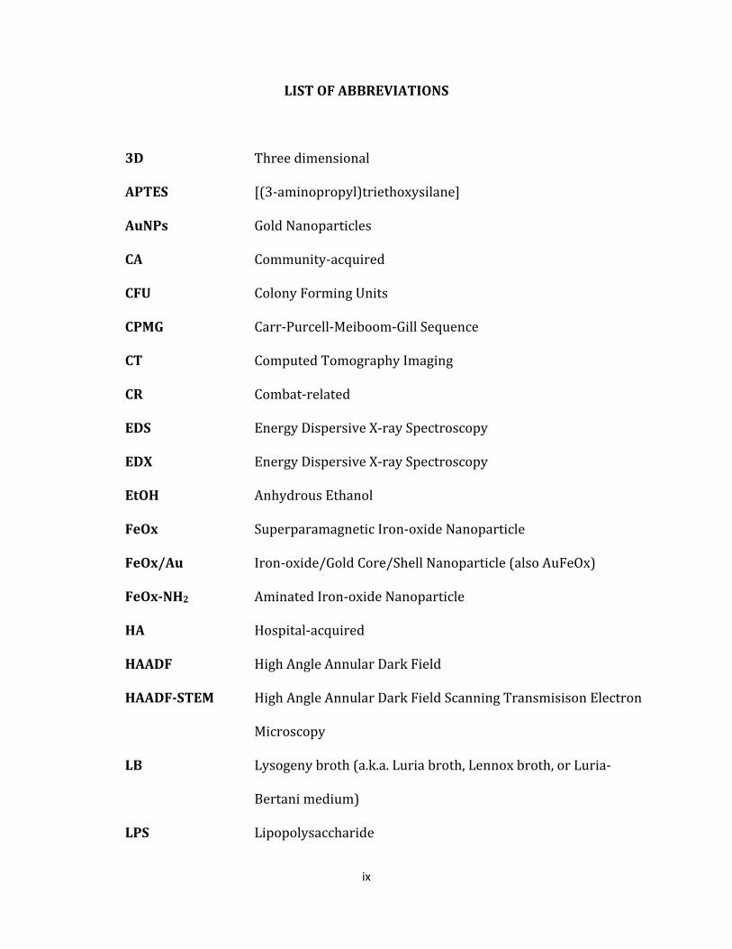
LIST OF ABBREVIATIONS
3D Three dimensional
APTES [(3-aminopropyl)triethoxysilane]
AuNPs Gold Nanoparticles
CA Community-acquired
CFU Colony Forming Units
CPMG Carr-Purcell-Meiboom-Gill Sequence
CT Computed Tomography Imaging
CR Combat-related
EDS Energy Dispersive X-ray Spectroscopy
EDX Energy Dispersive X-ray Spectroscopy
EtOH Anhydrous Ethanol
FeOx Superparamagnetic Iron-oxide Nanoparticle
FeOx/Au Iron-oxide/Gold Core/Shell Nanoparticle (also AuFeOx)
FeOx-NH2 Aminated Iron-oxide Nanoparticle
HA Hospital-acquired
HAADF High Angle Annular Dark Field
HAADF-STEM High Angle Annular Dark Field Scanning Transmisison Electron
Microscopy
LB Lysogeny broth (a.k.a. Luria broth, Lennox broth, or Luria-
Bertani medium)
LPS Lipopolysaccharide
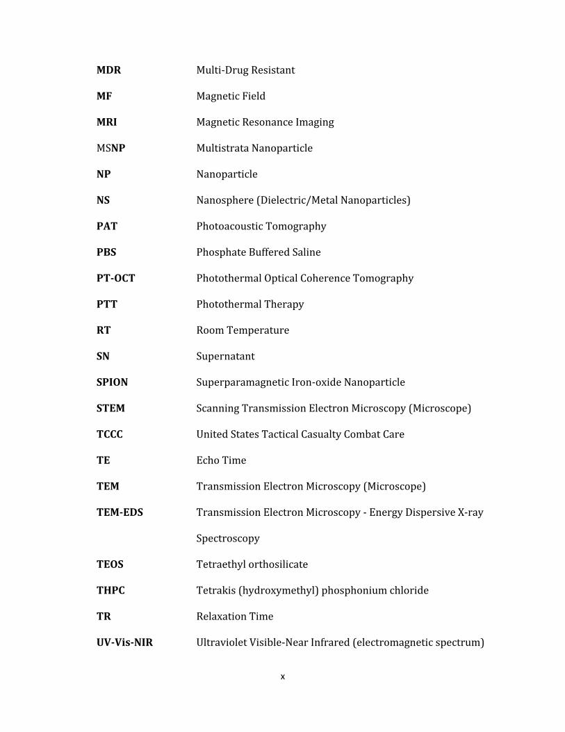
x
MDR Multi-Drug Resistant
MF Magnetic Field
MRI Magnetic Resonance Imaging
MSNP Multistrata Nanoparticle
NP Nanoparticle
NS Nanosphere (Dielectric/Metal Nanoparticles)
PAT Photoacoustic Tomography
PBS Phosphate Buffered Saline
PT-OCT Photothermal Optical Coherence Tomography
PTT Photothermal Therapy
RT Room Temperature
SN Supernatant
SPION Superparamagnetic Iron-oxide Nanoparticle
STEM Scanning Transmission Electron Microscopy (Microscope)
TCCC United States Tactical Casualty Combat Care
TE Echo Time
TEM Transmission Electron Microscopy (Microscope)
TEM-EDS Transmission Electron Microscopy - Energy Dispersive X-ray
Spectroscopy
TEOS Tetraethyl orthosilicate
THPC Tetrakis (hydroxymethyl) phosphonium chloride
TR Relaxation Time
UV-Vis-NIR Ultraviolet Visible-Near Infrared (electromagnetic spectrum)

1
CHAPTER 1
INTRODUCTION
For over half a century, nanotechnology has been the focal point of cutting edge technology
in a multitude of disciplines and fields (Feynman 1960; Editorial 2009). With the ability to
specifically synthesize structures less than 100 nm, investigators are able to control,
manipulate and explicitly interact with matter and substances on an atomic or molecular
scale. The ramifications of the implementation of nanotechnology are of great importance
in the medical field as biological processes occur mechanistically and molecularly at the
microscale and nanoscale, respectively (Roco 2003). With the advent of
bionanotechnology, bioengineers and medical scientists utilize these tools to better
characterize and modify biological process in an effort to enhance medical applications
(Ernest & Shetty 2005). Described as the convergence of materials-centric nanotechnology
and biology-focused biotechnology, bionanotechnology platforms normally include a
fabrication step where the nanomaterial is biofunctionalized with a specific molecule for
specific targeting or protection from unwanted interactions with biological materials. Over
the past two decades, the rise of bionanotechnology has accelerated innovations in
pathogen biodetection (Driskell & Tripp 2009; Kaittanis et al. 2010; Cheng et al. 2009),
disease diagnostics (Johnson et al. 2008; Wei et al. 2010), tissue engineering (Bose & Wui
Wong 2015; Pham et al. 2006), drug development and discovery (Chung et al. 2007), drug
delivery (de Villiers et al. 2009; Farokhzad & Langer 2009), protein detection and
extraction (Soman & Giorgio 2008; Soman & Giorgio 2009; Bordelon et al. 2013),

2
biomedical imaging (Tucker-Schwartz et al. 2012; Skala et al. 2008), tumor ablation (Huang
et al. 2008; Rozanova & Zhang 2009) and cell extraction (Radisic et al. 2006).
Most bionanotechnological applications are single-purpose and utilize only a single
property of the biofunctionalized nanomaterial. For example, nanotechnology-mediated
innovations in MRI imaging routinely use superparamagnetic iron oxide nanoparticles for
their image contrast characteristics alone (F. H. Wang et al. 2011; Ma et al. 2015).
Furthermore, nanotechnology applications in pathogen detection commonly utilize the
nanomaterials for only their magnetic properties (Chen et al. 2015) or optical properties
(Ali et al. 2012). These limitations are not only due to the focus of the investigators; but,
are also due to the narrow properties and characteristics of the nanomaterial. Whether the
material has been discovered to, or specifically designed to, exhibit a particular
characteristic, I propose that improvements in nanomaterial design through variation in
fabrication technique will allow multiple characteristics and properties to be combined
into a single, nanoscale entity.
From a chemical perspective, the combination of multiple nanoscale properties into
a single nanoparticle is achieved through the addition of functional layers. This assertion
forms the basis of core/shell nanotechnology. The combination of multiple characteristics
into a single nanoscale unit unleashes a host of potential combinatorial applications; many
of which are biomedical in nature. Most importantly, nanomaterials used solely for
diagnostics or therapeutics, individually, can be combined into a single theranostic
nanoconstruct.
3
Herein, we have developed multilayered, core/shell nanoparticles that possess
optical, magnetic-constrast and dual-plasmonic properties. From there, we modified the
fabrication techniques utilized in the development of this material to synthesize
magnetoplasmonic core/shell nanoparticles with the capacity for magnetomotion in the
presence of a magnetic field. Thereafter, we evaluated the core/shell nanomaterial in vitro
in a biomedical application.
Objectives
The central hypothesis of my dissertation is that inorganic, multilayered,
magnetoplasmonic core/shell nanomaterials can be specifically designed and synthesized
for biological applications which require both diagnostic detection and therapeutic
elimination of a disease causing agent. Furthermore, I hypothesize that these materials can
be fabricated in such a way that biomedical functionalization of the material can proceed
without detriment to the function of the biological component. Therefore, the objectives of
this work are to (1) develop multilayered nanomaterials that combine at least two
nanoscale characteristics into a single entity, (2) modify the synthesis strategy to enhance
the properties of the composite material while decreasing the synthesis duration such that
freshly synthesized materials can be rapidly utilized in biological applications, (3) ensure
the conservation of the activity of the biological component while binding and eliminating a
disease causing agent in vitro.
4
Specific Aims
Specific Aim 1: Novel multilayered, magneto-metallodielectric, core/shell nanoparticles will
be fabricated using a unique synthesis protocol. Characterization of the multimodal
nanoscale characteristics of the nanomaterial will be performed. Assessment of the
theranostic potential of the nanoparticulate will also be performed here.
Specific Aim 2: Multilayered, magnetoplasmonic core/shell nanoparticles will be fabricated
by adapting the synthesis protocol used in Aim 1. The magnetic properties of the material
will be enhanced to provide magnetomotive functionality to the nanostructure. Further,
the synthesis duration will be decreased enabling the rapid fabrication and use of fresh
materials for biological applications.
Specific Aim 3: The magnetoplasmonic core/shell nanoparticles fabricated in Aim 2 will be
biofunctionalized with an antibiotic which targets and binds gram negative bacteria. These
biofunctionalized nanostructures will then be characterized and utilized to magnetically
extract Acinetobacter baumannii from saline solution in vitro.
Background and Significance
For my dissertation, I proposed to develop magnetoplasmonic core/shell
nanoparticles that could be used for theranostic biomedical applications. These
magnetoplasmonic core/shell nanostructures, in particular, possess combinatorial
concentric configurations whose properties stem from the combination of two or more
nanoscale materials. Because of the unique, multimodal properties of nanostructure, the
5
nanomaterials were assessed as a potential theranostic for Acinetobacter baumannii
bacteremia – a disease where a strategy is urgently needed to improve patient outcomes.
In this section, I will discuss the clinical ramifications and therapeutic state-of-the-art for
Acinetobacter baumannii bacteremia to establish the biomedical impact of this work.
Thereafter, I will review previous nanotechnology-based strategies for both disease
detection and eradication which govern the design of both the novel and functional
magnetoplasmonic core/shell nanostructures described herein.
Bacterial Infection: The Impact on Humanity and the Rise of Resistance
Throughout history, human health has been influenced by a multitude of external factors,
invaders, and pathogens; but none more notable than the impact of bacterial infection. As
early as 3000 BCE, bacterial infections caused some of the most memorable pandemics
which lead to the decline of major cities, nations and diverted human history (Rasmussen
et al. 2015). The existence of multi-drug resistant organisms in healthcare facilities
predates the modern scientific era. Upon the advent of penicillin, its effectiveness pushed
the demand to such heights that scientists and clinicians had concerns regarding its
indiscriminate distribution (Falk 1945). Fears were realized when clinicians reported
‘profound changes in the number and character of infections that were occurring’ –
particularly those within hospitals (Finland et al. 1959). In fact, the first penicillin-resistant
Staphylococcus aureus emerged less than a year after the first administration of penicillin in
1945 (Prabaker & Weinstein 2011). From that point, nearly every antibacterial agent
developed has faced significant resistance problems. Within the last decade, there has been
a substantial rise in nosocomial and community-acquired multi-drug resistant infection
6
worldwide (Mumtaz et al. 2011). Multi-drug resistant (MDR) bacterial infection is of
critical concern to both foreign and domestic medical authorities due to its recent rise and
environmental persistence in mainstream, pediatric, elderly-care and urgent-care military
medical facilities (Rybak and R.L. Akins 2001; Lindsay and M.T. Holden 2004).
Acinetobacter baumannii: Impact and Resistance
The recent emergence of Acinetobacter baumannii as a resistant pathogen acquired by
wounded personnel in military field hospitals is of particular concern due to its recent
transition to domestic healthcare facilities (Scott et al. 2007). Known as the quintessential
gram-negative nosocomial pathogen for the past few decades (Prabaker & Weinstein
2011), A. baumannii is a significant MDR pathogen that causes a range of infections,
including respiratory and urinary tract infections, meningitis, endocarditis, wound
infections, and bacteremia (Jacobs et al. 2010).
A. baumannii is now responsible for up to 20% of all intensive care unit infections in
some regions of the world. Further, more than 6% of all Gram-negative infections in the
intensive care units of the United States can be attributed to A. baumannii. In the United
States, this incidence of the resistant strain of this organism is has risen from 6.7% in 1993
to 29.9% in 2004 – more than twice that for any other Gram-negative bacterium causing
infections in ICUs (Lockhart et al. 2007). Comparable to the rise of resistant S. aureus, the
clinical significance of A. baumannii has been exacerbated by its rapid acquisition of
resistance to virtually all antibiotics.

7
The uncanny ability of Acinetobacter baumannii to persist on dry surfaces for
extended periods of time prepares this resistant strain for survival in desert field hospital
conditions (Jawad et al. 1998). Even further, these microbes can be transferred from
foreign, military medical institutions and colonize domestic communities. Also, this
organism has the ability to form biofilms on abiotic surfaces which could create a favorable
environment for persistence (Getchell-White et al. 1989). These bacteria selectively persist
in host infections and also have the ability to biosynthesize lipopolysaccharide (LPS)
partially via the glycosyltransferase, LpsB (Luke et al. 2010). Further, these strains
increase their tolerance to antibiotics in response to the existence of monovalent cations
(as released by compounds such as NaCl) in their culture environment (Hood et al. 2010).
Monoclonal antibodies or antibiotics such as colistin (Levin et al. 1999) specifically target
these biochemical, surface moieties, however they remain generally inefficient against
resistant strains. The latter is of significant importance as it is informative of a conserved
targeting motif well suited for nanotechnology-mediated bactericidal theranostics.
Existence of such a motif is paramount when surface labelling any organism with a
particular nanostructure. Such a label will allow for the design of a number of
nanostructures that can possess many detection or therapeutic characteristics for specific
targeting to the bacterium.
Acinetobacter species can cause a number of disease states including pneumonia,
meningitis, urinary tract infections, peritonitis, skin and soft tissues infections and
bacteremia (Bergogne-Bérézin & Towner 1996). These primary ailments contribute to the
incidence of Acinetobacter species bloodstream infections (BSIs) which account for over

8
3% of all nosocomial acquired BSIs (Hidron et al. 2008). Disturbingly, the mortality rate
associated with A. baumannii infection is high: bacteremia-associated mortality is 52% and
pneumonia-associated mortality ranges from 23% to 73% (Rodriguez-Guardado et al.
2013).
Current Battlefield Treatment Strategies in Combat Related Infection
In both community-acquired (CA) and hospital-acquired (HA) infection types, multi-drug
resistant (MDR) pathogens gain access to host tissues when patients are either injured or
are in immuno-compromised states. Although CA and HA infections comprise the majority
of MDR infections in the world, over the last three decades, the incidence of combat-related
(CR) MDR infection is rising due to the increase in war-time activity in the Middle East in
concert with the intercontinental transport of infected military personnel to combat
installations worldwide. Due to an impending rise of CA and HA infections caused by the
return of military personnel to domestic homes and healthcare institutions, I will briefly
discuss the current state-of-the-field treatment of CR MDR infections as they are a point of
origin for the newest global MDR infections.
Historically, the extremities have been the most common sites of combat injury;
remaining true as current combat in the Middle East and Afghanistan continues (Murray,
Hospenthal, et al. 2011). CR extremity injuries – usually violent in occurrence - result in
gross contamination of the wound; along with anatomic and physiologic devastation of the
local tissue. Further, such a traumatic physiological event is likely to detrimentally alter the
patient’s immune response thus complicating their ability to resist infection (Stoecklein et
al. 2012). Thus, antimicrobial treatment, systemic or at the site of injury, must be
9
administered to prevent infection following combat injury. Currently, there are a number
of preventative, clinical treatments for post-injury control of infection. Infection through
the gross contamination of wounds can be impeded through the utilization of continuous
irrigation with debridement at the site of injury to prevent ongoing bacterial replication.
Further, surgical wound closure following irrigation, debridement, and a 5 day delay period
- to ensure no evidence of infection is apparent – has yielded positive outcomes as far as
inhibiting infection (Dufour et al. 1998). Failure to prevent infection progression at the
site of injury leads to the onset of bacteremia and septicemia.
Pharmaceutical Treatments for Point-of-Injury Infection and Bacteremia
Aside from physical treatment of the wound area, chemical treatments including
antimicrobials and antibiotics are administered to patients to impede the onset of infection.
The current recommendation by the United States Tactical Casualty Combat Care (TCCC)
committee is rapid delivery of intravenous or oral antimicrobial therapy at the point-of-
injury. The antimicrobial agents of choice to deliver at the site-of-injury vary depending on
the nation. Currently, the recommended antibacterial of choice is cephalosporin cefazolin
utilized due to its antibacterial coverage of possible infectious pathogens and its
appointment as the standard of care in the United States for extremity injuries (Murray et
al. 2011). The International Committee of the Red Cross (ICRC) recommends intravenous
penicillin for amputations, compound fractures, and major soft tissues wounds (Dufour et
al. 1998). For A. baumannii treatment in particular, Colistin (polymyxin E), a polymyxin
antibiotic produced by certain strains of Bacillus polymyxa var. colistinus, is the antibiotic
of choice and is thus delivered intrathecally and intraventricularly if infections have

10
reached the point of meningitis/ventriculitis. Nevertheless, some colistin resistance has
been described (Qureshi et al. 2014; Cai et al. 2012).
For treatment of A. baumannii bacteremia, aside from colistin, treatment options are
increasingly limited due to the rapid acquisition of multi-drug resistance to the few
antibiotics readily available (Fournier et al. 2006). Normally, upon presentation, empiric
therapy is implemented based on local susceptibility patterns. Broad-spectrum
cephalosporin with sulbactam is utilized until susceptibility tests from blood cultures are
available. Carbapenems are highly bactericidal against susceptible Acinetobacter strains
(Fishbain & Peleg 2010). However, multidrug resistant, extensively drug-resistant and
pandrug-resistant strains do not effectively respond to these therapeutics. Furthermore,
increasing dosage is correlated with decreased cytocompatibility and is a common problem
upon administration of toxic therapeutics. Because of this, many have investigated a
number of therapies which function as adjuvants to conventional antibiotic regimens in
order to effectively curtail bacteremia. Furthermore, combinatorial delivery of colistin
with sulbactam, rifampin and tigecycline proved suboptimal as it has been shown to lead to
the development of resistance (Vila & Pachón 2008).
Motivation for New Innovative Approaches
There are an inadequate number of antimicrobial pharmaceuticals which are active against
MDR (especially Gram-negative) pathogens in the pharmaceutical pipelines. This requires
renewed emphasis in the search for viable means of treating MDR pathogens (Murray et al.
2011). Further, it has been determined that the incidence of MDR infection increases as the
11
length of combat durations increases. Even further, when comparing the susceptibility
patterns of MDR organisms over time, that antimicrobial resistance increases with
hospitalization time (Matsumoto et al. 1969) leading to further hospitalization and
opportunities for the MDR organism to spread to other compromised patients. Even more
disturbing, the presence of MDR pathogens persisted during intercontinental travel and
remained in wounds upon arrival in the United States (Heggers et al. 1969). Due to these
facts, I assert that more novel means of bacterial treatment that do not rely on the
antimicrobial traits of antibiotics need to be developed.
Novel Treatments of Multi-drug Resistant Bacteria
Although the majority of traditional antibiotics can treat and somewhat manage drug-
resistant bacteria, many commonly used antibiotics are ineffective (Levy 1998; Wright
2010). In the past year, a number of novel compounds and strategies have been devised to
treat MDR bacterial infections (Kurek et al. 2011). Some of the more recent studies have
been utilizing bacteriophages as antibacterial agents able to naturally defeat MDR
resistance (Chibani-Chennoufi et al. 2004; Górski et al. 2009). This research is ongoing and
is limited by bacteriophage specificity. Recently, some investigators have turned to plant-
derived compounds – antibacterial phytochemicals. More specifically, certain terpenes and
phenolics/polyphenols interfere with bacterial peptidoglycan metabolism (Kurek et al.
2010) and cause cell wall/envelope disruption (Tsuchiya et al. 1996), respectively. The
difficulty with this natural approach is that drug delivery to the intended target is
challenging.

12
Nanotechnology-mediated Bactericidal Treatments
Another direct approach to antibacterial treatment is the use of nanotechnology. Silver ions
have shown antibacterial affects against various bacterial species since antiquity (Rai et al.
2009). Silver nanoparticles (AgNPs) have an even greater advantage due to their increased
surface area to volume ratio and 1.4-1.9 times higher antibacterial potential (Ingle et al.
2008). The mechanism of action of the AgNPs, like the some of the aforementioned
antibacterial phytochemicals, includes the disruption of bacterial envelope due to the
production of reactive oxygen species. Silver toxicity is one major problem with utilizing
silver nanoparticles in patients. Other novel ‘nanoantibiotics’ utilized for MDR infection
include zinc oxide NPs, titanium dioxide NPs, aluminum NPs, copper NPs, antimicrobial
peptides, chitosan, fullerenes (C60), carbon nanotubes (CNTs), nitric oxide (NO) releasing
NPs, surfactant based nanoemulsions, drug delivery based liposomes, polymeric NPs, and
dendrimers (Huh & Kwon 2011).
Alternatively, gold nanoparticles (AuNPs) have also been used as antimicrobial
agents. Unlike the use of AgNPs the mechanism of action of AuNPs does not involve the
delivery of antimicrobial ions to the surface of the cells. Instead, AuNPs have been
leveraged as either pharmaceutical agent delivery vehicles or photodynamic enhancement
mediators (Pissuwan et al. 2010). For instance, AuNP-vancomycin conjugates proved to be
50-fold more active than free antibiotics against bacterial populations (Gu et al. 2003). In
other examples, AuNP-ciprofloxacin (Tom et al. 2004) and AuNP-aminoglycosidic
antibiotics (Nirmala Grace & Pandian 2007) have also been described. In these examples,
the role of the AuNPs was to facilitate the attachment of the conjugated antibiotics to the
13
bacterium. Following attachment, the antibiotic constructs penetrate the cell wall. AuNPs
have also been utilized as stabilizers and enhancers of dye-mediated photodynamic
therapy. A four-fold increase in S. aureus eradication was observed when photodynamic
dyes (toluidine blue O and methylene blue dyes) were conjugated to AuNPs as compared to
lone administration (Gil-Tomás et al. 2007; Perni et al. 2009). The mechanism for this
enhancement lies in the ability of the AuNPs to enhance the absorption of light due to their
surface plasmon resonance (Pitsillides et al. 2003). The downfall of dye-mediated
photodynamic therapy is twofold where there exists host toxicity to the dye and
widespread cell death due to non-specific targeting after photoexposure.
Core/Shell Nanotechnology-mediated Antimicrobial Photothermal Therapy
Antibiotics are utilized in bacterial infections because they specifically target and
effectively treat bacterial strains without significant detriment to host tissue.
Nanotechnology-mediated photothermal (PT) therapy was designed for a similar purpose
where treatment only occurs at the site of nanoparticulate localization (Zharov et al. 2006).
Per protocol, specifically-targeted near-infrared (NIR; 700-1200 nm) sensitive
nanoparticles are delivered to the site of treatment. Following deposition and specific
binding to targeted moieties, the treatment area is washed to remove unbound
nanoparticles. Thereafter, coherent NIR laser light is administered to the treatment area
(Huang et al. 2008). NIR light is not significantly absorbed by albumin, hemoglobin, water
or other constituents making it biologically benign; however, the NIR sensitive
nanoparticles will strongly absorb this coherent light which safely penetrates up to one cm
into tissue (Hirsch et al. 2003). Due to the absorption of the laser energy, the nanoparticles
14
will convert the incident laser light energy to heat; thus depositing heavy doses of thermal
energy to the biological target.
A number of NIR sensitive nanoparticles have been utilized to successfully
administer PT therapy. The particles absorb light strongly in the visible region due to
coherent oscillations of the metal conduction band electrons in strong resonance with the
visible frequencies of light; a phenomenon called surface plasmon resonance (SPR) (Kerker
et al. 1983). The SPR frequency is dependent on the size and shape of metal particles.
Further, as the diameter size increases, the SPR wavelength maximum redshifts thus
providing the capacity for absorption tunability. When nanoparticles form aggregates or
assemblies, the SPR maximum shifts to the NIR. NIR-sensitive nanoparticles called gold
nanoshells (NSs) are silica-gold core-shell nanoparticles (silica core diameter: 100-200nm;
gold shell thickness: 5-20 nm) currently undergoing clinical assessment for tissue-specific
photothermal therapy (Lal et al. 2008).
Nanoshells also have tunable plasmonic extinction peaks in the NIR that can be
selected through the careful control of the core/shell thickness ratio of the silica/gold
layers. The NIR extinction peaks of the nanoshells allow for the optimal heating of
subdermal tissue for photothermal therapy upon co-localization with the targeted tissue
(Gobin et al. 2007). Due to their outer gold surface, nanoshells are water soluble and their
surfaces can be easily modified for cellular targeting and biocompatibility using thiol-
functionalized monoclonal antibodies, peptides, antibiotics and biocompatible polymers
linkers (Lowery et al. 2006). Because of this, nanoshells can be conjugated with surface

15
moieties that target resistant bacterial strains thus providing a platform for nanoparticle-
mediated photothermal therapy at the site of infection.
Nanoparticle-mediated PTT has been performed with a number of different
biological applications. Nanoshells have been co-conjugated with HER2-antibody and PEG
biocompatibility polymers to be used to detect and destroy breast carcinoma cells in vitro
(Loo et al. 2005). Further, PT tumor (subcutaneous injected murine colon carcinoma cells)
ablation in immune-competent mice was achieved using gold nanoshells (O’Neal et al.
2004) showing the in vivo application of PTT. Even further, the pathogenic bacteria
Pseudomonas aeruginosa, have been eradicated using targeted gold nanorods for PTT
(Norman et al. 2007) showing the bacterial application of PTT. The combination of this
background provides a suitable literature based backdrop that exposes the need for a novel
treatment of MDR bacterial infections.
Nanotechnology-mediated Magnetic Extraction of Bacteria
Superparamagnetic iron-oxide nanoparticles (SPION) are magnetized only in the presence
of an externally applied magnetic field. This is due to the presence of only one magnetic
domain in a single particle. Without an applied field, the magnetic moments in a
population of SPIONs are not aligned, resulting in no net magnetization and no particle-
particle magnetic attraction (Ortega & Giorgio 2012). Core-shell nanoparticles can be
formed when the SPIONs are coated with shells of gold possessing atomic-scale thickness
to provide a surface for molecular functionalization while retaining nearly all of the
superparamagnetic characteristics. Even further, surface functionalization (molecular

16
conjugation) can provide ligands for a specific target, producing a nanoparticle that can
specifically bind to a desired molecular structure. Thereafter, the nanoparticle can
transduce an applied magnetic field into a magnetomotive force which will mechanically
transport the target structure toward the externally applied magnetic field. These FeOx/Au
core/shell nanoparticles can also be self-assembled into larger particles through specific
fabrication techniques or crosslinking of bifunctional molecules, (i.e colistin). This
clustering increases the group magnetization thereby enhancing the magnetomotive
potential of the nanoconstruct.
Innovative approaches have been devised including the development of
extracorporeal devices aiming to mechanically cleanse bacteria from body fluids using
magnetic particles. Most investigations utilize pathogen and endotoxin targeted magnetic
nanotechnology with an external device acting as a filter or spleen mimic. A blood-
cleansing extracorporeal device, inspired by the spleen, which continuously removes
pathogens and toxins from blood has been previously described in the literature (Kang et
al. 2014). Magnetic nanoparticles conjugated with mannose-binding lectin were developed
forming a nano-engineered human opsonin. Magnets inside the device were used to pull
opsonin-bound pathogens from the blood flowing through a microfluidic device at a >90%
efficiency. Further, investigators have shown synthetic conjugates can been utilized for
highly-selective, rapid separation of bacteria from whole blood with almost 100%
clearance (Lee et al. 2014). These results support the relevance of extracorporeal adjuvant
therapies as a potentially relevant treatment of bacteremia.

17
The use of extracorpeal devices for the adjuvant treatment of A. baumannii has not
been described in the literature. Furthermore, no-robust motif for nanoparticulate
targeting, to the A. baumannii surface has been described for these purposes. To initiate
the development of such a treatment, robust nanoparticulate platforms possessing a
combination of composite characteristics must be developed.
Design and Use of Novel Multilayered Magnetoplasmonic NPs in Biomedical Applications
My dissertation is organized into several manuscripts, one of which is already published at
the time of this writing. These manuscripts outline the design of novel multilayered,
magnetoplasmonic nanoparticles for the binding and depletion of A. baumannii. This
dissertation identifies nanotechnology design problems, as well as a clinically-relevant
application, and thereafter provides the nanoengineering solutions to implement a novel
therapeutic approach. This was accomplished based on the following steps: (1) Design of
novel, multilayered, magnetoplasmonic, core/shell nanomaterials which surpass previous
constraints, (2) Optimization of nanoparticulate design and fabrication protocols to
enhance the nanoparticulate composite characteristics, (3) Implementation of the
multilayered, magnetoplasmonic nanomaterials to pioneer novel therapeutic strategies for
Acinetobacter baumannii treatment in vitro.
18
CHAPTER II
THE MULTISTRATA NANOPARTICLE: A FEOX/AU CORE/SHELL ENVELOPED IN A SILICA-GOLD SHELL
The utilization of biomedical diagnostics and therapeutic advances including magnetic
resonance imaging (MRI), computed (CT), photoacoustic (PAT) and photothermal optical
coherence tomography (PT-OCT), photothermal therapy (PTT) have been shown to
effectively detect and decrease pathological effects in a number of aggressive diseases such
as head and neck cancer, colorectal cancer, and microbial diseases. However, the use of
these modalities without the addition of contrast agents is limited as far as: (1) the lack of
tissue discernment at site of disease due to challenges in imaging contrast; and (2) poor
diagnostic and/or therapeutic localization at the site of action due to lack of active
targeting. Core-shell nanoparticles have been utilized as targeted contrast agents in order
to relieve the limitations of the modalities above. Core-shell nanoparticles are primarily
designed in order to provide new properties based on the characteristics of each individual
layer in a synergistic fashion. Specifically, metal nanoshells (NS, dielectric/metal
nanoparticles) and metal-plated iron-oxide nanoparticles (FeOx/Au nanoparticles) have
been implemented in order to both improve X-ray and magnetically based imaging contrast
(CT, MRI, PAT, PT-OCT) based on the inherent properties of the materials in the
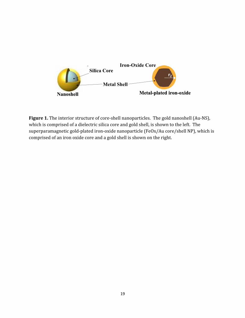
nanostructure (Figure 1). Even further, the surface of these nanoparticles can be
chemically modulated to target pathogenic tissue thus improving diagnostic imaging
capacity and photonics-based therapeutic treatment to the site of disease. For patients
who wish to undergo cross-modality diagnostic imaging and then request photonics-based
19
Figure 1. The interior structure of core-shell nanoparticles. The gold nanoshell (Au-NS), which is comprised of a dielectric silica core and gold shell, is shown to the left. The superparamagnetic gold-plated iron-oxide nanoparticle (FeOx/Au core/shell NP), which is comprised of an iron oxide core and a gold shell is shown on the right.
Iron-Oxide CoreSilica Core
Metal Shell
Nanoshell Metal-plated iron-oxide
20
therapeutic treatment, multiple imaging contrast and therapeutic agents must be
administered.
To alleviate this problem, I designed a nanomaterial that combines the properties
necessary to enhance the imaging and therapeutic capacity of the above modalities while
conserving the nanometer size and surface modification capability. The Multistrata
nanoparticle is the culmination of tunable, dual-peak, UV-Vis-NIR spectrum extinction
characteristics, tri-modal imaging contrast, simple synthesis and facile surface modification
capability into a single <60nm diameter, novel, multi-functional, multilayered nanosphere.
Seeking to relieve current methodological limitations by coupling diagnostics and
therapeutics into one single theranostic tool, herein I will specifically describe particle
fabrication and characterization. For further elucidation of the antimicrobial properties
and functionality of the designed nanoconstruct, multiple plasmonically active metals will
be included in the design of the nanomaterial, comparatively. For the purposes of this
dissertation, the results provided in this chapter accomplished an important objective
leading to the validation of the core hypothesis. Herein, we develop multilayered
nanomaterials that combine at least two nanoscale characteristics into a single, nanoscale
entity. Furthermore, results supporting the claims that: (1) a novel multilayered, magneto-
metallodielectric, core/shell nanoparticle can be fabricated using a unique synthesis
protocol; the (2) multimodal nanoscale characteristics and the (3) theranostic potential of
the nanomaterial can be elucidated and will be reinforced herein thus fulfilling Specific Aim
1 of this dissertation.

21
Introduction
Emerging materials and methods in biomedical imaging and biophotonics are
improving patient outcomes. More specifically, the utilization of biomedical diagnostics
and therapeutic advances in methods such as magnetic resonance imaging (MRI),
computed tomography (CT) imaging, photoacoustic tomography (PAT), photothermal
optical coherence tomography (PT-OCT) and targeted photothermal therapy (PTT) have
been shown to effectively detect and decrease pathological effects in head-neck cancer (R.
Popovtzer et al. 2008; M. Saksena et al. 2006), colorectal cancer (O. Will et al. 2006; T. Islam
& M. Harisinghani 2009), and breast cancer (M. Yezhelyev et al. 2006). Optical (V. Runge
1999), MRI (L. E. Ginsberg et al. 1998), and CT (G. Antoch 2002) based imaging contrast of
pathologic tissues, therapeutic localization at the site of action at the cellular level (N.
Portney & M. Ozkan 2006), and the inability to unite diagnosis and treatment into a single
entity (L. Johnson et al. 2010) continue to limit the practical power and application of these
current emerging technologies. In order to relieve these limitations and couple diagnostics
and therapeutics into a single theranostic material, we have prepared an enhancement
technology via the synthesis of a novel, multi-functional, multilayered nanomaterial. Our
approach provides tunable dual-peak (Vis-NIR) extinction characteristics, tri-modal
(optical, MRI and CT) imaging contrast with easy synthesis and surface modification as a
<60nm diameter nanosphere. This work describes our current progress regarding the
preparation and initial characterization of the Multistrata Nanoparticle (MSNP).
Multilayered, core/shell nanoparticles have been previously described (Schärtl
2010; Hirsch et al. 2006; R. Hao et al. 2010; M Melancon & C Li 2009). Classified as
inorganic or hybrid (organic-inorganic), core/shell nanoparticles are designed to provide

22
new properties based on the characteristics of each individual layer in a synergistic
fashion. Rational design principles can be employed to create nanomaterials with
functional applications such as tissue specific recognition, image contrast and therapeutic
delivery can be simultaneously enhanced with a single nanoscale platform. For example,
FeOx/Au, or iron-oxide/gold core/shell nanoparticles, have been synthesized to utilize
both the magnetic relaxivity of the FeOx and surface plasmon resonance properties of the
spherical gold shell (J. Aaron et al. 2006; B. Brinson C. Levin, et. al 2008; L. Wang et al.
2008). These nanoparticles have been implemented for simultaneous MR image contrast
with cancer phototherapy (D. Kirui & C. Batt 2010). SiO2/Au nanoshells, or silica/gold
core/shell nanoparticles, have been clinically assessed for tissue-specific photothermal
therapy (Hirsch et al. 2006) optimized for in vivo use by design of the surface plasmonic
properties of the nanomaterial. Unlike FeOx/Au nanoparticles, which have plasmonic
extinction peaks in the visible spectrum, extinction peaks in the near-infrared (NIR, 700-
1200 nm) can be achieved by control of the core/shell thickness ratio of the silica/gold
layers. Extinction peaks in the NIR allow for the optimal heating of subdermal tissue for
photothermal therapy (Hirsch et al. 2003) and efficient optical imaging. Harnessing the
surface plasmon resonance properties of core/shell materials, the nanosphere-in-a-
nanoshell (i.e. the “gold nanomatryushka”) was synthesized and demonstrated to provide
specific extinction maxima in the UV-Vis-NIR spectrum that are associated with the
nanoscale structure (Bardhan et al. 2010). A multilayered, metallodielectric nanostructure,
the nanomatryushka consists of a gold nanosphere surrounded by concentric silica/gold
shells. Governed by surface plasmon hybridization theory (Halas 2004), concentric metal
layers separated by a dielectric spacer layer causes plasmon interactions which generate

23
multi-peak extinction UV-Vis-NIR spectra. The location of the multi-extinction peaks are
controlled by the metal shell and dielectric layer geometric ratio allowing for specific
“tunability” of the optical characteristics of the nanostructure.
The Au Multistrata nanoparticle (Au-MSNP) is inspired by the material
characteristics of its predecessors, the FeOx/Au nanoparticle and Au-manomatryushka.
Designed to exhibit MRI contrast, X-ray contrast for CT, photonic contrast for OCT,
absorbance in the NIR for PTT, tunability of extinction characteristics during fabrication,
theranostic potential, easy surface modulation for cellular targeting and biocompatibility
and a nanostructure diameter of <60nm to support vascular extravasation ability, the
fabrication of each ‘primary’ and ‘subsidiary’ strata – or functional layer – must be carefully
controlled through fabrication methods. We report here our preliminary findings
regarding the multi-step fabrication and initial characterization of the Au-MSNP.
Furthermore, we specify methods necessary to fabricate extremely thin shells (as small as
1-2 nm to maintain an overall particle diameter less than 60 nm) and to ensure magnetic
material retention throughout the fabrication process. Relaxometry characterization,
suggesting MRI contrast capacity, is reported.
Materials and Methods
Fabrication of γ-Fe2O3 core Nanoparticles: γ-Fe2O3 with 12 ±1 nm diameter were fabricated
by a thermal decomposition, aeration and reflux protocol previously described (K. Woo et
al. 2004). Briefly, 20 ml of octyl ether (Sigma-Aldrich, St. Louis, MO) and 1.92 ml of oleic
acid (Sigma-Aldrich) were stirred under N2 gas flow and reflux. The sample was heated to
100ºC prior to addition of 0.4 ml Fe(CO)5 (Sigma-Aldrich). The reaction was heated from
24
150ºC to 280ºC where the reaction solution color changed from boil, to orange,
orange/colorless, to very dark orange. Sample was aerated at 80ºC for 14 hours and
refluxed while boiling for 2 hours. The γ-Fe2O3 cores were centrifuged (15 min, 770 rcf)
and washed in ethanol (EtOH, 200 proof, Sigma-Aldrich) twice, and dried under air.
Amination of γ-Fe2O3 core Nanoparticles: 150 mg of γ-Fe2O3 core (FeOx) were coated with
the first strata using a modified (3-aminopropyl)triethoxysilane (APTES, Sigma-Aldrich)
functionalization procedure. Core particles were added to 40 ml of tetrahydrofluran (THF,
Thermo Fisher Scientific, Waltham, MA) and stirred briskly using a magnetic stir plate and
stirring rod. 5 ml of APTES was added to the reaction solution and thereafter spiked with 5
μl of acetic acid (Sigma-Aldrich), 516 μl of MilliQ (18 MΩ) DI H2O, and stirred for 48 hours.
The reaction flask was then placed in an oil bath and heated to 80ºC. EtOH was used to
replace evaporated THF throughout the 2 hour boiling period. Reaction solution was
concentrated via rotovap to 40 ml of EtOH. Hexane was added to the solution in a 4:1 ratio
and centrifuged (10 min, 800 rcf). Recovered FeOx-NH2 nanoparticles (NPs) were
resuspended in EtOH and stored at room temperature where they remained stable
throughout the length of this study (>5 months).
Gold Plating of FeOx-NH2 Nanoparticles: FeOx-NH2 nanoparticles were plated with a gold
layer, the second strata, as adapted from the literature (W. Wu H. Chen, J. Tang and L. Nie
2007). Sonicated FeOx-NH2 nanoparticles (~1.5%wt) were added in equal volume to DI
H2O-based,1% HAuCl4 (Sigma-Aldrich, dark aged 24-72 hours) under ultrasonic
perturbation. 20 mM sodium citrate in DI H2O was added dropwise under sonication until

25
the light yellow-brown color of the reaction solution changes to a dark purple. The solution
of AuFeOx-NPs was washed via centrifugation (5 min, 800 rfc) and resuspended in EtOH
and stored for 18 hours at 4ºC where they remained stable for 5 months.
Silica Coating and Silanization of AuFeOx Nanoparticles: AuFeOx NPs were coated with a
silica layer, the third strata. 1 ml of AuFeOx NPs in EtOH from the previous step was added
to 5 ml of fresh EtOH. Under ultrasonic perturbation, 35 μl of 0.4% NH4OH and 25-50μl of
10 mM ethanolic tetraethyl orthosilicate (TEOS, Sigma-Aldrich) were added. Sonication
was continued at room temperature for 45 minutes and thereafter stored at 4ºC for 24
hours. SiO2AuFeOx NPs where coated with N-n-butyl-aza-dimethoxysilacyclopentane
(cyclic silane, Gelest, SIB1932.4), the fourth strata, as adapted from the literature (Bardhan
et al. 2010). 400 μl of 1 mM ethanolic cyclic silane was added under ultrasonic
perturbation to the ethanolic suspension of SiO2AuFeOx-NPs. The solution of NH2-
SiO2AuFeOx NPs was stored at 4ºC for 24 hours where they remained stable until
completely utilized (>3 months).
Decoration of NH2-SiO2AuFeOx Nanoparticles: Additionally adapted from literature (Hirsch
et al. 2005), NH2-SiO2AuFeOx NPs were decorated through emersion in Duff (D. Duff et al.
1993) gold colloid (2-4 nm, dark-aged for 3 weeks in 4ºC) in a 1:4, particle to colloid ratio.
Briefly, 1 ml of NH2-SiO2AuFeOx was mixed with 4 ml of Duff Au colloid. This mixture was
left unperturbed at room temperature (20-23ºC) for 24 to 96 hours, centrifuged (10 min,
800 rcf), supernatant removed via magnetic assisted aspiration and resuspended in 1 ml of
MilliQ DI H2O via ultrasonic sonication. More specifically, magnetic assisted aspiration is

26
conducted via a 1 Tesla neodymium 1” cube magnet (CMS Magnetics, Plano, TX) placed at
the bottom of the reaction vial in order to retain magnetic material in its pellet form during
aspiration. These decorated particles were immediately used for the next step.
Electroless Gold Plating of Decorated SiO2AuFeOx Nanoparticles: The final gold layer was
fabricated by seed-mediated electroless plating as from literature (Hirsch et al. 2005).
Decorated particles were vigorously mixed with a 1%HAuCl4-K2CO3 plating solution in a
1:10 ratio. Briefly, 25 mg of K2CO3 (Sigma-Aldrich) was added to 100 ml of H2O where 1%
HAuCl4 (dark-aged for 14 days prior) was added and dark-aged for 96 hours. 10 μl of H2CO
(Sigma-Aldrich) was added as a catalyst which began the release of Au ions thus causing a
color change from clear to bright pink. Following a 10 min reaction time, particles were
centrifuged (10 min, 800 rcf) and the supernatant was removed via magnetic assisted
aspiration. Completed Multistrata nanoparticles were re-suspended in 1 ml of EtOH, thus
quenching the plating solution, and stored at 4ºC for further characterization. For storage
longer than 10 days, MSNPs were re-suspended in 1 ml of 1.8 mM K2CO3 at 4ºC.
Silver Multistrata Nanoparticle Fabrication. Silver MSNPs were fabricated similarly to their
gold counterpart. Briefly, in fabrication steps where gold (1% HAuCl4) was added to a
reaction, silver (0.431% AgNO3) was added as a substitute. To fabricate FeOx/Ag
core/shell nanoparticles, 0.431% AgNO3 was added dropwise and mixed 1:1 with aminated
γ-Fe2O3 cores in the presence of sodium citrate and under vigorous ultrasonic agitation.
The Au Duff colloid analog, Ag-THPC nanoparticles, were synthesized. Briefly, THPC-
stabilized Ag nanoparticles were synthesized by adding 1.2 ml of 1M NaOH to 180 ml of

27
MilliQ H2O in a 250 ml beaker. A small stir bar was added and the reaction mixture was
allowed to stir at 50% max rate for 5 minutes. 4 ml of 0.95% THPC (Tetrakis
(hydroxymethyl) phosphonium chloride) solution was added, and the reaction mixture was
allowed to continue stirring for 5 minutes. The entire reaction solution was then added to a
fresh reaction vessel, and 69 μl of 0.04% NH4OH was added, and 6.75 ml of AgNO3 (0.431%
soln) was added under 100% vortex level. The color changed from clear to a brown, dark
brown “cola” color. To form Ag precursor particles, 400 μl of THPC-Ag nanoparticles were
added to 100 μl of FeOx-Ag-SiO2-NH2 precursor nanoparticles. Upon mixing, an immediate
color change occurred, causing the solution to turn dark amber red. After approximately
45-60 seconds, the color changed to redish purple, supporting the conclusion that FeOx-Au-
SiO2 nanoparticles were decorated with the Ag particles. These particles were immediately
measured for their extinction characteristics. After seeding the Ag precursor nanoparticles,
the final Ag layer was plated in a similar manner to the final gold layer in the Au-MSNPs. A
thickness of <3 nm of silver is preferred to maintain plasmonic interactions.
Particle Characterization: Multistrata nanoparticles were characterized by obtaining
transmission electron microscopy (TEM) Phillips CM20 microscope and spectroscopic
extinction measurements using a Varian Cary 50 UV-Vis-NIR spectrophotometer.
Relaxometry measurements were obtained on a Maran DRX-II 0.5T NMR spectroscopic
scanner following sample preparation using 5 ml of 1X PBS as a solvent. Whole sample
spectroscopic T2 relaxation measurements were made with a Carr-Purcell-Meiboom-Gill
(CPMG) sequence with the following parameters: relaxation time (TR) = 30 seconds, echo
time (TE) of first echo = 6ms, spacing of subsequent echoes = 2ms, and 1200 echoes. Zeta

28
potential measurements were performed on a Malvern Zetasizer (Malvern Instruments,
Westborough, MA) following sample preparation using 1 ml of 1.8 mM K2CO3 as a solvent.
MSNP size and statistical distributions were estimated from TEM images using Amt V600
and ImageJ software.
Results and Discussion
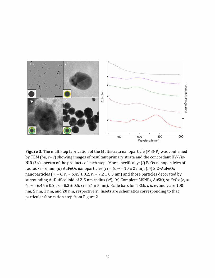
In order to generate a FeOx/Au enveloped in an SiO2/Au shell, the Multistrata nanoparticle
resembles a single core - five layer “onion,” each strata possessing a specific function
(Figure 2). The core fabrication begins with the generation of γ-Fe2O3 superparamagnetic
iron-oxide nanoparticles by thermal decomposition of Fe(CO)5 in the presence of oxygen
after aeration and reflux as described previously (K. Woo & 2004). Such synthesis provides
the oleic acid surface chemistry suitable for subsequent chemical modification. The first
subsidiary strata, APTES [(3-aminopropyl)triethoxysilane], displaces the oleic acid in an
exchange reaction and acts as a coupling layer between the γ-Fe2O3 (FeOx) core and the
initial gold layer to be applied. The APTES coating provides -NH2 groups that have a high
affinity for Au3+ ions to facilitate the initial gold coating. Conventional aminoxysilane
reactions, like those that utilize APTES, involve single solvents such as DI H2O, ethanol
(EtOH), toluene and tetrahydrofuran (THF) and are fully detailed in synthetic chemical
literature[23]. One particular modification to the conventional APTES reaction was the
necessity to perform multiple solvent exchanges throughout to optimize APTES deposition,
-NH2 availability, and magnetic material recovery. THF was used as the primary solvent
due to its ability to maximize APTES localization on the surface of the FeOx cores through
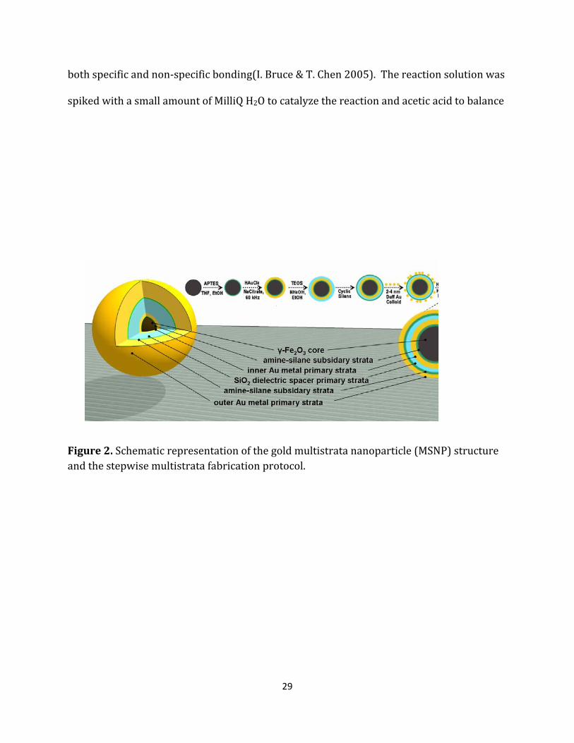
29
both specific and non-specific bonding(I. Bruce & T. Chen 2005). The reaction solution was
spiked with a small amount of MilliQ H2O to catalyze the reaction and acetic acid to balance
Figure 2. Schematic representation of the gold multistrata nanoparticle (MSNP) structure and the stepwise multistrata fabrication protocol.
30
the reaction solution at pH ~ 6.5. The THF was exchanged and washed with EtOH to
release the non-specific APTES adsorption. When suspended in EtOH, the aminated FeOx
cores are highly colloidal and difficult to sediment by centrifugation; therefore, the washed
cores were added to hexanes to prepare the material for purification and extraction
through centrifugation. Following three purification cycles, the FeOx-NH2 particles were
suspended and stored in EtOH in preparation for the deposition of the first gold layer.
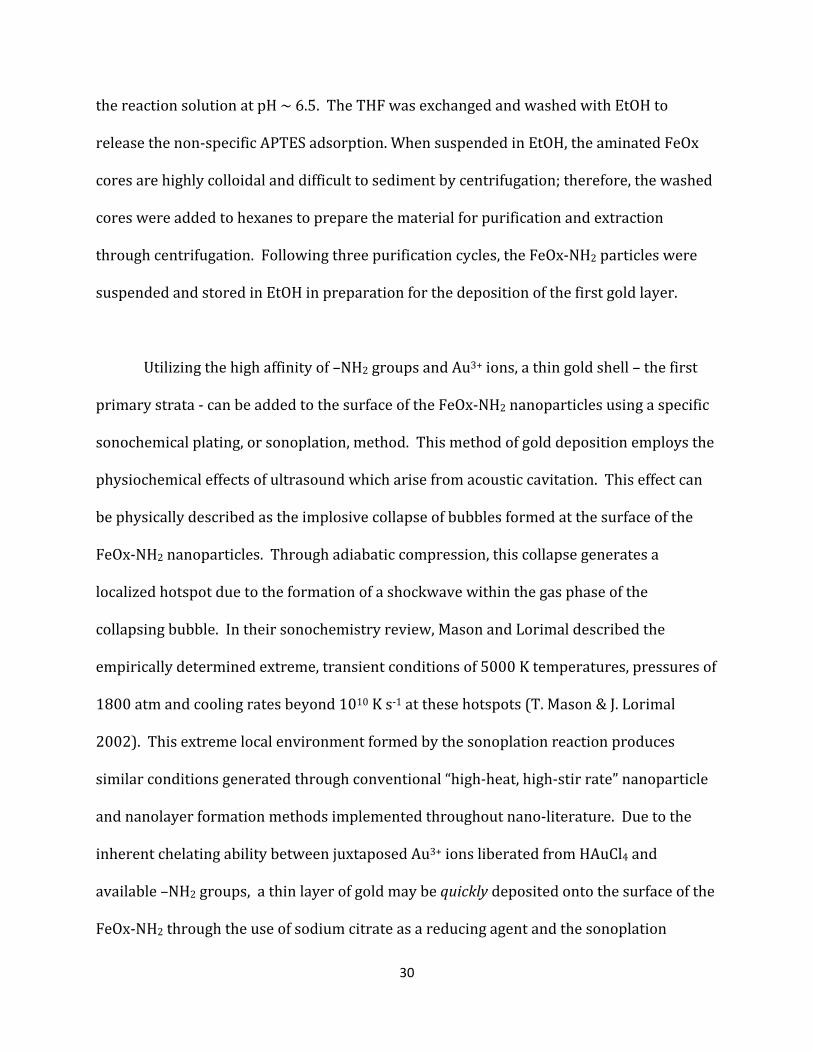
Utilizing the high affinity of –NH2 groups and Au3+ ions, a thin gold shell – the first
primary strata - can be added to the surface of the FeOx-NH2 nanoparticles using a specific
sonochemical plating, or sonoplation, method. This method of gold deposition employs the
physiochemical effects of ultrasound which arise from acoustic cavitation. This effect can
be physically described as the implosive collapse of bubbles formed at the surface of the
FeOx-NH2 nanoparticles. Through adiabatic compression, this collapse generates a
localized hotspot due to the formation of a shockwave within the gas phase of the
collapsing bubble. In their sonochemistry review, Mason and Lorimal described the
empirically determined extreme, transient conditions of 5000 K temperatures, pressures of
1800 atm and cooling rates beyond 1010 K s-1 at these hotspots (T. Mason & J. Lorimal
2002). This extreme local environment formed by the sonoplation reaction produces
similar conditions generated through conventional “high-heat, high-stir rate” nanoparticle
and nanolayer formation methods implemented throughout nano-literature. Due to the
inherent chelating ability between juxtaposed Au3+ ions liberated from HAuCl4 and
available –NH2 groups, a thin layer of gold may be quickly deposited onto the surface of the
FeOx-NH2 through the use of sodium citrate as a reducing agent and the sonoplation
31
ultrasonic frequency as a reaction catalyst (Wu et al. 2007). A distinct color change from a
flocculated (due to immediate repulsive electrostatic interactions prior to Au3+ liberation
by the sodium citrate) yellow-brown mixture to a black-purple, fully colloidal suspension,
following the induction of the reducing agent and catalyst, signals the generation of
FeOx/Au core/shell particles, the FeOx/Au nanoparticles. This addition of an outer gold
shell was substantiated by the appearance of a surface plasmon resonance extinction
maxima (λmax ~ 540-570 nm) in the sample absorbance spectra which is not evident in the
FeOx nanoparticles (Figure 3). In addition, high-resolution TEM images shown in Figure 3
document the detailed formation of gold “plates” on the iron oxide surface and the
development of gold fringe patterns (111 planes, 0.24 nm) consistent with well
characterized images in electron microscopy literature.[26]
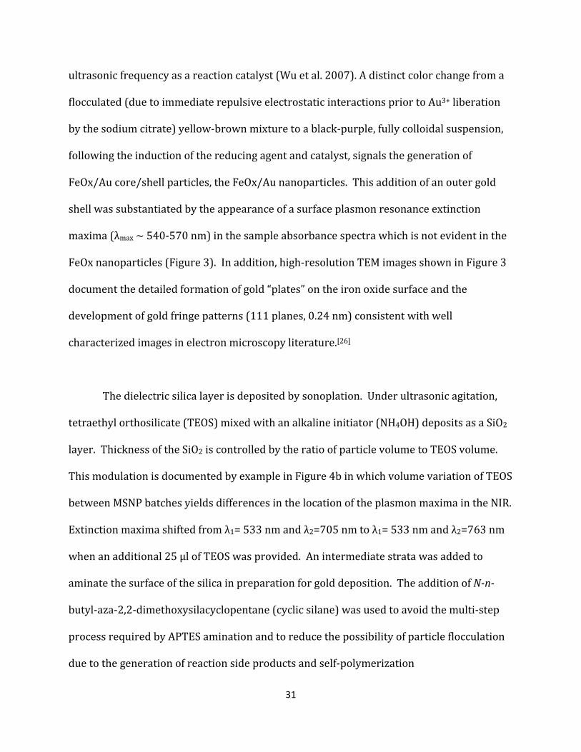
The dielectric silica layer is deposited by sonoplation. Under ultrasonic agitation,
tetraethyl orthosilicate (TEOS) mixed with an alkaline initiator (NH4OH) deposits as a SiO2
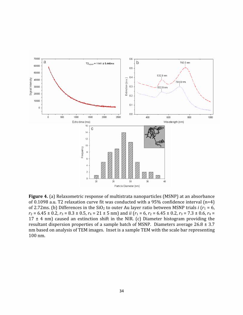
layer. Thickness of the SiO2 is controlled by the ratio of particle volume to TEOS volume.
This modulation is documented by example in Figure 4b in which volume variation of TEOS
between MSNP batches yields differences in the location of the plasmon maxima in the NIR.
Extinction maxima shifted from λ1= 533 nm and λ2=705 nm to λ1= 533 nm and λ2=763 nm
when an additional 25 μl of TEOS was provided. An intermediate strata was added to
aminate the surface of the silica in preparation for gold deposition. The addition of N-n-
butyl-aza-2,2-dimethoxysilacyclopentane (cyclic silane) was used to avoid the multi-step
process required by APTES amination and to reduce the possibility of particle flocculation
due to the generation of reaction side products and self-polymerization
32
Figure 3. The multistep fabrication of the Multistrata nanoparticle (MSNP) was confirmed by TEM (i-ii, iv-v) showing images of resultant primary strata and the concordant UV-Vis-NIR (i-v) spectra of the products of each step. More specifically: (i) FeOx nanoparticles of radius r1 = 6 nm; (ii) AuFeOx nanoparticles (r1 = 6, r2 = 10 ± 2 nm); (iii) SiO2AuFeOx nanoparticles (r1 = 6, r2 = 6.45 ± 0.2, r3 = 7.2 ± 0.3 nm) and those particles decorated by surrounding AuDuff colloid of 2-5 nm radius (vi); (v) Complete MSNPs, AuSiO2AuFeOx (r1 = 6, r2 = 6.45 ± 0.2, r3 = 8.3 ± 0.5, r4 = 21 ± 5 nm). Scale bars for TEMs i, ii, iv, and v are 100 nm, 5 nm, 1 nm, and 20 nm, respectively. Insets are schematics corresponding to that particular fabrication step from Figure 2.

33
(Bardhan et al. 2010). Following the silanization of the silica surface, the available -NH2
sites were decorated with 2-5 nm Duff Au colloids (D. Duff et al. 1993) which act as
nucleation or “seed” sites for subsequent gold deposition. We speculate that Au decoration
of these particles is consistent with the existence of a peak in the 900 nm wavelength range
suggestive of plasmonic interactions between the decorated gold particles, which are
aggregating over the surface area of the precursor particle, and the inner Au strata shown
in Figure 3. The final strata, a complete outer Au layer, was catalyzed onto the decorated
particles through the reduction of Au ions from a 1% HAuCl4 solution in the presence of
H2CO – a formaldehyde electroless plating reaction (B. Brinson et al. 2008). The deposition
of this final layer is supported by the change in surface plasmon extinction spectra of the
MSNP and the formation of a metallic-gold outer layer (Figure 3v). The characteristic
double-peak spectra of a multilayered, gold-dielectric-gold material appears following
electroless plating (Figure 3v and Figure 4b). In order to maintain the stability of the outer
Au strata, particles were resuspended in 1.8mM K2CO3 which acts as a stabilizing agent. A
zeta potential of -75.6 ± 0.902 was measured to confirm the stability of the MSNPs in this
solution. This negative charge is also consistent with the existence of -CO32- ions stabilizing
the Au surface. As for gold nanoparticles, which present the same surface chemistry, these
stabilizing ions are easily place-exchanged with a number of conjugates that proximally
present amine, sulfhydryl or other functional groups. From a surface modification
perspective, MSNP behavior is the same as for other gold surfaces, for which many robust
methods are well known to provide a wide range of molecular coatings. These fabrication
reactions yield monodisperse batches of MSNPs as shown in Figure 4c. In addition, these
reactions are scaleable making the fabrication of bulk quantities possible.
34
Figure 4. (a) Relaxometric response of multistrata nanoparticles (MSNP) at an absorbance of 0.1098 a.u. T2 relaxation curve fit was conducted with a 95% confidence interval (n=4) of 2.72ms. (b) Differences in the SiO2 to outer Au layer ratio between MSNP trials i (r1 = 6, r2 = 6.45 ± 0.2, r3 = 8.3 ± 0.5, r4 = 21 ± 5 nm) and ii (r1 = 6, r2 = 6.45 ± 0.2, r3 = 7.3 ± 0.6, r4 = 17 ± 4 nm) caused an extinction shift in the NIR. (c) Diameter histogram providing the resultant dispersion properties of a sample batch of MSNP. Diameters average 26.8 ± 3.7 nm based on analysis of TEM images. Inset is a sample TEM with the scale bar representing 100 nm.

35
For more than half a decade, gold nanotechnology has been shown to act as X-ray
and computed tomography contrast agents, increasing the utility of each technique imaging
biological samples (J. Hainfeld et al. 2006). MSNP capacity for MRI contrast can be
evaluated through relaxometric measurements. Prior to sample preparation, the extinction
peak located in the visible spectrum was measured at 0.1098 a.u. The MSNPs exhibit a T2
relaxation time at 1141 ± 5.4 ms fit via a 95% confidence interval based on four repetitions.
This relaxation time can be differentiated from the T2 values of healthy human tissue[30]
and are predicted to alter the relaxation times of proximal tissues. These results support
the MR contrast capacity of MSNPs.
Figure 5. Stepwise Comparison of Gold (top) and Silver Multistrata Fabrication Protocols
To further support the design and functionality of the MSNP, gold was replaced with
silver, another plasmonically active metal, during the synthesis process as shown in Figure
5. In this figure, the Au-MSNP and Ag-MSNP synthesis processes are compared in a
stepwise fashion. The gold was completely substituted for silver to such an extent that the
development of Ag-THPC nanoparticles (Woo 2014) was included as a substitute for the
Duff Au colloids. This stark substitution was deliberate to ensure that Ag-MSNP plasmonic

36
results could only be attributed to the effect of the silver-dielectric-silver concentric shells
and not due to the presence of gold in the particle architecture.
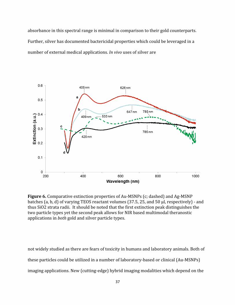
Upon comparing the extinction characteristics of the Au-MSNPs and Ag-MSNPs it is
clear that there is value in modifying the plasmonically-active metal which comprises the
nanoconstruct. As shown in Figure 6, Ag-MSNP extinction peaks deviate from Au-MSNP
peaks where the first peak appears at the interface of the long wave UV and visible
spectrum. Furthermore, the second peak appears in the visible but can be tuned into the
NIR. These results support the hypothesis that a multilayered, metallodielectric
nanoparticle which has combined multiple nanoscale characteristics into a single <60 nm
nanoconstruct could be developed. Furthermore, Ag-MSNPs could provide double the
therapeutic value due to the coupling of the inherent antimicrobial behavior of Ag at the
surface of the material with the ability to tune the plasmonic characteristics of
nanoconstruct to enable photothermal therapy.
It should be reiterated and appreciated that the fundamental difference between Au
and Ag containing MSNPs is their plasmon active metal (Bell & Yu 2011). Varying the
plasmonic metals that comprise the nanoconstruct generates different MSNPs for different
usage and applications. For example, silver MSNPs generally have secondary peaks in the
500-700 nm “visible” spectrum, thus eliminating their potential for use in NIR
photothermal therapy via NIR laser irradiation, In contrast, gold particles are generally
NIR sensitive and may be used in photothermal therapeutic procedures. However, as
shown above, silver MSNPs can be designed to absorb in the NIR. However, their
37
absorbance in this spectral range is minimal in comparison to their gold counterparts.
Further, silver has documented bactericidal properties which could be leveraged in a
number of external medical applications. In vivo uses of silver are
Figure 6. Comparative extinction properties of Au-MSNPs (c; dashed) and Ag-MSNP batches (a, b, d) of varying TEOS reactant volumes (37.5, 25, and 50 µl, respectively) - and thus SiO2 strata radii. It should be noted that the first extinction peak distinguishes the two particle types yet the second peak allows for NIR based multimodal theranostic applications in both gold and silver particle types.
not widely studied as there are fears of toxicity in humans and laboratory animals. Both of
these particles could be utilized in a number of laboratory-based or clinical (Au-MSNPs)
imaging applications. New (cutting-edge) hybrid imaging modalities which depend on the

38
combined biomagnetophotonic properties of their imaging contrast agents could employ
both Au-MSNPs and Ag-MSNPS as potential contrast agents.

39
Conclusions
In conclusion, we have demonstrated the fabrication of a single core, five layered
nanostructure with the potential capacity for both CT and MR imaging contrast. This
contrast agent may allow for the simultaneous use of both technologies as well as other
hybrid imaging modalities. Furthermore, we have characterized these particles for their
metallodielectric properties and have measured dual-peak UV-Vis-NIR extinction spectra.
We have also shown evidence of the geometric-dependent ‘tunability’ of the optical
extinction characteristics. This tunability will allow predictable optimization of the
nanoparticulate performance based on controllable synthesis conditions. We speculate
that this technology can be further applied for laser absorption and consequent thermal
characteristics in the NIR. Successful demonstration of significant optical absorption and
heat generation is consistent with future applications in theranostic disease treatment.
Summary
A nanoconstruct that combines the properties necessary to enhance the imaging and
therapeutic capacity of the above modalities while conserving the nanometer size and
surface chemical modification capability was designed, fabricated and characterized.
Furthermore, a comparable variant of the nanoconstruct was fabricated and characterized
to further substantiate the results and conclusions of the study. The Multistrata
nanoparticle is the culmination of tunable, dual-peak, UV-Vis-NIR spectrum extinction
characteristics, tri-modal imaging contrast, simple synthesis and facile surface modification
capability into a single <60nm diameter, novel, multi-functional, multilayered nanosphere.
Through careful design, we have coupled diagnostics and therapeutics into one single

40
Figure 7. The Multistrata Nanoparticle was featured on the Frontispiece, reserved for highlighting articles that present outstanding results, of the renowned, peer-review journal Small (ISI IF 2011: 8.349).

41
theranostic tool to alleviate current methodological limitations. For the purposes of this
dissertation, the results provided in this chapter accomplished an important objective
leading to the validation of the core hypothesis. We have developed multilayered
nanomaterials that combine at least two nanoscale characteristics into a single, nanoscale
entity. Furthermore, results support the claims that: (1) novel multilayered, magneto-
metallodielectric, core/shell nanoparticles were fabricated using a unique synthesis
protocol, the (2) multimodal nanoscale characteristics and the (3) theranostic potential of
the nanomaterial were successfully elucidated and described herein thus fulfilling Specific
Aim 1 of this dissertation. Even further, I am very proud that this work was featured on the
Frontispiece of Volume 7, Issue 9 of the premier nanoscale/microscale peer reviewed
journal, Small (Bell et al. 2011). This spot is reserved for highlighting articles that present
outstanding results. In summary, I have provided a sample of this achievement in Figure 7.
42
CHAPTER III
SONOCHEMICAL SYNTHESIS AND TOMOGRAPHIC CHARACTERIZATION OF SUPERPARAMAGNETIC FEOX/AU CORE/SHELL COMPOSITE NANOPARTICLES
Core/shell nanomaterials have become an increasingly significant sector of the
nanotechnology field (Chaudhuri & Paria 2012) and have been applied in a multitude of
applications, most notably in the biomedical domain (Chatterjee et al. 2014). Hybrid
materials can be designed to exhibit a multiplicity of properties depending on the
composite materials present and the orientation at which those materials comprise the
nanoparticle. This ability to integrate a multiplicity of properties into a single nanometer
length scale embodiment is of significant impact. Inorganic magentoplasmonic core/shell
nanomaterials are of considerable interest as they possess both the plasmonic and
magnetomotive properties necessary for a multitude of theranostic applications. However,
for biological applications, the ability to rapidly synthesize these nanomaterials to achieve
the desired nanoparticulate characteristics is paramount. A sonochemical synthesis
procedure will be utilized to reduce the overall duration of synthesis while ensuring the
decreased magnetic relaxivity and increasing the magnetomotivity of the nanoparticles.
The effects of this change in synthesis protocol on the plasmonic, magnetic and
morphologic characteristics of the nanoparticles will be elucidated. Furthermore, for the
first time, scanning transmission electron tomography will be utilized to further elucidate
the morphology of the resultant magnetoplasmonic iron-oxide/gold core/shell
nanomaterials. For the purposes of this dissertation, the results provided in this chapter
will accomplish an important objective leading to the validation of a portion of the core
43
hypothesis. Herein, we develop multilayered nanomaterials that combine at least two
nanoscale characteristics into a single, nanoscale entity. Furthermore, results from this
chapter will fulfill the goals in Specific Aim 2 of this dissertation. I will show that
multilayered, magnetoplasmonic core/shell nanoparticles can be fabricated by adapting
the synthesis protocol used in Chapter II. Furthermore, I will show that the magnetic
properties of the magnetoplasmonic nanomaterials will be enhanced to provide
magnetomotive functionality. I will also describe a revised fabrication strategy which will
decrease the overall synthetic period. This will enable rapid fabrication and use of fresh
nanomaterials for future biological applications.
44
Introduction
Core/shell nanomaterials have become an increasingly significant sector of the
nanotechnology field (Ghosh Chaudhuri & Paria 2012) and have been applied in a
multitude of applications, especially in the biomedical domain (Chatterjee et al. 2014).
Core/shell nanoparticles are primarily composed of two or more separate phases which
possess differing structure or chemical composition (Runowski 2014). Such hybrid
materials can be designed to exhibit a multiplicity of properties depending on the
composite materials present and the orientation at which those materials comprise the
nanoparticle. This ability to integrate a multiplicity of properties into a single nanometer
length scale embodiment is of significant impact and has remained state of the art for over
a decade (Wolfgang Schärtl 2010).
Classes of core/shell nanomaterials include: concentric-spherical (Shakhov et al.
2014), hexagonal (Yuan et al. 2014), multi-core/single shell (H. Wang et al. 2011),
nanomatryushka (Bardhan et al. 2010) or movable core within a hollow shell core/shell
nanoparticles (Shevchenko et al. 2008). Although the most notable of the group are
concentric-spherical and hexagonal core/shell materials, these classes of nanoparticles
possess varying morphologies which are dependent on: (1) the materials which comprise
the particle, (2) their orientation within the structure and (3) the synthesis procedures
utilized to develop the materials. As a result, in order to achieve the desired
multifunctional properties, core/shell nanoparticles which are comprised of the same
material with the same structural orientation may be forced to deviate in their
45
morphological appearance. Such deviations in morphology must be studied to further
understand the inherent nature of the nanomaterial.
Methods to enhance the study of the morphological appearance of materials at the
nanometer scale have vastly improved over the past decade. The enhancement of
conventional transmission electron microscopy to include aberration-corrected, electron
beam rastering is of great significance as it provides a technical basis for the
implementation of analysis techniques such as energy dispersive X-ray (EDX) spectroscopy
(EDS)(Herzing et al. 2008) and high angle annular dark field (HAADF) imaging (Jesson &
Pennycook 1995). The combination of HAADF-STEM imaging allows for the formation of
micrographs where contrast is directly correlated the atomic number of the elements in the
specimen. Because of this, following a shift-corrected tilt series, 3D tomograms can be
efficiently generated as HAADF-STEM image intensities vary with the mass-thickness of
low atomic number, microscale samples. Electron tomography allows for in depth, 3D
morphological analysis of submicron nanomaterials (Midgley et al. 2006) not previously
achievable.
Inorganic magnetoplasmonic core/shell nanomaterials are of considerable interest
as they possess the properties necessary for a multitude of theranostic applications. The
presence of a plasmonically-active, metallic surface provides the nanoparticle the ability to
act as a colorimetric probe. Based upon the nanoparticle size, structural morphology and
colloidal behavior, a solution of metallically coated core-shell nanomaterials will change
color by absorbing electromagnetic radiation at particular wavelengths. This phenomenon

46
can be attributed to the resonant absorption of the radiation by the metallic surface
plasmons – oscillating, surface valence electrons (Barnes et al. 2003). Even further, this
magnetic response is tunable where modification of nanoparticulate size, shape or
composite materials can yield a desired plasmonic characteristic (Link et al. 1999). These
characteristics are not only ideal for diagnostic strategies which utilize visible, colorimetric
detection (Zeng et al. 2014) or photonic image contrast (Brongersma 2003); but are also
suitable for photonic-based therapies once the appropriate tuning of the plasmonic
properties has been achieved. Coupling the plasmonic characteristics with the capacity for
magnetic manipulation (magnetomotive ability), as governed by the superparamagnetic
nanoparticle at the core of the nanomaterial, unleashes a host of possible applications.
As core/shell nanotechnology as a field matures, the ability to rapidly synthesize
these nanomaterials to achieve the desired nanoparticulate characteristics is paramount.
In this study, we sought to adapt the protocol used to synthesize the inner core/shell of the
multistrata metallodiaelectic nanoparticle (Bell et al. 2011) to rapidly synthesize
magnetoplasmonic iron-oxide/gold core/shell nanomaterials (Liang et al. 2009; Zhou et al.
2012; Li et al. 2014). Surface modification of nanoparticles can be rapidly achieved by
exploiting the liquid-based, acoustic cavitation phenomenon which occur during
sonochemical modification reactions. More specifically, acoustic cavitation occurs when
bubble formation, growth and collapse occur in liquid which is under ultrasonic
perturbation. This phenomenon occurs throughout the reaction volume but most notably
at liquid-nanostructure interfaces. The collapse of the bubbles is nearly an adiabatic
process where there is a significant accumulation of energy inside the bubble. Upon
47
implosion, this energy is rapidly released resulting in hotspots which possess high
temperatures and pressures at the liquid-nanoscale interface. Extreme, transient
conditions of 5000 K temperatures, pressures exceeding 1800 atm and cooling rates
beyond 1010 K s-1 were empirically determined (T. Mason 2002) at these microscale
hotspots. This local microenvironment results in the chemical excitation of matter inside
or proximal to the hotspot thus catalyzing chemical reactions between reagent ligands and
the nanoscale surface. Successful sonochemical synthesis, or sonoplation, of gold ions to
aminated FeOx nanoparticles has been previously described (W. Wu et al. 2007).
Furthermore, a rapid sonochemical protocol for the amination of the oleic acid capped
FeOx nanoparticles has been described (Sodipo & Aziz 2014). However, incorporating both
sonochemical synthesis processes with a <3hr core/shell synthesis period for the
development of a core/shell magnetophotonic nanomaterial has not been described.
This sonochemical synthesis procedure was utilized to reduce the overall duration
of synthesis while ensuring decreased magnetic relaxivity thus increasing the
magnetomotivity of the nanoparticles. The effects of this change in synthesis protocol on
the plasmonic, magnetic and morphologic characteristics of the nanoparticles were
elucidated. Furthermore, for the first time, scanning transmission electron tomography
was utilized to further elucidate the morphology of the resultant magnetoplasmonic iron-
oxide/gold core/shell nanomaterials.
48
Methods and Materials
Fabrication of γ-Fe2O3 Superparamagnetic Nanoparticles: γ-Fe2O3 (FeOx) with ~10 nm
diameter were fabricated via a thermal decomposition, aeration and reflux protocol
pioneered by Woo et al.(K. Woo et al. 2004) Octyl ether (Sigma-Aldrich, St. Louis, 200 mL)
and oleic acid (Sigma-Aldrich, 28.5 mL) were mixed in a 500 mL round-bottom flask under
reflux and Ar gas flow. The volume was heated to 100°C prior to the injection of Fe(CO)5
(Sigma-Aldrich, 2.0 mL). The vessel was quickly heated from 150°C to 295°C using a
heating mantle and glass wool to ensure insulation. During this heating process, the
solution color changed from yellow to orange, orange/colorless, to very dark orange then
black. The reaction was aerated at 80(±10)°C for 14 h and refluxed while boiling for an
additional 2 h. The γ-Fe2O3 nanoparticles were harvested using ethanol (EtOH, 200 proof,
Sigma-Aldrich) and purified via three iterations of centrifuge (20 min, 800 relative
centrifugal force (rcf)), supernatant extraction and EtOH resuspension. γ-Fe2O3
nanoparticle cores were dried under air for 48 h.
Conventional Amination of γ-Fe2O3 Core Nanoparticles: 150 mg of γ-Fe2O3 cores (FeOx) were
coated using a conventional (3-Aminopropyl)triethoxysilane (APTES; Sigma-Aldrich)
functionalization procedure(Bell et al. 2011). FeOx particles were added to
tetrahydrofluran (THF; Thermo Fisher Scientific, Waltham, MA, 40 mL) and briskly stirred
using a magnetic stir plate and stirring rod. APTES (5 mL) was added to the reaction
solution and thereafter spiked with acetic acid (Sigma-Aldrich, 5 μL), dH2O (516 μ L), and
stirred for 48 h. The reaction flask was then placed into a heating mantle, wrapped in steel
wool, and heated to 80 ° C. EtOH was used to incrementally replace evaporated THF

49
throughout the 2 h boiling period. This incremental replacement was tuned until the
reaction solution was concentrated to 40 mL of EtOH. Hexane was added to the solution in
a 4:1 ratio and centrifuged (10 min, 800 rcf). Recovered FeOx–NH2 nanoparticles were
resuspended in EtOH and stored at room temperature where they remained stable for at
least three months.
Sonochemical Amination of γ-Fe2O3 Core Nanoparticles: Aminated γ-Fe2O3 ~11 nm diameter
were fabricated via a place exchange reaction in an ultrasonic agitation reaction
environment. γ-Fe2O3 nanoparticles (10 mg) were dissolved into a 1% wt APTES (Sigma-
Aldrich) solution in anhydrous toluene (Sigma-Aldrich). This volume was reacted under
ultrasonic agitation (90 min, 20°C) and vortexed intermittently at 15 minute intervals to
ensure even mixing. Using a 1T neodymium 1” cube magnet, the aminated γ-Fe2O3 were
magnetically separated from the toluene based solvent and washed with anhydrous
ethanol (EtOH, 200 proof, Sigma-Aldrich). Aminated γ-Fe2O3 nanoparticles (10 mg/ml,
1ml) were stored at 4°C.
Gold Sonoplation of Conventionally Aminated γ-Fe2O3 Nanoparticles: FeOx–NH2 NPs were
plated with a gold layer using protocols adapted from Wu et al (Wu et al. 2007). Sonicated
FeOx–NH2 nanoparticles ( ≈ 1.5% wt.) were added in equal volume to aqueous, 1% HAuCl4
(Sigma-Aldrich, dark aged 72 h) in an ultrasonic perturbation reaction environment.
Sodium citrate (20 mM) in dH2O was added dropwise under sonication until the light
yellow–brown color of the reaction solution changes to a dark purple. The solution of
FeOx/Au core/shell nanoparticles (referred to hereafter as FeOx/Au) was washed via

50
centrifugation (800 rcf, 5 min) and resuspended in EtOH and stored for 18 h at 4°C where
they remained stable for at least 5 months.
Gold Sonoplation of Sonochemically Aminated γ-Fe2O3 Core Nanoparticles: Au, (γ-Fe2O3)
composite nanoclusters (FeOx-Au) were fabricated via an electroless plating reaction in an
ultrasonic agitation reaction environment. Adapted from a protocol described in the
literature (Bell et al. 2011), an aqueous 1% HAuCl4 solution was mixed with an ethanolic
solution of aminated γ-Fe2O3 nanoparticles (1 mg/ml) in ratios prescribed in Table 1.
Under an ultrasonic agitation environment, 12 mL of aqueous sodium citrate (20 mM,
Sigma-Aldrich) was added in 800 μL incremental injections every 30 seconds. The
resultant FeOx/Au nanoclusters were centrifuged (800 rcf, 10 min). The supernatant was
removed while the pellet was preserved via a 1T neodymium magnet. The pellet was
resuspended into 10 ml of dH2O and the resultant FeOx-Au nanomaterials (200 μg/mL)
were stored at 4°C.
Nanocluster Magnetomotive Characterization: Unconjugated FeOx/Au nanoclusters were
tested for their magnetomotivity in dH2O. Briefly, a 6 point standard curve was produced
through serial dilution from a stock concentration of 200 μg/mL FeOx/Au nanoclusters
was produced. A 2 mL microcentrifuge tube of FeOx/Au nanoculsters (200 μg/mL) was
fastened to a 1T neodymium 1” cube magnet. Every 5 min for 20 min then every 10 min
thereafter, 200 uL of the solution was removed from the side of the tube distal to the
magnet. Each separated solution was diluted into 800 μL of DI water and measured on the
51
Varian Cary 50 at a 595 nm wavelength. A 6 point curve was generated quantifying the
percent capture with respect to time.
Nanoparticle Characterization: Synthesized nanoparticles were characterized utilizing
multiple modalities. Relaxometric measurements were performed on a Maran DRX-II 0.5T
NMR spectroscopic scanner following the preparation of samples in Nanopure dH2O at a
diluted nanoparticulate concentration (40 μg/mL; 1 mL). Spectroscopic extinction
measurements were performed in dH2O using a Varian Cary 50 UV-vis-NIR
spectrophotometer (Agilent Technologies, Santa Clara, CA). Spectroscopic T2 relaxation
measurements were performed in a Carr-Purcell-Meiboom-Gill (CPMG) sequence with the
following parameters: relaxations time (TR) = 30 s, echo time (TE) of first echo = 6 ms,
spacing of subsequent echos = 2 ms, and 1200 echos. Size and zeta potential
measurements were performed on a Malvern Zetasizer (Malvern Instruments,
Westborough, MA) following nanoparticulate dilution (40 μg/mL; 1 mL) in dH2O. The
diameter of individual nanoparticles was measured from TEM images using ImageJ
software. For standard morphological analysis, standard TEM micrographs with EDX
(energy dispersive x-ray spectroscopy) elemental analysis were obtained on a FEI Tecnai
Osiris S/TEM Microscope (FEI, Hillsboro, OR).
Tomographic Imaging and Reconstruction: The surface structure of FeOx-Au nanoclusters
were examined in more detail by determining the 3D morphology of the individual
nanoparticles. STEM micrographs were collected on a FEI Tecnai Osiris S/TEM
Microscope. A group of nanoparticles possessing varying 2D morphologies was imaged.
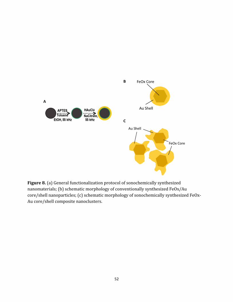
52
Figure 8. (a) General functionalization protocol of sonochemically synthesized nanomaterials; (b) schematic morphology of conventionally synthesized FeOx/Au core/shell nanoparticles; (c) schematic morphology of sonochemically synthesized FeOx-Au core/shell composite nanoclusters.

53
Micrographs were collected at 13.5 kx magnification and auto-focused at 24kx/56 kx
magnifications across a ±60° tilt series where single bright field and dark field images were
collected at every degree. Micrograph tilt series were reconstructed using Inspect 3D (FEI,
Hillsboro, OR) software using an iterative approach (n=30) to interpolate and approximate
the data outside of the ±60° tilt. The 3D data was imported into AMIRA 3D (FEI, Hillsboro,
OR) and the data was reconstructed into a volume and rendered in 3D. A ±60° dark field,
tilt series and 3D volume rendering of the morphology of the nanoclusters was produced.
Results and Discussion
FeOx/Au core/shell nanoparticles were successfully synthesized. The characterization of
the resultant nanoparticles is shown in Figure 9. The micrographs shown in Figure 9A
depict the morphology of the FeOx/Au nanoparticles. More specifically, bright field TEM
micrographs of FeOx-NH2 (9.47±1.56 nm dia.) and FeOx/Au nanoparticles (13.5±4.13 nm
dia.) are shown in Figure 9Aa and Figure 9Ab, respectively. The morphology of both
nanoparticle types is comparable to TEM micrographs depicted previously in literature
(Bell et al. 2011) where the micrograph of FeOx/Au nanoparticles depict the formation of
gold ‘plates’ on the iron oxide surface. The HAADF-STEM micrograph shown in Figure 9Ac
shows the spherical nature of the FeOx/Au nanoparticle synthesized using this method.
The formation of this gold coating is substantiated in Figure 9B where an extinction peak in
the visible spectral range (Figure 9Bb) is evidential of the surface plasmon resonance of the
gold coated material. This peak is only apparent post-synthesis and differs from the
extinction profile of non-plated FeOx-NH2 particles which lack an absorbance peak in the
visible spectral range. Measurement of the relaxivity (Figure 9C) of these nanomaterials
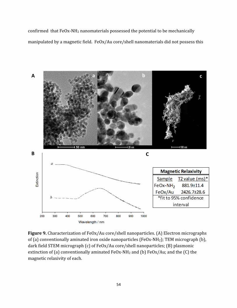
54
confirmed that FeOx-NH2 nanomaterials possessed the potential to be mechanically
manipulated by a magnetic field. FeOx/Au core/shell nanomaterials did not possess this
Figure 9. Characterization of FeOx/Au core/shell nanoparticles. (A) Electron micrographs of (a) conventionally aminated iron oxide nanoparticles (FeOx-NH2); TEM micrograph (b), dark field STEM micrograph (c) of FeOx/Au core/shell nanoparticles; (B) plasmonic extinction of (a) conventionally aminated FeOx-NH2 and (b) FeOx/Au; and the (C) the magnetic relaxivity of each.

55
magnetomotive ability. This was observed when a volume of the nanoparticles was
exposed to a 1T magnetic field and no significant bulk movement of the material was
observed for ~20 min. These observations were substantiated by the long T2 relaxivity
values (T2=2426.7±28.6) which support a lack of magnetomotive ability.
For applications which require plasmonic materials to possess magnetomotive
ability, the FeOx/Au core/shell nanoparticles, as synthesized, do not possess these
characteristics. From a design perspective, it was concluded that extremely thin shells
were necessary to enhance the magnetomotive ability of the core/shell materials. This is
due to the inverse relationship between the magnetic properties and shell thickness (i.e.
the distance from the magnetic core to the aqueous solution). Any magnetomotive particle
must possess a significant saturation magnetization, Msat, which is the maximum
magnetization a particle can experience. This characteristic is described in Eq. 1 and
defines a single core/shell magnetic nanoparticle, where the magnetic properties of the
particle arise from the core which is coated in a thin outer shell of magnetically inert
material,
𝑀𝑠𝑠𝑠 = 𝑚𝑀𝑠𝑠 �𝑟 − 𝑑𝑑 �
3
(1)
where Mso is the saturation magnetization of the bulk material in units of A/m/g, r is the
particle radius, d is shell thickness and m is the mass of the individual particle. Any
significant increase in the shell thickness, d, has significant detrimental effect on the

56
Figure 10. TEM micrographs and the corresponding plasmonic extinction of FeOx-NH2 nanoparticles and FeOx-Au core/shell composite nanoclusters. (i) FeOx-NH2 nanoparticles and FeOx-Au nanoclusters synthesized at HAuCl4:FeOx-NH2 reagent ratios of (ii) 1:13.3 (iii) 1:2 (iv) 3:4.

57
saturation magnetization of the particle. In order to successfully synthesize extremely thin
outer gold and intermediary silane shells, the amount of 1% HAuCl4 was decreased and a
sonochemical silanization protocol was implemented until the magnetomotive properties
were observed.
FeOx-Au composite nanoclusters were successfully synthesized by using a rapid,
sonochemical method as described at a high level in Figure 8. FeOx-Au nanoparticulates
were synthesized using decreased gold chloride reagent volumes as shown in Table 1.
These nanoparticles were characterized in Figure 10. The resultant polymorphous
morphology of the FeOx-Au differs from the aesthetically-uniform, spherical FeOx/Au
counterpart. While some spherical particles are observed, the appearance of oblong,
jagged and elliptical particles is apparent. It was deduced that the decrease in reagent gold
chloride was the cause of the significant changes in the morphology of the nanoparticles. As
shown in the micrographs in Figure 10A, this hypothesis is substantiated by the
reappearance of the particles possessing a spherical morphology with increasing
HAuCl4:FeOx reagent ratios from particle populations ii (13.01±1.56 nm dia), iii
(13.23±2.86 nm dia), and iv (14.17±4.35 nm dia). Although this increase in spherical
morphology was evident, the polymorphous particles continued to be observed. Even
further, it is apparent that all FeOx-Au nanoparticulates tend to cluster. This observation of
stabilized flocking is supported by hydrodynamic radii measurements on the order of
~O(100 nm) (Table 1). This is important as previous mathematical modeling of
superparamagnetic core/shell nanoparticle clustering has shown that the magnetization of
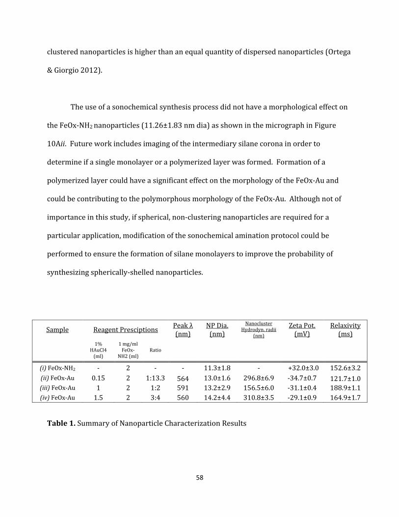
58
clustered nanoparticles is higher than an equal quantity of dispersed nanoparticles (Ortega
& Giorgio 2012).
The use of a sonochemical synthesis process did not have a morphological effect on
the FeOx-NH2 nanoparticles (11.26±1.83 nm dia) as shown in the micrograph in Figure
10Aii. Future work includes imaging of the intermediary silane corona in order to
determine if a single monolayer or a polymerized layer was formed. Formation of a
polymerized layer could have a significant effect on the morphology of the FeOx-Au and
could be contributing to the polymorphous morphology of the FeOx-Au. Although not of
importance in this study, if spherical, non-clustering nanoparticles are required for a
particular application, modification of the sonochemical amination protocol could be
performed to ensure the formation of silane monolayers to improve the probability of
synthesizing spherically-shelled nanoparticles.
Sample Reagent Presciptions Peak λ (nm)
NP Dia. (nm)
Nanocluster Hydrodyn. radii
(nm)
Zeta Pot. (mV)
Relaxivity (ms)
1%
HAuCl4 (ml)
1 mg/ml FeOx-
NH2 (ml) Ratio
(i) FeOx-NH2 - 2 - - 11.3±1.8 - +32.0±3.0 152.6±3.2 (ii) FeOx-Au 0.15 2 1:13.3 564 13.0±1.6 296.8±6.9 -34.7±0.7 121.7±1.0 (iii) FeOx-Au 1 2 1:2 591 13.2±2.9 156.5±6.0 -31.1±0.4 188.9±1.1 (iv) FeOx-Au 1.5 2 3:4 560 14.2±4.4 310.8±3.5 -29.1±0.9 164.9±1.7
Table 1. Summary of Nanoparticle Characterization Results

59
The plasmonic properties of FeOx-Au (Figure 10B) were observed as expected
where, in all three core/shell preparations, extinction peaks in the visible electromagnetic
spectrum were observed. Like the FeOx/Au nanoparticles, observation of visible spectrum
extinction peaks suggests that the grafting of the gold shell onto the iron oxide core was
successful. The observation of a small 700 nm extinction peak in the iv nanoparticle batch
suggests nanoparticulate aggregation or the presence of elliptical or rod-like morphologies
in the sample. In all FeOx-Au batches, small extinction peaks in the infrared spectrum from
900-1000nm appear which suggests nanoparticle-mediated light scattering at these
wavelengths due to incomplete shell formation and colloidal flocking (i.e. formation of
nanoparticulate clusters). The presence of these infrared extinction characteristics further
supports the assertion that the FeOx-Au composites were clustering.
To ensure the sonochemical fabrication protocol successfully fabricated core/shell
nanoparticles, a single spherical nanoparticle from the FeOx-Au (iv) preparation was
isolated and imaged as shown in the micrograph in Figure 11A. To determine the
elemental composition of the specimen, TEM-EDS was utilized and the resultant spectrum
is shown in Figure 5B. The elemental appearance of copper is expected as the TEM sample
grids are composed of the element. The detection of x-rays characteristic of gold, silicon,
and iron elements which have radiated from a single nanoparticle suggests the
encapsulation of an iron-oxide core by an intermediary silane and outer gold shell.
Magnetomotive capacity is inversely related to the HAuCl4:FeOx-NH2 reagent ratios.
The magnetomotivity of the FeOx-Au nanoclusters is depicted physically in Figure 11C
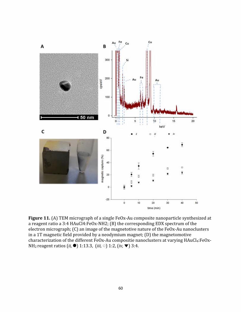
60
Figure 11. (A) TEM micrograph of a single FeOx-Au composite nanoparticle synthesized at a reagent ratio a 3:4 HAuCl4:FeOx-NH2; (B) the corresponding EDX spectrum of the electron micrograph; (C) an image of the magnetotive nature of the FeOx-Au nanoclusters in a 1T magnetic field provided by a neodymium magnet; (D) the magnetomotive characterization of the different FeOx-Au compositie nanoclusters at varying HAuCl4:FeOx-NH2 reagent ratios (ii, ) 1:13.3, (iii, ) 1:2, (iv, ) 3:4.

61
and quantitatively in Figure 11D. Figure 11D shows that the magnetomotivity is greatest
when the FeOx-Au nanoclusters are synthesized at lower HAuCl4:FeOx-NH2 reagent ratios.
We speculate that there is a threshold at which reagent ratios are too low to effectively
create core-shell materials; however, we assert that the magnetomotive capacity will
continue to increase as the lack of full shell coverage will increase the saturation
magnetization of the nanomaterials. The FeOx-Au nanoclusters were effectively separated
from solution with repeatability using a 1T magnet as shown in Figure 11C. The ability to
visualize the separation with the naked eye due to the plasmonic extinction properties of
the nanomaterial suggests that the FeOx-Au nanoclusters are viable nanoprobes for
applications which require visible, nanoscale tagging and extraction.
To assess the nanoparticulate morphology in a three dimensional space, 3D
tomographs of FeOx-Au composite nanoclusters from the ii nanoparticle batch were
generated and are shown in Figure 12A-B. A selected micrograph from the HAADF-STEM
±60° tilt series used to generate the tomographic volume is shown in Figure 12C. 3D
tomographic animations and videos of both the unaligned and aligned tilt series were
compiled. As previously shown in the micrographs in Figure 10, the nanoparticles shown
here are polymorphous and vary in shape which includes spherical, oblong, elliptical and
jagged forms. Tomographic analysis confirms these morphological characteristics are not
artifacts of a 2D transmission electron slice but are evident in a three dimensional volume.
The green and blue colors depict variations in elemental density throughout the cluster of
nanoparticles as detected by the high angle annular dark field detector in the STEM (Figure
12E). Gold atoms appear

62
Figure 12. Morphological analysis of FeOx-Au composite nanoclusters. Tomographic 3D reconstruction and rendering of FeOx-Au nanoclusters from a top view (A) and side view (B); (C) screen capture of the realigned tilt series reconstructed from single STEM images; (D) selected bright field and (E) dark field, high angle annular dark field (HAADF) STEM micrographs which comprise the reconstructed tilt series.

63
brighter than iron atoms because of their ability to incoherently scatter electrons at a high
angle due to their higher atomic number. It cannot be assumed that these elemental areas
of contrast are iron, silicon or gold until a tilt-series is performed at higher magnification
providing greater resolution. Future directions include utilizing a STEM-EDS mapping
technique during each tilt of the ±60° tilt series which would better elucidate and
enumerate the position and quantity of each element in the 3D tomograph, respectively.
Conclusion
We have successfully demonstrated that superparamagnetic FeOx-Au composite
nanoparticles, which manifest as magnetic nanoclusters, can be synthesized to effectively
exhibit both plasmonic and magnetomotive characteristics. The nanomaterials were
successfully fabricated where both the intermediary silane and outer gold shells were
grafted via a sonochemical synthesis process. Although the morphological aesthetics, in
comparison to FeOx/Au core/shell nanoparticles, were sacrificed for enhanced
functionality with respect to the magnetomotivity of the material, the nanomaterial
maintained its general magnetoplasmonic properties. For the first time, 3D tomographic
analysis was utilized in order to better understand the morphological characteristics of a
magnetoplasmonic composite material. The successful development and characterization
of these materials provides another robust nanotechnology-based platform for
applications, including biomedical theranostics, which can be enhanced by plasmonic
nanoprobes that possess the ability to be mechanically manipulated via an external,
contact-free command.

64
Summary
Inorganic magentoplasmonic core/shell nanomaterials are of considerable interest as they
possess both the plasmonic and magnetomotive properties necessary for a multitude of
theranostic applications. A sonochemical synthesis procedure was utilized to reduce the
overall duration of synthesis while ensuring the increased magnetomotivity of the
nanoparticles. The effects of this change in synthesis protocol on the plasmonic, magnetic
and morphologic characteristics of the nanoparticles were elucidated. Furthermore, for the
first time, scanning transmission electron tomography was utilized to further elucidate the
morphology of magnetoplasmonic iron-oxide/gold core/shell nanoconstructs. For the
purposes of this dissertation, the results provided in this chapter accomplished one of the
objectives. We (2) successfully modified the synthesis strategy to enhance the properties
of the composite material while decreasing the synthesis duration such that freshly
synthesized materials can be rapidly utilized in a biological application. We developed
multilayered nanomaterials that combine at least two nanoscale characteristics into a
single, nanoscale entity. Furthermore, we enhanced the magnetic properites of the
magnetoplasmonic nanomaterial enabling magnetomotive functionality. We showed that
multilayered, magnetoplasmonic core/shell nanoparticles were fabricated by adapting the
synthesis protocol used in Chapter II. This revised fabrication strategy was also shown to
decrease the overall fabrication period thus enabling rapid fabrication and use of fresh
nanomaterials for biological applications. These results validate my core hypothesis and
objective..
65
CHAPTER IV
NANOTECHNOLOGY-MEDIATED MAGNETIC SEPARATION OF ACINETOBACTER BAUMANNII USING LIPOPOLYSACCHARIDE TARGETED POLYMYXIN-E COATED γ-
FE2O3/AU CORE/SHELL COMPOSITE NANOCLUSTERS
Acinetobacter baumannii is a gram-negative bacterium of increasing interest due to its
significant virulence and enhanced persistence in combat and healthcare environments
(Peleg et al. 2008). Incidence in both community-acquired and nosocomial infection are on
the rise in both foreign and domestic US healthcare facilities (Dijkshoorn et al. 2007).
Treatment options are limited due to the rapid acquisition of multi-drug resistance to the
few antibiotics readily available (Fournier et al. 2006). Currently, the most effective
pharmaceutically-based treatment is the antibiotic colistin (polymyxin E) (Li et al. 2005).
With decreased cytocompatibility the common problem with administration of toxic
therapeutics, we propose the development of a nanotechnology-mediated treatment
strategy. Such a therapy will function as an adjuvant to conventional antibiotic regimens in
order to effectively curtail bacteremia.
In Chapters II and III, I have described how we have developed novel multilayered,
magnetoplasmonic nanoconstructs with the ability to be biofunctionalized for use in
biological applications. These nanomaterials will be biofunctionalized to bind to the
surface of Acinetobacter baumannii. This binding event will enable magnetic extraction of
the bacteria due to the magnetomotive characteristics of the nanomaterial. For the
purposes of this dissertation, this chapter will confirm the central hypothesis of my
dissertation that inorganic, multilayered, magnetoplasmonic core/shell nanomaterials can
66
be specifically designed and synthesized for biological applications which require both
diagnostic detection and therapeutic elimination of a disease causing agent. I will provide
results that support my hypothesis that these nanomaterials can be fabricated in such a
way that biomedical functionalization of the material can proceed without detriment to the
function of the biological component. These results will achieve our final objective where
the activity of the conjugated biological component will be conserved while binding and
extracting a disease causing agent in vitro.
In Chapter IV, the goals described in Aim 3, will be achieved. The magnetoplasmonic
core/shell nanoparticles fabricated in Aim 2 will be biofunctionalized with an antibiotic
which targets and binds gram negative bacteria. These biofunctionalized nanostructures
will then be characterized and utilized to magnetically extract Acinetobacter baumannii
from saline solution in vitro.
Introduction
Acinetobacter baumannii has emerged as a bacterial species of great interest due to its
significant virulence and enhanced persistence in combat and healthcare environments
(Peleg et al. 2008; Camp & Tatum 2010). A gram-negative bacterium initially found in the
Middle East, A. baumannii possess an enhanced ability to thrive in dry environments
(Wendt et al. 1997) and persist following conventional surface cleaning (Manian et al.
2013) and wound treatments (Maragakis & Perl 2008). Over the last decade, the recent
increase in incidence stems from US military combat in Iraq and Afghanistan (Aronson et
al. 2006). Incidence in both community-acquired and nosocomial infection are on the rise
67
in both foreign and domestic US healthcare facilities (Dijkshoorn et al. 2007); which can be
attributed to the return of infected cargo and veterans.
Acinetobacter species can cause a multitude of ailments including pneumonia,
bacteremia, meningitis, urinary tract infections, peritonitis, and skin and soft tissues
infections (Bergogne-Bérézin & Towner 1996). These primary ailments contribute to the
incidence of Acinetobacter species bloodstream infections (BSIs) which account for over
3% of all nosocomial acquired BSIs (Hidron et al. 2008). The mortality rate associated with
A. baumannii infection is high where A. baumannii bacteremia is associated with a 52%
mortality rate and pneumonia associated mortality ranges from 23% to 73% (Rodriguez-
Guardado et al. 2013).
Treatment options are increasingly limited due to the rapid acquisition of multi-
drug resistance to the few antibiotics readily available (Fournier et al. 2006). Currently,
the most effective pharmaceutically-based treatment is the antibiotic colistin
(colistimethate [colsitin sulfomethate] or polymixin E) (Li et al. 2005). Colistin is a cationic
polypeptide and potent broad-spectrum antibiotic. Previously not prescribed due to
reported nephrotoxicity and neurotoxicity, colistin has been revisited as a treatment due to
its potency against gram-negative bacilli. Polymyxins interact electrostatically with the
outer membrane of Gram negative bacterium by competitively displacing divalent cations –
most notably calcium and magnesium – from the negatively charged phosphate groups of
membrane lipids. This interaction disrupts the outer membrane and lipopolysaccharide is
released (Peterson et al. 1985). It has been shown that masking the five primary amine

68
groups on the lariat of the colistin molecule weakens the antibacterial activity. Therefore,
it is presumed that positively charged amine groups in the lariat of the antibiotic play an
important role in the interaction with the bacterial lipopolysaccharides as well as the
bactericidal effects of the molecule at physiological pH (Li et al. 2005). Although colistin
may be effective at high dosage, conservative administration is necessary due to the
nephrotoxicity, neurotoxicity, and broncho-restrictive effects that occur at aggressive
dosage (Justo & Bosso 2015). A non-aggressive administration regimen, however, can lead
to unchecked virulence thus leading to mortality.
With decreased cytocompatibility the common problem upon administration of
toxic therapeutics, many have investigated a number of therapies which function as
adjuvants to conventional antibiotic regimens in order to effectively curtail bacteremia.
Taking a pharmaceutically-centered approach, combinatorial delivery of colistin with
sulbactam, rifampin and tigecycline proved suboptimal as it has been shown to lead to the
development of resistance (Vila & Pachón 2008). Non-pharmaceutically centered
innovative approaches have been devised including the development of extracorporeal
devices aiming to mechanically cleanse bacteria from body fluids. Most investigations
utilize pathogen and endotoxin targeted magnetic nanotechnology with an external device
acting as a filter or spleen mimic. A blood-cleansing extracorporeal device, inspired by the
spleen, to continuously remove pathogens and toxins from blood has been described in the
literature (Kang et al. 2014). Magnetic nanoparticles conjugated with mannose-binding
lectin were developed forming a nano-engineered human opsonin. Magnets inside the
device were used to pull opsonin-bound pathogens from the blood flowing through a

69
microfluidic device at a >90% efficiency. Other investigators have shown synthetic ligands,
more specifically zinc-coordinated bis(dipicolylamine), can been utilized for highly
selective and rapid separation of bacteria from whole blood with almost 100% clearance
(Lee et al. 2014). These results support the relevance of extracorpeal adjuvant therapy as a
potentially relevant treatment of bacteremia.
The use of extracorpeal devices for the adjuvant treatment of A. baumannii has not
been described in literature. Furthermore, no-robust motif for nanoparticulate targeting,
with respect to binding affinity, to the A. baumannii surface has been described for these
purposes. To initiate the development of such a treatment, robust nanoparticulate
platforms must be developed and their capture ability in physiological phantoms must be
quantified. Here, for the first time, gold iron-oxide composite nanoclusters were
biologically modified and utilized for both their optical and magnetomotive properties.
Due to its mechanism of action, here we also describe the use of colistin as a targeting motif
to bind the magnetic gold-iron oxide nanoclusters to the surface of A. baumannii without
highly significant impact to bacterial survival. As a whole, we describe the separation
efficiency of A. baumannii using this antibiotic-enabled nanoparticulate platform in a
single-pass, macroscale system.

70
Materials and Methods
All materials were obtained from Sigma-Aldrich (St. Louis, MO) and used as received unless
otherwise noted. Deionized water (dH2O) was obtained through a dedicated, dual bed
laboratory installation. FeOx/Au nanocluster synthesis protocol is briefly summarized in
Figure 13.
Fabrication of oleic acid capped γ-Fe2O3 Superparamagnetic Nanoparticles: γ-Fe2O3 with
~10 nm diameter were fabricated by a thermal decomposition, aeration and reflux
protocol pioneered by Woo et al. (K. Woo et al. 2004). Briefly, octyl ether (Sigma-Aldrich,
St. Louis, 200 mL) and oleic acid (Sigma-Aldrich, 28.5 mL) were mixed in a 500 mL round-
bottom flask under reflux and Ar gas flow. The mixture was heated to 100°C prior to the
injection of Fe(CO)5 (Sigma-Aldrich, 2.0 mL). The reaction vessel was quickly heated from
150°C to 295°C using a heating mantle and glass wool to ensure insulation in the midst of
the high convection inside the fume hood. During this heating process, the reaction
solution color changed from yellow to orange, orange/colorless, to very dark orange then
black. The reaction was aerated at ~80(±10)°C for 14 h and refluxed while boiling for an
additional 2 h. The γ-Fe2O3 nanoparticles were harvested using ethanol (EtOH, 200 proof,
Sigma-Aldrich) and purified via three iterations of centrifuge (20 min, 800 relative
centrifugal force (rcf)), supernatant removal and EtOH wash. γ-Fe2O3 nanoparticle cores
were dried under air for 48 h.
Sonicatory Amination of γ-Fe2O3 Core Nanoparticles: Aminated γ-Fe2O3 ~11 nm diameter
were fabricated via a place exchange reaction in an ultrasonic agitation reaction
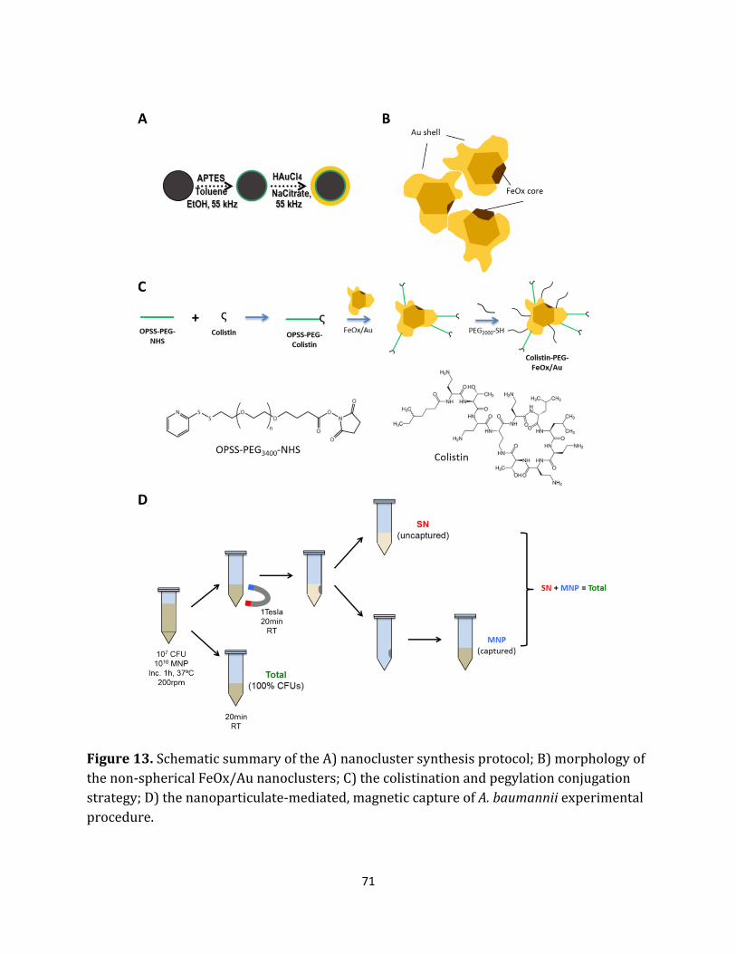
71
Figure 13. Schematic summary of the A) nanocluster synthesis protocol; B) morphology of the non-spherical FeOx/Au nanoclusters; C) the colistination and pegylation conjugation strategy; D) the nanoparticulate-mediated, magnetic capture of A. baumannii experimental procedure.
72
environment. Oleic acid capped γ-Fe2O3 nanoparticles (10 mg) were dissolved into a 1%
wt (3-Aminopropyl)triethoxysilane (APTES, Sigma-Aldrich) solution in anhydrous toluene.
This volume was reacted under ultrasonic agitation (90 min, 20°C) and vortexed
intermittently at 15 minute intervals to ensure homogenous mixing. Using a 1T
neodymium 1” cube magnet, the aminated γ-Fe2O3 were magnetically separated from the
toluene based solvent and washed with anhydrous ethanol (EtOH, 200 proof, Sigma-
Aldrich). Aminated γ-Fe2O3 nanoparticles (10 mg/ml, 1ml) were stored at 4°C.
Gold Sonoplation of Aminated γ-Fe2O3 Core Nanoparticles: Au-(γ-Fe2O3) composite
nanoclusters (FeOx/Au) were fabricated via an electroless plating reaction in an ultrasonic
agitation reaction environment. Adapted from a protocol described previously .(Bell et al.
2011), an aqueous 1% HAuCl4 (150 μl, Sigma-Aldrich) solution was mixed with an
ethanolic solution of aminated γ-Fe2O3 nanoparticles (1 mg/ml, 2 ml). Under an ultrasonic
agitation environment, 12 mL of aqueous sodium citrate (20 mM, Sigma-Aldrich) was
added in 800 μL increments every 30 seconds. The resultant FeOx/Au nanoclusters were
centrifuged (800 rcf, 10 min). The supernatant was extracted while the pellet was
preserved via a 1T neodymium magnet. The pellet was resuspended into 10 ml of dH2O
and the resultant FeOx/Au nanoclusters (200 μg/mL) were stored at 4°C.
Conjugation of colistin antibiotic to heterobifunctional PEG linker: Colistin (polymyxin-E;
Sigma-Aldrich) was conjugated to ortho-pyridine disulfide, N-hydroxysuccinimide
heterobifunctional polyethylene glycol polymer linker (OPSS-PEG3400-NHS; Creative PEG
Works) using a protocol adapted from Nanobiotechnology Protocols (L. Hirsch JL. West
73
2005). In a 100 mM NaHCO3 solution, OPSS-PEG3400-NHS (0.044 mg/ml, 9 mL) was mixed
with colistin (0.2 mg/ml; 900 μL) and allowed to react for 2 h at 20°C. OPSS-PEG3400-
colistin conjugates were stored at 4°C for further use for no more than 24 h. Fresh OPSS-
PEG3400-colistin conjugates were prepared for conjugations separated by 24 h or more.
Functionalization of FeOx/Au nanoclusters with OPSS-PEG3400-colistin conjugates and
PEG2000-SH: FeOx/Au nanoclusters were functionalized with OPSS-PEG3400-colistin
conjugates and thiolated PEG (PEG2000-SH; Laysan Bio, Inc.) using protocols adapted from
Nanobiotechnology Protocols (L. Hirsch & JL. West 2005). To prepare Col-PEG-FeOx/Au
nanoclusters, FeOx/Au nanoclusters (200 μg/mL; 4 ml) were mixed with OPSS-PEG3400-
colistin (100 μL; 18.8 μg/ml) for 1 h then mixed with PEG2000-SH (100 μL; 15.6 mg/mL) for
an additional hour. Simultaneously, PEG-FeOx/Au nanoclusters were prepared by mixing
FeOx/Au nanoclusters (200 μg/mL; 4 ml) with PEG2000-SH (100 μL; 15.6 mg/mL) for 1 h
then mixed with dH2O for an additional hour. To purify, both preparations were
centrifuged at 800 rcf for 10 min. The supernatant was removed using magnetic
separation to preserve the pellet and saved for unconjugated colistin enumeration. The
pellet was then resuspended in a 70% aqueous EtOH sterilization solution to restrict
bacteria contamination and promote sterility. Preparations were stored at 4°C until
utilized for bacterial separation experiments and characterization no more than 24 hours
thereafter.
Nanocluster Characterization: Both conjugated and unconjugated FeOx/Au nanoclusters
were characterized similarly. For morphological analysis, standard TEM micrographs with
74
EDX (energy dispersive x-ray spectroscopy) elemental analysis were obtained on an FEI
Tecnai Osiris S/TEM Microscope (FEI, Hillsboro, OR). Spectroscopic extinction
measurements were performed in dH2O using a Varian Cary 50 UV-vis-NIR
spectrophotometer (Agilent Technologies, Santa Clara, CA). Relaxometric measurements
were obtained on a Maran DRX-II 0.5T NMR spectroscopic scanner following sample
preparation in Nanopure dH2O at a diluted nanocluster concentration (40 μg/mL; 1 mL).
Spectroscopic T2 relaxation measurements were performed in a Carr-Purcell-Meiboom-Gill
(CPMG) sequence with the following parameters: relaxations time (TR) = 30 s, echo time
(TE) of first echo = 6 ms, spacing of subsequent echos = 2 ms, and 1200 echos. Size and
zeta potential measurements were performed on a Malvern Zetasizer (Malvern
Instruments, Westborough, MA) following nanoparticulate dilution (40 μg/mL; 1 mL) in
dH2O. The size of the individual nanoparticles which comprise the nanoclusters were
estimated from TEM images using ImageJ software.
Confirmation of ligand conjugation: A Colistin ELISA Test Kit (Bioo Scientific, Austin, TX)
was used to detect and quantify free, unconjugated colistin in the supernatant of the
reaction volume. The kit was used as prescribed by the manufacturer with no deviation
from the provided protocol.
1H NMR spectroscopy was used to confirm colistin conjugation to the FeOx/Au
nanoclusters. 1H NMR spectrums were obtained in deuterium oxide (D2O) and measured
using a Bruker 600 MHz spectrometer (Bruker, 600 MHz, Billerica, MA). Freshly
conjugated Col-PEG-FeOx/Au nanoclusters (2 mL, 2 mg/mL) were lyophilized for 24 h. The

75
sample was resuspended in 200 μL of D2O to prepare for the iodine-catalyzed nanocluster
decomposition reaction (iodination).
Briefly, four to seven iodine crystals were added directly to suspension of nanoclusters.
The iodinated sample was vortexed (10s, 100%) intermittently for 10 s, every 20 min, for 1
h. Following a nucleophilic substitution (place exchange reaction) between the iodine
molecules and the sulfur groups of the colistin-bound conjugate, the nanoclusters
spontaneously settled via precipitation out of solution. This event caused the colistin-
bound conjugates and free iodine to be the only remaining reagents in the solution. The
sample was stored at 20°C for 12 h to allow the iodine crystals to settle to the bottom of the
suspension. Only colistin-bound conjugates remained in the D2O suspension which was
measured using 1H NMR within 24 h.
Confirmation of nanocluster binding to A. baumannii: STEM-EDS (energy dispersive X-ray
spectrometry) micrographs of bacteria-nanocluster aggregates were also obtained from an
FEI Tecnai Osiris S/TEM Microscope using the following fixation protocol. Briefly,
following an 1 h incubation with A. baumannii in 1X PBS, FeOx/Au nanoclusters bound to
bacteria were centrifuged at 12,400 RPM for 5 min. The supernatant was discarded and
pellet was resuspended in sterile dH2O to wash the nanocluster-bound bacteria. The
washed sample was centrifuged at 12,400 RPM for 5 min and resuspended in 2%
formaldehyde for 30 min at 20°C. Approximately 2 μL of the nanocluster-bound bacteria
was dropcast onto carbon film-supported cooper grids (Ted Pella, Inc., Redding, CA) and
blotted to dry. Samples were allowed to air dry at 20°C for 2 h prior to imaging. X-rays

76
emitted from the specimen during EM imaging procedures were collected and analyzed for
their elemental composition in ESPRIT imaging software.
Cell culture: HepG2 cells (human liver carcinoma cancer cell line) used in this study were
cultured in Dulbecco's modified Eagle's medium (DMEM, Gibco Cell Culture, Carlsbad, CA)
supplemented with 10% fetal bovine serum (FBS, Gibco) at 37°C in a humidified
atmosphere containing 5% CO2. HUVEC cells (human umbilical vein endothelial cell line)
used in this study were cultured in vascular basal cell medium (ATCC PCS-100-300,
Manassas, VA) supplemented with endothelial cell growth kit-VEGF (ATCC PCS-100-41) at
37°C in a humidified atmosphere containing 5% CO2.
In vitro cytotoxicity studies: The cytotoxicity induced by Col-PEG-FeOx/Au nanoclusters,
PEG-FeOx/Au nanoclusters, and free colistin on mammalian cell cultures was determined
using the alamarBlue (Invitrogen Co., Carlsbad, CA) assay. HepG2 and HUVEC cells were
seeded in clear, flat 96-well plates at a density of 10,000 cells/well in 200 μL of medium
and incubated for 24 h at 37°C. Next, cells were treated with free colistin at concentrations
of 900, 90, 70, 15, 1 and 0 μM. Similarly, culture medium was removed and exchanged with
medium containing fresh Col-PEG-FeOx/Au and PEG-FeOx/Au at concentrations of 2, 1, 0.5,
0.25, and 0 mM, respectively. After incubation for 24 h at 37°C, cells were washed thrice
with phosphate buffer solution (PBS), pH 7.4 and treated with 200 μL of alamarBlue
containing media. Cells were then incubated for an additional 4 h at 37°C. Fluorescence was
quantified using a plate-reader (Tecan, Infinite M1000 Pro, Switzerland), with an excitation
wavelength of 540 nm and emission wavelength of 590 nm.

77
In vitro hemocompatibility studies: Human whole blood was collected from anonymous,
consenting human donors in accordance with an approved Institutional Review Board
(IRB) protocol (IRB 111251). Red blood cells (RBCs) were isolated according to well-
established protocols (Evans et al. 2013). Four different concentrations (1, 5, 40, and 200
ug/mL) of Col-PEG-FeOx/Au and PEG-FeOx/Au nanoclusters were prepared in PBS, pH 7.4,
respectively. Free colistin (900, 0.9, 0.4, 0.2, and 0 μM) was also prepared in PBS, pH 7.4.
Col-PEG-FeOx/Au, PEG-FeOx/Au, and free colistin were incubated with RBCs for 1h at 37°C,
each respectively, at the concentrations outlined above. Following incubation, the samples
were centrifuged and the supernatant was spectrophotometrically analyzed for
hemoglobin release using a plate-reader (Tecan, Infinite M1000 Pro) at 451 nm in order to
determine percent hemolysis relative to the positive control (Triton X-100 detergent). The
negative control used was PBS, pH 7.4. Percent of hemoglobin release was calculated
according to Equation 1.
𝐻𝐻𝑚𝑠𝐻𝐻𝑠𝐻𝐻𝐻 𝑟𝐻𝐻𝐻𝑠𝑠𝐻 % = (𝑆𝑆𝑆𝑆𝑆𝑆451 𝑛𝑛−𝑁𝑆𝑁𝑆𝑁𝑁𝑁𝑆 𝑐𝑐𝑐𝑁𝑐𝑐𝑆451 𝑛𝑛)(𝑃𝑐𝑃𝑁𝑁𝑁𝑁𝑆 𝑐𝑐𝑐𝑁𝑐𝑐𝑆451 𝑛𝑛−𝑁𝑆𝑁𝑆𝑁𝑁𝑁𝑆 𝑐𝑐𝑐𝑁𝑐𝑐𝑆451 𝑛𝑛)
× 100% (1)
Bacterial cell culture: 50 mL of LB broth (RPI, Mt. Prospect, IL) were inoculated with a
single colony of A. baumannii (ATCC 17978) using standard sterile techniques. The culture
was incubated overnight (18 h) at 37 ºC and agitated at 200 rpm in an orbital shaker.
Before using the bacteria for experiments, the culture was diluted 1/100 in fresh LB broth
and re-incubated in the same conditions for 2 hours.
78
Bacterial susceptibility tests: For survival analysis, FeOx/Au nanoclusters and A. baumannii
were mixed and incubated in LB. FeOx/Au nanoparticle concentration (both Col-PEG-
FeOx/Au and PEG-FeOx/Au) was varied from 0 to 1011 nanoparticles/mL, while the
concentration of A. baumannii was adjusted to be 106 CFU/mL in all samples. After a 1-hour
incubation at 37 ºC and agitation at 200 rpm in an orbital shaker, cultures were diluted,
plated on LB agar plates, and incubated for 18 h at 37 ºC. The percentage of surviving cells
compared to untreated controls was determined by CFU counting. A. baumannii
susceptibility to colistin was also analyzed using concentrations from 0 to 2 µg/mL, and
following the same protocol.
Bacterial magnetic capture: To determine the magnetic capture efficiency, FeOx/Au
nanoclusters (both Col-PEG-FeOx/Au and PEG-FeOx/Au) at 1010 nanoclusters and A.
baumannii at 107 CFU in 1 ml PBS were mixed and incubated for 1 h at 37°C and agitated at
200 rpm in an orbital shaker. Magnetic capture process is summarized in Figure 13. After
incubation, each sample was divided into two tubes (500 µl each). One of them was kept at
RT, as a control of the total concentration of bacteria after incubation (-MF tubes). A
permanent magnet (1 Tesla) was placed on the side of the other tube for 20 min at RT.
After magnetic separation, supernatants were collected (+MF SN tubes), and nanoclusters
were resuspended in 500 µl PBS (+MF NP tubes). 50 µl alamarBlue were added to each
tube. All samples were then incubated for 30 min at 37°C, centrifuged, and supernatants
were analyzed using a plate reader (Tecan, Infinite F500). Fluorescence was quantified
with an excitation wavelength of 535 nm and emission wavelength of 590 nm, and CFU/mL
in each sample was determined using a standard curve that correlated the number of
79
CFU/mL with the fluorescence intensity obtained from the alamarBlue assay. The standard
curve was generated and processed in parallel with all the samples. The percentage of
magnetic capture was determined by comparison of the CFU/ml in the -MF tubes and the
+MF NP tubes.
Statistical Analysis: All experiments were performed in triplicate unless otherwise noted.
The results were expressed as the arithmetic mean ± standard deviation (SD). A two-sided,
unpaired t-test was used to determine statistical significance, with significance levels
ranging from p < 0.05 to p < 0.001.
80
Results and Discussion
Colistinated FeOx/Au nanoclusters, utilized as a nanotechnology-enabled platform, were
used to successfully capture and magnetically extract A. baumannii from saline solution. As
shown in Figure 14, colistinated nanoclusters separated 28.8±7.15% of A. baumannii in the
sample, whereas the PEGylated control nanoclusters separated only 9.65±3.74% of the
bacteria. The difference in percent capture was highly significant (p<0.001) and suggests
that the presence of colistin on the surface of the nanomaterials enhanced both selectivity
and binding. This enhanced selectivity and binding is significantly influenced by
nanoparticulate surface charge which is mediated by the presence of surface conjugates.
This conjugate-mediated selectivity is supported by the 2-fold positive difference in zeta
potential between colistinated and PEGylated nanoclusters as shown in Table 3.
Bacteria capture was further elucidated with increased granularity in Figure 15.
Performance of capture was performed at a “per tube” level of quantification to confirm
that results were influenced by separation and neither by cell death nor some other
unforeseen variable. It was expected that significant differences between the number of
separated bacteria, per particulate population, should correlate to significant differences
between bacteria remaining in the supernatant. Notably, there were highly significant
differences between the captured and uncaptured bacteria populations internal and
between the PEGylated and colistinated nanoclusters treatments (***p<0.001, n=6).
Further, significant differences between supernatant CFU counts as well further validate
this strategy, as expected. Furthermore, summation of separated and remaining bacteria
should equal the total number of bacteria in the original sample.

81
Figure 14. Quantification of macroscale Acinetobacter baumannii separation by FeOx/Au nanoclusters. Results depict a highly significant difference in the magnetic capture percentage between colistinated (M=28.8, SD=7.15) and PEGylated (M=9.65, SD=3.74) nanoclusters; (***p<0.001; t(5)=0.000499; n=6).

82
Figure 15. Comparative quantitative analysis of macroscale Acinetobacter baumannii separation by FeOx/Au nanoclusters, pure tube. Notably, there were highly significant differences between the captured and uncaptured bacteria populations internal and between the captured and uncaptured bacteria populations internal and between the PEGylated and colistinated nanoclusters treatments (***p<0.001, n=6). The combination of the CFU/ml between the captured and uncaptured tubes yielded values near 100% (104.6% and 103.4% for PEG-FeOx/Au and Colistin-PEG-FeOx/Au, respectively) for both nanocluster treatments; thus indicating the conservation of bacterium throughout the entire separation procedure. This result is of key importance as it demonstrates that the differences between the captured and uncaptured samples is attributed to nanoparticulate-mediated magnetic separation and not an unaccounted for loss of bacteria in the samples (e.g. cell death).
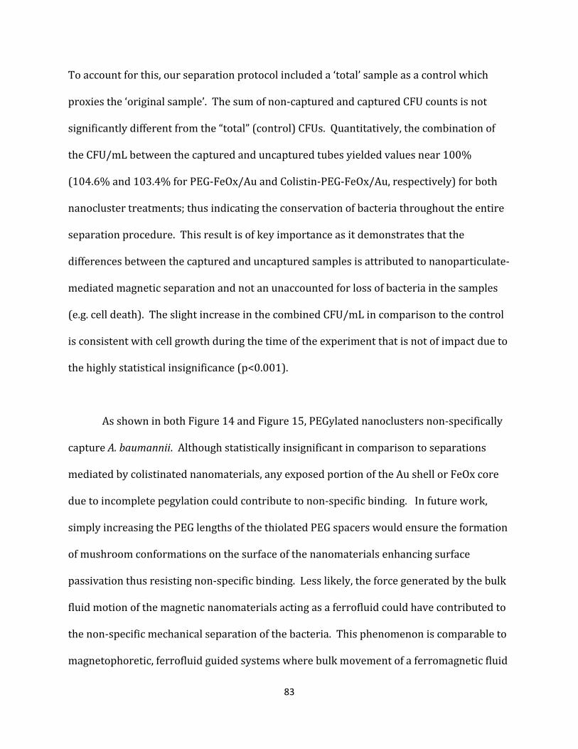
83
To account for this, our separation protocol included a ‘total’ sample as a control which
proxies the ‘original sample’. The sum of non-captured and captured CFU counts is not
significantly different from the “total” (control) CFUs. Quantitatively, the combination of
the CFU/mL between the captured and uncaptured tubes yielded values near 100%
(104.6% and 103.4% for PEG-FeOx/Au and Colistin-PEG-FeOx/Au, respectively) for both
nanocluster treatments; thus indicating the conservation of bacteria throughout the entire
separation procedure. This result is of key importance as it demonstrates that the
differences between the captured and uncaptured samples is attributed to nanoparticulate-
mediated magnetic separation and not an unaccounted for loss of bacteria in the samples
(e.g. cell death). The slight increase in the combined CFU/mL in comparison to the control
is consistent with cell growth during the time of the experiment that is not of impact due to
the highly statistical insignificance (p<0.001).
As shown in both Figure 14 and Figure 15, PEGylated nanoclusters non-specifically
capture A. baumannii. Although statistically insignificant in comparison to separations
mediated by colistinated nanomaterials, any exposed portion of the Au shell or FeOx core
due to incomplete pegylation could contribute to non-specific binding. In future work,
simply increasing the PEG lengths of the thiolated PEG spacers would ensure the formation
of mushroom conformations on the surface of the nanomaterials enhancing surface
passivation thus resisting non-specific binding. Less likely, the force generated by the bulk
fluid motion of the magnetic nanomaterials acting as a ferrofluid could have contributed to
the non-specific mechanical separation of the bacteria. This phenomenon is comparable to
magnetophoretic, ferrofluid guided systems where bulk movement of a ferromagnetic fluid
84
in a magnetic field could trap nonmagnetic particulates (Asmatulu et al. 2010). Future work
also includes modulating the nanoparticle to bacteria ratio to determine if the nanoparticle
concentration has a significant impact on the nonspecific ferrofluid separation potential.
The clustering effect of the nanomaterials is apparent upon binding to the surface of
gram-negative A. baumannii as shown in the STEM-EDS micrograph map in Figure 16A.
The presence of the bacterium is supported by the detection of phosphorous within the
cell. The location of the nanoclusters is substantiated by the co-localized detection of iron
and gold. We suggest that the binding of the nanoclusters is mediated by colistin as shown
with proposed biochemical detail in Figure 16B. The electrostatic interaction of the colistin
with the phosphate groups occurs due to the presence of positively-charged primary amine
groups. These positively-charged groups, especially those in the lariat of the molecule,
displace divalent cations (Mg2+ and Ca2+) which under normal conditions stabilize the
membrane lipids(Clifton et al. 2015), which compose the cell envelope of the bacterium. It
can be presumed that multiple colistin-lipid electrostatic interactions generate enough
force to resist detachment during magnetic separation. Furthermore, multiple
nanoparticulate binding events further multiplies this binding force.
The lipophilic portion of the colistin molecule penetrates the lipid layer of the
bacterium; however, only at high colistin concentrations will enough of these interactions
destabilize the lipid bilayer of the bacterium to the point of cell lysis. Even further, at high
enough nanoparticle concentrations, the nanoparticles begin to impinge on the cellular
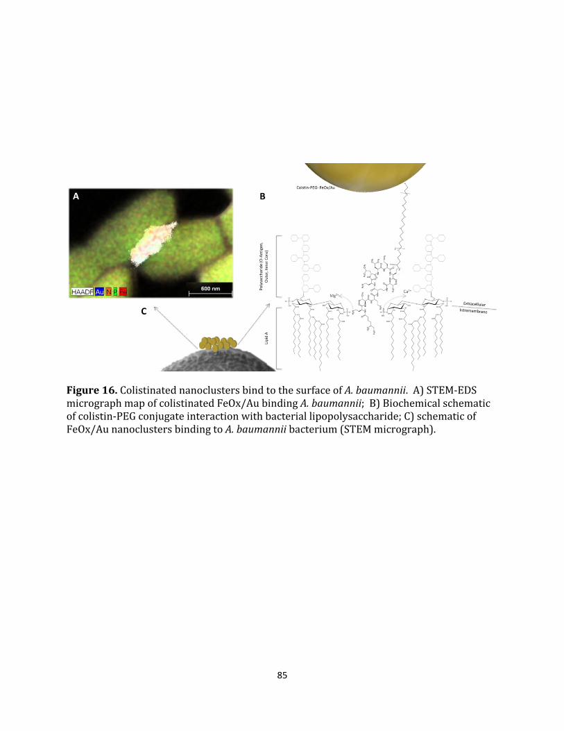
85
Figure 16. Colistinated nanoclusters bind to the surface of A. baumannii. A) STEM-EDS micrograph map of colistinated FeOx/Au binding A. baumannii; B) Biochemical schematic of colistin-PEG conjugate interaction with bacterial lipopolysaccharide; C) schematic of FeOx/Au nanoclusters binding to A. baumannii bacterium (STEM micrograph).
86
function of the bacterium by disrupting the cell membrane in a dose-dependent manner
(Seil & Webster 2012). Due to this, the highest nanoparticle to bacterium ratio that could
be tolerated by the bacteria had to be determined. The dose-dependent bacterial toxicity of
both PEGylated and colistinated nanoclusters with A. baumannii in vitro is described
quantitatively in Figure 17. PEGylated nanomaterials were nontoxic until becoming
significantly toxic (p<0.05) when nanoparticle to bacteria ratios exceeded 10000:1.
Colistinated particles were nontoxic in comparison to untreated populations until
becoming significantly toxic (p<0.05) in ratios which exceeded 10000:1. More importantly,
this data supports the chosen 1000:1 nanoparticle to bacterium ratio for the capture
experiments. From a therapeutic standpoint, cell lysis is to be avoided as bacterium release
cytotoxic agents into the bloodstream or into the local, internal environment during death.
In future devices, blood return to the patient should remain free of bacteria as well as free
of the remains of lysed cells.
At 1000:1 nanoparticle to bacterium ratios (NP:b), the greatest capture percentage
achieved was 38.5% with a minimum of 21.7%. Magnetic separations at these magnitudes
are acceptable for adjuvant therapies. However, we believe magnetic separations
exceeding 90% are achievable. Modification of the NP:b ratios may increase magnetic
separation. Augmentation of these ratios may require the simultaneous adjustment of
colistin surface density. Increasing the number of nanoparticles present in the sample
further increases the total concentration of colistin potentially impinging bacterial
survivability. Future directions include optimizing the balance between these ratios and
colistin surface coverage to maximize magnetic separation. Further, variation of separation

87
duration and an introduction of the sample into mesofluidic fluid flow may enhance
bacterial separation percentages.
Figure 17. Bacterial toxicity of PEGylated and colistinated nanoclusters with A. baumannii in vitro. PEGylated nanomaterials were nontoxic until becoming significantly toxic (p<0.05) when cell:NP ratios exceeded 1:10000. In comparison to untreated populations, colistinated particles were nontoxic until becoming statistically toxic (p<0.05) in ratios which exceeded 1:10000.
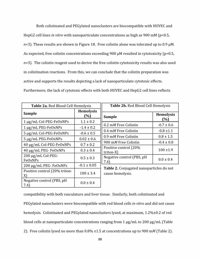
88
Both colistinated and PEGylated nanoclusters are biocompatible with HUVEC and
HepG2 cell lines in vitro with nanoparticulate concentrations as high as 900 mM (p<0.5,
n=3). These results are shown in Figure 18. Free colistin alone was tolerated up to 0.9 µM.
As expected, free colistin concentrations exceeding 900 μM resulted in cytotoxicity (p<0.5,
n=3). The colistin reagent used to derive the free colistin cytotoxicity results was also used
in colistination reactions. From this, we can conclude that the colistin preparation was
active and supports the results depicting a lack of nanoparticulate cytotoxic effects.
Furthermore, the lack of cytotoxic effects with both HUVEC and HepG2 cell lines reflects
compatibility with both vasculature and liver tissue. Similarly, both colistinated and
PEGylated nanoclusters were biocompatible with red blood cells in vitro and did not cause
hemolysis. Colistinated and PEGylated nanoclusters lysed, at maximum, 1.2%±0.2 of red
blood cells at nanoparticulate concentrations ranging from 1 µg/mL to 200 µg/mL (Table
2). Free colistin lysed no more than 0.8% ±1.5 at concentrations up to 900 mM (Table 2).
Table 2a. Red Blood Cell Hemolysis
Sample Hemolysis (%)
1 μg/mL Col-PEG-FeOxNPs 1.1 ± 0.2 1 μg/mL PEG-FeOxNPs -1.4 ± 0.2 5 μg/mL Col-PEG-FeOxNPs -0.6 ± 0.5 5 μg/mL PEG-FeOxNPs 0.02 ± 0.6 40 μg/mL Col-PEG-FeOxNPs 0.7 ± 0.2 40 μg/mL PEG- FeOxNPs 0.3 ± 0.4 200 μg/mL Col-PEG- FeOxNPs 0.5 ± 0.3
200 μg/mL PEG- FeOxNPs -0.1 ± 0.05 Positive control (20% triton-X) 100 ± 3.4
Negative control (PBS, pH 7.4) 0.0 ± 0.4
Table 2b. Red Blood Cell Hemolysis
Sample Hemolysis (%)
0.2 mM Free Colistin -0.7 ± 0.6 0.4 mM Free Colistin -0.8 ±1.1 0.9 mM Free Colistin 0.8 ± 1.5 900 mM Free Colistin -0.4 ± 0.8 Positive control (20% triton-X) 100 ±1.9
Negative control (PBS, pH 7.4) 0.0 ± 0.4
Table 2. Conjugated nanoparticles do not cause hemolysis.

89
These cytocompatibility results are of great importance with regard to future development
of a robust therapeutic. Failure to retain the magnetic nanomaterials and allowing them to
re-enter the patient is a major failure mode of an extracorporeal device. Upon entry into
the patient, the nanomaterials would have immediate interaction with the vascular tissue
and would accumulate in the liver because of their size and charge (Longmire et al. 2008).
Due to their cytocompatibility with these tissues, catastrophic failure of an extracorporeal
device would be better tolerated by a patient in comparison to devices which utilize
nanomaterials which are cytotoxic. The hemolysis results demonstrate that colisitnated
nanomaterials have the potential to cohabit with patient blood for 1 hr with no adverse risk
of hemolysis. Alleviation of strict ~O(min) flow durations aid in the development of
efficient extracorporeal devices which maximize the removal of bacteria and enhance
patient outcomes.
Following synthesis, the nanomaterials were fully characterized for their elemental
composition and their optical, magnetic and morphological properties. Following
sonicatory amination of γ-Fe2O3 nanoparticles, there were no significant changes in the
morphology of the nanomaterials (Figure 19A) where the materials remained
monomorphous. However, after gold sonoplation, the morphology of the nanoparticles
became polymorphous. While there appear to be many spherical particles, the formation of
jagged, oblong and elliptical particles is apparent following sonoplation (Figure 19Aii).
Furthermore, these polymorphous particles tended to cluster together forming on the
order of ~O(100 nm) clusters of core/shell nanomaterials.

90
Figure 18. Cytocompatibility results showing toxicity of free colistin, pegylated, and colistinated nanoclusters in HUVEC and HepG2 cells in vitro. Both pegylated and colistinated particulate populations caused no significant toxicity in both HUVEC and HepG2 cells in vitro. Both pegylated and colistinated particulate populations caused no significant toxicity in both HUVEC and HepG2 cells (p<0.05, n=3).
91
The formation of these particle morphologies is likely due to insufficient amounts of gold
chloride in the reaction volume. Although troublesome for therapeutic or diagnostic
strategies that require spherical gold-clad particles, our strategy requires nanomaterials
which are magnetomotive. Core/shell nanomaterials with significant shell thicknesses
have diminished magnetic properties as shown in Equation 2. This equation describes the
saturation magnetization, the maximum magnetization a particle can experience, Msat of a
single core/shell magnetic nanoparticle where the magnetic properties of the particle stem
from the core which is coated with a thin outer shell of magnetically inert material.
𝑀𝑃𝑆𝑁 = 𝑚𝑀𝑃𝑐 �𝑐−𝑑𝑑�3
(2)
where Mso is the saturation magnetization of the bulk material in units of A/m/g, m is the
mass of the individual particle, r is the particle radius, and d is shell thickness. Any
significant increase in the shell thickness, d, has significant detrimental effect on the
saturation magnetization of the particle. From this equation, we concluded that minimizing
the shell thickness in order to maximize the magnetic properties, and thus the probability
of magnetomotion in the presence of a magnetic field, of the nanomaterials was paramount.
Following statistical analysis of nanoparticle diameters, ~1.75nm gold shell thicknesses
were observed. It has been previously described that as magnetic nanomaterials cluster
together and form nanocluster units, the magnetization force of the nanocluster increases
(Ortega & Giorgio 2012). Here, this phenomenon increased the magnetization saturation of
the nanoclusters thus increasing their magnetomotive potential. This increase in
magnetization is supported by the decrease in the magnetic relaxivity of the FeOx/Au

92
nanoclusters shown in Table 3. This decrease in relaxivity suggests a higher nanocluster
magnetomotive potential; thus increasing the probability that the nanomaterials will be
able to effectively separate bacteria upon introduction into a magnetic field.
The optical properties of the nanomaterials change significantly upon the
sonochemical deposition , sonoplation, of gold to the surface of the γ-Fe2O3 nanoparticles.
Appearance of a surface plasmon resonance peak in the visible range of the spectrum is a
clear indicator of the formation of gold clad nanomaterials. Following conjugation with
PEG and colistin, the absorbance peak blue-shifts by ten nanometers signaling a change in
the surface plasmon resonance. This shift in the surface plasmon resonance is expected
as it signals that a chemical conjugation or ligand binding event has occurred on the gold
surface of the nanomaterial (Ghosh et al. 2004). The presence FeOx/Au core/shell
nanoparticles following synthesis was substantiated via HRTEM-EDS of an isolated
nanoparticle. The micrograph in Figure 19C shows an isolated nanoparticle with 2.4Å
fringe patterns – fringe spacing indicative of a gold clad material. This single nanoparticle
was characterized for its elemental composition via the EDS functionality of the S/TEM-
EDS microscope. Following 8 minutes of x-ray collection, the EDS spectrum (Figure 19D)
contained gold, as expected, but also both iron and silicon. The detection of elemental iron
and silicon substantiates the existence of both an iron core and the intermediary
aminosilane layers which comprise the core/shell nanoparticle.

93
Figure 19. FeOx/Au nanoparticulate characterization confirming nanomaterial synthesis. A) TEM micrographs of FeOx nanoparticles, (i) aminated FeOx nanoparticles, and (ii) gold-clad iron oxide (FeOx/Au) nanoparticles; B) UV-vis-NIR spectra of (i) FeOx-NH2, (ii) FeOx/Au, (iii) colistin-PEG-FeOx/Au, (iv) PEG-FeOx/Au; C) HRTEM micrograph of single FeOx/Au core-shell nanoparticles depicting Au lattice fringe patterns; D) EDS spectra of single FeOx/Au core-shell nanoparticles shown in (C) depicting the expected elemental presence of both Fe, Si and Au.

94
The dual, magnetoplasmonic nature of these nanomaterials enables their usefulness
in theranostic applications. Extracorpeal devices, while designed foremost to be
therapeutic devices, can also be utilized to specifically detect the species of bacteria leading
to rapid diagnosis. In the future, such a theranostic approach could be implemented in a
multiplexed fashion on patients who present with septic-like symptoms but remain
undiagnosed due to diagnostic limitations. Such a device could act as a “broad-spectrum”
therapeutic designed to extract a multitude of species while each species specific
nanomaterial could be a unique probe that identifies the extracted bacteria for early
diagnostic purposes.
The conjugation of colistin to the surface of the FeOx/Au nanoclusters was
confirmed. Following conjugation, the 2-fold, statistically significant (p<0.05) positive shift
Sample Peak λ (nm)
NP Dia. (nm)
Nanocluster Hydrodyn. radii (nm)
Zeta Pot. (mV)
Relaxivity (ms)
(i) FeOx-NH2 - 11.3±1.8 - +32.0±3.0 152.6±3.2 (ii) FeOx/Au 564 13.0±1.6 296.8±7.0 -34.7±0.7 121.7±1.0
(iii) FeOx/Au-Colistin 553 13.0±1.6 244.1±2.1 -10.5±0.3 148.0±1.8
(iv) FeOx/Au-PEG 552 13.0±1.6 275.6±5.1 -31.2±0.4 132.7±2.0
Table 3. Conjugated and Unconjugated Nanoparticle Characterization.
95
in zeta potential between colistinated and pegylated nanoclusters is an indicator of
conjugation as the colistin molecule has a higher positive charge than PEG (Table 3). There
was also a significant positive shift in zeta potential of both conjugated particle populations
when compared to the unconjugated particles, further confirming conjugation. The iodine-
catalyzed decomposition of the colstinated nanomaterials ensured that all bound colistin
would be liberated into solution from the gold surface. Thereafter, the iodinated gold
flocculated, losing its colloidal properties and settled in the tube. The inset of Figure 20A
depicts this observation, where colloidal, colistinated FeOx/Au nanoclusters (left) are
compared to iodinated gold with the colistin conjugates liberated into solution. 1H NMR
spectra of free colistin shows strong methyl-methyl peaks around 0.8-0.9 ppm whereas
primary amine groups are detected around 8 ppm. We expected to see a ppm shift in the
spectra due to diamagnetic shielding. The additional electrons surrounding the colistin,
which are contributed by the bound polymer, shield the molecule which causes a 0.5-1ppm
shift in the resultant spectrum. As shown in the ppm in Figure 8A, 1H NMR spectra of the
supernatant of this reaction depicts the presence of colistin substantiated by the
appearance of methyl-methyl peaks (0.5 -1 ppm). Most importantly, the appearance of
primary amines primary amines (8-8.5ppm) as shown in Figure 20B substantiates the
presence of liberated colistin.
To ensure no free colistin was contributing to the 1H NMR results, a free colistin
ELISA kit was performed on the first-wash supernatants of both colistination and
pegylation conjugation reactions. Per the ELISA kit, there was no free colistin in the sample
as the returned concentration (0.0015 ng/ml) was below the standard curve
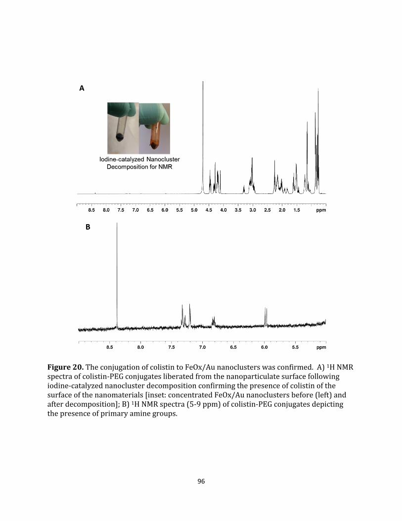
96
Figure 20. The conjugation of colistin to FeOx/Au nanoclusters was confirmed. A) 1H NMR spectra of colistin-PEG conjugates liberated from the nanoparticulate surface following iodine-catalyzed nanocluster decomposition confirming the presence of colistin of the surface of the nanomaterials [inset: concentrated FeOx/Au nanoclusters before (left) and after decomposition]; B) 1H NMR spectra (5-9 ppm) of colistin-PEG conjugates depicting the presence of primary amine groups.
97
recommendations (0.1 ng/ml) suggested by the manufacturer. As expected, there was no
colistin detected in PEGylated supernatants as the returned concentration was below the
concentration detected in the blank sample. Even further, 1H NMR detection of colistin at
concentrations less than 10 µg/mL was extremely difficult and would not have yielded
detectable signal at the levels detected by the ELISA kit.
Conclusion
We have successfully demonstrated that superparamagnetic FeOx/Au core/shell
nanoparticles, which form magnetic nanoclusters, can be used to magnetically capture and
separate Acinetobacter baumannii using colistin as the microbial targeting ligand. The
nanomaterials were successfully synthesized and characterized for their
magnetoplasmonic properties and potential for use in theranostic applications. Successful
conjugation was confirmed and nanocluster binding to the bacterial surface was observed.
This result, for the first time, demonstrates a robust, pharmaceutical-based motif for high
affinity nanoparticulate targeting to the A. baumannii surface. The antibiotic-activated
nanomaterials demonstrated cytocompatibility with human cells, bacterial tolerance below
1:10000 bacteria to nanoparticle concentrations, and bacterial toxicity at higher levels.
With maximal separation efficiencies exceeding 38%, this nanotechnology-mediated
platform is well suited for adjuvant therapies which assist in the treatment of sepsis.
These results reveal an opportunity for the development of an adjuvant treatment
option for patients suffering from Acinetonacter baumannii bacteremia and other gram-
negative bacteria. Possible adjuvant treatments include the development of extracorporeal

98
devices to mechanically remove bacteria while intravenous injections of the antibiotic
work concurrently to stifle the systemic infection. Our development and characterization
of a robust nanoparticulate platform for targeting and capture of Acinteobacter baumannii
in physiological phantoms paves the way for the translational development of such an
adjuvant treatment. Furthermore, due to the dual, magnetophotonic nature of the
nanotechnology platform, a theranostic approach could be developed in a multiplexed
fashion for patients who present with septic-like symptoms but remain undiagnosed due to
diagnostic limitations. In the future, development of “broad-spectrum” theranostics as well
as other adjuvant therapies based on this antibiotic-mediated nanotechnology platform
will significantly improve the clinical outcomes of septic patients.
Summary
This Chapter confirmed the central hypothesis of my dissertation: inorganic, multilayered,
magnetoplasmonic core/shell nanomaterials were specifically designed and synthesized
for biological applications which required both detection and therapeutic elimination of a
disease causing agent. More specifically, this work successfully demonstrated that
superparamagnetic FeOx/Au core/shell nanoparticles, which form magnetic nanoclusters,
can be used to magnetically capture and separate Acinetobacter baumannii using
polymyxin E as the microbial targeting ligand. The nanomaterials were synthesized and
assessed for their magnetoplasmonic properties and potential for use in theranostic
applications. Importantly, for the first time, a robust, pharmaceutical-based motif for high
affinity nanoparticulate targeting to the A. baumannii surface was demonstrated.
99
Furthermore, the goals described in Aim 3, were achieved in Chapter IV. The
magnetoplasmonic core/shell nanoparticles fabricated in Aim 2 were biofunctionalized
with antibiotic, polymyxin E, which targets and binds gram negative bacteria. These
biofunctionalized nanostructures were characterized and utilized to magnetically extract
Acinetobacter baumannii from saline solution in vitro. These results support my hypothesis
that these nanomaterials can be fabricated in such a way that biomedical functionalization
of the material can proceed without detriment to the function of the biological component.
These results achieve our final objective where the activity of the conjugated biological
component was conserved while binding and extracting a disease causing agent, A.
baumannii, in vitro.
100
CHAPTER V
SYNOPSIS AND CONCLUSIONS
In my dissertation work, I worked towards the development of novel, multilayered,
magnetoplasmonic nanoconstructs to enable nanotechnology-mediated, theranostic
biomedical applications. In order to develop these materials, I overcame a number of
limitations which introduced unfavorable size constraints and limited resultant
nanoparticle characteristics. My trial biomedical application, the nanotechnology-
mediated removal of A.baumannii, in vitro, introduced further challenges. Nevertheless, I
were able to develop the core/shell nanoconstructs necessary to bind and extract A.
baumannii in a saline solution. While these nanoconstructs and the results of my in vitro
strategy do not provide a comprehensive, clinical theranostic solution, my work has
enabled us to advance in other ways.
As outlined in the chapters of this dissertation, we began by developing novel
multilayered, metallodielectric multistrata nanoparticles which possess theranostic
characteristics in a less than 60 nm diameter nanoparticulate embodiment (Chapter II).
Thereafter, we modified the synthesis strategy to enhance the magnetic properties of the
composite material (thus enabling magnetomotion) while decreasing the synthesis
duration such that freshly synthesized materials can be rapidly utilized in a biological
application (Chapter III). Finally, we confirmed the central hypothesis of my dissertation
that inorganic, multilayered, magnetoplasmonic core/shell nanomaterials could be
101
specifically designed and synthesized for biological applications which require both
diagnostic detection and therapeutic elimination of a disease causing agent. More
specifically, this work successfully demonstrated that superparamagnetic FeOx/Au
core/shell nanoparticles, which form magnetic nanoclusters, can be used to magnetically
capture, visualize and separate Acinetobacter baumannii using polymyxin E as the
microbial targeting ligand (Chapter IV). Combined and utilized, this work provides a
launch pad for the development of nanotechnology-mediated, theranostic labeling,
detection and magnetic removal of pathogens from blood – or other body fluid. Built into a
continuous-flow corporeal device or a batch-process, macrovolume filtering device, the
impact of this technology would be extensive and positively affect patient outcomes across
the world.
Moving forward, research challenges exist for future work seeking to achieve the
development of this technology. Next steps include: (1) optimization of the parameters of
the in vitro experiment to maximize, or achieve a greater than 90%, separation efficiency;
(2) iteration of the in vitro magnetic separation experiments in whole, non-diluted blood;
(3) development of mesofludic or batch/macroscale instrumentation to facilitate the
nanotechnology-mediated separation in an automated fashion in a mechanical
environment; (4) iteration of all experiments on robust in vivo models; and (5) escalation of
the technology for clinical translation. Furthermore, as an entrepreneur, I would be remiss
if I did not mention the local and global regulatory challenges governing the clinical rollout
of such a technology to both domestic and foreign patients – including military veterans.
102
Nevertheless, core/shell nanotechnology remains a promising platform to enhance
current biomedical diagnostics, therapeutics and the combination thereof; as supported by
the rapid growth and implementation of such technology in both academia and industry.
Further development of core/shell nanotechnology in biomedical applications will
continue to require novel approaches to optimize the composite characteristics of
nanoconstructs. These efforts will bolster the technological impact of these biomedical
applications thus improving the way we deliver medical care to patients across the globe
and beyond.

103
BIBLIOGRAPHY
Ali, M.E. et al., 2012. Gold nanoparticle sensor for the visual detection of pork adulteration in meatball formulation. Journal of Nanomaterials, pp.1–7.
Aronson, N.E., Sanders, J.W. & Moran, K.A., 2006. In harm’s way: infections in deployed American military forces. Clinical infectious diseases : an official publication of the Infectious Diseases Society of America, 43, pp.1045–1051.
Asmatulu, R., Zhang, B. & Nuraje, N., 2010. A Ferrofluid Guided System for the Rapid Separation of the Non-Magnetic Particles in a Microfluidic Device. JOURNAL OF NANOSCIENCE AND NANOTECHNOLOGY, 10, pp.6383–6387.
B. Brinson C. Levin, R. Bardhan, N. Mirin and NJ. Halas, J.L., 2008. Nanoshells Made Easy: Improving Au Layer Growth on Nanoparticle Surfaces. Langmuir, 24(24), pp.14166–14171.
Bardhan, R. et al., 2010. Nanosphere-in-a-nanoshell: A simple nanomatryushka. Journal of Physical Chemistry C, 114, pp.7378–7383.
Barnes, W.L., Dereux, A. & Ebbesen, T.W., 2003. Surface plasmon subwavelength optics. Nature, 424, pp.824–30. Available at: http://www.nature.com/nature/journal/v424/n6950/full/nature01937.html.
Bell, C. & Yu, S.S., 2011. Multistrata nanoparticles and methods for making multistrata nanoparticles.
Bell, C.S., Yu, S.S. & Giorgio, T.D., 2011. Multistrata Nanoparticles: The Multistrata Nanoparticle: an FeOx/Au Core/Shell Enveloped in a Silica–Au Shell (Small 9/2011). Small, 7(9), p.1157. Available at: http://dx.doi.org/10.1002/smll.201190031.
Bergogne-Bérézin, E. & Towner, K.J., 1996. Acinetobacter spp. as nosocomial pathogens: Microbiological, clinical, and epidemiological features. Clinical Microbiology Reviews, 9, pp.148–165.
Bordelon, H. et al., 2013. A magnetic bead-based method for concentrating DNA from human urine for downstream detection. PloS one, 8, p.e68369. Available at: http://www.pubmedcentral.nih.gov/articlerender.fcgi?artid=3704639&tool=pmcentrez&rendertype=abstract.
Bose, A. & Wui Wong, T., 2015. Nanotechnology Applications for Tissue Engineering, Available at: http://www.sciencedirect.com/science/article/pii/B978032332889000011X.

104
Brongersma, M.L., 2003. Nanoscale photonics: Nanoshells: gifts in a gold wrapper. Nature materials, 2, pp.296–297.
Cai, Y. et al., 2012. Colistin resistance of Acinetobacter baumannii: Clinical reports, mechanisms and antimicrobial strategies. Journal of Antimicrobial Chemotherapy, 67, pp.1607–1615.
Camp, C. & Tatum, O.L., 2010. A Review of Acinetobacter baumannii as a Highly Successful Pathogen in Times of War. Laboratory Medicine, 41, pp.649–657.
Chatterjee, K. et al., 2014. Core/shell nanoparticles in biomedical applications. Advances in Colloid and Interface Science, 209, pp.8–39. Available at: http://dx.doi.org/10.1016/j.cis.2013.12.008.
Chen, Y. et al., 2015. One-Step Detection of Pathogens and Viruses: Combining Magnetic Relaxation Switching and Magnetic Separation. ACS Nano, 9(3), pp.3184–3191. Available at: http://dx.doi.org/10.1021/acsnano.5b00240.
Cheng, X., Chen, G. & Rodriguez, W.R., 2009. Micro- and nanotechnology for viral detection. Analytical and Bioanalytical Chemistry, 393, pp.487–501.
Chibani-Chennoufi, S. et al., 2004. In vitro and in vivo bacteriolytic activities of Escherichia coli phages: Implications for phage therapy. Antimicrobial Agents and Chemotherapy, 48, pp.2558–2569.
Chung, B.G., Kang, L. & Khademhosseini, A., 2007. Micro- and nanoscale technologies for tissue engineering and drug discovery applications. Expert Opinion on Drug Discovery, 2, pp.1653–1668.
Clifton, L.A. et al., 2015. The Effect of Divalent Cation Removal on the Structure of Gram-negative Bacterial Outer Membrane Models. Langmuir, 31, pp.404–412. Available at: http://pubs.acs.org/doi/abs/10.1021/la504407v.
D. Duff P. Edwards, A.B., 1993. A new hydrosol of gold clusters, 1: formation and particle size variation. Langmuir, 9(2301-2309).
D. Kirui C. Batt, D.R., 2010. Gold hybrid nanoparticles for targeted phototherapy and cancer imaging . Nanotechnology, 21(10).
Dijkshoorn, L., Nemec, A. & Seifert, H., 2007. An increasing threat in hospitals: multidrug-resistant Acinetobacter baumannii. Nature reviews. Microbiology, 5, pp.939–951.
Driskell, J.D. & Tripp, R. a., 2009. Emerging Technologies in Nanotechnology-Based Pathogen Detection. Clinical Microbiology Newsletter, 31, pp.137–144. Available at: http://dx.doi.org/10.1016/j.clinmicnews.2009.08.003.

105
Dufour, D. et al., 1998. Surgery for Victims of War, International Committee of the Red Cross.
Editorial, 2009. “Plenty of room” revisited. Nature nanotechnology, 4, p.781.
Ernest, H. & Shetty, R., 2005. Impact of Nanotechnology on Biomedical Sciences: Review of Current Concepts on Convergence of Nanotechnology With Biologye. AzoNano-Online Journal of Nanotechnology, 1, pp.1–14.
Evans, B.C. et al., 2013. Ex vivo red blood cell hemolysis assay for the evaluation of pH-responsive endosomolytic agents for cytosolic delivery of biomacromolecular drugs. Journal of visualized experiments : JoVE, p.e50166. Available at: http://www.pubmedcentral.nih.gov/articlerender.fcgi?artid=3626231&tool=pmcentrez&rendertype=abstract.
Falk, L.A. (1st L., 1945. Will penicillin be used indiscriminately? Journal of the American Medical Association, 127(11), p.672. Available at: http://dx.doi.org/10.1001/jama.1945.02860110052020.
Farokhzad, O.C. & Langer, R., 2009. Impact of nanotechnology on drug delivery. ACS Nano, 3, pp.16–20.
Feynman, R.P., 1960. There’s plenty of room at the bottom: An invitation to enter a new field of physics. Engineering and Science, 23, pp.22–35.
Finland, M., Jones Jr, W. & Barnes, M., 1959. Occurrence of serious bacterial infections since introduction of antibacterial agents. Journal of the American Medical Association, 170(18), pp.2188–2197. Available at: http://dx.doi.org/10.1001/jama.1959.63010180008012.
Fishbain, J. & Peleg, A.Y., 2010. Treatment of Acinetobacter infections. Clinical infectious diseases : an official publication of the Infectious Diseases Society of America, 51, pp.79–84. Available at: http://www.ncbi.nlm.nih.gov/pubmed/20504234.
Fournier, P.-E. et al., 2006. Comparative genomics of multidrug resistance in Acinetobacter baumannii. PLoS genetics, 2, p.e7.
G. Antoch T. Egelhof, J. Stattaus, W. Jentzen, J. Debatin and A. Bockisch, L.F., 2002. Focal Tracer Uptake: A Potential Artifact in Contrast-Enhanced Dual-Modality PET/CT Scans . The Journal of Nuclear Medicine, 43(10), p.1339.
Getchell-White, S.I., Donowitz, L.G. & Groschel, D.H., 1989. The inanimate environment of an intensive care unit as a potential source of nosocomial bacteria: evidence for long survival of Acinetobacter calcoaceticus. Infect Control Hosp Epidemiol, 10, pp.402–407.

106
Ghosh Chaudhuri, R. & Paria, S., 2012. Core/shell nanoparticles: Classes, properties, synthesis mechanisms, characterization, and applications. Chemical Reviews, 112, pp.2373–2433.
Ghosh, S.K. et al., 2004. Solvent and ligand effects on the localized surface plasmon resonance (LSPR) of gold colloids. Journal of Physical Chemistry B, 108, pp.13963–13971.
Gil-Tomás, J. et al., 2007. Lethal photosensitisation of Staphylococcus aureus using a toluidine blue O–tiopronin–gold nanoparticle conjugate. Journal of Materials Chemistry, 17, p.3739. Available at: http://discovery.ucl.ac.uk/168236/\nhttp://xlink.rsc.org/?DOI=b706615e.
Gobin, A.M. et al., 2007. Near-Infrared Resonant Nanoshells for Combined Optical Imaging and Photothermal Cancer Therapy. Nano Letters, 7(7), pp.1929–1934. Available at: http://dx.doi.org/10.1021/nl070610y.
Górski, A. et al., 2009. Bacteriophage therapy for the treatment of infections. Current opinion in investigational drugs (London, England : 2000), 10, pp.766–74. Available at: http://www.ncbi.nlm.nih.gov/pubmed/19649921.
Gu, H. et al., 2003. Presenting vancomycin on nanoparticles to enhance antimicrobial activities. Nano Letters, 3, pp.1261–1263.
Halas, C.R.N.J., 2004. Plasmonic Properties of Concentric Nanoshells. Nano Lett., 4(7), pp.1323–1327.
Heggers, J.P. et al., 1969. Microbial flora of orthopaedic war wounds. Military Medicine, 171, pp.602–603.
Herzing, A. a et al., 2008. Energy dispersive X-ray spectroscopy of bimetallic nanoparticles in an aberration corrected scanning transmission electron microscope. Faraday discussions, 138, pp.337–351; discussion 421–434.
Hidron, A. et al., 2008. update: antimicrobial-resistant pathogens associated with healthcare-associated infections: annual summary of data reported to the National Healthcare. Infect Control Hosp …, 29, pp.996–1011. Available at: http://www.jstor.org/stable/10.1086/591861\nhttp://www.jstor.org.ezp-prod1.hul.harvard.edu/stable/pdfplus/10.1086/591861.pdf?acceptTC=true\nhttp://scholar.google.com/scholar?hl=en&btnG=Search&q=intitle:NHSN+Annual+Update:+Antimicrobial?Resistant+Pathogens.
Hirsch, L. et al., 2006. Metal Nanoshells. Annals of Biomedical Engineering, 34(1), pp.15–22. Available at: http://dx.doi.org/10.1007/s10439-005-9001-8.
107
Hirsch, L., Halas, N. & West, J., 2005. Whole-Blood Immunoassay Facilitated by Gold Nanoshell–Conjugate Antibodies. In S. J. Rosenthal & D. Wright, eds. NanoBiotechnology Protocols. Springer, pp. 101–112.
Hirsch, L.R. et al., 2003. Nanoshell-mediated near-infrared thermal therapy of tumors under magnetic resonance guidance. Proceedings of the National Academy of Sciences, 100(23), pp.13549–13554. Available at: http://www.pnas.org/content/100/23/13549.abstract.
Hood, M.I. et al., 2010. Acinetobacter baumannii Increases Tolerance to Antibiotics in Response to Monovalent Cations. Antimicrobial Agents and Chemotherapy, 54(3), pp.1029–1041. Available at: http://aac.asm.org/content/54/3/1029.abstract.
Huang, X. et al., 2008. Plasmonic photothermal therapy (PPTT) using gold nanoparticles. Lasers in Medical Science, 23(3), pp.217–228. Available at: http://dx.doi.org/10.1007/s10103-007-0470-x.
Huh, A.J. & Kwon, Y.J., 2011. “Nanoantibiotics”: A new paradigm for treating infectious diseases using nanomaterials in the antibiotics resistant era. Journal of Controlled Release, 156, pp.128–145. Available at: http://dx.doi.org/10.1016/j.jconrel.2011.07.002.
I. Bruce, T. Sen, 2005. Surface Modification of Magnetic Nanoparticles with Alkoxysilanes and Their Application in Magnetic Bioseparations. Langmuir, 21(15), pp.7029–7035.
Ingle, A. et al., 2008. Mycosynthesis of Silver Nanoparticles Using the Fungus Fusarium acuminatum and its Activity Against Some Human Pathogenic Bacteria. Current Nanoscience, 4, pp.141–144.
J. Aaron T. Larson, S. Kumar, T. Milner, K. Sokolov, J.O., 2006. Increased optical contrast in imaging of epidermal growth factor receptor using magnetically actuated hybird gold/iron oxide nanoparticles. Optics Express, 14(26), pp.12930–12943.
J. Hainfeld T. Focella, H. Smilowitz, D.S., 2006. Gold nanoparticles: a new X-ray contrast agent. The British Journal of Radiology, 79, p.The British Journal of Radiology.
Jacobs, A.C. et al., 2010. Inactivation of Phospholipase D Diminishes Acinetobacter baumannii Pathogenesis. Infection and Immunity, 78(5), pp.1952–1962. Available at: http://iai.asm.org/content/78/5/1952.abstract.
Jawad, a. et al., 1998. Survival of Acinetobacter baumannii on dry surfaces: Comparison of outbreak and sporadic isolates. Journal of Clinical Microbiology, 36, pp.1938–1941.
Jesson, D.E. & Pennycook, S.J., 1995. Incoherent Imaging of Crystals Using Thermally Scattered Electrons. Proceedings of the Royal Society A: Mathematical, Physical and Engineering Sciences, 449, pp.273–293.
108
Johnson, C.J. et al., 2008. Proteomics, nanotechnology and molecular diagnostics. Proteomics, 8, pp.715–730.
Justo, J.A. & Bosso, J.A., 2015. Adverse reactions associated with systemic polymyxin therapy. Pharmacotherapy, 35, pp.28–33. Available at: http://www.ncbi.nlm.nih.gov/pubmed/25266910.
K. Woo S. Choi, H.W. Lee, J.P. Ahn, C.S. Kim, and S.W. Lee, J.H., 2004. Easy Synthesis and Magnetic Properties of Iron Oxide Nanoparticles. Chem. Mater., 16(14), pp.2814–2818.
Kaittanis, C., Santra, S. & Perez, J.M., 2010. Emerging nanotechnology-based strategies for the identification of microbial pathogenesis. Advanced Drug Delivery Reviews, 62, pp.408–423. Available at: http://dx.doi.org/10.1016/j.addr.2009.11.013.
Kang, J.H. et al., 2014. An extracorporeal blood-cleansing device for sepsis therapy. Nature Medicine, 20, pp.1211–1216. Available at: http://www.nature.com.ezp-prod1.hul.harvard.edu/nm/journal/v20/n10/full/nm.3640.html\nhttp://www.nature.com.ezp-prod1.hul.harvard.edu/nm/journal/v20/n10/pdf/nm.3640.pdf.
Kerker, M., Wang, D.-S. & Giles, C.L., 1983. Electromagnetic scattering by magnetic spheres. Journal of the Optical Society of America, 73, p.765. Available at: http://www.osapublishing.org/abstract.cfm?uri=josa-73-6-765.
Kurek, A. et al., 2011. New antibacterial therapeutics and strategies. Polish Journal of Microbiology, 60, pp.3–12.
Kurek, A. et al., 2010. Oleanolic acid and ursolic acid affect peptidoglycan metabolism in Listeria monocytogenes. Antonie van Leeuwenhoek, International Journal of General and Molecular Microbiology, 97, pp.61–68.
L. E. Ginsberg M.Hashmi, N. Leeds and D. Schomer, G.F., 1998. The Significance of Lack of MR Contrast Enhancement of Supratentorial Brain Tumors in Adults: Histopathological Evaluation of a Series . Surgical Neurology, 49(9), pp.436–440.
L. Hirsch JL. West, N.J.H., 2005. Whole-Blood Immunoassay Facilitated by Gold-Nanoshell-Conjugate Antibodies. In D. W. W. SJ. Rosenthal, ed. NanoBiotechnology Protocols. Totowa, New Jersey: Humana Press, pp. 101–111.
L. Johnson M. Douek, A.G., 2010. Applications of Nanotechnology in Cancer. Discovery Medicine, 9(47), pp.374–379.
L. Wang S. Lim, M. Schadt, D. Mott, J. Luo, X. Wang and CJ. Zhong, H.Y.P., 2008. Core@shell nanomaterials: gold-coated magnetic oxide nanoparticles. J. Mater. Chem., 18, pp.2629–2635.
109
Lal, S., Clare, S.E. & Halas, N.J., 2008. Nanoshell-Enabled Photothermal Cancer Therapy: Impending Clinical Impact. Accounts of Chemical Research, 41(12), pp.1842–1851. Available at: http://dx.doi.org/10.1021/ar800150g.
Lee, J.J. et al., 2014. Synthetic ligand-coated magnetic nanoparticles for microfluidic bacterial separation from blood. Nano Letters, 14, pp.1–5.
Levin, A.S. et al., 1999. Intravenous Colistin as Therapy for Nosocomial Infections Caused by Multidrug-Resistant Pseudomonas aeruginosa and Acinetobacter baumannii. Clinical Infectious Diseases, 28(5), pp.1008–1011. Available at: http://cid.oxfordjournals.org/content/28/5/1008.abstract.
Levy, S.B., 1998. The challenge of antibiotic resistance. Scientific American, 278, pp.46–53.
Li, J. et al., 2005. Evaluation of colistin as an agent against multi-resistant Gram-negative bacteria. International Journal of Antimicrobial Agents, 25, pp.11–25.
Li, Y. et al., 2014. Facile preparation of surface-exchangeable core@shell iron oxide@gold nanoparticles for magnetic solid-phase extraction: use of gold shell as the intermediate platform for versatile adsorbents with varying self-assembled monolayers. Analytica chimica acta, 811, pp.36–42. Available at: http://www.ncbi.nlm.nih.gov/pubmed/24456592.
Liang, C.H. et al., 2009. Iron oxide/gold core/shell nanoparticles for ultrasensitive detection of carbohydrate-protein interactions. Analytical Chemistry, 81, pp.7750–7756.
Lindsay and M.T. Holden, J.A., 2004. Staphylococcus aureus: superbug, super genome? Trends Microbiol, 12, pp.378–385.
Link, S., El-Sayed, M. a. & El-Sayed, M., 1999. Spectral Properties and Relaxation Dynamics of Surface Plasmon Electronic Oscillations in Gold and Silver Nanodots and Nanorods. Journal of Physical Chemistry B, 103, pp.8410–8426. Available at: http://pubs.acs.org/doi/abs/10.1021/jp9917648.
Lockhart, S.R. et al., 2007. Antimicrobial resistance among Gram-negative bacilli causing infections in intensive care unit patients in the United States between 1993 and 2004. Journal of clinical microbiology, 45, pp.3352–9. Available at: http://www.pubmedcentral.nih.gov/articlerender.fcgi?artid=2045364&tool=pmcentrez&rendertype=abstract.
Longmire, M., Choyke, P.L. & Kobayashi, H., 2008. Clearance properties of nano-sized particles and molecules as imaging agents: considerations and caveats. Nanomedicine (London, England), 3, pp.703–717.

110
Loo, C. et al., 2005. Immunotargeted Nanoshells for Integrated Cancer Imaging and Therapy. Nano Letters, 5(4), pp.709–711. Available at: http://dx.doi.org/10.1021/nl050127s.
Lowery AR ES Day, NJ Halas, and JL West, A.M.G., 2006. Immunonanoshells for targeted photothermal ablation of tumor cells. Int J Nanomedicine, 1(2), pp.149–154.
Luke, N.R. et al., 2010. Identification and Characterization of a Glycosyltransferase Involved in Acinetobacter baumannii Lipopolysaccharide Core Biosynthesis. Infection and Immunity, 78(5), pp.2017–2023. Available at: http://iai.asm.org/content/78/5/2017.abstract.
M Melancon C Li, W.L., 2009. Gold-Based Magneto/Optical Nanostructures: Challenges for In Vivo Applications in Cancer Diagnostics and Therapy. Mater Res Bull, 34(6), pp.415–421.
M. Saksena P. Hahn, J.Kim, A. Saokar, B. King and R. Weissleder, M.H., 2006. Comparison of Lymphotropic Nanoparticle-Enhanced MRI Sequences in Patients with Various Primary Cancers. American Journal of Roentgenology, 187, pp.582–588.
M. Yezhelyev Y. Xing, A. Al-Hajj, S. Nie, R. O’Regan, X.G., 2006. Emerging use of nanoparticles in diagnosis and treatment of breast cancer. The Lancet Oncology, 7, pp.657–667.
Ma, X. et al., 2015. Exploring a new SPION-based MRI contrast agent with excellent water-dispersibility, high specificity to cancer cells and strong MR imaging efficacy. Colloids and surfaces. B, Biointerfaces, 126, pp.44–49.
Manian, F. a., Griesnauer, S. & Senkel, D., 2013. Impact of terminal cleaning and disinfection on isolation of Acinetobacter baumannii complex from inanimate surfaces of hospital rooms by quantitative and qualitative methods. American Journal of Infection Control, 41, pp.384–385. Available at: http://dx.doi.org/10.1016/j.ajic.2012.04.321.
Maragakis, L.L. & Perl, T.M., 2008. Acinetobacter baumannii: epidemiology, antimicrobial resistance, and treatment options. Clinical infectious diseases : an official publication of the Infectious Diseases Society of America, 46, pp.1254–1263.
Matsumoto, T. et al., 1969. Combat surgery in communication zone. I. War wound and bacteriology (preliminary report). Military Medicine, 134(9), pp.655–665.
Midgley, P.A. et al., 2006. Nanoscale scanning transmission electron tomography. Journal of Microscopy, 223, pp.185–190.
Mumtaz, S. et al., 2011. Burden of meticillin-resistant Staphylococcus aureus colonization and infection in London acute hospitals: retrospective on a voluntary surveillance programme. The Journal of hospital infection, 79(4), pp.309–12. Available at:

111
http://www.sciencedirect.com/science/article/pii/S0195670111003677 [Accessed October 27, 2015].
Murray, C.K., Hospenthal, D.R., et al., 2011. Efficacy of Point-of-Injury Combat Antimicrobials. The Journal of Trauma, 71(2), pp.S307–S313 10.1097/TA.0b013e318227af79. Available at: http://journals.lww.com/jtrauma/Fulltext/2011/08002/Efficacy_of_Point_of_Injury_Combat_Antimicrobials.11.aspx.
Murray, C.K., Obremskey, W.T., et al., 2011. Prevention of Infections Associated With Combat-Related Extremity Injuries. Journal of Trauma and Acute Care Surgery, 71(2). Available at: http://journals.lww.com/jtrauma/Fulltext/2011/08002/Prevention_of_Infections_Associated_With.4.aspx.
N.Portney, M.O., 2006. Nano-oncology: drug delivery, imaging, and sensing . Analytical and Bioanalytical Chemistry, 384(3), pp.620–630.
Nirmala Grace, a. & Pandian, K., 2007. Antibacterial efficacy of aminoglycosidic antibiotics protected gold nanoparticles-A brief study. Colloids and Surfaces A: Physicochemical and Engineering Aspects, 297, pp.63–70.
Norman, R.S. et al., 2007. Targeted Photothermal Lysis of the Pathogenic Bacteria, Pseudomonas aeruginosa, with Gold Nanorods. Nano Letters, 8(1), pp.302–306. Available at: http://dx.doi.org/10.1021/nl0727056.
O. Will C. Chan, T. Athanasiou, A. Darzi, W. Gedroyc and P. P Tekkis, S.P., 2006. Diagnostic precision of nanoparticle-enhanced MRI for lymph-node metastases: a meta-analysis . The Lancet Oncology, 7(1), pp.52–60.
O’Neal, D.P. et al., 2004. Photo-thermal tumor ablation in mice using near infrared-absorbing nanoparticles. Cancer Letters, 209(2), pp.171–176. Available at: http://www.sciencedirect.com/science/article/pii/S0304383504001442.
Ortega, R. a. & Giorgio, T.D., 2012. A mathematical model of superparamagnetic iron oxide nanoparticle magnetic behavior to guide the design of novel nanomaterials. Journal of Nanoparticle Research, 14, p.1282. Available at: http://link.springer.com/10.1007/s11051-012-1282-x.
Peleg, A.Y., Seifert, H. & Paterson, D.L., 2008. Acinetobacter baumannii: Emergence of a successful pathogen. Clinical Microbiology Reviews, 21, pp.538–582.
Perni, S. et al., 2009. The antimicrobial properties of light-activated polymers containing methylene blue and gold nanoparticles. Biomaterials, 30, pp.89–93. Available at: http://dx.doi.org/10.1016/j.biomaterials.2008.09.020.

112
Peterson, A.A., Hancock, R.E. & McGroarty, E.J., 1985. Binding of polycationic antibiotics and polyamines to lipopolysaccharides of Pseudomonas aeruginosa. Journal of Bacteriology, 164, pp.1256–1261. Available at: http://jb.asm.org/content/164/3/1256.abstract.
Pham, Q.P., Sharma, U. & Mikos, A.G., 2006. Electrospinning of polymeric nanofibers for tissue engineering applications: a review. Tissue engineering, 12, pp.1197–211. Available at: http://www.ncbi.nlm.nih.gov/pubmed/16771634.
Pissuwan, D. et al., 2010. Functionalised gold nanoparticles for controlling pathogenic bacteria. Trends in Biotechnology, 28(4), pp.207–213. Available at: http://www.sciencedirect.com/science/article/pii/S0167779909002364.
Pitsillides, C.M. et al., 2003. Selective cell targeting with light-absorbing microparticles and nanoparticles. Biophysical journal, 84, pp.4023–4032. Available at: http://dx.doi.org/10.1016/S0006-3495(03)75128-5.
Prabaker, K. & Weinstein, R.A., 2011. Trends in antimicrobial resistance in intensive care units in the United States. Current opinion in critical care, 17, pp.472–9. Available at: http://www.ncbi.nlm.nih.gov/pubmed/21900766.
Qureshi, Z. et al., 2014. Colistin-Resistant Acinetobacter baumannii: Beyond Carbapenem Resistance Accepted. Clinical infectious diseases, pp.1–33.
R. Hao Z. Xu, Y. Hou, S. Gao, S. Sun, R.X., 2010. Synthesis, functionalization, and biomedical applications of multifunctional magnetic nanoparticles. Advanced Materials, 22(25), pp.2729–2742.
R. Popovtzer N. Kotov, A. Popovtzer, J. Balter, T. Carey, R. Kopelman, A.A., 2008. Targeted Gold Nanoparticles enable Molecular CT Imaging of Cancer. Nano Lett., 8(12), pp.4593–4596.
Radisic, M., Iyer, R.K. & Murthy, S.K., 2006. Micro- and nanotechnology in cell separation. International journal of nanomedicine, 1, pp.3–14.
Rai, M., Yadav, A. & Gade, A., 2009. Silver nanoparticles as a new generation of antimicrobials. Biotechnology advances, 27, pp.76–83. Available at: http://www.ncbi.nlm.nih.gov/pubmed/18854209.
Rasmussen, S. et al., 2015. Early Divergent Strains of Yersinia pestis in Eurasia 5,000 Years Ago. Cell, 163(3), pp.571–582. Available at: http://www.sciencedirect.com/science/article/pii/S0092867415013227 [Accessed October 23, 2015].
Roco, M.C., 2003. Nanotechnology: Convergence with modern biology and medicine. Current Opinion in Biotechnology, 14, pp.337–346.

113
Rodriguez-Guardado, A. et al., 2013. Acinetobacter infections: an emerging problem in the neurosurgical intensive care unit. In D. A. Godoy, ed. Intensive Care in Neurology and Neurosurgery: Pathophysiological Basis for the Management of Acute Cerebral Injury. SEEd, pp. 1053–1092.
Rozanova, N. & Zhang, J., 2009. Photothermal ablation therapy for cancer based on metal nanostructures. Science In China Series B Chemistry, 52, pp.1559–1575.
Runge, V., 1999. Contrast media research. Invest. Radiol., 34, pp.785–790.
Runowski, M., 2014. Nanotechnology – nanomaterials, nanoparticles and multifunctional core/shell type nanostructures. Chemik, 68(9), pp.766–775.
Rybak and R.L. Akins, M.J., 2001. Emergence of methicillin-resistant Staphylococcus aureus with intermediate glycopeptide resistance: clinical significance and treatment options. Drugs, 61, pp.1–7.
Schärtl, W., 2010. Current directions in core-shell nanoparticle design. Nanoscale, 2, pp.829–843.
Schärtl, W., 2010. Current directions in core-shell nanoparticle design. Nanoscale, 2, pp.829–843.
Scott, P. et al., 2007. An Outbreak of Multidrug-Resistant Acinetobacter baumannii-calcoaceticus Complex Infection in the US Military Health Care System Associated with Military Operations in Iraq. Clinical Infectious Diseases, 44(12), pp.1577–1584. Available at: http://cid.oxfordjournals.org/content/44/12/1577.abstract.
Seil, J.T. & Webster, T.J., 2012. Antimicrobial applications of nanotechnology: Methods and literature. International Journal of Nanomedicine, 7, pp.2767–2781.
Shakhov, A.M. et al., 2014. Spherical gold nanoparticles and SiO2/Au core/shell microparticles under intense femtosecond laser excitation: relaxation dynamics of gold nanoparticles and nanostructuring of borosilicate glass using SiO2/Au microparticles. Quantum Electronics, 44(9).
Shevchenko, E. V et al., 2008. Gold/Iron Oxide Core/Hollow-Shell Nanoparticles. Advanced Materials, 20(22).
Skala, M.C. et al., 2008. Photothermal Optical Coherence Tomography of Epidermal Growth Factor Receptor in Live Cells Using Immunotargeted Gold Nanospheres. Nano Letters, 8(10), pp.3461–3467. Available at: http://dx.doi.org/10.1021/nl802351p.
Sodipo, B.K. & Aziz, A.A., 2014. No TitleA sonochemical approach to the direct surface functionalization of superparamagnetic iron oxide nanoparticles with (3-aminopropyl)triethoxysilane. Beilstein J. Nanotechnol., 5, pp.1472–1476.
114
Soman, C. & Giorgio, T., 2009. Kinetics of molecular recognition mediated nanoparticle self-assembly. Nano Research, 2, pp.78–84.
Soman, C.P. & Giorgio, T.D., 2008. Quantum Dot Self-Assembly for Protein Detection with Sub-Picomolar SensitivityTitle. Langmuir, 24(8), pp.4399–4404.
Stoecklein, V.M., Osuka, A. & Lederer, J.A., 2012. Trauma equals danger—damage control by the immune system. Journal of Leukocyte Biology, 92(3), pp.539–551. Available at: http://www.ncbi.nlm.nih.gov/pmc/articles/PMC3427603/.
T. Islam, M.H., 2009. Overview of nanoparticle use in cancer imaging . Cancer Biomarkers, 5(2), pp.61–67.
T. Mason, J.L., 2002. Applied Sonochemistry, New York: Wiley.
Tom, R.T. et al., 2004. Ciprofloxacin-protected gold nanoparticles. Langmuir, 20, pp.1909–1914.
Tsuchiya, H. et al., 1996. Comparative study on the antibacterial activity of phytochemical flavanones against methicillin-resistant Staphylococcus aureus. Journal of Ethnopharmacology, 50, pp.27–34.
Tucker-Schwartz, J.M. et al., 2012. In vivo photothermal optical coherence tomography of gold nanorod contrast agents. Biomedical Optics Express, 3, p.2881.
Vila, J. & Pachón, J., 2008. Therapeutic options for Acinetobacter baumannii infections. Expert opinion on pharmacotherapy, 9, pp.587–599.
De Villiers, M.M., Aramwit, P. & Kwon, G.S., 2009. Nanotechnology in Drug Delivery,
W. Wu H. Chen, J. Tang and L. Nie, Q.H., 2007. Sonochemical synthesis, structure and magnetic properties of air-stable Fe3O4/Au nanoparticles . Nanotechnology, 18(14).
Wang, F.H. et al., 2011. Diffusion and clearance of superparamagnetic iron oxide nanoparticles infused into the rat striatum studied by MRI and histochemical techniques. Nanotechnology, 22, p.015103.
Wang, H. et al., 2011. Preparation of multi-core/single-shell OA-Fe3O4/PANI bifunctional nanoparticles via miniemulsion polymerization. Colloids and Surfaces A: Physicochemical and Engineering Aspects, 384, pp.624–629. Available at: http://dx.doi.org/10.1016/j.colsurfa.2011.05.031.
Wei, F., Lillehoj, P.B. & Ho, C.M., 2010. DNA diagnostics: Nanotechnology-enhanced electrochemical detection of nucleic acids. Pediatric Research, 67, pp.458–468.

115
Wendt, C. et al., 1997. Survival of Acinetobacter baumannii on dry surfaces. J. Clin. Microbiol., 35, pp.1394–1397. Available at: http://jcm.asm.org/cgi/content/abstract/35/6/1394.
Woo, K., 2014. Prompt and synergistic antibacterial activity of silver nanoparticle-decorated silica hybrid particles on air filtration. Journal of Materials Chemistry B: Materials for biology and medicine, 2, pp.6714–6722. Available at: http://dx.doi.org/10.1039/C4TB01068J.
Wright, G.D., 2010. Antibiotic resistance in the environment: A link to the clinic? Current Opinion in Microbiology, 13, pp.589–594. Available at: http://dx.doi.org/10.1016/j.mib.2010.08.005.
Wu, W. et al., 2007. Sonochemical synthesis, structure and magnetic properties of air-stable Fe 3 O 4 / Au nanoparticles. Nanotechnology, 18, p.145609. Available at: http://stacks.iop.org/0957-4484/18/i=14/a=145609.
Yuan, C. et al., 2014. Simultaneous multiple wavelength upconversion in a core-shell nanoparticle for enhanced near infrared light harvesting in a dye-sensitized solar cell. ACS applied materials & interfaces, 6, pp.18018–18025. Available at: http://www.ncbi.nlm.nih.gov/pubmed/25238319.
Zeng, J.-B. et al., 2014. A colorimetric agarose gel for formaldehyde measurement based on nanotechnology involving Tollens reaction. Chemical communications (Cambridge, England), pp.10–12. Available at: http://www.ncbi.nlm.nih.gov/pubmed/24846681.
Zharov, V.P. et al., 2006. Photothermal Nanotherapeutics and Nanodiagnostics for Selective Killing of Bacteria Targeted with Gold Nanoparticles. Biophysical Journal, 90(2), pp.619–627. Available at: http://www.sciencedirect.com/science/article/pii/S0006349506722408.
Zhou, T., Wu, B. & Xing, D., 2012. Bio-modified Fe3O4 core/Au shell nanoparticles for targeting and multimodal imaging of cancer cells. Journal of Materials Chemistry, 22, p.470.