NOTES AND MEMORANDA. 245 - Journal of Cell Science · NOTES AND MEMORANDA. 245 In the Nauplius...
25
NOTES AND MEMORANDA. 245 In the Nauplius larvae of Cyclops and Diaptomus the working is slightly different. The rectum is a subspherical muscular sac, which at regular intervals contracts so as to leave a linear cavity (along the long axis of the animal), and immediately dilates, sucking up the water from without. An anal respiration, such as that of Cyclops, is found widely among Crustacea—even those which have well developed gills like Astacus, which is one of the highest forms. It has been demonstrated in Phyllopoda and Cladocera, and is probably the exclusive mode in Leptodora, as shown by Weismann. That it is therefore primitive, and should be expected to occur in the primitive or at least very generalised group of the Copepoda, is an obvious deduction. Hence I anticipate that the hotnceomor- phic zoasa larvae of the Decapoda will prove to have this same mode of respiration. If there be any connection between Eotifers and Nauplius, it is easy to make out the origin of the arrangement in the latter. The ciliated funnels and lateral canals of the former can only be of service when there is a thin unchitinised anterior surface through which water can transude into the ccelom; by the ex- tension of chitinisation over the whole surface these organs lose their function and abort, while the cloacal " contractile vesicle " takes on an inspiratory as as well as an expiratory function, and becomes more or less confounded with the rectum, from which probably, even in Rotifers, it takes origin. Here must be noticed the wide diffusion of anal respiration in aquatic Insect larvae (alternate inspiration and expiration by the pumping movements of the rectum). This would point to a common origin with Crustacea. A list of the groups in which anal respiration is made out may be added. Vermes: Rotifera. Oligochcsio Limicola. Holothuroidea. Crustacea (general). Insecta (most aquatic larvae). Mollusca. Dr. Or. von Koch's Method of Preparing Sections of Corals,—When working at the structure of corals during the Challenger expedition I found very great difficulty in deter- mining the exact relations of the hard to the soft parts. It is
Transcript of NOTES AND MEMORANDA. 245 - Journal of Cell Science · NOTES AND MEMORANDA. 245 In the Nauplius...

NOTES AND MEMORANDA. 2 4 5
In the Nauplius larvae of Cyclops and Diaptomus the workingis slightly different. The rectum is a subspherical muscular sac,which at regular intervals contracts so as to leave a linear cavity(along the long axis of the animal), and immediately dilates,sucking up the water from without.
An anal respiration, such as that of Cyclops, is found widelyamong Crustacea—even those which have well developed gillslike Astacus, which is one of the highest forms. I t has beendemonstrated in Phyllopoda and Cladocera, and is probablythe exclusive mode in Leptodora, as shown by Weismann. Thatit is therefore primitive, and should be expected to occur in theprimitive or at least very generalised group of the Copepoda, isan obvious deduction. Hence I anticipate that the hotnceomor-phic zoasa larvae of the Decapoda will prove to have this samemode of respiration.
If there be any connection between Eotifers and Nauplius, itis easy to make out the origin of the arrangement in the latter.The ciliated funnels and lateral canals of the former can only beof service when there is a thin unchitinised anterior surfacethrough which water can transude into the ccelom; by the ex-tension of chitinisation over the whole surface these organs losetheir function and abort, while the cloacal " contractile vesicle "takes on an inspiratory as as well as an expiratory function, andbecomes more or less confounded with the rectum, from whichprobably, even in Rotifers, it takes origin.
Here must be noticed the wide diffusion of anal respiration inaquatic Insect larvae (alternate inspiration and expiration by thepumping movements of the rectum). This would point to acommon origin with Crustacea.
A list of the groups in which anal respiration is made out maybe added.
Vermes:Rotifera.
Oligochcsio Limicola.
Holothuroidea.
Crustacea (general).Insecta (most aquatic larvae).
Mollusca.
Dr. Or. von Koch's Method of Preparing Sections ofCorals,—When working at the structure of corals during theChallenger expedition I found very great difficulty in deter-mining the exact relations of the hard to the soft parts. I t is
2 4 6 NOTES AND MEMORANDA.
easy enough to prepare sections of the soft tissues after decalci-fication, and of the hard dried corallum by the old process ofgrinding, but the results thus obtained have then to be com-bined more or less by guess work. Dr. G. von Koch has latelysent me a series of microscopic sections of corals prepared byhim by means of a method which he described in the ' Zoolo-gischer Anzeiger/ No. 2, 1878, S. 36. In these sections thehard and soft tissues are maintained in their exact relations toone another, and both are reduced to a sufficient thinness toexhibit their minute structure in all essential details. Amongstthem, for example, is a complete section across a Caryophyllia, in•which the arrangement of the mesenteries with regard to thesepta is most fully and clearly exhibited. Dr. von Koch hasdescribed the results at which he has arrived by means of hismethod of section cutting, in a series of papers published in the' Morphologisches Jahrbuch/ and elsewhere. His latest paper,which gives an account of some points in the structures ofCaryophyllia, is contained in the ' Morpbol. Jahrb./ Bd. v, S.316 (" Bemerkuugen iiber das Skelett der Korallen").
My object in writing this note is to testify to the great successof Dr. von Koch's method. It will yield valuable results notonly in the case of corals, but also in all other problems ofhistology or minute anatomy in which the relations of hard andsoft parts have to be determined. It might, perhaps, be em-ployed with advantage in the examination of the structure ofCorti's organ. Sections could thus be prepared of the undecal-cified cochlea in which the components of the organ of Cordwould be seen in situ, and unaltered by the action of acids.Sections of injected bone, showing the relations of the blood-vessels to the Haversian system, could also thus be made.Sections across the arms of undecalcified Crinoids and Starfish,and many similar preparations, suggest themselves as likely toyield valuable results. Dr. von Koch's method is described infull in the ' Zoologischer Anzeiger' in the notice quoted above.The hardened objects of which sections are to be cut are stainedand treated with absolute alcohol, and then placed in a solutionof gum copal in chloroform. The objects are then slowly driedby means of artificial heat till they are stony hard. They arethen cut into sections with a fine saw, and the sections arerubbed smooth on. a bone on one side, then fastened to the slidewith the copal solution, and ground down with a grindstoneand hone on the other, just as in the case of ordinary sections ofbones and teeth.—H. N. MOSELKY.

HEMILEIA VASTATRIX.

Fig. 1.
D
HEMILEIA VASTATRIK.

JOURNAL OP MICROSCOPICAL SCIENCE.
EXPLANATION OF PLATES IX—XIV,
Illustrating the paper on the " Coffee-leaf Disease ofCeylon," by W. T. Thiselton Dyer, M.A., F.L.S.
The whole of the drawings were made by D. Morris, M.A., F.G.S., forhis forthcoming Handbook.
EXPLANATION OF PLATE IX.
FIG. 1.—Under side of coffee leaf, showing disease spots in variousstages of development. The orange-coloured sporangia are arranged innumberless clusters on each spot. A. An old.'disease spot subsequentlyattacked by a second fungus (?Aspergillus). B. A disease spot traversedby nerves of the leaf. In such cases the filaments cross over the barriersformed by the nerves from one stoma to another, on the outside of theleaf.
FIG. 2.—Disease spots coalescing and forming an individual patch.FIG. 3.—Portion of coffee leaf, twice natural size, with old disease
spots in the centre. The immature sporangia are orange coloured,•whilst the more mature are colourless. The larva of a male dipterousinsect is represented feeding on the sporangia. See fig. 7.
FIG. 4.—Disease spot, magnified about ten times, showing the arrange-ment of the sporangia in clusters.
FIG. 5.—Portion of under surface of coffee leaf magnified, so as toshow :—A. Stomata. B. Cluster of sporangia coming through and occu-pying the area of a stoma. x 200.
FIG. 6.—Single cluster of sporangia: The more mature have fallenoff, exposing their points of attachment. X 500.
FIG. 7.—Enlarged drawing of larva of dipterous insect, found feedingon sporangia of Hemileia. See Fig. 3.
EXPLANATION OF PLATE X.
FIG. 1.—Sporangia found on fallen leaves and moist surfaces undercoffee trees, giving rise to wide-spreading mycelial filaments. X 250.
FIG. 2.—Sporangia sown on glass slide, and kept in a moist atmo-sphere for thirty-six hours. Mycelial filaments abundantly produced.A. Unbroken cluster of ripe sporangia, B. Detached orange-colouredsporangia, c. Sporidia escaped from the sporangia, developing fila-ments, c. Sporidia developing filaments whilst still enclosed in thesporangia, x 600.
VOL. XX. NEW SEK.

EXPLANATION OF PLATE XI.List of References.
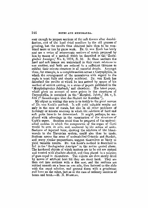
A. Area of epidermal cell. B. "Wall of same. c. Filaments ofHemileia. D. Stomata. All X 600.
FIG. 1.—Portion of epidermis of young bark of coffee tree, showingthe ramifications of the filaments externally. The fainter lines indicatethe epidermal cells. The darker lines represent the filaments of theHemileia.
FIG. 2.—Portion of the under surface of a coffee leaf, showing thearrangement of the stomata and distribution of the filaments of theHemileia.
EXPLANATION OF PLATE XII.
•" FIG. 1.—Section through coffee leaf, showing mycelium of theHemileia as an internal parasite, A. Upper surface of leaf, B. Verticalcells (palisade tissue). c. Spongy tissue, D. Intercellular air-pas-sages, E. Lower surface of leaf. F. Cluster of Sporangia of Hemileia.a. Mycelium of Hemileia. H. Cavity caused by the destruction ofspongy tissueless mycelium of Hemileia. X 150.
FIG. 2.—Cluster of sporangia borne upon mycelial filaments, extri-cated from the leaf. a. Sporangia. 6. Line representing surface ofthe leaf. c. Mycelium dissected from the interior of the leaf. X 250.
FIG. 3.—Diagram representing a section of a coffee leaf, with portionof lower surface seen in perspective. References as in Fig. 1. X 250.
EXPLANATION OF PLATE XIII .References A to D as in Plate XL X 1000.
FIG. 1.—Portion of epidermis of young bark of coffee tree, showingfilaments of Hemileia before treatment with mixture of sulphur analime.
FIG. 2.—Same preparation, showing the filaments thirty-six hoursafter treatment with a mixture of sulphur and caustic lime in the pro-portion of 1 to 3. The yellow particles represent the sulphur grains;the irregular particles are those of lime. B. Portion of filament corrodedby the action of caustic lime. p. Portion of filament shrivelled by theaction of sulphurous acid evolved by the sulphur.
EXPLANATION OF PLATE XIV.References A to xt as in Plate XL X 1000.
FIG. 1.—Portion of under surface of coffee leaf, showing filaments ofdisease before treatment with a mixture of sulphur and caustic lime.E. Growing point of a filament.
FIG. 2.—Same preparation, showing the filaments forty-eight hoursafter treatment with a mixture of sulphur and caustic lime in the pro-portion of 1 to 2. Less lime being used than in the case shown inPL XIII, fig. 2, the filaments are not so broken. They owe theirdestruction almost entirely to the action of the sulphur.

HEMILEIA VASTATRIX

Fi| . 2.
- A
HEMILEIA VASTATRIX
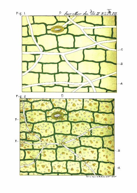
Fig- 1
-A
J4!Fa.rUn. &. Er.Vm.- U.tli" 5Tdn

Fig. 1.
-c
Fig. 2. D

J D. Siddall ,ad. nat. del . - En lmi Lilh" Kdin"
^HEPHEARDIA T.ENIF0RM1S.

;\>5.VTf .V.. ft r.y—.. . . „ _ _ _
»'*
1-7 ,SHEPHEARDIA, 8-12 LlEBERKUHNIA,

JOURNAL OF MICROSCOPICAL SCIENCE.
EXPLANATION OF PLATES XV & XVI,
Illustrating Mr. J. D. Siddall's " Memoir on Shepheardella,an undescribed Type of Marine Rhizopoda; with afew Observations on Lieberkiihnia."
PLATE XV,Illustrating Shepheardella taniformis life-history.
FIG. 1.—Three specimens drawn natural size.FIG. 2.—A living Shepheardella, with pseudopodia extended from the two
end apertures, and also from the investing layer of sarcode. Oval nucleusnear the middle. X 15 diameters.
PIG. 3 a. b.—The middle and two ends of same specimen, showingnucleus, apertures, integument, and yellowish granular sarcode. X 170diams.
FIG. 4.—The varying appearances presented by the nucleus as it iscarried along by the rotating protoplasm (sarcode). X 120 diams.'
FIG. 5.—A very small and somewhat abnormal specimen, x 60 diams.a. aud b. The end showing aperture and first protrusion of pseudopodia.
FIG. 6.—A many-apertured form, probably " Shepheardella,"'in first stageof " breaking up." Pseudopodia drawn to actual length, x 27 diams.
FIG. 7.—Fig. 2 at nine a.m., December 18th. a. Detached portion ofsarcode.
FIG. 8 a. b.—Fig. 2 at seven p.m., December 19th. Viewed from bothsides of the cell.
FIG. 9 a. 6.—Fig. 2 at nine a.m., December 20tb. Viewed from bothsides of the cell. Sarcode all naked, having been exuded from thewrinkled empty integument t.
FIG. 10 a. b. c. d.—The naked sarcode of another " Shepheardella," which,having passed through preliminary alterations in form, somewhat similar tothose represented by Figs. 2, 7, 8, and 9, broke up into four separate portionson December 17th at nine a.m., twenty-four hours previously having beenjust as Fig. 9 in form, but still enclosed in its integument.
FIG. 11.—Fig. 10 at 10.30 p.m., December 17th.FIG. 12.—Fig. 10 at nine a.m., December 18th. a. b. Detached amoeboid
portions. Figs. 7 to 12 X 20 diameters.FIG. 13 a. b. c.—Fig. 10 on December 27tb. Breaking up. a. The
empty integument, b. A protruded mass of still sarcode, containing anumber of nucleated granules, e. One of the latter which has developedActinophrys-like rays.
FIG. 14 a. b.—Actinopbrys-form some days after liberation. x 250diams. a. Dividing into three separate individuals, b. Large example,containing nucleus and contractile vesicle.
FIG. 15.—A minute portion of Fig. 12, detached on December 18th,containing a large number of granules exceedingly small, yet much largerand more definite in form than the ordinary granules of the sarcode.

EXPLANATION OF PLATE XV.—Continued.
a. As first given off. b. Its appearance on December 20th, showing" lobose" pseudopodia and granules given off from it. By January 25thit had discharged all the granules, which could not be distinguished fromthe other orgauic matter on the slide.
FIGS. 16, 17, IS, and 19.—Amcebaj, as given off from Shepheardella;16 containing nucleus, n. Nucleus, c. v. Contractile vesicle. 17.Another form, occasionally nucleated. 18. Non-nucleated, very activeform. 19. Hesting condition, as now assumed, four to seven weeks afterliberation.
PLATE XVI,
Illustrating Shepheardella taniformis histology, and Lieberkuhnia.
FIG. 1.—Camera tracing of the subcutaneous layer of the sarcode of" Shepheardella" mounted in glycerine jelly (without any other treatment)and containing, imbedded in its clear, structureless protoplasm, nucleatedgranules of various sizes, and large clear masses of firmer protoplasm.X 600 diams.
FIG. 2.—Camera tracing of a similarly mounted specimen, after treat-ment with osmio acid, alcohol, and picro-carmine, showing every granulecontained in the clear protoplasm to be of definite form and nucleated.X 600 diams.
FIG. 3.—The nucleus of a glycerine-mounted specimen (only), showing,the nucleus, a, proper, enveloped in a membrane, be. X 300 diams.
FIG. 4.—The nucleus of another specimen, after treatment with aceticacid and carmine, showing the nucleus, a, proper, filled with granularprotoplasm (stained deeply), and an irregular edged denser mass near itsceutre (nucleolus), the nucleus being embraced by a delicate membrane,b. X 300 diams.
FIG. 5.—Another nucleus, treated and mounted as Fig. 4, showing a,nucleus; b, the protoplasmic contents contracted upon its walls; c, theembracing membrane; and d, the very transparent enveloping sac. x 300diams.
FIG. 6.—Another, mounted in glycerine jelly only, showing c, theembracing membrane; and d, the enveloping sac wrinkled upon it.X 300 diams.
FIG. 7-—Shepheardella ttsnia, from a living specimen, x 40 diams.FIG. 8.—Lieberkuhnia Wageneri. X 55 diameters.FIG. 9.—Aperture of ditto. Base of the principal stem of pseudopodia,
shown as an oval spot within the square mouth, x 55 diams.FIG. 10.—The same specimen shortly after being transferred to the cell,
showing the living worm entangled among the pseudopodia. x 38 diams.FIG. 11.—The same, with the worm swallowed as far as possible.FIG. 12.—Marginal portion of the same when mounted in glycerine
jelly, viewed in optical section, showing transparent integument, a ; vesi-cular nuclei, b; granular protoplasm, c; and subcutaneous layer of finerprotoplasm, d. x 1000 diams.


ty-.>. 21. ,--
i / -•
k.t.
: . v - •-<.)•,
SI
V) >
/ '•)
A ,y
•\
/ '
JOURNAL OF MICROSCOPICAL SCIENCE.
EXPLANATION OF PLATES XVII AND XVIII,
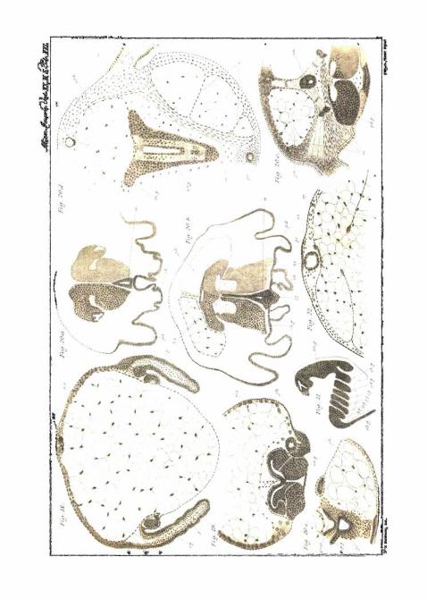
Illustrating Mr. Sedgwick's Memoir on " Development ofthe Kidney in its Relation to the Wolffian Body iu theChick."
Complete List of Reference Letters.
Ao. Aorta. Al. Alimentary canal, cl. Cloaca, c. v. Cardinal vein.ep. Epiblast. hy. Hypoblast. i. c. m. Intermediate cell mass. i. c. m.'Cell mass, which later becomes the intermediate cell mass. m. e.Mesentery. M. d. Miillerian duct. k. b. Kidney blastema. k. t.Kidney tubule, n. c. Notochord, p. Protovertebra. p'. Cell mass,which later becomes a protovertebra. p. v. Body-cavity, p. e. Peri-toneal epithelium. t. Testis. u. Ureter, ve. Vertebral body. to.Wolffian body. to. b. Wolffian blastema, to. d. Wolffian duct. to. t.yPrimary Wolffian tubule, to. t? Secondary ditto, to. t? Tertiary ditto.
FIG. 1.—Section between the fifteenth and sixteenth protovertebra)of a chick with twenty-three protovertebra;, showing the rudimentarycontinuation of the body-cavity into the intermediate cell mass and theconnection which the latter has obtained with the Wolffian duct. Theintermediate cell mass in anterior and posterior neighbouring sectionshas separated from the peritoneal epithelium.
FIGS. 2, 3, 4, and 5.—Sections taken from a duck embryo with aboutthirty-two protovertebrse, illustrating the development of the Wolffiantubules. Hart, cam., ob. 4.
FIG. 2.—Section through the thirtieth segment, intermediate cell masscontinuous with peritoneal epithelium, and containing a rudimentaryprolongation of the body-cavity. Lumen of Wolffian duct doubtful.
FIG. 3.—Section through the twenty-ninth segment, intermediatecell mass separate from peritoneal epithelium.
FIG. 4.—Section through the twenty-sixth protovertebra, showingfeatures similar to above.
FIG. 5.—Section through the twenty-second protovertebra ; com-mencing differentiation of Wolffian tubule.
FIGS. 6—10.—Sections illustrating the more modified development ofthe Wolffian blastema, as seen in the chick behind the twentieth segment.
FIG. 6.—Section through a chick with twenty-six protovertebrasbehind the four last-formed segment, showing the thick peritonealepithelium, the Wolffian blastema in connection with the mass of cells•which will become a protovertebra. Hart, cam., ob. 4.
FIG. 7.—Section through the twenty-ninth protovertebra of a chickwith twenty-nine protovertebrse, showing the thick peritoneal epi-thelium and the Wolffian blastema in connection with the provertebrse.Hart, cam., ob. 4.
FIG. 8.—Section through the twenty-fourth segment of a chick withtwenty-six protovertebra), showing the Wolffian blastema separate fromprotovertebra) and thick peritoneal epithelium. Hart, cam., ob. 3.

EXPLANATION" OF PLATES XVII AND XVIII— Continued.
FIG. 9.—Section through the twenty-fourth segment of a chick withtwenty-nine protovertebrse, showing YVolfiian blastema and thin peri-toneal epithelium. Hart, cam., ob. 3.
FIG. 10.—Section through the twenty-ninth segment of a chick withthirty-four protovertebrse, showing the commencing development of aprimary Wolffian tubule from Wolffian blastema. Hart, cam., ob. 4.
FIG. 11.—Section through a chick, end of third day or beginning offourth, showing earliest appearance of a secondary tubule. Hart,cam., ob. 3.
FIG. 12.—Section through the thirty-second protovertebra of achick with thirty-four protovertebrse, showing the kidney blastema.
FIGS. 13—17.—A series of sections from the hind end of a chick offourth day, illustrating the continuity of the Wolffian body with thecells forming the kidney blastema. Hart, cam., ob. 3.
FIG. 13.—Last section, in which a tertiary tubule was seen.FIG. 14.—Last section, in which a secondary tubule was seen. The
tubules in figs. 13 and 14 are contiguous.FIG. 15.~Next section but one behind fig. 14.FIG. 16.—Next section but one to fig. 15.FIG. 17 A.—Section some distance behind that drawn in fig. 16.FIGS. 15, 16, and 17 show kidney blastema.FIG. 17 shows opening of Wolffian duct into horn of cloaca.FIGS. 18—20.—Sections through a slightly older embryo than that
from which above series was taken. Hart, cam., ob. 3. Showing (fig.20), ureter opening into Wolffian duct, with shifted kidney blastemalying just internal to it.
FIG. 19.—Showing developing ureter and kidney blastema.FIG. 20. — Section just anterior to ureter through the anterior end of
the kidney blastema.FIG. 21.—A longitudinal vertical section through the hind end of a
four-dav chick, showing continuity of kidney blastema with hindermostpart of Wolffian blastema, in which the development of Wolffian tubuleis taking place. No line of demarcation can be drawn between the two.
FIG. 22.—Section through a chick of seventh day or late in sixth,showing the portion of the ureter (a.) and its dorsal dilatation (». t.)with regard to the Wolffian body («>.).
FIGS. 23 and 24 are from sections of the chick from which fig. 22 waataken.
FIG. 23.—Section next but one to fig. 22. It shows the kidney tubuledorsal to the ureter, surrounded by the blastema.
FIG. 24.—Section next to fig. 22, shows the dilated termination of thekidney tubule and the continuity of its lining cells with those of thekidney blastema.
FIG. 25.—From a section through the kidney of an eight-day chick,showing the termination of a kidney tubule. It presents the samefeature as fig. 24.

U. S'MJDL.
A.Li Hali'vur- aUl. MP 1'arkerlUh


JOURNAL OF MICROSCOPICAL SCIENCE.
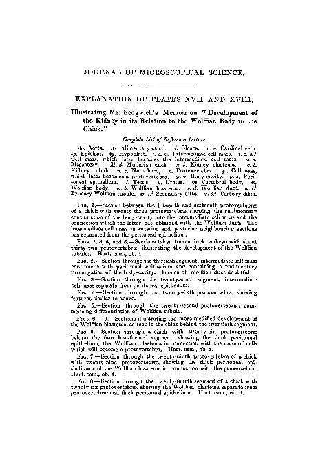
EXPLANATION OF PLATES XIX, XX, AND XXI,
Illustrating Mr. F. M. Balfour's Notes on the Develop-ment of the Araneina.
PLATE XIX.Complete List of Reference letters.
eh. g. Ganglion of chelicerse. c. I. Caudal lobe. ch. Chelicene. pd.Pedipalpi. pr. 1. Prseoral lobe. pp1. pp1. etc. Provisional appendages.p. c. Primitive cumulus, sp. Spinnerets, si. Stomodieuin.
I—IV. Ambulatory appendages. 1—16. Postoral segments.FIG. 1.—Ovum, with primitive cumulus and streak proceeding from it>FIG. 2.—Somewhat later stage, in which the primitive cumulus is still
visible. Near the opposite end of the blastoderm is a white area, whioh isprobably the rudiment of the procephalic lobe.
FIG. 3a and 3b.—View of an embryo from the ventral surface and fromthe side when six segments have become established.
FIG. 4.—View of an embryo, ideally unrolled, when the first rudi-ments of the appendages become visible.
FIG. 5.—Embryo ideally unrolled at the stage when all the appendageshave become established.
FIG. 6.—Somewhat older stage, when the limbs begin to be jointed.Viewed from the side.
FIG. 7.—Later stage, viewed from the side.FIG. 7a.—Same embryo as fig. 7, ideally unrolled.FIG. 8« and Sd.—View from the ventral surface and from the side of
an embryo, after the ventral flexure has considerably advanced.FIG. 9.—Somewhat older embryo, viewed from the ventral surface.
PLATES XX AND XXI.Complete List of Reference Letters.
ao. Aorta, ab. g. Abdominal nerve cord. ch. Chelicerse. ch. g. Gan-glion of chelicerae. ep. Epiblast. At. Heart, hs. Hemispherical lobeof supra-oEsophageal ganglion. /. I. Lower lip. m. Muscles, me. Meso-blast. mes. Mesenteron. mp.g. Malpighian tube. ms. Mesoblasticsomite, m. (Esophagus, p. c. Pericardium, pr. Proctodjeum (rectum).pd. Pedipalpi. pd. g. Ganglion of pedipalpi. pr. c. Primitive cumulus.s. Septum in abdomen, so. Somatopleure. sp. Splanchnopleure. at.Stomodseum. su. Suctorial apparatus, su. g. Supra-cesophageal ganglion.th. g. Thoracic ganglion, v. g. Ventral nerve cord. yk. Yolk. y. c.Cells derived from yolk. y. n. Nuclei of yolk cells.
Iy—IV g. Ganglia of ambulatory limbs. 1—16. Postoral segments.

PLATE XX & XXL—Continued.
FIG. 10.—Section through an ovum, slightly younger than fig. 1.Showing the primitive cumulus and the columnar character of the celUof one half of the blastoderm.
FIG. 11.—Section through an embryo 'of > the same age as fig, 2.Showing the median thickening of the blastoderm.
FIG. 12.—Transverse section through the ventral plate of a somewhatolder embryo, Showing the division of the ventral plate into epiblast andmesoblast.
FIG. 13.—Section through the ventral plate of an embryo of the sameage as fig. 3, showing the division of the mesoblast of the ventral plateinto two mesoblastic bands.
FIG. 14.—Transverse section through an embryo of the same age asfig. 5, passing through an abdominal segment above and a thoracicsegment below.
FIG. 15—Longitudinal section slightly to one side of the middle linethrough an embryo of the same age.
FIG. 16.—Tranverse section through the ventral plate in the thoracicregion of an embryo of the same age as fig. 7.
FIG. 17.—Transverse section through the procephalic lobes of anembryo of the same age. gr. Section of hemicircular groove in pro-cephalic lobe.
FIG. 18.—Transverse section through the thoracic region of anembryo of the same age as fig. 8.
FIG. 19.—Section through the procephalic lobes of an embryo of thesame age.
FIG. 20 a, b, c, d, e.—Five sections through an embryo of the sameage as fig. 9. a and b are sections through the procephalic lobes,c through the front part of the thorax, d cuts transversely theposterior parts of the thorax, and longitudinally and horizontally theventral surface of the abdomen, e cuts the posterior part of the ab-domen longitudinally and horizontally, and shows the commencementof the mesenteron.
FIG. 21.—Longitudinal and vertical section of an embryo of the sameage. The section passes somewhat to one side of the middle line, andshows the structure of the nervous system.
FIG. 22.—Transverse section through the dorsal part of the abdomenof an embryo of the same stage as fig. 9.

IZ
"Tf ' • ;s$$t'*y'
•vfty
til
/ ' " " u I
6
' " ' .V •
7 ?
L '~ " ^ ' ^
:v.-:-i&i
y
' ' ' S' \ "
/ •v 'v V
i;l';;-."-v;.';,' K
fsi ?•
f'fTl-'
V- x-J
/
- , ' • • . ' - * . ™
•Li; k v-v
I V1 \
-<
• > •
• * • /
ill
z
\t
w
: c c o r t>'.'•Off.
* ( re '•*

JOURNAL OF MICROSCOPICAL SCIENCE.
EXPLANATION OF PLATE XXII,
Illustrating Professor Giard's " Memoir on tie Ottho-nectida."
FIG. 1.—Rhopalura ophiocomce (natural state).PIG. 2.—Short form, or young phase (natural state).FIG. 3.—Adult animal, treated by reagents and showing the muscular
bands.FIG. 4.—Immature animal (acetic acid and carmine).FIG. 5.—Intoshia gigas (profile view).FIG. 6.— The same seen from the dorsal aspect, and treated in such a
way as to show the ectoderinal cells.FIG. 7.—The same with the ectoderm removed, in order to show the
endoderm.FIG. 8.—Very young blaslula of Intoshia.FIG. 9.—The same more advanced.FIG. 10.—Commencement of delamination.FIGS. 11, 12, and 13.—Formation of planula.FIGS. 14 and 15.—Very young sporocysts of Intoshia still retaining
remains of the ectoderm.FIG. 16.—More advanced sporocyst, opened in order to show its buds.FIG. 17.—Portion of the same very highly magnified.FIG. 18.—Bud in the condition of a planula.FIGS. 19, 20, and 21.—Stages in the segmentation of the egg of
Rhopalura.


![BRzTou] MEMORANDA-, MEDICAL ... · 374MzBRzTou] MEMORANDA. [FER. i6, 1907. tosay,thebodyfluidsareinaconditionofsuper-saturation. The explanation of these remarkable results cannot](https://static.fdocuments.in/doc/165x107/60604012bbe4ff435e367cb1/brztou-memoranda-medical-374mzbrztou-memoranda-fer-i6-1907-tosaythebodyfluidsareinaconditionofsuper-saturation.jpg)


![PERSONAL MEMORANDA [Office Copy]](https://static.fdocuments.in/doc/165x107/61bd3a5561276e740b10a203/personal-memoranda-office-copy.jpg)