Nose reconstructions using pinna cartilage and ...
5
IP Journal of Otorhinolaryngology and Allied Science 2020;3(2):72–76 Content available at: https://www.ipinnovative.com/open-access-journals IP Journal of Otorhinolaryngology and Allied Science Journal homepage: www.ipinnovative.com Case Report Nose reconstructions using pinna cartilage and interpolated paramedian forehead flap: our experience Manish Gupta 1, *, Ginni Datta 1 , Gurchand Singh 1 1 Dept. of ENT, Maharishi Markandeshwar Institute of Medical Sciences & Research, MMDU, Ambala, Haryana, India ARTICLE INFO Article history: Received 28-01-2020 Accepted 04-05-2020 Available online 16-08-2020 Keywords: Nasal reconstruction Forehead flap Cartilage Trauma Basal cell carcinoma ABSTRACT Intact nose plays important role in perceptible beauty of face. Any amount of loss of nose has ruinous effect on quality of life. Surgical correction is by reconstruction of nasal pyramid by various surgical techniques. Six hundred years before Christ, Sushruta had described mid-forehead flap, which has an important role in reconstruction of the nose. Here we present our experience of two cases, where reconstruction of nose was done using interpolated paramedian forehead flap (IPFF) and cartilage from conchal region of pinna. © 2020 Published by Innovative Publication. This is an open access article under the CC BY-NC license (https://creativecommons.org/licenses/by-nc/4.0/) 1. Introduction In antiquity the nose was considered as “the organ of reputation” and it was amputated as a form of punishment, with the certainty that this would irreparably compromise the social life of the convicted. 1 Partial or total loss of nose, as a result of trauma or tumor excision, has great impact on beauty of a face resulting in limited social life. Since the Vedic period (2000-500BC) in ancient India, amputation of the nose was punishment for infidelity. 2 In 600 BC, Sushruta, mentioned a procedure for total nasal reconstruction using mid-forehead flap, also called “Indian flap”. 2 In 1597, Gaspare Tagliacozzi, gave a technique of using upper third of the inner side of the arm for nose reconstruction. It is now called “Italian method”. 2 Gillies, in 1943 and Converse in 1956, advocated forming inner lining by, chondrocutaneous composite graft from pinna, chondromucosal graft from nasal septum and nasolabial flaps thus supporting the mid forehead flap. 2 We present a series of two cases with nasal reconstruction using the forehead flap and conchal cartilage grafts. * Corresponding author. E-mail address: [email protected] (M. Gupta). 2. Case Report 1 A 28 year old male, presented to ENT out patient, with complaint of loss of tip nose, following dog bite 9 months back. On examination, the lower lateral alar cartilage medial crura, lobule and part of lateral crura were missing, bilaterally. There was also loss of septal quadrilateral cartilage, as seen on lateral view (Figure 1). There was no open wound, granulations or mass seen. The area was non tender with normal temperature. After routine investigations and informed consent, patient was taken up for reconstruction with cartilage grafting and right interpolated paramedian frontal flap. The skin overlying residual tip and dorsum was elevated antero-inferiorly based and used to form inner lining. Septal extension graft and batten graft was placed using posterior septal cartilage (Figure 2). Bilateral conchal cartilages were used for reconstruction of lower lateral cartilages. Right side IPFF, based on supratrochlear artery was raised and sutured in place. After 3 weeks flap pedicle was cut and replaced (Figure 3). The patient was satisfied with end result. https://doi.org/10.18231/j.ijoas.2020.016 2582-4147/© 2020 Innovative Publication, All rights reserved. 72
Transcript of Nose reconstructions using pinna cartilage and ...

IP Journal of Otorhinolaryngology and Allied Science 2020;3(2):72–76
Content available at: https://www.ipinnovative.com/open-access-journals
IP Journal of Otorhinolaryngology and Allied Science
Journal homepage: www.ipinnovative.com
Case Report
Nose reconstructions using pinna cartilage and interpolated paramedian foreheadflap: our experience
Manish Gupta1,*, Ginni Datta1, Gurchand Singh1
1Dept. of ENT, Maharishi Markandeshwar Institute of Medical Sciences & Research, MMDU, Ambala, Haryana, India
A R T I C L E I N F O
Article history:Received 28-01-2020Accepted 04-05-2020Available online 16-08-2020
Keywords:Nasal reconstructionForehead flapCartilageTraumaBasal cell carcinoma
A B S T R A C T
Intact nose plays important role in perceptible beauty of face. Any amount of loss of nose has ruinous effecton quality of life. Surgical correction is by reconstruction of nasal pyramid by various surgical techniques.Six hundred years before Christ, Sushruta had described mid-forehead flap, which has an important role inreconstruction of the nose. Here we present our experience of two cases, where reconstruction of nose wasdone using interpolated paramedian forehead flap (IPFF) and cartilage from conchal region of pinna.
© 2020 Published by Innovative Publication. This is an open access article under the CC BY-NC license(https://creativecommons.org/licenses/by-nc/4.0/)
1. Introduction
In antiquity the nose was considered as “the organ ofreputation” and it was amputated as a form of punishment,with the certainty that this would irreparably compromisethe social life of the convicted.1 Partial or total loss of nose,as a result of trauma or tumor excision, has great impact onbeauty of a face resulting in limited social life.
Since the Vedic period (2000-500BC) in ancient India,amputation of the nose was punishment for infidelity.2 In600 BC, Sushruta, mentioned a procedure for total nasalreconstruction using mid-forehead flap, also called “Indianflap”.2 In 1597, Gaspare Tagliacozzi, gave a technique ofusing upper third of the inner side of the arm for nosereconstruction. It is now called “Italian method”.2 Gillies,in 1943 and Converse in 1956, advocated forming innerlining by, chondrocutaneous composite graft from pinna,chondromucosal graft from nasal septum and nasolabialflaps thus supporting the mid forehead flap.2
We present a series of two cases with nasal reconstructionusing the forehead flap and conchal cartilage grafts.
* Corresponding author.E-mail address: [email protected] (M. Gupta).
2. Case Report 1
A 28 year old male, presented to ENT out patient,with complaint of loss of tip nose, following dog bite9 months back. On examination, the lower lateral alarcartilage medial crura, lobule and part of lateral crurawere missing, bilaterally. There was also loss of septalquadrilateral cartilage, as seen on lateral view (Figure 1).There was no open wound, granulations or mass seen.The area was non tender with normal temperature. Afterroutine investigations and informed consent, patient wastaken up for reconstruction with cartilage grafting and rightinterpolated paramedian frontal flap.
The skin overlying residual tip and dorsum was elevatedantero-inferiorly based and used to form inner lining. Septalextension graft and batten graft was placed using posteriorseptal cartilage (Figure 2). Bilateral conchal cartilages wereused for reconstruction of lower lateral cartilages. Right sideIPFF, based on supratrochlear artery was raised and suturedin place. After 3 weeks flap pedicle was cut and replaced(Figure 3). The patient was satisfied with end result.
https://doi.org/10.18231/j.ijoas.2020.0162582-4147/© 2020 Innovative Publication, All rights reserved. 72

Gupta, Datta and Singh / IP Journal of Otorhinolaryngology and Allied Science 2020;3(2):72–76 73
3. Case Report 2
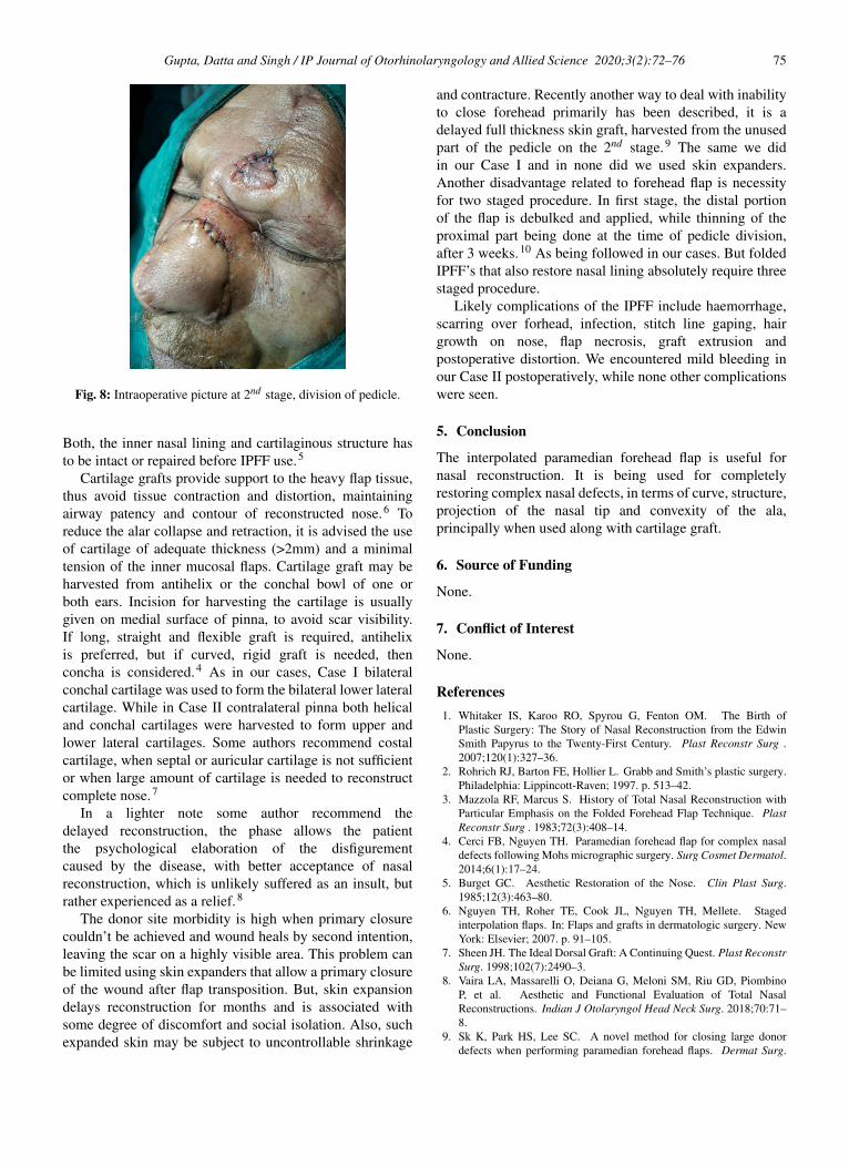
73 year old male patient came with non healing ulcerover tip, dorsum and left ala (Figure 4). The marginswere rounded and biopsy confirmed basal cell carcinoma.After routine investigations and informed consent, themass was excised under general anaesthesia (Figure 5)repair was done with conchal cartilage (Figure 6) and leftIPFF (Figure 7). After 3 weeks pedicle was divided andflap reposited back (Figure 8 ), with good result and norecurrence in one year follow up.
Both the patients expressed that decent nasalreconstruction both functionally and aesthetically havebeen achieved, with no evidence of ischemia, infection ornecrosis.
Fig. 1: Preoperative clinical picture of 1st case, showing loss oflower lateral cartilage bilaterally and lateral picture showing lossof tip and septum.
4. Discussion
Following nose, tumor resection or accidental chopping,the most common complaint of these patients is cosmeticdeformity. The three layered repair is considered ideal,for the restoration. In this, outer layer reconstruction byforehead flap, gives tissue with identical quality as original.
The interpolated paramedian forehead flap (IPFF) isbeing used since centuries for nasal reconstruction. Overthe years, various alterations have been specified to improveits end result.3 The IPFF chief indication is large and deepwound based on the distal nose (tip and ala). It can entirelyreconstruct nasal contour, texture of skin, tip projection andalar convexity, when used along with cartilage graft. Thelarge bulk of IPFF makes it less preferable for upper nosereconstruction, where thin skin is required.
The use of IPFF for nose reconstruction needs goodknowledge of anatomy, surgical skill and plan. The IPFFnutrition relies on the supratrochlear artery, which is locatedat the medial border of the eyebrow, 1.5 to 2 cm fromthe vertical facial midline.4 Sometimes, Doppler may beused to further confirm the location of the artery. Aftercoming out from supratrochlear foramen, the artery liesdeep to periorbital muscles (orbicularis oculi and frontalis).
Fig. 2: Intraoperative picture showing inner lining of vestibule andreconstructed cartilage framework.
Fig. 3: Postoperative, after 2nd surgery front view and lateral view,at stitch removal.
Above the superior orbital rim, the artery passes through thefrontalis muscle and progressively comes to lie superficialwithin subcutaneous plane, halfway up the forehead. Theflap is raised in three different planes.4 At the freeend epithelium and subcutaneous tissue, middle includingmuscle and the lower third, near the orbital rim, includingperiosteum. It is preferred to raise flap ipsilaterally.4 Itgives less torsion and twisting to the flap, has to cover lessdistance to reach the defect with no postoperative visualobstruction and increased flap reach. In our Case I since thedefect was bilateral, so laterality of flap didn’t matter, but inCase II we preferred ipsilateral forehead flap due to abovementioned reason.
The IPFF provides robust surface covering andadequately thick soft tissue but lacks structural support.

74 Gupta, Datta and Singh / IP Journal of Otorhinolaryngology and Allied Science 2020;3(2):72–76
Fig. 4: Preoperative clinical picture of 2nd case, showing irregularulcer with rolled out edges.
Fig. 5: Intraoperative picture after excision of growth with safemargins.
Fig. 6: Cartilage framework reconstruction following suturing.
Fig. 7: Interpolatedparamedian forehead flap in position, afterforehead closure.
Gupta, Datta and Singh / IP Journal of Otorhinolaryngology and Allied Science 2020;3(2):72–76 75
Fig. 8: Intraoperative picture at 2nd stage, division of pedicle.
Both, the inner nasal lining and cartilaginous structure hasto be intact or repaired before IPFF use.5
Cartilage grafts provide support to the heavy flap tissue,thus avoid tissue contraction and distortion, maintainingairway patency and contour of reconstructed nose.6 Toreduce the alar collapse and retraction, it is advised the useof cartilage of adequate thickness (>2mm) and a minimaltension of the inner mucosal flaps. Cartilage graft may beharvested from antihelix or the conchal bowl of one orboth ears. Incision for harvesting the cartilage is usuallygiven on medial surface of pinna, to avoid scar visibility.If long, straight and flexible graft is required, antihelixis preferred, but if curved, rigid graft is needed, thenconcha is considered.4 As in our cases, Case I bilateralconchal cartilage was used to form the bilateral lower lateralcartilage. While in Case II contralateral pinna both helicaland conchal cartilages were harvested to form upper andlower lateral cartilages. Some authors recommend costalcartilage, when septal or auricular cartilage is not sufficientor when large amount of cartilage is needed to reconstructcomplete nose.7
In a lighter note some author recommend thedelayed reconstruction, the phase allows the patientthe psychological elaboration of the disfigurementcaused by the disease, with better acceptance of nasalreconstruction, which is unlikely suffered as an insult, butrather experienced as a relief.8
The donor site morbidity is high when primary closurecouldn’t be achieved and wound heals by second intention,leaving the scar on a highly visible area. This problem canbe limited using skin expanders that allow a primary closureof the wound after flap transposition. But, skin expansiondelays reconstruction for months and is associated withsome degree of discomfort and social isolation. Also, suchexpanded skin may be subject to uncontrollable shrinkage
and contracture. Recently another way to deal with inabilityto close forehead primarily has been described, it is adelayed full thickness skin graft, harvested from the unusedpart of the pedicle on the 2nd stage.9 The same we didin our Case I and in none did we used skin expanders.Another disadvantage related to forehead flap is necessityfor two staged procedure. In first stage, the distal portionof the flap is debulked and applied, while thinning of theproximal part being done at the time of pedicle division,after 3 weeks.10 As being followed in our cases. But foldedIPFF’s that also restore nasal lining absolutely require threestaged procedure.
Likely complications of the IPFF include haemorrhage,scarring over forhead, infection, stitch line gaping, hairgrowth on nose, flap necrosis, graft extrusion andpostoperative distortion. We encountered mild bleeding inour Case II postoperatively, while none other complicationswere seen.
5. Conclusion
The interpolated paramedian forehead flap is useful fornasal reconstruction. It is being used for completelyrestoring complex nasal defects, in terms of curve, structure,projection of the nasal tip and convexity of the ala,principally when used along with cartilage graft.
6. Source of Funding
None.
7. Conflict of Interest
None.
References1. Whitaker IS, Karoo RO, Spyrou G, Fenton OM. The Birth of
Plastic Surgery: The Story of Nasal Reconstruction from the EdwinSmith Papyrus to the Twenty-First Century. Plast Reconstr Surg .2007;120(1):327–36.
2. Rohrich RJ, Barton FE, Hollier L. Grabb and Smith’s plastic surgery.Philadelphia: Lippincott-Raven; 1997. p. 513–42.
3. Mazzola RF, Marcus S. History of Total Nasal Reconstruction withParticular Emphasis on the Folded Forehead Flap Technique. PlastReconstr Surg . 1983;72(3):408–14.
4. Cerci FB, Nguyen TH. Paramedian forehead flap for complex nasaldefects following Mohs micrographic surgery. Surg Cosmet Dermatol.2014;6(1):17–24.
5. Burget GC. Aesthetic Restoration of the Nose. Clin Plast Surg.1985;12(3):463–80.
6. Nguyen TH, Roher TE, Cook JL, Nguyen TH, Mellete. Stagedinterpolation flaps. In: Flaps and grafts in dermatologic surgery. NewYork: Elsevier; 2007. p. 91–105.
7. Sheen JH. The Ideal Dorsal Graft: A Continuing Quest. Plast ReconstrSurg. 1998;102(7):2490–3.
8. Vaira LA, Massarelli O, Deiana G, Meloni SM, Riu GD, PiombinoP, et al. Aesthetic and Functional Evaluation of Total NasalReconstructions. Indian J Otolaryngol Head Neck Surg. 2018;70:71–8.
9. Sk K, Park HS, Lee SC. A novel method for closing large donordefects when performing paramedian forehead flaps. Dermat Surg.

76 Gupta, Datta and Singh / IP Journal of Otorhinolaryngology and Allied Science 2020;3(2):72–76
2013;39(10):1549–50.10. Burget GC, Menick FJ. Nasal support and lining: the marriage of
beauty and blood supply. Plast Reconstr Surg. 1989;84(2):189–202.
Author biography
Manish Gupta Professor & Head
Ginni Datta Professor
Gurchand Singh Assistant Professor
Cite this article: Gupta M, Datta G, Singh G. Nose reconstructionsusing pinna cartilage and interpolated paramedian forehead flap:our experience. IP J Otorhinolaryngol Allied Sci 2020;3(2):72-76.