Northern California Emergency Ultrasound Course … EFAST Lecture... · Northern California...
16
Northern California Emergency Ultrasound Course UC C SF The Extended FAST Exam Rimon Bengiamin, MD, RDMS Objectives • Discuss the components of the EFAST exam • Evaluate the utility of the EFAST • Review how to obtain and interpret the images • Discuss the strengths and weaknesses of the EFAST F F ocused d A A ssessment With F F o S S S onography In essme e n n T T rauma (FAST) • Abdominal sonography can detect as little as 50 cc of free fluid but generally you need about 200-250cc on average for a positive view • An analysis of 62 publications with 18,167 patients revealed an overall sensitivity of 79% and a specificity of 99.2% for detecting free fluid, organ damage, or both. (1) • Another study of emergency physicians showed a sensitivity of 90% and specificity of 99%. (2) • Sensitivity increases with a repeat exam at 30 minutes. (3) • Sensitivity is nearly 100 percent in the setting of hypotension and trauma • What does this mean? • It’s a good screening tool • Not a good definitive test if your suspicion is high and the test is negative F F ocused d A A ssessment With F F o S S S onography In essme e n n T T rauma (FAST) • Advantages of the FAST compared to DPL and CT • Accurate - as a screening tool • Rapid - the average time to perform a complete FAST examination of the thoracic and abdominal cavities is 2.1 to 4.0 minutes. (4,2) • Noninvasive - less risk of infection/bleeding/other complications • Repeatable - increases the sensitivity of the study • Portable - convenient in unstable patients • No contrast or radiation - renal failure and pregnant patients
Transcript of Northern California Emergency Ultrasound Course … EFAST Lecture... · Northern California...

Northern California Emergency Ultrasound Course
UCCSF
The Extended FAST Exam
Rimon Bengiamin, MD, RDMS
Objectives
• Discuss the components of the EFAST exam
• Evaluate the utility of the EFAST
• Review how to obtain and interpret the images
• Discuss the strengths and weaknesses of the EFAST
FFocused d AAssessment With FFo
SSSonography In
essmee
nn TTrauma (FAST)
• Abdominal sonography can detect as little as 50 cc of free fluid but generally you need about 200-250cc on average for a positive view
• An analysis of 62 publications with 18,167 patients revealed an overall sensitivity of 79% and a specificity of 99.2% for detecting free fluid, organ damage, or both. (1)
• Another study of emergency physicians showed a sensitivity of 90% and specificity of 99%. (2)
• Sensitivity increases with a repeat exam at 30 minutes. (3)
• Sensitivity is nearly 100 percent in the setting of hypotension and trauma
• What does this mean?
• It’s a good screening tool
• Not a good definitive test if your suspicion is high and the test is negative
FFocused d AAssessment With FFo
SSSonography In
essmee
nn TTrauma (FAST)
• Advantages of the FAST compared to DPL and CT
• Accurate - as a screening tool
• Rapid - the average time to perform a complete FAST examination of the thoracic and abdominal cavities is 2.1 to 4.0 minutes. (4,2)
• Noninvasive - less risk of infection/bleeding/other complications
• Repeatable - increases the sensitivity of the study
• Portable - convenient in unstable patients
• No contrast or radiation - renal failure and pregnant patients

FFocused d AAssessment With FFo
SSSonography In
essmee
nn TTrauma (FAST)
• Disadvantages of the FAST
• Inability to determine the exact etiology of free fluid in some cases
• Technically difficult in cases of obesity or bowel gas
• Cannot evaluate the retroperitoneum as well as CT
m
FFocused d AAssessment With FFo
SSSonography In
essmee
nn TTrauma (FAST)
• When should it be done?• Part of the primary survey, particularly when evaluating
circulation
• Examples
• Hypotensive, tachycardic patient and you find pericardial tamponade
• Hypotensive, tachypnic patient and you find a pneumo/hemothorax
• Do not let it hinder your treatment and stabilization!
• Should be done in conjunction/simultaneous with resuscitation
• FAST can be performed simultaneously as other things are being done!
aall all
FFocused d AAssessment With FFo
SSSonography In
essmee
nn TTrauma (FAST)
• What about cases when there isn’t trauma?
• AAA
• Ruptured ectopic pregnancy
What Is The Extended
FAST Exam?
• Also known as the EFAST
• The traditional FAST exam with the addition of evaluation of the thorax
• Thoracic exam includes looking for:
• Pneumothoraces
• Hemothoraces

Probe Choice
• You are looking at deep structures so you need a medium to low frequency probe
• These have better penetrance but lower resolution
Medium Low High
Right Upper Quadrant
• Evaluate three areas:
• Morison’s Pouch
• Most sensitive for detecting free fluid particularly if the patient is in Trendelenburg (6,7)
• Tip of the liver and pericolic gutter
• Slide the probe caudally
• Diaphragm
• Slide the probe cephalad and it may help to rotate the probe tip posteriorly (counterclockwise) to get through the ribs
Right Upper Quadrant
-Probe Orientation-
Right Upper Quadrant
-Morison’s Pouch-
Morison’s Pouch
Liver
Kidney
Right Upper Quadrant
-Positive Free Fluid-
Text
Free fluid appears black
RUQ Diaphragm
-Probe Orientation-
Counterclockwise probe rotation will help decrease rib shadows
Right Upper Quadrant
-Diaphragm-
Diaphragm
Look for “mirror” artifact - equal echo on both sides of the diaphragm
Pelvic View
• The traditional view is the transverse view
• However, evaluation of the pelvis in the saggital view, with the probe dot toward the head, can be more helpful
• Better delineation of the anatomy
• Helps with differentiation of free fluid
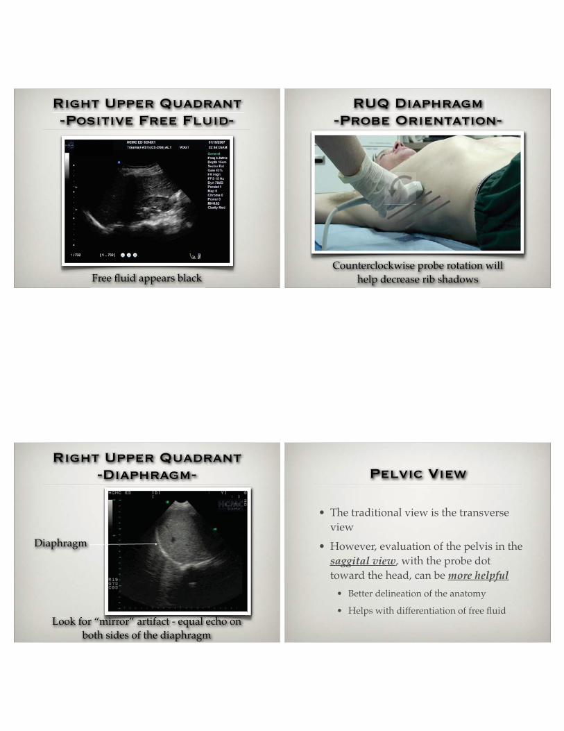
Pelvic View
-Probe Orientation-
Transverse vs.
Longitudinal
Longitudinal Transverse
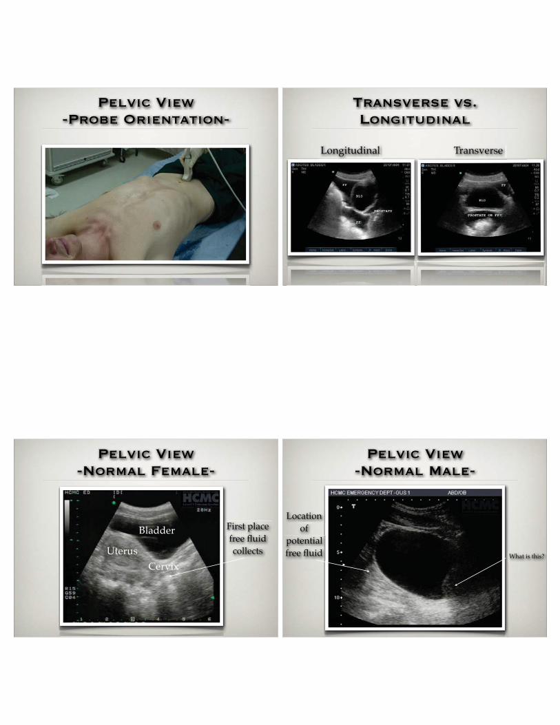
Pelvic View
-Normal Female-
First place free fluid collects
Bladder
UterusCervix
Pelvic View
-Normal Male-
What is this?
Location of
potential free fluid

Pelvic View
-Free Fluid-
Clot
Free Fluid
Left Upper Quadrant
• Unlike Morison’s view, evaluation of the interface between the kidney and spleen is not as important
• Free fluid does not commonly collect in this space because of the phrenicocolic ligament running in this area
• Fluid commonly collects around the tip of the spleen, base of the spleen, or between the spleen and diaphragm
Left Upper Quadrant
-Probe Orientation- Left Upper Quadrant
• Need to have the probe oriented more cephalad and posterior than with Morison’s view
• Also may help to rotate the probe tip posteriorly (clockwise) to get through the ribs

Left Upper Quadrant
-Normal View-
Spleen
Diaphragm
Left Upper Quadrant
-Free Fluid-
Left Upper Quadrant
-Free Fluid-
Cardiac
• Two views
• Subxiphoid long axis
• Parasternal long axis
• Try to get comfortable with both windows
• Looking mainly for pericardial effusion

Cardiac (Subxiphoid)
-Probe Orientation-
Cardiac Subxiphoid
-Is this normal?-
Cardiac Subxiphoid
-Pericardial Effusion-
Is this tamponade?
Cardiac (Parasternal)
-Probe Orientation-

Cardiac
-Parasternal Long Axis-
Cardiac Parasternal
-Effusion with Clot-
Tamponade and
pericardiocentesis
• If there is tamponade you need to act now through pericardiocentesis
• Look for right atrial and ventricular collapse
• If there is clot, you should probably go straight to thoracotomy
Pericardiocentesis
• Direct visualization dynamic technique has far less complications
• Traditional subxiphoid approach has complication rate nearing 50% including wall puncture, coronary artery laceration, pneumothorax, diaphragm or organ injury

Pericardiocentesis
• Mayo Clinic 2002 (Teresa et al).
• 1127 ultrasound guided pericardiocentesis over the course of 21 years
• 97% success rate and 4.7% complication rate
• Much lower than blind
• Not an ideal study but take home message is it’s safer
Thoracic Ultrasound
• What does it add?
• Evaluation for pnuemothorax
• Sliding lung sign
• Leading edge
• Comet tail artifact
• Evaluation for hemothorax
• RUQ and LUQ windows
Thoracic Ultrasound
-Kirkpatrick et al. (8)-
• “EFAST has comparable specificity to CXR but is more sensitive for the detection of occult pneumothorax after trauma”
• Study of 225 patients
• EFAST more sensitive than CXR
• Picked up 63% of pneumothoraces missed on CXR
Chest X-ray vs
Ultrasound
• Why is ultrasound better in the setting of trauma?
• Patient is supine
• Air will layer anteriorly
• Blood will layer posteriorly

Thoracic Ultrasound
-Comparison to CXR-
• Makes sense that it would be more sensitive particularly in the supine patient since the air will be anterior
• Can be life saving in the case of the unstable patient
• Zhang et al. (9)
• 135 trauma patients, 83 mech. ventilated
• 29 had a pneumothorax
• US: sensitivity 86%, NPV 96%
• CXR: sensitivity 28%, NPV 84%
• US: 2.3 minutes CXR:10.3 minutes
Sliding Lung Sign
• Curvilinear (abdominal) or vascular probe
• The vascular probe tends to provide better quality images
• Position the probe:
• with the dot toward the head
• at the 2nd intercostal space
• at the midclavicular line
• Can use M mode to confirm - “waves crashing on a beach”
• Should see pleural lines
Thoracic Ultrasound
-Probe Position- Probe Position
• Should probably look at two spots
• Try to look at the most anterior part of the chest (likely around the nipple line - 4th intercostal space)

Thoracic Ultrasound
-Sliding Lung Sign-
Sliding Lung Sign
-M Mode-
Thoracic Ultrasound
-Lichtenstein et al (10)-
• “Ultrasound was a sensitive test for detection of pneumothorax, although false-positive cases were noted. The principal value of this test was that it could immediately exclude anterior pneumothorax.”
• 43 patients in an ICU setting
• Examination of sliding lung sign
• 95.3% sensitivity and 91.1% specificity
Thoracic Ultrasound
-Sliding Lung-
• Can use sliding lung to estimate the size of a pneumothorax
• Actually need to map out the lung by evaluating for sliding lung at each of the intercostal spaces
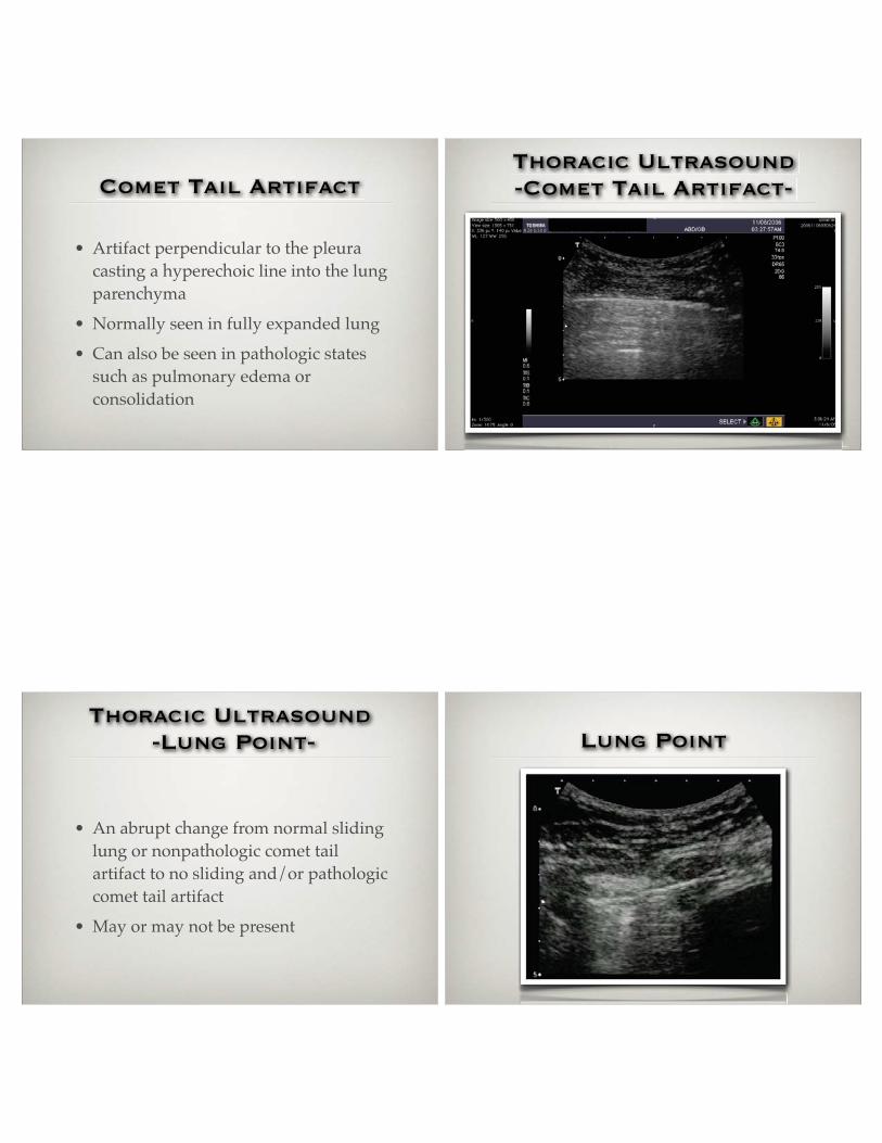
Comet Tail Artifact
• Artifact perpendicular to the pleura casting a hyperechoic line into the lung parenchyma
• Normally seen in fully expanded lung
• Can also be seen in pathologic states such as pulmonary edema or consolidation
Thoracic Ultrasound
-Comet Tail Artifact-
Thoracic Ultrasound
-Lung Point-
• An abrupt change from normal sliding lung or nonpathologic comet tail artifact to no sliding and/or pathologic comet tail artifact
• May or may not be present
Lung Point

Thoracic Ultrasound
-Hemothorax-
• Upright CXR - 100 cc
• Supine CXR - 200-300 cc
• CXR can miss large effusions
• US can pick up as little as 20 cc of effusion
Thoracic Ultrasound
-Hemothorax-
• Hemothorax
• Ma et al. (11)
• 240 patients
• Blunt and penetrating trauma
• Hemothorax confirmed by chest tube output or CT
• 99.6% accuracy
• Sisley et al. (12)
• 360 patients
• Blunt and penetrating trauma
• 40 hemothoraces confirmed by CT or chest tube
• Thoracic US had 97.5% sensitivity and 99.7% specificity
• Plain CXR had 92.5% sensitivity and 99.7% specificity
• Time for US: 1.3 min. Time for CXR: 14.8 min.
Thoracic ultrasound
-Hemothorax-
EFAST
-A Case-

EFAST
-A Case-
EFAST
-A Case-
EFAST
-A Case-
EFAST
-A Case-

EFAST
-A Case- Quiz!
• The most common place to see free fluid in the left upper quadrant is:
• A. Between the spleen and the kidney
• B. Between the spleen and the diaphragm
• C. Around the tip of the spleen
• D. B and C
EFAST
Questions?
References
• (1) Stengel D, Bauwens K, Rademacher G, et al: Association between compliance with methodological standards of diagnostic research and reported test accuracy: Meta-analysis of focused assessment of US for trauma. Radiology 2005; 236:102–111 nol 1996; 166:317–321 95.
• (2) Ma OJ, Mateer JR, Ogata M, Kefer MP, et al. Prospective analysis of a rapid trauma ultrasound examination performed by emergency physicians. J Trauma 1995;38:879-885.
• (3) Blackbourne LH, Soffer D, McKenney M, et al: Secondary ultrasound examination increases the sensitivity of the FAST exam in blunt trauma. J Trauma 2004; 57:934-938.
• (4) Thomas B, Falcone RE, Vasquez D, et al. Ultrasound evaluation of blunt abdominal trauma: program implementation, initial experience, and learning curve. J Trauma 1997;42:38-388.
• (5) Hoff WS, Holevar M, Nagy KK, et al. Practice management guidelines for the evaluation of blunt abdominal trauma: The EAST practice management guidelines work group. J Trauma 2002;53:602-615.
• (6) Sisley A, Rozycki G, Ballard R, et al: Rapid detection of traumatic effusion using surgeon performed ultrasound. J Trauma 1998;44:291-297.
• (7) Jehle D, Guarino J, Karamanoukian H: Emergency department ultrasound in the evaluation of blunt abdominal trauma. Am J Emerg Med 1993;11:342-346.
• (8) Kirkpatrick AW, Sirois M, Laupland KB, Liu D, et al: Hand-held thoracic sonography for detecting post-traumatic pneumothoraces: the extended focused assessment with sonography for trauma (EFAST). J Trauma 2004;57(2):288-295.
• (9) Zhang M, Liu ZH, Yang JX, Gan, JX, et al: Rapid detection of pneumothorax by ultrasonography in patients with multiple trauma. Critical Care 2006;10:R112.
• (10) Lichtenstein DA and Menu Y: A bedside ultrasound sign ruling out pneumothorax in the critically ill. Lung sliding. Chest 1995;108:1345-1348.
• (11) Ma OH, Mateer JR: Trauma ultrasound examination versus chest radiography in the detection of hemothorax. Annals of emergency medicine 2007;29:312-316.
• (12) Sisley A, Rozycki G, et al: Rapid detection of traumatic effusion using surgeon-performed ultrasonography. J Trauma 1998;44(2):291-297.