Normative Vertebral Hounsfield Unit Values and Correlation with … · Cross-sectional slices were...
7
Normave Vertebral Hounsfield Unit Values and Correlaon with Bone Mineral Density Shaun P. Patel 1* , John J. Lee 2 , Garin G. Hecht 3 , Sven A. Holcombe 4 , Stewart C. Wang 4 and James A. Goulet 5 1 Harvard Combined Orthopaedic Residency Program, Boston, MA, USA 2 Department of Orthopaedic Surgery, Memorial Healthcare System, Hollywood, FL, USA 3 Department of Orthopaedic Surgery, University of California - Davis, Sacramento, CA, USA 4 Department of Surgery, University of Michigan, Ann Arbor, MI, USA 5 Department of Orthopaedic Surgery, University of Michigan, Ann Arbor, MI, USA * Corresponding author: Patel SP, Harvard Combined Orthopaedic Residency Program, 55 Fruit Street, Boston, MA 02114, USA, Tel: 617-726-2942; E-mail: [email protected] Received date: March 25, 2016; Accepted date: April 07, 2016; Published date: April 11, 2016 Copyright: © 2016, Patel SP, et al. This is an open-access arcle distributed under the terms of the Creave Commons Aribuon License, which permits unrestricted use, distribuon, and reproducon in any medium, provided the original author and source are credited. Abstract Background: We hypothesized that vertebral bone mineral density as measured in Hounsfield units (HU) by computed tomography (CT) can correlate with values obtained by dual x-ray absorpometry (DXA). Our study aimed to report such a correlaon along with the normave HU values within a large, heterogeneous populaon. Methods: We idenfied 2,020 trauma paents with a CT scan encompassing any part of their thoracic or lumbar spine. Mean HU values within each vertebral body were reported with respect to vertebral level, sex, and age. Using linear regression analysis, our data were also compared to DXA results in paents having both CT and DXA scans. Results: Analyses of CT scans yielded 28,083 unique vertebrae (18,489 male and 9,594 female) for which HU values were obtained. There were significant correlaons between lumbar vertebral body HU and both DXA bone mineral density esmates (R2 = 0.50) and T-scores (p<0.001 and p<0.01, respecvely). Using DXA T-scores as the reference standard, the sensivity, specificity, and posive and negave predicve values of CT HU were 0.93, 0.75, 0.74 and 0.94, respecvely. Conclusion: CT can be used to measure vertebral bone mineral density and may aid in the diagnosis and management of osteoporosis. Keywords: Bone mineral density; Osteoporosis; Osteopenia; Computed tomography; Hounsfield units; Dual x- ray absorpometry; DXA Background The World Health Organizaon (WHO) defines osteoporosis as a bone mineral density more than 2.5 standard deviaons below the young adult mean for a given populaon [1]. Paents with decreased bone mineral density have increased bone fragility and are at increased fracture risk [2]. As a consequence of our aging populaon, the public health burden of osteoporosis has reached epidemic proporons [3]. In 2004, 10 million Americans carried the diagnosis of osteoporosis, and the prevalence of osteopenia reached 34 million [4]. Every third postmenopausal woman and every fiſth man older than 50 years is esmated to have osteoporosis [5]. Consequently, the incidence of osteoporoc fractures was greater than 2 million in 2005, including over 500,000 vertebral compression fractures [6]. The economic burden of osteoporoc fractures accrues $18 billion in direct medical costs annually [7], and this number is expected to grow by 150% by the year 2025 [6]. Bone densitometry is an important component of assessing fracture risk [8]. When compared to women at peak bone mass, results of densitometry tesng have been shown to correlate to both fracture risk and efficacy of treatment [9]. Dual x-ray absorpometry (DXA) is widely accepted to be the gold standard for measuring bone mineral density. Recently, the WHO’s Fracture Risk Assessment Tool (FRAX) has incorporated DXA T-scores into a more comprehensive fracture risk assessment [10]. However, DXA is an expensive test and is not without radiaon. Accordingly, roune use of DXA screening in the presence of other significant indicators of osteoporosis has come into queson [11]. Other methods of bone mineral density quanficaon include calcaneal ultrasound and quantave computed tomography (qCT) [12]. A less expensive modality, peripheral bone ultrasound has been shown to correlate with fracture risk. Similar to DXA, though, it is a unique imaging study dedicated only to assessing bone mineral density. Conversely, Research Article iMedPub Journals http://www.imedpub.com/ Journal of Clinical & Experimental Orthopaedics ISSN 2471-8416 Vol.2 No.1:14 2016 © Copyright iMedPub | This article is available from: 10.4172/2471-8416.100014 1
Transcript of Normative Vertebral Hounsfield Unit Values and Correlation with … · Cross-sectional slices were...

Normative Vertebral Hounsfield Unit Values and Correlation with BoneMineral DensityShaun P. Patel1*, John J. Lee2, Garin G. Hecht3, Sven A. Holcombe4, Stewart C. Wang4 and James A.Goulet5
1Harvard Combined Orthopaedic Residency Program, Boston, MA, USA2Department of Orthopaedic Surgery, Memorial Healthcare System, Hollywood, FL, USA3Department of Orthopaedic Surgery, University of California - Davis, Sacramento, CA, USA4Department of Surgery, University of Michigan, Ann Arbor, MI, USA5Department of Orthopaedic Surgery, University of Michigan, Ann Arbor, MI, USA*Corresponding author: Patel SP, Harvard Combined Orthopaedic Residency Program, 55 Fruit Street, Boston, MA 02114, USA, Tel:617-726-2942; E-mail: [email protected]
Received date: March 25, 2016; Accepted date: April 07, 2016; Published date: April 11, 2016
Copyright: © 2016, Patel SP, et al. This is an open-access article distributed under the terms of the Creative Commons Attribution License,which permits unrestricted use, distribution, and reproduction in any medium, provided the original author and source are credited.
AbstractBackground: We hypothesized that vertebral bonemineral density as measured in Hounsfield units (HU) bycomputed tomography (CT) can correlate with valuesobtained by dual x-ray absorptiometry (DXA). Our studyaimed to report such a correlation along with thenormative HU values within a large, heterogeneouspopulation.
Methods: We identified 2,020 trauma patients with a CTscan encompassing any part of their thoracic or lumbarspine. Mean HU values within each vertebral body werereported with respect to vertebral level, sex, and age.Using linear regression analysis, our data were alsocompared to DXA results in patients having both CT andDXA scans.
Results: Analyses of CT scans yielded 28,083 uniquevertebrae (18,489 male and 9,594 female) for which HUvalues were obtained. There were significant correlationsbetween lumbar vertebral body HU and both DXA bonemineral density estimates (R2 = 0.50) and T-scores(p<0.001 and p<0.01, respectively). Using DXA T-scores asthe reference standard, the sensitivity, specificity, andpositive and negative predictive values of CT HU were0.93, 0.75, 0.74 and 0.94, respectively.
Conclusion: CT can be used to measure vertebral bonemineral density and may aid in the diagnosis andmanagement of osteoporosis.
Keywords: Bone mineral density; Osteoporosis;Osteopenia; Computed tomography; Hounsfield units; Dual x-ray absorptiometry; DXA
BackgroundThe World Health Organization (WHO) defines osteoporosis
as a bone mineral density more than 2.5 standard deviationsbelow the young adult mean for a given population [1].Patients with decreased bone mineral density have increasedbone fragility and are at increased fracture risk [2]. As aconsequence of our aging population, the public healthburden of osteoporosis has reached epidemic proportions [3].In 2004, 10 million Americans carried the diagnosis ofosteoporosis, and the prevalence of osteopenia reached 34million [4]. Every third postmenopausal woman and every fifthman older than 50 years is estimated to have osteoporosis [5].Consequently, the incidence of osteoporotic fractures wasgreater than 2 million in 2005, including over 500,000vertebral compression fractures [6]. The economic burden ofosteoporotic fractures accrues $18 billion in direct medicalcosts annually [7], and this number is expected to grow by150% by the year 2025 [6].
Bone densitometry is an important component of assessingfracture risk [8]. When compared to women at peak bonemass, results of densitometry testing have been shown tocorrelate to both fracture risk and efficacy of treatment [9].Dual x-ray absorptiometry (DXA) is widely accepted to be thegold standard for measuring bone mineral density. Recently,the WHO’s Fracture Risk Assessment Tool (FRAX) hasincorporated DXA T-scores into a more comprehensive fracturerisk assessment [10]. However, DXA is an expensive test and isnot without radiation. Accordingly, routine use of DXAscreening in the presence of other significant indicators ofosteoporosis has come into question [11].
Other methods of bone mineral density quantificationinclude calcaneal ultrasound and quantitative computedtomography (qCT) [12]. A less expensive modality, peripheralbone ultrasound has been shown to correlate with fracturerisk. Similar to DXA, though, it is a unique imaging studydedicated only to assessing bone mineral density. Conversely,
Research Article
iMedPub Journalshttp://www.imedpub.com/
Journal of Clinical & Experimental Orthopaedics
ISSN 2471-8416Vol.2 No.1:14
2016
© Copyright iMedPub | This article is available from: 10.4172/2471-8416.100014 1

qCT is not a stand-alone study and can be used to assessfracture risk in patients already undergoing such imaging [13].To date, the majority of studies comparing qCT to DXA havereported lower accuracy and precision. However, this waslikely due to the need for regular phantom calibration as ameans to establish reference measures for CT scanners [14].Since the advent of automatic exposure control in qCT, modernscanners now automatically account for a patient’s bodyhabitus, leading to a more homogenous x-ray beamencountered by bone. This theoretically eliminates the needfor the phantom calibration that was used in most qCT studiesand allows a more accurate and precise bone mineral densitymeasurement [15]. As such, Schreiber et al. [15] were able toshow that modern clinical qCT scanners can report Hounsfieldunit (HU) measurements that correlate well with bone mineraldensity of trabecular bone and DXA T-scores.
While the financial cost and radiation exposure of CT makeit unreasonable to order solely for assessing bone mineraldensity [12], numerous patients at risk for osteoporosisundergo CT scans of the chest, abdomen, or pelvis for otherclinical reasons [16]. The goal of our study is to reportnormative HU measurements per vertebral level of a large,heterogeneous population of otherwise healthy traumapatients. This normative set of data does not currently exist fora large cohort representing the general population.Additionally, no data exist on vertebral bone density outsidethe L1-L4 range reported by DXA, which could be useful forassessing fracture risk if the population norms are known. Ourstudy’s data will help standardize CT HU measurements to DXAbone mineral density estimates in order to aid in the diagnosisand management of osteoporosis.
MethodsThis study was approved by the University of Michigan
Medical Center Institutional Review Board. We identified 2,020trauma patients over the age of 19 years at the University ofMichigan Medical Center between 2001 and 2011 whounderwent a CT scan encompassing any part of their thoracicor lumbar spine. We did not exclude any of these patients inorder to maintain a large, heterogeneous populationrepresentative of the general population. The CT scannersused during this study period were equipped with automaticexposure control (General Electric, Waukesha, WI).
We developed an automatic processing script using MATLABR2011a (Math Works, Natick, MA) that generated cross-sectional slices of each vertebral body from which the averageHU values were obtained. This involved the identification ofthe curve of the spine from the spinal canal, identifying andlabelling individual vertebral levels and identifying the anteriorand posterior vertebral bodies as points of reference (Figure1a). Cross-sectional slices were then taken of each vertebrausing these points of reference.
Figure 1a: Peaks and troughs are then represented to theuser as initial labelling of vertebral levels (starting at L5).
Identifying the spinal canal begins with an automatedprocess based on the proximity of the spinal canal to anysurrounding vertebral bone. A 3-dimensional distance map iscalculated from bone (specified as 200 HU or higher).Candidate canal centers are identified as planar local maximain the distance map that are closer than 20 mm from anysurrounding bone. These candidate locations are connectedvia image ridges in the distance map volume, and an estimated3-dimensional curve is fitted, approximating the spinal canal. Agraphical interface is provided to the user who may adjust thefinal curve manually in the sagittal and coronal planes. Usinginterpolated slices perpendicular to the spinal canal, the axialrotation of the spine as a function of caudal level is specifiedas the angle of the axis of symmetry of slice pixels within 40mm of the spinal canal center.
Next, a curved planar reformation (CPR) is created in thesagittal plane based on the 3-dimensional canal curvepreviously identified, aligned with the local axial twist.Traveling cranially along the CPR in 1 mm increments from thelowest spinal level imaged, density samples are taken between5 and 30 mm anterior to the canal. Peaks in this signalcorrespond to vertebral body endplates, and hypodensetroughs correspond to disc spaces. Peaks and troughs are thenrepresented to the user as initial labelling of vertebral levels(starting at L5), and the user can manually correct any labellingerrors (Figure 1a). At each specified vertebral level, two 3-dimensional points are recorded along the medial axis of the
Journal of Clinical & Experimental Orthopaedics
ISSN 2471-8416 Vol.2 No.1:14
2016
2 This article is available from: 10.4172/2471-8416.100014

vertebral body at the most anteroinferior and mostposteroinferior aspects (Figure 1b).
Figure 1b: The medial axis of the vertebral body at the mostanteroinferior and most posteroinferior aspects.
Vertebral HU measures are taken from a reconstructedcross-sectional slice as defined by the point selection processdescribed above. These points define a coordinate system onthe vertebral body with the origin located at the midpoint ofthe points and the Z axis representing the cranial-caudal axisof the spine. A new image slice is then created through thebody X and Y axes at the original axial resolution of the CTscan, using cubic linear interpolation of the original volume(Figure 1c). A circular sample region of this slice is used,centered at the body origin and having a diameter equal to75% of the average of distances posteroinferior-to-anteroinferior, and posterosuperior-to-anterosuperior. Thedensity of the vertebra is calculated as the average imageintensity within the sample region, reported in CT HU (Figure1d).
Figure 1c: The body X and Y axes at the original axialresolution of the CT scan.
Figure 1d: The average image intensity within the sampleregion, reported in CT HU.
L1-L4 bone mineral densities from DXA scans were reportedin two formats: automated reads from the scanners as well asradiology reads from clinical radiologists. These radiologistsremoved vertebra outliers from the final analysis to provide amore accurate report (e.g., removed vertebra with significantosteophytes or adjacent tissue calcification).
Mean HU are reported with respect to vertebral level, sexand age. Correlations between HU values and bone mineraldensity from DXA scans were calculated with use of thePearson correlation coefficient. The relationship between T-score groups (as defined by the WHO) and corresponding HUvalues was determined using one-way analysis of variance(Microsoft Excel, Redmond, WA). The sensitivity, specificity,and positive and negative predictive values for HU werecalculated using the automated DXA report as the referencestandard. Bone density was dichotomized as normal orabnormal on the basis of whether the HU value was less thanthe 95% confidence interval for subjects with normal T-scores≥ -1.0.
ResultsOf the 2,020 CT scans meeting our inclusion criteria, 1304
were from males (65%) and 716 were from females (35%). Themean age of the study population was 45 ± 18 years, with theage distribution shown in Figure 2a. Caucasians made up thelargest portion of our population (86%), as demonstrated inFigure 2b. After processing the CT scans using the algorithmsdescribed earlier, we obtained data on 28,083 uniquevertebrae (18,489 male and 9,594 female).
Figures 3a and 3b illustrate the mean HU of our studypopulation as stratified by age decade and vertebral level formales and females, respectively. To validate our CT results and
Journal of Clinical & Experimental Orthopaedics
ISSN 2471-8416 Vol.2 No.1:14
2016
© Copyright iMedPub 3

correlate them to DXA results, we identified a subset ofpostmenopausal female patients who underwent both a CTscan and a DXA scan within 90 days of each other (n=35). Afterlinear regression analysis, we found a significant correlationbetween HU and bone mineral density as measured by DXA(Figure 4). This significant correlation was present when usingboth the DXA automated reports of L1-L4 (R2=0.44, p<0.001)and the DXA radiology reports (R2=0.50, p<0.01) where aradiologist reviewed the DXA scans and modified theautomated reports using the methods noted above.Additionally, Table 1 describes the relationship between HUvalues and T-scores produced by both DXA automated andradiologist-modified reports corresponding to normal,osteopenia, and osteoporotic bone mineral densities.
Figure 2a: The mean age of the study population was 45 ±18 years, with the age distribution.
Figure 2b: Caucasians made up the largest portion of ourpopulation (86%).
Of those who had DXA and CT studies within 90 days of eachother, each was determined to have either normal orabnormal bone density as determined by the lower limit of the
normal 95% confidence interval from the DXA radiologyreports (159.5 HU), using the T-score as the standard. Thesensitivity, specificity, and positive and negative predictivevalues were 0.93, 0.75, 0.74 and 0.94, respectively, using theautomated L1-L4 DXA scores as the reference and 0.77, 0.67,0.72 and 0.71 using the radiologist’s DXA score report as thereference. Three DXA studies (8.6%, 3/35) were consideredunreliable by a clinical radiologist secondary to significantsclerosis and/or osteophytes. The automated read differedfrom the radiology read 12.5% (4/32) of the time. When theunreliable DXA scans were also included, 20% (7/35) of theautomated DXA reads were either considered unreliable orhad differing reads from the radiologist. In the four cases inwhich the radiology read differed from the automated read,three of them were reported normal by the automated readbut were considered osteopenia by the radiologist’s read. Thesingle remaining study was considered osteopenia by theautomated read and osteoporotic by the radiology read.
Table 1: Mean and 95% confidence intervals of normal,osteopenia, and osteoporotic subjects.
Hounsfield Units
DXA AutomatedReports
DXA RadiologyReports
Classification T-Score Mean ±SD 95% CI Mean ±
SD 95% CI
Normal Greaterthan -1.0
195.7 ±55.5
171.4–220.0
189.3 ±58.9
159.5–219.1
OsteopeniaBetween-1.0 and-2.5
118.9 ±29.1
98.7 –139.0
139.4 ±48.8
109.2–169.7
Osteoporosis Lessthan -2.5
97.9 ±58.8
54.4 –141.5
107.2 ±60.4
65.3 –149.0
Notes: Values of P < 0.001 between automated groups and P < 0.01 betweenradiology groups. DXA, dual x-ray absorptiometry.
Figure 3a: The mean HU of our study population asstratified by age decade and vertebral level for males.
Journal of Clinical & Experimental Orthopaedics
ISSN 2471-8416 Vol.2 No.1:14
2016
4 This article is available from: 10.4172/2471-8416.100014
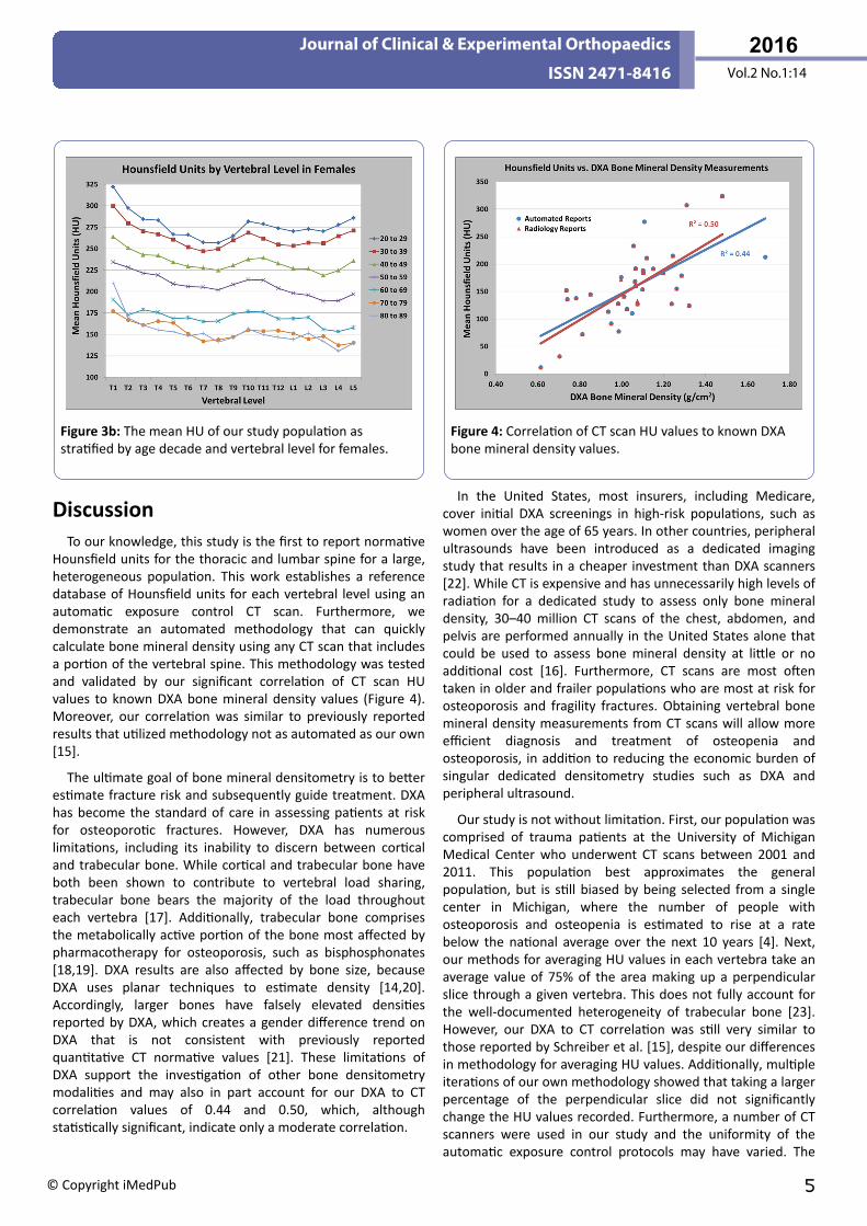
Figure 3b: The mean HU of our study population asstratified by age decade and vertebral level for females.
DiscussionTo our knowledge, this study is the first to report normative
Hounsfield units for the thoracic and lumbar spine for a large,heterogeneous population. This work establishes a referencedatabase of Hounsfield units for each vertebral level using anautomatic exposure control CT scan. Furthermore, wedemonstrate an automated methodology that can quicklycalculate bone mineral density using any CT scan that includesa portion of the vertebral spine. This methodology was testedand validated by our significant correlation of CT scan HUvalues to known DXA bone mineral density values (Figure 4).Moreover, our correlation was similar to previously reportedresults that utilized methodology not as automated as our own[15].
The ultimate goal of bone mineral densitometry is to betterestimate fracture risk and subsequently guide treatment. DXAhas become the standard of care in assessing patients at riskfor osteoporotic fractures. However, DXA has numerouslimitations, including its inability to discern between corticaland trabecular bone. While cortical and trabecular bone haveboth been shown to contribute to vertebral load sharing,trabecular bone bears the majority of the load throughouteach vertebra [17]. Additionally, trabecular bone comprisesthe metabolically active portion of the bone most affected bypharmacotherapy for osteoporosis, such as bisphosphonates[18,19]. DXA results are also affected by bone size, becauseDXA uses planar techniques to estimate density [14,20].Accordingly, larger bones have falsely elevated densitiesreported by DXA, which creates a gender difference trend onDXA that is not consistent with previously reportedquantitative CT normative values [21]. These limitations ofDXA support the investigation of other bone densitometrymodalities and may also in part account for our DXA to CTcorrelation values of 0.44 and 0.50, which, althoughstatistically significant, indicate only a moderate correlation.
Figure 4: Correlation of CT scan HU values to known DXAbone mineral density values.
In the United States, most insurers, including Medicare,cover initial DXA screenings in high-risk populations, such aswomen over the age of 65 years. In other countries, peripheralultrasounds have been introduced as a dedicated imagingstudy that results in a cheaper investment than DXA scanners[22]. While CT is expensive and has unnecessarily high levels ofradiation for a dedicated study to assess only bone mineraldensity, 30–40 million CT scans of the chest, abdomen, andpelvis are performed annually in the United States alone thatcould be used to assess bone mineral density at little or noadditional cost [16]. Furthermore, CT scans are most oftentaken in older and frailer populations who are most at risk forosteoporosis and fragility fractures. Obtaining vertebral bonemineral density measurements from CT scans will allow moreefficient diagnosis and treatment of osteopenia andosteoporosis, in addition to reducing the economic burden ofsingular dedicated densitometry studies such as DXA andperipheral ultrasound.
Our study is not without limitation. First, our population wascomprised of trauma patients at the University of MichiganMedical Center who underwent CT scans between 2001 and2011. This population best approximates the generalpopulation, but is still biased by being selected from a singlecenter in Michigan, where the number of people withosteoporosis and osteopenia is estimated to rise at a ratebelow the national average over the next 10 years [4]. Next,our methods for averaging HU values in each vertebra take anaverage value of 75% of the area making up a perpendicularslice through a given vertebra. This does not fully account forthe well-documented heterogeneity of trabecular bone [23].However, our DXA to CT correlation was still very similar tothose reported by Schreiber et al. [15], despite our differencesin methodology for averaging HU values. Additionally, multipleiterations of our own methodology showed that taking a largerpercentage of the perpendicular slice did not significantlychange the HU values recorded. Furthermore, a number of CTscanners were used in our study and the uniformity of theautomatic exposure control protocols may have varied. The
Journal of Clinical & Experimental Orthopaedics
ISSN 2471-8416 Vol.2 No.1:14
2016
© Copyright iMedPub 5

calibration of these devices is unknown to us but wasperformed on a routine basis per the manufacturer’srecommendations.
The underlying comorbidities of patients were not includedin our analyses, and many of these patients may have been onmetabolic bone medications such as hormone replacementtherapy, bisphosphonates, or teriparatide, potentiallyelevating the normative HU values for this group of patients.Separately analysing those who were on metabolic bonemedications from those naïve may have elucidated the effectsof these medications on vertebral HU. Furthermore,menopause could roughly be observed between the fifth andseventh decades of life, as seen in a proportionally larger lossof HU in figure 3b between these age groups. Between thefourth and fifth, fifth and sixth, and sixth and seventh decadesof life, there was an average percentage loss of HU across allvertebral levels of 10.8%, 11.4%, and 18.2%, respectively.
When using the lower limit of the normal population HUfrom our correlation analysis with DXA bone mineral density(Table 1), 41% of those 50 years of age or older, 37% of those60-69 years old, 60% of those 70-79 years old, and 67% ofthose 80-89 years old were considered osteopenia orosteoporotic based on their L1-L4 mean HU. These figures arelower than the estimate provided by the NationalOsteoporosis Foundation of 55% of those age 50 years or olderhaving osteopenia or osteoporosis [4]. This may be due to theaforementioned regional bias in our patient population andour lower female to male ratio of 0.7:1, with fewer maleshaving abnormal bone density. When stratified by sex, 45%(138/308) of females and 37% (166/443) of males 50 years orolder were considered osteopenia or osteoporotic by HU.
The severe morbidity and mortality of osteoporoticfractures are often underappreciated, and the societal cost ofthese fractures is enormous [3,24]. While population studieshave revealed that treating patients with osteopenia andosteoporosis can reduce fracture rate, there is still much tolearn about fracture risk at different vertebral levels [7,9,10].Our study identified varying bone mineral densities acrossdifferent vertebral levels within not only similar age ranges butalso within the same patient. Further studies are necessary tohelp identify particular vertebral levels at increased risk forfracture, which could create opportunities for prophylacticsupport systems and stabilization devices rather than solereliance on metabolic therapy. This increased understanding ofbone mineral density at different levels may also haveimplications for predicting hardware failure in instrumentation[25].
Fracture prevention is the ultimate goal of clinical bonedensitometry. With this work, we introduce a novelmethodology to ascertain bone mineral density using CT scans.Our resultant findings suggest that CT scans can be used as asupplemental tool for measuring bone mineral density, andmay subsequently aid in the diagnosis and management ofosteoporosis.
REFERENCES1. World Health Organization (1994) Assessment of fracture risk
and its application to screening for postmenopausalosteoporosis. Report of a WHO Study Group. World HealthOrgan Tech Rep Ser. 843: 1-129.
2. Prentice A (2004) Diet, nutrition and the prevention ofosteoporosis. Public Health Nutr. 7: 227-243.
3. Office of the Surgeon General (2004) Bone health andosteoporosis: a report of the Surgeon General. Rockville, MD:Office of the Surgeon General (US.
4. National Osteoporosis Foundation (2002) America’s bonehealth: the state of osteoporosis and low bone mass in ournation. Washington, DC: National Osteoporosis Foundation.
5. Olszynski WP, Davison KS, Adachi JD (2004) Osteoporosis in men:epidemiology, diagnosis, prevention, and treatment. Clin Ther.26: 15-28.
6. Burge R, Dawson-Hughes B, Solomon DH (2007) Incidence andeconomic burden of osteoporosis-related fractures in the UnitedStates, 2005-2025. J Bone Miner Res. 22: 465-475.
7. Dell RM, Greene D, Anderson D (2009) Osteoporosis diseasemanagement: What every orthopaedic surgeon should know. JBone Joint Surg Am. 6: 79-86.
8. NIH Consensus Development Panel (2001) Osteoporosisprevention, diagnosis, and therapy. JAMA 285: 785-795.
9. Marshall D, Johnell O, Wedel H (1996) Meta-analysis of how wellmeasures of bone mineral density predict occurrence ofosteoporotic fractures. BMJ 312: 1254-1259.
10. Kanis JA, Johnell O, Oden A (2008) FRAX and the assessment offracture probability in men and women from the UK.Osteoporos Int. 19: 385-397.
11. Mueller D, Gandjour A (2009) Cost-effectiveness of using clinicalrisk factors with and without DXA for osteoporosis screening inpostmenopausal women. Value Health 12: 1106-1117.
12. Lewiecki EM, Borges JL (2006) Bone density testing in clinicalpractice. Arq Bras Endocrinol Metabol 50: 586-595.
13. Lang TF, Keyak JH, Heitz MW (1997) volumetric quantitativecomputed tomography of the proximal femur: precision andrelation to bone strength. Bone 21: 101-108.
14. Guglielmi G, Lang TF (2002) Quantitative computed tomography.Semin Musculoskelet Radiol. 6: 219-227.
15. Schreiber JJ, Anderson PA, Rosas HG (2011) Hounsfield units forassessing bone mineral density and strength: a tool forosteoporosis management. J Bone Joint Surg Am. 93:1057-1063.
16. Brenner DJ, Hall EJ (2007) Computed tomography--an increasingsource of radiation exposure. N Engl J Med. 357: 2277-2284.
17. Eswaran SK, Gupta A, Adams MF (2006) Cortical and trabecularload sharing in the human vertebral body. J Bone Miner Res. 21:307-314.
18. Black DM, Greenspan SL, Ensrud KE (2003) the effects ofparathyroid hormone and alendronate alone or in combinationin postmenopausal osteoporosis. N Engl J Med. 349: 1207-1215.
19. Rubin MR, Cosman F, Lindsay R (2002) the anabolic effects ofparathyroid hormone. Osteoporos Int. 13: 267-277.
Journal of Clinical & Experimental Orthopaedics
ISSN 2471-8416 Vol.2 No.1:14
2016
6 This article is available from: 10.4172/2471-8416.100014

20. Kanis JA, McCloskey EV, Johansson H (2008) A referencestandard for the description of osteoporosis. Bone 42: 467-475.
21. Genant HK, Block JE, Steiger P (1987) Quantitative computedtomography in assessment of osteoporosis. Semin Nucl Med.17: 316-333.
22. Magkos F, Manios Y, Babaroutsi E (2005) Quantitative ultrasoundcalcaneus measurements: normative data for the Greekpopulation. Osteoporos Int. 16: 280-288.
23. Keaveny TM, Hayes WC (1993) a 20-year perspective on themechanical properties of trabecular bone. J Biomech Eng. 115:534-542.
24. National Osteoporosis Foundation (2009) Osteoporosis AnnualReport. Washington, DC: National Osteoporosis Foundation.
25. Turkyilmaz I, Ozan O, Yilmaz B (2008) Determination of bonequality of 372 implant recipient sites using Hounsfield unit fromcomputerized tomography: a clinical study. Clin Implant DentRelat Res. 10: 238-244.
Journal of Clinical & Experimental Orthopaedics
ISSN 2471-8416 Vol.2 No.1:14
2016
© Copyright iMedPub 7