Normal Sinus Rhythm MIAMI DADE FIRE RESCUE · All patients with a 12 lead EKG displaying ST segment...
4
Normal Sinus Rhythm Rate 60 – 100 Rhythm Regular P Wave? Yes. Upright and normal PR Interval? Less than 4 small boxes QRS Complex? Narrow. Less than 3 small boxes Ectopy? Sinus Tachycardia Rate Above 100 Sinus Bradycardia Rate Less than 60 Sinus Arrhythmia Rate 60 - 100 Rhythm Irregular Sinus Arrest Rate 60 - 100 Rhythm Irregular –period of inactivity (Arrest) P Wave? Yes. Upright and normal PR Interval? Less than 4 small boxes QRS Complex? Narrow. Less than 3 small boxes Ectopy? MIAMI DADE FIRE RESCUE MIAMI, FL Always Ready, Proud To Serve Wandering Pacemaker Rate 60 - 100 Rhythm May be irregular P Wave? Yes. Different appearance PR Interval? Less than 4 small boxes, but can have varying PR intervals QRS Complex? Narrow. Less than 3 small boxes Ectopy? Atrial Tachycardia Rate 150-250 Rhythm May be irregular P Wave? Yes. May be multiform PR Interval? Less than 4 small boxes, but can have varying PR intervals QRS Complex? Narrow. Less than 3 small boxes Ectopy? Supraventricular Tachycardia Rate 150-250 Rhythm Regular P Wave? Unable to see P waves – buried in T-wave PR Interval? Usually unable to see QRS Complex? Narrow. Less than 3 small boxes Ectopy? Atrial Flutter Rate 60 - 100 Rhythm May be irregular P Wave? Multiple p waves for each QRS PR Interval? Less than 4 small boxes, but can have varying PR intervals QRS Complex? Narrow. Less than 3 small boxes Ectopy? Atrial Fibrillation Rate Atrial - ≥350, Ventricular rate may vary Rhythm Irregularly irregular P Wave? “Fib waves” showing atrial activity PR Interval? Unidentifiable QRS Complex? Narrow. Less than 3 small boxes Ectopy? Junctional Rhythm Rate 40 - 60 Rhythm Regular P Wave? Absent – may be inverted or buried PR Interval? None (short if inverted) QRS Complex? Narrow. Less than 3 small boxes Ectopy? Accelerated Junctional Rhythm Rate 60 - 100 Rhythm Regular P Wave? Absent – may be inverted or buried PR Interval? None (short if inverted) QRS Complex? Narrow. Less than 3 small boxes Ectopy?
Transcript of Normal Sinus Rhythm MIAMI DADE FIRE RESCUE · All patients with a 12 lead EKG displaying ST segment...
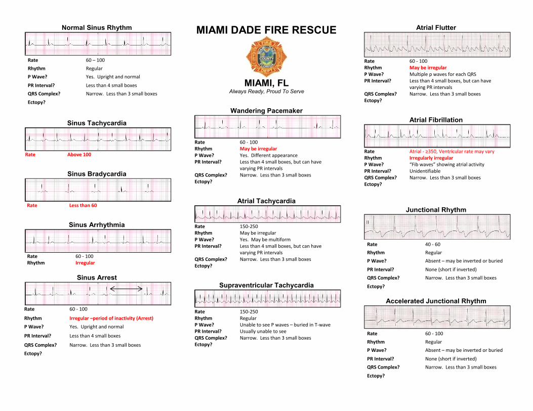
Normal Sinus Rhythm
Rate 60 – 100Rhythm RegularP Wave? Yes. Upright and normal
PR Interval? Less than 4 small boxesQRS Complex? Narrow. Less than 3 small boxesEctopy?
Sinus Tachycardia
Rate Above 100
Sinus Bradycardia
Rate Less than 60
Sinus Arrhythmia
Rate 60 - 100Rhythm Irregular
Sinus Arrest
Rate 60 - 100
Rhythm Irregular –period of inactivity (Arrest)P Wave? Yes. Upright and normal
PR Interval? Less than 4 small boxes
QRS Complex? Narrow. Less than 3 small boxesEctopy?
MIAMI DADE FIRE RESCUE
MIAMI, FLAlways Ready, Proud To Serve
Wandering Pacemaker
Rate 60 - 100Rhythm May be irregularP Wave? Yes. Different appearancePR Interval? Less than 4 small boxes, but can have
varying PR intervalsQRS Complex? Narrow. Less than 3 small boxesEctopy?
Atrial Tachycardia
Rate 150-250Rhythm May be irregularP Wave? Yes. May be multiformPR Interval? Less than 4 small boxes, but can have
varying PR intervalsQRS Complex? Narrow. Less than 3 small boxesEctopy?
Supraventricular Tachycardia
Rate 150-250Rhythm RegularP Wave? Unable to see P waves – buried in T-wavePR Interval? Usually unable to seeQRS Complex? Narrow. Less than 3 small boxesEctopy?
Atrial Flutter
Rate 60 - 100Rhythm May be irregularP Wave? Multiple p waves for each QRSPR Interval? Less than 4 small boxes, but can have
varying PR intervalsQRS Complex? Narrow. Less than 3 small boxesEctopy?
Atrial Fibrillation
Rate Atrial - ≥350, Ventricular rate may varyRhythm Irregularly irregularP Wave? “Fib waves” showing atrial activityPR Interval? UnidentifiableQRS Complex? Narrow. Less than 3 small boxesEctopy?
Junctional Rhythm
Rate 40 - 60Rhythm RegularP Wave? Absent – may be inverted or buried
PR Interval? None (short if inverted)QRS Complex? Narrow. Less than 3 small boxes
Ectopy?
Accelerated Junctional Rhythm
Rate 60 - 100Rhythm RegularP Wave? Absent – may be inverted or buried
PR Interval? None (short if inverted)QRS Complex? Narrow. Less than 3 small boxes
Ectopy?
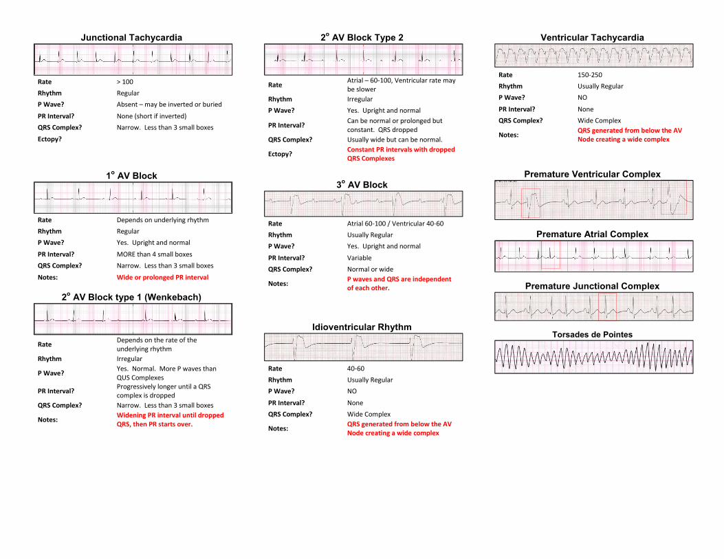
Junctional Tachycardia
Rate > 100Rhythm RegularP Wave? Absent – may be inverted or buried
PR Interval? None (short if inverted)QRS Complex? Narrow. Less than 3 small boxesEctopy?
1o AV Block
Rate Depends on underlying rhythmRhythm RegularP Wave? Yes. Upright and normal
PR Interval? MORE than 4 small boxesQRS Complex? Narrow. Less than 3 small boxes
Notes: Wide or prolonged PR interval
2o AV Block type 1 (Wenkebach)
Rate Depends on the rate of theunderlying rhythm
Rhythm Irregular
P Wave? Yes. Normal. More P waves thanQUS Complexes
PR Interval? Progressively longer until a QRScomplex is dropped
QRS Complex? Narrow. Less than 3 small boxes
Notes: Widening PR interval until droppedQRS, then PR starts over.
2o AV Block Type 2
Rate Atrial – 60-100, Ventricular rate maybe slower
Rhythm IrregularP Wave? Yes. Upright and normal
PR Interval? Can be normal or prolonged butconstant. QRS dropped
QRS Complex? Usually wide but can be normal.
Ectopy? Constant PR intervals with droppedQRS Complexes
3o AV Block
Rate Atrial 60-100 / Ventricular 40-60Rhythm Usually RegularP Wave? Yes. Upright and normal
PR Interval? VariableQRS Complex? Normal or wide
Notes: P waves and QRS are independentof each other.
Idioventricular Rhythm
Rate 40-60Rhythm Usually RegularP Wave? NO
PR Interval? NoneQRS Complex? Wide Complex
Notes: QRS generated from below the AVNode creating a wide complex
Ventricular Tachycardia
Rate 150-250Rhythm Usually RegularP Wave? NO
PR Interval? NoneQRS Complex? Wide Complex
Notes: QRS generated from below the AVNode creating a wide complex
Premature Ventricular Complex
Premature Atrial Complex
Premature Junctional Complex
Torsades de Pointes
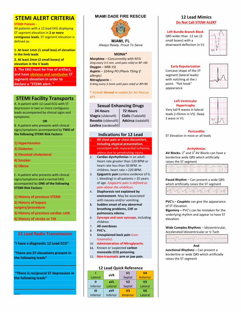
STEMI ALERT CRITERIASTEMI Patient –All patients with a 12 lead EKG displayingST segment elevation in 2 or morecontiguous leads. ST segment elevation isdefined as:
1. At least 1mm (1 small box) of elevationin the limb leads2. At least 2mm (2 small boxes) ofelevation in the V leads3. The EKG must be free of artifact,and have obvious and conclusive STsegment elevation in order todeclare a “STEMI alert. “
STEMI Facility TransportsA. A patient with 12-Lead ECG with STdepression in two or more contiguousleads accompanied by clinical signs andsymptoms.
ORB. A patient who presents with clinicalsigns/symptoms accompanied by TWO ofthe following STEMI Risk Factors:
1) Hypertension2) Diabetes3) Elevated cholesterol4) Smoker5) Obese
C. A patient who presents with clinicalsigns/symptoms and a normal EKGaccompanied by ONE of the followingSTEMI Risk Factors:
1) History of previous STEMI2) History of bypasssurgery/procedure3) History of previous cardiac cath4) History of stroke or TIA
12 Lead Radio Transmission
“I have a diagnostic 12 Lead ECG”
“There are ST elevations present inthe following leads”
“There is reciprocal ST depression inthe following leads”
__________________________________
MIAMI DADE FIRE RESCUE
MIAMI, FLAlways Ready, Proud To Serve
MONA*Morphine – Concurrently with NTG2mg every 3-5 min. until pain relief or BP <90Oxygen – NRB 15LAspirin – 324mg PO (Plavix 75mg ifallergic)Nitroglycerin –0.4mg every 3-5min until pain relief or BP<90
* Include Versed to sedate for Air Rescuept’s
Sexual Enhancing Drugs24 Hours 72 Hours
Viagra (sildenafil) Cialis (Tadalafil)Revatio (sildenafil) Adcirca (tadalafil)Levitra (vardenafil)
Indications for 12 Lead1. All chest pain or chest discomfort,
including atypical presentation,consistent with myocardial ischemia,unless due to penetrating injury.
2. Cardiac dysrhythmias in an adult:Heart rate greater than 120 BPM orheart rate less than 50 BPM. Inchildren, heart rate > 220 BPM.
3. Epigastric pain (unless evidence of G.I. bleeding) in all patients > 35 yearsof age. Epigastric pain is defined aspain above the umbilicus.
4. Diaphoresis not explained byenvironment. May be associatedwith nausea and/or vomiting.
5. Sudden onset of any abnormalbreathing problems, CHF orpulmonary edema.
6. Syncope and near syncope, includingchildren.
7. All overdoses.8. PVC’s.9. Unexplained back pain (non-
traumatic).10. Administration of Nitroglycerin.11. Known or suspected carbon
monoxide (CO) poisoning.12. Non-traumatic arm or jaw pain.
12 Lead Quick ReferenceI
Lateral aVR V1Septal
V4Anterior
IIInferior
aVLLateral
V2Septal
V5Lateral
IIIInferior
aVFInferior
V3Anterior
V6Lateral
12 Lead MimicsDo Not Call STEMI ALERT
Left Bundle Branch BlockQRS wider than .12 sec (3small boxes) with adownward deflection in V1
Early RepolarizationConcave shape of the STsegment (lateral leads)with notching at the Jpoint. “fish hook”appearance
Left VentricularHypertrophy
Very tall R waves in lateralleads (>35mm in V5) DeepS wave in V1
PericarditisST Elevation in most or all leads
ArrhythmiasAV Blocks- 2o and 3o AV Blocks can have aborderline wide QRS which artificiallyraises the ST segment
Paced Rhythm – Can present a wide QRSwhich artificially raises the ST segment
PVC’s – Couplets can give the appearanceof ST Elevation.Bigeminy – PVC’s can be mistaken for theunderlying rhythm and appear to have STelevation
Wide Complex Rhythms – Idioventricular,Accelerated Idioventricular or V-Tach
AndJunctional Rhythms – Can present aborderline or wide QRS which artificiallyraises the ST segment
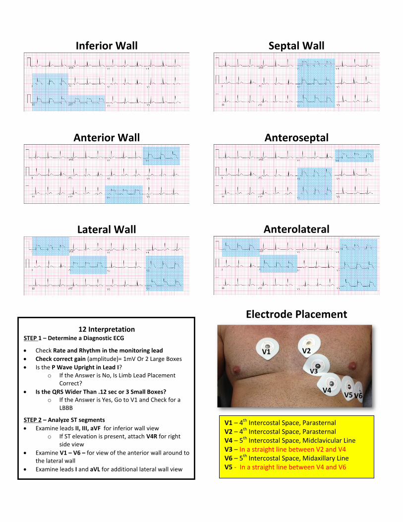
Inferior Wall
Anterior Wall
Lateral Wall
12 InterpretationSTEP 1 – Determine a Diagnostic ECG
Check Rate and Rhythm in the monitoring lead Check correct gain (amplitude)= 1mV Or 2 Large Boxes Is the P Wave Upright in Lead I?
o If the Answer is No, Is Limb Lead PlacementCorrect?
Is the QRS Wider Than .12 sec or 3 Small Boxes?o If the Answer is Yes, Go to V1 and Check for a
LBBB
STEP 2 – Analyze ST segments Examine leads II, III, aVF for inferior wall view
o If ST elevation is present, attach V4R for rightside view
Examine V1 – V6 – for view of the anterior wall around tothe lateral wall
Examine leads I and aVL for additional lateral wall view
Septal Wall
Anteroseptal
Anterolateral
Electrode Placement
V6V5
V3
V4
V2V1
V1 – 4th Intercostal Space, ParasternalV2 – 4th Intercostal Space, ParasternalV4 – 5th Intercostal Space, Midclavicular LineV3 – In a straight line between V2 and V4V6 – 5th Intercostal Space, Midaxillary LineV5 - In a straight line between V4 and V6