Normal Range of Human Left Ventricular Volumes and Mass Using Steady State Free Precession MRI in...
5
Magn Reson Mater Phy (2006) 19: 41–45 DOI 10.1007/s10334-005-0025-8 RESEARCH ARTICLE Sarah Clay Khaled Alfakih Aleksandra Radjenovic Timothy Jones John P. Ridgway Mohan U. Sinvananthan Normal range of human left ventricular volumes and mass using steady state free precession MRI in the radial long axis orientation Received: 6 June 2005 Accepted: 22 December 2005 Published online: 14 February 2006 © ESMRMB 2006 S. Clay (B ) · K. Alfakih · T. Jones M. U. Sinvananthan BHF Cardiac MRI Unit, B Floor, Clarendon Wing, Leeds General Infirmary, Great George Street, Leeds LS1 3EX, UK E-mail: [email protected] Tel.: +44-113-3925167 Fax: +44-113-3925690 A. Radjenovic · J. P. Ridgway Academic Unit of Medical Physics, University of Leeds, Leeds, UK Abstract Background: The radial long-axis orientation for the measurement of left ventricular (LV) volume and mass has been shown to have advantages over the short-axis orientation. Previous work has highlighted the need for technique specific normal ranges. The purpose of this study was therefore to establish normal ranges of LV volume and mass for the radial long-axis orientation. Materials and methods: Forty normal subjects (20 males, average age 32.3, age range 19–58; 20 females, average age 37.4, age range 21–54) were examined utilising a steady state free precession (SSFP) pulse sequence. Two observers analysed the images independently using in-house validated software. Results: The normal ranges for LV end-diastolic volume measurements after adjustment to body surface area (BSA) were 62–120 ml for males and 58–103 ml for females. LV mass indexed to BSA ranged from 50–86 g for males and 36–72 g for females. The normal range for ejection fraction was 49–73 % for males and 54–73 % for females. Conclusion:A gender specific normal range using SSFP in the radial long-axis orientation was established. Keywords Cardiovascular magnetic resonance · SSFP · Radial long axis orientation · Normal range · Left ventricular volume and mass Introduction Cardiac MRI has now become the technique of choice for the measurement of left ventricular(LV) function, vol- ume and mass. It is both a highly accurate and reproduc- ible technique due to the excellent definition of both the endocardial and epicardial borders of the ventricle and avoids the need for mathematical formulae. The pres- ence and degree of left ventricular hypertrophy, present in approximately 30% of patients with hypertension, can be detected by calculating LV mass [1]. There is a direct linear relationship between cardiovascular morbidity and mortality and LV mass [2]. There is also evidence that the regression of LV mass reduces the risk of cardiovascu- lar events [3, 4]. Assessing LV function accurately is also a useful clinical tool for the diagnosis and management of heart failure and is routinely performed by echocar- diography[1]. As echocardiography values are dependent on geometric assumptions, they are inherently inaccu- rate in the abnormal ventricle; comparative studies have shown that cardiac magnetic resonance (CMR) is both a more accurate and reproducible technique [3, 5–7]. Accu- rate measurements of LV mass and ejection fraction are not only invaluable for research purposes but also in the long-term follow up of an individual patient. CMR is also capable of assessing several aspects of heart disease in a single study [8, 9]. Magnetic resonance images obtained in the short axis orientation were shown to give the most accurate mea- surements of LV mass [10]. Short axis slices therefore be- came the conventional orientation for image acquisition
-
Upload
sarah-clay -
Category
Documents
-
view
212 -
download
0
Transcript of Normal Range of Human Left Ventricular Volumes and Mass Using Steady State Free Precession MRI in...
Magn Reson Mater Phy (2006) 19: 41–45DOI 10.1007/s10334-005-0025-8 RESEARCH ARTICLE
Sarah ClayKhaled AlfakihAleksandra RadjenovicTimothy JonesJohn P. RidgwayMohan U. Sinvananthan
Normal range of human left ventricularvolumes and mass using steady state freeprecession MRI in the radial long axisorientation
Received: 6 June 2005Accepted: 22 December 2005Published online: 14 February 2006© ESMRMB 2006
S. Clay (B) · K. Alfakih · T. JonesM. U. SinvananthanBHF Cardiac MRI Unit,B Floor, Clarendon Wing,Leeds General Infirmary,Great George Street,Leeds LS1 3EX, UKE-mail: [email protected].: +44-113-3925167Fax: +44-113-3925690
A. Radjenovic · J. P. RidgwayAcademic Unit of Medical Physics,University of Leeds, Leeds, UK
Abstract Background: The radiallong-axis orientation for themeasurement of left ventricular (LV)volume and mass has been shown tohave advantages over the short-axisorientation. Previous work hashighlighted the need for techniquespecific normal ranges. The purposeof this study was therefore toestablish normal ranges of LVvolume and mass for the radiallong-axis orientation. Materials andmethods: Forty normal subjects (20males, average age 32.3, age range19–58; 20 females, average age 37.4,age range 21–54) were examinedutilising a steady state freeprecession (SSFP) pulse sequence.Two observers analysed the imagesindependently using in-house
validated software. Results: Thenormal ranges for LV end-diastolicvolume measurements afteradjustment to body surface area(BSA) were 62–120 ml for males and58–103 ml for females. LV massindexed to BSA ranged from 50–86 gfor males and 36–72 g for females.The normal range for ejectionfraction was 49–73 % for males and54–73 % for females. Conclusion: Agender specific normal range usingSSFP in the radial long-axisorientation was established.
Keywords Cardiovascular magneticresonance · SSFP · Radial long axisorientation · Normal range ·Left ventricular volume and mass
Introduction
Cardiac MRI has now become the technique of choicefor the measurement of left ventricular(LV) function, vol-ume and mass. It is both a highly accurate and reproduc-ible technique due to the excellent definition of both theendocardial and epicardial borders of the ventricle andavoids the need for mathematical formulae. The pres-ence and degree of left ventricular hypertrophy, presentin approximately 30% of patients with hypertension, canbe detected by calculating LV mass [1]. There is a directlinear relationship between cardiovascular morbidity andmortality and LV mass [2]. There is also evidence that theregression of LV mass reduces the risk of cardiovascu-lar events [3,4]. Assessing LV function accurately is also
a useful clinical tool for the diagnosis and managementof heart failure and is routinely performed by echocar-diography[1]. As echocardiography values are dependenton geometric assumptions, they are inherently inaccu-rate in the abnormal ventricle; comparative studies haveshown that cardiac magnetic resonance (CMR) is both amore accurate and reproducible technique [3,5–7]. Accu-rate measurements of LV mass and ejection fraction arenot only invaluable for research purposes but also in thelong-term follow up of an individual patient. CMR is alsocapable of assessing several aspects of heart disease in asingle study [8,9].
Magnetic resonance images obtained in the short axisorientation were shown to give the most accurate mea-surements of LV mass [10]. Short axis slices therefore be-came the conventional orientation for image acquisition

42
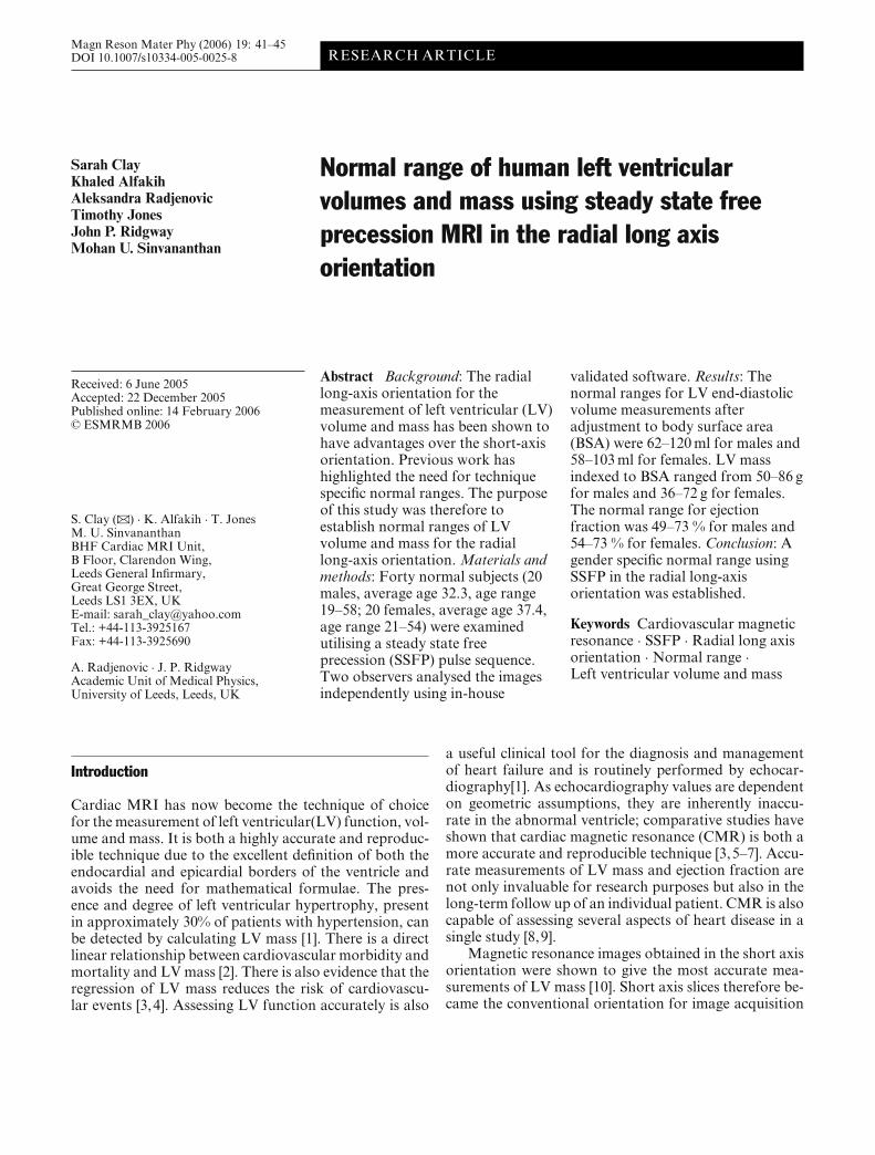
Fig. 1 A representative end-diastolic image with endocardial andepicardial contours shown
and analysis. The accuracy of the results from short axisimages are however limited by the difficulties encounteredwhen defining the most basal slice adjacent to the mitralvalve plane due to partial volume effects. The basal sliceshave a large cross sectional area so can have a consider-able effect on both the volume and the mass. Determi-nation of the true basal slice can therefore be subjectiveand can be a significant cause of interobserver variabil-ity, with one study showing a highly significant difference(p<0.001) between inclusion and exclusion of the basalslice for all the ventricular parameters [11].
Radially orientated long axis slices avoid the basalslice problem and have intrinsic advantages as they givea clear view of the mitral and aortic valve planes, thusreducing the partial volume effects which hinder basalslice determination and also improve localisation of theapex (see Fig. 1). A previous study by our group assessedthe use of a steady state free precession (SSFP) sequenceand radial slices to calculate LV volume and mass [10]Using a phantom and porcine hearts to validate the tech-nique volume estimates were shown to be within 3% ofthe actual value. The group investigated the minimumnumber of radial slices required for an accurate volumeand mass measurement by comparing the analysis of var-ious numbers of slices to the full 36 slice set. When nineor more slices were included, the difference was < 1%in all cases [10]. Six slices has also been shown to beenough to accurately cover the entire ventricle in anothervalidated study in the radial long-axis orientation [12].A larger measured LV volume and lower LV mass was
observed in the short-axis orientation compared to theradial-long axis measurements despite showing good cor-relation when comparing the two techniques, highlightingthe need for technique-specific normal ranges [10].
Normal ranges of LV volumes and mass have beenpublished for short-axis images acquired by different tech-niques. In this study, gender specific normal ranges wereestablished for LV volume and mass in the radial long-axis orientation and were indexed to body surface area(BSA) and height.
Methods
Subjects
Forty normal subjects (20 males, average age 32.3, age range19–58; 20 females, average age 37.4, age range 21–54) with anormal blood pressure, defined as a systolic BP < 140 mmHgand diastolic BP < 90 mmHg. They also had a normal cardiovas-cular examination, normal resting 12-lead ECG and no historyof cardiovascular disease or diabetes and were not taking anymedications. Subjects were excluded if found to have contrain-dications to CMR scanning, e.g. pacemakers, aneurysm clips orclaustrophobia. Other exclusion criteria included arrhythmias,age over 65 and under 18, elite athletes and pregnancy. The studyreceived local ethics committee approval and informed consentwas obtained from all subjects.
Acquisition protocol
The CMR studies were performed on a 1.5-T Philips Intera CVMRI system (Philips Medical Systems, Best, The Netherlands).This system is equipped with master gradients with maximumgradient amplitude of 30 mT/m and a maximum slew rate of150 mT/m/ms. All subjects were imaged in the supine positionusing a vectocardiographic method for ECG synchronisation anda five-element cardiac phased array coil. An SSFP pulse sequencewas used to obtain the images. (TR = 3.34 ms, TE = 1.67 ms,flip angle = 55◦, bandwidth = 1042 Hz/pixel, acquisition matrix= 192× 163, FOV = 360 × 288 mm, half Fourier acquisitionmatrix 6-mm slice thickness, 4-mm interslice gap, 18 phases/car-diac cycle, with 2 slices acquired within a 10- to 12-s breath hold.)Standard survey images were first acquired followed by breath-hold cine acquisitions in the LV long axis, a short axis slice at themid-ventricular level and a double oblique four chamber view toset up the series of radial long axis slices. The radial long axisslices have equal angular separation with a common axis alignedfrom the apex to the mid-point of the mitral valve orifice [10].Nine slices were acquired and analysed to adequately cover theentire ventricle.
Image analysis
Commercially available analysis software (MASS version 5.0,Medis Medical Imaging Systems, Leiden, The Netherlands) was

43
Table 1 The mean values ± one standard deviation and the upper and lower limits of normal for left ventricular (LV) parameters with andwithout adjustment to body surface area
Male (n =20) Female (n =20)
Mean ± SD Normal range Mean ± SD Normal range
EDV (ml) 181.1±30.3 121–242 144.9±21.1 103–187EDV/BSA (ml/m2) 90.9±14.3 62–120 80.7±11.2 58–103EDV/HT (ml/m) 95.8±27.2 41–150 88.4±10.1 68–109ESV (ml) 70.7±18.0 35–107 52.5±12.4 28–77MASS (g) 136.2±20.5 95–177 96.2±15.5 65–127MASS/BSA (g/m2) 68.0±8.8 50–86 53.7±8.9 36–72MASS/HT (g/m) 76.0±10.4 55–89 54.9±15.0 25–85EF (%) 61.1±6.0 49–73 63.6±4.8 54–73
SD standard deviation, E DV end diastolic volume, BS A body surface area, E SV end systolic volume, E F ejection fraction, H Theight
Table 2 The interobserver variability for LV measurements (n =40)
Interobserver variability EDV (ml) ESV (ml) EF (%) Mass (g)
Bias −2.6 −1.3 0.002 5.7SDD 7.0 5.6 0.05 6.8SDD (%) 4.8 9.7 4.6 5.7Pearson’s correlation 0.97 0.95 0.85 0.97Limits of agreement −16.7 to 11.4 −12.4 to 9.8 −0.1 to 0.1 −7.9 to 19.3
SDD standard deviation of the difference between two means, SDD(%) The SDD expressed as a percentage of the mean result,E DV end diastolic volume, E SV end systolic volume, E F ejection fraction
used off-line on a workstation (UltraSPARC 10, SUN Microsys-tems) by two observers independently to trace epicardial andendocardial contours. Contours were closed in straight linesacross both the mitral and aortic valve rings and were not tracedaround papillary muscles. Volumetric analysis and mass calcula-tions were carried out using software developed in the departmentspecifically for the radial long-axis orientation and previouslyvalidated with both phantom and porcine models before imple-mentation [10]. LV EDV and LV mass were indexed to BSA andheight.
Statistical analysis
The mean ± one standard deviation (SD) was calculated for eachof the LV parameters measured. The normal range was expressedas two standard deviations below the mean to two standard devi-ations above. The Bland–Altman method was used to calculatebias and the limits of agreement [13]. Observer variability wasexpressed as the SD of the difference (SDD) between two meansand the co-efficient of variation expressed as the SDD of the tworeadings divided by the average value of the two readings. Pear-son’s correlation was also calculated to evaluate how closely theresults correlated between the two observers.
Results
Diagnostic quality data sets were obtained in all of the40 subjects. Table 1 shows the mean ±1 standard devia-tion for the measured LV parameters for both men andwomen and the normal ranges, calculated as two stan-dard deviations below to two standard deviations abovethe mean. As expected the mean values were greater inmen than women for all LV parameters measured exceptthe ejection fraction. This gender difference remained forEDV and mass after indexation to BSA and height.
To assess the reproducibility of the measurements forthis normal range, the inter- and intraobserver variabil-ity was calculated using the Bland–Altman method. Ta-bles 2 and 3 show the inter- and intraobserver variabilityexpressed as the mean bias, the limits of agreement, thestandard deviation of the difference (SDD) and the coeffi-cient of variation.
Discussion
Cardiac magnetic resonance (CMR) normal ranges forLV volumes and mass have been previously defined for

44
Table 3 The intraobserver variability for LV measurements (n =40)
Intraobserver variability EDV (ml) ESV (ml) EF (%) MASS (g)
Bias 1.4 0.2 0.003 0.3SDD 4.5 4.1 0.05 5.7SDD(%) 2.4 7.9 4.3 5.1Pearson’s correlation 0.99 0.96 0.88 0.98Limits of agreement −7.5 to 10.3 −9.2 to 9.6 −0.1 to 0.1 −11.6 to 12.2
SDD standard deviation of the difference between two means, SDD(%) The SDD expressed as a percentage of the mean result,E DV end diastolic volume, E SV end systolic volume, E F ejection fraction
images acquired in the short-axis orientation [14–16]. Thisis the first study to define normal ranges for LV volumesand mass in the radial long-axis orientation using SSFP.The previous study from our group demonstrated thatusing the SSFP sequence in the radial long-axis orien-tation could rapidly assess LV function and mass, pro-ducing accurate and reproducible results [10]. The mainlimitation of this study was the small number of volun-teers, nine in total.
One limitation of the radial long-axis orientation isthat closing contours across the mitral valve plane usu-ally results in a slight overestimate of LV end systolic vol-ume (ESV) because a small volume of blood on the atrialside of the valve is included in the contour. However, thisoverestimate will be systematic and have a much smalleroverall effect than the incorrect inclusion or exclusion ofthe most basal slice for short axis studies.
A recent study describing a normal range using SSFPin the short axis orientation by Alfakih et al. [14] reporteda similar range of LV dimensions to our results. The LVmass indexed to BSA in men was 46–83 g/m2 compared to50–86 g/m2 in this study and in women 37–67 g/m2 com-pared to 36–72 g/m2 in this study. LV end distolic volume(EDV) indexed to BSA was also similar between the twostudies with a range of values of 53–112 ml/m2 comparedto 62–120 ml/m2 in men and 56–99 ml/m2 compared to58–103 ml/m2 in women. The previous study from ourgroup compared the radial long-axis orientation to theshort-axis orientation and showed that EDV, ESV andmass were all slightly greater in the radial orientation [10].It is thought that the slight over-estimation of mass in theradial long-axis orientation with respect to the short-axismass occurs because in the short-axis orientation part ofthe basal portion of the ventricle is excluded from analysisat the atrioventricular boundary [10]. The likely explana-tion for the smaller difference between this study and therecent study by Alfakih et al. [14] is that different criteriawere used when determining the basal slice in the shortaxis orientation. The previous study from our group how-ever only included a slice if there was at least 75% of themyocardium visible around the blood pool whereas Al-fakih et al. followed the Lorenz criteria stating that anyslice with at least 50% of the myocardium visible should
be included which have since become the gold-standardcriteria [10,14,15]. The similarity between the results ofthe two orientations is a promising finding.
On the whole, the results for the intraobserver and in-terobserver variability are lower than those in the short-axis normal range published by Alfakih et al. [14]. Thisimprovement can be attributed to the fact that basal slicesand papillary muscles did not have to be contended within the radial long-axis images. Papillary muscles were ig-nored because a limitation of the radial long-axis orienta-tion is that a papillary muscle can be missed between slicesor that the papillary muscle contour can be integratedthroughout the whole sector represented by the radialslice resulting in an overestimation of the LV mass [10].The previous radial paper from our group only tabulatedinterobserver variability and expressed it as a percentageof the mean result for their work with radial long-axisimages [10]. Their interobserver variability ranged from2.0 to 7.0% compared to 4.8 to 9.7% in this study. Withthe exception of ESV the interobserver variability for allmeasurements was lower than the results previously pub-lished by our group, Bloomer et al. [10]. In this instancethe difference between the variability for ESV is likely tobe due to the difficulty in defining the end-systolic phase,a problem also previously highlighted by our earlier workand not specific to the radial long-axis orientation [10].They overcame the problem by drawing contours on morethan one phase but this lengthens analysis time. These arerelatively minor limitations in comparison to the problemof determining the basal slice in the short-axis orientation.
The gender differences observed for all LV parameterswere consistent with many previously reported differences[14–16]. The age range of the normal volunteers are alsoimportant as Alfakih et al. [14] demonstrated a differencein results of LV parameters between older and youngerage groups. The volunteers used in this study covered awide age range from 19 to 58 and would therefore beacceptable to use for a normal range.
In conclusion, a gender specific normal range in theradial long-axis orientation was established.
Acknowledgements This work was carried out in the BritishHeart Foundation cardiac MRI unit at the Leeds General Infirmary.

45
References
1. Missouris CG, Forbat SM, Singer DR,Markandu ND, Underwood R,MacGregor GA (1996)Echocardiography overestimates leftventricular mass: a comparative studywith magnetic resonance imaging inpatients with hypertension. JHypertens 14(8):1005–1010
2. Verdecchia P, Scillaci G, Borgioni C,Ciucci A, Gattobigio R, Zampi I,Reboldi G, Porcellati C (1998)Prognostic significance of serialchanges in left ventricular mass inessential hypertension. Circulation97:48–54
3. Dahlof B, Devereux RB, Kjeldsen SE,Julius S, Beevers G, Faire U, FyhrquistF, Ibsen H, Kristiansson K,Lederballe-Pederson O, Lindholm LH,Nieminen MS, Omvik P, Oparil S,Wedel H (2002) The LSG.Cardiovascular morbidity andmortality in the Losartan interventionfor endpoint reduction in hypertensionstudy (LIFE): a randomised trialagainst atenolol. (comment, summaryfor patients. J Fam Pract; 51(7):599;PMID: 12160492). Lancet359:995–1003
4. St John Sutton M, Otterstat JE,Plappert T, Parker a, Sekarski D,Keane MG, Poole-Wilson P, Lubsen K(1998) Quantification of left ventricularvolumes and ejection fraction inpost-infarction patients from biplaneand single plane two-dimensionalechocardiograms. A prospectivelongitudinal study of 371 patients. EurHeart J 19:808–816
5. Hibberd MG, Chuang ML, BleaudinRA, Riley MF, Mooney MG,Fearnside JT, Manning WJ, DouglasPS (2000) Accuracy ofthree-dimensional echocardiographywith unrestricted selection of imagingplanes for measurement of leftventricular volumes and ejectionfraction. Am Heart J 140:469–475
6. Buck T, Hunold P, Wentz KU, TkalecW, Nesser HJ, Erbel R (1997)Tomographic three-dimensionalechocardiographic determination ofchamber size and systolic function inpatients with left ventricular aneurysm:comparison to magnetic resonanceimaging, cineventriculography, andtwo-dimensional echocardiography.Circulation 96:4286–4297
7. Salcedo EE, Gockowski K, Tarazi RC(1979) Comparison of M Mode andtwo-dimensional echocardiography intwo experimental models. AmJ Cardiol 4:936–940
8. Alfakih K, Thiele H, Plein S,Bainbridge GJ, Ridgway J,Sinvananthan UM (2002) Comparisonof right ventricular volumemeasurement between segmentedk-space gradient-echo and steady statefree precession magnetic resonanceimaging. J Magn Reson 16:253–258
9. Aebischer N, Meuli R, Jeanrenaud X,Koerfer J, Kappenberger L (1998) Anechocardiographic and magneticresonance imaging comparative studyof right ventricular volumedetermination. Int J Cardiac Imaging14:271–278
10. Bloomer T, Plein S, Radjenovic A,Higgins DM, Jones TR, Ridgway JP,Sinvananthan UM (2001) Cine MRIusing steady state free precession in theradial long axis orientation is a fastaccurate method for obtainingvolumetric data of the left ventricle.J Magn Reson 14:685–692
11. Marcus JT, Gotte MJ, DeWaal LK,Stam MR, Van der Geest RJ, HeethaarRM (1999) The influence ofthrough-plane motion on leftventricular volumes measured bymagnetic resonance imaging:implications for image acquisition andanalysis. J Cardiovasc Magn Reson1(1):1–6
12. Bloomgarden DC, Fayad ZA, FerrariVA, Chin B, St. John Sutton MG, AxelL (1997) Global cardiac function usingfast breath-hold MRI: Validation ofnew acquisition and analysistechniques. MRM 37:683–692
13. Bland JM (1995) An introduction tomedical statistics. Oxford Medicalpublications, Oxford, pp 266–273
14. Alfakih K, Plein S, Thiele H, Jones T,Ridgway J, Sinvananthan MU (2003)Normal human left and rightventricular dimensions for MRI asassessed by turbo gradient echo andsteady-state free precession imagingsequences. J Magn Reson 17:323–329
15. Lorenz CH, Walker ES, Morgan VL,Klein SS, Graham TP (1999) Normalhuman right and left ventricular mass,systolic function, and genderdifferences by cine magnetic resonanceimaging. J Cardiovasc Magn Reson1(1):7–21
16. Marcus JT, DeWaal LK, Gotte MJ,Van der Geest RJ, Heethaar RM, VanRossum AC (1999) MRI-derived leftventricular function parameters andmass in healthy young adults: relationwith gender and body size. IntJ Cardiac Imaging 15:411–419