
Normal labour and delivery
91
NORMAL LABOUR AND DELIVERY Dr Jograjiya PG Student Department of Obstetrics and Gynecology, ESIC-PGIMSR, Basaidarapur, New Delhi
-
Upload
jograjiya-gelabhai-raghubhai -
Category
Healthcare
-
view
556 -
download
3
Transcript of Normal labour and delivery
NORMAL LABOUR
AND
DELIVERY
Dr Jograjiya PG Student
Department of Obstetrics and Gynecology, ESIC-PGIMSR, Basaidarapur, New Delhi
CONTENTS
1. Definition of normal labour
2. Factors influencing progress of labour
3. Diagnosis of labour
4. Stages of labour
5. Mechanisms of labour
6. Management of labour
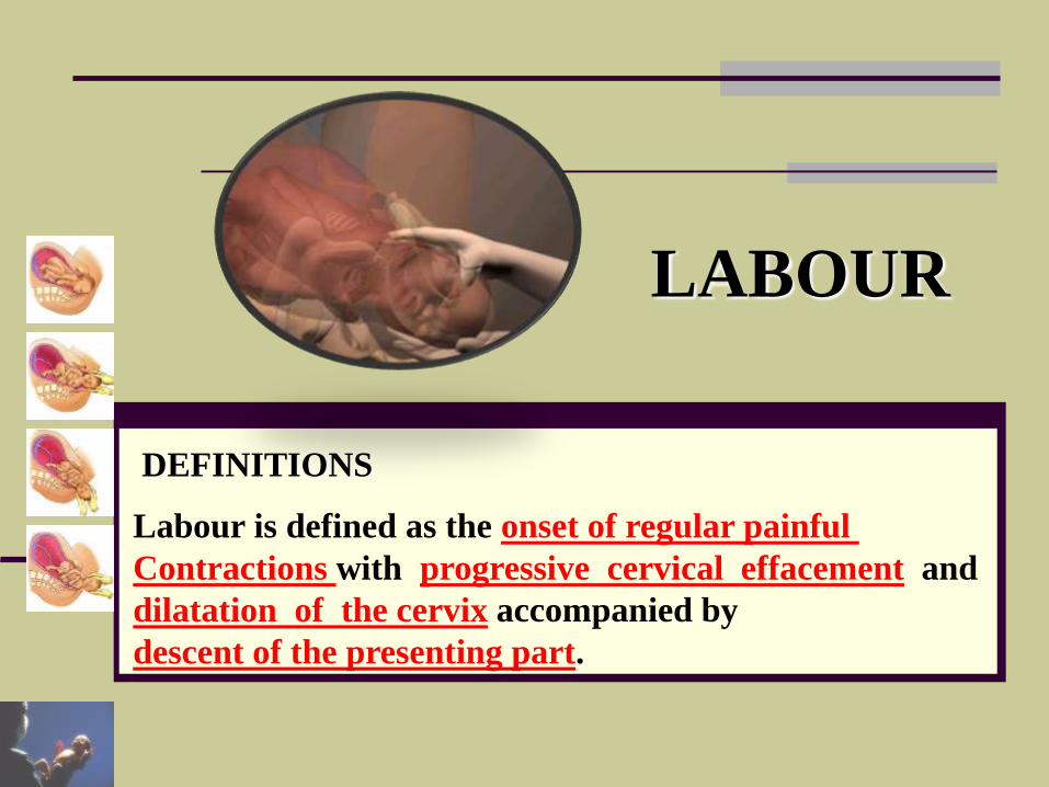
LABOUR
Labour is defined as the onset of regular painful
Contractions with progressive cervical effacement and
dilatation of the cervix accompanied by
descent of the presenting part.
DEFINITIONS
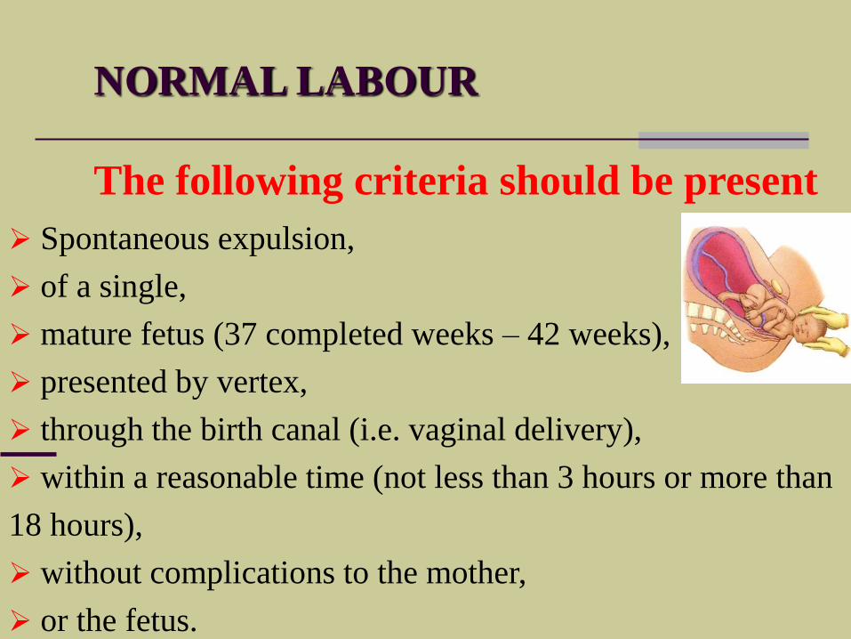
NORMAL LABOUR
Spontaneous expulsion,
of a single,
mature fetus (37 completed weeks – 42 weeks),
presented by vertex,
through the birth canal (i.e. vaginal delivery),
within a reasonable time (not less than 3 hours or more than
18 hours),
without complications to the mother,
or the fetus.
The following criteria should be present
NORMAL LABOUR
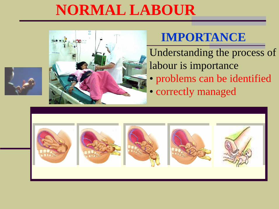
Understanding the process of
labour is importance
• problems can be identified
• correctly managed
IMPORTANCE
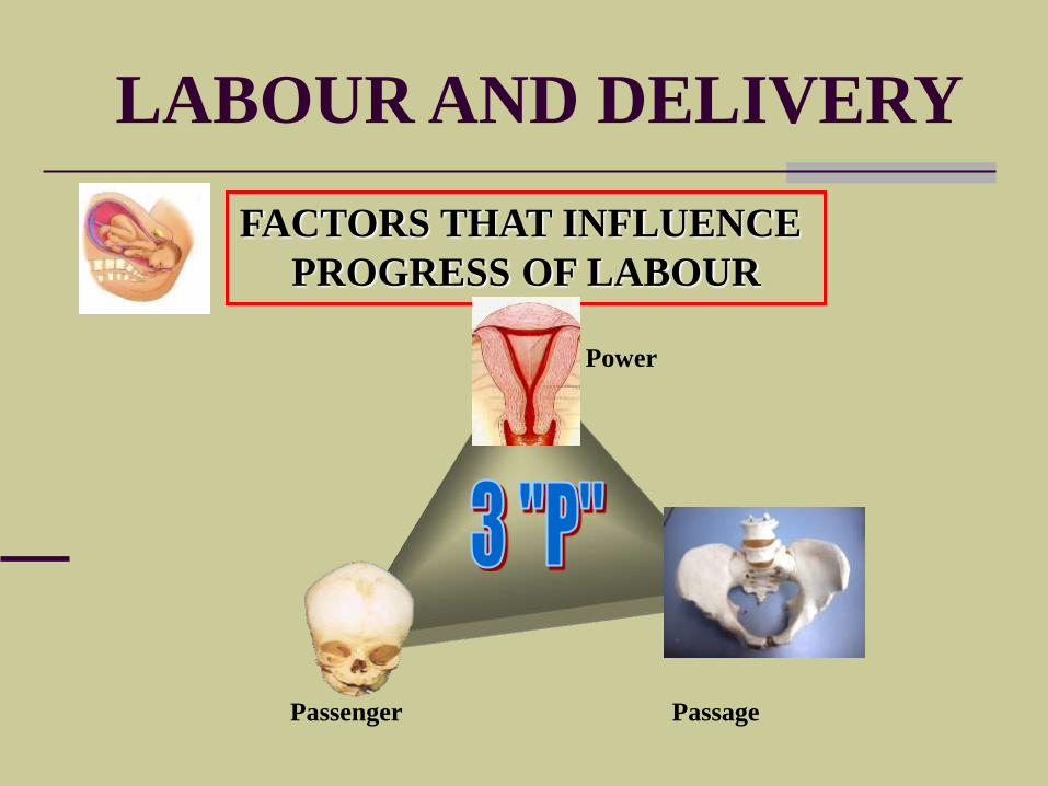
LABOUR AND DELIVERY
FACTORS THAT INFLUENCE
PROGRESS OF LABOUR
Passenger Passage
Power
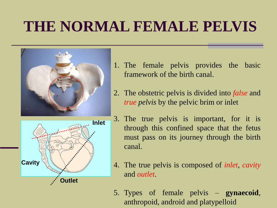
THE NORMAL FEMALE PELVIS
1. The female pelvis provides the basic
framework of the birth canal.
2. The obstetric pelvis is divided into false and
true pelvis by the pelvic brim or inlet
3. The true pelvis is important, for it is
through this confined space that the fetus
must pass on its journey through the birth
canal.
4. The true pelvis is composed of inlet, cavity
and outlet.
5. Types of female pelvis – gynaecoid,
anthropoid, android and platypelloid
Outlet
Cavity
Inlet
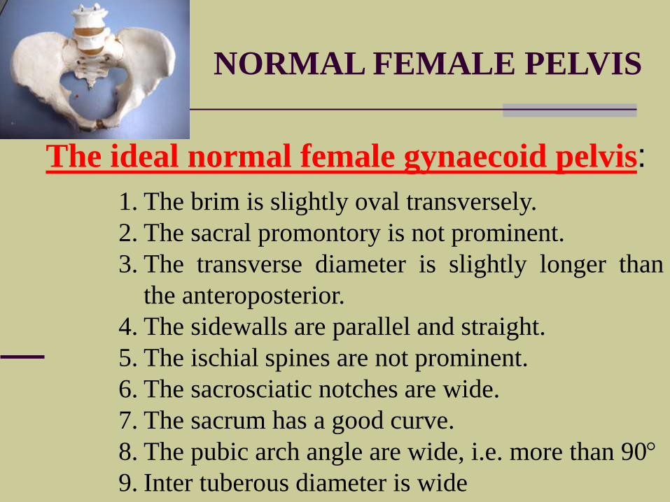
NORMAL FEMALE PELVIS
1. The brim is slightly oval transversely.
2. The sacral promontory is not prominent.
3. The transverse diameter is slightly longer than
the anteroposterior.
4. The sidewalls are parallel and straight.
5. The ischial spines are not prominent.
6. The sacrosciatic notches are wide.
7. The sacrum has a good curve.
8. The pubic arch angle are wide, i.e. more than 90
9. Inter tuberous diameter is wide
The ideal normal female gynaecoid pelvis:
THE NORMAL FEMALE PELVIS
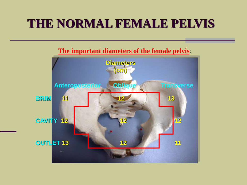
The important diameters of the female pelvis:
Anteroposterior Oblique Transverse
BRIM 11 12 13
CAVITY 12 12 12
OUTLET 13 12 11
Diameters
(cm)
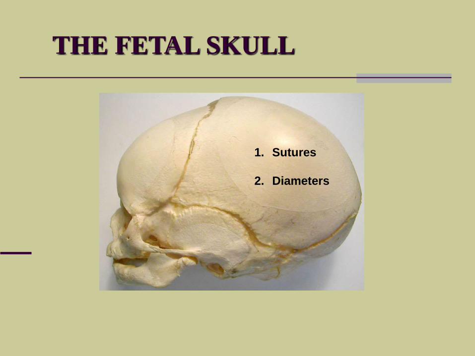
THE FETAL SKULL
1. Sutures
2. Diameters
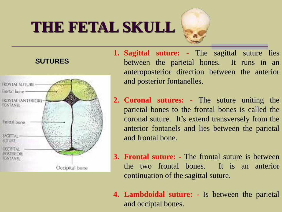
THE FETAL SKULL
1. Sagittal suture: - The sagittal suture lies
between the parietal bones. It runs in an
anteroposterior direction between the anterior
and posterior fontanelles.
2. Coronal sutures: - The suture uniting the
parietal bones to the frontal bones is called the
coronal suture. It’s extend transversely from the
anterior fontanels and lies between the parietal
and frontal bone.
3. Frontal suture: - The frontal suture is between
the two frontal bones. It is an anterior
continuation of the sagittal suture.
4. Lambdoidal suture: - Is between the parietal
and occiptal bones.
SUTURES
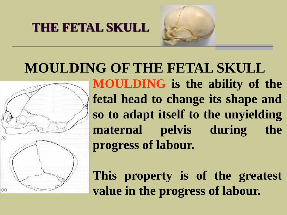
THE FETAL SKULL
MOULDING OF THE FETAL SKULLMOULDING is the ability of the
fetal head to change its shape and
so to adapt itself to the unyielding
maternal pelvis during the
progress of labour.
This property is of the greatest
value in the progress of labour.
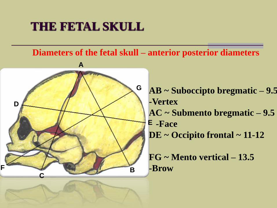
THE FETAL SKULL
Diameters of the fetal skull – anterior posterior diameters
A
BC
D
E
F
G AB ~ Suboccipto bregmatic – 9.5
-Vertex
AC ~ Submento bregmatic – 9.5
-Face
DE ~ Occipito frontal ~ 11-12
FG ~ Mento vertical – 13.5
-Brow
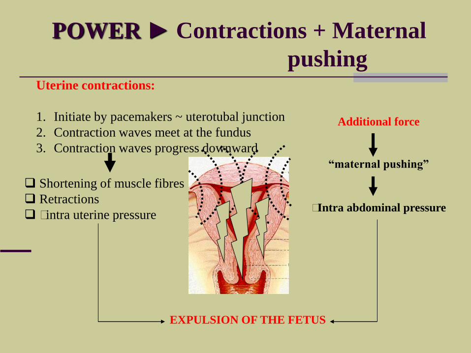
POWER ► Contractions + Maternal
pushingUterine contractions:
1. Initiate by pacemakers ~ uterotubal junction
2. Contraction waves meet at the fundus
3. Contraction waves progress downward
Shortening of muscle fibres
Retractions
intra uterine pressure
EXPULSION OF THE FETUS
Additional force
“maternal pushing”
Intra abdominal pressure
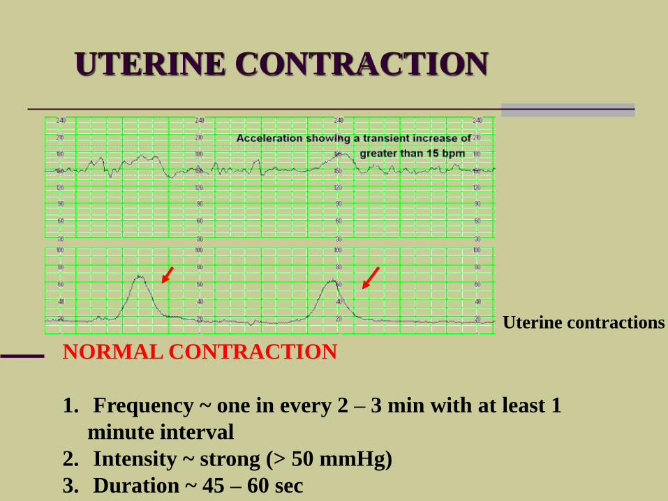
UTERINE CONTRACTION
NORMAL CONTRACTION
1. Frequency ~ one in every 2 – 3 min with at least 1
minute interval
2. Intensity ~ strong (> 50 mmHg)
3. Duration ~ 45 – 60 sec
Uterine contractions
LABOUR AND DELIVERY
WHAT INITIATE LABOUR
“ONSET OF LABOUR”
NORMAL LABOUR
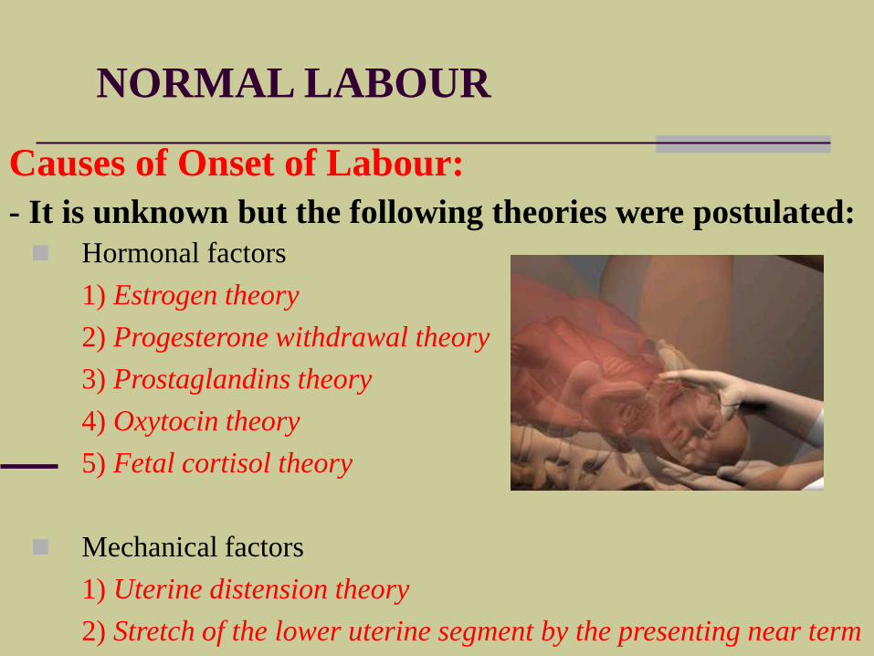
Hormonal factors
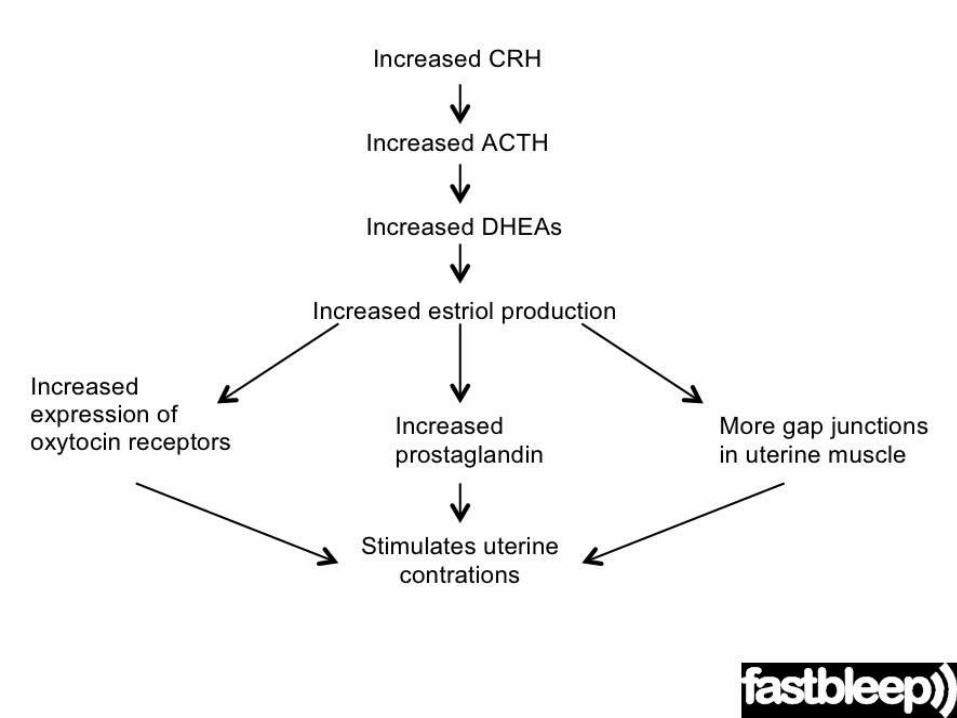
1) Estrogen theory
2) Progesterone withdrawal theory
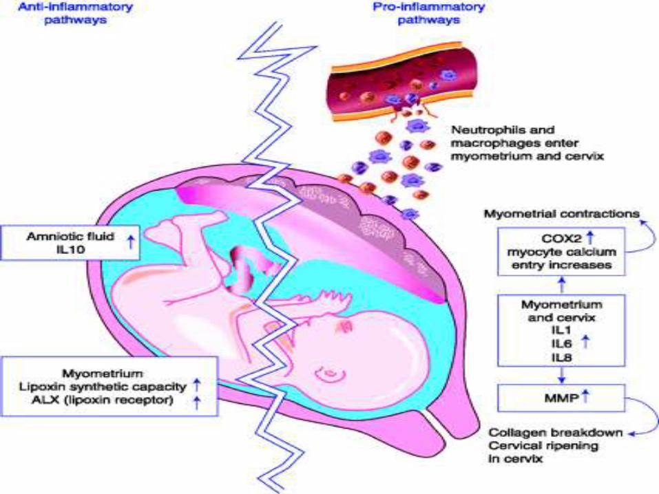
3) Prostaglandins theory
4) Oxytocin theory
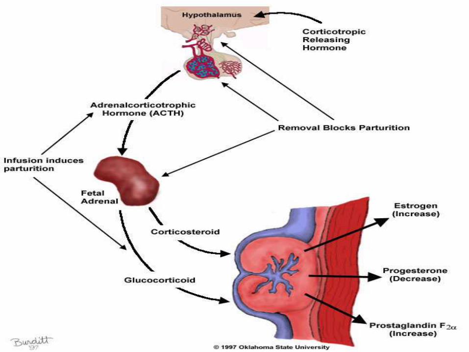
5) Fetal cortisol theory
Mechanical factors
1) Uterine distension theory
2) Stretch of the lower uterine segment by the presenting near term
Causes of Onset of Labour:
- It is unknown but the following theories were postulated:
LABOUR AND DELIVERY
DIAGNOSIS OF LABOUR
NORMAL LABOUR AND DELIVERY
Painful regular uterine contractions
– as evidence by contraction at least
one in ten minutes
Show – as evidence by mucus mixed
with blood
Rupture of membranes – as
evidence by leaking liquor
Progressive shortening and
SYMPTOMS AND SIGNS OF LABOURBefore labour begins, women usually notice one or more premonitory, or
warnings, signs that labour is about to begin.
They are:

LABOUR AND DELIVERY
DESCRIBE THE STAGES OF
LABOUR
NORMAL LABOUR AND DELIVERY
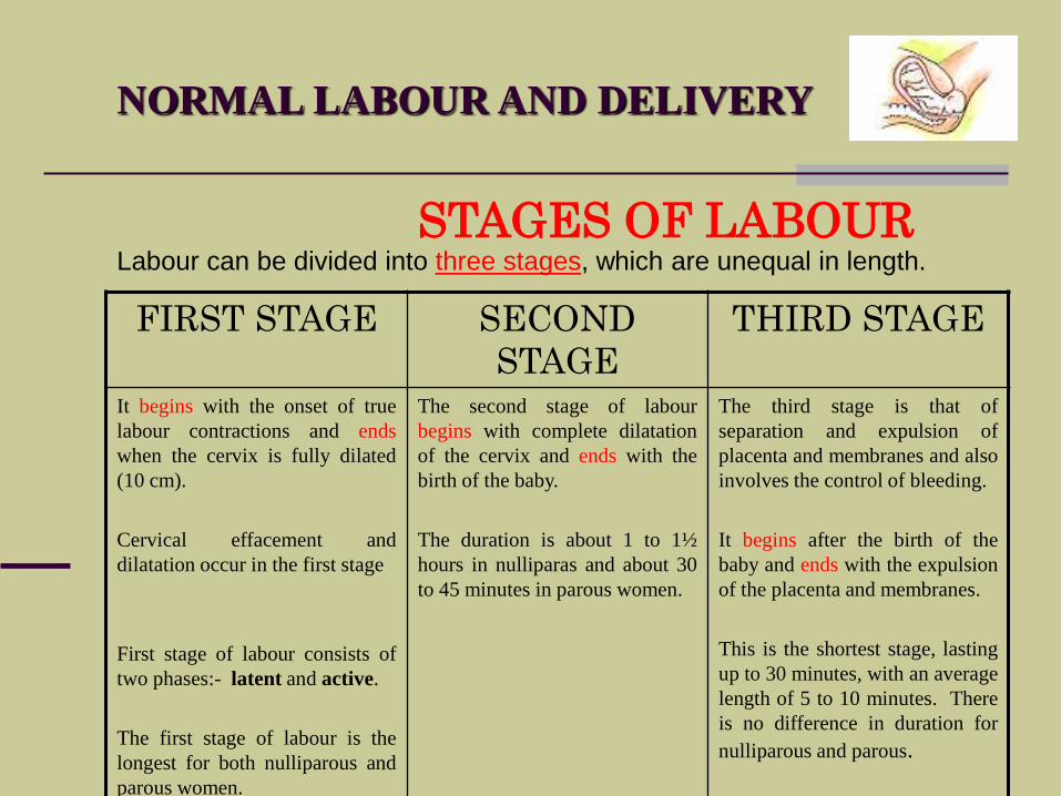
STAGES OF LABOUR
FIRST STAGE SECOND
STAGE
THIRD STAGE
It begins with the onset of true
labour contractions and ends
when the cervix is fully dilated
(10 cm).
Cervical effacement and
dilatation occur in the first stage
First stage of labour consists of
two phases:- latent and active.
The first stage of labour is the
longest for both nulliparous and
parous women.
The second stage of labour
begins with complete dilatation
of the cervix and ends with the
birth of the baby.
The duration is about 1 to 1½
hours in nulliparas and about 30
to 45 minutes in parous women.
The third stage is that of
separation and expulsion of
placenta and membranes and also
involves the control of bleeding.
It begins after the birth of the
baby and ends with the expulsion
of the placenta and membranes.
This is the shortest stage, lasting
up to 30 minutes, with an average
length of 5 to 10 minutes. There
is no difference in duration for
nulliparous and parous.
Labour can be divided into three stages, which are unequal in length.
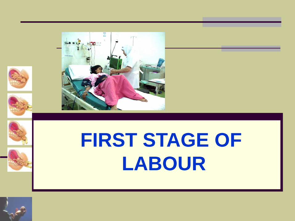
FIRST STAGE OF
LABOUR
NORMAL LABOUR AND DELIVERY
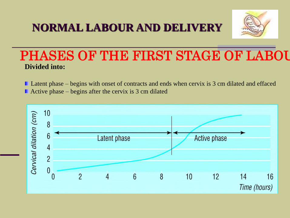
PHASES OF THE FIRST STAGE OF LABOURDivided into:
Latent phase – begins with onset of contracts and ends when cervix is 3 cm dilated and effaced
Active phase – begins after the cervix is 3 cm dilated
NORMAL LABOUR AND DELIVERY
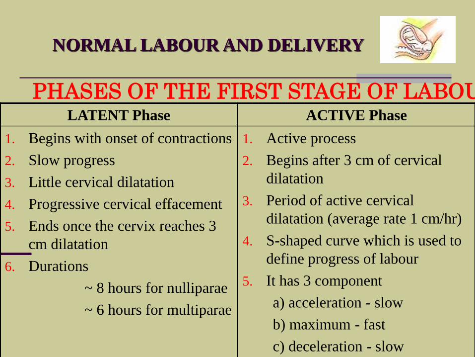
PHASES OF THE FIRST STAGE OF LABOURLATENT Phase ACTIVE Phase
1. Begins with onset of contractions
2. Slow progress
3. Little cervical dilatation
4. Progressive cervical effacement
5. Ends once the cervix reaches 3
cm dilatation
6. Durations
~ 8 hours for nulliparae
~ 6 hours for multiparae
1. Active process
2. Begins after 3 cm of cervical
dilatation
3. Period of active cervical
dilatation (average rate 1 cm/hr)
4. S-shaped curve which is used to
define progress of labour
5. It has 3 component
a) acceleration - slow
b) maximum - fast
c) deceleration - slow

NORMAL LABOUR AND DELIVERY
WHAT HAPPEN DURING
THE FIRST STAGE OF LABOUR
NORMAL LABOUR AND DELIVERY
WHAT HAPPEN DURING THE FIRST STAGE OF LABOUR
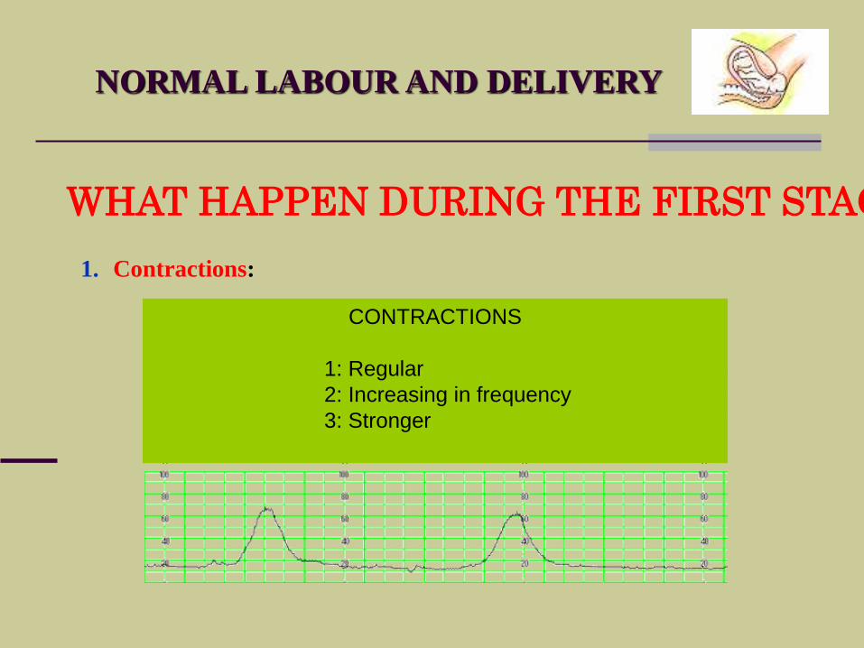
1. Contractions:
CONTRACTIONS
1: Regular
2: Increasing in frequency
3: Stronger
NORMAL LABOUR AND DELIVERY
WHAT HAPPEN DURING THE FIRST STAGE OF LABOUR
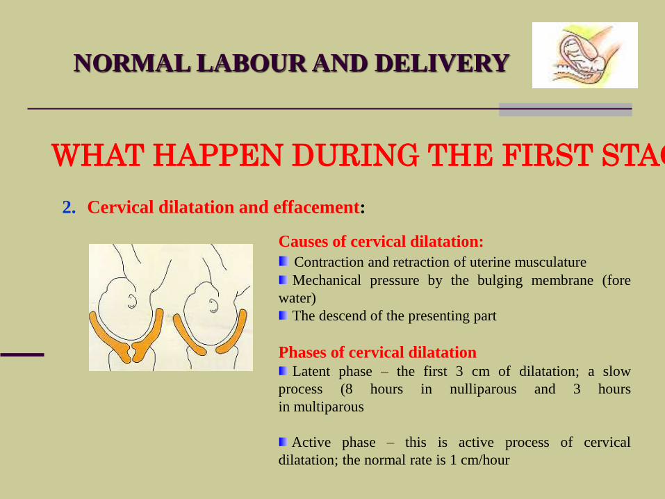
2. Cervical dilatation and effacement:
Causes of cervical dilatation:
Contraction and retraction of uterine musculature
Mechanical pressure by the bulging membrane (fore
water)
The descend of the presenting part
Phases of cervical dilatationLatent phase – the first 3 cm of dilatation; a slow
process (8 hours in nulliparous and 3 hours
in multiparous
Active phase – this is active process of cervical
dilatation; the normal rate is 1 cm/hour
NORMAL LABOUR AND DELIVERY
WHAT HAPPEN DURING THE FIRST STAGE OF LABOUR
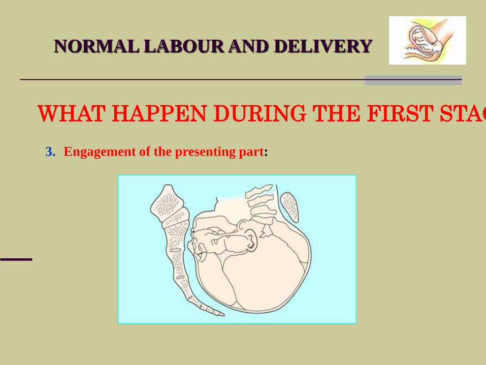
3. Engagement of the presenting part:
NORMAL LABOUR AND DELIVERY
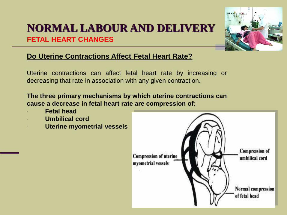
Do Uterine Contractions Affect Fetal Heart Rate?
Uterine contractions can affect fetal heart rate by increasing or
decreasing that rate in association with any given contraction.
The three primary mechanisms by which uterine contractions can
cause a decrease in fetal heart rate are compression of:
· Fetal head
· Umbilical cord
· Uterine myometrial vessels
FETAL HEART CHANGES
NORMAL LABOUR AND DELIVERY
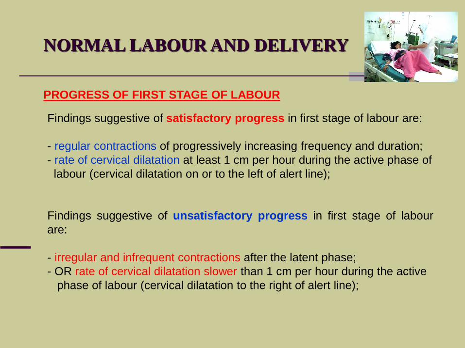
PROGRESS OF FIRST STAGE OF LABOUR
Findings suggestive of satisfactory progress in first stage of labour are:
- regular contractions of progressively increasing frequency and duration;
- rate of cervical dilatation at least 1 cm per hour during the active phase of
labour (cervical dilatation on or to the left of alert line);
Findings suggestive of unsatisfactory progress in first stage of labour
are:
- irregular and infrequent contractions after the latent phase;
- OR rate of cervical dilatation slower than 1 cm per hour during the active
phase of labour (cervical dilatation to the right of alert line);
SECOND STAGE OF
LABOUR
NORMAL LABOUR AND DELIVERY
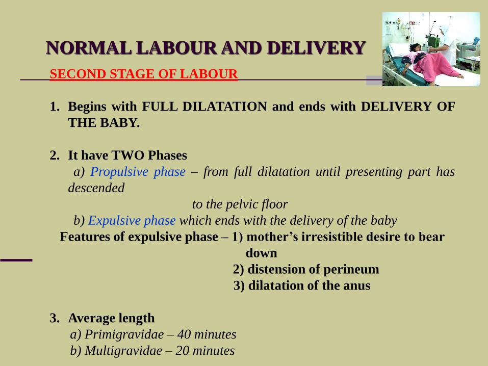
SECOND STAGE OF LABOUR
1. Begins with FULL DILATATION and ends with DELIVERY OF
THE BABY.
2. It have TWO Phases
a) Propulsive phase – from full dilatation until presenting part has
descended
to the pelvic floor
b) Expulsive phase which ends with the delivery of the baby
Features of expulsive phase – 1) mother’s irresistible desire to bear
down
2) distension of perineum
3) dilatation of the anus
3. Average length
a) Primigravidae – 40 minutes
b) Multigravidae – 20 minutes
NORMAL LABOUR AND DELIVERY
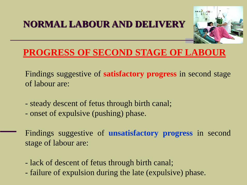
PROGRESS OF SECOND STAGE OF LABOUR
Findings suggestive of satisfactory progress in second stage
of labour are:
- steady descent of fetus through birth canal;
- onset of expulsive (pushing) phase.
Findings suggestive of unsatisfactory progress in second
stage of labour are:
- lack of descent of fetus through birth canal;
- failure of expulsion during the late (expulsive) phase.

THIRD STAGE OF
LABOUR
NORMAL LABOUR AND DELIVERY
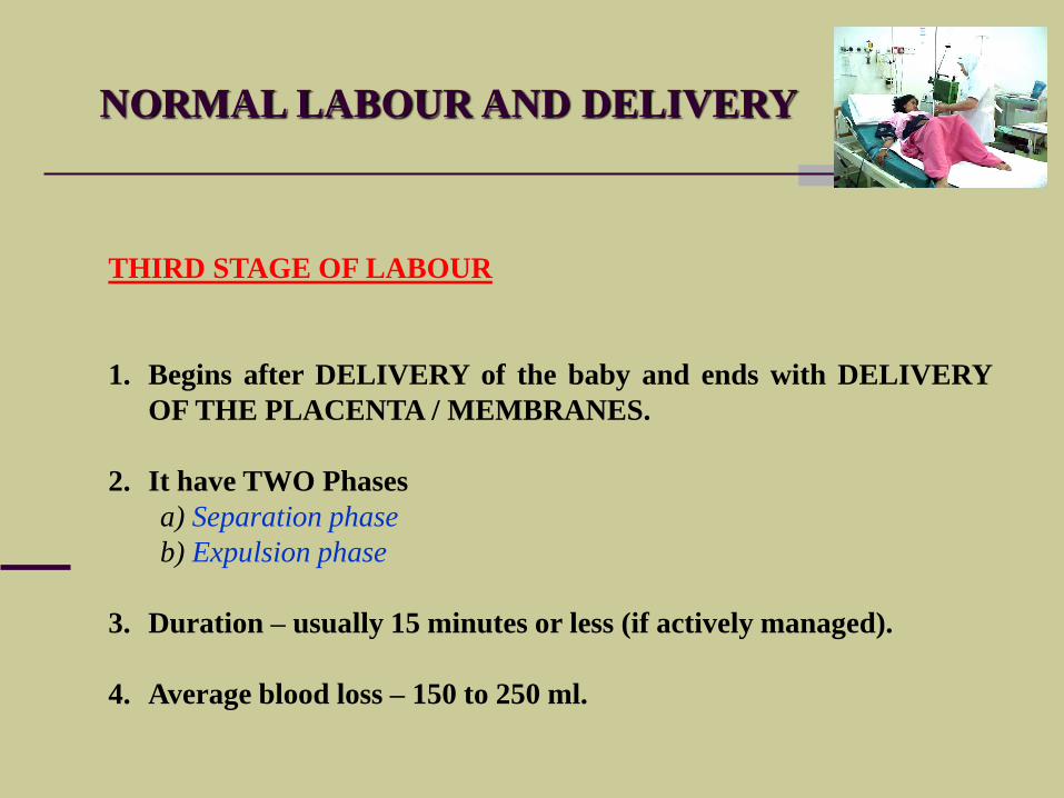
THIRD STAGE OF LABOUR
1. Begins after DELIVERY of the baby and ends with DELIVERY
OF THE PLACENTA / MEMBRANES.
2. It have TWO Phases
a) Separation phase
b) Expulsion phase
3. Duration – usually 15 minutes or less (if actively managed).
4. Average blood loss – 150 to 250 ml.
NORMAL LABOUR AND DELIVERY
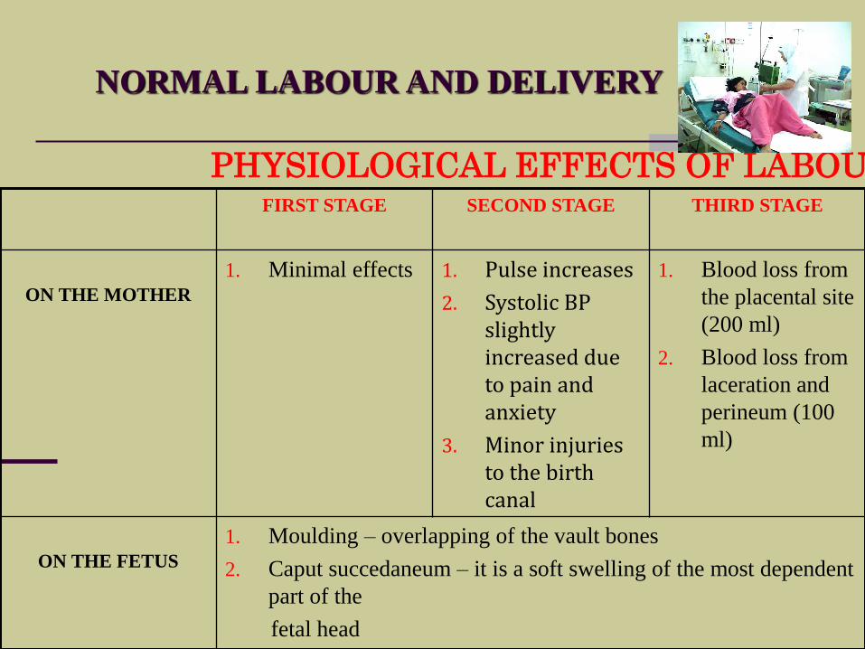
PHYSIOLOGICAL EFFECTS OF LABOURFIRST STAGE SECOND STAGE THIRD STAGE
ON THE MOTHER
1. Minimal effects 1. Pulse increases
2. Systolic BP slightly increased due to pain and anxiety
3. Minor injuries to the birth canal
1. Blood loss from
the placental site
(200 ml)
2. Blood loss from
laceration and
perineum (100
ml)
ON THE FETUS
1. Moulding – overlapping of the vault bones
2. Caput succedaneum – it is a soft swelling of the most dependent
part of the
fetal head

MANAGEMENT
OF
LABOUR
AIMS IN THE MANAGEMENT OF LABOUR
To achieve delivery of a normal healthychild
To anticipate, recognize and treatpotential abnormal conditions beforesignificant hazard develops for the motheror the fetus.
PRINCIPLES IN THE MANAGEMENT OF LABOUR
Diagnosis of labour
Monitoring the progress of labour
Ensuring maternal well-being
Ensuring fetal well-being.
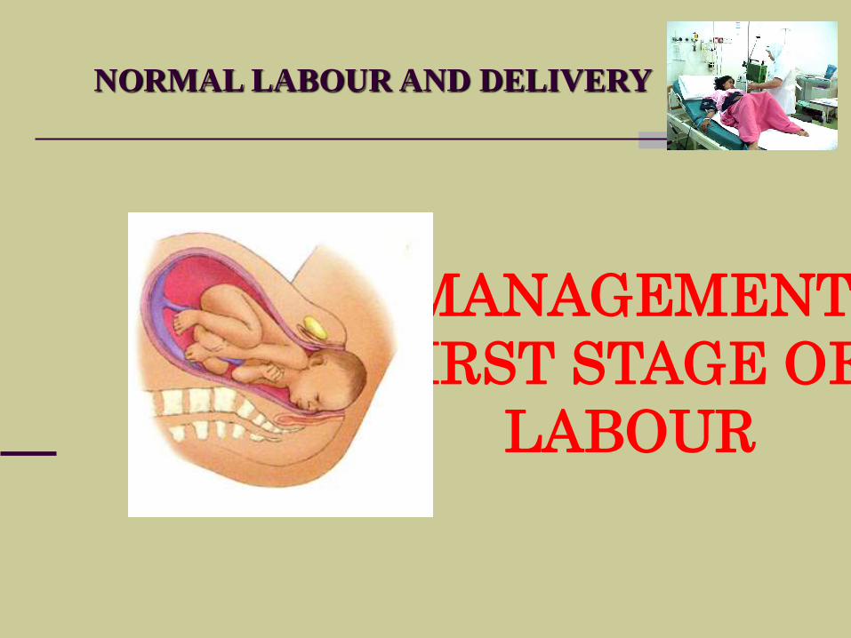
NORMAL LABOUR AND DELIVERY
MANAGEMENT
FIRST STAGE OF
LABOUR
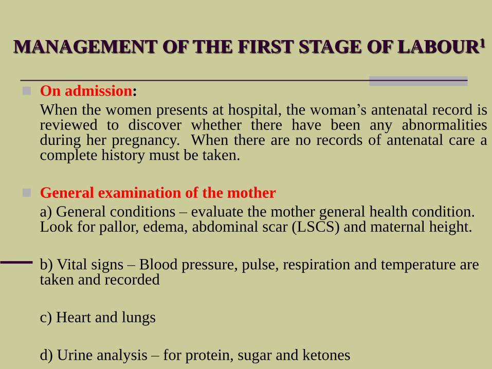
MANAGEMENT OF THE FIRST STAGE OF LABOUR1
On admission:
When the women presents at hospital, the woman’s antenatal record isreviewed to discover whether there have been any abnormalitiesduring her pregnancy. When there are no records of antenatal care acomplete history must be taken.
General examination of the mother
a) General conditions – evaluate the mother general health condition. Look for pallor, edema, abdominal scar (LSCS) and maternal height.
b) Vital signs – Blood pressure, pulse, respiration and temperature are taken and recorded
c) Heart and lungs
d) Urine analysis – for protein, sugar and ketones
MANAGEMENT OF THE FIRST STAGE OF LABOUR2
Abdominal examination:
a) A detailed abdominal examination should be carried out and recorded.
b) Determine the presentation and position of the fetus and also theengagement
c) Auscultate the fetal heart
d) Evaluate the uterine contraction
Vaginal examination – the purpose is to
a) To make a positive diagnosis of labour
b) To make a positive identification of presentation
c) To determine whether the fetal head is engaged in case of doubt
d) To ascertain whether the fore waters have ruptured or to rupture them artificially
e) To exclude cord prolapse after rupture of the fore waters
f) To confirm the degree of cervical dilatation and position of the presenting part
g) To assess progress of labour.
h) To assess the adequacy of the pelvis.
MANAGEMENT OF THE FIRST STAGE OF LABOUR3
Bowel preparation:
If there has been no bowel action for 24 hours or the rectum feels loaded on vaginalexamination an enema is given.
Bladder care
A full bladder may initially prevent the fetal head from entering the pelvic brim and later impede descent of the fetal head. It will also inhibit effective uterine action.
The woman should be encouraged to empty her bladder every 1½ - 2 hours during labour.
The quantity of urine passed should be measured and recorded and a specimen obtained for testing.
Nutrition in early labour
No food is permitted after labour is established – to prevent regurgitation and aspiration
It is important to maintain adequate hydration - via intravenous routes
MANAGEMENT OF THE FIRST STAGE OF LABOUR4
Position of labouring mother:
As long as the patient is healthy, the presentation normal, the presenting partengaged, and the fetus in good condition, the patient may walk about or may be inbed, as she wishes
Monitoring the progress of labour
Once labour has become established, all events during labour should be recorded on a partogram.
a) The well-being of the fetus
b) The well-being of the mother
c) The progress of the labour
Pain relief
When the pains are severe an analgesic preparation may be given.
a) Opiate drugs – e.g. Pethidine given intramuscularly every 4 hour
b) Inhalational analgesia – e.g. Entonox
c) Epidural analagesia
NORMAL LABOUR AND DELIVERY
Pain in labour
The pain experienced by the woman in labour is caused by the:
1): Uterine contractions and uterine ischaemia.
2): Cervical dilatation. Dilatation and stretching of the cervix and
lower uterine segment stimulate nerve ganglia and are a major
source of pain.
3): Distention of the vagina and perineum. Marked distention of the
vagina and perineum occurs with fetal descent, especially during the
second stage.
LABOUR PAIN – causes1
NORMAL LABOUR AND DELIVERY
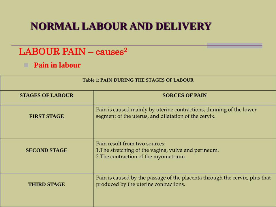
Pain in labour
LABOUR PAIN – causes2
Table 1: PAIN DURING THE STAGES OF LABOUR
STAGES OF LABOUR SORCES OF PAIN
FIRST STAGE
Pain is caused mainly by uterine contractions, thinning of the lower segment of the uterus, and dilatation of the cervix.
SECOND STAGE
Pain result from two sources:1.The stretching of the vagina, vulva and perineum.2.The contraction of the myometrium.
THIRD STAGE
Pain is caused by the passage of the placenta through the cervix, plus that produced by the uterine contractions.
NORMAL LABOUR AND DELIVERY
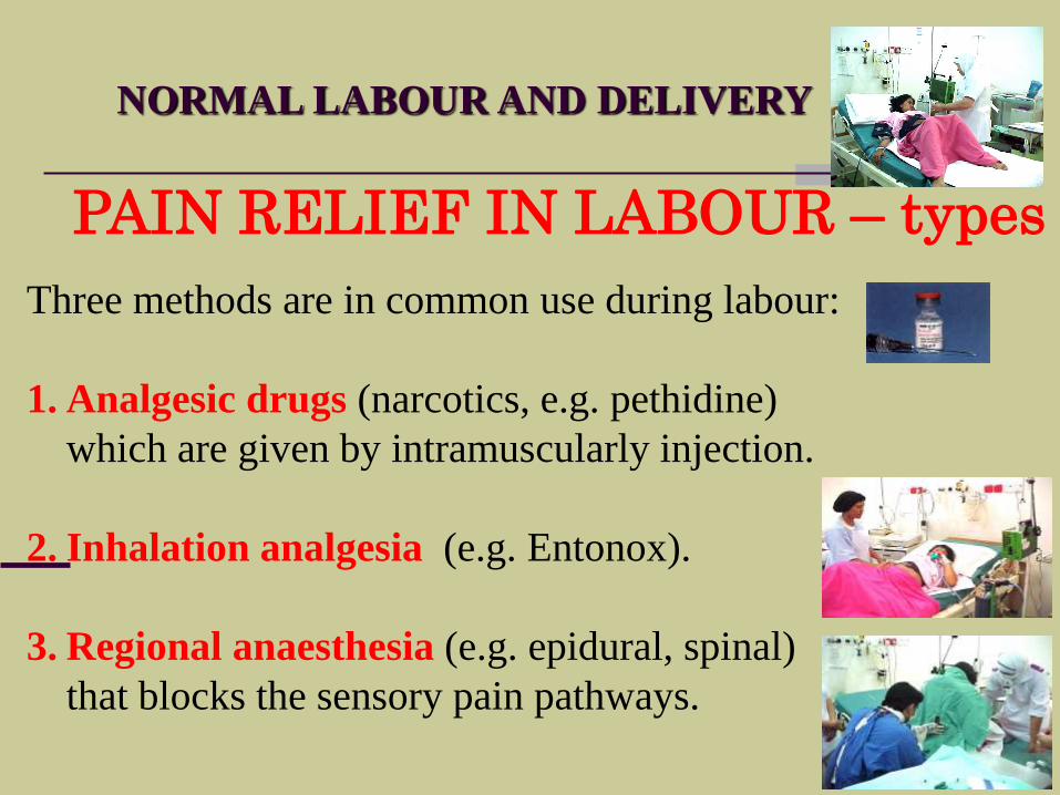
PAIN RELIEF IN LABOUR – types
Three methods are in common use during labour:
1. Analgesic drugs (narcotics, e.g. pethidine)
which are given by intramuscularly injection.
2. Inhalation analgesia (e.g. Entonox).
3. Regional anaesthesia (e.g. epidural, spinal)
that blocks the sensory pain pathways.
NORMAL LABOUR AND DELIVERY
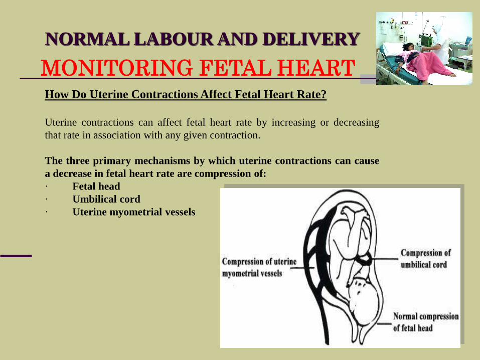
MONITORING FETAL HEARTHow Do Uterine Contractions Affect Fetal Heart Rate?
Uterine contractions can affect fetal heart rate by increasing or decreasing
that rate in association with any given contraction.
The three primary mechanisms by which uterine contractions can cause
a decrease in fetal heart rate are compression of:
· Fetal head
· Umbilical cord
· Uterine myometrial vessels
NORMAL LABOUR AND DELIVERY
MONITORING FETAL HEART
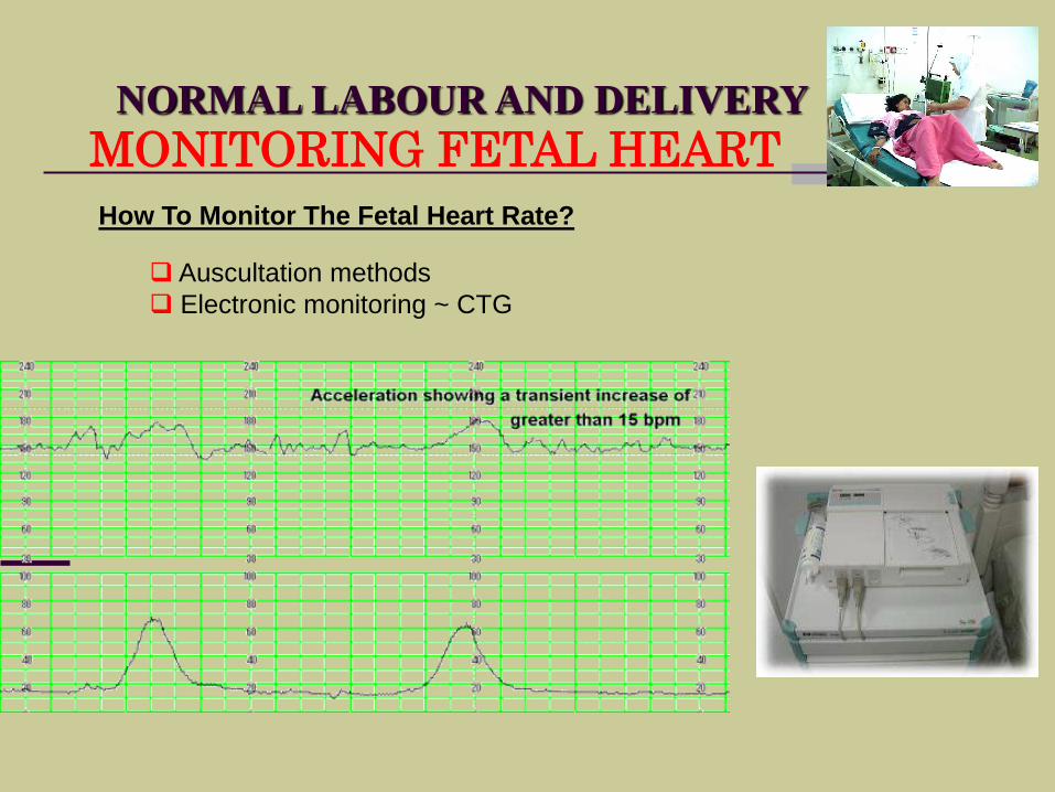
How To Monitor The Fetal Heart Rate?
Auscultation methods
Electronic monitoring ~ CTG
NORMAL LABOUR AND DELIVERY
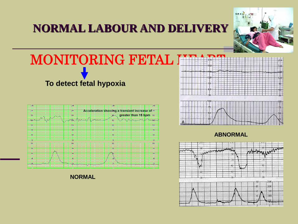
MONITORING FETAL HEART
To detect fetal hypoxia
NORMAL
ABNORMAL
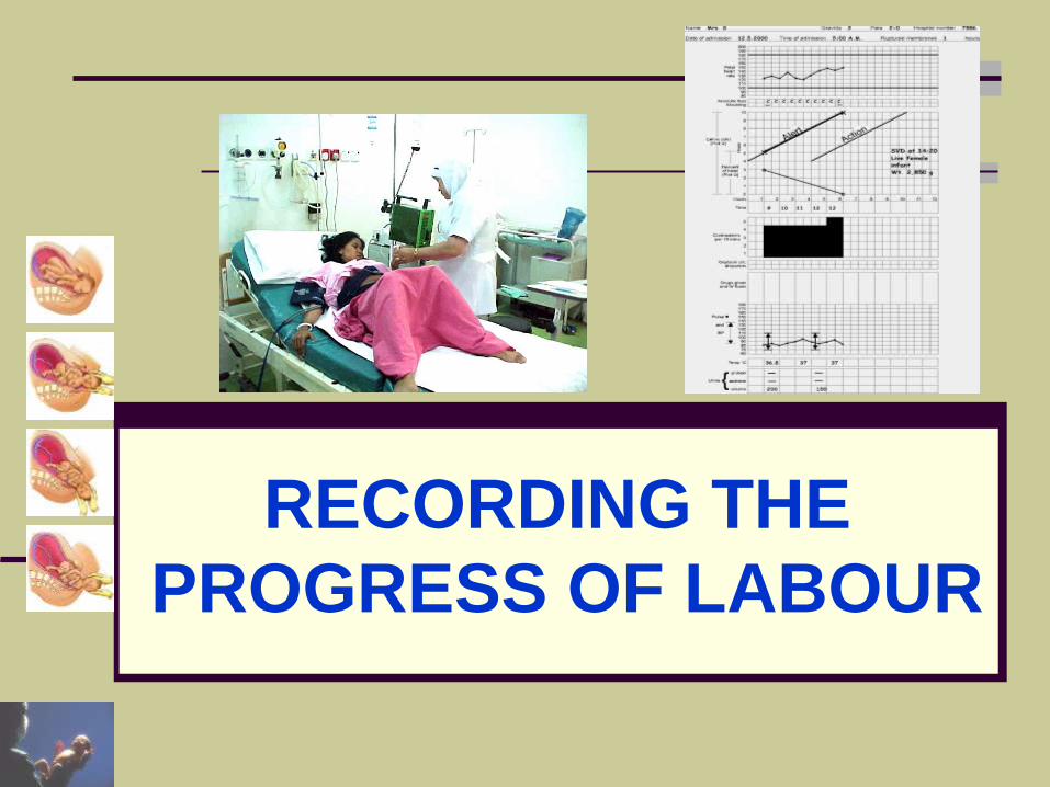
RECORDING THE
PROGRESS OF LABOUR
NORMAL LABOUR AND DELIVERY
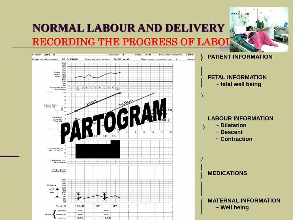
RECORDING THE PROGRESS OF LABOUR
PATIENT INFORMATION
FETAL INFORMATION
~ fetal well being
LABOUR INFORMATION
~ Dilatation
~ Descent
~ Contraction
MEDICATIONS
MATERNAL INFORMATION
~ Well being
NORMAL LABOUR AND DELIVERY
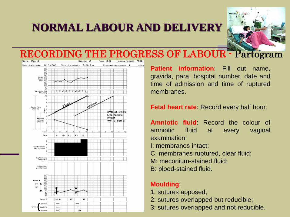
RECORDING THE PROGRESS OF LABOUR - Partogram
Patient information: Fill out name,
gravida, para, hospital number, date and
time of admission and time of ruptured
membranes.
Fetal heart rate: Record every half hour.
Amniotic fluid: Record the colour of
amniotic fluid at every vaginal
examination:
I: membranes intact;
C: membranes ruptured, clear fluid;
M: meconium-stained fluid;
B: blood-stained fluid.
Moulding:
1: sutures apposed;
2: sutures overlapped but reducible;
3: sutures overlapped and not reducible.
NORMAL LABOUR AND DELIVERY
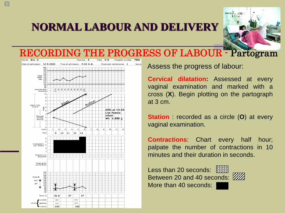
RECORDING THE PROGRESS OF LABOUR - Partogram
Cervical dilatation: Assessed at every
vaginal examination and marked with a
cross (X). Begin plotting on the partograph
at 3 cm.
Station : recorded as a circle (O) at every
vaginal examination.
Contractions: Chart every half hour;
palpate the number of contractions in 10
minutes and their duration in seconds.
Less than 20 seconds:
Between 20 and 40 seconds:
More than 40 seconds:
Assess the progress of labour:
NORMAL LABOUR AND DELIVERY
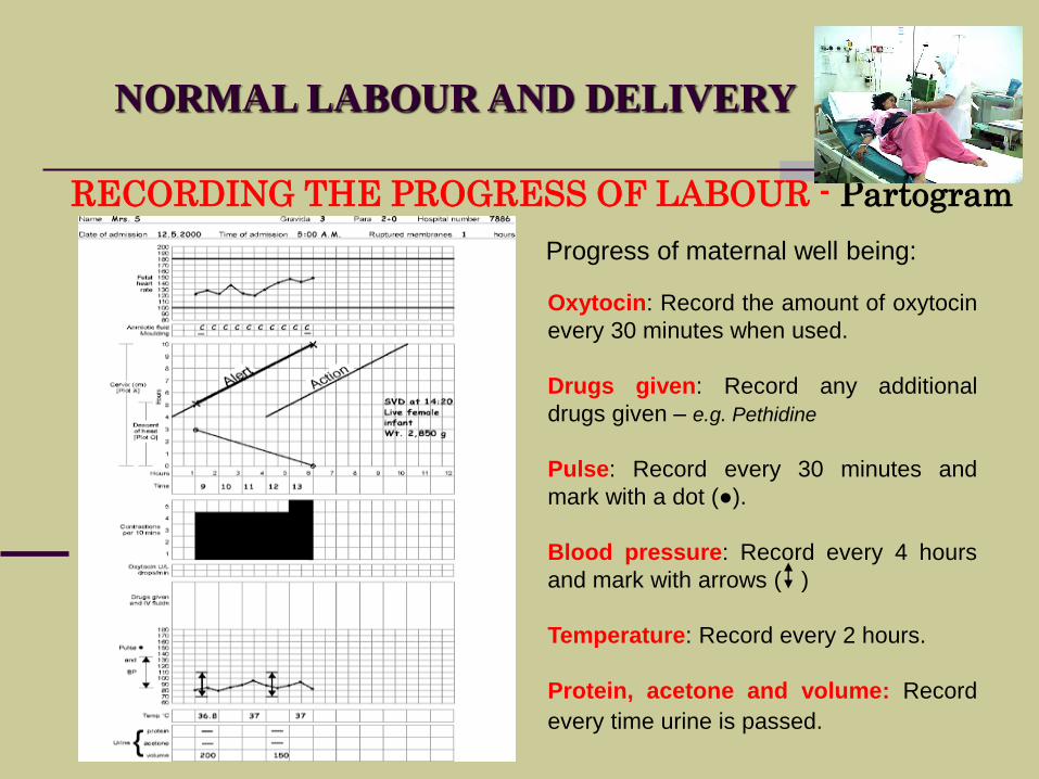
RECORDING THE PROGRESS OF LABOUR - Partogram
Oxytocin: Record the amount of oxytocin
every 30 minutes when used.
Drugs given: Record any additional
drugs given – e.g. Pethidine
Pulse: Record every 30 minutes and
mark with a dot (●).
Blood pressure: Record every 4 hours
and mark with arrows ( )
Temperature: Record every 2 hours.
Protein, acetone and volume: Record
every time urine is passed.
Progress of maternal well being:
NORMAL LABOUR AND DELIVERY
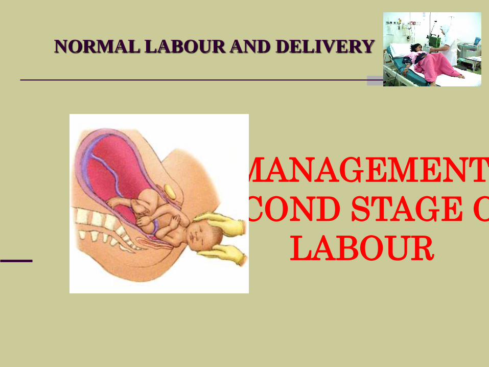
MANAGEMENT
SECOND STAGE OF
LABOUR
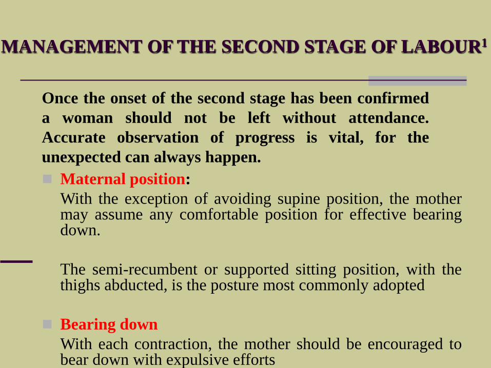
MANAGEMENT OF THE SECOND STAGE OF LABOUR1
Maternal position:
With the exception of avoiding supine position, the mothermay assume any comfortable position for effective bearingdown.
The semi-recumbent or supported sitting position, with thethighs abducted, is the posture most commonly adopted
Bearing down
With each contraction, the mother should be encouraged tobear down with expulsive efforts
Once the onset of the second stage has been confirmed
a woman should not be left without attendance.
Accurate observation of progress is vital, for the
unexpected can always happen.
MANAGEMENT OF THE SECOND STAGE OF LABOUR2
Observation during the second stage:
Four factors determine whether the second stage may be safely continued andthese must be carefully monitored throughout the second stage of labour.
1. Maternal conditions
Observation includes an appraisal of the mother’s ability to cope emotionally as well as an assessment of her physical wellbeing. A maternal pulse rate is usually recorded quarter-hourly and bloods pressure hourly
2. Fetal conditions - During the second stage, the fetal heart should be monitored either continuously or after each contraction. stage may be associated with fetal distress.
The liquor amnii is observed for signs of meconium staining.
3. Uterine contractions - The strength, length and frequency of contractions should be assessed continuously.
4. The progress of descent - The progress should be recorded approximately every 30 minutes during the second stage.
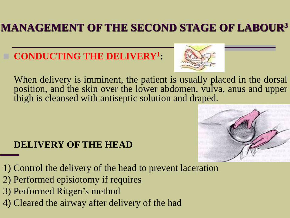
MANAGEMENT OF THE SECOND STAGE OF LABOUR3
CONDUCTING THE DELIVERY1:
When delivery is imminent, the patient is usually placed in the dorsalposition, and the skin over the lower abdomen, vulva, anus and upperthigh is cleansed with antiseptic solution and draped.
DELIVERY OF THE HEAD
1) Control the delivery of the head to prevent laceration
2) Performed episiotomy if requires
3) Performed Ritgen’s method
4) Cleared the airway after delivery of the had
MANAGEMENT OF THE SECOND STAGE OF LABOUR3
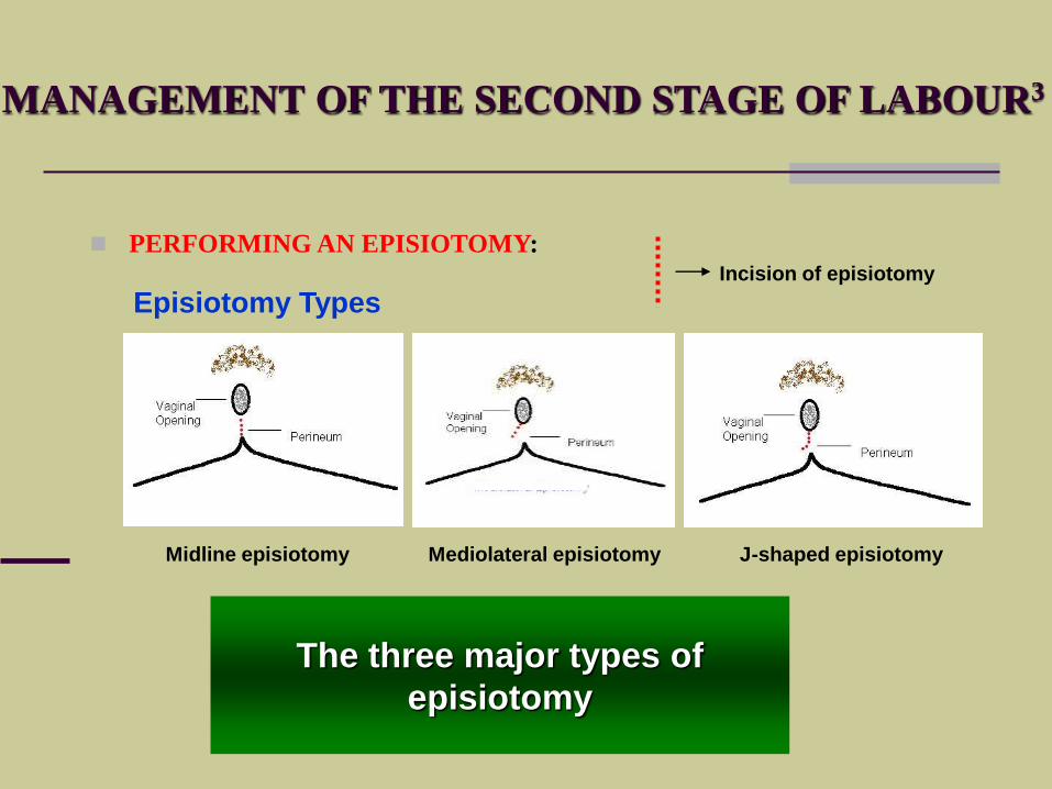
PERFORMING AN EPISIOTOMY:
"..is a surgical incision into the perineum to enlarge the space at the
outlet
EPISIOTOMY
IS EPSIOTOMY REALLY NEEDED?
Episiotomies are said to provide the following benefits:
1. Speed up the birth
2. Prevent Tearing
3. Protects against incontinence
4. Protects against pelvic floor relaxation
5. Heals easier than tears
medical research has not proven
any of these benefits
MANAGEMENT OF THE SECOND STAGE OF LABOUR3
PERFORMING AN EPISIOTOMY:
Episiotomies are not always necessary
Episiotomy should be considered only in the case of:
• Complicated vaginal delivery (breech, shoulder
dystocia, forceps,
vacuum);
• Scarring of the perineum;
• Fetal distress.
MANAGEMENT OF THE SECOND STAGE OF LABOUR3
PERFORMING AN EPISIOTOMY:
Episiotomy Types
Midline episiotomy Mediolateral episiotomy J-shaped episiotomy
Incision of episiotomy
The three major types of
episiotomy
MANAGEMENT OF THE SECOND STAGE OF LABOUR3
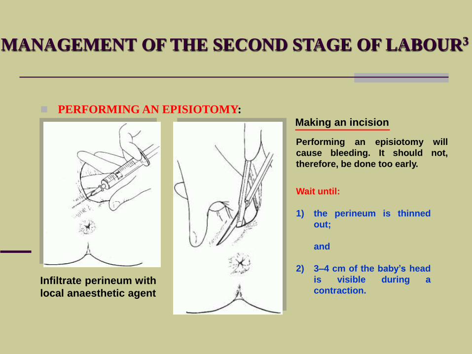
PERFORMING AN EPISIOTOMY:
Infiltrate perineum with
local anaesthetic agent
Making an incision
Wait until:
1) the perineum is thinned
out;
and
2) 3–4 cm of the baby’s head
is visible during a
contraction.
Performing an episiotomy will
cause bleeding. It should not,
therefore, be done too early.
MANAGEMENT OF THE SECOND STAGE OF LABOUR3
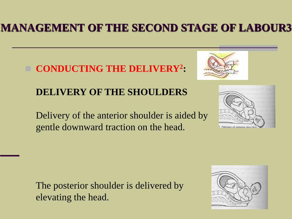
CONDUCTING THE DELIVERY2:
DELIVERY OF THE SHOULDERS
Delivery of the anterior shoulder is aided by
gentle downward traction on the head.
The posterior shoulder is delivered by
elevating the head.
MANAGEMENT OF THE SECOND STAGE OF LABOUR3
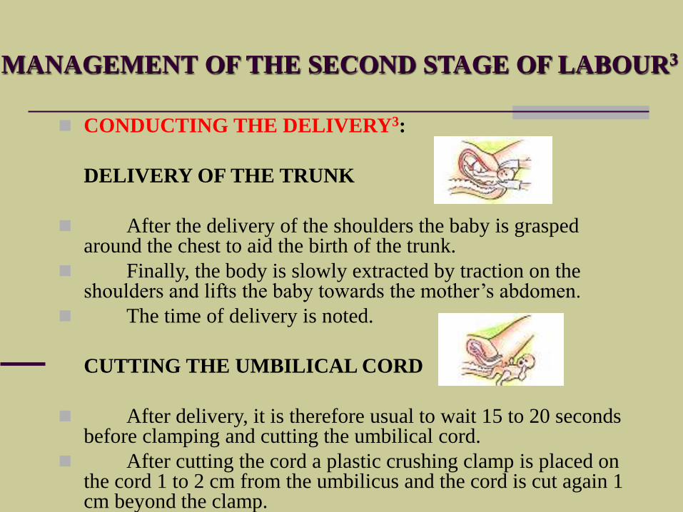
CONDUCTING THE DELIVERY3:
DELIVERY OF THE TRUNK
After the delivery of the shoulders the baby is grasped around the chest to aid the birth of the trunk.
Finally, the body is slowly extracted by traction on the shoulders and lifts the baby towards the mother’s abdomen.
The time of delivery is noted.
CUTTING THE UMBILICAL CORD
After delivery, it is therefore usual to wait 15 to 20 seconds before clamping and cutting the umbilical cord.
After cutting the cord a plastic crushing clamp is placed on the cord 1 to 2 cm from the umbilicus and the cord is cut again 1 cm beyond the clamp.
MANAGEMENT OF THE SECOND STAGE OF LABOUR3
CONDUCTING THE DELIVERY4:
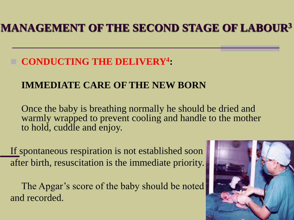
IMMEDIATE CARE OF THE NEW BORN
Once the baby is breathing normally he should be dried and warmly wrapped to prevent cooling and handle to the mother to hold, cuddle and enjoy.
If spontaneous respiration is not established soon
after birth, resuscitation is the immediate priority.
The Apgar’s score of the baby should be noted
and recorded.
LABOUR AND DELIVERY
THE MECHANISMS OF
NORMAL LABOUR
- Occiput anterior -
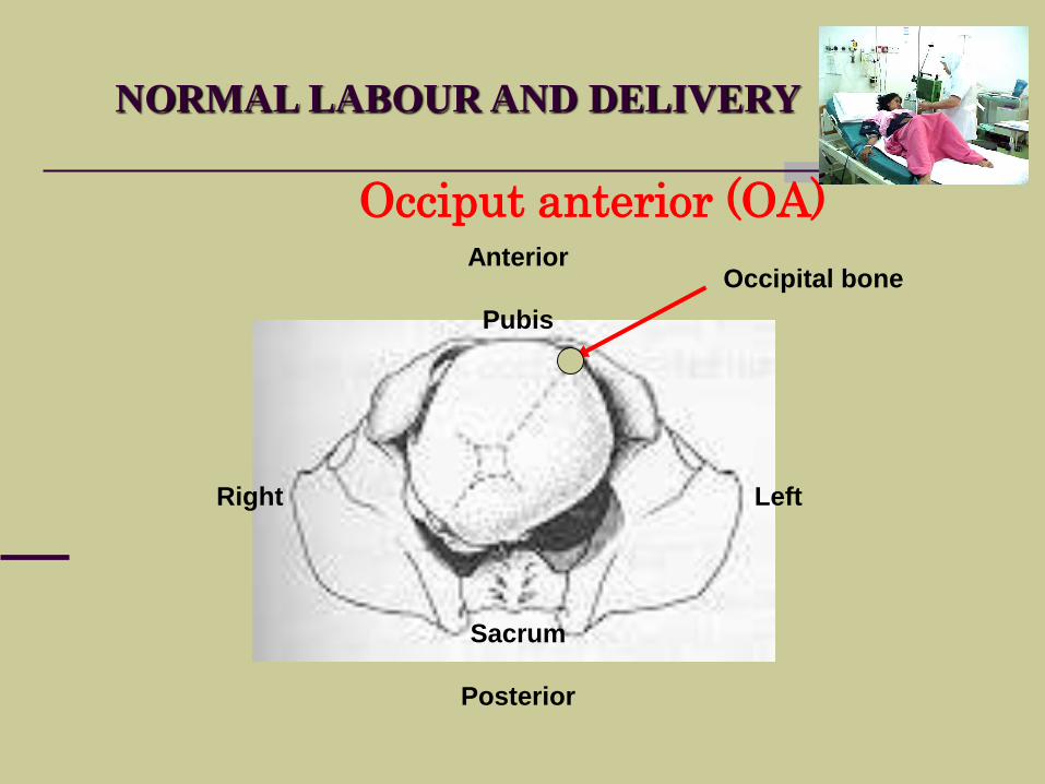
NORMAL LABOUR AND DELIVERY
Occiput anterior (OA)Anterior
Pubis
Sacrum
Posterior
Right Left
Occipital bone
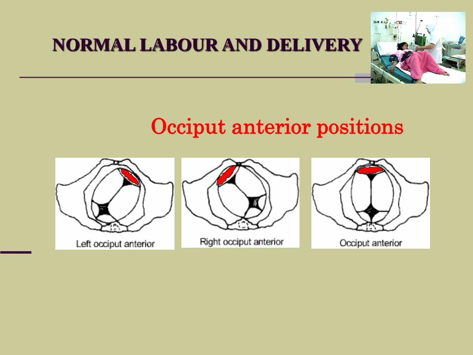
NORMAL LABOUR AND DELIVERY
Occiput anterior positions
NORMAL LABOUR AND DELIVERY
MECHANISM OF LABOUR for occiput anterior
The “mechanism of labour” refers to the sequencing of
events related to posturing and positioning that allows the
baby to find the “easiest way out”.
For a normal mechanism of labour to occur, both the fetal
and maternal factors must be harmonious.
DEFINITION:
NORMAL LABOUR AND DELIVERY
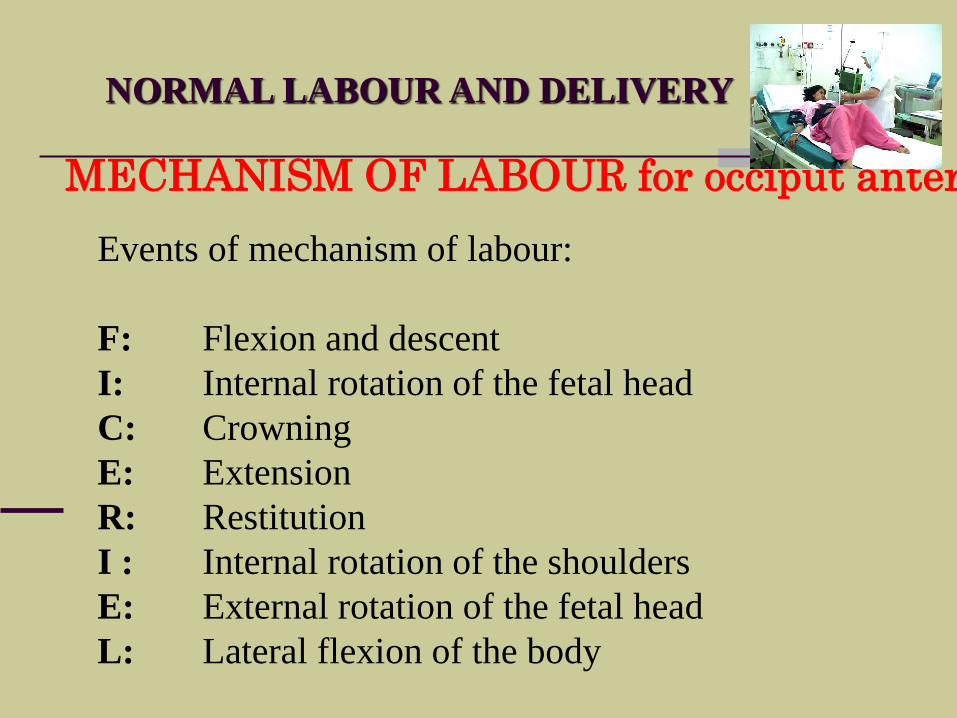
MECHANISM OF LABOUR for occiput anterior
Events of mechanism of labour:
F: Flexion and descent
I: Internal rotation of the fetal head
C: Crowning
E: Extension
R: Restitution
I : Internal rotation of the shoulders
E: External rotation of the fetal head
L: Lateral flexion of the body
NORMAL LABOUR AND DELIVERY
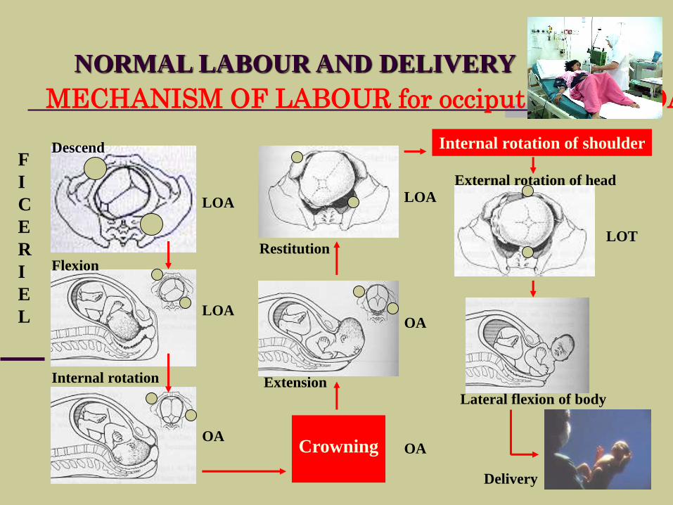
MECHANISM OF LABOUR for occiput anterior (OA)
Descend
Flexion
Internal rotation
Crowning
Extension
Restitution
Internal rotation of shoulder
External rotation of head
Lateral flexion of body
LOA
LOA
OA
LOA
OA
OA
LOT
Delivery
F
I
C
E
R
I
E
L
NORMAL LABOUR AND DELIVERY
MANAGEMENT
THIRD STAGE OF
LABOUR
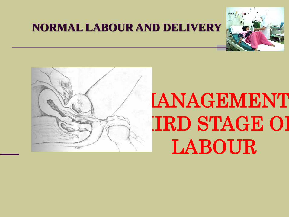
MANAGEMENT OF THE THIRD STAGE OF LABOUR
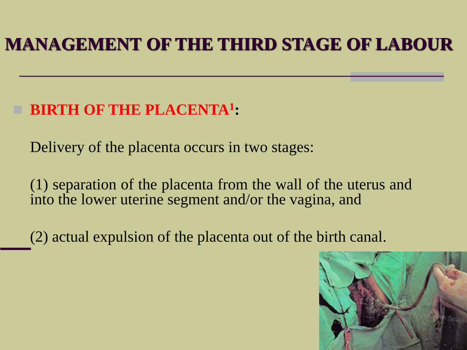
BIRTH OF THE PLACENTA1:
Delivery of the placenta occurs in two stages:
(1) separation of the placenta from the wall of the uterus andinto the lower uterine segment and/or the vagina, and
(2) actual expulsion of the placenta out of the birth canal.
THE THIRD STAGE OF LABOUR
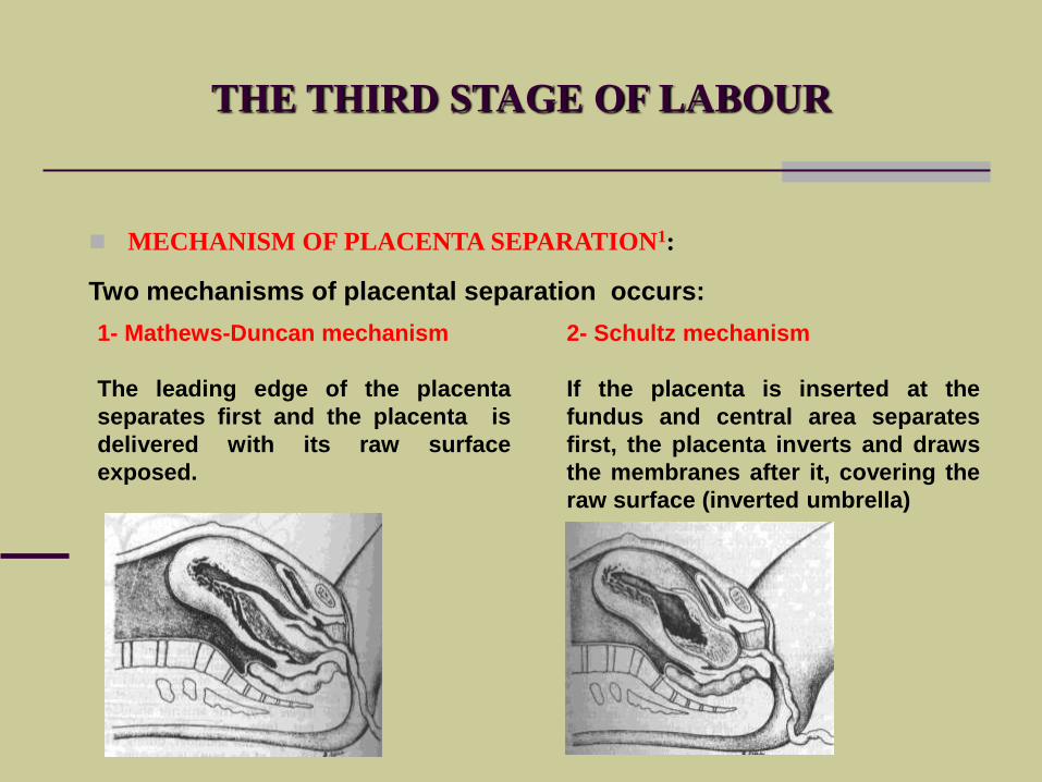
MECHANISM OF PLACENTA SEPARATION1:
Two mechanisms of placental separation occurs:
1- Mathews-Duncan mechanism
The leading edge of the placenta
separates first and the placenta is
delivered with its raw surface
exposed.
2- Schultz mechanism
If the placenta is inserted at the
fundus and central area separates
first, the placenta inverts and draws
the membranes after it, covering the
raw surface (inverted umbrella)
LABOUR AND DELIVERY
WHAT ARE THE SIGNS OF
PLACENTA SEPARATION
MANAGEMENT OF THE THIRD STAGE OF LABOUR
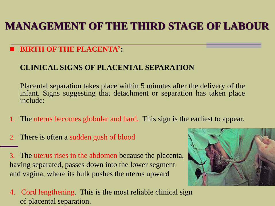
BIRTH OF THE PLACENTA2:
CLINICAL SIGNS OF PLACENTAL SEPARATION
Placental separation takes place within 5 minutes after the delivery of theinfant. Signs suggesting that detachment or separation has taken placeinclude:
1. The uterus becomes globular and hard. This sign is the earliest to appear.
2. There is often a sudden gush of blood
3. The uterus rises in the abdomen because the placenta,
having separated, passes down into the lower segment
and vagina, where its bulk pushes the uterus upward
4. Cord lengthening. This is the most reliable clinical sign
of placental separation.
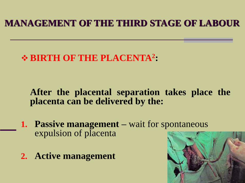
MANAGEMENT OF THE THIRD STAGE OF LABOUR
BIRTH OF THE PLACENTA2:
After the placental separation takes place theplacenta can be delivered by the:
1. Passive management – wait for spontaneous expulsion of placenta
2. Active management
LABOUR AND DELIVERY
ACTIVE MANAGEMENT OF
THE THIRD STAGE OF LABOUR
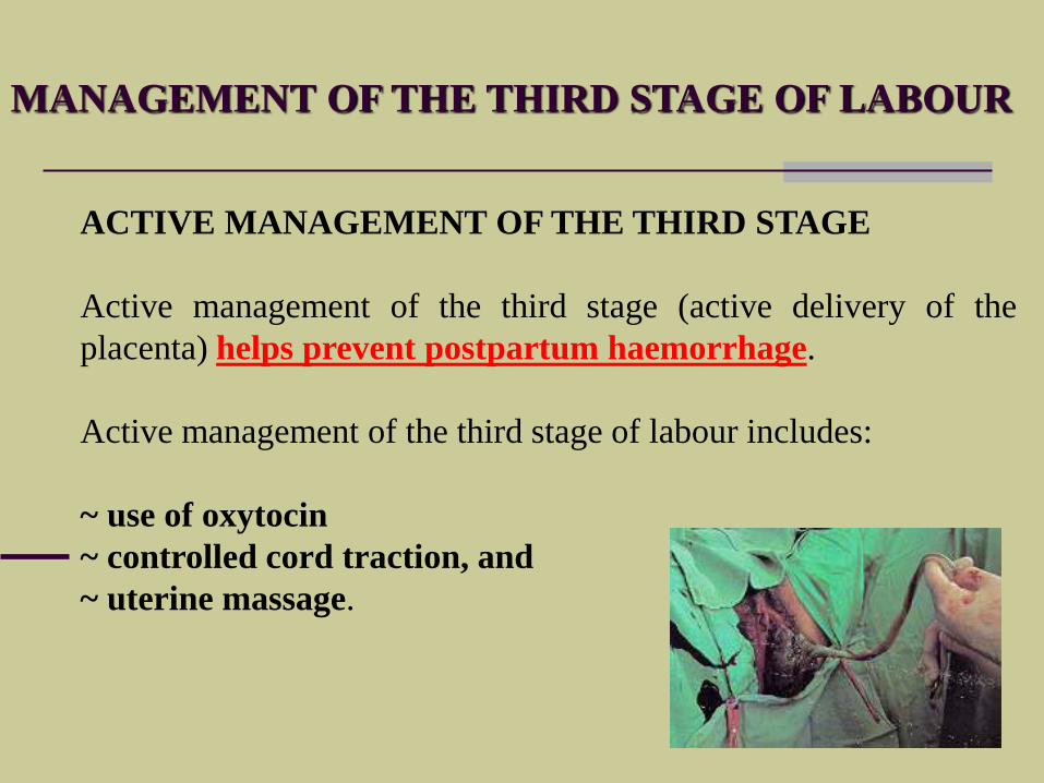
MANAGEMENT OF THE THIRD STAGE OF LABOUR
ACTIVE MANAGEMENT OF THE THIRD STAGE
Active management of the third stage (active delivery of the
placenta) helps prevent postpartum haemorrhage.
Active management of the third stage of labour includes:
~ use of oxytocin
~ controlled cord traction, and
~ uterine massage.
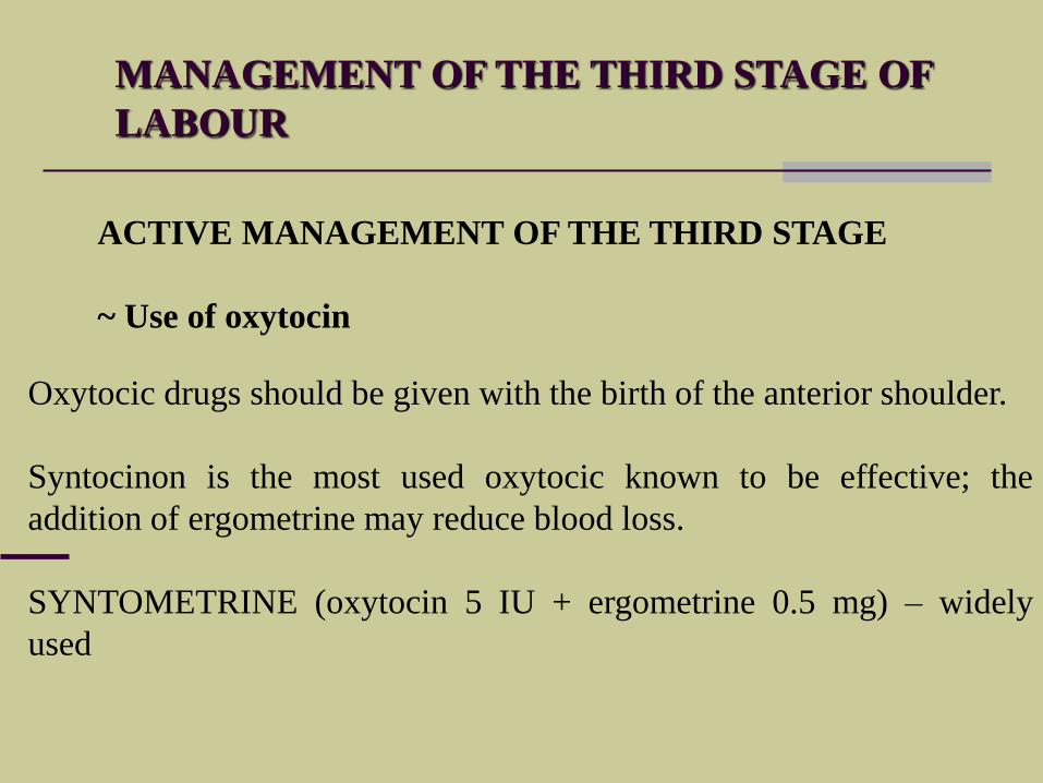
MANAGEMENT OF THE THIRD STAGE OF
LABOUR
ACTIVE MANAGEMENT OF THE THIRD STAGE
~ Use of oxytocin
Oxytocic drugs should be given with the birth of the anterior shoulder.
Syntocinon is the most used oxytocic known to be effective; the
addition of ergometrine may reduce blood loss.
SYNTOMETRINE (oxytocin 5 IU + ergometrine 0.5 mg) – widely
used
MANAGEMENT OF THE THIRD STAGE OF LABOUR
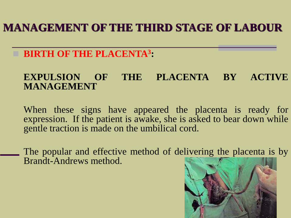
BIRTH OF THE PLACENTA3:
EXPULSION OF THE PLACENTA BY ACTIVEMANAGEMENT
When these signs have appeared the placenta is ready forexpression. If the patient is awake, she is asked to bear down whilegentle traction is made on the umbilical cord.
The popular and effective method of delivering the placenta is byBrandt-Andrews method.
MANAGEMENT OF THE THIRD STAGE OF LABOUR
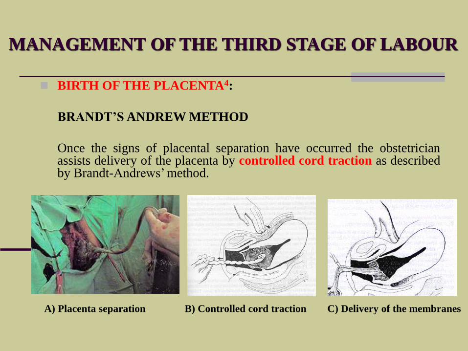
BIRTH OF THE PLACENTA4:
BRANDT’S ANDREW METHOD
Once the signs of placental separation have occurred the obstetricianassists delivery of the placenta by controlled cord traction as describedby Brandt-Andrews’ method.
A) Placenta separation B) Controlled cord traction C) Delivery of the membranes
MANAGEMENT OF THE THIRD STAGE OF LABOUR
BIRTH OF THE PLACENTA5:
EXAMINATION OF THE PLACENTA
The placenta, membranes, and umbilical cord should be examinedfor completeness and for anomalies.
EXAMINATION OF THE PERINEUM
At the same time, the perineal region, vulva outlet, vaginal canal, andthe cervix should be carefully examined for lacerations.
If the perineum has been torn or an episiotomy made, tear or incisionshould be repaired immediately.
MANAGEMENT OF THE THIRD STAGE OF LABOUR
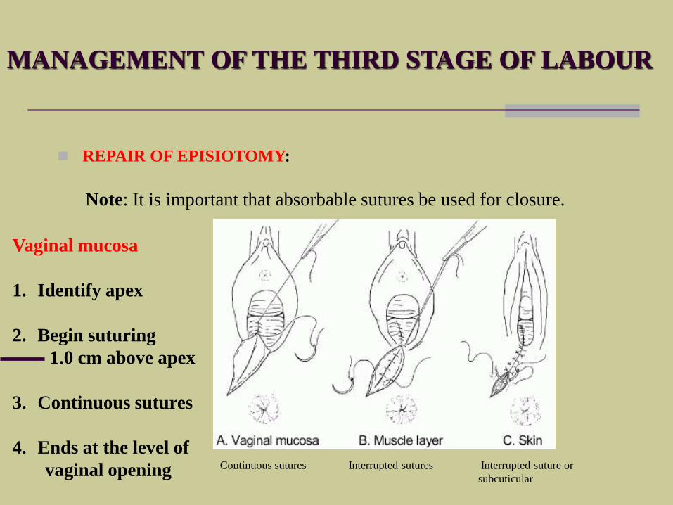
REPAIR OF EPISIOTOMY:
Note: It is important that absorbable sutures be used for closure.
Continuous sutures Interrupted sutures Interrupted suture or
subcuticular
Vaginal mucosa
1. Identify apex
2. Begin suturing
1.0 cm above apex
3. Continuous sutures
4. Ends at the level of
vaginal opening
MANAGEMENT AFTER
DELIVERY
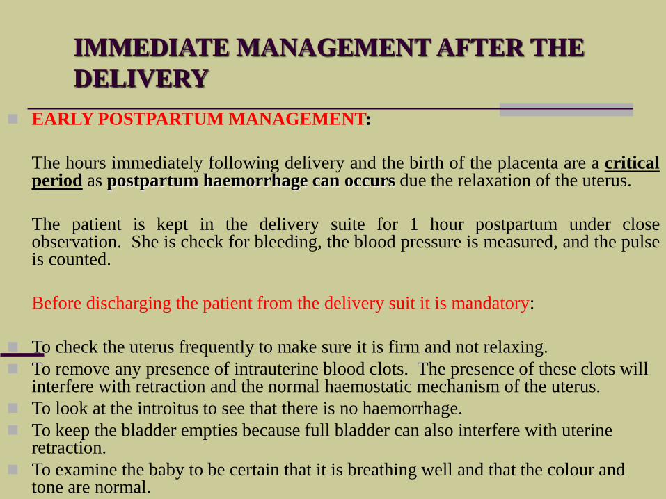
IMMEDIATE MANAGEMENT AFTER THE
DELIVERY
EARLY POSTPARTUM MANAGEMENT:
The hours immediately following delivery and the birth of the placenta are a criticalperiod as postpartum haemorrhage can occurs due the relaxation of the uterus.
The patient is kept in the delivery suite for 1 hour postpartum under closeobservation. She is check for bleeding, the blood pressure is measured, and the pulseis counted.
Before discharging the patient from the delivery suit it is mandatory:
To check the uterus frequently to make sure it is firm and not relaxing.
To remove any presence of intrauterine blood clots. The presence of these clots will interfere with retraction and the normal haemostatic mechanism of the uterus.
To look at the introitus to see that there is no haemorrhage.
To keep the bladder empties because full bladder can also interfere with uterine retraction.
To examine the baby to be certain that it is breathing well and that the colour and tone are normal.


![[PPT]PowerPoint Presentation - Trinity College, Dublin · Web viewSpecial Tutorial programme Professor Deirdre Murphy Trinity College Labour & Delivery Objectives Normal Labour Abnormal](https://static.fdocuments.in/doc/165x107/5b00989f7f8b9a6a2e8ce3d1/pptpowerpoint-presentation-trinity-college-viewspecial-tutorial-programme-professor.jpg)