![Research Article The Epidemiology of Pulmonary ...downloads.hindawi.com/journals/pm/2014/894976.pdfDiagnosis, Treatment, and Prevention of Nontuberculous Mycobacterial Diseases [ ]..](https://static.fdocuments.in/doc/165x107/604c7f25b5ac753490666de3/research-article-the-epidemiology-of-pulmonary-diagnosis-treatment-and-prevention.jpg)
Nontuberculous Mycobacterial Keratitis - InTech -...
26
Chapter 8 Nontuberculous Mycobacterial Keratitis Ana Lilia Pérez-Balbuena, David Arturo Ancona-Lezama, Lorena Gutiérrez-Sánchez and Virginia Vanzzini-Zago Additional information is available at the end of the chapter http://dx.doi.org/10.5772/45964 1. Introduction Mycobacterium species that are considered typical are the tuberculosis species such as M.tu‐ berculosis, M.bovis, M.africarium and M.leprae. These species have only human or animal res‐ ervoirs and are not transmitted by water. In contrast, the species Non-Tuberculosis or “atypical”, naturally are ubiquitous in soil and water and have been found as normal flora of skin, sputum, and gastric contents. These bacteria are resistant to common, disinfectants, chlorine, formaldehyde and glutaraldehyde. NTM can cause infections on all adnexal and ocular tissues including the cornea, iris, lens, retina, choroid and optic nerve. Most NTM infections are caused by M.chelonae and M. fortu‐ itum, that as we will discuss later, belong to the rapid growers group. In this chapter, we will focus on keratitis caused by atypical mycobacterium, since a great number of recent clinical reports of NTM ocular infections are of keratitis. In common gener‐ al ophthalmology procedures like refractive surgery, for example laser in situ keratomileusis (LASIK), Laser epithelial keratomileusis (LASEK), photorefractive keratectomy (PRK), and other specialized procedures such as penetrating keratoplasty (PKP), a transgression to nat‐ ural barriers occurs, this constitutes a risk factor for infection by these organisms. In addi‐ tion, LASIK is one of the most commonly performed procedures in ophthalmology practice. Several factors may contribute to the development of mycobacterial keratitis following LA‐ SIK, making it difficult to determine the true origin of the infection in most cases. This pro‐ cedure is often performed utilizing aseptic, but non sterile techniques. Mycobacterium © 2013 Pérez-Balbuena et al.; licensee InTech. This is an open access article distributed under the terms of the Creative Commons Attribution License (http://creativecommons.org/licenses/by/3.0), which permits unrestricted use, distribution, and reproduction in any medium, provided the original work is properly cited.
Transcript of Nontuberculous Mycobacterial Keratitis - InTech -...

Chapter 8
Nontuberculous Mycobacterial Keratitis
Ana Lilia Pérez-Balbuena,David Arturo Ancona-Lezama,Lorena Gutiérrez-Sánchez andVirginia Vanzzini-Zago
Additional information is available at the end of the chapter
http://dx.doi.org/10.5772/45964
1. Introduction
Mycobacterium species that are considered typical are the tuberculosis species such as M.tu‐berculosis, M.bovis, M.africarium and M.leprae. These species have only human or animal res‐ervoirs and are not transmitted by water. In contrast, the species Non-Tuberculosis or“atypical”, naturally are ubiquitous in soil and water and have been found as normal floraof skin, sputum, and gastric contents. These bacteria are resistant to common, disinfectants,chlorine, formaldehyde and glutaraldehyde.
NTM can cause infections on all adnexal and ocular tissues including the cornea, iris, lens,retina, choroid and optic nerve. Most NTM infections are caused by M.chelonae and M. fortu‐itum, that as we will discuss later, belong to the rapid growers group.
In this chapter, we will focus on keratitis caused by atypical mycobacterium, since a greatnumber of recent clinical reports of NTM ocular infections are of keratitis. In common gener‐al ophthalmology procedures like refractive surgery, for example laser in situ keratomileusis(LASIK), Laser epithelial keratomileusis (LASEK), photorefractive keratectomy (PRK), andother specialized procedures such as penetrating keratoplasty (PKP), a transgression to nat‐ural barriers occurs, this constitutes a risk factor for infection by these organisms. In addi‐tion, LASIK is one of the most commonly performed procedures in ophthalmology practice.
Several factors may contribute to the development of mycobacterial keratitis following LA‐SIK, making it difficult to determine the true origin of the infection in most cases. This pro‐cedure is often performed utilizing aseptic, but non sterile techniques. Mycobacterium
© 2013 Pérez-Balbuena et al.; licensee InTech. This is an open access article distributed under the terms of theCreative Commons Attribution License (http://creativecommons.org/licenses/by/3.0), which permitsunrestricted use, distribution, and reproduction in any medium, provided the original work is properly cited.

chelonei, M. abscessus, M. fortuitum, M. szulgai, and M. mucogenicum have been reported as theresult of improper asepsis.
Atypical Mycobacteria corneal infections are rare, but devastating complications. Althoughrare, are a diagnostic and therapeutic challenge. Mycobacterium have been involved in sev‐eral isolated cases as well as in outbreaks.[4-12]
2. Microbiological and laboratory profile
Mycobacterium species that are considered typical are the tuberculosis specie; M.tuberculosis.Many species enclosed in genus Mycobacteriaceae are true human pathogens as Mycobacteri‐um tuberculosis complex, that include M tuberculosis, M bovis, non pathogenic M bovis BCG,M africanum, M caprae, M microti, and M pinnipedii are characterized by different phenotypesand mammalian host ranges, displays the most extreme genetic homogeneity with 0.01 to0.03% nucleotides variation only. Growth rate in this group is 6 to 12 weeks. M leprae is theonly non cultivable in vitro specie and has some genetic variations in relation to M tuberculo‐sis complex.
The only genus of the Mycobacteriaceae family is the Mycobacterium, the Mycobacteriaceae be‐longs to the order Actinomycetales. Mycobacteria is an unusual ocular pathogen that has thefollowing characteristics: intracellular bacilli, slow growing organisms, obligate aerobic,non-motile, non-capsulated, non-sporing, present a large amounts of lipids and true waxesin their cell walls, and are considered gram-positive and acid-fast.
Other places where NTM have been isolated are: contaminated tap water, saline solutions,disinfectant solutions, and hemodialyzers. Mycobacteria influences a number of ocularstructures, including the cornea,iris, lens,retina, choroid and optic nerve.
Clinical manifestations of the typical mycobacteria are : lupus vulgaris on eyelid, phlycte‐nule, scleritis, lacrimal gland involvement, orbital periostitis, granulomatous panuveitis, sec‐ondary glaucoma and cataract, chorioretinal plaque or nodule, nerve palsies.
The incidence of tuberculosis has increased due to the growth in homelessness, the upsurgeof intravenous drug abuse, neglect of tuberculosis programs, acquired immunodeficiencysyndrome.
Runyon classified nontuberculous mycobacteria into four groups, described in [Table 1].Runyon Classification of tuberculous and non-tuberculous Mycobacterium is based, on thegrowth rate, and pigment production. Groups I to III are slow growers that require approxi‐mately 2 to 3 or more weeks to form visible colonies in culture at 27°C. Group IV organismsare rapid growers, forming non-pigmented colonies in culture in one week.[1,14,15]
Out of the more than 130 actually validated species of non-tuberculousmycobacteria, 60 areslowly growingmycobacteria, that shows in solid culture media growth rates of 2 to 4weeks, the most clinically significance and most frequently in isolated human samples are Mavium, M intracellulare, M kansasii, M marinum, M xenopi, M malmoense and M ulcerans. In the
Common Eye Infections148

rapidly growing mycobacteria group with 7 -10 days of growing rate on solid culture media,there are three major clinically important species responsible for 80% of diseases in humansM chelonae, M abscessus and M fortuitum, that are too frequently located in tap water andhave been related with sepsis in bone marrow transplant, post-traumatic, surgical ocularand other surgical wound infections.
SlowGrowers Rapid Growers
Group I Group II Group III Group IV
(photochromogens)
6-12 weeks
(scotochromogens)
2-4 weeks
(nonchromogens)
2-4 weeks
(nonchromogens)
7-10 days
M. marinum
M. kansasii
M. simiae
M. asiaticum
M. scrofulaceum
M. szulgai
M. gordonae
M. xenopi
M. flavescens
M. avium
M. intracellulare
M. haemophilum
M. paratuberculosis
M. gastri
M. malmoense
M. nonchromogenicum
M. terrae
M. triviale
M. fortuitum group
M. fortuitum
M. peregrinum
M. mucogenicum
M. Senegalese
M. septicum
M. mageritense
M. chelonae-abscessus group
M. chelonae
M. abscessus
M. immunogenum
M. smegmatis group
M. smegmatis
M. goodii
M. wolinskyi
Problem statement: To describe the experience in México (Asociación Para Evitar La Ceguera I.A.P. “Hospital Dr. LuisSánchez Bulnes” [APEC]) in the management of keratitis caused by nontuberculous mycobacteria.
Application area: Cornea and Refractive Service and Mycrobiology Service
Research course: To describe of atypical Mycobacterium keratitiscases diagnosed and attended in the Cornea Serviceof our Hospital in the last 10 years.
Methods: This is a descriptive retrospective case series of five patients treated in our service.
Table 1. Runyon’s Classification of Nontuberculous Mycobacterium
3. Laboratory diagnosis and bacteriology
In ophthalmological infections traumatic or post-surgical in origin, are frequently involvedin non-tuberculous or atypical fast growing Mycobacteria, the species M. chelonae, M. chelo‐nae /abscesus, M. fortuitum have been isolated in many cases. These rapidly growing Mycobac‐teria share the cellular characteristics of Mycobacterium genus,like mycolic acids esters in its
Nontuberculous Mycobacterial Keratitishttp://dx.doi.org/10.5772/45964
149
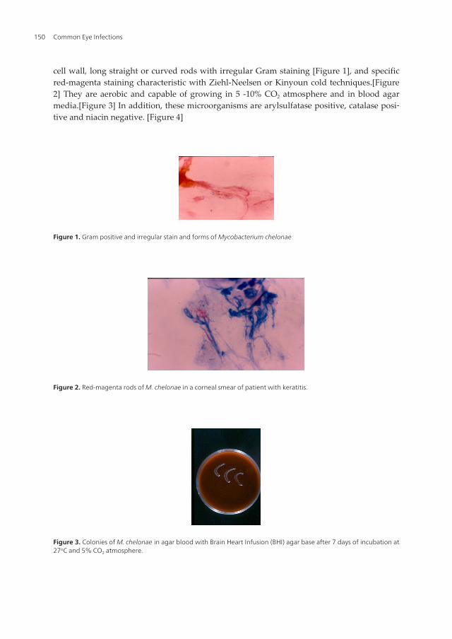
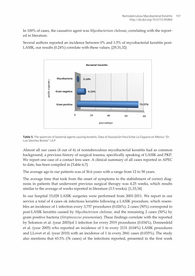
cell wall, long straight or curved rods with irregular Gram staining [Figure 1], and specificred-magenta staining characteristic with Ziehl-Neelsen or Kinyoun cold techniques.[Figure2] They are aerobic and capable of growing in 5 -10% CO2 atmosphere and in blood agarmedia.[Figure 3] In addition, these microorganisms are arylsulfatase positive, catalase posi‐tive and niacin negative. [Figure 4]
Figure 1. Gram positive and irregular stain and forms of Mycobacterium chelonae
Figure 2. Red-magenta rods of M. chelonae in a corneal smear of patient with keratitis.
Figure 3. Colonies of M. chelonae in agar blood with Brain Heart Infusion (BHI) agar base after 7 days of incubation at27oC and 5% CO2 atmosphere.
Common Eye Infections150

Figure 4. Catalase 65oC positive test (O2 bubbles) for Mycobacterium chelonae.
To identify the microorganism, its phenotypic characteristics were used, such as pigmenta‐tion of colonies growing in the darkness (presented in Table 1) on Lowenstein-Jensen media.
The most common species of rapidly growing Mycobacteria belong to group IV of Runyon’sclassification, also known as colorless or nonchromatogens.[Figure 5]
Figure 5. Mycobacterium chelonae colonies in Lowenstein-Jensen medium after 7 days of incubation at 27oC.
For genotypic characterization, the 16Sr RNA gene sequencing, high performance liquidchromatography and polymerase chain reaction has been used.
4. Clinical features
Nontuberculous Mycobacteria can cause infections of all adnexal and ocular tissues. Mostatypical Mycobacteria infections are caused by M. chelonae, and M. fortuitum.
Dacryocystitis and Canaliculitis: Present as epiphora and erythematous swelling in the me‐dial canthal area, purulent material can be expressed with massage of the lacrimal sac.
Orbital Infections: Present with a gradual development of periorbital edema, without a sig‐nificant proptosis and a superficial skin lesion may be present. The visual acuity will de‐pend on the involvement of the optic nerve. [18,19]
Conjuntivitis and Scleritis: Present as conjunctival or as scleral injection and tenderness ac‐companied with chronic redness, irritation, discharge and pain. Sometimes, marked scleralthinning may develop. Scleral abscesses manifest late in the course of the disease as subcon‐junctival nodules. [20,21]
Nontuberculous Mycobacterial Keratitishttp://dx.doi.org/10.5772/45964
151

Endoftalmitis: Present with severe pain, decreased vision, and redness and discharge, mayexist hypopyon, and variable amounts of granulomatous keratitic precipitates. Moderate vit‐reous inflammation is present in most cases.
Keratitis: The greatest number of recent clinical reports of nontuberculous Mycobacteria oc‐ular infections are of keratitis, as seen in our hospital (Asociación Para Evitar La Ceguera enMéxico “Dr. Luis Sánchez Bulnes” I.A.P. [APEC]). Keratitis most commonly follows traumaor surgery and has been associated with penetrating keratoplasty and refractive surgery.
Nontuberculous Mycobacteria keratitis is characterized by a delayed onset of symptomsthat range typically from 1 to 3 weeks following the exposing event. There is decreased vi‐sion and an indolent course and some cases various degrees of pain, ranging from indolentto severe.
Presenting symptoms can include any of the following: pain, redness, photophobia, de‐creased vision, foreign body sensation and/or mild irritation. Presenting clinical signs in‐clude infiltrates in the corneal interface that can either be multiple white granular opacities<0.5mm in diameter with well defined borders or radiating projections, or a single whiteround lesion (0.1-2 mm in diameter) which may progress to satellite lesions. These infiltratesspread subsequently into the corneal stroma posteriorly and anteriorly and can result in per‐foration though the flap to surface. [Table 2].A hypopyon is often found in untreated orpoorly treated cases. [25,26]
Lazar and colleagues first described the presence of a “cracked windshield” appearance tothe cornea around the edge of the central area of ulceration and infiltrate, seen transientlyearly in the course of the infection. [25,27,28] This sign consist of radiating lines from thecentral infiltrate in the middle third of the corneal stroma. It is important to mention thatNTM keratitis has also been noted in the abscence of epithelial defect with deep stromal ker‐atitis. The corneal infiltrate may show irregular margins.
Signs Symptoms
Single or multiple white granular opacities with well
defined borders or radiating projections
Satellite infiltrates
Hypopyon
Mild or absent anterior chamber reaction
“Cracked windshield” appearance
Pain (mild)
Redness
Photophobia
Tearing
Foreign body sensation
Decreased visual acuity
Table 2. Signs and symptoms of keratitis caused by mycobacterias
Common Eye Infections152

5. Predisposing factors
Nontuberculous Mycobacteria are opportunistic pathogens that require an alteration inthe ocular barriers to produce infection. In nearly all reports, a previous history of minorto severe trauma is the common denominator.Men and women are equally affectedamong NTM keratitis patients who have had LASIK, in contrast to a 70% male prepon‐derance among patients who have not had LASIK, the result of a higher prevalence oftrauma in males. [Table 3] [5,29]
Risk factors associated with NTM keratitis
Trauma
Surgical trauma
Refractive surgeries
Laser in situ keratomileusis (LASIK)
Laser epithelial keratomileusis (LASEK)
Corneal transplantation
Radial keratotomy
Photorefractive keratectomy (PRK)
Penetrating keratoplasty (PKP)
Other ophthalmologic surgeries
Extracapsular cataract extraction
Small incision corneal cataract surgery
Suture removal
Contact lens wear
Corticosteroid use
Improper aseptic technique or sterilization of surgical instrumentation
Table 3. Risk factors for the development of nontuberculous mycobacterial keratitis.
Post-LASIK NTM keratitis: Laser in situ keratomileusis (LASIK) is the most commonlyperformed refractive surgical procedure, since it offers rapid visual rehabilitation, de‐creased stromal scarring, less postoperative pain, and the ability to treat a wider range ofrefractive disorders. LASIK preserves the integrity of Bowman’s membrane and the overly‐ing epithelium, thus decreasing the risk for microbial keratitis. Several studies have report‐ed an incidence of bacterial infection following LASIK procedures varying between 0% to1.5%. [29,31,32] Solomon et. al published the first survey that provides information about
Nontuberculous Mycobacterial Keratitishttp://dx.doi.org/10.5772/45964
153

post-LASIK infectious keratitis. The most common organisms cultured were nontubercu‐lous mycobacteria (48%) and staphylococci (33%).. These findings are consistent withChang’s research, where he found that nearly 47% of infectious keratitis cases after LASIKappear to be caused by NTM; 32% being caused by Mycobacterium chelonei alone. In con‐trast to the acute or subacute onset of symptoms generally seen postoperatively in bacteri‐al and fungal keratitis, rapid growing atypical mycobacteria may present with a sloweronset of clinical disease, from 3 to 14 weeks (3.5 weeks in average) after the procedure. Itis important to keep in mind that this is not a rule, and more rapidly growing NTM suchas the Mycobacterium chelonae-abscessus group may present as soon as 10 days posterior tothe refractive surgery. [1,33,34]
Innoculation of NTM to the flap-stromal interface probably takes place at the time of sur‐gery, therefore, it is infrequent to find an epithelial defect, being present in less than half ofcases. Corneal infiltrates appear to be entirely within the lamellar flap or at the flap inter‐face and may be either multiple, tiny, white, granular opacities less than 0.5mm in diame‐ter or a single white lesion ranging between 0.1-0.2mm in diameter. Anterior extension ofinfiltrate with ulceration or anterior perforation of the corneal flap or posterior extension in‐to the stroma is a rare finding and is usually associated with a delay in diagnosis and thebeginning of therapy. Anterior chamber reaction is not a common finding, occurring in on‐ly 20% of cases.[1,29]
6. Differential diagnosis
NTM keratitis can often be mistaken with other bacterial infections that cause nonsuppura‐tive keratitis. Several authors suggest to keep in mind other causative organisms that maypresent, in the course of disease, similar clinical features such as fungal keratitis, infec‐tious crystalline keratopathy, Nocardia keratitis, herpes simplex virus, and rarely Acantha‐moeba keratitis. In our experience at APEC, the principal differential diagnosis must bemade between fungal and Nocardia keratitis.
Fungal keratitis: Often preceded by history of trauma involving plants or foreign bodies.Like NTM, mycotic keratitis may worsen with the use of topical corticosteroids. Thesekeratitis often do not respond to topical antibiotics, as seen with NTM keratitis. Multiplecorneal fungal abscesses may emulate the multifocal presentation of NTM keratitis. Sabo‐uraud’s agar is essential for the identification of the causative fungus. [Figure 6]
Infectious Crystalline Keratopathy (ICK): “Cracked windshield” corneal appearance maybe also seen in this keratitis caused most commonly by Streptococcus species, but unlike thisentity, NTM keratitis presents with this sign transiently early in the course. Gorovoy et alfirst described Infectious crystalline keratopathy in 1984, describing it as a unique cornealinfection characterized by and indolent, progressive course: a paucity of inflammation; and
Common Eye Infections154

the formation of sharply demarcated, gray-white, branching, round, stellate, or needle-likeopacities in the corneal stroma. Although the duration of the relatively recalcitrant course ofthe infectious crystalline keratopathy may mimic NTM keratitis, the crystalline appearancepersists in ICK but is transient in NTM keratitis. Among post-LASIK patients, crystallineNTM keratitis occurs rarely (less than 10%).
Nocardiaasteroides infection: should also be considered, since it is an acid-fast microorgan‐ism capable of producing bacterial keratitis. The best way to differentiate Nocardia infectionfrom NTM keratitis is with a Gram stain. Nocardia keratitis is more fulminant than NTMkeratitis.[Figure 7]
Figure 7. Nocardial keratitis as a differential diagnosis of NTM keratitis.
Deep lamellar keratitis can be confused with post-LASIK NTM keratitis. It usually presentswithin the first 7 days post-LASIK, and unlike NTM keratitis, it clears with topical cortico‐steroids. If the wrong diagnosis is made, the improper use of such medications contribute tothe delay in diagnosis of post-LASIK NTM keratitis.
Acanthamoeba keratitis generally presents with out-of-proportion pain in comparison tothe clinical findings. It is common to see ring ulcers in Acanthamoeba keratitis. This agentresponds, unlike NTM, to topical biguanides and diamidines, and topical corticosteroidsmay be of some benefit.
Figure 6. Candida keratitis after penetrating keratoplasty for keratoconus.
Nontuberculous Mycobacterial Keratitishttp://dx.doi.org/10.5772/45964
155

Herpetic keratitis. In necrotizing stromal keratitis, herpetic keratitis can cause dense whitestromal infiltrates that may be confused with NTM keratitis. Special features that are moretypically found in herpetic keratitis are decreased corneal sensation and previous or con‐comitant history of herpes labialis lesions. NTM keratitis may simulate a non-suppurativeherpetic keratitis, especially in cases caused by Mycobacterium marinum. There may also bea dendritic or geographic epithelial defect with minimal stromal infiltration, misleading theclinician and prompting treatment with antivirals. This can lead to the development of a se‐vere, wide corneal infiltrate.
7. Our experience
Keratitis caused by atypical Mycobacteria is characterized by an indolent course and poor re‐sponse to antibiotics. The diagnosis requires a high index of suspicion and their treatment isusually very difficult. The early diagnosis of nontuberculous mycobacterial keratitis follow‐ing LASIK is not easy, because the overlying, noninvolved stroma hinders the collection ofsufficient material for culture. In addition, such organisms are only detectable by culture inspecial media, such as Lowenstein-Jensen, and special stains like Ziehl-Neelsen, which maynot be included among routine cultures in the microbiology service.
Table 4. Profile of microorganisms causing infectious keratitis; 2025 cases, during 10 years (2000-2010). Data ofAsociación Para Evitar La Ceguera en México “Dr. Luis Sánchez Bulnes” I.A.P.
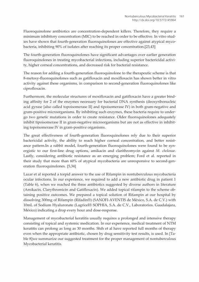
In our hospital, our service found an incidence of 2025 cases of infectious keratitis in the last10 years (2000-2010). We found that 83.03% corresponded to infections caused by bacteria,6.67% mycotic, and 10.3% originated by virus. [Table 4] Out of this percentage of bacterialkeratitis, we report a frequency of 73.57% caused by gram positive, 9.22% caused by gramnegative and 0.24% originated by nontuberculous mycobacteria. [Table 5]
Common Eye Infections156
In 100% of cases, the causative agent was Mycobacterium chelonae, correlating with the report‐ed in literature.
Several authors reported an incidence between 0% and 1.5% of mycobacterial keratitis post-LASIK, our results (0.24%) correlate with these values. [29,31,32]
Table 5. The spectrum of bacterial agents causing keratitis. Data of Asociación Para Evitar La Ceguera en México “Dr.Luis Sánchez Bulnes” I.A.P
Almost all our cases (4 out of 6) of nontuberculous mycobacterial keratitis had as commonbackground, a previous history of surgical trauma, specifically speaking of LASIK and PKP.We report one case of a contact lens user. A clinical summary of all cases reported in APECto date, has been compiled in [Table 6,7]
The average age in our patients was of 36.6 years with a range from 12 to 58 years.
The average time that took from the onset of symptoms to the stabishment of correct diag‐nosis in patients that underwent previous surgical therapy was 4.25 weeks, which resultssimilar to the average of weeks reported in literature (3.5 weeks). [1,33,34]
In our hospital 15,028 LASIK surgeries were performed from 2001-2011. We report in ourservice a total of 4 cases ok infectious keratitis following a LASIK procedure, which resem‐bles an incidence of 1 infection every 3,757 procedures (0.026%). 2 cases (50%) correspond topost-LASIK keratitis caused by Mycobacterium chelonae, and the remaining 2 cases (50%) bygram positive bacteria (Streptococcus pneumoniae). These findings correlate with the reportedby Solomon et al. (year 2003)of 1 infection for every 2919 procedures (0.034%), Donnenfeldet al. (year 2005) who reported an incidence of 1 in every 2131 (0.04%) LASIK proceduresand LLovet et al. (year 2010) with an incidence of 1 in every 2841 cases (0.035%). The studyalso mentions that 65.5% (76 cases) of the infections reported, presented in the first week
Nontuberculous Mycobacterial Keratitishttp://dx.doi.org/10.5772/45964
157

postoperatively. 6.03% (7 cases) presented in the second week, 14.65% (17 cases) presentedbetween the second and fourth week and lastly 13.79% (16 cases) presented after 1 month. 2of our cases, the ones caused by Streptococcus pneumoniae, presented in the first week postop‐eratively. 1 nontuberculous mycobacterial case presented between the second and fourthweek (3 weeks), and lastly the remaining NTM keratitis case presented presented after 1month (7 weeks). Speaking of ethiological factors, Solomon et al. reported that the mostcommon microorganisms involved in post-LASIK keratitis are mycobacteria (48%) and coc‐cus (33%), we found similar data in our retrospective analysis; Mycobacterial keratitis 50%and Streptococcus 50%.[30,39,40]
F=female, M=male, CF=count fingers, OS=left eye, OD=right eye, PO=per oral, BID=twice daily, DM=Diabetes Mellitus,VA= visual acuity, PKP=penetrating keratoplasty, LASIK=laser in situ keratomileusis
Table 6. Nontuberculousmycobacterialkeratitis in patients of Asociación Para Evitar La Ceguera en México “Dr. LuisSánchez Bulnes“ I.A.P.
Velotta reported that nearly 90% of NTM keratitis after LASIK cases are unilateral, all of ourcases presented in just one eye.
Infectious keratitis after penetrating keratoplasty (PKP) is not a frequent complication withan incidence ranging from 1.8% to 11.0%; however, this infection has a high risk of loss ofcorneal clarity. In our present analysis, the remaining 2 patients that underwent surgicalprocedures, developed nontuberculous mycobacterial keratitis posterior to penetrating kera‐toplasty. Both cases were promptly diagnosed after onset of symptoms, resulting in satisfac‐tory outcomes and good final visual acuity [Table 7] [Figure 8,9]
Common Eye Infections158

PKP=penetrating keratoplasty, ECCE=extracapsular, IOL=intraocular lens.
Table 7. Surgical treatment and outcome in nontuberculousmycobacterial keratitis in patients of Asociación ParaEvitar La Ceguera en México “Dr. Luis Sánchez Bulnes“ I.A.P.
Figure 8. Patient 2, clinical examination 4 weeks after penetrating keratoplasty with conjunctival hyperemia and cor‐neal infiltrate (3.0 x 2.0 mm) in graft-host junction caused by Mycobacterium chelonae.
8. Treatment
Management of this type of infectious keratitis often traduces in a medical challenge. In cases of
identified NTM corneal infection, there is considerable benefit from the use of combined antibi‐
otics, since atypical mycobacteria have a slower growth rate compared to other bacteria and
may become resistant to a single antibiotic class during the course of extended treatment.
Nontuberculous Mycobacterial Keratitishttp://dx.doi.org/10.5772/45964
159

Figure 9. Patient 2, eighteen months after therapy discontinuation. Corneal graft is infection-free and clear in the vis‐ual axis; best-corrected vision of 20/30 was attained with a +3.50-D contact lens.
The base of treatment consists of a double approach; appropriate antibiotic and judicioussurgical intervention. Such antimicrobial choice becomes complicated since a poor correla‐tion exists between in vitro susceptibility profiles and the final clinical response. We recom‐mend surgical debridement, depending on the case, to facilitate drug penetration to theinterlamellar space. In some cases, flap amputation may be necessary, the rationale for thisprocedure is to lower the bacterial load, remove necrotic as well as infected tissue, and per‐mit better antibiotic penetration. We recommend this surgical procedures in recalcitrantpost-LASIK NTM keratitis to maintain the infection under control.
De La Cruz et al. suggest initial combined antibiotic therapy that includes at least 2 of the 3most susceptible agents (clarithromycin, amikacin, and fourth-generation fluoroquinolones)for rapidly growing mycobacteria specially if known resistance has been documented. Theinitial therapy recommended for many years has been the use of topical Amikacin sulfate20-40mg/mL.This antibiotic is the most frequently used agent in the treatment of NTM kera‐titis. In our institution we use amikacin sulfate (Amikin® 500mg injectable solution. Bristol-Myers Squibb de México S. de R.L. de C.V.)diluted to a concentration of 20mg/mL, one dropevery hour and dose-response. Even though this antibiotic constitutes the first line of treat‐ment against atypical mycobacterial keratitis, only a success rate of 30-40% has been report‐ed. This therapeutic agent has also been associated with high epithelium toxicity when it isapplied for a prolonged course.
We recommend the addition of two additional antibiotics to the drug scheme, such as amacrolide like clarithromycin and a fourth-generation fluoroquinolone like gatifloxacin.[Ta‐ble 6] In our hospital we employ oral clarithromycin Klaricid H.P.® 500mg (Abbott Labora‐tories de México S.A. de C.V. México, D.F.) twice daily, and Zymar® (gatifloxacin 0.3%Allergan Labs, Irvine, CA).
Common Eye Infections160
Fluoroquinolone antibiotics are concentration-dependent killers. Therefore, they require aminimum inhibitory concentration (MIC) to be reached in order to be effective. In vitro stud‐ies have shown that fourth-generation fluoroquinolones are effective against atypical myco‐bacteria, inhibiting 90% of isolates after reaching its proper concentration.[23,43]
The fourth-generation fluoroquinolones have significant advantages over earlier generationfluoroquinolones in treating mycobacterial infections, including superior bactericidal activi‐ty, higher corneal concentrations, and decreased risk for bacterial resistance.
The reason for adding a fourth-generation fluoroquinolone to the therapeutic scheme is that8-metoxy-fluoroquinolones such as gatifloxacin and moxifloxacin has shown better in vitroactivity against these organisms, in comparison to second-generation fluoroquinolones likeciprofloxacin.
Furthermore, the molecular structures of moxifloxacin and gatifloxacin have a greater bind‐ing affinity for 2 of the enzymes necessary for bacterial DNA synthesis (deoxyribonucleicacid gyrase [also called topoisomerase II] and tipoisomerase IV) in both gram-negative andgram-positive microorganisms. By inhibiting such enzymes, these bacteria require to under‐go two genetic mutations in order to create resistance. Older fluoroquinolones adequatelyinhibit tipoisomerase II in gram-negative microorganisms but are not as effective in inhibit‐ing topoisomerase IV in gram-positive organisms.
The great effectiveness of fourth-generation fluoroquinolones rely due to their superiorbactericidal activity, the ability to reach higher corneal concentration, and better resist‐ance pattern.In a rabbit model, fourth-generation fluoroquinolones were found to be syn‐ergistic to our first-line drug options, amikacin and clarithromycin against M. chelonae.Lastly, considering antibiotic resistance as an emerging problem; Ford et al. reported intheir study that more than 60% of atypical mycobacteria are unresponsive to second-gen‐eration fluoroquinolones. [5,34]
Lazar et al reported a torpid answer to the use of Rifampin in nontuberculous mycobacteriaocular infections. In our experience, we required to add a new antibiotic drug in patient 1(Table 6), when we reached the three antibiotics suggested by diverse authors in literature(Amikacin, Clarythromicin and Gatifloxacin). We added topical rifampin to the scheme ob‐taining positive outcomes. We prepared a topical solution of Rifampin at our hospital bydissolving 300mg of Rifampin (Rifadin®) (SANOFI-AVENTIS de México, S.A. de C.V.) with10mL of Sodium Hyaluronate (Lagricel® SOPHIA, S.A. de C.V., Laboratorios. Guadalajara,México) indicating a drop every hour and dose-response.
Management of mycobacterial keratitis usually requires a prolonged and intensive therapyconsisting of topical and systemic medication. In our experience, medical treatment of NTMkeratitis can prolong as long as 30 months. Shih et al have reported full months of therapyeven when the appropriate antibiotic, chosen by drug sensitivity test results, is used. In [Ta‐ble 8]we summarize our suggested treatment for the proper management of nontuberculousMycobacterial keratitis.
Nontuberculous Mycobacterial Keratitishttp://dx.doi.org/10.5772/45964
161
Suggested treatment
Triple
Antibotic Therapy
Topical1. Amikacin 20 mg/mL
2. Fourth-generation fluoroquinolone (gatifloxacin)
Systemic 3. Clarithromycin 500mg PO BID
* In case of resistance addition of Rifampin 30mg/mL.
SurgicalTherapy
1. Flap lift and irrigation
2. Flap amputation in post-LASIK
3. Biopsy and culture
4. Penetrating keratoplasty
Table 8. We suggest a triple antibiotic treatment combined if needed with surgical therapy.
9. Modification to initial therapy
The medical response of mycobacterial keratitis to antibiotic therapy can be achieved by con‐stant clinical observance. This can be difficult to appreciate in the first days of treatment due toincrease in inflammation and local reaction to topical agents. The clinical response varies de‐pending on the microorganism and pathogenicity of the mycobacteria, duration of the infec‐tion, risk factors involved and the patient’s individual response (immunosuppresion).
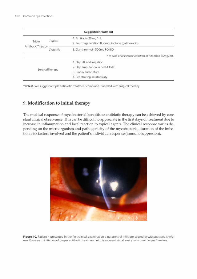
Figure 10. Patient 4 presented in the first clinical examination a paracentral infiltrate caused by Mycobacteria chelo‐nae. Previous to initiation of proper antibiotic treatment. At this moment visual acuity was count fingers 2 meters.
Common Eye Infections162

If the chosen therapy is effective, some response should manifest within the first of 24 to 72hours of appropriate treatment. [Figure 10,11]. Said response manifests with the decrease ofstromal infiltrates and less anterior chamber inflammation in case it exists. [Figure 12, 13]
Figure 11. Patient 4 at 3 months follow-up after proper antibiotic treatment was applied. Final visual acuity was20/30.
Figure 12. Patient 1 with preceding hypopyon (black arrow) and anterior chamber reaction who underwent a thera‐peutic flap amputation procedure.
Nontuberculous Mycobacterial Keratitishttp://dx.doi.org/10.5772/45964
163
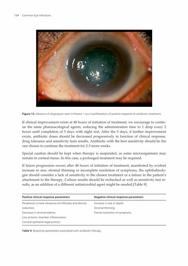
Figure 13. Absence of ahypopyon seen in Patient 1 as a manifestation of positive response to antibiotic treatment.
If clinical improvement exists at 48 hours of initiation of treatment, we encourage to contin‐ue the same pharmacological agents, reducing the administration time to 1 drop every 2hours until completion of 5 days with night rest. After the 5 days, if further improvementexists, antibiotic doses should be decreased progressively in function of clinical response,drug tolerance and sensitivity tests results. Antibiotic with the best sensitivity should be theone chosen to continue the treatment for 2-3 more weeks.
Special caution should be kept when therapy is suspended, as some microorganisms mayremain in corneal tissue. In this case, a prolonged treatment may be required.
If lesion progression occurs after 48 hours of initiation of treatment, manifested by evidentincrease in size, stromal thinning or incomplete resolution of symptoms, the ophthalmolo‐gist should consider a lack of sensitivity to the chosen treatment or a failure in the patient’sattachment to the therapy. Culture results should be rechecked as well as sensitivity test re‐sults, as an addition of a different antimicrobial agent might be needed.[Table 9]
Positive clinical response parameters Negative clinical response parameters
Peripheral corneal clearance of infiltrates and density
reduction.
Decrease in stromal edema.
Less anterior chamber inflammation.
Corneal epithelial regeneration.
Increase in size or depth.
Stromal thinning.
Partial resolution of symptoms.
Table 9. Response parameters associated with antibiotic therapy
Common Eye Infections164
10. Complementary therapy
Pain management: The cornea is a highly innervated tissue. Despite most of the times, theselesions tend to have an indolent course, on occasions, patients can refer any degree of pain,ranging from mild to severe. The clinician should administer a cyclopegic agent to ease thesymptoms caused by ciliary spasms and to prevent the formation of sinequiae. We recom‐mend the employment of cyclopentolate 1% eye drops or Atropine 1% collyrium every 12hours.
Topical corticosteroids: Its role and appropriate moment of use is a controversial topic. Cor‐ticosteroids are applied to diminish the host’s inflammatory response, capable of producingtissue destruction. Its use is also aimed to decrease the subsequent corneal cicatrization.Nevertheless, some potential adverse effects of these agents include bacterial growth stimu‐lation by local immunosuppression, decrease in phagocytic activity, inhibition of collagensynthesis, drug-induced glaucoma and secondary cataract formation. Several experimentalstudies have shown a lack of harmful effects associated by addition of steroids to the preex‐istent bactericidal regime in keratitis. However, other studies documented an increase inbacterial growth with the addition of topical steroids to previous therapy. Due to the uncer‐tain role of these agents in keratitis caused by nontuberculous mycobacteria, we recommendthe use of low doses of steroids like fluorometholone (Flumetol® SOPHIA, S.A. de C.V.,Laboratorios. Guadalajara, México) if it is considered appropriate, only when certainty ex‐ists of the infectious process being under control or in an inactive phase.
Alternate medical treatment: Authors have recommended the use of Azithromycin 2mg/mLor 1%, prepared Clarithromycin eye drops 10mg/mL, imipenem, tobramycin and systemicdoxyciclin. We do not have experience with these drugs. [1,5]
Surgical treatment Conjunctival flap: Its purpose is aimed to provide blood vessels to theinfected area, thus promoting curation. It is indicated in uncontrolled progression of the cor‐neal lesion or infiltrates, limbal compromise with imminent scleritis or elevated risk of cor‐neal perforation.
Therapeutic penetrating keratoplasty: It is difficult to perform in the initial stages of myco‐bacterium keratitis, furthermore it involves a higher incidence of complications and an infe‐rior graft survival rate in comparison to optical PKP in an inactive process. We recommendto avoid this surgical option when possible. The indications for urgent therapeutic PKP are:
• Uncontrolled progression of the infection.
• Imminent risk of corneal perforation
• Confirmed cornealperforation.
We recommend maximal antibiotic therapy for 48 hours prior to surgery to decrease thenumber of bacterial colonies as much as possible and consequently the diminish the riskof endophthalmitis. Additional to topical antibiotics, we suggest the use of systemic anti‐microbial and antiinflammatory agents in the preoperative period. The trepan employed
Nontuberculous Mycobacterial Keratitishttp://dx.doi.org/10.5772/45964
165

on the recipient’s cornea should be of enough size to extract the entire infected area, andthe donor’s corneal graft should be 0.5mm bigger than the measurement made on the re‐cipient’s cornea. It is advisable to obtain cultures from one half of the obtained cornea tis‐sue (including stains and special culture media), and the other half should be sent forhistopathological study. Sutures should be placed separately due to intense inflammatoryreaction. In the postoperative period, corticosteroid therapy should be continued as wellas specific antibiotics. Systemic therapy should continue. Posterior to the complete resolu‐tion of corneal infection, an optical PKP is an option of treatment to seek visual rehabilita‐tion, as seen in out patient that appears on [Table 7]. As a consequence of the long terminfectious process caused by mycobacterium keratitis, secondary cataract formation can beinduced by the production of toxins, iridocyclitis, treatment toxicity and corticosteroid us‐age. For this complication, and optic PKP combined with a cataract extraction and Ahmedvalve implantation can be considered as a treatment option, as seen in patient 1 who de‐veloped glaucoma.[Table 6,7]
Figure 14. Patient 1 treated with optic PKP combined with Ahmed valve implantation and cataract extraction withcolocation of intraocular lens posterior to the resolution of nontuberculous mycobacterial keratitis.
11. Conclusion
We describe our experience in patients who developed keratitis caused by nontuberculousmycobacteria. As the most common cause of post-LASIK keratitis is NTM, a greater degreeof suspicion, recognition of typical clinical course and presentation, and knowledge of simi‐lar cluster of NTM keratitis prompts rapid institution of appropriate antibiotic therapy,granting this cases with a better prognosis in comparison with those of late diagnosis. Anti‐biotic resistance continues to be an emerging problem, thus a limitation in the coverage ofthis pharmacological agents exists. We emphasize the need for vigilance in the follow-up ofpatients. Appropriate adjustment of antimicrobial therapy may be required based on cul‐tures and sensitivity tests when atypical mycobacteria are responsible for corneal infection.
Common Eye Infections166

We believe that fourth-fluoroquinolones adequately combined with first-line antibiotics con‐stitute the best option so far to treat keratitis caused by atypical mycobacteria.
Acknowledgements
We express our gratitude to the cornea service and pathology service at Asociación ParaEvitar La Ceguera en México “Hospital Dr. Luís Sánchez Bulnes” for their valuable contri‐bution with images that helped making this chapter possible. Also to Miss. Elia Portugal forher assistance in the translation of this work.
Author details
Ana Lilia Pérez-Balbuena1, David Arturo Ancona-Lezama1, Lorena Gutiérrez-Sánchez1 andVirginia Vanzzini-Zago2
1 Cornea Service. Asociación para Evitar la Ceguera en México I.A.P. “Hospital. Dr. LuisSánchez Bulnes”. Vicente García Torres,México D.F., México
2 Laboratory of Microbiology. Asociación para Evitar la Ceguera en México I.A.P. “Hospital.Dr. Luis Sánchez Bulnes”. Vicente García Torres, México D.F., México
References
[1] Moorthy RS, Valluri S, Rao NA. Major Review; Nontuberculous Mycobacterial Ocu‐lar and Adnexal Infections.SurvOphthalmol 2012;57:202-235.
[2] Pallikaris IG, Papatzanaki ME, Stathi EZ, et al. Laser in situ keratomileusis. LasersSurg Med 1990;10:463-468.
[3] Krachmer JH, Mannis MJ, Holland EJ. Cornea, Chapter 82. Nontuberculous Myco‐bacteria Keratitis. 3rd edition. ElSevier Mosby.
[4] Chandra NS, Torres MF, Winthrop KL, Bruckner DA, Heidemann DG, Calvet HM,Yakrus M, Mondino BJ, Holland GN. Cluster of Mycobacterium chelonae keratitiscases following laser in-situ keratomileusis. Am J Ophthalmol. 2001:132(6):819-830.
[5] Ford JG, Huang AJ, Pflugfelder SC, Alfonso EC, Forster RK, Miller D. Nontubercu‐lous mycobacterial keratitis in south Florida. Ophthalmology. 1998:105(9):1652-1658.
[6] Freitas D, Alvarenga L, Sampaio J, Mannis M, Sato E, Sousa L, Vieira L, Yu MC, Mar‐tins MC, Hoffling-Lima A, Belfort R Jr. An outbreak of Mycobacterium chelonae in‐fection after LASIK. Ophthalmology. 2003:110(2):276-285.
Nontuberculous Mycobacterial Keratitishttp://dx.doi.org/10.5772/45964
167
[7] Garg P, Bansal AK, Sharma S, Vemuganti GK. Bilateral infectious keratitis after laserin situ keratomileusis: a case report and review of the literature. Ophthalmology.2001;108(1):121-125.
[8] Kouyoumdjian GA, Forstot SL, Durairaj VD, Damiano RE. Infectious keratitis afterlaser refractive surgery. Ophthalmology. 2001 Jul;108(7):1266-1268.
[9] Maloney RK. Cluster of Mycobacterium chelonae keratitis cases following laser insitu keratomileusis. Am J Ophthalmol. 2002:134(2):298-299.
[10] Solomon A, Karp CL, Miller D, Dubovy SR, Huang AJ, Culbertson WW. Mycobacte‐rium interface keratitis after laser in situ keratomileusis. Ophthalmology.2001:108(12):2201-2208.
[11] Sossi N, Feldman RM, Feldman ST, Frueh BE, McGuiere G, Davis C. Mycobacteriumgordonae keratitis after penetrating keratoplasty Arch. Ophthalmol. 1991:109(8):1064-1065.
[12] Winthrop KL, Steinberg EB, Holmes G, Kainer MA, Werner SB, Winquist A, VugiaDJ. Epidemic and sporadic cases of nontuberculous mycobacterial keratitis associat‐ed with laser in situ keratomileusis. Am J Ophthalmol. 2003;135(2):223-224.
[13] Pfyffer GE, Palicova F. Mycobacterium: General characteristics, laboratory detection,and staining procedures. In Versalovic J, Carrol KC, Funke G, Jorgensen JH, LandryML, Warnock DW. Manual of Clinical Microbiology 10th ed. ASM press. WashingtonDC. 472-502.
[14] Runyon EH. Identification of mycobacterial pathogens using colony characteristics.Am J ClinPathol. 1970;54:578-586.
[15] Vincent V, Brown-Elliot BA, Jost KC Jr, et al. Mycobacterium: Phenotypic and Geno‐typic Identification, in Murray PR, Baron EJ, Jorgensen JH, Pfaller MA, Yolken RH(eds) Manual of Clinical Microbiology. Vol. 1. Washington, DC, ASM Press, 2003, 8thed, pp 560-658.
[16] Broadway DC, Kerr-Muir MG, Eykyn SJ, Pambakian H. Mycobacterium chelonei ker‐atitis: a case report and review of previously reported cases. Eye 1994; 8: 134-142.
[17] Fowler AM, Dutton JJ, Fowler WC, et al. Mycobacterium chelonaecanaliculitis associ‐ated with SmartPlug use. Ophthal Plast ReconstrSurg. 2008;24:241-243.
[18] Chang WJ, Tse DT, Rosa RH Jr, et al. Periocular atypical mycobacterial infections.[see comment]. Ophthalmology. 1999;106:86-90.
[19] Mauriello JA Jr. Atypical Mycobacterial Study G. Atypical mycobacterial infection ofthe periocular region after periocular and facial surgery. OphthalPlastReconstr Surg.2003;19:182-188
[20] Margo CE, Pavan PR. Mycobacterium chelonae conjunctivitis and scleritis followingvitrectomy. Arch Ophthalmol. 2000;118:1125—1128.
Common Eye Infections168
[21] Nash KA, Zhang Y, Brown-Elliott BA, et al. Molecular basis of intrinsic macrolide re‐sistance in clinical isolates of Mycobacterium fortuitum. J AntimicrobChemother.2005; 55:170-177.
[22] Benz MS, Murray TG, Dubovy SR, et al. Endophthalmitis caused by Mycobacteriumchelonaeabscessus after intravitreal injection of triamcinolone. Arch Ophthalmol.2003;121:271-273.
[23] Velotta JT. Nontuberculous (atypical) mycobacterial keratitis after LASIK: currentstatus and clinical implications. Cornea. 2005;24:245-255
[24] Rola NH, Baha N, Hayham IS,Randa H, Johnny MK. Recalcitrant post-LASIK Myco‐bacterium chelonae Keratitis Eradicates after the Use of Fourtn–Generation Fluoro‐quinolone .Ophthalmology 2006;113:950-954.
[25] Lazar M, Nemet P, Bracha R, et al. Mycobacterium fortuitum keratitis. Am J Ophthal‐mol 1974;78:530-532.
[26] Reviglio V, Rodriguez ML, Picotti GS, et al. Mycobacterium Chelonae Keratitis Fol‐lowing Laser in situ Keratomileusis. J Refract Surg 1998;14:357-360.
[27] Mirate D, Hull D, Steel J, et al. Mycobacterium chelonei keratitis: a case report. Br JOphthalmol 1983;67:324-327.
[28] Zabel R, Mintsioulis G, MacDonald I. Mycobacterium keratitis in a soft contact lenswearer. Can J Ophthalmol. 1988;23:315-317.
[29] Chang MA, Jain S, Azar DT. Infections following laser in situ keratomileusis: an inte‐gration of the published literature. SurvOphthalmol. 2004;49:269-280.
[30] Solomon R, Donnenfeld ED, Azar DT. Infectious keratitis after laser in situ keratomi‐leusis: Results of an ASCRS survey. J Cataract Refract Surg 2003;29:2001-2006
[31] Lin RT, Maloney RK- Flap complications associated with lamellar refractive surgery.Am J Ophthalmol 1999;127(2):129-136.
[32] Machat J. LASIK complications and their management. In Machat J, editor: Excimerlaser refractive surgery: practice and principles, Thorofare, NJ 1996, Slacc 359-400.
[33] Alvarenga L, Fretias D, Hofling-Lima AL, et al. Infectious post-LASIK crystalline ker‐atopathy caused by nontuberculous mycobacteria. Cornea. 2002;21:426-429.
[34] De La Cruz J, Behlau I, Pineda R. Atypical mycobacteria keratitis after laser in situkeratomileusis unresponsive to fourth-generation fluoroquinolone therapy. J Cata‐ract ReractSurg 2007;33:1318-1321.
[35] Shukla PK, Kumar M, Keshava GBS. Mycotic keratitis: an overview of diagnosis andtherapy. Mycoses. 2008;51:183-199.
[36] Dart JKG, Saw VPJ, Kilvington S. Acanthamoeba keratitis: diagnosis and treatmentupdate 2009. Am J Ophthalmol. 2009;148:487-499.
Nontuberculous Mycobacterial Keratitishttp://dx.doi.org/10.5772/45964
169
[37] Knickelbein JE, Hendricks RL, Charukamnoetkanok P. Management of herpes sim‐plex virus stromal keratitis: An evidence-based review. SurvOphthalmol.2009;54:226-234.
[38] Schonherr U, Naumann GO, Lang GK, et al. Sclerokeratitis caused by Mycobacteri‐um marinum. Am J Ophthalmol. 1989;108:607-608
[39] Donnenfeld ED, Kim TK, Holland EJ Azar DT, Palmon FR, Rubenstein JB, Daya S,Yoo SH. American Society of Cataract and Refractive Sugery Cornea Clinical Com‐mittee. ASCRS White Paper. Management of infectious keratitis following laser insitu keratomileusis. J Cataract Refract Surg 2005;31:2008-2011.
[40] Llovet F, de Rojas V, Interlandi E, Martín C, Cobo-Soriano R, Ortega-Usobiaga J, Ba‐viera J. Infectiouskeratitia in 204586 LASIK procedures. Ophthalmology2010;117:232-238.
[41] Pérez-Balbuena AL, Vanzzini-Zago V, Garza M, Cuevas-Cancino D. Atypical Myco‐bacterium Keratitis Associated With Penetrating Keratoplasty: Case Report of Suc‐cessful Therapy With Topical Gatifloxacin 0.3%. Cornea 2010;29:468-470.
[42] Bullington RH Jr, Lanier JD, Font RL. Nontuberculous mycobacteria keratitis; reportof two cases and review of the literature. Arch Ophthalmol 1992;110:519-524.
[43] Hu F-R, Luh K-T. Topical ciprofloxacin for treating nontuberculous mycobacterialkeratitis. Ophthalmology 1998;105:269-272.
[44] Schlech BA, Alfonso E. Overview of the potency of moxifloxacin ophthalmic solution0.5% (VIGAMOX®). SurvOphthalmol 2005;50(suppl):S7-S15.
[45] Hamam RN, Noureddin B, Salti H, et al. Recalcitrant post-LASIK Mycobacteriumchelonae keratitis eradicated after the use of fourth-generation fluoroquinolones.Ophthalmology 2006;113:950-954.
[46] Hyon JY, Joo MJ, Hose S, et al comparative efficacy of topical gatifloxacin with cipro‐floxacin, amikacin, and clarithromycin in the treatment of experimental Mycobacteri‐um chelonae keratitis. Arch Ophthalmol 2004,122:1166-1169.
[47] Shih et al have reported full months of therapy even when the appropriate antibiotic,chosen by drug sensitivity test results, is used. (Shih MH, Huang FC. Effects of Pho‐todynamic Therapy on Rapidly Growing Nontuberculous Mycobacteria Keratitis. In‐vest Ophthalmol Vis Sci. 2011; 52:223-229.
[48] Banoch PR, Hay GJ, McDonellPJ, et al. A rat model of bacterial keratitis :effects of an‐tibiotics and corticosteroids. Arch Ophthalmol 1980,98:718-20.
[49] Leibowitz HM, Kupferman A. Topically administered corticosteroids: effect with an‐tibiotic treated bacterial keratitis. Arch opthalmol 1980, 98: 1287-1290.
Common Eye Infections170

[50] Pérez-Balbuena AL, Santander-García D, Vanzzini-Zago V. Therapeutic Keratoplastyfor Microbial Keratitis. In: MoscaL. Keratoplasties. Surgical Techinques and Compli‐cations. Rijeka: InTech; 2011.
Nontuberculous Mycobacterial Keratitishttp://dx.doi.org/10.5772/45964
171