Noncontact Evaluation of Articular Cartilage Degeneration ...
35
Noncontact Evaluation of Articular Cartilage Degeneration 1 Using a Novel Ultrasound Water Jet Indentation System 2 3 Lu MH 1, 3 , Zheng YP 1, 2 , Huang QH 1 , Ling HY 1 , Wang Q 1 , Bridal SL 4 , Qin L 5 , Mak AFT 1 . 4 1 Department of Health Technology and Informatics, 2 Research Institute of Innovative 5 Products and Technologies, The Hong Kong Polytechnic University, Hong Kong, China. 6 3 Department of Biomedical Engineering, Shen Zhen University, Guangdong 7 Province, China 8 4 Laboratoire d’Imagerie Paramétrique, UMR CNRS 7623 – University of Paris VI, Paris, 9 France 10 5 Department of Orthopaedics and Traumatology, The Chinese University of Hong Kong, 11 Hong Kong, China 12 13 14 15 Corresponding author: 16 Yong-Ping Zheng 17 Department of Health Technology and Informatics, 18 The Hong Kong Polytechnic University, 19 Hung Hom, Kowloon, Hong Kong SAR, P. R. China. 20 Tel: 852-27667664 21 Fax: 852-23624365 22 Email: [email protected] 23 24 25 This is the Pre-Published Version.
Transcript of Noncontact Evaluation of Articular Cartilage Degeneration ...
Noncontact Evaluation of Articular Cartilage Degeneration 1
Using a Novel Ultrasound Water Jet Indentation System 2
3 Lu MH1, 3, Zheng YP1, 2, Huang QH1, Ling HY1, Wang Q1, Bridal SL4, Qin L5, Mak AFT1. 4
1 Department of Health Technology and Informatics, 2 Research Institute of Innovative 5
Products and Technologies, The Hong Kong Polytechnic University, Hong Kong, China. 6 3 Department of Biomedical Engineering, Shen Zhen University, Guangdong 7
Province, China 8
4Laboratoire d’Imagerie Paramétrique, UMR CNRS 7623 – University of Paris VI, Paris, 9
France 10 5Department of Orthopaedics and Traumatology, The Chinese University of Hong Kong, 11
Hong Kong, China 12
13
14 15
Corresponding author: 16
Yong-Ping Zheng 17
Department of Health Technology and Informatics, 18
The Hong Kong Polytechnic University, 19
Hung Hom, Kowloon, Hong Kong SAR, P. R. China. 20
Tel: 852-27667664 21
Fax: 852-23624365 22
Email: [email protected] 23
24
25
This is the Pre-Published Version.
1
Abstract 1
We previously reported a noncontact ultrasound water jet indentation system for 2
measuring and mapping tissue mechanical properties. The key idea was to utilize a water 3
jet as an indenter as well as the coupling medium for high-frequency ultrasound. In this 4
paper, the system was employed to assess articular cartilage degeneration, using stiffness 5
ratio as an indicator of the mechanical properties of samples. Both the mechanical and 6
acoustical properties of intact and degenerated bovine patellar articular cartilage (n = 8) 7
were obtained in situ. It was found that the stiffness ratio was reduced by 44 ± 17% after 8
the articular cartilage was treated by 0.25% trypsin at 37oC for 4 hours while no 9
significant difference in thickness was observed between the intact and degenerated 10
samples. A significant decrease of 36 ± 20% in the peak-to-peak amplitude of ultrasound 11
echoes reflected from the cartilage surface was also found for the cartilage samples 12
treated by trypsin. The results also showed that the stiffness obtained with the new 13
method highly correlated with that measured using a standard mechanical testing protocol. 14
A good reproducibility of the measurements was demonstrated. The present results 15
showed that the ultrasound water jet indentation system may provide a potential tool for 16
the non-destructive evaluation of articular cartilage degeneration by simultaneously 17
obtaining mechanical properties, acoustical properties, and thickness data. 18
19
Keywords: articular cartilage, tissue, indentation, ultrasound indentation, water jet, 20
degeneration, osteoarthritis 21
22
2
1. Introduction 1
Articular cartilage has unique material properties that enable it to perform unique 2
physiological functions over lifetime and under a wide range of loading conditions. The 3
degenerative morphological and structural changes in articular cartilage and subchondral 4
bone, i.e. osteoarthritis (OA), is a complex and progressive musculoskeletal disorder and 5
is common among humans, particularly in the elderly. One of the first signs of early OA 6
is the decrease in superficial proteoglycan (PG) concentration which results in the 7
softening of articular cartilage.3,5,15-16,37-38,44-45,55,57,64 In contrast to disruption of the 8
collagen fibrils, PG loss may be reversible via appropriate medicines and adequate 9
physical exercises at sufficiently early stage.16,27,29 Therefore, early diagnosis of OA is 10
important for the prevention of further destruction of the tissue as well as the alleviation 11
of pain and disability. Recent improvements of non-invasive imaging techniques, such as 12
diffraction-enhanced x-ray imaging,48 microscopic MRI,17,69 and quantitative MRI55 have 13
provided more detailed information for better diagnosis of tissue disorders. However, 14
these techniques still lack the sensitivity to detect the cartilage stiffness at the initial stage 15
of degeneration. 16
Indentation is a widely used technique to measure the mechanical properties of 17
articular cartilage. To perform the indentation on articular cartilage, a number of 18
mechanical, arthroscopic and ultrasound indentation instruments have been developed. 19
Mechanical indentation instruments can provide an accurate and repeatable indentation 20
measurement,6-9,33,36,49,51,62 but they are not convenient for clinical use. In addition, as 21
mechanical instruments employ a needle probe to penetrate into the cartilage to measure 22
the tissue thickness which is important information for the estimation of stiffness, 23
destruction of the tissue structure might be caused. Arthroscopic indentation apparatuses 24
3
can perform experiments in vivo,45,52,53 however, they can’t provide the tissue thickness 1
thus difficult to provide the accurate elastic modulus of cartilage. 2
As ultrasound is capable of measuring cartilage thickness and deformation non-3
destructively, ultrasound indentation instruments have been developed with a 4
combination of a force sensor and an ultrasound transducer.1,39,63,68,72 By recording 5
indentation force simultaneously, the instantaneous Young’s modulus of articular 6
cartilage can be calculated. Since most of the available indentation instruments use a 7
contact way to assess the articular cartilage, tissue damage caused by the measurement 8
instrumentation usually cannot be avoided. Therefore, a diagnostic device which may 9
avoid direct contacts and use low loading forces is desirable to quantify the stiffness of 10
articular cartilage. Besides of the capability to measure the mechanical properties of 11
cartilage, recent studies have reported that ultrasound can also be used for characterizing 12
cartilage structural properties such as surface roughness or fibrillation and PG 13
depletion.2,19,20,21,22,24,31,54,66 If a probe can measure mechanical properties, acoustic 14
properties, and thickness data for articular cartilage simultaneously,38 it will be beneficial 15
for the diagnosis of cartilage degeneration. 16
This study introduced a noncontact ultrasound indentation system that is capable of 17
determining the material properties of the cartilage without a direct contact between the 18
testing probe and cartilage. The key idea was to use a water jet as the indenter and 19
simultaneously as the coupling medium for ultrasound to propagate through. By 20
analyzing the response of articular cartilage to water jet loading and the ultrasound 21
echoes reflected from the cartilage, both mechanical and acoustical properties of articular 22
cartilage could be obtained. Our previous studies have proved that this novel indentation 23
system could successfully measure and map the stiffness of tissue-mimicking 24
4
phantoms.42,43 In this paper, the performance of the ultrasound water jet indentation 1
system in evaluation of the softening of degenerated cartilage was tested and validated 2
against a standard mechanical testing protocol, and the potential of the system for the 3
assessment of the cartilage degradation using the change of acoustical properties was also 4
discussed. 5
6
2. Methods 7
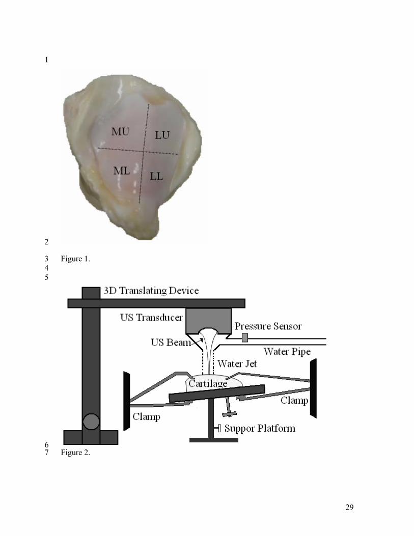
2.1 Samples 8
Eight fresh mature bovine patellae without obvious lesions were obtained from local 9
slaughterhouse within 2 hours post mortem and stored at -20oC before use. During the 10
specimen preparation, the patellae were first thawed in normal saline solution (0.15 M 11
NaCl) at the room temperature of approximately 20oC for 1 hour, then the lateral lower 12
part (1/4 of the patella) of each patella (Figure 1) was cut for the experiment by a 13
bandsaw (Buehler, Lake Bluff, IL, USA), and the bone was cut into a flat layer with a 14
thickness of 3 mm to 8 mm. During processing, the cartilage surface was kept moist with 15
normal saline solution without immersing the sample. All the samples were wrapped with 16
bandage and wetted with normal saline, then stored at -20oC before later use. 17
Simulated articular cartilage degeneration was achieved by a trypsin digestion for 18
the cartilage (n = 8). Before digestion, the sample was first thawed in normal saline 19
solution at room temperature 20oC for 1 hour; it was then put into a container and 20
immersed in the 0.25% trypsin solution (Gibco Invitrogen Corp., Grand Island, NY, 21
USA). The container was kept for 4 hours inside an incubator (MMM, Medcenter 22
Einrichtungen GmbH, Schulstrasse, DE) at 37oC to induce degeneration of articular 23
cartilage. It has been reported that trypsin reduces the content of PGs in cartilage and a 24
5
trypsin digestion process is thought to simulate cartilage degeneration.4,44,57 The 1
concentration and duration of the digestion treatment were selected according to the 2
previously developed protocols.57 All samples were washed using normal saline solution 3
after the digestion finished. 4
5
2.2 The Ultrasound Water Jet Indentation System 6
A noncontact ultrasound indentation system was developed using a water jet as the 7
indenter. As shown in Figure 2, a bubbler was used to eject the water jet by controlling 8
the water flow. The diameter of the water ejecting nozzle was 1.94 mm. A 20 MHz 9
focused ultrasound transducer (GE Panametrics, Inc., OH, USA) was fixed with the 10
bubbler, i.e., the water ejector. The focused ultrasound beam could propagate through the 11
bubbler when it was full of water as the coupling medium. The transducer and the 12
bubbler were installed to a 3-D translating device (Parker Hannifin Corporation, Irvine, 13
CA, USA) which was used to adjust the distance between the nozzle and the specimen 14
surface and to perform the 2-D scanning over a tissue specimen or phantom with a spatial 15
resolution at 1 μm. To focus the ultrasound beam at the specimen surface to obtain the 16
maximal echoes, the distances from the specimen surface to the nozzle outlet and the 17
transducer surface were adjusted to be 5.0 mm and 19.5 mm in current study, 18
respectively. A fixation device was carefully designed to fix the cartilage sample rigidly 19
and keep its surface perpendicular to the ultrasound transducer. As shown in Figure 2, the 20
sample was placed on the head of a tripod (#115, Manfrotto, Italy) which provided a rigid 21
support and meanwhile allowed for 3 degrees of adjustment and was fixed by two clamps 22
to avoid the slipping during test. This fixation device could be easily adjusted and thus 23
was convenient for experimental use. 24
6
A pressure sensor (EPB-C12, Entran Devices, Inc., Fairfield, NJ, USA) was used to 1
measure the water pressure within the water pipe. By calibrating the relationship between 2
the overall force applied on a load cell located under the platform and the pressure within 3
the water pipe, the pressure applied on the sample surface could be calculated using the 4
water pressure measured by the pressure sensor.43 We confirmed that the change of force 5
applied on the tissue could be negligible when the change of distance between the nozzle 6
outlet and the tissue surface was within approximately ± 0.5 mm. When the distance 7
increased, the ultrasound echo reflected from the tissue surface reduced significantly, as 8
the surface moved away from the focal point. In addition, the calibration for the force 9
measurement had to be re-conducted under this situation. A program was developed 10
using Microsoft VC++ to control the 3D translating device and to collect, process and 11
display the ultrasound signals, along with the pressure value, in real time during the 12
indentation process. Thus the movement of the transducer and the acquisition of the 13
radio-frequency (RF) ultrasound signal and pressure data were synchronized. The 14
ultrasound echoes reflected from the articular cartilage under different loading conditions 15
were tracked using a cross-correlation algorithm.70 The original tissue thickness and the 16
subsequent change of thickness, i.e., the deformation of the tissue under indentation, were 17
derived from the time information. The stiffness ratio, defined as the ratio of the pressure 18
applied on the sample surface to the local strain of the sample, was used as an indicator of 19
the mechanical properties of articular cartilage since it was found well correlated with 20
Young’s modulus from previous phantom studies.42,43 21
22
2.3 Stiffness, Thickness and Ultrasound Reflection Measurement Using Ultrasound Water 23
Jet Indentation 24
7
The bovine patella sample was first thawed in normal saline solution at room 1
temperature 20oC for 1 hour, and then it was fixed with its cartilage surface 2
perpendicularly facing the ultrasound transducer. For each sample, three indentation sites 3
were carefully selected through the following procedures: 1) the sample was scanned 4
across by the 20 MHz ultrasound beam to form a sequence of B-mode images, and those 5
regions with strong echoes reflected from both the cartilage surface and the 6
cartilage/subchondral bone interface were selected; 2) three sites in the selected regions 7
were chosen with the distance between each pair of them and the distance from each test 8
site to the edges of the sample both larger than 8 mm; 3) at each test site, fine adjustment 9
of the orientation of the platform was made to obtain the maximal echo amplitude. Such a 10
selection criterion could fulfill the boundary condition of indentation proposed by 11
Galbraith and Bryant28 and also make the ultrasound beam perpendicular to the cartilage 12
surface. The indentation sites were marked using a permanent marker pen (Lumocolor, 13
Staedtler, Germany). 14
During the measurements, the thickness and deformation of the sample were 15
determined with the 20 MHz ultrasound by calculating the time of flight and its 16
deflection of the ultrasound signals reflected from the cartilage surface and the cartilage-17
substrate interface. The inner parts of cartilage were mostly unechoic so it was 18
straightforward to determine the echoes from the interfaces. It has been earlier confirmed 19
using optical measurements that the second ultrasonic reflection was from the tidemark, 20
i.e. uncalcified-calcified cartilage interface.35,47,66 The cartilage thickness was determined 21
based on a constant ultrasound speed in bovine cartilage ccartilage of 1636 m/s.56 22
The sample was first scanned using the water jet with the pressure applied on the 23
sample surface no more than 1 kPa after the indentation sites were determined. This 24
8
process last for about 15 minutes which allowed the cartilage swelling caused by the 1
change of the concentration of solution from 0.15 M to approximate 0 M.67,71 Towards 2
the end of this period, the ultrasound echo reflected from the articular surface was 3
recorded for six times at each selected site for each sample. The peak-to-peak echo 4
amplitudes recorded from the intact cartilage sample Aintact and the sample after digestion 5
treatment Adigest at the identical indentation site were compared to investigate if there was 6
significant difference between them. 7
During indentation, the cartilage was first preloaded with a pressure of 20 kPa for 3 8
seconds, and then it was indented with the pressure increased to approximate 180 kPa 9
within 1 second. The loading and unloading process was repeated for 3 cycles in an 10
indentation. The stiffness ratio of cartilage was determined by calculating the slope of 11
pressure applied on the cartilage to the local strain induced in the loading phase. As 12
suggested by previous researchers, instantaneously induced deformation of a biphasic 13
tissue such as articular cartilage, can be modelled as that of an equivalent incompressible 14
single phase elastic material.32,46 Therefore, the load-indentation curve was fit by a linear 15
regression to obtain its slope since the strain was small (no more than 2%). 16
The cartilage sample was immersed in normal saline for 2 hours for full recovery49 17
before the next indentation. The indentation was repeated for three times at each selected 18
site, and the mean values of stiffness ratio and thickness were calculated. 19
20
2.4 Evaluation of the Effect of Repositioning 21
The accuracy of the ultrasonic evaluation highly depends on the position of the 22
ultrasound transducer relative to the indentation site. As the samples were repositioned to 23
the previous sites after digestion during the test, the effect of sample repositioning should 24
9
be evaluated. Two samples were used for this test. The sample was first assessed by the 1
water jet system on the selected test sites and taken away from the platform and 2
immersed into the saline solution for 2 hours. Then it was repositioned to the original 3
place for the repeated test. During this process, the orientation of the platform was kept 4
unchanged and the position of the sample was marked on the platform. The three-5
dimensional translating device could retrieve the transducer back to the recorded position 6
of each test site. Thus the same test sites were indented. This process was repeated for 7
three times. The obtained stiffness ratio, thickness and peak-to-peak ultrasound echo 8
amplitude of each test site were used to evaluate the variation caused by the repositioning. 9
10
2.5 Comparison with Mechanical Indentation 11
To compare with the measurements using the ultrasound water jet indentation, 12
mechanical indentation was also conducted on the same samples using a custom made 13
computer controlled material testing device. This system consisted of a 10N load cell, 14
precision motion controller and a computerized data acquisition system with a cylindrical, 15
plane-ended, impermeable, stainless steel indenter with a diameter of 1.6 mm. The 16
indenter was connected with a load cell (ELFS-T3M, Entran Devices, Inc., Fairfield, NJ, 17
USA, calibrated for a range of 10N) and installed to the three dimensional translating 18
device. During mechanical indentation, the indenter was first moved to the marked test 19
site, and a preload of 0.04 N (a pre-stress of 20 kPa) was applied to the sample. After 20
holding for 3 seconds, the sample was loaded up to a maximal strain of 10%. The loading 21
was recorded by the load cell synchronously with the movement of the indenter by the 22
program and the accuracy of the force collected by the data acquisition card was better 23
than 1 μN. The indentation rate was controlled at approximately 0.2 mm/s. 24
10
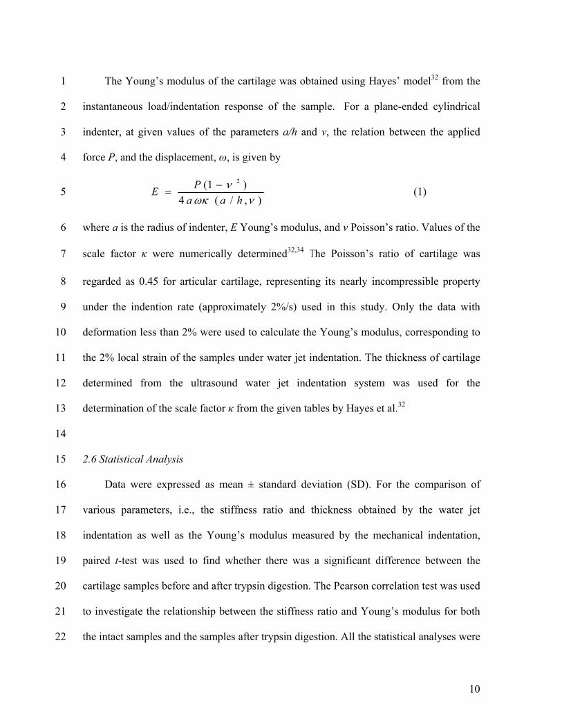
The Young’s modulus of the cartilage was obtained using Hayes’ model32 from the 1
instantaneous load/indentation response of the sample. For a plane-ended cylindrical 2
indenter, at given values of the parameters a/h and ν, the relation between the applied 3
force P, and the displacement, ω, is given by 4
),/(4
)1( 2
νωκν
haaPE −
= (1) 5
where a is the radius of indenter, E Young’s modulus, and ν Poisson’s ratio. Values of the 6
scale factor κ were numerically determined32,34 The Poisson’s ratio of cartilage was 7
regarded as 0.45 for articular cartilage, representing its nearly incompressible property 8
under the indention rate (approximately 2%/s) used in this study. Only the data with 9
deformation less than 2% were used to calculate the Young’s modulus, corresponding to 10
the 2% local strain of the samples under water jet indentation. The thickness of cartilage 11
determined from the ultrasound water jet indentation system was used for the 12
determination of the scale factor κ from the given tables by Hayes et al.32 13
14
2.6 Statistical Analysis 15
Data were expressed as mean ± standard deviation (SD). For the comparison of 16
various parameters, i.e., the stiffness ratio and thickness obtained by the water jet 17
indentation as well as the Young’s modulus measured by the mechanical indentation, 18
paired t-test was used to find whether there was a significant difference between the 19
cartilage samples before and after trypsin digestion. The Pearson correlation test was used 20
to investigate the relationship between the stiffness ratio and Young’s modulus for both 21
the intact samples and the samples after trypsin digestion. All the statistical analyses were 22
11
conducted by using the commercial software SPSS (SPSS Inc., Chicago, IL, USA). P < 1
0.05 was used to indicate a significant difference. 2
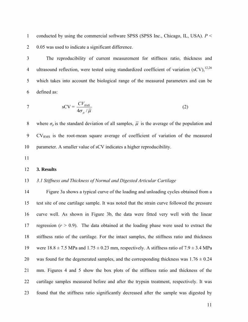
The reproducibility of current measurement for stiffness ratio, thickness and 3
ultrasound reflection, were tested using standardized coefficient of variation (sCV),12,26 4
which takes into account the biological range of the measured parameters and can be 5
defined as: 6
sCV = μσ μ /4
RMSCV (2) 7
where σμ is the standard deviation of all samples, μ is the average of the population and 8
CVRMS is the root-mean square average of coefficient of variation of the measured 9
parameter. A smaller value of sCV indicates a higher reproducibility. 10
11
3. Results 12
3.1 Stiffness and Thickness of Normal and Digested Articular Cartilage 13
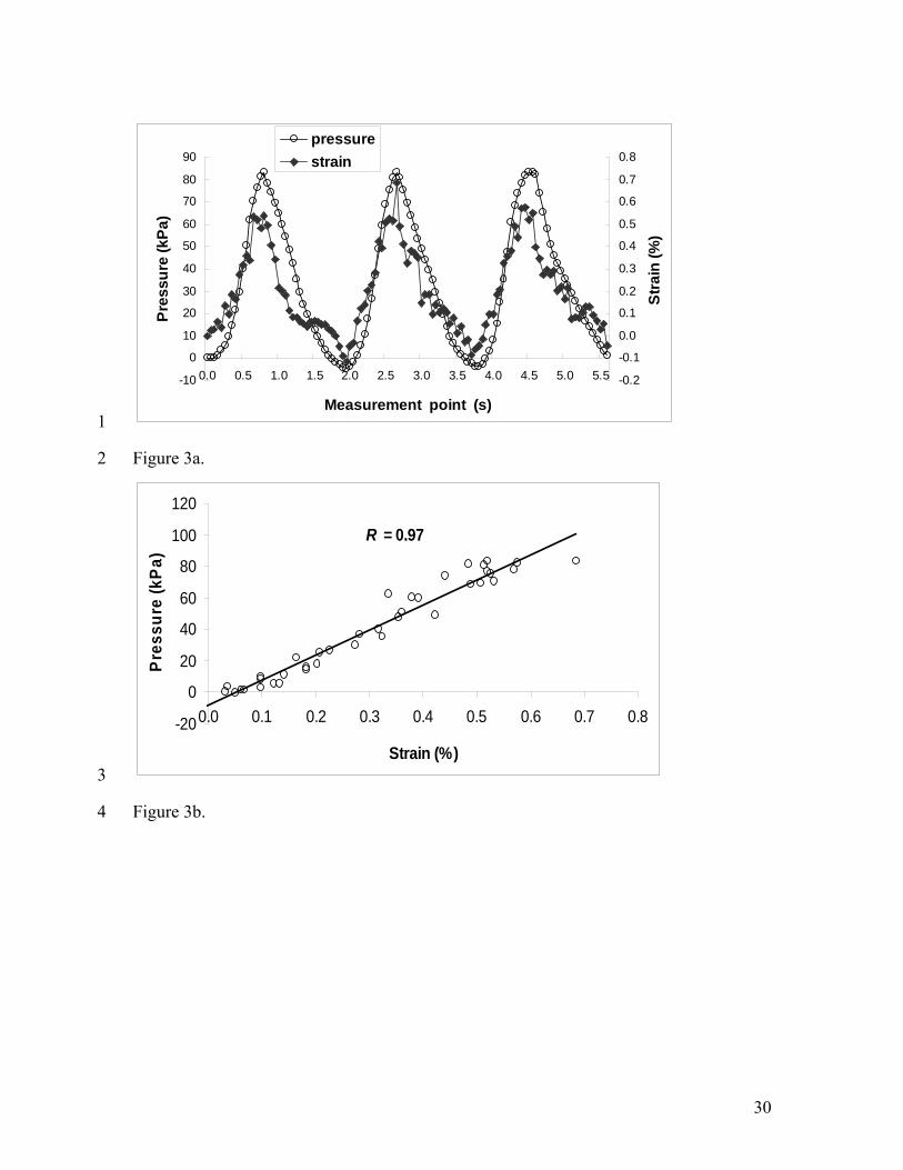
Figure 3a shows a typical curve of the loading and unloading cycles obtained from a 14
test site of one cartilage sample. It was noted that the strain curve followed the pressure 15
curve well. As shown in Figure 3b, the data were fitted very well with the linear 16
regression (r > 0.9). The data obtained at the loading phase were used to extract the 17
stiffness ratio of the cartilage. For the intact samples, the stiffness ratio and thickness 18
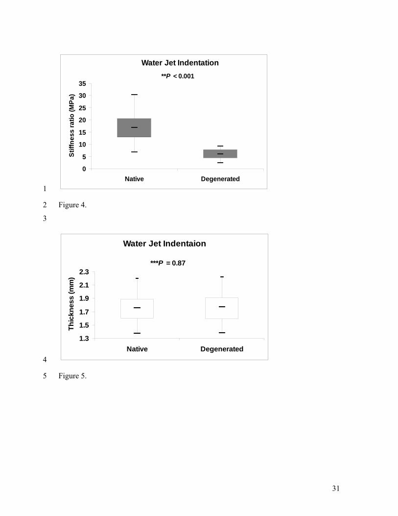
were 18.8 ± 7.5 MPa and 1.75 ± 0.23 mm, respectively. A stiffness ratio of 7.9 ± 3.4 MPa 19
was found for the degenerated samples, and the corresponding thickness was 1.76 ± 0.24 20
mm. Figures 4 and 5 show the box plots of the stiffness ratio and thickness of the 21
cartilage samples measured before and after the trypsin treatment, respectively. It was 22
found that the stiffness ratio significantly decreased after the sample was digested by 23
12
trypsin (P < 0.001) while no significant difference was observed between the thickness 1
values of the cartilage samples before and after the trypsin treatment (P = 0.87). 2
The reproducibility of the measurement using the ultrasound water jet indentation 3
system was assessed by the standardized variation of three repeated measurements of the 4
stiffness ratio and thickness on each test site of four cartilage samples (number of test 5
sites n = 12). The values of sCV were 8.0% for the stiffness ratio measurement and 1.6% 6
for the thickness measurement. 7
8
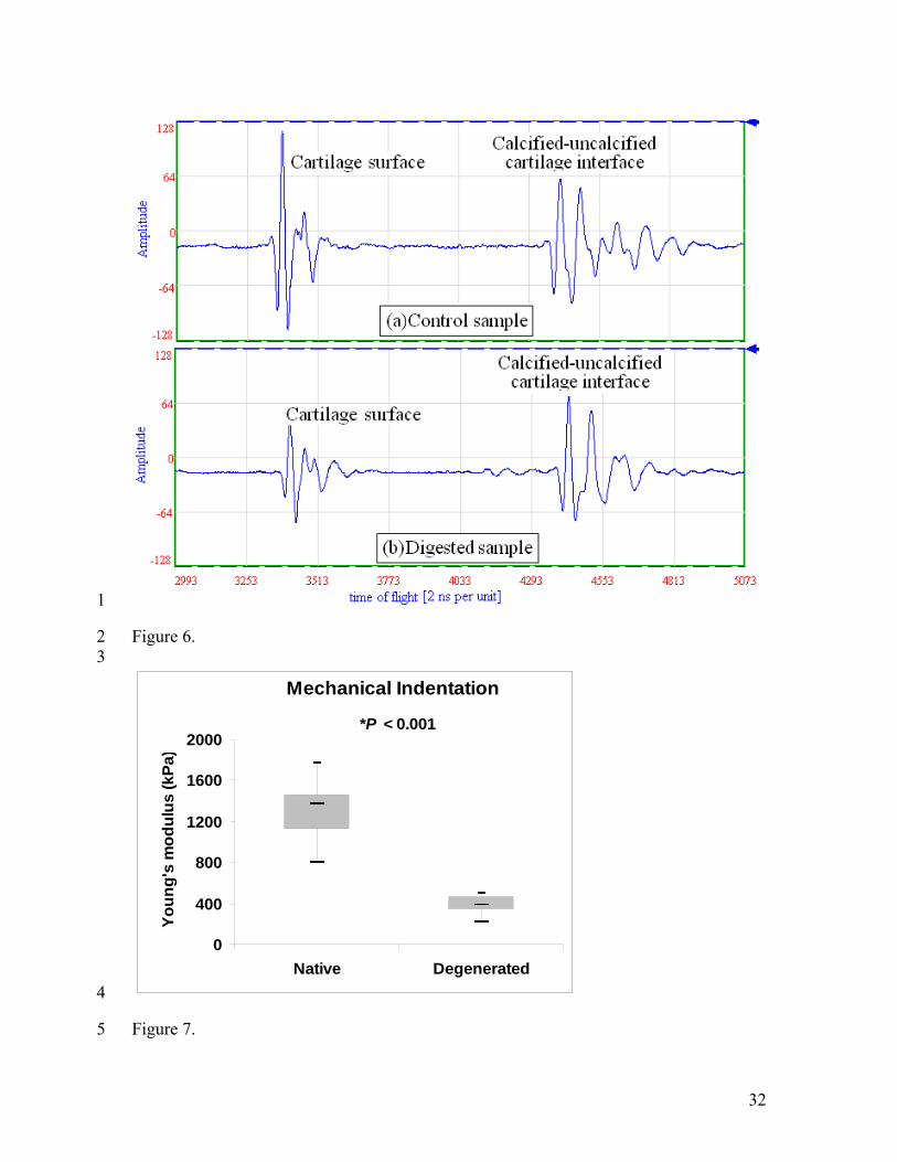
3.2 Ultrasound Reflection Measurements 9
For the ultrasound reflection measurements, the standardized coefficient of variation 10
for the repeatability test was 4.9% for the peak-to-peak amplitude measurement of all test 11
sites. Figure 6 shows typical A-mode ultrasound signals from the sample before (Figure 12
6a) and after the trypsin digestion (Figure 6b). A significant decrease of 36 ± 20% of the 13
peak-to-peak amplitude at the articular surface (P < 0.001) was found after the cartilage 14
sample was treated by the trypsin, however, no significant decrease of the signal 15
amplitude was found (P = 0.053) at the interface of uncalcified-calcified cartilage. The 16
decrease of the ultrasound reflection at the cartilage surface indicated that trypsin induced 17
not only PGs degradation but also small degradation of collagens 38 18
19
3.3 Effect of Repositioning on the Measurement 20
For the repositioning test, the values of the standardized coefficient of variation were 21
6.6%, 0.6% and 4.9% for the stiffness ratio, the cartilage thickness and the peak-to-peak 22
amplitude measurements, respectively. The small coefficients of variation demonstrated 23
13
that the reproducibility of the measurement was good and the effect of repositioning was 1
not significant. 2
3
3.4 Comparison with Mechanical Indentation 4
Using the mechanical indentation system, an instantaneous Young’s modulus of 5
1.59 ± 0.49 MPa was obtained for the native cartilage samples and 0.47 ± 0.13 MPa for 6
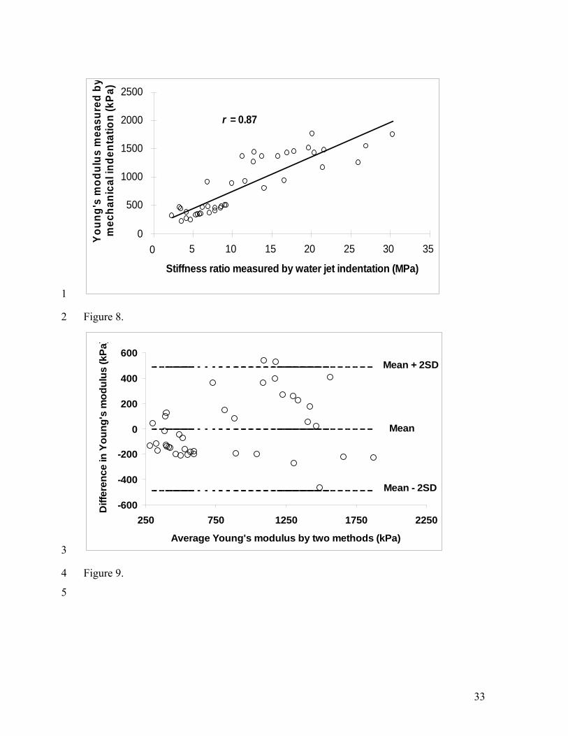
the degenerated samples (Figure 7). It was correlated with the stiffness ratio obtained 7
using the water jet system and a good correlation was found between them with r = 0.87 8
(P < 0.001, Figure 8). We estimated the Young’s modulus of cartilage, Ew, from the 9
obtained relationship between the stiffness ratio and the instantaneous Young’s modulus, 10
E, of the cartilage. A Bland-Altman plot (Figure 9) was also used to give an indication of 11
the size of errors between Ew and E. The mean difference d was 0 kPa and the standard 12
deviation s was 243.1 kPa. From the Bland-Altman plot, it can be observed most of the 13
differences lied between d - 2s and d + 2s. Such a result was good enough for clinical 14
applications.13 15
It was found that the elastic modulus was significantly reduced after the treatment 16
with trypsin, independent of the measurement methods used. The stiffness ratio was 17
reduced by 44 ± 17% (P < 0.001) obtained by the ultrasound water jet indentation system, 18
and the instantaneous Young’s modulus decreased by 30.5 ± 7.4% (P < 0.001) measured 19
using the mechanical indentation system. The decrease of the mechanical properties 20
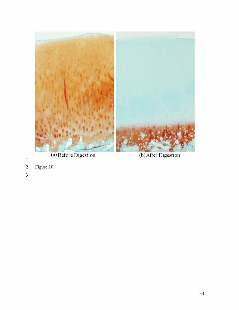
might be due to the loss of the PG. As shown in Figure 10, most of the PGs of the sample 21
were digested after the trypsin treatment. However, no significant difference was found 22
14
between the thickness values of the samples before and after digestion (P = 0.87), as 1
measured using ultrasound. 2
3
4. Discussion 4
In this study, the mechanical and acoustical properties of bovine patella articular 5
cartilage were quantified using the noncontact ultrasound water jet indentation system in 6
situ. Clinical findings show that during the development of osteoarthritic joint disease, 7
the cartilage swells, the level of aggrecan appears to change, especially that the PG 8
concentration decreases and the collagen network disrupts in the superficial zone.3,16,23 9
Such changes in cartilage quality can be determined by stiffness measurement at an early 10
stage,40 therefore, the determination of the mechanical properties of articular cartilage 11
becomes of great importance in trauma and joint repair surgery. In comparison to other 12
indentation systems, the current ultrasound water jet indentation system offers the 13
advantage of no direct contact between the testing probe and the tissue. Thus, the 14
potential damage to the tissues caused by the testing probe is minimized compared with 15
those contact indentation measurements. Further analysis of cell viability is required to 16
investigate whether any cellular damage might result from the water jet application.11 In 17
addition, the potential penetration of water into the cartilage should also be investigated 18
and its influence to the tissue should be studied. 19
According to the results, the ultrasound water jet indentation system demonstrated 20
its ability to effectively assess bovine articular cartilage by measuring both mechanical 21
and acoustical properties as well as tissue thickness. Unlike the ultrasound indentation 22
instrument developed by Laasanen et al.,38 no additional apparatus was needed in the 23
water-jet measurements when changing from the mechanical to acoustical measurement 24
15
modes. The repeated measurements of the stiffness ratio, the thickness, and the amplitude 1
echo signal from the articular cartilage were found highly reproducible with low values of 2
sCV (< 10%). The repositioning test showed similar reproducibility, suggesting the 3
system was reliable when used for the assessment of cartilage degeneration. 4
It was found in the current study that the stiffness ratio obtained using the water jet 5
system correlated well with the instantaneous Young’s modulus measured from the 6
mechanical indentation (r = 0.87), and the estimated Young’s moduli were well within 7
the range of previous studies.10,30,61 The Bland-Altman plot indicated that the estimation 8
was acceptable for clinical applications. It was found that it was not easy to keep the 9
pressure as a constant value due to the fluctuation of the flow rate when it was manual 10
controlled. A medical pump will be tried in the future studies to have a better control of 11
the water flow. 12
A significant decrease was found in both the stiffness ratio and the peak-to-peak 13
amplitude of the echo from the articular surface between the intact and trypsin treated 14
samples. The decrease of the mechanical properties may result from the PG content 15
reduction induced by trypsin treatment.57 The result of histological measurement revealed 16
that most of the PGs in the digested sample had been lost. More studies will be conducted 17
for the investigation on the relationship between the histological imaging quantification 18
and the water-jet detection of mechanical properties of articular cartilage. The amplitude 19
of the echo signal from the articular surface is related to the ratio of acoustic impedance 20
of cartilage and water. The surface roughness may also control the acoustic scattering, 21
and thereby affect the amplitude of the reflected signal.2 After the trypsin treatment, the 22
articular surface appeared normal without any fibrillation or other macroscopic signs of 23
degradation. Therefore, the significant decrease of the echo amplitude from the digested 24
16
articular surface should indicate that trypsin might have not only digested most of PG 1
contents, but also caused slight degradation of collagens. 2
It is well known that articular cartilage swells when the concentration of the external 3
bathing solution decreases. The swelling is caused by the change of the Donnon osmotic 4
pressure.25,50 In our test on the articular cartilage, water (nearly 0 M NaCl) was used to 5
eject a water jet as the indenter to deform the cartilage after it was moved away from the 6
normal saline solution (0.15 M NaCl). As reported by Zheng et al.,71,74 the intact cartilage 7
sample reached its maximal swelling strain (less than 0.1%) with an average swelling 8
duration of approximately 300 seconds when the bathing solution was suddenly changed 9
from 0.15 M NaCl to 0.015 M NaCl. In our experiments, the cartilage sample was 10
scanned under a very low constant pressure by the water jet for 15 minutes before it was 11
deformed. The scan duration should have been long enough to allow the cartilage to 12
equilibrate in the new solution. The strain caused by swelling during the indentation 13
process was small and could be neglected. However, it is better to keep the sample in its 14
physiological conditions to maintain its material properties. Therefore, normal saline 15
solutions will be used in the future instead of water when a medical pump is employed in 16
the system to eject a water jet as an indenter. 17
One of the main sources of error in the conventional indentation measurements is the 18
imperfect contact between the indenter and the articular cartilage. This may also affect 19
the water-jet indentation. In our measurements, the ultrasound beam was kept 20
perpendicular to the cartilage to maintain the water jet loading being normally applied so 21
as to minimize the error. The test sites were carefully selected by moving the transducer 22
and scanning across the cartilage to find those points with maximal echo amplitude 23
reflected from both the articular surface and the uncalcified-calcified cartilage interface. 24
17
Such a selection criterion further ensured the perpendicularity between the ultrasound 1
beam and the cartilage. 2
Cartilage thickness is an essential clinical parameter for the assessment of cartilage 3
degeneration and necessary for the accurate measurement of the tissue modulus. In this 4
study, the tissue thickness was calculated using a predetermined speed of sound and the 5
time-of-flight in samples. The time-of-flight was determined from the echo signals using 6
a cross-correlation technique. The measurement error of time-of-flight of the ultrasound 7
signal in cartilage may be induced by the distortion of the echoes reflected from different 8
interfaces.58 In our study, a small strain was applied on cartilage by the water jet 9
ultrasound indentation, and high correlations (R > 0.98) between the echoes were 10
achieved for most of the samples. Therefore, the measurement error caused by the 11
frequency-dependent attenuation may probably be a small value. The uncertainty in 12
determining the thickness was mainly caused by the variation of sound velocity. Previous 13
research work reported a variation of speed of sound between different measurement sites 14
and degenerative states.41,56,65 The speed of sound is slightly lower in OA cartilage than in 15
normal cartilage65 and digestion of PGs may reduce the speed of sound in cartilage as 16
well.63 As reported by Toyras et al.,65 with considering the variable speed of sound, the 17
use of a predefined speed of sound, 1636 m/s,56 may induce an error of (mean ± SD) 2.9 18
± 1.9% for the measurement of cartilage thickness, which would further induce an error 19
of 0.9 ± 0.6% for the stiffness ratio. These small errors should be clinically acceptable. 20
The accuracy of the deformation measurement depends rather on the sampling rate 21
of the A/D converter than on the ultrasound frequency. With the 500 MHz A/D converter 22
we used, a resolution of 2 ns, i.e. 1.6 μm, could be reached. Such a resolution is adequate 23
for the determination of cartilage deformation during indentation. Higher frequency (e.g. 24
18
50 MHz) ultrasound transducers would enable a higher axial resolution, and thereby 1
improve the resolution of thickness measurements. Utilizing a high frequency ultrasound 2
transducer in current system may also help to measure the layered material properties of 3
articular cartilage without damaging its biomechanical integrity, for the analysis of zonal 4
variation in articular cartilage biomechanical properties.57,73 This will potentially enable 5
the system to identify the grade of degeneration of the cartilage. 6
The ultrasound water jet indentation system could be easily used for the A-mode 7
reflection measurement. The condition of the cartilage sample could be differentiated 8
based on the change of the amplitude of the A-mode echo signals reflected from the 9
articular surface. Recent studies on the ultrasonic evaluation of articular cartilage have 10
suggested that quantitative ultrasound imaging could sensitively diagnose degeneration of 11
articular surface and changes in the subchondral bone, related to early 12
osteoarthritis18,31,59,60 In addition to the A-mode measurement, this system is capable of 13
performing scanning to obtain B-mode ultrasound images which would provide 14
additional, visual and quantitative information of the cartilage and subchondral bone 15
structure and composition41. However, to obtain a deeper insight of the acoustic 16
properties of articular cartilage, it is necessary to develop a theoretical model which could 17
describe satisfactorily the behaviour of ultrasound in cartilage. 18
Based on the present results, it is concluded that the ultrasound water jet indentation 19
system is capable of providing quantitative evaluation of both mechanical and acoustical 20
properties of intact and degenerated articular cartilages. Compared to other ultrasound 21
indentation devices used to assess cartilage, this new system could minimize the risk of 22
surface damage to the cartilage caused by mechanical loading therefore provided a 23
nondestructive way to measure the cartilage degeneration. However, the system needs to 24
19
be further improved before it can be clinically used to assess the cartilage degeneration, 1
such as the miniaturization of the water jet ultrasound probe for arthroscopic use, better 2
water pressure control mechanism, and use of physiological saline. 3
4
Acknowledgements 5
This work was partially supported by the Research Grants Council of Hong Kong 6
(PolyU 5245/03E, PolyU 5318/05E, PolyU5354/08E) and The Hong Kong Polytechnic 7
University. 8
9
References 10 1 Adam, C., F. Eckstein, S. Milz, E. Schulte, C. Becker, and R. Putz. The distribution 11
of cartilage thickness in the knee-joints of old-aged individuals - measurement by A-12
mode ultrasound. Clin. Biomech. 13: 1-10, 1998. 13 2 Adler, R. S., D. K. Dedrick, T. J. Laing, E. H. Chiang, C. R Meyer., P. H. Bland, J. 14
M. Rubin. Quantitative assessment of cartilage surface roughness on osteoarthritis 15
using high frequency ultrasound. Ultrasound Med. Biol. 18: 51-58, 1992. 16 3 Altman, R. D., J. Tenenbaum, L. Latta, W. Riskin, L. N. Blanco, and D. S. Howell. 17
Biomechancial and biochemical properties of dog cartilage in experimentally 18
induced osteoarthritis. Ann Rheum Dis 43: 83-90, 1984 19 4 Andrew, J. G., J. Hoyland, and A. J. Freemont. Insulin-like growth-factor gene-20
expression in human fracture callus. Calcif. Tissue Int. 53: 97-102, 1993. 21 5 Armstrong, S. and V. C. Mow. Variations of the intrinsic mechanical properties of 22
human articular cartilage with age, degeneration and water content. J. Bone Joint 23
Surg. Am. 64: 88-94, 1982. 24 6 Arokoski, J., J. Jurvelin, I. Kiviranta, M. Tammi, and H. J. Helminen. Softening of 25
the lateral condyle aricular-cartilage in the canine knee-joint after long-distance (up 26
to 40KM/day) running training lasting one-year. Int. J. Sports Med. 15: 254-260, 27
1994. 28
20
7 Arokoski, J. P. A., M. M. Hyttinen, H. J. Helminen, and J. S. Jurvelin. 1
Biomechanical and structural characteristics of canine femoral and tibial cartilage. J. 2
Biomed. Mater. Res. 48: 99-107, 1999. 3 8 Athanasiou, K. A., A. Agarwal, A. Muffoletto, F. J. Dzida, G. Constantinides, and M. 4
Clem. Biomechanical properties of hip cartilage in experimental animal-models. Clin. 5
Orthop. Res. 316: 254-266, 1995. 6 9 Athanasiou, K. A., J. G. Fleischli, J. Bosma, T. J. Laughlin, C. F. Zhu, C. M. 7
Agrawal, and L. A. Lavery. Effects of diabetes mellitus on the biomechanical 8
properties of human ankle cartilage. Clin. Orthop. Res. 368: 182-189, 1999. 9 10 Athanasiou, K. A., M. P. Rosenwasser, J. A. Buckwalter, T. I. Malinin, V. C. Mow. 10
Interspecies comparisons of insitu intrinsic mechanical-properties of distal femoral 11
cartilage. J. Orthop. Res. 9: 330-340, 1991. 12 11 Bae, W. C., C. W. Lewis, and R. L. Sah. Intra-tissue strain distribution in normal 13
human cartilage during clinical indentation testing. [49th annual meeting of the 14
Orthopaedic Research Society; New Orleans, La]. Trans. Orthop. Res. Soc. 28: 1, 15
2003. 16 12 Blake, G. M., H. W. Wahner, and I. Fogelman. Assessment of instrument 17
performance: precision, installation of new equipment and radiation dose. In: The 18
Evaluation of Osteoporosis: Dual Energy X-ray absorptiometry and Ultrasound in 19
Clinical Practice. Martin Dunitz Ltd., London, 147-157. 20 13 Bland, J. M. and D. G. Altman. Statistical methods for assessing agreement between 21
two methods of clinical measurement. Lancet i: 307-310, 1986. 22 14 Bonassar, L. J., E. H. Frank, J. C. Murray, C. G. Paguio, V. L. Moore, M. W. Lark, J. 23
D. Sandy, J. J. Wu, D. R. Eyre, and A. J. Grodzinsky. Changes in cartilage 24
composition and physical properties due to stromelysin degradation. Arthritis Rhuem. 25
38: 173-183, 1995. 26 15 Brandt, K., L. S. Lohmander, and M. Doherty. Pathogenesis of osteoarthritis. In: 27
Osteoarthritis. Oxford Univ. Press, Oxford, 1998. 28 16 Buckwalter, J. A. and H. J. Mankin. Articular cartilage, Part II: degeneration and 29
osteoarthritis, repair, regeneration and transplantation. J. Bone Joint Surg. Am. 79: 30
612-632, 1997. 31
21
17 Cohen, Z. A., D. M. Mccarthy, S. D. Kwak, P. Legrand, F. Fogarasi, E. J. Ciaccio, 1
and G. A. Ateshian. Knee cartilage topography, thickness, and contact areas from 2
MRI: in-vitro calibration and in-vivo measurements. Osteoarthritis Cartilage. 7: 95-3
109, 1999. 4 18 Chaffai, S., F. Peyrin, S. Nuzzo, R. Porcher, G. Berger, and P. Laugier. Ultrasonic 5
characterization of human cancellous bone using transmission and backscatter 6
measurements: relationship to density and microstructure. Bone. 30: 229-237, 2002. 7 19 Chiang, E. H., T. J. Laing, C. R. Meyer, J. L. Boes, J. M. Rubin, and R. S. Adler. 8
Ultrasonic characterization of in vitro osteoarthritic articular cartilage with validation 9
by confocal microscopy. Ultrasound Med. Biol. 23: 205-213, 1997. 10 20 Chiang, E. H., R. S. Adler, C. R. Meyer, J. M. Rubin, D. K. Dedrick, and T. J. Laing. 11
Quantitative assessment of surface-roughness using backscattered ultrasound – the 12
effects of finite surface curvature. Ultrasound Med. Biol. 20: 123-135, 1994. 13 21 Cherin, E., A. Saied, P. Laugier, P. Netter and G. Berger. Evaluation of acoustical 14
parameter sensitivity to age-related and osteoarthritic changes in articular cartilage 15
using 50-MHz ultrasound. Ultrasound Med. Biol. 24: 341-354, 1998. 16 22 Cherin, E., A. Saied, B. Pellaumail, D. Loeuille, P. Laugier, P. Gillet, P. Netter, and 17
G. Berger. Assessment of rat articular cartilage maturation using 50-MHz 18
quantitative ultrasonography. Osteoarthritis Cartilage. 9: 178-186, 2001. 19 23 Dean, D. D., J. Martel Pelletier, J. P. Pelletier, D. S. Howell, J. F. Jr. Woessner. 20
Evidence for metalloproteinase and metalloproteinase inhibitor imbalance in human 21
osteoarthritic cartilage. J. Clin. Invest. 84 (2): 678-85, 1989. 22 24 Disler, D. G., E. Raymond, D. A. May, J. S. Wayne, and T. R. McCauley. Articular 23
cartilage defects: In vitro evaluation of accuracy and interobserver reliability for 24
detection and grading with US. Radiology. 215: 846-851, 2000. 25 25 Donnon, F. G. The theory of membrane equilibria. Chem. Rev. 1: 73-90, 1924. 26 26 Gluer, C. C., G. Blake, Y. Lu, B. A. Blunt, M. Jergas, and H. K. Genant. Accurate 27
assessment of precision errors: how to measure the reproducibility of bone 28
densitometry techniques. Osteoporos. Int. 5: 262-270, 1995. 29 27 Freeman, M. A. Is collagen fatigue failure a cause of osteoarthrosis and prosthetic 30
component migration? A hypothesis. J. Orthop. Res. 17: 3-8, 1999. 31
22
28 Galbraith, P. C., and J. T. Bryant. Effect of grid dimensions on finite element models 1
of an articular surface. J. Biomech. 22: 385-393, 1989. 2 29 Haapala, J., J. P. A. Arokoski, M. M. Hyttinen, M. Lammi, M. Tammi, V. Kovanen, 3
H. J. Helminen, and I. Kiviranta. Remobilization does not fully restore 4
immobilization induced articular cartilage atrophy. Clin. Orthop. Res. 362: 218-229, 5
1999 6 30 Hale, J. E. M. J., Rudert, and T. D. Brown. Indentation assessment of biphasic 7
mechanical property deficits in size-dependent osteochondral defect repair. J. 8
Biomech. 26: 1319-1325, 1993. 9 31 Hattori, K., Y. Takakura, M. Ishimura, T. Habata, K. Uematsu, and K. Ikeuch. 10
Quantitative arthroscopic ultrasound evaluation of living human cartilage. Clin. 11
Biomech. 19: 213-216, 2004. 12 32 Hayes, W. C., L. M. Keer, G. Herrmann, and L. F. Mockros. A mathematical 13
analysis for indentation tests of articular cartilage. J. Biomech. 5: 541-551, 1972. 14 33 Hori, R. Y., and L. F. Mockros. Indentation tests of human articular-cartilage. J. 15
Biomech. 9: 259-268, 1976. 16 34 Jurvelin, J., I. Kiviranta, A. M. Saamanen, M. Tammi, and H. J. Helminen. 17
Indentation stiffness of young canine knee articular-cartilage – influence of strenuous 18
joint loading. J. Biomech. 23: 1239-1246, 1990. 19 35 Jurvelin, J. S., T. Rasanen, P. Kolmonen, and T. Lyyra. Comparison of optical needle 20
probe and ultrasonic techniques for measurement of articular cartilage thickness. J. 21
Biomech. 28: 231-235, 1995 22 36 Kempson, G. E., M. A. R. Freeman, and S. A. V. Swanson. Determination of a creep 23
modulus for articular cartilage from indentation tests on human femoral head. J. 24
Biomech. 4: 239-250, 1971. 25 37 Kempson, G. E. Mechanical properties of articular cartilage and their relationship to 26
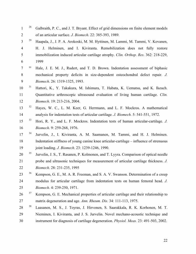
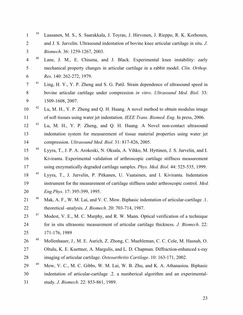
matrix degeneration and age. Ann. Rheum. Dis. 34: 111-113, 1975. 27 38 Laasanen, M. S., J. Toyras, J. Hirvonen, S. Saarakkala, R. K. Korhonen, M. T. 28
Nieminen, I. Kiviranta, and J. S. Jurvelin. Novel mechano-acoustic technique and 29
instrument for diagnosis of cartilage degeneration. Physiol. Meas. 23: 491-503, 2002. 30
23
39 Laasanen, M. S., S. Saarakkala, J. Toyras, J. Hirvonen, J. Rieppo, R. K. Korhonen, 1
and J. S. Jurvelin. Ultrasound indentation of bovine knee articular cartilage in situ. J. 2
Biomech. 36: 1259-1267, 2003. 3 40 Lane, J. M., E. Chisena, and J. Black. Experimental knee instability: early 4
mechanical property changes in articular cartilage in a rabbit model. Clin. Orthop. 5
Res. 140: 262-272, 1979. 6 41 Ling, H. Y., Y. P. Zheng and S. G. Patil. Strain dependence of ultrasound speed in 7
bovine articular cartilage under compression in vitro. Ultrasound Med. Biol. 33: 8
1509-1608, 2007. 9 42 Lu, M. H., Y. P. Zheng and Q. H. Huang. A novel method to obtain modulus image 10
of soft tissues using water jet indentation. IEEE Trans. Biomed. Eng. In press, 2006. 11 43 Lu, M. H., Y. P. Zheng, and Q. H. Huang. A Novel non-contact ultrasound 12
indentation system for measurement of tissue material properties using water jet 13
compression. Ultrasound Med. Biol. 31: 817-826, 2005. 14 44 Lyyra, T., J. P. A. Arokoski, N. Oksala, A. Vihko, M. Hyttinen, J. S. Jurvelin, and I. 15
Kiviranta. Experimental validation of arthroscopic cartilage stiffness measurement 16
using enzymatically degraded cartilage samples. Phys. Med. Biol. 44: 525-535, 1999. 17 45 Lyyra, T., J. Jurvelin, P. Pitkanen, U. Vaatainen, and I. Kiviranta. Indentation 18
instrument for the measurement of cartilage stiffness under arthroscopic control. Med. 19
Eng.Phys. 17: 395-399, 1995. 20 46 Mak, A. F., W. M. Lai, and V. C. Mow. Biphasic indentation of articular-cartilage .1. 21
theoretical -analysis. J. Biomech. 20: 703-714, 1987. 22 47 Modest, V. E., M. C. Murphy, and R. W. Mann. Optical verification of a technique 23
for in situ ultrasonic measurement of articular cartilage thickness. J. Biomech. 22: 24
171-176, 1989 25 48 Mollenhauer, J., M. E. Aurich, Z. Zhong, C. Muehleman, C. C. Cole, M. Hasnah, O. 26
Oltulu, K. E. Kuettner, A. Margulis, and L. D. Chapman. Diffraction-enhanced x-ray 27
imaging of articular cartilage. Osteoarthritis Cartilage. 10: 163-171, 2002. 28 49 Mow, V. C., M. C. Gibbs, W. M. Lai, W. B. Zhu, and K. A. Athanasiou. Biphasic 29
indentation of articular-cartilage .2. a numberical algorithm and an experimental-30
study. J. Biomech. 22: 853-861, 1989. 31
24
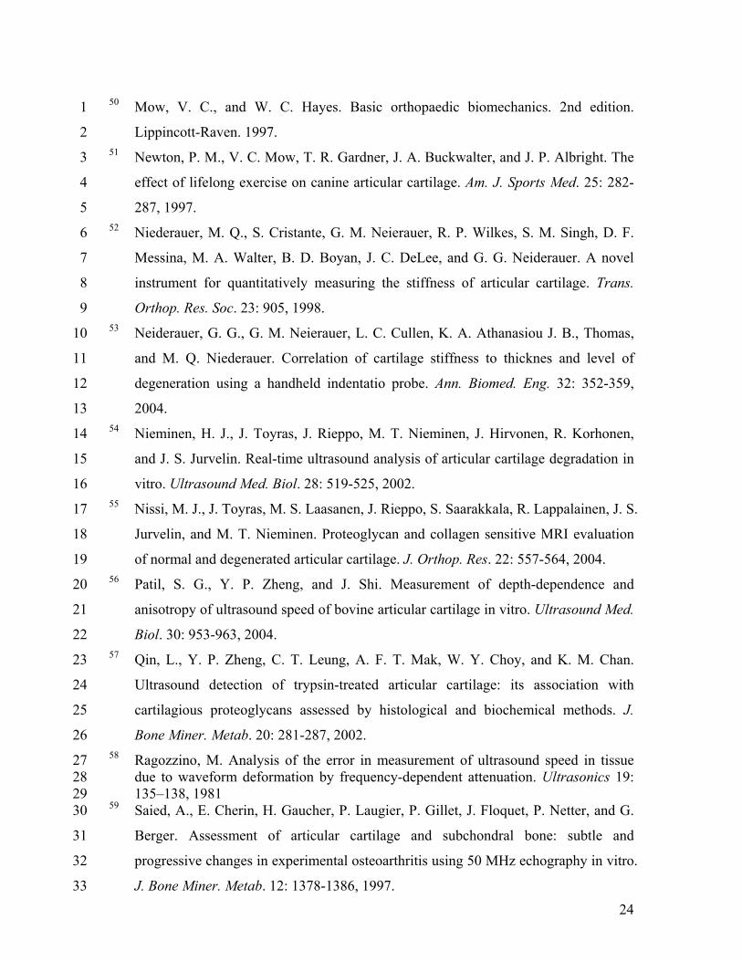
50 Mow, V. C., and W. C. Hayes. Basic orthopaedic biomechanics. 2nd edition. 1
Lippincott-Raven. 1997. 2 51 Newton, P. M., V. C. Mow, T. R. Gardner, J. A. Buckwalter, and J. P. Albright. The 3
effect of lifelong exercise on canine articular cartilage. Am. J. Sports Med. 25: 282-4
287, 1997. 5 52 Niederauer, M. Q., S. Cristante, G. M. Neierauer, R. P. Wilkes, S. M. Singh, D. F. 6
Messina, M. A. Walter, B. D. Boyan, J. C. DeLee, and G. G. Neiderauer. A novel 7
instrument for quantitatively measuring the stiffness of articular cartilage. Trans. 8
Orthop. Res. Soc. 23: 905, 1998. 9 53 Neiderauer, G. G., G. M. Neierauer, L. C. Cullen, K. A. Athanasiou J. B., Thomas, 10
and M. Q. Niederauer. Correlation of cartilage stiffness to thicknes and level of 11
degeneration using a handheld indentatio probe. Ann. Biomed. Eng. 32: 352-359, 12
2004. 13 54 Nieminen, H. J., J. Toyras, J. Rieppo, M. T. Nieminen, J. Hirvonen, R. Korhonen, 14
and J. S. Jurvelin. Real-time ultrasound analysis of articular cartilage degradation in 15
vitro. Ultrasound Med. Biol. 28: 519-525, 2002. 16 55 Nissi, M. J., J. Toyras, M. S. Laasanen, J. Rieppo, S. Saarakkala, R. Lappalainen, J. S. 17
Jurvelin, and M. T. Nieminen. Proteoglycan and collagen sensitive MRI evaluation 18
of normal and degenerated articular cartilage. J. Orthop. Res. 22: 557-564, 2004. 19 56 Patil, S. G., Y. P. Zheng, and J. Shi. Measurement of depth-dependence and 20
anisotropy of ultrasound speed of bovine articular cartilage in vitro. Ultrasound Med. 21
Biol. 30: 953-963, 2004. 22 57 Qin, L., Y. P. Zheng, C. T. Leung, A. F. T. Mak, W. Y. Choy, and K. M. Chan. 23
Ultrasound detection of trypsin-treated articular cartilage: its association with 24
cartilagious proteoglycans assessed by histological and biochemical methods. J. 25
Bone Miner. Metab. 20: 281-287, 2002. 26 58 Ragozzino, M. Analysis of the error in measurement of ultrasound speed in tissue 27
due to waveform deformation by frequency-dependent attenuation. Ultrasonics 19: 28 135–138, 1981 29
59 Saied, A., E. Cherin, H. Gaucher, P. Laugier, P. Gillet, J. Floquet, P. Netter, and G. 30
Berger. Assessment of articular cartilage and subchondral bone: subtle and 31
progressive changes in experimental osteoarthritis using 50 MHz echography in vitro. 32
J. Bone Miner. Metab. 12: 1378-1386, 1997. 33
25
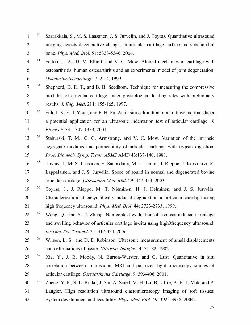
60 Saarakkala, S., M. S. Laasanen, J. S. Jurvelin, and J. Toyras. Quantitative ultrasound 1
imaging detects degenerative changes in articular cartilage surface and subchondral 2
bone. Phys. Med. Biol. 51: 5333-5346, 2006. 3 61 Setton, L. A., D. M. Elliott, and V. C. Mow. Altered mechanics of cartilage with 4
osteoarthritis: human osteoarthritis and an experimental model of joint degeneration. 5
Osteoarthritis cartilage. 7: 2-14, 1999. 6 62 Shepherd, D. E. T., and B. B. Seedhom. Technique for measuring the compressive 7
modulus of articular cartilage under physiological loading rates with preliminary 8
results. J. Eng. Med. 211: 155-165, 1997. 9 63 Suh, J. K. F., I. Youn, and F. H. Fu. An in situ calibration of an ultrasound transducer: 10
a potential application for an ultrasonic indentation test of articular cartilage. J. 11
Biomech. 34: 1347-1353, 2001. 12 64 Stahurski, T. M., C. G. Armstrong, and V. C. Mow. Variation of the intrinsic 13
aggregate modulus and permeability of articular cartilage with trypsin digestion. 14
Proc. Biomech. Symp. Trans. ASME AMD 43:137-140, 1981. 15 65 Toyras, J., M. S. Laasanen, S. Saarakkala, M. J. Lammi, J. Rieppo, J. Kurkijarvi, R. 16
Lappalainen, and J. S. Jurvelin. Speed of sound in normal and degenerated bovine 17
articular cartilage. Ultrasound Med. Biol. 29: 447-454, 2003. 18 66 Toyras, J., J. Rieppo, M. T. Nieminen, H. J. Helminen, and J. S. Jurvelin. 19
Characterization of enzymatically induced degradation of articular cartilage using 20
high frequency ultrasound. Phys. Med. Biol. 44: 2723-2733, 1999. 21 67 Wang, Q., and Y. P. Zheng. Non-contact evaluation of osmosis-induced shrinkage 22
and swelling behavior of articular cartilage in-situ using high0frequency ultrasound. 23
Instrum. Sci. Technol. 34: 317-334, 2006. 24 68 Wilson, L. S., and D. E. Robinson. Ultrasonic measurement of small displacements 25
and deformations of tissue. Ultrason. Imaging. 4: 71–82, 1982. 26 69 Xia, Y., J. B. Moody, N. Burton-Wurster, and G. Lust. Quantitative in situ 27
correlation between microscopic MRI and polarized light microscopy studies of 28
articular cartilage. Osteoarthritis Cartilage. 9: 393-406, 2001. 29 70 Zheng, Y. P., S. L. Bridal, J. Shi, A. Saied, M. H. Lu, B. Jaffre, A. F. T. Mak, and P. 30
Laugier. High resolution ultrasound elastomicroscopy imaging of soft tissues: 31
System development and feasibility. Phys. Med. Biol. 49: 3925-3938, 2004a. 32
26
71 Zheng, Y. P., J. Shi, L. Qin, S. G. Patil, V. C. Mow, and K. Y. Zhou. Dynamic depth-1
dependent osmotic swelling and solute diffusion in articular cartilage monitored 2
using real-time ultrasound. Ultrasound Med. Biol. 30: 841-849, 2004b. 3 72 Zheng, Y. P., and A. F. T. Mak. An ultrasound indentation system for biomechanical 4
properties assessment of soft tissues in-vivo. IEEE Trans. Biomed. Eng. 43: 912-918, 5
1996. 6 73 Zheng, Y. P., H. J. Niu, F. T. A. Mak, and Y. P. Huang. Ultrasonic measurement of 7
depth-dependent transient behaviors of articular cartilage under compression. J. 8
Biomech. 38 (9): 1830-1837, 2005. 9 74 Zheng, Y. P., J. Shi, S. G. Patil, L. Qin, and A. F. T. Mak. Ultrasonic measurement of 10
articular cartilage swelling: preliminary results. SPIE Medical Imgaing: Ultrasonic 11
Imaging and Signal Processing Conf., San Diego, CA, USA. 501-512, 2003. 12
13
14 15 16 17 18
19
20
21
22
27
Figure Captions: 1
Figure 1. The lateral lower (LL) quarter of normal bovine patella without obvious lesions 2
was cut as the sample for experiments. 3
Figure 2. Diagram of the noncontact ultrasound indentation system using water jet 4
compression. The water jet was used as the indenter and focused high-frequency 5
ultrasound was employed to monitor the deformation of the cartilage. 6
Figure 3. (a) A typical curve of the loading and unloading cycles obtained from a test site 7
of one cartilage sample using the water jet indentation system. (b) The relationship 8
between the pressure and strain which was fitted by a linear regression model. 9
Figure 4. Box plot of the stiffness ratios of the cartilage samples obtained using the 10
ultrasound water jet indentation. Significant difference was found between the stiffness 11
ratios of intact and degenerated samples (P < 0.001). The box represents the inter-quartile 12
range. The upper and lower limits of the box indicate the 75th and 25th percentile. The 13
horizontal line in the box represents the median. 14
Figure 5. Box plot of the thickness of the cartilage samples obtained using the ultrasound 15
water jet indentation. No significant difference was found between the thickness values of 16
the samples before and after trypsin digestion (P = 0.87). 17
Figure 6. Typical A-mode ultrasound signals from (a) the control sample and (b) the 18
digested sample. The first echo indicates the cartilage surface and the second echo 19
indicates the calcified-uncalcified cartilage interface. 20
Figure 7. Box plot of the Young’s modulus obtained using mechanical indentation. It was 21
found that the Young’s modulus significantly decreased after trypsin treatment (P < 22
0.001). 23
Figure 8. Correlation between the stiffness ratio obtained using the ultrasound water jet 24
indentation system and the Young’s modulus obtained from the mechanical indentation 25
for all the cartilage samples, both intact and degenerated (r = 0.87, P < 0.001). 26
Figure 9. Bland-Altman plot to test the agreement between the estimated Young’s 27
modulus of cartilage samples by ultrasound water jet indentation and the measured 28
modulus by the mechanical indentation. 29

28
Figure 10. Representative histological images of articular cartilage stained with Safranine 1
O (red in coloured images) and fast green of a typical sample: (a) intact status; (b) after 2
trypsin treatment. 3
29
1
2
Figure 1. 3 4 5
6 Figure 2. 7
30
-10
0
10
20
30
40
50
60
70
80
90
0.0 0.5 1.0 1.5 2.0 2.5 3.0 3.5 4.0 4.5 5.0 5.5
Measurement point (s)
Pres
sure
(kPa
)
-0.2
-0.1
0.0
0.1
0.2
0.3
0.4
0.5
0.6
0.7
0.8
Stra
in (%
)
pressurestrain
1
Figure 3a. 2
R = 0.97
-20
0
20
40
60
80
100
120
0.0 0.1 0.2 0.3 0.4 0.5 0.6 0.7 0.8
Strain (%)
Pres
sure
(kPa
)
3
Figure 3b. 4
31
Water Jet Indentation
0
5
10
15
20
25
30
35
Native Degenerated
Stiff
ness
ratio
(MPa
)**P < 0.001
1
Figure 4. 2
3
Water Jet Indentaion
1.3
1.5
1.7
1.9
2.1
2.3
Native Degenerated
Thic
knes
s (m
m)
***P = 0.87
4
Figure 5. 5
32
1
Figure 6. 2 3
Mechanical Indentation
0
400
800
1200
1600
2000
Native Degenerated
Youn
g's
mod
ulus
(kPa
)
*P < 0.001
4
Figure 7. 5
33
r = 0.87
0
500
1000
1500
2000
2500
0 5 10 15 20 25 30 35
Stiffness ratio measured by water jet indentation (MPa)
Youn
g's
mod
ulus
mea
sure
d by
mec
hani
cal i
nden
tatio
n (k
Pa)
1
Figure 8. 2
-600
-400
-200
0
200
400
600
250 750 1250 1750 2250
Average Young's modulus by two methods (kPa)
Diff
eren
ce in
You
ng's
mod
ulus
(kPa
)
Mean
Mean - 2SD
Mean + 2SD
3
Figure 9. 4
5
34
1
Figure 10. 2
3
![Eng Metrology Topic 4 [Noncontact Inspection]](https://static.fdocuments.in/doc/165x107/563db9b3550346aa9a9f1d40/eng-metrology-topic-4-noncontact-inspection.jpg)