Non-steroidal anti-inflammatory drugs in an …...id of patients affected by endometriosis...
10
8487 Abstract. – OBJECTIVE: Endometriosis is a debilitating disease characterized by chronic inflammation. The transporter multidrug resis- tance-associated protein 4 (MRP4/ABCC4) is ex- pressed in human endometrial tissue; it is over- expressed in ectopic endometrial tissue, and is modulated by the anti-inflammatory lipid Lipox- in A4 (LXA4). Recently, it was demonstrated that aspirin induces platelet MRP4 over-expression, through genomic modulation in megakaryocytes. Since patients with endometriosis frequent- ly use aspirin or other non-aspirin Non-Steroi- dal Anti-Inflammatory Drugs (NSAIDs), the aim of this study was to verify whether aspirin and other NSAIDs enhance MRP4 expression in 12Z human endometriotic epithelial cells and wheth- er this was peroxisome proliferator-activated re- ceptor alpha (PPARa) dependent. MATERIALS AND METHODS: MRP4 and PPA- Ra expression was analyzed by Q-RT-PCR us- ing TaqMan ® Master Mix and TaqMan ® Assay Reagents (Life Technologies, Monza, Italy) and Western blot. RESULTS: In 12Z cells, aspirin and other NSAIDs enhanced MRP4 mRNA and protein ex- pression; these treatments also induced PPARa expression. Aspirin and diclofenac-induced in- creases in MRP4 expression were not observed in cells where PPARa was knocked down using siRNA. NSAIDs-induced MRP4 expression was correlated with augmented PGE2 secretion, in- dicating functional relevance. CONCLUSIONS: MRP4 expression was in- creased in cells treated with NSAIDs and the nu- clear receptor PPARa is involved. Elevated PGE2 levels in cell supernatants correlate with its in- creased transport by MRP4 after NSAID treat- ment. More importantly, we provide evidence that in endometriotic epithelial cells aspirin and non-aspirin NSAIDs treatments alter gene ex- pression. Key Words: Endometriosis, MRP4/ABCC4, Pain, Aspirin, NSAID. Introduction Endometriosis is a debilitating disease with features of chronic inflammation that affects ap- proximately 10% of women in reproductive age. It is defined as the presence of functional endome- trial glands and stroma outside the uterine cavity 1 . As one of the most common benign gynecological conditions, endometriosis is a debilitating disease with detrimental effects on social, occupational and psychological functioning 2 . Despite its pre- valence, this disease remains poorly understood and current studies have proved that there is no relationship between the extent of the disease and its symptomatology 1 . The most frequent symptom is a chronic pelvic pain, which may include: dy- smenorrhea (painful periods), periovulatory pain, dyspareunia (pain during or after sexual inter - course), dysuria, dyschezia, and leg pain. For this European Review for Medical and Pharmacological Sciences 2018; 22: 8487-8496 I. MASSIMI 1 , F.M. PULCINELLI 2 , V.P. PISCITELLI 2 , L. ALEMANNO 1 , T. MALTESE 1 , M.L. GUARINO 1 , R. MARCI 3 , G.O. CANNY 4 , L. FRATI 5 , M. MALLOZZI 2 , A. FREGA 2 , D. CASERTA 2 1 Department of Experimental Medicine, Sapienza University of Rome, Rome, Italy 2 Department of Medical-Surgery Sciences and Translation Medicine, Sapienza University of Rome, Rome, Italy 3 Department of Morphology, Surgery and Experimental Medicine, Sant’Anna University Hospital, Ferrara, Italy 4 University College Dublin, Dublin 4, Ireland 5 I.R.C.C.S Neuromed, Pozzilli (IS), Italy Isabella Massimi and Francesca Maria Pulcinelli contributed equally Corresponding Author: Donatella Caserta, MD, Ph.D; e-mail: [email protected] Non-steroidal anti-inflammatory drugs increase MRP4 expression in an endometriotic epithelial cell line in a PPARa dependent manner
Transcript of Non-steroidal anti-inflammatory drugs in an …...id of patients affected by endometriosis...

8487
Abstract. – OBJECTIVE: Endometriosis is a debilitating disease characterized by chronic inflammation. The transporter multidrug resis-tance-associated protein 4 (MRP4/ABCC4) is ex-pressed in human endometrial tissue; it is over-expressed in ectopic endometrial tissue, and is modulated by the anti-inflammatory lipid Lipox-in A4 (LXA4). Recently, it was demonstrated that aspirin induces platelet MRP4 over-expression, through genomic modulation in megakaryocytes. Since patients with endometriosis frequent-ly use aspirin or other non-aspirin Non-Steroi-dal Anti-Inflammatory Drugs (NSAIDs), the aim of this study was to verify whether aspirin and other NSAIDs enhance MRP4 expression in 12Z human endometriotic epithelial cells and wheth-er this was peroxisome proliferator-activated re-ceptor alpha (PPARa) dependent.
MATERIALS AND METHODS: MRP4 and PPA-Ra expression was analyzed by Q-RT-PCR us-ing TaqMan® Master Mix and TaqMan® Assay Reagents (Life Technologies, Monza, Italy) and Western blot.
RESULTS: In 12Z cells, aspirin and other NSAIDs enhanced MRP4 mRNA and protein ex-pression; these treatments also induced PPARa expression. Aspirin and diclofenac-induced in-creases in MRP4 expression were not observed in cells where PPARa was knocked down using siRNA. NSAIDs-induced MRP4 expression was correlated with augmented PGE2 secretion, in-dicating functional relevance.
CONCLUSIONS: MRP4 expression was in-creased in cells treated with NSAIDs and the nu-
clear receptor PPARa is involved. Elevated PGE2 levels in cell supernatants correlate with its in-creased transport by MRP4 after NSAID treat-ment. More importantly, we provide evidence that in endometriotic epithelial cells aspirin and non-aspirin NSAIDs treatments alter gene ex-pression.
Key Words: Endometriosis, MRP4/ABCC4, Pain, Aspirin, NSAID.
Introduction
Endometriosis is a debilitating disease with features of chronic inflammation that affects ap-proximately 10% of women in reproductive age. It is defined as the presence of functional endome-trial glands and stroma outside the uterine cavity1. As one of the most common benign gynecological conditions, endometriosis is a debilitating disease with detrimental effects on social, occupational and psychological functioning2. Despite its pre-valence, this disease remains poorly understood and current studies have proved that there is no relationship between the extent of the disease and its symptomatology1. The most frequent symptom is a chronic pelvic pain, which may include: dy-smenorrhea (painful periods), periovulatory pain, dyspareunia (pain during or after sexual inter-course), dysuria, dyschezia, and leg pain. For this
European Review for Medical and Pharmacological Sciences 2018; 22: 8487-8496
I. MASSIMI1, F.M. PULCINELLI2, V.P. PISCITELLI2, L. ALEMANNO1, T. MALTESE1, M.L. GUARINO1, R. MARCI3, G.O. CANNY4, L. FRATI5, M. MALLOZZI2, A. FREGA2, D. CASERTA2
1Department of Experimental Medicine, Sapienza University of Rome, Rome, Italy2Department of Medical-Surgery Sciences and Translation Medicine, Sapienza University of Rome, Rome, Italy3Department of Morphology, Surgery and Experimental Medicine, Sant’Anna University Hospital, Ferrara, Italy4University College Dublin, Dublin 4, Ireland5I.R.C.C.S Neuromed, Pozzilli (IS), Italy
Isabella Massimi and Francesca Maria Pulcinelli contributed equally
Corresponding Author: Donatella Caserta, MD, Ph.D; e-mail: [email protected]
Non-steroidal anti-inflammatory drugs increase MRP4 expression in an endometriotic epithelial cell line in a PPARa dependent manner

I. Massimi, F.M. Pulcinelli, V.P. Piscitelli, L. Alemanno, T. Maltese, M.L. Guarino, R. Marci, et al.
8488
reason, patients with endometriosis frequently use either non-aspirin Non-Steroidal Anti-Inflamma-tory Drugs (NSAIDs) or aspirin. Endometriosis is divided by the classification system of the Ame-rican Fertility Society into four stages of severity from stage I, that is represented by minimal dise-ase, to stage IV, which means severe disease. Sta-ging does not correspond with degree or severity of symptoms but assesses the extension of disease visible by laparoscopy. In addition, an elevated le-vel of CA125 (epithelial ovarian cancer associated antigen) in patients with endometriosis is associa-ted with the progression of the disease3. Women’s quality of life, including their careers, everyday activities, sexual and non-sexual relationships and fertility may be really affected by endome-triosis. The United Kingdom – Endometriosis UK (www.endometriosis-uk.org/), which represents a patient support organization, confirmed that the 65% of women with endometriosis had negatively influenced their employment, the 10% of women reduced the hours of work, and 30% of them lost the job, which brought them to perceive a fee-ling of distress and low self-esteem4. Moreover, infertility has been identified as one of the most relevant complications related to endometriosis, which has been observed in 15-25% of patients. In 2018, has been demonstrated that the levels of inflammatory cytokines (IL-6, IL-10, IL-13, and TNF) are particularly increased in peritoneal flu-id of patients affected by endometriosis compli-cated with infertility. Therefore, the high level of those cytokines may determinate embryo toxicity and could affect sperm motility, fertilization, and implantation leading to an elevated rate of infer-tility among patients affected by endometriosis5,6. Endometriosis can be suspected in reproductive age women with clinical symptoms. Transvaginal ultrasonography can reliably detect endometrio-mas but failure to reveal cystic structures does not exclude the diagnosis of endometriosis. Ma-gnetic resonance imaging (MRI) is increasingly used to identify subperitoneal deposits, although retroversion, endometrioma, and bowel structu-res may mask small nodules7. Risk factors gene-rally relate to menstruation: early menarche, late menopause, nulliparity and first pregnancy late in life increase the risk whereas the use of oral contraceptives is associated with a reduced risk of developing the disease. While the etiology of endometriosis still remains unclear, the most wi-dely accepted hypothesis first advanced by Samp-son8 is that viable endometrial tissue fragments are refluxed through the fallopian tubes into the
pelvic cavity during retrograde menstruation, and after being migrated adhere and invade other tissues. Different studies9-11 indicate that growth factors, cytokines, and prostaglandins promote the establishment and maintenance of endome-triosis. In endometriosis patients, concentrations of prostaglandin E2 (PGE2) in the peritoneal fluid are higher compared to that of endometriosis-free women9; PGE2 plays a pivotal role in endometrio-sis-associated inflammation and pain12,13. COX-2 protein is abundantly expressed in ectopic en-dometrium and in the eutopic endometrium of women with endometriosis. Banu et al14, showed that inhibition of COX-2 and of PGE2 receptors PTGER2 and PTGER4 decrease survival and invasion of human endometriotic epithelial and stromal cells through multiple mechanisms. Their results support that COX-2/PGE2 promotes the pathophysiology and pathogenesis of endome-triosis in humans. The inhibition of PTGER2 and PTGER4 in vitro, inhibits adhesion of human en-dometriotic epithelial cells 12Z and stromal cells 22B to collagen I, vitronectin, fibronectin, colla-gen IV, and laminin in a substrate- and epithe-lial-stromal cell-specific manner of the peritoneal extracellular matrix (ECM)15. Together, these re-sults suggest the importance of the PTGER2 and PTGER4 associated signaling pathways in the pa-thogenesis of endometriosis.
Nonsteroidal anti-inflammatory drugs (NSAIDs) are the most frequently used first-line drugs for pain in women affected by endome-triosis. Nevertheless, there is no sufficient evi-dence that showed whether or not they are really effective in relieving endometriosis-associated pain. Moreover, it has not been demonstrated if there is a NSAID more effective than another4. Currently, has been affirmed that the expression of COX-2 in ectopic endometrial cells is higher than in eutopic endometrium. Nonsteroidal an-ti-inflammatory drugs inhibit the function of the enzyme COX-1 and COX-2, preventing the production of PGs, which are implicated in the genesis of endometriosis-associated pain. Fur-thermore, have been registered several side ef-fects related with NSAIDs including nausea, diarrhoea, headache, dryness of the mouth16. In 2016 the Cochrane Gynecology and Fertili-ty Group Specialized Register of Controlled Trials published a comparison performed from January 2008 to October 2016, by using NSAIDs (naproxen) vs. placebo in women with endome-triosis-related pain. Data revealed no evidence of a positive effect on pain relief (odds ratio (OR)

Non-steroidal anti-inflammatory drugs in an endometriotic epithelial cell line
8489
3.27, 95% confidence interval (CI) 0.61 to 17.69; one trial, 24 women; very low-quality evidence). In addition, the work did not demonstrate impro-vements concerning the quality of life, effects on daily activities, absence from work or school in patients treated with NSAIDs17. Clinical expe-riences of our center evaluated the impact of NSAIDs, which are used as pain therapy in en-dometriosis. It showed that 39 (12.8%) out of 305 patients, did not require any therapy, 84 (27.5%) patients did not report if they needed drugs, 18 patients (5.9%) stated they needed treatment but did not specify which therapy. About the remai-ning patients: 111 (36.4%) take only NSAIDs, 41 (13.4%) take NSAIDs in combination with other drugs, and 12 (3.9%) use drugs different from NSAIDs. In our opinion, these data are very im-portant because, if we sum up all the patients who use NSAIDs, alone and in combination, the total of the patients who take NSAIDs, increa-ses to 152 (49.8%). The transporter multidrug resistance-associated protein 4 (MRP4) is also expressed in human endometrial tissue; it is ove-rexpressed in the ectopic endometrial tissue, and is modulated by the anti-inflammatory lipid li-poxin A4 (LXA4)
11. Increased MRP4 expression in the ectopic endometrium of women suffering from endometriosis was confirmed by a subse-quent study18. MRP4, an ATP binding cassette (ABCC4) and unidirectional transporter of en-dogenous molecules including several PGs, plays a key role in cellular communication and signa-ling. Elevated MRP4 expression may result in increased extracellular PGE2, which binds its re-ceptors and activates various signaling pathways in endometriosis. MRP4, as well as PGE2 itself, is implicated in proliferative conditions and could, therefore, serves as a potential biomarker for disease severity, as well as a possible drug target for endometriosis therapy13. We demon-strated that aspirin causes MRP4 overexpression in human platelets, through an activation of the nuclear receptor PPARa in megakaryocytes19. Recently we reported that non-aspirin Non-Ste-roidal Anti-Inflammatory Drugs (NSAIDs) can induce MRP4 overexpression in human pla-telets. Patients with endometriosis frequently suffer from dysmenorrhea, which negatively affects daily activities, and for this reason, they frequently take aspirin or non-aspirin NSAIDs20. We can assume that MRP4 can be modulated by the action of aspirin and non-aspirin NSAIDs even in the endometrium, leading to enhance-ment of PGE2 secretion. In this study the role
of aspirin and non-steroidal anti-inflammatory drugs (NSAIDs) in regulating MRP4 expression in human endometriotic epithelial cells (12Z), the involvement of nuclear receptor PPARa and PGE2 secretion were investigated.
Materials and Methods
Cell CultureThe simian virus 40 T-antigen-transformed
human ectopic endometriotic epithelial cell line, 12Z (kindly provided by Dr. M. Beste, Massachu-setts Institute of Technology), was maintained in Dulbecco’s Modified Eagle Medium (DMEM)/Ham’s F12 medium supplemented with 10% he-at-inactivated fetal bovine serum (FBS), 20 mM L-glutamine, 100 units/ml of penicillin G sodium, and 100 μg/ml streptomycin sulphate in a humi-dified atmosphere containing 5% CO2 at 37°C21. For stimulation experiments, cells (250,000/well) were seeded in 12-well plates; the following day, stimulation with various factors was carried out in media for 48 h. Cells were treated with either aspirin (25 μM, 50 μM, and 100 μM), or diclofe-nac (25 μM, 50 μM, and 100 μM), or nimesulide (25 μM, 50 μM, and 100 μM), or acetaminophen (50 μM), or ketoprofen (50 μM), or ibuprofen (50 μM), or naproxen (50 μM). In a mock culture, an equivalent amount of DMSO, the vehicle, was added. After treatment, cells were processed for RNA and protein extraction. For PGE2 analysis and measurement of prostaglandin H synthase activity, 12Z cells (250,000/well) were seeded in 12-well plates; the following day, stimulation with various factors was carried out in media for 48 h (50 uM aspirin or diclofenac or nimesulide). After 48 hours of treatment, cells were washed and maintained in medium without any drugs for 24 hours, after which supernatants were collected for PGE2 and cells collected for the measurement of prostaglandin H synthase activity and MRP4 expression.
Protein Extraction and Western BlotTo analyze MRP4 protein, cells were washed
twice with cold phosphate-buffered saline (PBS), collected and centrifuged at 400 g for 10 min. Cell pellets were then resuspended in lysis buffer (RIPA buffer: 10 mM Tris-HCl pH 7.6, 160 mM NaCl, 1 mM EGTA, 1% Deoxycholic acid, 1% Triton, 0.1% SDS), incubated on ice for 30 min and centrifuged at 12,000 g for 30 min, the super-natant was then collected. Protein extracts (30 μg)

I. Massimi, F.M. Pulcinelli, V.P. Piscitelli, L. Alemanno, T. Maltese, M.L. Guarino, R. Marci, et al.
8490
were incubated at 37°C for 30 min and separated on 4-12% sodium dodecyl sulphate-polyacryla-mide gel electrophoresis (SDS-PAGE) gel, tran-sferred onto polyvinylidene difluoride (PVDF) membrane (GE Healthcare, Milano, Italy), and probed with rat anti-MRP4 (Alexis, Plymouth Meeting, PA, USA) and mouse anti-actin (San-ta Cruz Biotechnology, Santa Cruz, CA, USA) monoclonal antibodies. Immunoreactive bands were visualized by enhanced chemiluminescence (PerkinElmer, Waltham, MA, USA). The densito-metric analysis was performed with the National Institutes of Health ImageJ analyzer program.
RNA Preparation and Real-Time Quantitative PCR Analysis
Total RNA from human cell lines was extracted using TRIzol reagent (Invitrogen, Carlsbad, CA, USA). For mRNA detection 1 μg of total RNA was transcribed using the GeneAmp Gold RNA PCR Reagent Kit pAW109 (Life Technologies, Mon-za, Italy) according to the manufacturer’s proto-col. Gene expression analysis was carried out by means of Q-RT-PCR using TaqMan® Master Mix and TaqMan® Assay Reagents (Life Technologies, Monza, Italy). The amplification program, moni-tored using ABI Prism 7900 Sequencer Detector (Life Technologies, Monza, Italy), was as follows: 50°C for 2 min, 95°C for 10 min, 95°C for 15 sec, and 60°C for 1 min, the latter two temperatures were repeated for 40 cycles. All amplification re-actions were performed in duplicate using 25 ng of cDNA. Changes in MRP4, PPARa and ACTIN mRNA amounts were quantified using the ΔΔCt method for relative quantification of gene expres-sion using SDS software version 2.3 (Life Tech-nologies, Monza, Italy).
RNA InterferenceDouble strand interfering RNA (siRNA) tar-
geting human PPARa and control non-specific siRNA (Santa Cruz Biotechnology, Santa Cruz, CA, USA) were transfected using Lipofectamine RNAi MAX Reagent (Life Technologies, Monza, Italy). 24 hours after siRNA administration, cells were treated with aspirin (50 µM) or diclofenac (50 µM) (Sigma-Aldrich Chemicals Company, St. Louis, MO, USA). 48 hours after treatment, cells were processed for RNA and protein analysis.
PGE2 QuantificationFor PGE2 analysis, 12Z cells (250,000/well)
were seeded in 12-well plates and treated as de-scribed above. Supernatants were then collected
and PGE2 levels were quantified using a mono-clonal PGE2 EIA kit (Cayman Chemical Co., Ann Arbor, MI, USA) according to the manufacturer’s instructions. PGE2 levels (pg/105 cells) are repor-ted as ratio between treated and untreated cells (relative levels).
Measurement of Prostaglandin H Synthase Activity
Prostaglandin H synthase activity was per-formed as previously described 22. Briefly, cells were washed with PBS and pre-loaded with 2 µM 5-(and-6)-carboxyl-2’,7’-dichlorodihydrofluore-scein (CDCF) (Molecular Probes Inc., Eugene, OR, USA), serving as a reducing substrate for the peroxidase activities of COX-1. Platelet COX acti-vity was measured as arachidonic acid induced (8 µM) fluorescence enhancement in a Victor-3 spectrofluorimeter (PerkinElmer, Waltham, MA, USA), thermostatically regulated (37°C). 5 µM Nicotinamide adenine dinucleotide phosphate oxidase inhibitor (diphenyliodonium) was added to avoid reactive oxygen species production-me-diated interference. The results are reported as the rise in CDCF fluorescence (Arbitrary Units) recorded for 5 min.
Statistical AnalysisData are presented as mean ±SD. The level of
significance was determined employing unpaired, 2-tailed Student’s t-test (KaleidaGraph software 3.6). Results were considered statistically signi-ficant if a p-value of less than 0.05 was reached.
Results
MRP4 Expression in NSAID Treated 12Z CellsThe effect of aspirin and other NSAIDs on
MRP4 over-expression has recently been demon-strated in a human megacaryoblastic cell line (DAMI)19,20. In order to examine whether these drugs also induced MRP4 expression in endo-metriotic cells, we treated 12Z cells with aspirin, diclofenac, nimesulide, acetaminophen, ketopro-fen, ibuprofen, and naproxen for 48 hours. Among the NSAIDs used, aspirin (1.6 fold increase), di-clofenac (1.6 fold increase), acetaminophen (1.6 fold increase), ketoprofen (1.9 fold increase) and nimesulide (2.3 fold increase) induced a signifi-cant increase in MRP4 mRNA expression in 12Z cells compared to the control culture (all p<0.05) (Figure 1). In contrast, in 12Z cells treated with naproxen and ibuprofen, no increase in MRP4

Non-steroidal anti-inflammatory drugs in an endometriotic epithelial cell line
8491
expression was observed (Figure 1). In the same experimental conditions, Western Blot analysis revealed augmented MRP4 protein expression in 12Z cells treated with aspirin, diclofenac, aceta-minophen, ketoprofen, and nimesulide (50 μM for 48 h) (Figure 2A). Densitometric analysis showed a statistically significant increase in MRP4 pro-tein expression in 12Z cells treated with aspirin (1.6 fold increase), diclofenac, acetaminophen, ketoprofen, and nimesulide (1.4 fold increase) in comparison with untreated cells (p<0.05) (Figu-re 2B). We recently demonstrated that the nuclear receptor PPARa is involved in the aspirin-indu-ced MRP4 overexpression in megakaryocytes19. To explore whether PPARa was responsible for the MRP4 expression increases in NSAID treated 12Z cells, we analyzed PPARα mRNA expression. The results obtained show that treatment with all NSAIDs used, resulted in a significant increase in PPARα mRNA expression compared with untre-ated cells (1.6 fold increase) (p<0.05), except for naproxen and ibuprofen (Figure 3). Figure 4 (A-F) shows a correlation between MRP4 and PPARα expression and scalar increasing doses of aspirin, diclofenac, and nimesulide (from 25 μM to 100 μM). MRP4 expression was strongly induced in a dose-dependent manner after aspirin (Figure 4A),
diclofenac (Figure 4B) and nimesulide (Figure 4C) treatment. Indeed, the highest MRP4 mRNA increase is evident after treatment with 100 μM of these drugs. These increases are also evident for PPARα expression levels after a different dose of drug treatment, for aspirin (Figure 4D), diclofe-nac (Figure 4E) and nimesulide (Figure 4F).
PPARα siRNA Inhibits MRP4 Expression Induced by NSAIDs
To confirm the involvement of PPARa in MRP4 up-regulation, 12Z cells were transfected with PPARa specific siRNA (PPAR-si); cells transfected with PPAR-si didn’t exhibit any signi-ficant increase in PPARa mRNA expression after aspirin treatment; while cells transfected with control, non specific siRNA (CTR-si), exhibited the same aspirin-induced expression changes as untransfected cells (1.8 fold increase; p<0.05) (Figure 5A). Similarly, no change in aspirin de-pendent MRP4 expression was observed in cells treated with PPAR-si, unlike those found in CTR-si transfected cells (1.8 fold increase; p<0.05) (Fi-
Figure 1. Aspirin and NSAIDs induce MRP4 mRNA expression in 12Z cells. Q-RT-PCR analysis of endogenous MRP4 expression in 12Z cells treated with aspirin, diclo-fenac, acetaminophen, ketoprofen, nimesulide, naproxen and ibuprofen (50 μM for 48 h) compared to vehicle con-trol (Ctr). Data were normalized with ACTB expression and reported as mean ±SD of 3 experiments (*p<0.05; NS: not significant; t-test).
Figure 2. Aspirin and NSAIDs increase MRP4 protein expression in 12Z cells. (A) A representative Western Blot, of 3 performed, of MRP4 expression in 12Z cells treated with aspirin, diclofenac, acetaminophen, ketoprofen and nimesulide (50 μM for 48 h). (B) Densitometric analysis reported as the ratio between treated (aspirin, diclofenac, acetaminophen, ketoprofen and nimesulide) and untreated cells (Ctr). Data are reported as mean ±SD; statistical si-gnificance was evaluated using Student’s t-test for unpaired samples (*p<0.05).

I. Massimi, F.M. Pulcinelli, V.P. Piscitelli, L. Alemanno, T. Maltese, M.L. Guarino, R. Marci, et al.
8492
gure 5B). To confirm that aspirin induces PPARa activation through COX1 inhibition, we studied diclofenac-induced PPARa-MRP4 up-regulation in siRNA transfected cells. Among all non-aspi-rin NSAIDs previously reported, diclofenac was chosen because it induces a similar increase in MRP4 expression to that caused by aspirin. Also in this case, no significant difference in PPARα mRNA expression in cells transfected with PPAR-si was observed after diclofenac treatment; while cells transfected with control, non specific siRNA (CTR-si), exhibited the same diclofenac induced expression changes as untransfected cells (2.1 fold increase; p<0.05) (Figure 5A). Similarly, no increase in diclofenac dependent MRP4 expres-sion was detected in cells treated with PPAR-si, unlike those found in CTR-si transfected cells (1.8 fold increase; p<0.05) (Figure 5B).
NSAID-Induced MRP4 Expression Correlates with Increased PGE2 Secretion by Endometriotic Epithelial Cells
MRP4 may be relevant to endometriosis patho-physiology as increases extracellular secretion
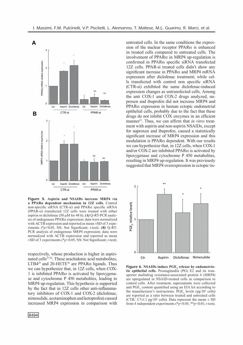
of PGE2, which binds its receptors and activates various signaling pathways13. PGE2 is long known to promote inflammation and pain, and it is con-sidered to be a pivotal mediator for endometriosis development and progression13,14. To determine whether NSAIDs induced MRP4 up-regulation was functionally relevant in endometriotic cells, we measured PGE2 in 12Z cell supernatants after 24 hours suspension of NSAID treatment (50 μM for 48 hours). In order to avoid a possible role of NSAID cell treatment in the inhibition of PGE2 formation, PGE2 levels were evaluated 48 hours after the last drug treatment, when the MRP4 expression is still high and prostaglandin H syn-thase activity is not affected by drug treatment. In fact, in treated cells, MRP4 protein expression after 48h was still up-regulated and prostaglandin H synthase activity was equal to untreated cells (data not shown). As shown in Figure 6, PGE2 lev-els were significantly elevated after treatment with aspirin (1.75 fold increase; p<0.01), diclofenac (1.20 fold increase; p<0.05) and nimesulide (1.48 fold increase; p<0.05) compared to control cells (3.7± 1.1 pg/105 cells). These data demonstrate that NSAID-induced MRP4 enhancement is cor-related with enhanced PGE2 secretion.
Discussion
Patients with endometriosis frequently suffer from dysmenorrhea, periovulatory pain, dyspareu-nia, dyspareunia, chronic pelvic pain, dysuria, dy-schezia, and leg pain. Schlincke in 1946 illustrated for the first time a case of endometriosis-induced sciatica, which is considered one of the most com-mon causes in women affected by sciatica. Leg pain has been reported by 4% of women with chro-nic pelvic pain and it is described that 40% of wo-men affected by endometriosis have leg pain23,24. Furthermore, leg pain is a common and disabling symptom related to endometriosis but it is still difficult to determine its cause. We recognize two main causes of endometriosis related leg pain: referred pain and neuropathic pain. Our studies proved that leg pain in patients with endometrio-sis may be caused to nerve injury or it may be a referred dysmenorrheal pain. The most commonly implicated nerve is the sciatic nerve. In 2013, we showed in a case-control study that pain generally affects the left leg (70% of patients versus 30% of patients in which pain interests the right leg) and it has a usual distribution in the crural and the lateral region of the thigh. In our study, 30% of women
Figure 3. Aspirin and NSAIDs also augment endoge-nous PPARα mRNA expression in 12Z cells. Q-RT-PCR analysis of endogenous PPAR-alpha expression in 12Z cells treated with aspirin, diclofenac, acetaminophen, ketopro-fen, nimesulide, naproxen and ibuprofen (50 μM for 48 h) compared to control culture (Ctr). Data were normalized with ACTB expression and reported as mean ±SD of 3 expe-riments (*p<0.05; NS: not significant; t-test).

Non-steroidal anti-inflammatory drugs in an endometriotic epithelial cell line
8493
demonstrated a decreased nociceptive and tactile sensibility in the crural region of the thigh and a positive Wasserman test, suggesting the hypothesis of femoral nerve involvement25. Therefore, endo-metriosis may be considered a debilitating disease characterized by chronic pain. This is the reason why patients with endometriosis often use non-a-spirin Non-Steroidal Anti-Inflammatory Drugs (NSAIDs). Furthermore, it has been confirmed that Thrombin-activated fibrinolytic inhibitor (TAFI) is largely expressed in endometriosis and causes epi-thelial mesenchymal transition (EMT), which in-creases the cell proliferation and invasion26. MRP4 is expressed in the human endometrium and its expression can be up-regulated in ectopic endome-trium of women suffering from endometriosis and modulated by the anti-inflammatory lipid Lipoxin A4 (LXA4)
13,18. This transporter likely plays an im-portant role in the pathogenesis of endometriosis, because it facilitates PGE2 secretion. We recently demonstrated that aspirin and non-aspirin NSAIDs drugs induce platelet MRP4 over-expression throu-
gh the activation of the nuclear receptor PPARa in megakaryocytes18,19. In this work we report for the first time that aspirin and other non-aspirin NSAIDs induce an enhancement of MRP4 expres-sion in a human ectopic endometrial epithelial cell line (12Z cells). In addition we demonstrate that the nuclear receptor involved is PPARa. In fact, MRP4 and PPARa mRNA expression are enhanced in 12Z cells treated with aspirin. The involvement of PPARa in MRP4 up-regulation was confirmed by the fact that 12Z cells transfected with PPARa specific siRNA didn’t show a significant increase of aspirin dependent on both PPARa and MRP4 mRNA up-regulation; while cells transfected with control non specific siRNA (CTR-si) show the same aspirin-induced MRP4 up-regulation. Arachidonic acid is metabolized through three different enzy-matic pathways: cyclooxygenase, lipoxygenase and cytochrome P450 dependent. Both lipoxygenase and cytochrome P450 enzymes are insensitive to aspirin and non-aspirin NSAIDs action and they produce leukotriene B4 and 20-HETE metabolites
Figure 4. Scalar doses of Aspirin and NSAIDs stimulate endogenous MRP4 and PPARα mRNA expression in 12Z cells. (A-C) Q-RT-PCR analysis of endogenous MRP4 expression in 12Z cells treated with different doses of aspirin (A), diclofenac (B) and nimesulide (C) (25 μM, 50 μM and 100 μM for 48 h) compared to control culture (Ctr); data were normalized with ACTB expression and reported as mean ±SD of 2 experiments (*p<0.05, **p<0.01, ***p<0.0001, NS: Not Significant; t-test). (D-F) Q-RT-PCR analysis of endogenous PPARa expression in 12Z cells treated with different doses of aspirin (D), diclofe-nac (E) and nimesulide (F) (25 μM, 50 μM and 100 μM for 48h) compared to control culture (Ctr); data were normalized with ACTB expression and reported as mean ±SD of 2 experiments; (*p<0.05, **p<0.01, ***p<0.0001, NS: Not Significant; t-test).
I. Massimi, F.M. Pulcinelli, V.P. Piscitelli, L. Alemanno, T. Maltese, M.L. Guarino, R. Marci, et al.
8494
respectively, whose production is higher in aspiri-nated cells27,28. These arachidonic acid metabolites, LTB429 and 20-HETE30 are PPARa ligands. Thus we can hypothesize that, in 12Z cells, when COX-1 is inhibited PPARa is activated by lipoxygena-se and cytochrome P 450 metabolites, leading to MRP4 up-regulation. This hypothesis is supported by the fact that in 12Z cells other anti-inflamma-tory inhibitors of COX-1 and COX-2 (diclofenac, nimesulide, acetaminophen and ketoprofen) caused increased MRP4 expression in comparison with
untreated cells. In the same conditions the expres-sion of the nuclear receptor PPARa is enhanced in treated cells compared to untreated cells. The involvement of PPARa in MRP4 up-regulation is confirmed in PPARa specific siRNA transfected 12Z cells. PPAR-si treated cells didn’t show any significant increase in PPARa and MRP4 mRNA expression after diclofenac treatment; while cel-ls transfected with control non specific siRNA (CTR-si) exhibited the same diclofenac-induced expression changes as untransfected cells. Among the anti COX-1 and COX-2 drugs analyzed, na-proxen and ibuprofen did not increase MRP4 and PPARa expression in human ectopic endometrial epithelial cells, probably due to the fact that these drugs do not inhibit COX enzymes in an efficient manner31. Thus, we can affirm that in vitro treat-ment with aspirin and non-aspirin NSAIDs, except for naproxen and ibuprofen, caused a statistically significant increase of MRP4 expression and this modulation is PPARa dependent. With our results we can hypothesize that, in 12Z cells, when COX-1 and/or COX-2 are inhibited PPARa is activated by lipoxygenase and cytochrome P 450 metabolites, resulting in MRP4 up-regulation. It was previously suggested that MRP4 overexpression in ectopic tis-
Figure 5. Aspirin and NSAIDs increase MRP4 via a PPARα dependent mechanism in 12Z cells. Control non-specific siRNA (CTR-si) and PPARa specific siRNA (PPAR-si) transfected 12Z cells were treated with either aspirin or diclofenac (50 μM for 48 h). (A) Q-RT-PCR analy-sis of endogenous PPARa expression; data were normalized with ACTB expression and reported as mean ±SD of 3 expe-riments (*p<0.05, NS: Not Significant; t-test). (B) Q-RT-PCR analysis of endogenous MRP4 expression; data were normalized with ACTB expression and reported as mean ±SD of 3 experiments (*p<0.05, NS: Not Significant; t-test).
Figure 6. NSAIDs induce PGE2 release by endometrio-tic epithelial cells. Prostaglandin (PG) E2 and its tran-sporter multidrug resistance-associated protein 4 (MRP4) are upregulated in NSAID-treated cells in comparison to control cells. After treatment, supernatants were collected and PGE2 content quantified using an EIA kit according to the manufacturer’s instructions. PGE2 levels (pg/105 cells) are reported as a ratio between treated and untreated cells (CTR: 3.7±1.1 pg/105 cells). Data represent the mean ± SD from 4 independent experiments (*p<0.05, **p<0.01; t-test).
Non-steroidal anti-inflammatory drugs in an endometriotic epithelial cell line
8495
sue plays a pivotal role in endometriosis, because it facilitates PGE2 secretion13, as it was transported with high affinity by MRP432. Prostaglandin E2 is a pro-inflammatory lipid mediator derived from arachidonic acid metabolism which plays a pivotal role in endometriosis-associated inflammation and pain33, and its production is augmented in lesions and in the peritoneal cavity34,35. NSAIDs are often the first-line treatment for endometriosis36. As the NSAID dependent MRP4 over-expression persi-sts for a longer period than its capability to redu-ce COX-1 and/or COX-2 activity, we can suggest that after a high dose of NSAIDs there is a period in which the ratio between MRP4 expression and PGs production is unbalanced, leading to augmen-ted PGs levels in peritoneal fluid and consequently increasing the risk of developing endometriosis. Lipoxin A4 may reduce MRP4 over-expression by reducing PPAR-activation, and our results further support its capability to inhibit endometriosis pro-gression in a mouse model37. Our suggestion was corroborated by the fact that 24 hours after drug removal from the medium, when MRP4 levels are still high, NSAIDs do not affect COX-1 and COX-2 activity and PGE2 secretion is enhanced. These results indicate that the NSAID-induced MRP4 over-expression leads to enhancement of PGE2 secretion. These PGE2 increase may be important to promote endometriosis progression by affecting cell proliferation, angiogenesis and the immune re-sponse.
Conclusions
Many women use NSAIDs; treatment with NSAIDs was investigated because they are bene-ficial in women with primary dysmenorrhea and are relatively safe36, but we showed that these drugs modify gene expression in endometrial cells. Our data further support the consideration of PPARa or MRP4 as potential targets for endometriosis the-rapy.
Conflict of InterestThe Authors declare that they have no conflict of interest.
References
1) Mehedintu C, Plotogea Mn, ionesCu s, antonoviCi M. Endometriosis still a challenge. J Med Life 2014; 7: 349-357.
2) Caserta d, Mallozzi M, PulCinelli FM, Mossa B, Mo-sCarini M. Endometriosis allergic or autoimmune disease: pathogenetic aspects, a case control study. Clin Exp Obstet Gynecol 2016; 43: 354-357.
3) ruan YQ, liang Wg, huang sh. Analysis of la-paroscopy on endometriosis patients with high expression of CA125. Eur Rev Med Pharmacol Sci 2015; 8: 1334-1337.
4) allen C, hoPeWell s, PrentiCe a, gregorY d. Nonste-roidal anti-inflammatory drugs for pain in women with endometriosis. Cochrane Database Syst Rev 2009; 2: CD004753.
5) Wang XM, Ma zY, song n. Inflammatory cytokines IL-6, IL-10, IL-13, TNF-α and peritoneal fluid flora were associated with infertility in patients with en-dometriosis. Eur Rev Med Pharmacol Sci 2018; 22: 2513-2518.
6) eggert-Kruse W, KieFer i, BeCK C, deMiraKCa t, stroWitzKi t. Role for tumor necrosis factor alpha (TNF-alpha) and interleukin 1-beta (IL-1beta) de-termination in seminal plasma during infertility in-vestigation. Fertil Steril 2007; 87: 810-823.
7) FarQuhar C. Endometriosis. BMJ 2007; 334: 249-253.
8) saMPson Ja. Metastatic or embolic endometriosis, due to the menstrual dissemination of endome-trial tissue into the venous circulation. Am J Pa-thol 1927; 3: 93-110.
9) Wu Mh, shoJi Y, Chuang PC, tsai sJ. Endometriosis: disease pathophysiology and the role of prosta-glandins. Expert Rev Mol Med 2007; 9: 1-20.
10) lasChKe MW, elitzsCh a, vollMar B, vaJKoCzY P, Menger Md. Combined inhibition of vascular en-dothelial growth factor (VEGF), fibroblast growth factor and platelet-derived growth factor, but not inhibition of VEGF alone, effectively suppresses angiogenesis and vessel maturation in endome-triotic lesions. Hum Reprod 2006; 21: 262-268.
11) lasChKe MW, Menger Md. In vitro and in vivo ap-proaches to study angiogenesis in the pathophy-siology and therapy of endometriosis. Hum Re-prod Update 2007; 13: 331-342.
12) engeMise s, gordon C, KonJe JC. Endometriosis. BMJ 2010; 340: c2168.
13) gori i, rodriguez Y, Pellegrini C, aChtari C, hornung d, Chardonnens e, Wunder d, FiChe M, CannY go. Augmented epithelial multidrug resistance-asso-ciated protein 4 expression in peritoneal endome-triosis: regulation by lipoxin A(4). Fertil Steril 2013; 99: 1965-1973.
14) Banu sK, lee J, sPeights vo Jr, starzinsKi-PoWitz a, arosh Ja. Cyclooxygenase-2 regulates survival, migration, and invasion of human endometriotic cells through multiple mechanisms. Endocrinolo-gy 2008; 149: 1180-1189.
15) lee J, Banu sK, Burghardt rC, starzinsKi-PoWitz a, arosh Ja. Selective inhibition of prostaglandin E2 receptors EP2 and EP4 inhibits adhesion of human endometriotic epithelial and stromal cells

I. Massimi, F.M. Pulcinelli, V.P. Piscitelli, L. Alemanno, T. Maltese, M.L. Guarino, R. Marci, et al.
8496
through suppression of integrin-mediated mecha-nisms. Biol Reprod 2013; 88: 77.
16) haYes eC, roCK J a. COX-2 inhibitors and their role in gynecology. Obstet Gynecol Surv 2002; 57: 768-780.
17) BroWn J, CraWFord tJ, allen C, hoPeWell s, PrentiCe a. Nonsteroidal anti-inflammatory drugs for pain in women with endometriosis. Cochrane Databa-se Syst Rev 2017; 1: CD004753.
18) raKhila h, BourCier n, aKouM a, Pouliot M. Abnor-mal expression of prostaglandins E2 and F2alpha receptors and transporters in patients with endo-metriosis. Biomed Res Int 2015; 2015: 808146.
19) MassiMi i, guerriero r, lotti lv, lulli v, Borgognone a, roMani F, Barillà F, gaudio C, gaBBianelli M, Frati l, PulCinelli FM. Aspirin influences megakaryocytic gene expression leading to up-regulation of multi-drug resistance protein-4 in human platelets. Br J Clin Pharmacol 2014; 78: 1343-1353.
20) teMPerilli F, di FranCo M, MassiMi i, guarino Ml, guzzo MP, valesini g, Frati l, PulCinelli FM. Non-steroidal anti-inflammatory drugs in-vitro and in-vivo treatment and multidrug resistance pro-tein 4 expression in human platelets. Vascul Pharmacol 2016; 76: 11-17.
21) MarJoriBanKs J, ProCtor M, FarQuhar C, derKs rs. Nonsteroidal anti-inflammatory drugs for dysme-norrhoea. Cochrane Database Syst Rev 2010; 20: CD001751.
22) zeitvogel a, BauMann r, starzinsKi-PoWitz a. Iden-tification of an invasive, N-cadherin-expressing epithelial cell type in endometriosis using a new cell culture model. Am J Pathol 2001; 159: 1839-1852.
23) Ballard K, lane h, hudelist g, BanerJee s, Wright J. Can specific pain symptoms help in the diagnosis of endometriosis? A cohort study of women with chronic pelvic pain. Fertil Steril 2010; 94: 20-27.
24) MissMer sa, Bove gM. A pilot study of the prevalen-ce of leg pain among women with endometriosis. J Bodyw Mov Ther 2011; 15: 304-308.
25) PaCChiarotti a, Milazzo gn, Biasiotta a, truini a, an-tonini g, Frati P, gentile v, Caserta d, MosCarini M. Pain in the upper anterior-lateral part of the thigh in women affected by endometriosis: study of sensiti-ve neuropathy. Fertil Steril 2013; 100: 122-126.
26) Cai Y, h Jin, l-Q Cao, Q gao, J tao. Overexpres-sion of TAFI promotes epithelial mesenchymal
transition in endometriosis. Eur Rev Med Phar-macol Sci 2017; 21: 5527-5533.
27) Morita i, sChindler M, regier MK, otto JC, hori t, deWitt dl, sMith Wl. Different intracellular locations for prostaglandin endoperoxide H syn-thase-1 and -2. J Biol Chem 1995; 270: 10902-10908.
28) elliott gr, lauWen aP, Bonta il. Prostaglandin E2 inhibits and indomethacin and aspirin enhan-ce, A23187-stimulated leukotriene B4 synthesis by rat peritoneal macrophages. Br J Pharmacol 1989; 96: 265-270.
29) narala vr, adaPala rK, suresh Mv, BroCK tg, Pe-ters-golden M, reddY rC. Leukotriene B4 is a phy-siologically relevant endogenous peroxisome pro-liferator-activated receptor-alpha agonist. J Biol Chem 2010; 285: 22067-22074.
30) ng vY, huang Y, reddY lM, FalCK Jr, lin et, Kroetz dl. Cytochrome P450 eicosanoids are activators of peroxisome proliferator-activated receptor al-pha. Drug Metab Dispos 2007; 35: 1126-1134.
31) CaPone Ml, taCConelli s, di FranCesCo l, saCChetti a, sCiulli Mg, Patrignani P. Pharmacodynamic of cyclooxygenase inhibitors in humans. Prostaglan-dins Other Lipid Mediat 2007; 82: 85-94.
32) deeleY rg, WestlaKe C, Cole sP. Transmembrane transport of endo and xenobiotics by mammalian ATP-binding cassette multidrug resistance pro-teins. Physiol Rev 2006; 86: 849-899.
33) Wu Mh, lu CW, Chuang PC, tsai sJ. Prostaglandin E2: the master of endometriosis? Exp Biol Med (Maywood) 2010; 235: 668-677.
34) saCCo K, Portelli M, PollaCCo J, sCheMBri-WisMaYer P, CalleJa-agius J. The role of prostaglandin E2 in endo-metriosis. Gynecol Endocrinol 2012; 28: 134-138.
35) Khan Kn, KitaJiMa M, YaMaguChi n, FuJishita a, naKa-shiMa M, ishiMaru t, MasuzaKi h. Role of prostaglan-din E2 in bacterial growth in women with endome-triosis. Hum Reprod 2012; 27: 3417-3424.
36) sChrager s, Falleroni J, edgoose J. Evaluation and treatment of endometriosis. Am Fam Physician 2013; 87: 107-113.
37) KuMar r, ClerC aC, gori i, russell r, Pellegrini C, govender l, WYss JC, golshaYan d, CannY go. Lipoxin A4 prevents the progression of de novo and established endometriosis in a mouse model by attenuating prostaglandin E2 production and estrogen signaling. PLoS One 2014; 9: e89742.