
Non-pharmacological heart failure therapies - Universiteit Leiden
216
Non-pharmacological heart failure therapies Evaluation by ventricular pressure-volume loops Sven A.F. Tulner
Transcript of Non-pharmacological heart failure therapies - Universiteit Leiden
Non-pharmacological heart failure therapies
Evaluation by ventricular pressure-volume loops
Sven A.F. Tulner

Non-pharmacological heart failure therapies
Evaluation by ventricular pressure-volume loops
PROEFSCHRIFT
ter verkrijging van
de graad van Doctor aan de Universiteit Leiden,
op gezag van de Rector Magnificus Dr. D.D. Breimer,
hoogleraar in de faculteit der Wiskunde en
Natuurwetenschappen en die der Geneeskunde,
volgens besluit van het College voor Promoties
te verdedigen op woensdag 8 maart 2006
te klokke 14:15 uur
door
Sven Arjen Friso Tulner
geboren te Gouda
in 1973

Promotiecommissie Promotores: Prof. dr. R.A.E. Dion
Prof. dr. E.E. van der Wall Co-promotor: Dr. P. Steendijk Referent: Prof. dr. A.S. Wechsler (Drexel University College of Medicine, Philadelphia, USA) Overige commissieleden: Prof. dr. V.M. Dor (Cardiothoracic Center of Monaco, Monaco)
Prof. dr. L.A. van Herwerden (Universitair Medisch Centrum Utrecht) Prof. dr. M.J. Schalij Prof. dr. J.J. Bax
The research described in this thesis was performed at the Departments of Cardiology (Head: Prof. dr. E.E. van der Wall) and Cardiothoracic Surgery (Head: Prof. dr. R.A.E. Dion) of the Leiden University Medical Center, Leiden, the Netherlands. The study described in this thesis was supported by a grant of the Netherlands Heart Foundation (NHF-2002B133). Financial support by the Netherlands Heart Foundation for the publication of this thesis is gratefully acknowledged.

"How could you describe the heart in words without filling a whole book"
Leonardo da Vinci, 1513
Voor mijn ouders
Aan Mies
© 2006 S.A.F. Tulner, Leiden, The Netherlands All rights reserved. No part of this publication may be reproduced or transmitted in any form or by any means, electronic or mechanical, including photocopy, recording, or any information storage and retrieval system, without permission from the copyright owner. ISBN: 90-9020330-3 Cover image: Front: "Study of the valves and muscles of the heart"; Back: "Section of the heart with the left ventricle and mitral valve" c. 1513 by Leonardo da Vinci, Windsor Castle Royal Library. Printed by: Febodruk B.V. te Enschede Financial contribution to the publication of this thesis was kindly provided by Bio Implant Service, Jacques H. de Jong Stichting, J.E. Jurriaanse Stichting, GE Healthcare Medical Diagnostics, Siemens Nederland N.V., Servier Nederland Farma B.V., Biotronik Nederland B.V., Einthoven Foundation, Johnson & Johnson Medical B.V., Datascope B.V., Boston Scientific Nederland, Guidant Nederland B.V., St. Jude Medical Nederland B.V., Bristol-Meyers Squibb B.V., AstraZeneca B.V., Yamanouchi Pharma B.V., Medtronic B.V., Pfizer B.V., Edwards Lifesciences, Solvay Pharma B.V., Vitatron Nederland B.V., Merck Sharp & Dohme B.V, Scheringh-Plough B.V.
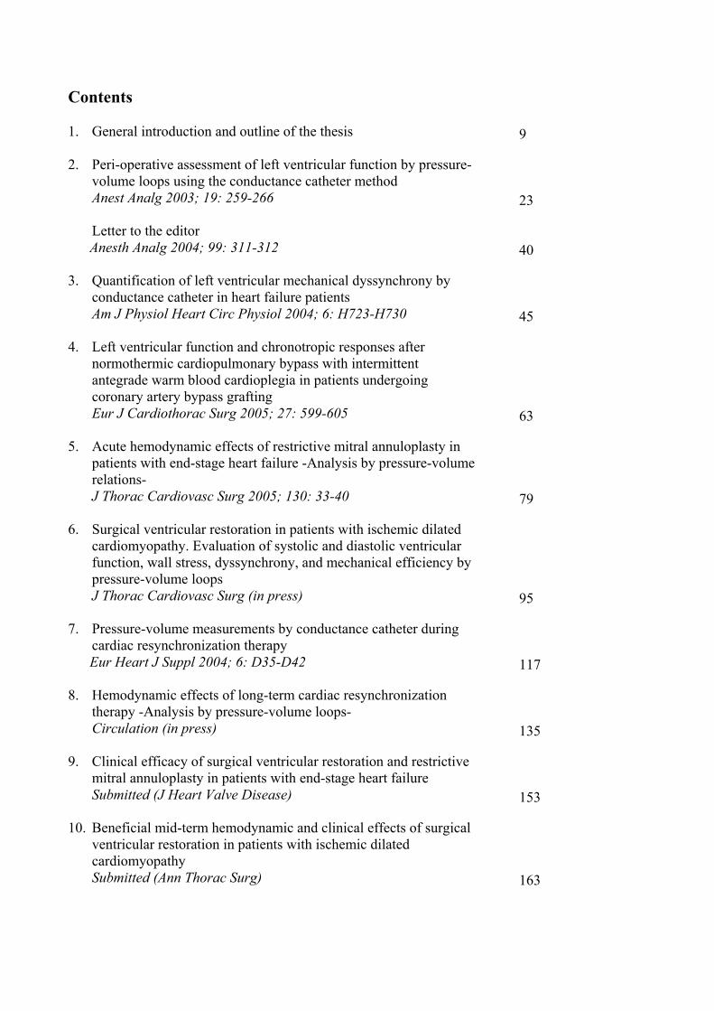
Contents 1. General introduction and outline of the thesis 2. Peri-operative assessment of left ventricular function by pressure-
volume loops using the conductance catheter method Anest Analg 2003; 19: 259-266
Letter to the editor
Anesth Analg 2004; 99: 311-312 3. Quantification of left ventricular mechanical dyssynchrony by
conductance catheter in heart failure patients Am J Physiol Heart Circ Physiol 2004; 6: H723-H730
4. Left ventricular function and chronotropic responses after
normothermic cardiopulmonary bypass with intermittent antegrade warm blood cardioplegia in patients undergoing coronary artery bypass grafting Eur J Cardiothorac Surg 2005; 27: 599-605
5. Acute hemodynamic effects of restrictive mitral annuloplasty in
patients with end-stage heart failure -Analysis by pressure-volume relations- J Thorac Cardiovasc Surg 2005; 130: 33-40
6. Surgical ventricular restoration in patients with ischemic dilated
cardiomyopathy. Evaluation of systolic and diastolic ventricular function, wall stress, dyssynchrony, and mechanical efficiency by pressure-volume loops J Thorac Cardiovasc Surg (in press)
7. Pressure-volume measurements by conductance catheter during
cardiac resynchronization therapy Eur Heart J Suppl 2004; 6: D35-D42 8. Hemodynamic effects of long-term cardiac resynchronization
therapy -Analysis by pressure-volume loops- Circulation (in press)
9. Clinical efficacy of surgical ventricular restoration and restrictive
mitral annuloplasty in patients with end-stage heart failure Submitted (J Heart Valve Disease)
10. Beneficial mid-term hemodynamic and clinical effects of surgical
ventricular restoration in patients with ischemic dilated cardiomyopathy
Submitted (Ann Thorac Surg)
9 23 40 45 63 79 95 117 135 153 163

11. Sustained left ventricular reverse remodeling, improved systolic function and unchanged diastolic function six months after surgical ventricular restoration -Analysis by pressure-volume loops- Submitted
12. Summary and conclusions / Samenvatting en conclusies List of publications Acknowledgements Curriculum Vitae
179 191 207 209 211

CHAPTER 1
General introduction and outline of the thesis
Chapter 1
10
END-STAGE HEART FAILURE
Chronic heart failure is one of the major healthcare problems in the world both in terms
of patient numbers, hospitalizations, and economic costs. In the United States, 4 to 5
million people have chronic heart failure, which leads to more than 2 million
hospitalizations each year.1,2 Recently, the Rotterdam study showed an overall incidence
of chronic heart failure of 1.4% in the Netherlands with an overall prevalence of 7.0%.3
Despite optimal medical therapy (β-blockers, angiotensin-converting enzyme inhibitors,
spironolactone), many patients develop end-stage heart failure and remain severely
symptomatic.
In these patients, cardiac transplantation remains the most effective surgical therapy
with 1-, 5- and 10-year survival rates of 94, 78, and 46 percent, respectively.4,5
Although effective, heart transplantation is hindered by donor shortage and its limited
applicability. The International Society of Heart and Lung Transplantation has reported
a progressive worldwide decline of cardiac transplantation.6
Given the limitations of medical therapy and cardiac transplantation, several alternative
therapies for end-stage heart failure have been adopted in the last decade. Most
prominent is cardiac resynchronization therapy (CRT), after the first implant in 1995,
large multi-center trials have been performed indicating improved symptoms, exercise
tolerance and quality of life.7 A recent study shows an additional survival benefit in
patients treated by CRT and pharmacological therapy above patients treated with only
pharmacological therapy.8 In addition, new surgical therapies such as restrictive mitral
annuloplasty and surgical ventricular restoration have evolved and are currently widely
performed in patients with end-stage heart failure.9,10 These therapies aim to correct
frequently observed end-stage complications as mitral regurgitation and left ventricular
(LV) aneurysm. If not treated, these complications have important adverse effects on
long-term survival.11-13
The long-term survival rates of patients with end-stage heart failure treated with several
therapies are summarized in table 1. Obviously, comparison is hampered by the fact that
the etiology of heart failure is different in the various subgroups.
Other alternative therapies in patients with end-stage heart failure involve the use of LV
devices. Heerdt et al. showed that chronic unloading by LV assist devices reverses
contractile dysfunction and alters gene expression in patients with end-stage heart
failure.14 Recently, the cardiac support device (Acorn device) was introduced, which
seems to reverse LV dilatation and improves functional capacity of heart failure
General introduction and Outline of the thesis
11
patients.15 However, long-term studies with these devices implanted in more patients
should be awaited. Finally, preliminary data suggest that cell transplantation or stem
cell therapy may be applied for repairing damaged myocardium.16-18 These therapies are
currently under clinical investigation and future data should define their clinical
efficacy.
Table 1. Survival in patients with NYHA III/IV heart failure after different treatments
Follow-up (years)
Therapy (ref) 1-year 5-year 10-year
Medical 3,19 63% 35% 9%
HTX 4,5,20 94% 78% 46%
CRT 8,21 86% 75% -
RMA 22-25 84% 50% -
SVR 26 88% 69% -
Ref: references; HTX: cardiac transplantation; CRT: cardiac resynchronization therapy; RMA:
restrictive mitral annuloplasty; SVR: surgical ventricular restoration
PHARMACOLOGICAL THERAPIES
Currently, angiotensin-converting-enzym inhibitors and beta-blockers constitute the
most important pharmacological therapies for heart failure and large trials have shown
their capacity to improve survival and to lower morbidity.27-32 Aldosterone antagonists
and angiotensin receptor blockers may provide additional benefit.33-35,36,37 However, the
sustained benefit of medical treatment appears relatively short-lived.38 Non-
pharmacological therapies such as heart transplantation and implantable assist devices
are only considered in the late stage of the disease and access to such therapies is
limited.39 Alternative non-pharmacological treatments for the failing heart such as CRT,
mitral valve repair and surgical ventricular restoration are currently widely performed.
NON-PHARMACOLOGICAL THERAPIES
Cardiac resynchronization therapy
LV mechanical dyssynchrony in patients with end-stage heart failure is related to
electrical, structural, and morphological features.40,41 Mechanical dyssynchrony is
Chapter 1
12
present in the normal heart, but becomes more apparent in pathological conditions such
as heart failure. 42,43 In patients with heart failure, LV electrical dyssynchrony typically
results from left bundle-branch block. Notably, left bundle-branch block changes LV
contraction patterns, leading to early and late contraction.44,45 This, in turn, impairs
systolic function, reduces cardiac output, and increases end-systolic volume and LV
wall stress.40
CRT is a novel treatment option in symptomatic patients with end-stage heart failure
and LV mechanical dyssynchrony. Current indications for CRT in patients with drug-
refractory end-stage heart failure are NYHA class III/IV symptoms, LV ejection
fraction below 35 percent, QRS duration above 120 ms and left bundle branch block
configuration. Large randomized placebo controlled studies have demonstrated the
beneficial effects of CRT on symptoms, exercise capacity, and quality of life.46,47 In
addition, a recent prospective randomized study showed that CRT substantially reduced
the risk of complications and death among patients with heart failure and cardiac
dyssynchrony.8 In this study, a total of 404 patients were assigned to receive medical
therapy alone and 409 patients to receive medical therapy plus cardiac
resynchronization therapy and all patients were evaluated in a mean follow-up period of
29 months. The mortality rate in the medical-therapy group was 13% at one year and
25% at two years, as compared with 10% and 18%, respectively, in the CRT group.
This study therefore concluded that implantation of CRT should routinely be considered
in patients with moderate to severe heart failure and cardiac dyssynchrony. Several
studies have demonstrated that CRT has beneficial effects on LV hemodynamics
including reverse LV remodeling.48-50 Recently, Yu et al. demonstrated that LV reverse
remodeling is a strong predictor of lower long-term mortality and heart failure events.51
In addition, CRT is associated with reduced sympathetic nervous activity, suggesting
potentially favourable neurohormonal effects.40 These benefits are pacing dependent,
because discontinuation of pacing resulted in a rapid loss of cardiac improvement.
Penicka et al. have recently demonstrated that the degree of baseline LV dyssynchrony
is the main predictive factor for LV functional recovery and reversed remodeling after
CRT.52 Therefore, LV dyssynchrony assessed by tissue Doppler imaging may be an
important additional selection criterium for CRT.53 Bax et al. have recently shown that
patients with septal to lateral delay above 65 ms will respond to CRT and will have an
excellent prognosis after CRT. Furthermore, CRT also has beneficial effects on mitral
regurgitation.54,55 Improved coordinated timing of mechanical activation of papillary
muscle insertion sites appears to be a mechanistic contributor to immediate reduction of
General introduction and Outline of the thesis
13
mitral regurgitation by CRT in patients with heart failure. Despite the clear clinical
benefit, accurate hemodynamic data, i.e. effects on systolic and diastolic LV function,
remain largely limited to the acute effects of CRT. Long-term effects are reported
mainly in terms of ejection fraction and reversed remodeling. More detailed
hemodynamic studies would provide potentially important insight in the working
mechanisms of long-term CRT.
Restrictive mitral annuloplasty
Patients with chronic heart failure due to LV systolic dysfunction frequently develop
mitral regurgitation.56 Several studies have shown that coaptation failure arises in these
patients as a consequence of geometric alterations, which affects mitral annular size and
the geometric position of the subvalvular apparatus.57,58 Previously, surgical treatment
of mitral regurgitation was avoided in patients with heart failure owing to concerns
about operative risk and peri-operative complications.59 However, patients with mitral
regurgitation have a significantly decreased survival at 2 years follow-up versus patients
without mitral regurgitation.11 More recently, with improvements in surgical techniques,
surgical mitral annuloplasty for mitral regurgitation in the setting of heart failure has
become a more popular treatment option. Bolling et al. have demonstrated the
feasibility of mitral valve repair in patients with heart failure by downsizing the annulus
using a flexible ring.23 Their initial results in 48 patients who underwent restrictive
mitral annuloplasty showed an early mortality rate of approximately 5% with 1- and 2-
year survival rates of 82% and 71% respectively. Several recent studies have confirmed
that early mortality is low (between 5 and 7%), heart failure symptoms are ameliorated,
LV size and ejection fraction improve, and intermediate outcome is favorable.24,25
However, several studies in patients treated with mitral annuloplasty demonstrated a
high recurrence rate (30%) of mitral regurgitation after six months follow-up.60,61 In
contrast to these results, Bax et al. reported no recurrences of mitral regurgitation in 51
patients with ischemic LV dysfunction at 2-years follow-up.22 Similarly, Szalay et al.
reported in 121 patients with end-stage heart failure a recurrent rate of 3% with a mean
mitral regurgitation grade 0.6 at 1-year follow-up.25 The low recurrence rates in these
latter studies may be associated with a more truly restrictive annuloplasty performed in
these patients.
The effects of restrictive mitral annuloplasty on systolic and diastolic LV performance
are relatively unknown. Bolling and coworkers hypothesized that restrictive mitral
annuloplasty leads to LV systolic improvement by acute remodeling of the base of the

Chapter 1
14
heart and re-establishing the ellipsoid shape.62,63 Recent data from Bax et al. reported
that 50% of patients showed significant reduction in LV end-systolic diameter over
time.22 Of note, a substantial percentage (60%) of patients in this study especially those
with a preoperative LV end-diastolic diameter and LV end-systolic diameter of 65 mm
and 51 mm, respectively, showed reverse remodeling at late follow-up. These findings
indicate that the process of reverse remodeling may need substantial time in some
patients. These issues are clinically relevant, since a reduction of LV dimensions and an
increase in LV ejection fraction are associated with a favorable prognosis.64,65 However,
until now there is no randomized clinical trial that demonstrates that surgical correction
of mitral regurgitation by mitral annuloplasty improves survival or leads to reverse LV
remodeling. Wu and colleagues have recently demonstrated that there is no clearly
demonstrable survival benefit conferred by mitral annuloplasty for significant mitral
regurgitation in patients with chronic heart failure.66 In addition, Enomoto et al.
demonstrated in an animal model that mitral regurgitation might not contribute
significantly to adverse remodeling suggesting that it is likely a manifestation rather
than an important impetus for post-infarction remodeling.67
In summary, current data demonstrates that restrictive mitral annuloplasty is safe in
patients with heart failure. Still, data about long-term survival benefits, recurrent mitral
regurgitation, and LV reverse remodeling is inconclusive. Future prospective
randomized controlled trials should answer these questions. In addition, hemodynamic
studies may provide insight in the effects of restrictive mitral annuloplasty on LV
systolic and diastolic function.
Surgical ventricular restoration
In patients with ischemic heart failure, structural changes like LV aneurysm, may
contribute to substantial mechanical LV dyssynchrony. At least 88% of dyskinetic LV
aneurysms result from anterior-septal infarctions, while the remainder follow after
inferior infarction.68 The LV nonuniformity of contraction and relaxation reduces
mechanical efficiency of LV filling and ejection and contributes to diastolic and systolic
dysfunction.42,69 Furthermore, scarring and LV dilatation associated with aneurysm
formation may provide a substrate for LV arrhythmias. Surgical ventricular restoration
is increasingly applied in patients with heart failure and LV aneurysm. Controversy still
exists regarding the question whether similar techniques may also be useful in treating
patients with dilated ventricles and scarred regions of the heart when the shape is not
seriously distorted by an LV aneurysm. Dor et al. described the endoventricular circular
General introduction and Outline of the thesis
15
patch plasty for LV reconstruction and demonstrated that the results of this technique
were just as good in patients with akinetic regions as in patients with dyskinetic
regions.70 Several studies further advocated the use of the endoventricular circular patch
technique above the simple linear technique in patients with LV aneurysm.71,72
Although surgical ventricular restoration is increasingly performed, it has not yet found
general acceptance. Possible reasons include a lack of evidence that demonstrates
improvement in morbidity and mortality with this technique in patients with ischemic
heart failure. A recent retrospective analysis has demonstrated that the outcome was
significantly better in patients who received CABG plus surgical ventricular restoration
compared to patients who received CABG alone.73 In most studies, operative mortality
ranges between 0 and 20% and the reported 1- and 5-year survival hovers around 85%
and 70%, respectively.74-76 Patients in these studies had a subjective clinical benefit, as
indicated by a significant improvement of their NYHA classification (from III-IV to I-
III) with significant improvement of LV ejection fraction and reduction in end-diastolic
and end-systolic volumes. However, none of these studies has been conducted in a
prospective, randomized manner with an acceptable number of patients.
Initial results with surgical ventricular restoration have recently been published in a 3-
year observational study by the RESTORE group.26 The surgeons in this international
group performed the surgical ventricular restoration in 662 patients who mainly had
akinetic defects of the anterior wall. The results have been promising, although any
conclusions on the incremental efficacy of surgical ventricular restoration relative to
CABG must be made with caution because of the absence of a control group in the
RESTORE registry. LV ejection fraction was improved on an average of 10% and all
patients had significant improvement of NYHA classification. Despite these promising
data, Elefteriades et al. demonstrated a similar improvement in contractile function in a
small and selected group of patients who underwent isolated CABG.77 Therefore,
controversy remains regarding the question whether surgical ventricular restoration or
CABG alone provide additional benefit above medical therapy. These questions will not
be answered unless they are investigated in a prospective randomized fashion. The
STICH (Surgical Treatment for IschemiC Heart failure) trial is the first prospective
randomized study in the history of coronary artery surgery to specifically assess the
potential benefit of CABG in patients with ischemic heart failure. This trial is designed
and powered to answer fundamental clinical questions regarding the ischemic heart
failure population. The trial tests two hypotheses: (1) CABG combined with intensive
medical therapy improves long-term survival compared with medical therapy alone and
Chapter 1
16
(2) surgical ventricular restoration combined with CABG and medical therapy improves
survival free of cardiac events compared to CABG and medical therapy without surgical
ventricular restoration.
Several studies demonstrated beneficial hemodynamic effects of surgical ventricular
restoration in patients with ischemic heart failure. These studies reported acute
improvements in contractile state, energy efficiency, and relaxation, together with a
decrease in LV mechanical dyssynchrony in patients with heart failure.78,79 Buckberg et
al. emphasized the importance of considering size, shape and LV fiber orientation in
patients with heart failure.80-82 It has been proposed that surgical ventricular restoration
of the dilated LV will restore myofibers in the diseased ventricle to a normal, oblique
orientation.83 However, this issue remains still controversial and data supporting these
claims are lacking.84,85
In conclusion, despite the promising results of these alternative therapies in patients
with end-stage heart failure, the working mechanisms and effects on LV function are
relatively poorly defined.
AIM AND OUTLINE OF THE THESIS
The aim of this thesis was to study the hemodynamic effects of CRT, surgical
ventricular restoration and restrictive mitral annuloplasty in patients with end-stage
heart failure by use of pressure-volume loops derived by the conductance catheter. An
important rational for this approach is that pressure-volume derived indices reflect
intrinsic systolic and diastolic LV function in a relative load-independent fashion,
whereas conventional methods are importantly influenced by changes in loading
conditions. This may be particular relevant during cardiac procedures such as valve
surgery and surgical ventricular restoration where loading conditions may change
substantially. Moreover, it is increasingly recognized that mechanical dyssynchrony,
importantly influence LV function and that benefit of CRT and surgical therapies may
be partly explained by reduced mechanical dyssynchrony. The ability of the
conductance catheter to quantify mechanical dyssynchrony in an objective and on-line
fashion may therefore add to the diagnostic power of this methodology.
The quantification of effects of these therapies on global and intrinsic LV systolic and
diastolic function and mechanical dyssynchrony may provide further insight in the
General introduction and Outline of the thesis
17
working mechanisms of these therapies. This may help to explain improved survival,
functional status and exercise tolerance in heart failure patients treated with these
therapies. In this thesis, acute effects of surgical therapies on LV function were assessed
by peri-operative measurements by the conductance catheter in the operating room,
whereas chronic effects of CRT and surgical therapies were assessed in the
catheterization laboratory at baseline and at 6 months follow-up.
REFERENCES 1. Nohria A, Lewis E, Stevenson LW. Medical management of advanced heart failure. JAMA.
2002;287:628-640. 2. Jessup M, Brozena S. Heart failure. N Engl J Med. 2003;348:2007-2018. 3. Bleumink GS, Knetsch AM, Sturkenboom MC, Straus SM, Hofman A, Deckers JW, Witteman JC,
Stricker BH. Quantifying the heart failure epidemic: prevalence, incidence rate, lifetime risk and prognosis of heart failure The Rotterdam Study. Eur Heart J. 2004;25:1614-1619.
4. Copeland JG, McCarthy M. University of Arizona, Cardiac Transplantation: changing patterns in selection and outcomes. Clin Transpl. 2001;203-207.
5. Robbins RC, Barlow CW, Oyer PE, Hunt SA, Miller JL, Reitz BA, Stinson EB, Shumway NE. Thirty years of cardiac transplantation at Stanford university. J Thorac Cardiovasc Surg. 1999;117:939-951.
6. Taylor DO, Edwards LB, Boucek MM, Trulock EP, Keck BM, Hertz MI. The Registry of the International Society for Heart and Lung Transplantation: twenty-first official adult heart transplant report--2004. J Heart Lung Transplant. 2004;23:796-803.
7. Auricchio A, Stellbrink C, Sack S, Block M, Vogt J, Bakker P, Mortensen P, Klein H. The Pacing Therapies for Congestive Heart Failure (PATH-CHF) study: rationale, design, and endpoints of a prospective randomized multicenter study. Am J Cardiol. 1999;83:130D-135D.
8. Cleland JG, Daubert JC, Erdmann E, Freemantle N, Gras D, Kappenberger L, Tavazzi L. The Effect of Cardiac Resynchronization on Morbidity and Mortality in Heart Failure. N Engl J Med. 2005.
9. Bolling SF, Smolens IA, Pagani FD. Surgical alternatives for heart failure. J Heart Lung Transplant. 2001;20:729-733.
10. Dor V. The endoventricular circular patch plasty ("Dor procedure") in ischemic akinetic dilated ventricles. Heart Fail Rev. 2001;6:187-193.
11. Grigioni F, Enriquez-Sarano M, Zehr KJ, Bailey KR, Tajik AJ. Ischemic mitral regurgitation: long-term outcome and prognostic implications with quantitative Doppler assessment. Circulation. 2001;103:1759-1764.
12. Koelling TM, Aaronson KD, Cody RJ, Bach DS, Armstrong WF. Prognostic significance of mitral regurgitation and tricuspid regurgitation in patients with left ventricular systolic dysfunction. Am Heart J. 2002;144:524-529.
13. Robbins JD, Maniar PB, Cotts W, Parker MA, Bonow RO, Gheorghiade M. Prevalence and severity of mitral regurgitation in chronic systolic heart failure. Am J Cardiol. 2003;91:360-362.
14. Heerdt PM, Holmes JW, Cai B, Barbone A, Madigan JD, Reiken S, Lee DL, Oz MC, Marks AR, Burkhoff D. Chronic unloading by left ventricular assist device reverses contractile dysfunction and alters gene expression in end-stage heart failure. Circulation. 2000;102:2713-2719.
15. Oz MC, Konertz WF, Kleber FX, Mohr FW, Gummert JF, Ostermeyer J, Lass M, Raman J, Acker MA, Smedira N. Global surgical experience with the Acorn cardiac support device. J Thorac Cardiovasc Surg. 2003;126:983-991.
16. Menasche P. Cell transplantation in myocardium. Ann Thorac Surg. 2003;75:S20-S28. 17. Perin EC, Geng YJ, Willerson JT. Adult stem cell therapy in perspective. Circulation.
2003;107:935-938. 18. Perin EC, Dohmann HF, Borojevic R, Silva SA, Sousa AL, Silva GV, Mesquita CT, Belem L,
Vaughn WK, Rangel FO, Assad JA, Carvalho AC, Branco RV, Rossi MI, Dohmann HJ, Willerson JT. Improved exercise capacity and ischemia 6 and 12 months after transendocardial injection of autologous bone marrow mononuclear cells for ischemic cardiomyopathy. Circulation. 2004;110:II213-II218.

Chapter 1
18
19. Copeland JG, Smith RG, Arabia FA, Nolan PE, Sethi GK, Tsau PH, McClellan D, Slepian MJ. Cardiac replacement with a total artificial heart as a bridge to transplantation. N Engl J Med. 2004;351:859-867.
20. Vitali E, Colombo T, Fratto P, Russo C, Bruschi G, Frigerio M. Surgical therapy in advanced heart failure. Am J Cardiol. 2003;91:88F-94F.
21. Auricchio A, Stellbrink C, Sack S, et al. Long-term benefit as a result of pacing resynchronization in congestive heart failure: results of the PATH-CHF trial (Abstract). 102 Suppl II, 693. 2000.
22. Bax JJ, Braun J, Somer ST, Klautz R, Holman ER, Versteegh MI, Boersma E, Schalij MJ, van der Wall EE, Dion RA. Restrictive annuloplasty and coronary revascularization in ischemic mitral regurgitation results in reverse left ventricular remodeling. Circulation. 2004;110:II103-II108.
23. Bolling SF, Pagani FD, Deeb GM, Bach DS. Intermediate-term outcome of mitral reconstruction in cardiomyopathy. J Thorac Cardiovasc Surg. 1998;115:381-386.
24. Gummert JF, Rahmel A, Bucerius J, Onnasch J, Doll N, Walther T, Falk V, Mohr FW. Mitral valve repair in patients with end stage cardiomyopathy: who benefits? Eur J Cardiothorac Surg. 2003;23:1017-1022.
25. Szalay ZA, Civelek A, Hohe S, Brunner-LaRocca HP, Klovekorn WP, Knez I, Vogt PR, Bauer EP. Mitral annuloplasty in patients with ischemic versus dilated cardiomyopathy. Eur J Cardiothorac Surg. 2003;23:567-572.
26. Athanasuleas CL, Buckberg GD, Stanley AW, Siler W, Dor V, Di Donato M, Menicanti L, Almeida dO, Beyersdorf F, Kron IL, Suma H, Kouchoukos NT, Moore W, McCarthy PM, Oz MC, Fontan F, Scott ML, Accola KA. Surgical ventricular restoration in the treatment of congestive heart failure due to post-infarction ventricular dilation. J Am Coll Cardiol. 2004;44:1439-1445.
27. Effects of enalapril on mortality in severe congestive heart failure. Results of the Cooperative North Scandinavian Enalapril Survival Study (CONSENSUS). The CONSENSUS Trial Study Group. N Engl J Med. 1987;316:1429-1435.
28. Pfeffer MA, Braunwald E, Moye LA, Basta L, Brown EJ, Jr., Cuddy TE, Davis BR, Geltman EM, Goldman S, Flaker GC, . Effect of captopril on mortality and morbidity in patients with left ventricular dysfunction after myocardial infarction. Results of the survival and ventricular enlargement trial. The SAVE Investigators. N Engl J Med. 1992;327:669-677.
29. Pfeffer MA, Swedberg K, Granger CB, Held P, McMurray JJ, Michelson EL, Olofsson B, Ostergren J, Yusuf S, Pocock S. Effects of candesartan on mortality and morbidity in patients with chronic heart failure: the CHARM-Overall programme. Lancet. 2003;362:759-766.
30. Bristow MR. beta-adrenergic receptor blockade in chronic heart failure. Circulation. 2000;101:558-569.
31. Packer M, Coats AJ, Fowler MB, Katus HA, Krum H, Mohacsi P, Rouleau JL, Tendera M, Castaigne A, Roecker EB, Schultz MK, DeMets DL. Effect of carvedilol on survival in severe chronic heart failure. N Engl J Med. 2001;344:1651-1658.
32. Poole-Wilson PA, Swedberg K, Cleland JG, Di Lenarda A, Hanrath P, Komajda M, Lubsen J, Lutiger B, Metra M, Remme WJ, Torp-Pedersen C, Scherhag A, Skene A. Comparison of carvedilol and metoprolol on clinical outcomes in patients with chronic heart failure in the Carvedilol Or Metoprolol European Trial (COMET): randomised controlled trial. Lancet. 2003;362:7-13.
33. Zannad F. [Anti-aldosterone: the evidence of the RALES study]. Arch Mal Coeur Vaiss. 2000;Spec No:8-9, 15.
34. Tsutamoto T, Wada A, Maeda K, Mabuchi N, Hayashi M, Tsutsui T, Ohnishi M, Sawaki M, Fujii M, Matsumoto T, Horie H, Sugimoto Y, Kinoshita M. Spironolactone inhibits the transcardiac extraction of aldosterone in patients with congestive heart failure. J Am Coll Cardiol. 2000;36:838-844.
35. Tsutamoto T, Wada A, Maeda K, Mabuchi N, Hayashi M, Tsutsui T, Ohnishi M, Sawaki M, Fujii M, Matsumoto T, Matsui T, Kinoshita M. Effect of spironolactone on plasma brain natriuretic peptide and left ventricular remodeling in patients with congestive heart failure. J Am Coll Cardiol. 2001;37:1228-1233.
36. Cohn JN, Tognoni G. A randomized trial of the angiotensin-receptor blocker valsartan in chronic heart failure. N Engl J Med. 2001;345:1667-1675.
37. Jong P, Demers C, McKelvie RS, Liu PP. Angiotensin receptor blockers in heart failure: meta-analysis of randomized controlled trials. J Am Coll Cardiol. 2002;39:463-470.
38. Cleland JG, Swedberg K, Poole-Wilson PA. Successes and failures of current treatment of heart failure. Lancet. 1998;352 Suppl 1:SI19-SI28.
39. Rose EA, Gelijns AC, Moskowitz AJ, Heitjan DF, Stevenson LW, Dembitsky W, Long JW, Ascheim DD, Tierney AR, Levitan RG, Watson JT, Meier P, Ronan NS, Shapiro PA, Lazar RM, Miller LW, Gupta L, Frazier OH, Desvigne-Nickens P, Oz MC, Poirier VL. Long-term mechanical left ventricular assistance for end-stage heart failure. N Engl J Med. 2001;345:1435-1443.

General introduction and Outline of the thesis
19
40. Leclercq C, Kass DA. Retiming the failing heart: principles and current clinical status of cardiac resynchronization. J Am Coll Cardiol. 2002;39:194-201.
41. Barold SS. What is cardiac resynchronization therapy? Am J Med. 2001;111:224-232. 42. Brutsaert DL. Nonuniformity: a physiologic modulator of contraction and relaxation of the normal
heart. J Am Coll Cardiol. 1987;9:341-348. 43. Curry CW, Nelson GS, Wyman BT, Declerck J, Talbot M, Berger RD, McVeigh ER, Kass DA.
Mechanical dyssynchrony in dilated cardiomyopathy with intraventricular conduction delay as depicted by 3D tagged magnetic resonance imaging. Circulation. 2000;101:E2.
44. Prinzen FW, Hunter WC, Wyman BT, McVeigh ER. Mapping of regional myocardial strain and work during ventricular pacing: experimental study using magnetic resonance imaging tagging. J Am Coll Cardiol. 1999;33:1735-1742.
45. Wyman BT, Hunter WC, Prinzen FW, Faris OP, McVeigh ER. Effects of single- and biventricular pacing on temporal and spatial dynamics of ventricular contraction. Am J Physiol Heart Circ Physiol. 2002;282:H372-H379.
46. Abraham WT, Fisher WG, Smith AL, Delurgio DB, Leon AR, Loh E, Kocovic DZ, Packer M, Clavell AL, Hayes DL, Ellestad M, Trupp RJ, Underwood J, Pickering F, Truex C, McAtee P, Messenger J. Cardiac resynchronization in chronic heart failure. N Engl J Med. 2002;346:1845-1853.
47. Auricchio A, Stellbrink C, Block M, Sack S, Vogt J, Bakker P, Klein H, Kramer A, Ding J, Salo R, Tockman B, Pochet T, Spinelli J. Effect of pacing chamber and atrioventricular delay on acute systolic function of paced patients with congestive heart failure. The Pacing Therapies for Congestive Heart Failure Study Group. The Guidant Congestive Heart Failure Research Group. Circulation. 1999;99:2993-3001.
48. Nelson GS, Berger RD, Fetics BJ, Talbot M, Spinelli JC, Hare JM, Kass DA. Left ventricular or biventricular pacing improves cardiac function at diminished energy cost in patients with dilated cardiomyopathy and left bundle-branch block. Circulation. 2000;102:3053-3059.
49. Ukkonen H, Beanlands RS, Burwash IG, de Kemp RA, Nahmias C, Fallen E, Hill MR, Tang AS. Effect of cardiac resynchronization on myocardial efficiency and regional oxidative metabolism. Circulation. 2003;107:28-31.
50. Sundell J, Engblom E, Koistinen J, Ylitalo A, Naum A, Stolen KQ, Kalliokoski R, Nekolla SG, Airaksinen KE, Bax JJ, Knuuti J. The effects of cardiac resynchronization therapy on left ventricular function, myocardial energetics, and metabolic reserve in patients with dilated cardiomyopathy and heart failure. J Am Coll Cardiol. 2004;43:1027-1033.
51. Yu CM, Bleeker GB, Fung JW, Schalij MJ, Zhang Q, van der Wall EE, Chan YS, Kong SL, Bax JJ. Left Ventricular Reverse Remodeling but Not Clinical Improvement Predicts Long-Term Survival After Cardiac Resynchronization Therapy. Circulation. 2005.
52. Penicka M, Bartunek J, de Bruyne B, Vanderheyden M, Goethals M, De Zutter M, Brugada P, Geelen P. Improvement of left ventricular function after cardiac resynchronization therapy is predicted by tissue Doppler imaging echocardiography. Circulation. 2004;109:978-983.
53. Bax JJ, Marwick TH, Molhoek SG, Bleeker GB, Van Erven L, Boersma E, Steendijk P, van der Wall EE, Schalij MJ. Left ventricular dyssynchrony predicts benefit of cardiac resynchronization therapy in patients with end-stage heart failure before pacemaker implantation. Am J Cardiol. 2003;92:1238-1240.
54. Kanzaki H, Bazaz R, Schwartzman D, Dohi K, Sade LE, Gorcsan J, III. A mechanism for immediate reduction in mitral regurgitation after cardiac resynchronization therapy: insights from mechanical activation strain mapping. J Am Coll Cardiol. 2004;44:1619-1625.
55. Lancellotti P, Melon P, Sakalihasan N, Waleffe A, Dubois C, Bertholet M, Pierard LA. Effect of cardiac resynchronization therapy on functional mitral regurgitation in heart failure. Am J Cardiol. 2004;94:1462-1465.
56. Yiu SF, Enriquez-Sarano M, Tribouilloy C, Seward JB, Tajik AJ. Determinants of the degree of functional mitral regurgitation in patients with systolic left ventricular dysfunction: A quantitative clinical study. Circulation. 2000;102:1400-1406.
57. Aikawa K, Sheehan FH, Otto CM, Coady K, Bashein G, Bolson EL. The severity of functional mitral regurgitation depends on the shape of the mitral apparatus: A three-dimensional echo analysis. Journal of Heart Valve Disease. 2002;11:627-636.
58. Kumanohoso T, Otsuji Y, Yoshifuku S, Matsukida K, Koriyama C, Kisanuki A, Minagoe S, Levine RA, Tei C. Mechanism of higher incidence of ischemic mitral regurgitation in patients with inferior myocardial infarction: quantitative analysis of left ventricular and mitral valve geometry in 103 patients with prior myocardial infarction. J Thorac Cardiovasc Surg. 2003;125:135.
59. Harris KM, Sundt TM, III, Aeppli D, Sharma R, Barzilai B. Can late survival of patients with moderate ischemic mitral regurgitation be impacted by intervention on the valve? Ann Thorac Surg. 2002;74:1468-1475.

Chapter 1
20
60. McGee EC, Gillinov AM, Blackstone EH, Rajeswaran J, Cohen G, Najam F, Shiota T, Sabik JF, Lytle BW, McCarthy PM, Cosgrove DM. Recurrent mitral regurgitation after annuloplasty for functional ischemic mitral regurgitation. J Thorac Cardiovasc Surg. 2004;128:916-924.
61. Tahta SA, Oury JH, Maxwell JM, Hiro SP, Duran CM. Outcome after mitral valve repair for functional ischemic mitral regurgitation. J Heart Valve Dis. 2002;11:11-18.
62. Bolling SF, Deeb GM, Brunsting LA, Bach DS. Early outcome of mitral valve reconstruction in patients with end-stage cardiomyopathy. J Thorac Cardiovasc Surg. 1995;109:676-682.
63. Smolens IA, Pagani FD, Bolling SF. Mitral valve repair in heart failure. Eur J Heart Fail. 2000;2:365-371.
64. Udelson JE, Konstam MA. Relation between left ventricular remodeling and clinical outcomes in heart failure patients with left ventricular systolic dysfunction. J Card Fail. 2002;8:S465-S471.
65. White HD, Norris RM, Brown MA, Brandt PW, Whitlock RM, Wild CJ. Left ventricular end-systolic volume as the major determinant of survival after recovery from myocardial infarction. Circulation. 1987;76:44-51.
66. Wu AH, Aaronson KD, Bolling SF, Pagani FD, Welch K, Koelling TM. Impact of mitral valve annuloplasty on mortality risk in patients with mitral regurgitation and left ventricular systolic dysfunction. J Am Coll Cardiol. 2005;45:381-387.
67. Enomoto Y, Gorman JH, III, Moainie SL, Guy TS, Jackson BM, Parish LM, Plappert T, Zeeshan A, John-Sutton MG, Gorman RC. Surgical treatment of ischemic mitral regurgitation might not influence ventricular remodeling. J Thorac Cardiovasc Surg. 2005;129:504-511.
68. Mills NL, Everson CT, Hockmuth DR. Technical advances in the treatment of left ventricular aneurysm. Ann Thorac Surg. 1993;55:792-800.
69. Aoyagi T, Pouleur H, Van Eyll C, Rousseau MF, Mirsky I. Wall motion asynchrony is a major determinant of impaired left ventricular filling in patients with healed myocardial infarction. Am J Cardiol. 1993;72:268-272.
70. Dor V. Surgery for left ventricular aneurysm. Curr Opin Cardiol. 1990;5:773-780. 71. Sinatra R, Macrina F, Braccio M, Melina G, Luzi G, Ruvolo G, Marino B. Left ventricular
aneurysmectomy; comparison between two techniques; early and late results. Eur J Cardiothorac Surg. 1997;12:291-297.
72. Lundblad R, Abdelnoor M, Svennevig JL. Surgery for left ventricular aneurysm: early and late survival after simple linear repair and endoventricular patch plasty. J Thorac Cardiovasc Surg. 2004;128:449-456.
73. Maxey TS, Reece TB, Ellman PI, Butler PD, Kern JA, Tribble CG, Kron IL. Coronary artery bypass with ventricular restoration is superior to coronary artery bypass alone in patients with ischemic cardiomyopathy. J Thorac Cardiovasc Surg. 2004;127:428-434.
74. Di Donato M, Toso A, Maioli M, Sabatier M, Stanley AW, Jr., Dor V. Intermediate survival and predictors of death after surgical ventricular restoration. Semin Thorac Cardiovasc Surg. 2001;13:468-475.
75. Isomura T, Suma H, Yamaguchi A, Kobashi T, Yuda A. Left ventricular restoration for ischemic cardiomyopathy - comparison of presence and absence of mitral valve procedure. Eur J Cardiothorac Surg. 2003;23:614-619.
76. Suma H, Isomura T, Horii T, Hisatomi K. Left ventriculoplasty for ischemic cardiomyopathy. Eur J Cardiothorac Surg. 2001;20:319-323.
77. Elefteriades JA, Tolis G, Jr., Levi E, Mills LK, Zaret BL. Coronary artery bypass grafting in severe left ventricular dysfunction: excellent survival with improved ejection fraction and functional state. J Am Coll Cardiol. 1993;22:1411-1417.
78. Di Donato M, Toso A, Dor V, Sabatier M, Barletta G, Menicanti L, Fantini F. Surgical ventricular restoration improves mechanical intraventricular dyssynchrony in ischemic cardiomyopathy. Circulation. 2004;109:2536-2543.
79. Schreuder JJ, Castiglioni A, Maisano F, Steendijk P, Donelli A, Baan J, Alfieri O. Acute decrease of left ventricular mechanical dyssynchrony and improvement of contractile state and energy efficiency after left ventricular restoration. J Thorac Cardiovasc Surg. 2005;129:138-145.
80. Buckberg GD, Coghlan HC, Torrent-Guasp F. The structure and function of the helical heart and its buttress wrapping. V. Anatomic and physiologic considerations in the healthy and failing heart. Semin Thorac Cardiovasc Surg. 2001;13:358-385.
81. Buckberg GD. Congestive heart failure: treat the disease, not the symptom--return to normalcy. J Thorac Cardiovasc Surg. 2001;121:628-637.
82. Buckberg GD. Basic science review: the helix and the heart. J Thorac Cardiovasc Surg. 2002;124:863-883.
83. Buckberg GD, Coghlan HC, Torrent-Guasp F. The structure and function of the helical heart and its buttress wrapping. VI. Geometric concepts of heart failure and use for structural correction. Semin Thorac Cardiovasc Surg. 2001;13:386-401.

General introduction and Outline of the thesis
21
84. Buckberg GD. Imaging, models, and reality: A basis for anatomic-physiologic planning. J Thorac Cardiovasc Surg. 2005;129:243-245.
85. Walker JC, Guccione JM, Jiang Y, Zhang P, Wallace AW, Hsu EW, Ratcliffe MB. Helical myofiber orientation after myocardial infarction and left ventricular surgical restoration in sheep. J Thorac Cardiovasc Surg. 2005;129:382-390.

CHAPTER 2
Peri-operative assessment of left ventricular function by
pressure-volume loops using the conductance catheter
S.A.F. Tulner
R.J.M. Klautz
G.L. van Rijk-Zwikker
F.H.M. Engbers
J.J. Bax
J. Baan
E.E. van der Wall
R.A.E. Dion
P. Steendijk
Anest Analg 2003; 19: 259-266
Poster presentation at Cardiac Techniques and Technologies, March 2003, Miami,
Florida, USA
Chapter 2
24
ABSTRACT
Interpretation of peri-operative measurements of cardiac function during cardiac surgery
is complicated by changes in loading conditions induced by anesthesia,
cardiopulmonary bypass (CPB) and the surgical procedure itself. Quantification of left
ventricular (LV) function by pressure-volume relations as obtained by the conductance
catheter would be advantageous because load-independent indices can be determined.
Accordingly, we evaluated methodological aspects of the conductance catheter
technique and documented LV function pre- and post-CPB in 8 patients undergoing
CABG. LV pressure-volume loops by TEE-guided trans-aortic application of the
conductance catheter were obtained at steady state and during preload reduction by
temporary occlusion of the inferior caval vein. All patients remained hemodynamically
stable and no complications occurred. Complete data were acquired within 15 minutes
pre- and post-CPB. Cardiac output (5.2±1.3 to 6.0±1.4 L/min) and LV ejection fraction
(46±17 to 48±19%) did not change, but end-diastolic pressure increased significantly
post-CPB (8±2 to 16±7mmHg, p<0.05). Load-independent systolic indices remained
constant (end-systolic elastance: 1.31±1.20 to 1.13±0.59mmHg/mL). Diastolic function
changed significantly post-CPB, as Tau decreased from 64±6 to 52±5ms (p<0.05) and
the chamber stiffness constant increased from 0.016±0.014 to 0.038±0.016/mL
(p<0.05). We conclude that the conductance catheter method provides detailed data on
peri-operative myocardial function. Therefore, the conductance catheter method may be
used to evaluate the effects of new surgical and anesthetic procedures for which the
present data may serve as reference data.
INTRODUCTION
Recently, several new approaches were introduced in cardiac surgery such as restrictive
mitral annuloplasty, endoventricular circular patch plasty, and off-pump CABG.
Generally, the efficacy of new techniques is assessed by long-term follow-up of
patients. However, the acute effects on left ventricular (LV) function of these
procedures are not well documented and may be predictive for long-term outcome. Peri-
operative assessment of LV function may allow better evaluation of new surgical
procedures and may help post-operative management by providing insight in the cardiac
pathophysiology. During cardiac surgery cardiac output, aortic pressure, central venous
Peri-operative assessment of left ventricular function
25
pressure and the pulmonary arterial wedge pressure usually assess hemodynamic status.
In addition, transesophageal echocardiography (TEE) is used to assess regional
contractile function. However, interpretation of all these parameters is complicated by
their load-dependency. Therefore, given the substantial changes in loading conditions
that may occur during the operation, these parameters may not reflect intrinsic
myocardial function. Pressure-volume relations as obtained by the conductance catheter,
have been shown to provide load-independent indices of systolic and diastolic
function.1,2 Accordingly, the aim of present study was twofold. Firstly, we described
and evaluated the application of the conductance technique in the operating room
including catheter placement, calibration procedures and heart rate-controlled
measurement of systolic and diastolic pressure-volume relations. Secondly, we
compared various indices of LV function before and after CPB in patients undergoing
CABG. These data obtained in patients with relatively normal LV function may provide
reference data for future studies in which more complex cardiac surgical procedures are
evaluated.
METHODS
The study protocol was approved by the Local Ethics Committee and all patients gave
informed consent. Eight patients with multivessel coronary artery disease elected for
CABG were included. Patients with severely depressed LV function (LVEF < 35%),
unstable angina or atrial fibrillation were excluded.
Anesthesia
After 2mg lorazepam as sublingual premedication two hours before surgery, all patients
received total intravenous anesthesia with target-controlled infusion of propofol,
remifentanil and sufentanil.3-5 Hypnotic state was monitored with a Bispectral Index
(BIS) monitor (Aspect medical systems, Newton, MA). Induction of anesthesia was
started with targeted concentration of 1.5μg/ml propofol and 3ng/ml remifentanil.
Before intubation the remifentanil-targeted concentration was increased to 9ng/ml and
the targeted propofol concentration to 2μg/ml. A single dose of pancuronium bromide
(0.1mg/kg) was given to facilitate intubation. During surgery the propofol concentration
was adjusted between 1.5μg/ml and 2.0μg/ml to maintain a BIS value below 60.
Remifentanil was titrated between 5 and 10ng/ml in response to the patient's
Chapter 2
26
hemodynamic reaction on surgical stimuli. Sufentanil was started at a targeted
concentration of 0.1ng/ml after start of surgery to allow smooth transition of the patient
analgesic state from the operating room to the ICU. The patients were ventilated with an
oxygen/air mixture (FiO2=40%) at a ventilatory rate of 12-15/min and ventilatory
volume was adjusted to maintain PaCO2 between 4.5 and 5.5kPa (34-41mmHg). A
thermal filament catheter was placed in the pulmonary artery via the right internal
jugular vein for semi-continuous cardiac output measurements (Edwards Lifesciences,
Uden, The Netherlands). To monitor cardiac function and facilitate positioning of the
conductance catheter peri-operatively a multiplane TEE-probe was inserted.
Conductance catheter technique
We used a 7F integrated pressure-conductance catheter (CD-Leycom, Zoetermeer, The
Netherlands) incorporating a solid-state pressure sensor and 12 electrodes with an inter-
electrode spacing of 10mm. A pigtail facilitates placement through the aortic valve and
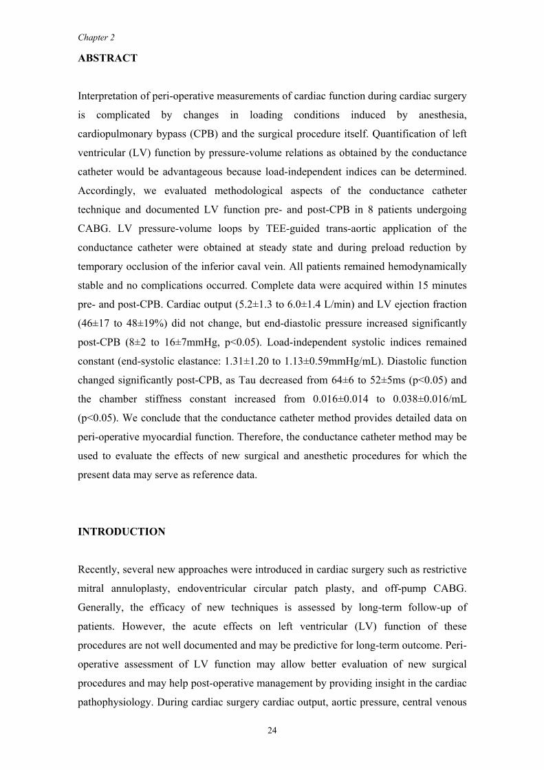
positioning within the LV apex (Figure 1).
Figure 1. Left: The optimal position of the conductance catheter along the long-axis of the left ventricle.
Right: the conductance catheter viewed by long-axis view by TEE peri-operatively
The catheter is connected to a Leycom Cardiac Function Lab (CFL) signal-processor.
Between the two most proximal and two most distal electrodes a dual electric field
(20kHz, 30μA) is generated.6 The remaining 8 electrodes are used to measure 5
segmental volume signals. The user may select from three settings the best match with
the LV long axis: by skipping electrodes one or two 1-cm segments may be converted to
2-cm segments thereby extending the effective length of the catheter. The optimal
setting is selected based on inspection of the segmental volume signals. An aortic
Peri-operative assessment of left ventricular function
27
volume signal is easily distinguished from a ventricular signal because it resembles an
aortic pressure signal and is out-of-phase with the ventricular volume signals. The
segmental conductance’s are summed to yield total conductance G(t) and, taking into
account the specific resistivity of blood and the electrode spacing, converted to a time-
varying volume signal, V(t), which follows through the equation:
V(t) = (1/α)⋅(rho⋅L²)⋅(G(t)-GP)
where α is a slope factor, L is the inter-electrode spacing, rho is the specific resistivity
of blood measured from a 5ml blood sample using a special 4-electrode cuvette
connected to the CFL, and GP is the parallel conductance. G(t) is the sum of the
conductance of the blood in the LV and GP. The latter results from the conductance of
the ventricular wall, other cardiac chambers and to some extent all electrically
conductive structures outside the LV cavity. Baan et al. devised a method to determine
GP by injecting a small bolus (7ml) of hypertonic saline solution (10%) in the distal port
of the pulmonary artery catheter.1 The highly conductive saline transiently changes
blood conductivity, which is measured only in the LV. By analyzing the conductance
signal registered during passage of the bolus through the LV, GP can be determined.1
The correction volume (Vc) corresponding to GP equals:
Vc = (rho⋅L²)⋅GP
After correction for GP the volume signal is directly proportional to actual ventricular
volume, but generally underestimates true volume by a fixed factor. There are two main
causes for this underestimation. First, there may be a mismatch between the measured
segments and the LV long-axis. Secondly, the conversion of conductance to volume
assumes that the electric field is homogeneous within the cavity. In reality this is not
entirely the case resulting in underestimation. The development of dual field excitation
has substantially improved electric field homogeneity, but some underestimation
remains especially in large hearts.6 To correct for this underestimation the factor α was
introduced, which is obtained by comparing conductance-derived stroke volume (SV)
with an independent measure of SV. In most studies α is calculated by dividing SV of
the conductance catheter by SV obtained by thermodilution: α =
SVconductance/SVthermodilution. In the present study we used the 'stat' cardiac output
Chapter 2
28
measurements recorded from a Vigilance® Continuous Cardiac Output Monitoring
System (Edwards Lifesciences, Uden, The Netherlands).
Instrumentation and surgical technique
After harvesting bypass material, the pericardium was opened and epicardial pacemaker
leads were placed on the right atrium. A caval tourniquet was applied around the
inferior caval vein to perform temporary preload reductions by caval vein occlusion.
After systemic heparinization, a sheath (F8, Cordis, Roden, The Netherlands) was
introduced in the ascending aorta for placement of the conductance catheter.
Subsequently the conductance catheter was inserted into the LV and positioned along
the long axis toward the LV apex. Catheter introduction and positioning was guided and
verified by TEE and inspection of the segmental conductance signals. Positioning was
aimed at locating the pigtail in the apex while the most proximal electrodes should be
located just above the aortic valve. Measurements were started if 5 segmental LV
volume signals were obtained.
Measurement protocol and data acquisition
The protocol included measurements at a paced heart rate of 80bpm pre- and post-CPB.
If intrinsic rate was above 80bpm the pacemaker was set slightly above the intrinsic
rate. Pressure-volume loops were measured at steady state and during transient caval
vein occlusion (typical pressure drop of 20mmHg within 5-10s) in order to obtain
systolic and diastolic pressure-volume relationships. The ventilator was turned off to
exclude the effects of respiration. Rho was measured just before data acquisition, both
before and after CPB. Additional acquisitions (before and after CPB) were done for
determination of GP after injection of 7ml 10% hypertonic saline solution through the
distal port of the pulmonary artery catheter. Independent cardiac output measurements
by thermodilution were obtained during steady state. The thermodilution catheter
provides update measurements approximately every minute indicating average cardiac
output over the preceding period. An analog signal reflecting the 'stat' signal was
recorded simultaneously with the pressure-volume signals for off-line calculation of α.
Data analysis
Baseline hemodynamic data were calculated from steady state pressure-volume loops:
heart rate (HR), end-systolic volume (ESV), end-diastolic volume (EDV), end-systolic
pressure (ESP), end-diastolic pressure (EDP), cardiac output (CO), stroke volume (SV),
Peri-operative assessment of left ventricular function
29
stroke work (SW), maximal and minimal rate of LV pressure change (dP/dtMAX,
dP/dtMIN), ejection fraction (EF) and the relaxation time constant (Tau). Tau, reflecting
the early active relaxation process, was calculated as the time constant of mono-
exponential pressure decay during isovolumic relaxation. The isovolumic period was
defined as the period between the time-point of dP/dtMIN and the time-point at which
dP/dt reached 10% of the dP/dtMIN value. From pressure-volume loops during caval vein
occlusion indices of systolic and diastolic function were derived. For systolic function,
the end-systolic pressure-volume relation (ESPVR), the dP/dtMAX-EDV relation and the
preload recruitable stroke work relation (PRSW: SW versus EDV) were determined as
for diastolic function the chamber stiffness constant (CS) was determined. The systolic
relationships were characterized by their slope and volume intercept. The slope of the
ESPVR (Ees) as well as its volume intercept, at a fixed systolic pressure of 75mmHg
(V75) have been shown to be indices of contractility, largely independent of loading
conditions.7,8 The ESPVR was determined by linear regression of end-systolic pressure-
volume points obtained during caval vein occlusion. Similarly, the PRSW slope (S-
PRSW) was determined by plotting SW against EDV and the same was done for the
slope of the dP/dtMAX-EDV relation (S-dP/dt). The slopes of these two relationships
have also been shown to reflect contractility.9,10 The chamber stiffness constant (CS)
was determined by exponential regression of the end-diastolic pressure-volume relation
(EDPVR) by means of the following equation:
EDP = yo+A⋅eCS·EDV
where yo is the pressure asymptote and A is a constant.
Statistical analysis
Pre- and post-CPB data were compared with paired t-tests. Statistical significance was
assumed at p<0.05. All data are presented as the mean±SD.
RESULTS
Patients
Patient characteristics are shown in table 1. All patients underwent normothermic CPB
and received intermittently antegrade warm oxygenated blood cardioplegia. The
surgical procedure and postoperative intensive care stay were uncomplicated. Peri-
operative and post-operative ECGs did not show signs of ischemia. Furthermore
Chapter 2
30
troponin T levels were measured at least up to 12 hours post-surgery and did not exceed
0.6 μg/L at any time point indicating that in none of the patients peri-operative
myocardial infarction occurred.11
Table 1. Patient-characteristics
Variable Mean ± SD Range
Age (yr.) 63 ± 11 42-75
Male sex (%) 88 -
EF (%) 58 ± 9 40-68
CPB-time (min) 100 ± 31 60-162
Aox-time (min) 70 ± 22 49-80
Duration of surgery (min) 301 ± 72 200-381
Grafts (number) 4 ± 1 2-5
EF = Ejection fraction; CPB = Cardiopulmonary bypass; Aox = Aortic cross clamp
Technical considerations
In all patients complete pressure-volume data were acquired before and after CPB.
Preparation of the pacemaker wires, application of the caval tourniquet and introduction
of the sheath were uncomplicated. The introduction of the conductance catheter through
the aortic valve and catheter placement required careful monitoring by use of TEE
(figure 1) to reduce the risks of perforation and to obtain an optimal catheter position.
The optimal transesophageal long-axis view was obtained with the multiplane TEE-
probe from the midesophageal transducer position with the array at 135 º of rotation.
Occasionally, placement of the catheter within the apex caused ventricular extrasystolic
beats, but a stable catheter position without arrhythmias could always be obtained. After
the pre-CPB measurements the conductance catheter was withdrawn, rinsed with
normal saline, and placed on a sterile table to be re-used post-CPB. During the CPB, the
introducer sheath on the ascending aorta was used to infuse cardioplegia. Catheter
placement and measurements before and after CPB were completed within
approximately 15 minutes.
Calibration of the conductance measurements
Rho measurements, assessment of Vc and α were performed in each patient before and
after CPB. Results are summarized in table 2. Rho decreased significantly post-CPB as
Peri-operative assessment of left ventricular function
31
expected due to hemodilution. On the average, Vc and α were not significantly altered
post-CPB but showed a substantial interindividual variability.
Table 2. Conductance catheter calibration factor, hemoglobin and hematocrit, pre- and post CPB
Variable Pre-CPB Post-CPB P
Vc (ml) 129 ± 54 139 ± 50 0.696
α 0.54 ± 0.24 0.67 ± 0.21 0.267
Rho (ohm⋅cm) 129 ± 23 105 ± 9 0.015
Hemoglobin (mmol/L) 7.5 ± 1.1 5.3 ± 0.7 <0.001
Hematocrit (%) 0.40 ± 0.05 0.26 ± 0.03 <0.001
VC = Parallel conductance correction volume; α = slope factor; rho = blood resistivity
Hemodynamic data
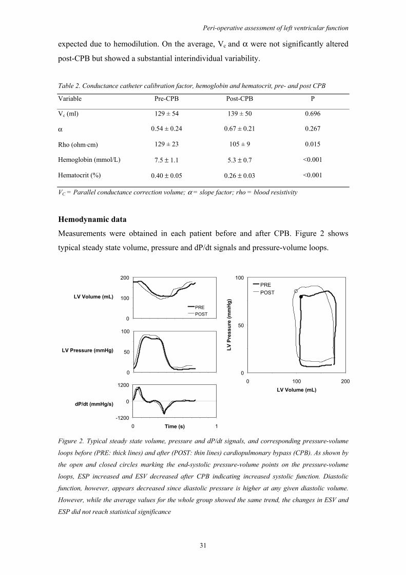
Measurements were obtained in each patient before and after CPB. Figure 2 shows
typical steady state volume, pressure and dP/dt signals and pressure-volume loops.
LV Volume (mL)
LV Pressure (mmHg)
dP/dt (mmHg/s)
0
100
200
PREPOST
0
50
100
-1200
0
1200
0 1Time (s)
0
50
100
0 100 200LV Volume (mL)
LV P
ress
ure
(mm
Hg)
PREPOST
Figure 2. Typical steady state volume, pressure and dP/dt signals, and corresponding pressure-volume
loops before (PRE: thick lines) and after (POST: thin lines) cardiopulmonary bypass (CPB). As shown by
the open and closed circles marking the end-systolic pressure-volume points on the pressure-volume
loops, ESP increased and ESV decreased after CPB indicating increased systolic function. Diastolic
function, however, appears decreased since diastolic pressure is higher at any given diastolic volume.
However, while the average values for the whole group showed the same trend, the changes in ESV and
ESP did not reach statistical significance

Chapter 2
32
Systolic and diastolic pressure-volume relations (ESPVR, EDPVR, PRSW and
dP/dtMAX-EDV) in the same patient derived from pressure-volume loops during caval
vein occlusion are shown in Figure 3.
0
25
50
75
100
0 50 100 150 200LV Volume (mL)
LV P
ress
ure
(mm
Hg)
PRE
POST
0
3500
7000
100 150 200EDV (mL)
SW (m
mH
g.m
L) PREPOST
0
600
1200
100 150 200EDV (mL)
dP/d
t Max
(mm
Hg/
s)
PREPOST
Figure 3. Example of pressure-volume relations derived by caval vein occlusion before and after CPB.
The ESPVRs (left panel) show the increased contractile performance after CPB in this patient: although
Ees is slightly decreased, the position of all end-systolic P-V points to the left and above the pre-CPB
ESPVR suggests higher contractility. The dotted lines indicate the position of the ESPVR at 75-mmHg
(V75). The same holds for the PRSW relation (upper-right panel) and the dP/dtMAX-EDV relation (lower-
right panel) although the differences are much less pronounced. The EDPVRs (left panel) provide clear
evidence for substantial increase in chamber stiffness after CPB, as observed in all patients. As shown in
table 3, the average position and slope of the ESPVR were not significantly altered after CPB in this
group of patients
All patients had sinus rhythm and were paced at 80-90bpm during measurements.
Hemodynamic data are summarized in table 3: Only EDP, Tau and CS changed
significantly post-CPB
DISCUSSION
Assessment of peri-operative ventricular function during cardiac surgery is complicated
by the fact that substantial changes in loading conditions may occur. Therefore the
quantification of systolic and diastolic function requires load-independent indices,
which can be determined from ventricular pressure-volume relations as obtained by the
Peri-operative assessment of left ventricular function
33
conductance catheter. Accordingly, the purpose of this study was twofold: we evaluated
methodological aspects of peri-operative application of the conductance catheter and
documented changes of various indices of LV function pre- and post-CPB in patients
undergoing CABG.
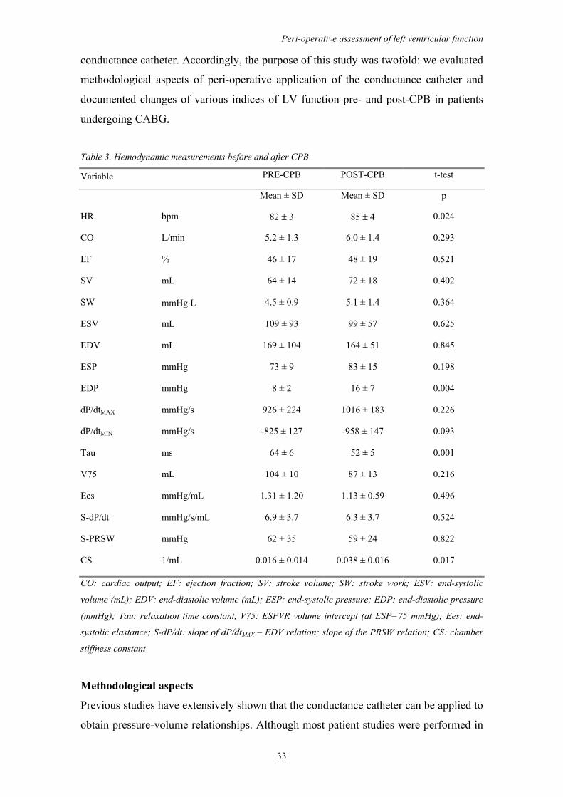
Table 3. Hemodynamic measurements before and after CPB
Variable PRE-CPB POST-CPB t-test
Mean ± SD Mean ± SD p
HR bpm 82 ± 3 85 ± 4 0.024
CO L/min 5.2 ± 1.3 6.0 ± 1.4 0.293
EF % 46 ± 17 48 ± 19 0.521
SV mL 64 ± 14 72 ± 18 0.402
SW mmHg⋅L 4.5 ± 0.9 5.1 ± 1.4 0.364
ESV mL 109 ± 93 99 ± 57 0.625
EDV mL 169 ± 104 164 ± 51 0.845
ESP mmHg 73 ± 9 83 ± 15 0.198
EDP mmHg 8 ± 2 16 ± 7 0.004
dP/dtMAX mmHg/s 926 ± 224 1016 ± 183 0.226
dP/dtMIN mmHg/s -825 ± 127 -958 ± 147 0.093
Tau ms 64 ± 6 52 ± 5 0.001
V75 mL 104 ± 10 87 ± 13 0.216
Ees mmHg/mL 1.31 ± 1.20 1.13 ± 0.59 0.496
S-dP/dt mmHg/s/mL 6.9 ± 3.7 6.3 ± 3.7 0.524
S-PRSW mmHg 62 ± 35 59 ± 24 0.822
CS 1/mL 0.016 ± 0.014 0.038 ± 0.016 0.017
CO: cardiac output; EF: ejection fraction; SV: stroke volume; SW: stroke work; ESV: end-systolic
volume (mL); EDV: end-diastolic volume (mL); ESP: end-systolic pressure; EDP: end-diastolic pressure
(mmHg); Tau: relaxation time constant, V75: ESPVR volume intercept (at ESP=75 mmHg); Ees: end-
systolic elastance; S-dP/dt: slope of dP/dtMAX – EDV relation; slope of the PRSW relation; CS: chamber
stiffness constant
Methodological aspects
Previous studies have extensively shown that the conductance catheter can be applied to
obtain pressure-volume relationships. Although most patient studies were performed in
Chapter 2
34
the catheterization laboratory, several groups have demonstrated feasibility of the
technique in the operating room under various conditions.12-14 Consistent with these
previous studies, our study demonstrates that peri-operative pressure-volume
measurements by the conductance catheter can be used to quantify detailed intrinsic
systolic and diastolic function within an acceptable time-window. Measurements were
uncomplicated and no technical difficulties during instrumentation; catheter placement
and loading interventions were encountered. New technical aspects of our study were
the use of retrograde insertion of the conductance catheter using TEE guidance
compared to the trans-mitral approach used in previous studies in the operating room.
Both approaches may have theoretical advantages and disadvantages: The trans-aortic
approach provides a better match of the catheter position with the LV long axis.
Compared with the anterograde placement this gives a better registration especially of
the volume changes in the basal segments. In contrast anterograde placement through
the mitral valve may complicate interpretation of segmental volume signals because of
changes in the mitral valve plane during ejection and filling. On the other hand with
retrograde placement eccentric (antero-medial) displacement of the catheter at the base
of the heart may occur but the electric field is such that the measurement electrodes will
move approximately parallel to the equipotential planes field and thus the eccentric
movement is unlikely to strongly influence the conductance signal. Another reason for
using the trans-aortic approach is that we aim to apply this methodology in future
studies to evaluate the effects of mitral valve surgery, in which case placement through
the aortic valve is clearly preferable. Furthermore we analyzed the changes in the
calibration factors. As a disadvantage, substantial between-patient variability was found
for calibration factors (rho, α and Vc) indicating the need for careful assessment of these
factors in each individual patient. In addition, after CPB calibration factors rho and, to a
lesser extent α and Vc, were changed due to reduced hematocrit, fluid shifts and
possibly altered catheter position with re-insertion. Although the average α and Vc were
not significantly changed, substantial differences were present in individual patients
indicating that re-assessment is required at the various stages of surgery. Besides
influencing between and within-patient variability, the calibration factors importantly
determine the absolute accuracy of the conductance-derived volumes. Calibration
factors α and Vc are both obtained by means of indicator-dilution methods:
thermodilution and, respectively, saline dilution. Thermodilution is widely used in the
surgical setting and the accuracy is generally found to be acceptable.15 In the present
study we used 'stat' continuous cardiac output measurements using a thermal filament
Peri-operative assessment of left ventricular function
35
catheter which has been shown to have accuracy comparable to the bolus injection
method.16,17 The saline dilution method has been used extensively to obtain parallel
conductance and was found to be accurate with a slight tendency to underestimate
parallel conductance obtained by alternative methods.18 An important advantage of
these indicator-dilution methods compared to imaging modalities such as TEE is that
they do not require assumptions regarding the geometry of the ventricle. This may be
relevant especially when comparing conditions in which geometrical changes would be
anticipated such as after ventricular reconstruction or mitral valve surgery. Furthermore
the inter- and intra-observer variability of indicator-dilution methods is very limited.
Physiological aspects
Our main physiological findings were that systolic function was unchanged after CPB
in these patients undergoing CABG, whereas early relaxation was improved and
diastolic stiffness was increased. Previous pressure-volume studies comparing pre- and
post-CPB cardiac function in patients undergoing CABG have shown conflicting data.
Schreuder et al. reported unchanged systolic function and increased diastolic stiffness,
while Wallace et al. found a decrease in systolic function, but no changes in relaxation
or diastolic stiffness.13,14 Both studies used cold cardioplegia whereas our study was
performed with warm blood cardioplegic arrest, which may explain the preserved
systolic function in our study as compared to the decrease found by Wallace et al. The
unchanged systolic function found by Schreuder et al. may be explained by the fact that
during their pre-CPB measurement the temperature was lowered below 35OC, which
according to a recent study significantly reduces Ees by approximately 50%. 19 Since
the post-CPB measurements in Schreuder's study were done at 37OC this may have
masked an actual reduction in systolic function. With regard to diastolic function all
studies report an increase in diastolic stiffness although in Wallace's study this effect did
not reach statistical significance.14 Also in Schreuder's study the increase was less
pronounced as compared to our study (39% increase vs. 138%).13 However, Schreuder
et al. described the end-diastolic pressure-volume relation as linear, whereas we derived
the diastolic stiffness constant from an exponential relation. The increase is most likely
due to myocardial edema post-CPB as myocardial lymph flow has been shown to
almost cease during cardioplegic arrest.20 De Hert et al. have shown that a more rapid
normalization of diastolic stiffness may be obtained by optimizing preload conditions
prior to weaning from CPB.21 Furthermore, Allen et al. demonstrated that increasing
contractility by dobutamine infusion enhanced myocardial lymphatic function, thus
Chapter 2
36
speeding edema removal post-CPB.22 Thus, for patients who are difficult to wean from
CPB due to increased diastolic stiffness, inotropic support could be considered.
However it should be used with caution because it may adversely affect energetics, raise
heart rate, and induce ischemia.23 In addition several pharmacological substances added
to the cardioplegia composition have been shown to be associated with reduced edema
formation.24-26 Remarkably, although diastolic stiffness was increased, early relaxation
was improved in our study as shown by the significantly reduced Tau. After
revascularization, enhanced oxygen dependent re-uptake of calcium into the
sarcoplasmic reticulum would indeed be expected to improve active relaxation.27 Our
findings are consistent with the results of Humphrey et al. who demonstrated a reduced
Tau post-CPB in patients undergoing CABG.28 In contrast, De Hert et al. found an
increased Tau in a similar patient group.21 Differences may be due to the applied
anesthetic and cardioplegic protocol which influence post-CPB relaxation directly or
indirectly via changes in contractility or loading, which are tightly coupled with
relaxation.23,29 Thus unchanged or even increased Tau as found in some studies may be
related to post-CPB changes in systolic function and/or loading conditions. In our study
EDV, ESP, dP/dtMAX and Ees were not significantly altered after CPB, whereas De Hert
et al. report a reduced dP/dtMAX indicating reduced contractile state.21
Comparison with TEE
As an alternative to invasive volume measurements several groups have used TEE to
obtain on-line area determination.30-33 This method is less invasive but when used to
construct pressure-area loops it still requires a LV catheter for pressure measurements,
and a loading intervention. Schmidlin et al. tested whether pressure-area relations may
be used as a surrogate for pressure-volume relations to detect changes in contractile
state and they concluded that pressure-area analysis provides the same changes as
pressure-volume analysis.33 However the calculations derived from area estimates have
several limitations. During the cardiac cycle the through-plane motion of the LV
complicates volume calculations by short axis area estimates. This effect is even more
prominent during acute loading interventions. On the contrary, the intraventricular
placement of the conductance catheter provides on-line volume measurements of almost
the whole ventricle unaffected by translations or rotations of the heart within the thorax.
In general, on-line area determination by TEE requires optimal image quality and the
stability and reproducibility of measurements is more successful at higher preload
conditions by minimizing effects of tracing errors.31 Area estimates derived during caval
Peri-operative assessment of left ventricular function
37
vein occlusion could become very small thereby decreasing precision of the digital
echocardiographic quantification method for calculation of pressure-area relations. In
addition the precision is reduced in the presence of regional wall motion
abnormalities.30 Conventional assessment of diastolic function by TEE (i.e. without
simultaneous LV pressure measurement) has two disadvantages compared with the
conductance catheter method. First, assessment of both active and passive components
requires two separate TEE views, being the midpapillary esophageal long-axis and
transgastric short-axis view, respectively.32 Second, the active diastolic relaxation
measured by mitral Doppler flow analysis is heart-rate and load-dependent.
In conclusion, despite the above limitations, the limitations of TEE are outweighed by
its proven clinical value to visualize the endoventricular wall and to quantify segmental
wall motion. On the other hand, the important value of the conductance catheter is that
it yields accurate, load-independent quantitative data on basic systolic and diastolic
function. The possibility to measure these fundamental quantities in addition to the data
provided by TEE may prove to be important in selected patient-groups and is ideal to
evaluate e.g. new surgical techniques or anesthetic agents or procedures. The
physiological effects on systolic and diastolic function reported in this study will be
useful reference data for future studies in patients with depressed LV function
undergoing cardiac surgery.
REFERENCES
1. Baan J, van der Velde ET, de Bruin HG, Smeenk GJ, Koops J, van Dijk AD, Temmerman D,
Senden J, Buis B. Continuous measurement of left ventricular volume in animals and humans by conductance catheter. Circulation. 1984;70:812-823.
2. Kass DA, Maughan WL, Guo ZM, Kono A, Sunagawa K, Sagawa K. Comparative influence of load versus inotropic states on indexes of ventricular contractility: experimental and theoretical analysis based on pressure-volume relationships. Circulation. 1987;76:1422-1436.
3. Bovill JG, Sebel PS, Blackburn CL, Oei-Lim V, Heykants JJ. The pharmacokinetics of sufentanil in surgical patients. Anesthesiology. 1984;61:502-506.
4. Coetzee JF, Glen JB, Wium CA, Boshoff L. Pharmacokinetic model selection for target controlled infusions of propofol. Assessment of three parameter sets. Anesthesiology. 1995;82:1328-1345.
5. Minto CF, Schnider TW, Shafer SL. Pharmacokinetics and pharmacodynamics of remifentanil. II. Model application. Anesthesiology. 1997;86:24-33.
6. Steendijk P, van der Velde ET, Baan J. Left ventricular stroke volume by single and dual excitation of conductance catheter in dogs. Am J Physiol. 1993;264:H2198-H2207.
7. Little WC, Cheng CP, Peterson T, Vinten-Johansen J. Response of the left ventricular end-systolic pressure-volume relation in conscious dogs to a wide range of contractile states. Circulation. 1988;78:736-745.
8. Suga H, Sagawa K, Shoukas AA. Load independence of the instantaneous pressure-volume ratio of the canine left ventricle and effects of epinephrine and heart rate on the ratio. Circ Res. 1973;32:314-322.
Chapter 2
38
9. Glower DD, Spratt JA, Snow ND, Kabas JS, Davis JW, Olsen CO, Tyson GS, Sabiston DC, Jr., Rankin JS. Linearity of the Frank-Starling relationship in the intact heart: the concept of preload recruitable stroke work. Circulation. 1985;71:994-1009.
10. Little WC. The left ventricular dP/dtmax-end-diastolic volume relation in closed- chest dogs. Circ Res. 1985;56:808-815.
11. Fransen EJ, Diris JH, Maessen JG, Hermens WT, Dieijen-Visser MP. Evaluation of "new" cardiac markers for ruling out myocardial infarction after coronary artery bypass grafting. Chest. 2002;122:1316-1321.
12. Al Khalidi AH, Townend JN, Bonser RS, Coote JH. Validation of the conductance catheter method for measurement of ventricular volumes under varying conditions relevant to cardiac surgery. Am J Cardiol. 1998;82:1248-1252.
13. Schreuder JJ, Biervliet JD, van der Velde ET, ten Have K, van Dijk AD, Meyne NG, Baan J. Systolic and diastolic pressure-volume relationships during cardiac surgery. J Cardiothorac Vasc Anesth. 1991;5:539-545.
14. Wallace A, Lam HW, Nose PS, Bellows W, Mangano DT. Changes in systolic and diastolic ventricular function with cold cardioplegic arrest in man. The Multicenter Study of Perioperative Ischemia (McSPI) Research Group. J Card Surg. 1994;9:497-502.
15. Stetz CW, Miller RG, Kelly GE, Raffin TA. Reliability of the thermodilution method in the determination of cardiac output in clinical practice. Am Rev Respir Dis. 1982;126:1001-1004.
16. Singh A, Juneja R, Mehta Y, Trehan N. Comparison of continuous, stat, and intermittent cardiac output measurements in patients undergoing minimally invasive direct coronary artery bypass surgery. J Cardiothorac Vasc Anesth. 2002;16:186-190.
17. Sun Q, Rogiers P, Pauwels D, Vincent JL. Comparison of continuous thermodilution and bolus cardiac output measurements in septic shock. Intensive Care Med. 2002;28:1276-1280.
18. Steendijk P, Staal E, Jukema JW, Baan J. Hypertonic saline method accurately determines parallel conductance for dual-field conductance catheter. Am J Physiol Heart Circ Physiol. 2001;281:H755-H763.
19. Lewis ME, Al Khalidi AH, Townend JN, Coote J, Bonser RS. The effects of hypothermia on human left ventricular contractile function during cardiac surgery. J Am Coll Cardiol. 2002;39:102-108.
20. Mehlhorn U, Geissler HJ, Laine GA, Allen SJ. Myocardial fluid balance. Eur J Cardiothorac Surg. 2001;20:1220-1230.
21. De Hert SG, ten Broecke PW, Mertens E, Van Sommeren EW, De Blier IG, Stockman BA, Rodrigus IE. Sevoflurane but not propofol preserves myocardial function in coronary surgery patients. Anesthesiology. 2002;97:42-49.
22. Allen SJ, Geissler HJ, Davis KL, Gogola GR, Warters RD, de Vivie ER, Mehlhorn U. Augmenting cardiac contractility hastens myocardial edema resolution after cardiopulmonary bypass and cardioplegic arrest. Anesth Analg. 1997;85:987-992.
23. Zile MR, Brutsaert DL. New concepts in diastolic dysfunction and diastolic heart failure: Part II: causal mechanisms and treatment. Circulation. 2002;105:1503-1508.
24. Jayawant AM, Stephenson ER, Jr., Damiano RJ, Jr. 2,3-Butanedione monoxime cardioplegia: advantages over hyperkalemia in blood-perfused isolated hearts. Ann Thorac Surg. 1999;67:618-623.
25. Kevelaitis E, Oubenaissa A, Peynet J, Mouas C, Menasche P. Preconditioning by mitochondrial ATP-sensitive potassium channel openers: An effective approach for improving the preservation of heart transplants. Circulation. 1999;100:II345-II350.
26. Tritto FP, Inserte J, Garcia-Dorado D, Ruiz-Meana M, Soler-Soler J. Sodium/hydrogen exchanger inhibition reduces myocardial reperfusion edema after normothermic cardioplegia. J Thorac Cardiovasc Surg. 1998;115:709-715.
27. Halow JM, Figueredo VM, Shames DM, Camacho SA, Baker AJ. Role of slowed Ca(2+) transient decline in slowed relaxation during myocardial ischemia. J Mol Cell Cardiol. 1999;31:1739-1748.
28. Humphrey LS, Topol EJ, Rosenfeld GI, Borkon AM, Baumgartner WA, Gardner TJ, Maruschak G, Weiss JL. Immediate enhancement of left ventricular relaxation by coronary artery bypass grafting: intraoperative assessment. Circulation. 1988;77:886-896.
29. Brutsaert DL, Sys SU, Gillebert TC. Diastolic dysfunction in post-cardiac surgical management. J Cardiothorac Vasc Anesth. 1993;7:18-20.
30. De Hert SG, Rodrigus IE, Haenen LR, De Mulder PA, Gillebert TC. Recovery of systolic and diastolic left ventricular function early after cardiopulmonary bypass. Anesthesiology. 1996;85:1063-1075.
31. Gorcsan J, III, Gasior TA, Mandarino WA, Deneault LG, Hattler BG, Pinsky MR. Assessment of the immediate effects of cardiopulmonary bypass on left ventricular performance by on-line pressure-area relations. Circulation. 1994;89:180-190.

Peri-operative assessment of left ventricular function
39
32. Houltz E, Hellstrom A, Ricksten SE, Wikh R, Caidahl K. Early effects of coronary artery bypass surgery and cold cardioplegic ischemia on left ventricular diastolic function: evaluation by computer- assisted transesophageal echocardiography. J Cardiothorac Vasc Anesth. 1996;10:728-733.
33. Schmidlin D, Aschkenasy S, Vogt PR, Schmidli J, Jenni R, Schmid ER. Left ventricular pressure-area relations as assessed by transoesophageal echocardiographic automated border detection: comparison with conductance catheter technique in cardiac surgical patients. Br J Anaesth. 2000;85:379-388.
Chapter 2
40
LETTER TO THE EDITOR
Left ventricular function after cardiopulmonary bypass is related to the length-
dependent regulation of myocardial function
Stefan G. De Hert, MD, PhD
Philippe J. Van der Linden, MD, PhD
Anesth Analg 2004; 99: 311-312
We read with interest the paper of Tulner and colleagues, in which they reported, in
eight coronary surgery patients, the use of the conductance catheter method for the peri-
operative assessment of left ventricular (LV) function.1 After cardiopulmonary bypass
(CPB), the authors observed a preserved systolic function, an acceleration of LV
pressure fall, and an increase in end-diastolic pressure (EDP). They suggested that these
data may constitute useful reference values for further studies in patients undergoing
cardiac surgery. We think that some caution is indicated with respect to this statement.
Recovery of LV function after CPB is a complex phenomenon and various patterns
have been described over the years, most of them reporting a transient decrease in
cardiac function. Different factors may be responsible for this variability. Apart from
differences in patient population and cardioprotective strategies, specific weaning
procedures and the choice of the anesthetic regimen may also influence post-CPB
myocardial recovery. For instance, early restoration of preload conditions can prevent
the transient depression of both systolic and diastolic dysfunction after weaning from
CPB (ref. 30 in the article by Tulner et al.).2 Similarly, the use of a volatile anesthetic
regimen was associated with a better early recovery of myocardial function than a total
intravenous regimen.3,4
More important however is the individual variability in cardiac functional reserve. It has
been shown in coronary surgery patients that an increase in cardiac load resulted in a
variable hemodynamic response that could not be explained by differences in
preoperative variables. Some patients showed an improvement, whereas other patients
showed either no change or even an impairment of LV function. These patients
developed a decrease in maximal rate of pressure development (dP/dtmax), a delayed
myocardial relaxation (increase in tau) with enhanced load dependence of LV pressure
fall, and a major increase in EDP. These patients showed systolic and diastolic

Letter to the editor
41
dysfunction post-CPB, and necessitated inotropic support to be weaned from CPB.5
This latter response has been attributed to a deficient length-dependent regulation of
myocardial function.6 On the other hand, patients who developed improvement of
myocardial function with an increase in cardiac load (manifested by an increase in
dP/dtmax, an acceleration of LV pressure fall with a decrease in tau, less load
dependence of LV pressure fall and a minor change in EDP), typically showed no (or
only minor) decrease in myocardial function post-CPB.5
In view of these data, it seems that the results reported by Tulner et al. concern a
subgroup of patients with good cardiac functional reserve and an adequate length-
dependent regulation of myocardial function, resulting in a preserved myocardial
function post-CPB. Therefore, this particular response, although present in some
patients, cannot be withheld as the sole reference for the patient population undergoing
coronary surgery with CPB.
REFERENCES
1. Tulner SA, Klautz RJ, Rijk-Zwikker GL, Engbers FH, Bax JJ, Baan J, van der Wall EE, Dion RA,
Steendijk P. Peri-operative assessment of left ventricular function by pressure-volume loops using the conductance catheter method. Anesth Analg. 2003;97:950-7, table.
2. De Hert SG, Rodrigus IE, Haenen LR, De Mulder PA, Gillebert TC. Recovery of systolic and diastolic left ventricular function early after cardiopulmonary bypass. Anesthesiology. 1996;85:1063-1075.
3. De Hert SG, ten Broecke PW, Mertens E, Van Sommeren EW, De Blier IG, Stockman BA, Rodrigus IE. Sevoflurane but not propofol preserves myocardial function in coronary surgery patients. Anesthesiology. 2002;97:42-49.
4. De Hert SG, Cromheecke S, ten Broecke PW, Mertens E, De Blier IG, Stockman BA, Rodrigus IE, Van Der Linden PJ. Effects of propofol, desflurane, and sevoflurane on recovery of myocardial function after coronary surgery in elderly high-risk patients. Anesthesiology. 2003;99:314-323.
5. De Hert SG, Gillebert TC, ten Broecke PW, Mertens E, Rodrigus IE, Moulijn AC. Contraction-relaxation coupling and impaired left ventricular performance in coronary surgery patients. Anesthesiology. 1999;90:748-757.
6. De Hert SG, Gillebert TC, ten Broecke PW, Moulijn AC. Length-dependent regulation of left ventricular function in coronary surgery patients. Anesthesiology. 1999;91:379-387.
Chapter 2
42
IN RESPONSE
We thank De Hert and Van der Linden for their insightful comments on our paper and
we would like to respond on some of the issues brought forward.1 The aim of our study
was two-fold: First to describe our approach to quantify peri-operative LV function, and
second to obtain a reference data set for future studies in patients undergoing cardiac
surgery. The comments of De Hert and Van der Linden focus on the latter aspect of our
study.
We fully agree that the published literature indicates a substantial variability in recovery
of LV function after cardiopulmonary bypass and we acknowledge the extensive list of
possible factors influencing this variable outcome. In fact, this is exactly the reason why
we felt it was necessary to generate a data set that would be applicable to the anesthetic
and cardioprotective approach followed in our institute. Specifically, we use low-dose
target-controlled infusion of propofol, remifentanil and sufentanil, and intermittent
antegrade warm-blood cardioplegic arrest during normothermic cardiopulmonary
bypass. The metabolic advantages of this approach have already been published, but
few data are available on the acute hemodynamic effects. Our study was performed in
patients with relatively preserved LV function undergoing elective CABG, to ensure
that the possible changes in LV function could be contributed mainly to the effects of
anesthesia and cardioplegic arrest, rather than to the surgical intervention. This selection
may partly explain the preserved post-operative systolic function in our patient group.
However, De Hert et al. studied a similar patient group and reported a more variable
outcome that could not be explained by pre-operative LV function.2 Therefore
differences in anesthesia and cardioplegic approaches between our study and the studies
by De Hert et al. may need to be considered. One such difference is the use of
normothermic arrest with blood cardioplegia in our study, whereas the studies of de
Hert et al. included the use of hypothermia and crystalloid cardioplegia. This may be
important because recent studies indicate less myocardial cell damage after
normothermic blood cardioplegia.3 Furthermore, the use of propofol in both studies may
not be comparable because hypothermia has an important influence on propofol
pharmacokinetics.4 However, we certainly agree that extrapolation of our findings to
patients with poor baseline LV function and prolonged cardiac arrest should be done
with caution. But despite this, we would still conclude that the new data provided by our
study constitute valuable background information when interpreting the acute
Letter to the editor: In response
43
hemodynamic effects of complex surgical interventions such as LV reconstruction in
heart failure patients in whom the same anesthesia and cardioplegia approach is used.
Sven A.F. Tulner and Paul Steendijk.
Leiden University Medical Center, Leiden, The Netherlands
REFERENCES
1. Tulner SA, Klautz RJ, Rijk-Zwikker GL, Engbers FH, Bax JJ, Baan J, van der Wall EE, Dion RA,
Steendijk P. Peri-operative assessment of left ventricular function by pressure-volume loops using the conductance catheter method. Anesth Analg. 2003;97:950-7.
2. De Hert SG, Gillebert TC, ten Broecke PW, Mertens E, Rodrigus IE, Moulijn AC. Contraction-relaxation coupling and impaired left ventricular performance in coronary surgery patients. Anesthesiology. 1999;90:748-757.
3. Jacquet LM, Noirhomme PH, Van Dyck MJ, El Khoury GA, Matta AJ, Goenen MJ, Dion RA. Randomized trial of intermittent antegrade warm blood versus cold crystalloid cardioplegia. Ann Thorac Surg. 1999;67:471-477.
4. Leslie K, Sessler DI, Bjorksten AR, Moayeri A. Mild hypothermia alters propofol pharmaco-kinetics and increases the duration of action of atracurium. Anesth Analg. 1995;80:1007-1014.

CHAPTER 3
Quantification of left ventricular mechanical dyssynchrony by
conductance catheter in heart failure patients
P. Steendijk
S.A.F. Tulner
J.J. Schreuder
J.J. Bax
L. van Erven
E.E. van der Wall
R.A.E. Dion
M.J. Schalij
J. Baan
Am J Physiol Heart Circ Physiol 2004; 6: H723-H730
Poster presentation at European Society of Heart Failure 2003, Strasbourg,
Luxembourg
Chapter 3
46
ABSTRACT
Mechanical dyssynchrony is an important co-determinant of cardiac dysfunction in
heart failure. Treatment, either medical, surgical, or by pacing, may improve cardiac
function to a large extent by improving mechanical synchrony. Consequently the
quantification of ventricular mechanical dyssynchrony may have important diagnostic
and prognostic value and may help to determine optimal therapy. Therefore we
introduced new indices to quantify temporal and spatial aspects of mechanical
dyssynchrony derived from on-line segmental conductance catheter signals obtained
during diagnostic cardiac catheterization.
To test the feasibility and usefulness of our approach we determined cardiac function
and left ventricular mechanical dyssynchrony by the conductance catheter in heart
failure patients with intraventricular conduction delay (n=12) and in patients with
coronary artery disease (n=6) and relatively preserved left ventricular function.
The heart failure patients showed depressed systolic and diastolic function. However,
the most marked hemodynamic differences between the groups were found for
mechanical dyssynchrony indicating a high sensitivity and specificity of the new
indices. Comparison of conductance catheter derived indices with septal-to-lateral
dyssynchrony derived by tissue-Doppler velocity imaging showed highly significant
correlations.
The proposed indices provide additional, new and quantitative information on temporal
and spatial aspects of mechanical dyssynchrony. They may refine diagnosis of cardiac
dysfunction and evaluation of interventions, and ultimately help to select optimal
therapy.
INTRODUCTION
In addition to intrinsic myocardial abnormalities and abnormal loading conditions,
cardiac dysfunction in heart failure patients is determined by mechanical
nonuniformities (dyssynchrony), which lead to inefficient pump performance and
energy expenditure. There is increasing evidence that pharmacological, surgical and
pacemaker therapies of heart failure partly exert their beneficial effects by reducing left
ventricular (LV) dyssynchrony. Consequently, quantification of LV dyssynchrony will
provide diagnostic and prognostic data, which should help to select and guide therapy.
Quantification of left ventricular mechanical dyssynchrony
47
Currently, various indices based on magnetic resonance imaging or echocardiographic
measurements are being used. In the present study we introduce indices, which quantify
temporal and spatial aspects of dyssynchrony based on measurements obtained during
cardiac catheterization using conductance catheter methodology. To test the feasibility
and usefulness of our approach we compared data from congestive heart failure (CHF)
patients with left bundle branch block (LBBB) with those from patients with coronary
artery disease (CAD) who had relatively preserved LV function. In addition we
compared the conductance catheter derived dyssynchrony indices with septal to lateral
delay in peak systolic velocity as obtained by tissue-Doppler imaging.
METHODS
Patients
All patients gave informed consent and procedures were conducted in accordance with
institutional guidelines. The investigation conforms with the principles outlined in the
Declaration of Helsinki.1 Twelve CHF patients (NYHA class III/IV) with LBBB were
studied during diagnostic catheterization. Six CAD patients were studied in the
operating room prior to coronary artery bypass grafting.
Protocol
CHF patients underwent diagnostic catheterization including thermodilution cardiac
output, left ventriculography and coronary angiography. In addition, a conductance
catheter was placed in the LV via the femoral artery, and a temporary pacing lead was
positioned in the right atrium.
Prior to catheterization the CHF patients were studied by echocardiography. We
performed tissue-Doppler imaging as described in detail elsewhere2 to determine
myocardial velocities in basal septal and lateral segments. The time delay between peak
systolic velocity in the septum and the lateral wall was determined as an index of
mechanical dyssynchrony.
CAD patients received total intravenous anesthesia with target-controlled infusion of
propofol and remifentanyl (1.5-2μg/ml, resp. 5-10ng/ml blood concentration). A
continuous cardiac output catheter was placed in the pulmonary artery via the jugular
vein. Following midline sternotomy and before starting cardiopulmonary bypass a
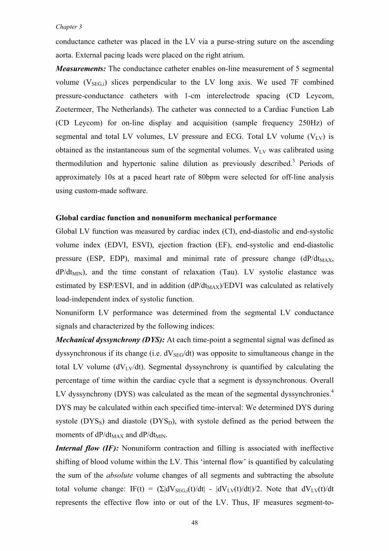
Chapter 3
48
conductance catheter was placed in the LV via a purse-string suture on the ascending
aorta. External pacing leads were placed on the right atrium.
Measurements: The conductance catheter enables on-line measurement of 5 segmental
volume (VSEG,i) slices perpendicular to the LV long axis. We used 7F combined
pressure-conductance catheters with 1-cm interelectrode spacing (CD Leycom,
Zoetermeer, The Netherlands). The catheter was connected to a Cardiac Function Lab
(CD Leycom) for on-line display and acquisition (sample frequency 250Hz) of
segmental and total LV volumes, LV pressure and ECG. Total LV volume (VLV) is
obtained as the instantaneous sum of the segmental volumes. VLV was calibrated using
thermodilution and hypertonic saline dilution as previously described.3 Periods of
approximately 10s at a paced heart rate of 80bpm were selected for off-line analysis
using custom-made software.
Global cardiac function and nonuniform mechanical performance
Global LV function was measured by cardiac index (CI), end-diastolic and end-systolic
volume index (EDVI, ESVI), ejection fraction (EF), end-systolic and end-diastolic
pressure (ESP, EDP), maximal and minimal rate of pressure change (dP/dtMAX,
dP/dtMIN), and the time constant of relaxation (Tau). LV systolic elastance was
estimated by ESP/ESVI, and in addition (dP/dtMAX)/EDVI was calculated as relatively
load-independent index of systolic function.
Nonuniform LV performance was determined from the segmental LV conductance
signals and characterized by the following indices:
Mechanical dyssynchrony (DYS): At each time-point a segmental signal was defined as
dyssynchronous if its change (i.e. dVSEG/dt) was opposite to simultaneous change in the
total LV volume (dVLV/dt). Segmental dyssynchrony is quantified by calculating the
percentage of time within the cardiac cycle that a segment is dyssynchronous. Overall
LV dyssynchrony (DYS) was calculated as the mean of the segmental dyssynchronies.4
DYS may be calculated within each specified time-interval: We determined DYS during
systole (DYSS) and diastole (DYSD), with systole defined as the period between the
moments of dP/dtMAX and dP/dtMIN.
Internal flow (IF): Nonuniform contraction and filling is associated with ineffective
shifting of blood volume within the LV. This ‘internal flow’ is quantified by calculating
the sum of the absolute volume changes of all segments and subtracting the absolute
total volume change: IF(t) = (Σ|dVSEG,i(t)/dt| - |dVLV(t)/dt|)/2. Note that dVLV(t)/dt
represents the effective flow into or out of the LV. Thus, IF measures segment-to-
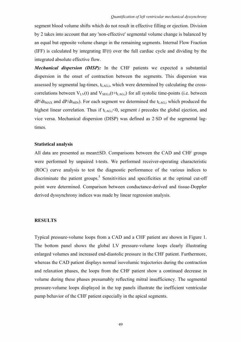
Quantification of left ventricular mechanical dyssynchrony
49
segment blood volume shifts which do not result in effective filling or ejection. Division
by 2 takes into account that any 'non-effective' segmental volume change is balanced by
an equal but opposite volume change in the remaining segments. Internal Flow Fraction
(IFF) is calculated by integrating IF(t) over the full cardiac cycle and dividing by the
integrated absolute effective flow.
Mechanical dispersion (DISP): In the CHF patients we expected a substantial
dispersion in the onset of contraction between the segments. This dispersion was
assessed by segmental lag-times, tLAG,i, which were determined by calculating the cross-
correlations between VLV(t) and VSEG,i(t+tLAG,i) for all systolic time-points (i.e. between
dP/dtMAX and dP/dtMIN). For each segment we determined the tLAG,i which produced the
highest linear correlation. Thus if tLAG,i<0, segment i precedes the global ejection, and
vice versa. Mechanical dispersion (DISP) was defined as 2⋅SD of the segmental lag-
times.
Statistical analysis
All data are presented as mean±SD. Comparisons between the CAD and CHF groups
were performed by unpaired t-tests. We performed receiver-operating characteristic
(ROC) curve analysis to test the diagnostic performance of the various indices to
discriminate the patient groups.5 Sensitivities and specificities at the optimal cut-off
point were determined. Comparison between conductance-derived and tissue-Doppler
derived dyssynchrony indices was made by linear regression analysis.
RESULTS
Typical pressure-volume loops from a CAD and a CHF patient are shown in Figure 1.
The bottom panel shows the global LV pressure-volume loops clearly illustrating
enlarged volumes and increased end-diastolic pressure in the CHF patient. Furthermore,
whereas the CAD patient displays normal isovolumic trajectories during the contraction
and relaxation phases, the loops from the CHF patient show a continued decrease in
volume during these phases presumably reflecting mitral insufficiency. The segmental
pressure-volume loops displayed in the top panels illustrate the inefficient ventricular
pump behavior of the CHF patient especially in the apical segments.
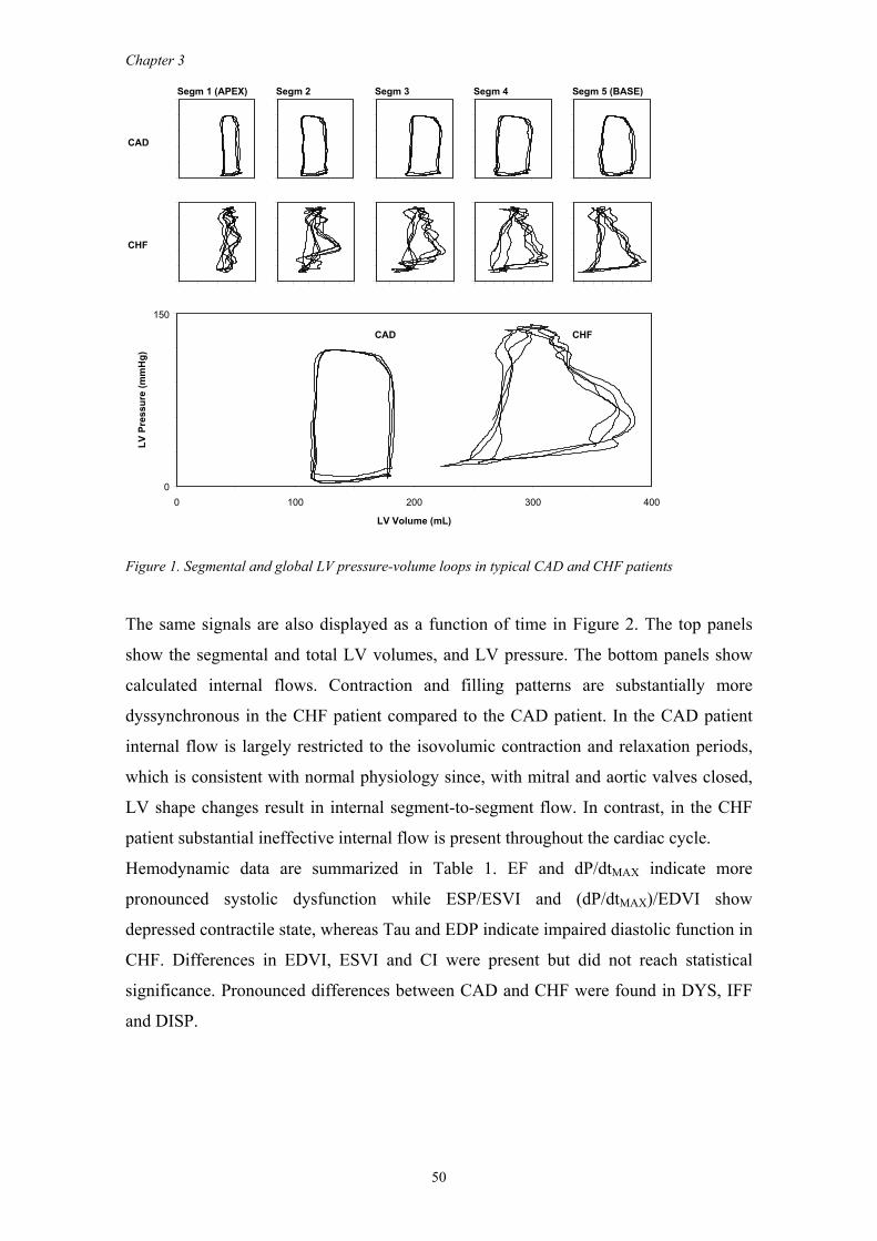
Chapter 3
50
Segm 1 (APEX) Segm 2 Segm 3 Segm 4 Segm 5 (BASE)
CAD
CHF
CAD CHF
0
150
0 100 200 300 400
LV Volume (mL)
LV P
ress
ure
(mm
Hg)
Figure 1. Segmental and global LV pressure-volume loops in typical CAD and CHF patients
The same signals are also displayed as a function of time in Figure 2. The top panels
show the segmental and total LV volumes, and LV pressure. The bottom panels show
calculated internal flows. Contraction and filling patterns are substantially more
dyssynchronous in the CHF patient compared to the CAD patient. In the CAD patient
internal flow is largely restricted to the isovolumic contraction and relaxation periods,
which is consistent with normal physiology since, with mitral and aortic valves closed,
LV shape changes result in internal segment-to-segment flow. In contrast, in the CHF
patient substantial ineffective internal flow is present throughout the cardiac cycle.
Hemodynamic data are summarized in Table 1. EF and dP/dtMAX indicate more
pronounced systolic dysfunction while ESP/ESVI and (dP/dtMAX)/EDVI show
depressed contractile state, whereas Tau and EDP indicate impaired diastolic function in
CHF. Differences in EDVI, ESVI and CI were present but did not reach statistical
significance. Pronounced differences between CAD and CHF were found in DYS, IFF
and DISP.

Quantification of left ventricular mechanical dyssynchrony
51
CAD DYS (%) CHF DYS (%)
apex 33.0 36.6
21.6 34.8
mid 16.3 35.4
10.8 32.9
base 16.1 29.4
mean meanVLV 19.6 33.8
PLV
IFF (%) IFF (%)
Internal 17.5 105.3Flow
time (s) time (s)
Figure 2. Typical examples of segmental and total LV volume signals and calculated internal flow in
CAD and CHF patients. DYS: mechanical dyssynchrony; IFF: internal flow fraction
For both groups dyssynchrony and internal flows were highest in diastole, and the
apical segments were the most affected (Figure 3). In both groups mechanical
dispersion in the long-axis direction was present, but it was twice as large in CHF.
Figure 3 (right panel) shows that contraction started in the basal segment and, on the
average, subsequent segments (1cm-slices) followed after 5.9ms for CAD and after
12.4ms in CHF patients.
The diagnostic value of the various indices to discriminate the two patient groups was
tested using ROC analysis. Table 1 shows the results with the optimal cut-off values,
and corresponding sensitivities and specificities. As expected QRS duration accurately
delineates the groups with a cut-off value of 107ms. The dyssynchrony indices DYS
and IFF show excellent sensitivity/specificity values, which are higher than the best
hemodynamic indices EF and dP/dtMAX. The other hemodynamic indices show lower
sensitivity/specificity reflecting a substantial overlap of the values between the two
groups.
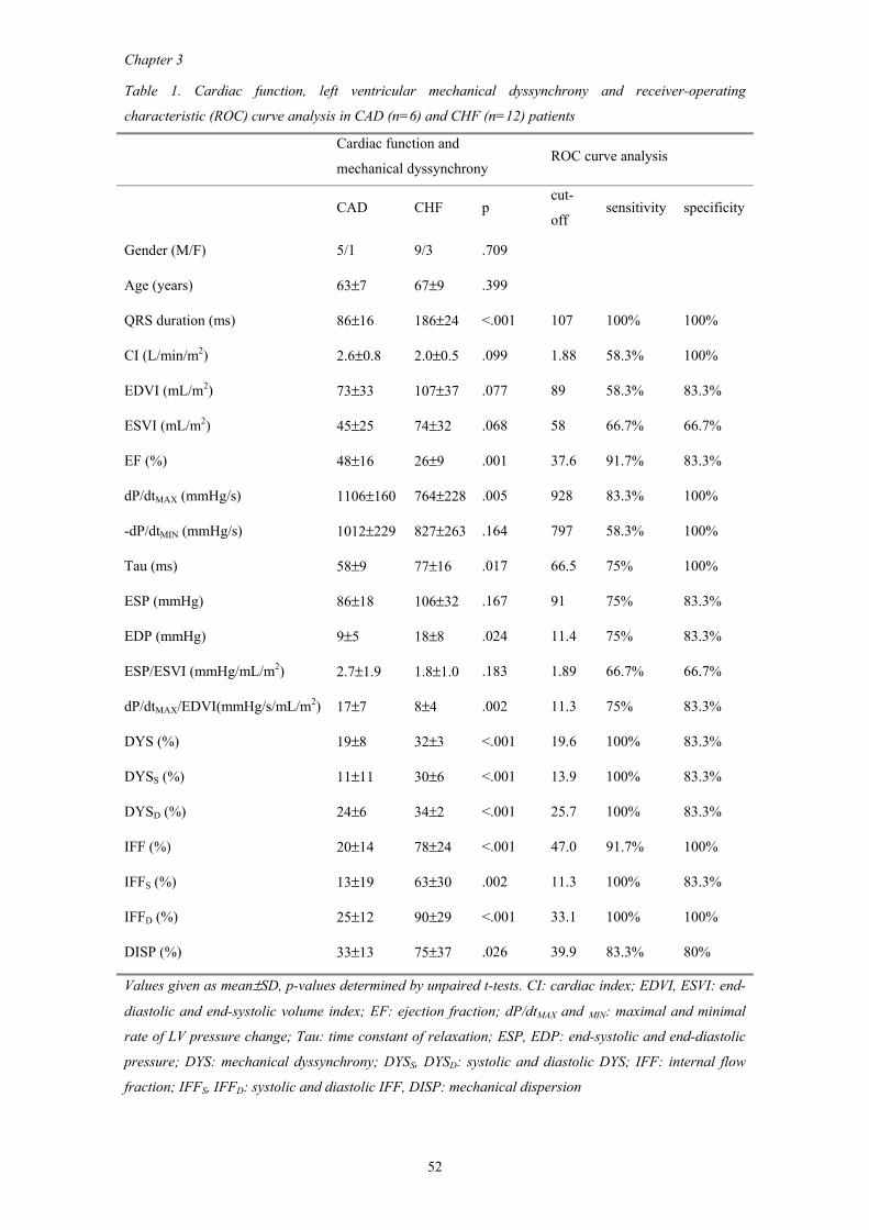
Chapter 3
52
Table 1. Cardiac function, left ventricular mechanical dyssynchrony and receiver-operating
characteristic (ROC) curve analysis in CAD (n=6) and CHF (n=12) patients
Cardiac function and
mechanical dyssynchrony ROC curve analysis
CAD CHF p cut-
off sensitivity specificity
Gender (M/F) 5/1 9/3 .709
Age (years) 63±7 67±9 .399
QRS duration (ms) 86±16 186±24 <.001 107 100% 100%
CI (L/min/m2) 2.6±0.8 2.0±0.5 .099 1.88 58.3% 100%
EDVI (mL/m2) 73±33 107±37 .077 89 58.3% 83.3%
ESVI (mL/m2) 45±25 74±32 .068 58 66.7% 66.7%
EF (%) 48±16 26±9 .001 37.6 91.7% 83.3%
dP/dtMAX (mmHg/s) 1106±160 764±228 .005 928 83.3% 100%
-dP/dtMIN (mmHg/s) 1012±229 827±263 .164 797 58.3% 100%
Tau (ms) 58±9 77±16 .017 66.5 75% 100%
ESP (mmHg) 86±18 106±32 .167 91 75% 83.3%
EDP (mmHg) 9±5 18±8 .024 11.4 75% 83.3%
ESP/ESVI (mmHg/mL/m2) 2.7±1.9 1.8±1.0 .183 1.89 66.7% 66.7%
dP/dtMAX/EDVI(mmHg/s/mL/m2) 17±7 8±4 .002 11.3 75% 83.3%
DYS (%) 19±8 32±3 <.001 19.6 100% 83.3%
DYSS (%) 11±11 30±6 <.001 13.9 100% 83.3%
DYSD (%) 24±6 34±2 <.001 25.7 100% 83.3%
IFF (%) 20±14 78±24 <.001 47.0 91.7% 100%
IFFS (%) 13±19 63±30 .002 11.3 100% 83.3%
IFFD (%) 25±12 90±29 <.001 33.1 100% 100%
DISP (%) 33±13 75±37 .026 39.9 83.3% 80%
Values given as mean±SD, p-values determined by unpaired t-tests. CI: cardiac index; EDVI, ESVI: end-
diastolic and end-systolic volume index; EF: ejection fraction; dP/dtMAX and MIN: maximal and minimal
rate of LV pressure change; Tau: time constant of relaxation; ESP, EDP: end-systolic and end-diastolic
pressure; DYS: mechanical dyssynchrony; DYSS, DYSD: systolic and diastolic DYS; IFF: internal flow
fraction; IFFS, IFFD: systolic and diastolic IFF, DISP: mechanical dispersion
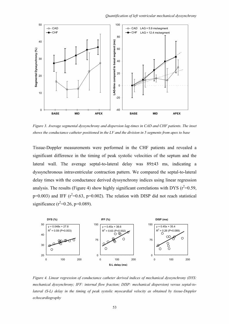
Quantification of left ventricular mechanical dyssynchrony
53
0
10
20
30
40
50
BASE MID APEX
Segm
enta
l Dys
sync
hron
y (%
)
CADCHF LAG = 12.4 ms/segment
LAG = 5.9 ms/segment
-40
-20
0
20
40
60
80
100
BASE MID APEXLA
G-ti
me
com
pare
d to
bas
al s
egm
ent (
ms)
CADCHF
Figure 3. Average segmental dyssynchrony and dispersion lag-times in CAD and CHF patients. The inset
shows the conductance catheter positioned in the LV and the division in 5 segments from apex to base
Tissue-Doppler measurements were performed in the CHF patients and revealed a
significant difference in the timing of peak systolic velocities of the septum and the
lateral wall. The average septal-to-lateral delay was 89±43 ms, indicating a
dyssynchronous intraventricular contraction pattern. We compared the septal-to-lateral
delay times with the conductance derived dyssynchrony indices using linear regression
analysis. The results (Figure 4) show highly significant correlations with DYS (r2=0.59,
p=0.003) and IFF (r2=0.63, p=0.002). The relation with DISP did not reach statistical
significance (r2=0.26, p=0.089).
DYS (%) IFF (%) DISP (ms)
S-L delay (ms)
y = 0.048x + 27.8R2 = 0.59 (P=0.003)
20
30
40
50
0 100 200
y = 0.45x + 38.6R2 = 0.63 (P=0.002)
0
75
150
0 100 200
y = 0.45x + 35.4R2 = 0.26 (P=0.089)
0
75
150
0 100 200
Figure 4. Linear regression of conductance catheter derived indices of mechanical dyssynchrony (DYS:
mechanical dyssynchrony; IFF: internal flow fraction; DISP: mechanical dispersion) versus septal-to-
lateral (S-L) delay in the timing of peak systolic myocardial velocity as obtained by tissue-Doppler
echocardiography
Chapter 3
54
DISCUSSION
Dyssynchrony plays a regulating role already in normal physiology, but is especially
important in pathological conditions such as hypertrophy, ischemia, infarction, or heart
failure.6,7,8,9,10 Currently, cardiac resynchronization by biventricular pacing is emerging
as an important therapy for heart failure.11,12 Recently, MRI and echocardiography have
been used to visualize mechanical dyssynchrony, further emphasizing the important role
of mechanical dyssynchrony in cardiac dysfunction.10,13,14-18 However, these methods
are laborious and require substantial operator interaction and expertise.
We introduce novel indices to quantify dyssynchrony based on volume signals acquired
with the conductance catheter during cardiac catheterization. The conductance catheter
was validated previously and the segmental signals reflect instantaneous volume slices
perpendicular to the LV long-axis as obtained by cine-CT.3,19 Currently, the
conductance catheter is used mainly to assess global systolic and diastolic function.20-23
Quantification of nonuniform mechanical function and dyssynchrony may lead to a
more complete diagnosis of ventricular dysfunction.24,25 Moreover, it may guide
therapy, since patients with extensive dyssynchrony are likely to benefit from
resynchronization therapy.26
We compared CHF versus CAD patients. The groups show pronounced differences for
DYS, IFF and DISP, which indicates a high sensitivity and specificity of these
dyssynchrony indices. QRS duration, dP/dtMAX and Tau show a similar discrimination
between the groups and may also partly reflect dyssynchrony. However, whereas the
conductance catheter derived indices directly measure regional mechanical events
throughout the cardiac cycle, QRS duration reflects the underlying electrical activation
and studies indicate that mechanical and electrical synchrony may diverge.27 Tau and
dP/dtMAX have also been shown to be markers of dyssynchrony but they more indirectly
reflect the integrated effects of spatially dispersed mechanical (de)activation during the
isovolumic relaxation and contraction periods. Dyssynchrony is likely to be most
pronounced in the isovolumic phases, which explains the sensitivity of parameters that
reflect these periods. However, the consequences of dyssynchrony on the effectiveness
of ejection and filling are important for cardiac pump performance, so that indices
selectively reflecting those cardiac phases may be of high value.
Quantification of left ventricular mechanical dyssynchrony
55
In the CHF patients we compared the conductance derived dyssynchrony indices with
the delay in timing of peak systolic velocity between the septal and lateral wall as
obtained by tissue-Doppler echocardiography. Septal-to-lateral delay has recently been
introduced as an index of mechanical dyssynchrony. We found a significant correlation
for both DYS and IFF, but DISP did not reach a statistically significant correlation. The
various indices measure different characteristics. Whereas the tissue-Doppler method
compares the timing of peak velocity between two regions that are likely to show the
largest phase shift, the conductance-derived indices are based on a comparison of the
volume changes of short axis slices and global LV volume changes. Apparently patients
with a larger septal-to-lateral delay also show more segmental dyssynchrony as
reflected by DISP and IFF. Whether this correspondence is specific for LBBB-CHF
patients or is more generally valid requires further study. The lack of correlation with
DISP is unclear. It may be because the index is less sensitive than DISP or IFF as
shown in the comparison between CAD and CHF patients, or the index may inherently
be more prone to errors. Interestingly, within the group of CHF patients neither septal-
to-lateral delay nor the conductance derived indices showed a significant correlation
with QRS duration (Figure 5). This finding is consistent with other reports indicating
that electrical dyssynchrony does not necessarily predict mechanical dyssynchrony,
which prompts a need for methods to accurately detect mechanical dyssynchrony.10,28
S-L delay (ms) DYS (%) IFF (%) DISP (ms)
QRS duration (ms)
R2 = 0.044
0
100
200
150 200 250
R2 = 0.020
0
75
150
150 200 250
R2 = 0.021
20
30
40
50
150 200 250
R2 = 0.097
0
75
150
150 200 250
Figure 5. Linear regression of indices of mechanical dyssynchrony (S-L delay: septal-to-lateral delay of
peak systolic velocity obtained by tissue-Doppler echocardiography; DYS: mechanical dyssynchrony;
IFF: internal flow fraction; DISP: mechanical dispersion) vs QRS duration as index of electrical
dyssynchrony
Our approach may offer several technical advantages. After catheter placement, the
signals are obtained continuously without operator interaction. In the present study the
analysis was performed off-line, but real-time display of dyssynchrony indices is
technically feasible and should enable immediate quantification of the effects of
Chapter 3
56
interventions and, e.g., effects of changes in pacemaker settings. The method is
invasive, but positioning of the catheter in the LV largely eliminates problems with
through-plane motion inherent in most imaging methods. Heart failure is often
associated with substantial beat-to-beat hemodynamic variations due to changes in cycle
length, cardio-pulmonary interaction and conduction disturbances. Thus, techniques -
like MRI- that require hemodynamic steady state and beat-averaging to increase signal-
to-noise may filter out important components of dyssynchrony. Furthermore, the
temporal resolution of the conductance signals (4ms) is relatively high.
Determination of absolute LV volume from the conductance catheter requires careful
calibration.3 In the present study calibration factor parallel conductance was obtained by
the hypertonic saline method and slope factor alpha by thermodilution. Slope factor
alpha was significantly lower in the CHF patients than in the CAD patients (0.38±0.22
vs 0.67±0.08, p<0.01) and parallel conductance was significantly higher (214±60 vs
131±48mL, p<0.01). These findings are consistent with previous studies and reflect
more electrical field inhomogeneity in the enlarged hearts in the CHF group. However,
the conductance catheter has been used extensively in enlarged hearts and validation
studies show that accurate volumes estimates can be obtained provided that appropriate
calibration is performed.29 As an advantage the dyssynchrony indices can be calculated
from the raw segmental conductance signals and do not require calibration. Correction
for parallel conductance (offset factor) is not required because the calculations are based
on volume changes, and correction for slope factor alpha is not required because
segmental volume changes are judged relative to the global LV volume changes. The
latter however implicitly assumes that the segmental slope factors are all the same (and
thus equal to the slope factor for global volume). This assumption may be a concern
because theoretical studies indicate that volume in the segments closest to the current
electrodes may be relatively underestimated due to electric field inhomogeneity
especially in enlarged hearts.30-32 To test the effects of such underestimations, if present,
on our dyssynchrony indices we recalculated DYS, IFF and DISP after correcting
segments 1 and 5 for an assumed underestimation of 20% and segments 2 and 4 for an
assumed underestimation of 10%. Theoretical studies indicate that underestimation in
this order of magnitude may be present.30,32 The results were compared with the original
data using Bland-Altman analysis (Figure 6).33
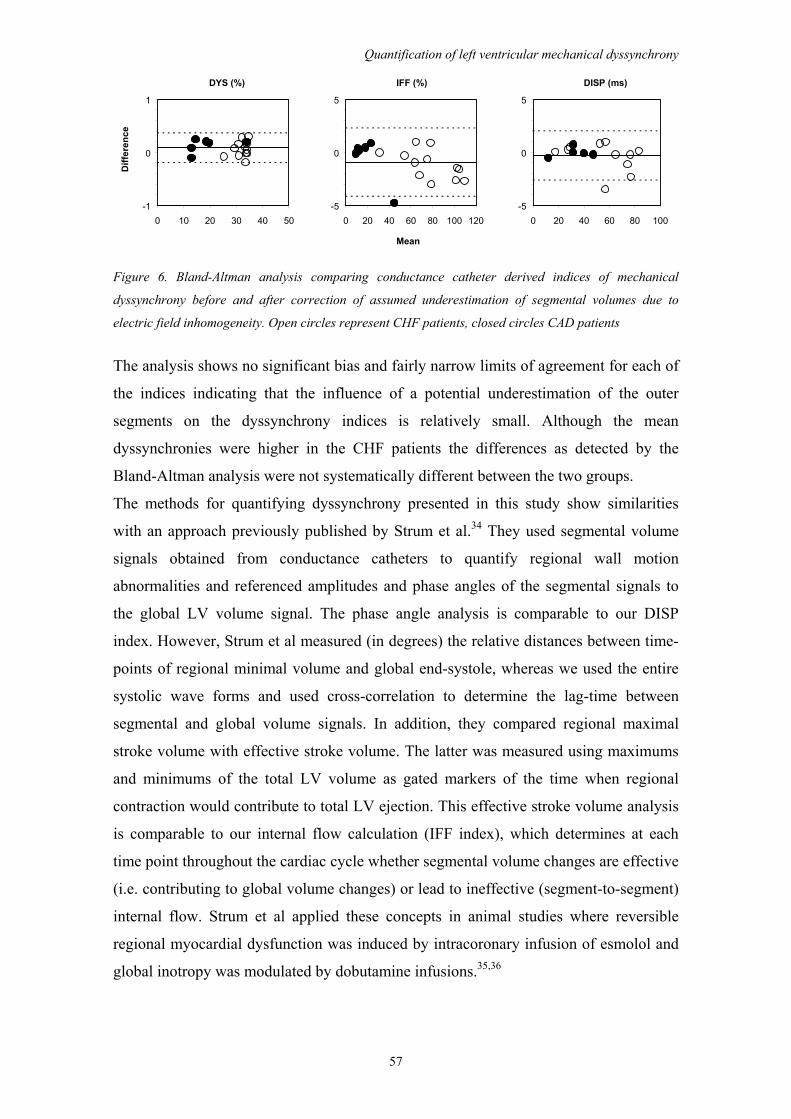
Quantification of left ventricular mechanical dyssynchrony
57
DYS (%) IFF (%) DISP (ms)
Mean
-1
0
1
0 10 20 30 40 50
Diff
eren
ce
-5
0
5
0 20 40 60 80 100 120-5
0
5
0 20 40 60 80 100
Figure 6. Bland-Altman analysis comparing conductance catheter derived indices of mechanical
dyssynchrony before and after correction of assumed underestimation of segmental volumes due to
electric field inhomogeneity. Open circles represent CHF patients, closed circles CAD patients
The analysis shows no significant bias and fairly narrow limits of agreement for each of
the indices indicating that the influence of a potential underestimation of the outer
segments on the dyssynchrony indices is relatively small. Although the mean
dyssynchronies were higher in the CHF patients the differences as detected by the
Bland-Altman analysis were not systematically different between the two groups.
The methods for quantifying dyssynchrony presented in this study show similarities
with an approach previously published by Strum et al.34 They used segmental volume
signals obtained from conductance catheters to quantify regional wall motion
abnormalities and referenced amplitudes and phase angles of the segmental signals to
the global LV volume signal. The phase angle analysis is comparable to our DISP
index. However, Strum et al measured (in degrees) the relative distances between time-
points of regional minimal volume and global end-systole, whereas we used the entire
systolic wave forms and used cross-correlation to determine the lag-time between
segmental and global volume signals. In addition, they compared regional maximal
stroke volume with effective stroke volume. The latter was measured using maximums
and minimums of the total LV volume as gated markers of the time when regional
contraction would contribute to total LV ejection. This effective stroke volume analysis
is comparable to our internal flow calculation (IFF index), which determines at each
time point throughout the cardiac cycle whether segmental volume changes are effective
(i.e. contributing to global volume changes) or lead to ineffective (segment-to-segment)
internal flow. Strum et al applied these concepts in animal studies where reversible
regional myocardial dysfunction was induced by intracoronary infusion of esmolol and
global inotropy was modulated by dobutamine infusions.35,36
Chapter 3
58
Limitations
Optimally the conductance catheter is placed in a straight position from the aortic valve
to the LV apex. In the operating room we used transesophageal echocardiography and in
the catheterization laboratory we used angiography to guide positioning.37 However
occasionally arrhythmias necessitate pulling back the catheter slightly from the apical
position. In addition the distance from the pigtail to the first measurement electrode is
approximately 2 cm. Thus volume changes in the most apical part of the LV are not
measured. If this region is highly dyssynchronous, as might be the case in patients with
apical infarcts, underestimation of dyssynchrony by our methodology may be expected.
The patient groups in our study were investigated under different conditions. For
practical purposes we studied the CAD patients in the operating room during anesthesia
and after sternotomy, whereas the CHF patients were awake and studied in the
catheterization laboratory. These differences may have affected the comparisons
between the two groups. Propofol-remifentanyl anesthesia is known to have myocardial
depressant and vasodilating properties, whereas sternotomy and pericardiotomy are
associated with alterations in loading conditions.38,39,40 Given the anesthesia-related
cardiodepression in the CAD patients, one may expect that the differences in the
hemodynamic indices would have been more pronounced in case both groups had been
studied awake. Whether these changes affect the level of dyssynchrony is not well
known, but studies in dogs with regional stunning show unchanged LV wall asynchrony
after systemic inotropic stimulation.41 Thus we do not expect that the differences in
mechanical dyssynchrony between the groups were importantly influenced by the
different experimental conditions.
Furthermore, we did not study normal subjects. Thus, future studies are required to
establish a 'normal' range for the dyssynchrony indices.
Finally, the segmental conductance catheter signals do not provide an anatomical view
but represent the total volume of slices perpendicular to the LV long-axis. Thus, e.g. in
CAD patients, abnormal regional wall motion might be obscured by compensatory wall
motions within the same circumferential segment. The proposed dyssynchrony indices
therefore reflect intersegmental differences in contraction and filling and may
underestimate phase changes obtained by comparing regional lateral and septal wall
motions, e.g. using tissue Doppler imaging.
Quantification of left ventricular mechanical dyssynchrony
59
In conclusion, the proposed indices quantify various aspects of mechanical
dyssynchrony using conductance catheter methodology which, at the same time, can be
used for assessment of global systolic and diastolic (dys)function. Diagnostic and
prognostic value of the dyssynchrony indices requires further investigation.
REFERENCES 1. World Medical Association Declaration of Helsinki. Recommendations guiding physicians in
biomedical research involving human subjects. Cardiovasc Res. 1997;35:2-3. 2. Bax JJ, Molhoek SG, van Erven L, Voogd PJ, Somer S, Boersma E, Steendijk P, Schalij MJ, van
der Wall EE. Usefulness of myocardial tissue Doppler echocardiography to evaluate left ventricular dyssynchrony before and after biventricular pacing in patients with idiopathic dilated cardiomyopathy. Am J Cardiol. 2003;91:94-97.
3. Baan J, Van Der Velde ET, De Bruin H, Smeenk G, Koops J, Van Dijk AD, Temmerman D, Senden J, Buis B. Continuous measurement of left ventricular volume in animals and humans by conductance catheter. Circulation. 1984;70:812-823.
4. Schreuder JJ, Steendijk P, Van der Veen FH, Alfieri O, Van der Nagel T, Lorusso R, van Dantzig JM, Prenger KB, Baan J, Wellens HJ, Batista RJ. Acute and short-term effects of partial left ventriculectomy in dilated cardiomyopathy: assessment by pressure-volume loops.[In Process Citation]. J Am Coll Cardiol. 2000;36:2104-2114.
5. Zweig MH, Campbell G. Receiver-operating characteristic (ROC) plots: a fundamental evaluation tool in clinical medicine. Clin Chem. 1993;39:561-577.
6. Brutsaert DL. Nonuniformity: a physiologic modulator of contraction and relaxation of the normal heart. J Am Coll Cardiol. 1987;9:341-8.
7. Villari B, Vassalli G, Betocchi S, Briguori C, Chiariello M, Hess OM. Normalization of left ventricular nonuniformity late after valve replacement for aortic stenosis. Am J Cardiol. 1996;78:66-71.
8. Heyndrickx GR, Paulus WJ. Effect of asynchrony on left ventricular relaxation. Circulation. 1990;81:41-7.
9. Gepstein L, Goldin A, Lessick J, Hayam G, Shpun S, Schwartz Y, Hakim G, Shofty R, Turgeman A, Kirshenbaum D, Ben-Haim SA. Electromechanical characterization of chronic myocardial infarction in the canine coronary occlusion model. Circulation. 1998;98:2055-64.
10. Nelson GS, Curry CW, Wyman BT, Kramer A, Declerck J, Talbot M, Douglas MR, Berger RD, McVeigh ER, Kass DA. Predictors of systolic augmentation from left ventricular preexcitation in patients with dilated cardiomyopathy and intraventricular conduction delay. Circulation. 2000;101:2703-9.
11. Saxon LA, De Marco T. Cardiac resynchronization: a cornerstone in the foundation of device therapy for heart failure*. J Am Coll Cardiol. 2001;38:1971-1973.
12. Leclercq C, Kass DA. Retiming the failing heart: principles and current clinical status of cardiac resynchronization. J Am Coll Cardiol. 2002;39:194-201.
13. Wyman BT, Hunter WC, Prinzen FW, Faris OP, McVeigh ER. Effects of single- and biventricular pacing on temporal and spatial dynamics of ventricular contraction. Am J Physiol Heart Circ Physiol. 2002;282:H372-H379.
14. Ansalone G, Giannantoni P, Ricci R, Trambaiolo P, Fedele F, Santini M. Doppler myocardial imaging to evaluate the effectiveness of pacing sites in patients receiving biventricular pacing. J Am Coll Cardiol. 2002;39:489-499.
15. Breithardt OA, Stellbrink C, Kramer AP, Sinha AM, Franke A, Salo R, Schiffgens B, Huvelle E, Auricchio A. Echocardiographic quantification of left ventricular asynchrony predicts an acute hemodynamic benefit of cardiac resynchronization therapy. J Am Coll Cardiol. 2002;40:536-545.
16. Kawaguchi M, Murabayashi T, Fetics BJ, Nelson GS, Samejima H, Nevo E, Kass DA. Quantitation of basal dyssynchrony and acute resynchronization from left or biventricular pacing by novel echo-contrast variability imaging. J Am Coll Cardiol. 2002;39:2052-2058.
17. Sogaard P, Egeblad H, Pedersen AK, Kim WY, Kristensen BO, Hansen PS, Mortensen PT. Sequential versus simultaneous biventricular resynchronization for severe heart failure: evaluation by tissue Doppler imaging. Circulation. 2002;106:2078-2084.
18. Yu CM, Chau E, Sanderson JE, Fan K, Tang MO, Fung WH, Lin H, Kong SL, Lam YM, Hill MR, Lau CP. Tissue Doppler echocardiographic evidence of reverse remodeling and improved
Chapter 3
60
synchronicity by simultaneously delaying regional contraction after biventricular pacing therapy in heart failure. Circulation. 2002;105:438-445.
19. Van Der Velde ET, Van Dijk AD, Steendijk P, Diethelm L, Chagas T, Lipton MJ, Glantz SA, Baan J. Left ventricular segmental volume by conductance catheter and Cine-CT. Eur Heart J. 1992;13 (Suppl E):15-21.
20. Hayward CS, Kalnins WV, Kelly RP. Gender-related differences in left ventricular chamber function. Cardiovasc Res. 2001;49:340-350.
21. Kass DA, Grayson R, Marino P. Pressure-volume analysis as a method for quantifying simultaneous drug (amrinone) effects on arterial load and contractile state in vivo. J Am Coll Cardiol. 1990;16:726-732.
22. Kass DA, Midei M, Graves W, Brinker JA, Maughan WL. Use of a conductance (volume) catheter and transient inferior vena caval occlusion for rapid determination of pressure-volume relationships in man. Cathet Cardiovasc Diagn. 1988;15:192-202.
23. MacGowan GA, Haber HL, Cowart TD, Tedesco C, Wu C, Feldman MD. Direct myocardial effects of OPC-18790 in human heart failure: beneficial effects on contractile and diastolic function demonstrated by intracoronary infusion with pressure-volume analysis. J Am Coll Cardiol. 1998;31:1344-1351.
24. Schreuder JJ, Van der Veen FH, Van Der Velde ET, Delahaye F, Alfieri O, Jegaden O, Lorusso R, Jansen JRC, Hoeksel SAA, Finet G, Volterrani M, Kaulbach HG, Baan J, Wellens HJJ. Left ventricular pressure volume relationships before and after cardiomyoplasty in patients with heart failure. Circulation. 1997;96:2978-2986.
25. Schreuder JJ, Steendijk P, Van der Veen FH, Alfieri O, Van der Nagel T, Lorusso R, van Dantzig JM, Prenger KB, Baan J, Wellens HJ, Batista RJ. Acute and short-term effects of partial left ventriculectomy in dilated cardiomyopathy: assessment by pressure-volume loops.[In Process Citation]. J Am Coll Cardiol. 2000;36:2104-2114.
26. Leclercq C, Kass DA. Retiming the failing heart: principles and current clinical status of cardiac resynchronization. J Am Coll Cardiol. 2002;39:194-201.
27. Leclercq C, Faris O, Tunin R, Johnson J, Kato R, Evans F, Spinelli J, Halperin H, McVeigh E, Kass DA. Systolic improvement and mechanical resynchronization does not require electrical synchrony in the dilated failing heart with left bundle- branch block. Circulation. 2002;106:1760-1763.
28. Leclercq C, Faris O, Tunin R, Johnson J, Kato R, Evans F, Spinelli J, Halperin H, McVeigh E, Kass DA. Systolic improvement and mechanical resynchronization does not require electrical synchrony in the dilated failing heart with left bundle- branch block. Circulation. 2002;106:1760-1763.
29. Odake M, Takeuchi M, Takaoka H, Hata K, Hayashi Y, Yokoyama M. Determination of left ventricular volume using a conductance catheter in the diseased human heart. Eur Heart J. 1992;13 Suppl E:22-27.
30. Steendijk P, Van Der Velde ET, Baan J. Single and dual excitation of the conductance-volume catheter analysed in a spheroidal mathematical model of the canine left ventricle. Eur Heart J. 1992;13 (Suppl E):28-34.
31. Steendijk P, Van Der Velde ET, Baan J. Left ventricular stroke volume by single and dual excitation of conductance catheter in dogs. Am J Physiol. 1993;264 (Heart Circ Physiol 33):H2198-H2207.
32. Wu CC, Skalak TC, Schwenk TR, Mahler CM, Anne A, Finnerty PW, Haber HL, Weikle RM2, Feldman MD. Accuracy of the conductance catheter for measurement of ventricular volumes seen clinically: effects of electric field homogeneity and parallel conductance. IEEE-Trans-Biomed-Eng. 1997;44:266-77.
33. Bland JM, Altman DG. Statistical methods for assessing agreement between two methods of clinical measurement. Lancet. 1986;1:307-10.
34. Strum DP, Pinsky MR. Modeling of asynchronous myocardial contraction by effective stroke volume analysis. Anesthesia & Analgesia. 2000;90:243-51.
35. Strum DP, Pinsky MR. Esmolol-induced regional wall motion abnormalities do not affect regional ventricular elastances. Anesthesia & Analgesia. 2000;90:252-61.
36. Strum DP, Pinsky MR. Does dobutamine improve ventricular function in dogs with regional myocardial dysfunction? Anesth Analg. 2002;95:19-25, table.
37. Tulner SA, Klautz RJ, Rijk-Zwikker GL, Engbers FH, Bax JJ, Baan J, van der Wall EE, Dion RA, Steendijk P. Peri-operative assessment of left ventricular function by pressure-volume loops using the conductance catheter method. Anesth Analg. 2003;97:950-957.
38. Lehmann A, Boldt J, Rompert R, Thaler E, Kumle B, Weisse U. Target-controlled infusion or manually controlled infusion of propofol in high-risk patients with severely reduced left ventricular function. J Cardiothorac Vasc Anesth. 2001;15:445-450.

Quantification of left ventricular mechanical dyssynchrony
61
39. Schmidt C, Roosens C, Struys M, Deryck YL, Van Nooten G, Colardyn F, Van Aken H, Poelaert JI. Contractility in humans after coronary artery surgery. Anesthesiology. 1999;91:58-70.
40. De Hert SG, ten Broecke PW, Rodrigus IE, Mertens E, Stockman BA, Vermeyen KM. The effects of the pericardium on length-dependent regulation of left ventricular function in coronary artery surgery patients. J Cardiothorac Vasc Anesth. 2001;15:300-305.
41. Schlack W, Ebel D, Thamer V. Effect of inotropic stimulation on the synchrony of left ventricular wall motion in a dog model of myocardial stunning. Acta Anaesthesiol Scand. 1996;40:621-630.

CHAPTER 4
Left ventricular function and chronotropic responses after
normothermic cardiopulmonary bypass with intermittent
antegrade warm blood cardioplegia in patients undergoing
coronary artery bypass grafting
S.A.F. Tulner
R.J.M. Klautz
F.H.M. Engbers
J.J. Bax
J. Baan
E.E. van der Wall
R.A.E. Dion
P. Steendijk
Eur J Cardiothorac Surg 2005; 27: 599-605
Poster presentation at Europace, July 2003, Paris, France
Chapter 4
64
ABSTRACT
Background. Recent studies indicate that normothermic cardiopulmonary bypass
(CPB) with intermittent antegrade warm blood cardioplegia (IAWBC) may have
metabolic and clinical advantages, but limited data exist on its effects on myocardial
function. Therefore, we investigated the acute effects of this approach on systolic and
diastolic left ventricular function and on chronotropic responses.
Methods. In 10 patients undergoing isolated CABG we obtained on-line left ventricular
pressure-volume loops using the conductance catheter before and after normothermic
CPB with IAWBC. Steady state and load-independent indices of left ventricular
function derived from pressure-volume relations were obtained during right atrial
pacing (80-100-120 beats/min) to determine baseline systolic and diastolic function and
chronotropic responses.
Results. The mean time of CPB was 105±36 min (median 103, range 60-167 min) with
a mean aortic cross-clamp time of 75±27 min (median 69, range 43-129 min). Baseline
(80 beats/min) end-systolic elastance (EES) did not change after CPB (1.22±0.53 to
1.12±0.28 mmHg/ml, P>0.2), while the diastolic chamber stiffness constant (kED)
significantly increased (0.014±0.005 to 0.040±0.007 ml-1, p=0.018) and relaxation time
constant (τ) significantly decreased (61±3 to 49±2 ms, p=0.004). Before CPB,
incremental atrial pacing had no significant effects on EES and τ but significant negative
effects on kED (0.014±0.005 to 0.045±0.012 ml-1, p=0.013). After CPB, atrial pacing had
significant positive effects on EES, τ and kED (EES: 1.12±0.28 to 2.60±1.54 mmHg/ml,
p=0.021; τ: 49±2 to 45±2 ms, p=0.009; kED: 0.040±0.007 to 0.026±0.005 mmHg,
p=0.010), indicating improved systolic and diastolic chronotropic responses.
Conclusion. On-pump normothermic CABG with IAWBC preserved systolic function,
increased diastolic stiffness, and improved systolic and diastolic chronotropic responses.
Normalization of the chronotropic responses post-CPB is likely due to effects of
successful revascularization and subsequent relief of ischemia.
Left ventricular function and chronotropic responses after normothermic CABG
65
INTRODUCTION
Coronary artery bypass grafting (CABG) using cardiopulmonary bypass (CPB) is a
routine and safe procedure with a mortality rate of approximately 2% in elective cases.
As traditional cold heart and cold cardioplegic arrest have been shown to reduce post-
operative myocardial function, improvements in cardioplegic approaches are still
valuable.1 Moreover, the number of patients with heart failure who are eligible for
surgical intervention is rapidly increasing and preservation of left ventricular function
by cardioprotection in these patients should be optimal. Warm blood cardioplegia
represents an accepted alternative method for myocardial protection. Recent studies
indicate that warm blood cardioplegia results in less myocardial damage than cold
crystalloid cardioplegia, whereas comparisons against cold blood cardioplegia indicated
metabolic advantages, a reduced rate of low output syndrome, and improved post-
operative LV function.2,3,4,5 To facilitate construction of distal coronary anastomoses
intermittent antegrade warm blood cardioplegia (IAWBC) is currently used by many
surgeons and has shown to be a safe approach with potentially important metabolic
advantages.6,7,8,9 However, the acute effects of IAWBC on post-operative myocardial
function have not been studied extensively. With on-pump CABG, postoperative
myocardial function may be affected by at least three factors: the extracorporal
circulation, the revascularization and the cardioplegic cardiac arrest. In addition, the
interpretation of postoperative hemodynamic measurements is complicated by possible
alterations in loading conditions and heart rate in comparison to preoperative values.
The aim of the present study was to quantify the physiological effects of on-pump
CABG using IAWBC on systolic and diastolic left ventricular function. To this end, we
measured pressure-volume loops by conductance catheter and quantified systolic and
diastolic left ventricular function by load-independent parameters derived from
pressure-volume relations. To assess chronotropic responses the measurements pre- and
post-CPB were performed during right atrial pacing at 80, 100 and 120 beats/min.

Chapter 4
66
METHODS
Patients
Patients undergoing elective isolated CABG were studied pre- and post-CPB. All
patients had multi-vessel coronary artery disease and a relatively normal left ventricular
ejection fraction (> 40%). The ejection fraction was derived from preoperative
echocardiography. Patients included in the study had regular sinus rhythm and none of
them had significant valvular disease. The study protocol was reviewed and approved
by the medical ethics committee of our institute board and all included patients gave
informed consent.
Anaesthesia
Patients received premedication (2 mg Lorazepam, sublingual) two hours before
surgery. All patients received total intravenous anesthesia with target-controlled
infusion of propofol, remifentanyl and sufentanyl. Pancuronium bromide 0.1 mg/kg was
given to facilitate intubation. No further muscle relaxation was used. To monitor cardiac
function and facilitate positioning of the conductance catheter a transesophageal
multiplane echo (TEE) probe was inserted after induction of anesthesia. Subsequently, a
thermal filament catheter was placed in the pulmonary artery via the right internal
jugular vein for semi-continuous cardiac output “stat” measurements (Edwards
Lifesciences, Uden, The Netherlands). The patients were ventilated with an oxygen/air
mixture (FiO2 = 40%) at a ventilatory rate of 12-15/min and ventilatory volume was
adjusted to maintain arterial CO2 tension between 3.5 and 4 kPa.
Cardiopulmonary bypass and cardioplegic arrest
The cardiopulmonary bypass system consisted of a centrifugal pump (Stockert SIII,
Stockert instrumente GmbH, Munchen, Germany), a closed venous reservoir, a Trillium
coated Affinity hollow fiber oxygenator (Medtronic Cardiac Surgery, Kerkrade, The
Netherlands), a cardiotomy reservoir, and an arterial filter (Dideco, Mirandola Italy).
The systems were primed with 1300 ml Ringer solution, 200 ml 20% Human albumin
Cealb®solution (Sanquin, Amsterdam, The Netherlands), 100 ml 20% Mannitol and
5000 IU of heparin. CPB was performed with a nonpulsatile flow of 2.4 L/min/m2 and
the core temperature was maintained at 35 0C. Heparin (300 IU/kg) was administered
before cannulation. Additional heparin was administered if the activating clotting time
(ACT, Hemochron, Edison, USA) was less than 400 seconds. After cessation of CPB

Left ventricular function and chronotropic responses after normothermic CABG
67
protamine sulfate was administered (1 mg/ 100 IU heparin). All patients received
intermittent antegrade warm blood cardioplegia as described by Calafiore et al.6
Normothermic blood (temperature 35-37°C) was collected from the oxygenator and was
infused into the aortic root using a roller pump with a mean mean flow of 280 ml/min.
The tubing was connected to a syringe pump containing potassium in a concentration of
2 mmol/ml. The first dose (2 min duration, or longer if necessary to obtain a flat ECG)
was given immediately after aorta cross-clamping and subsequent doses (2 min
duration) after construction of each distal anastomosis or after 15 minutes. During the
first dose an initial 2 ml bolus of potassium solution was given and subsequently the
syringe pump was set to 150 ml/hr. During the second dose the syringe pump speed was
set to 120 ml/hr, and to 60 ml/hr during all subsequent doses. Consequently, 14 mmol
potassium was given during the first infusion, 8 mmol during the second, and 4 mmol in
all subsequent infusions.
Study protocol
Before and directly after CPB, conductance catheter measurements were performed as
described previously: Briefly, temporary epicardial pacemaker wires were placed on the
right atrium to enable measurement at fixed heart rates.10 A tourniquet was placed
around the inferior vena cava to enable temporary preload reductions. An 8F sheath was
placed in the ascending aorta for introduction of the conductance catheter. The
conductance catheter was introduced under TEE guidance and placed along the long
axis of the left ventricle. Position was optimized by inspection of the segmental volume
signals. Conductance catheter calibration was performed using calibration factors alpha
(α) derived from thermodilution and parallel conductance correction volume (Vc)
determined by hypertonic saline injections.11,12 Continuous left ventricular pressure and
volume signals derived from the conductance catheter were displayed and acquired at a
250 Hz sampling rate using a Leycom CFL (CD Leycom, Zoetermeer, The
Netherlands). Data were acquired during steady state and during temporary caval vein
occlusion, all with the ventilator turned off at end-expiration. Acquisition was repeated
at atrial pacing rates (80, 100 and 120 beats/min). From these signals hemodynamic
indices were derived as described below.
Pressure-volume analysis
Post-process data analysis was performed by custom-made software. Indices of global,
systolic and diastolic left ventricular function (heart rate, cardiac output, stroke volume,
Chapter 4
68
stroke work, ejection fraction, dP/dtMAX, dP/dtMIN, end-diastolic volume, end-systolic
volume, end-diastolic pressure, end-systolic pressure, relaxation time constant τ) were
calculated from steady state pressure-volume loops at 80, 100 and 120 beats/min.
Systolic and diastolic pressure-volume relations were derived from pressure-volume
loops acquired during caval vein occlusion at heart rates of 80, 100 and 120 beats/min.
The slope of the end-systolic pressure-volume relationship (end-systolic elastance, EES)
was used as relatively load-independent index of systolic left ventricular contractility.13
Exponential regression of the end-diastolic pressure-volume relationship was used to
determine the stiffness constant kED as a measure of diastolic chamber stiffness.14
Ischemic markers
We evaluated post-operative troponin T levels at regular intervals up to 48 hours (1, 3,
6, 12, 24 and 48 hours). Twelve-lead electrocardiographic recordings before and after
CPB were routinely performed and assessed by the cardiologist for signs of myocardial
infarction. Peri- and postoperative myocardial ischemia or infarction was defined as
serum troponin T levels above 1 μg/l, ECG changes suspective for myocardial
infarction, and new echocardiographic regional left ventricular wall motion
abnormalities.
Statistical analysis
The pre- and post-CPB data were compared with paired t-tests and we used a multiple
linear regression implementation of repeated measures analysis of variance to analyze
the effects of chronotropic stimulation pre-CPB and post-CPB, respectively.15 Data are
presented as mean ± SEM. A p-value less than 0.05 was considered statistically
significant.
RESULTS
Ten patients (9 men; age 62±10 years) were enrolled in this study. All patients had
multi-vessel disease (mean number of affected vessels 2.7±0.5) and four had previous
myocardial infarction. The mean pre-operative echocardiographic left ventricular
ejection fraction was 58±9%. Mean CPB-time was 105±36 min (median 103, range 60-
167 min) with a mean aortic cross-clamp time of 75±27 min (median 69, range 43-129
min). Note that the actual ischemic time is less because approximately 15% of cross-
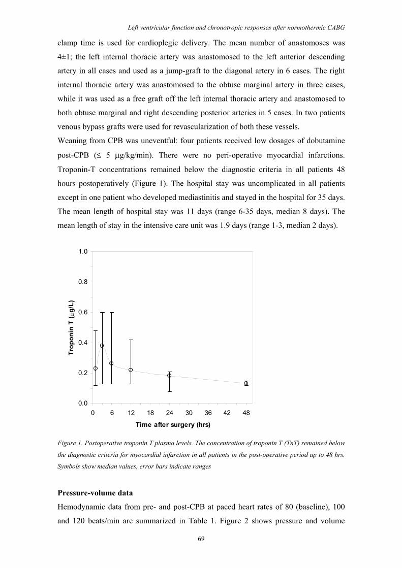
Left ventricular function and chronotropic responses after normothermic CABG
69
clamp time is used for cardioplegic delivery. The mean number of anastomoses was
4±1; the left internal thoracic artery was anastomosed to the left anterior descending
artery in all cases and used as a jump-graft to the diagonal artery in 6 cases. The right
internal thoracic artery was anastomosed to the obtuse marginal artery in three cases,
while it was used as a free graft off the left internal thoracic artery and anastomosed to
both obtuse marginal and right descending posterior arteries in 5 cases. In two patients
venous bypass grafts were used for revascularization of both these vessels.
Weaning from CPB was uneventful: four patients received low dosages of dobutamine
post-CPB (≤ 5 μg/kg/min). There were no peri-operative myocardial infarctions.
Troponin-T concentrations remained below the diagnostic criteria in all patients 48
hours postoperatively (Figure 1). The hospital stay was uncomplicated in all patients
except in one patient who developed mediastinitis and stayed in the hospital for 35 days.
The mean length of hospital stay was 11 days (range 6-35 days, median 8 days). The
mean length of stay in the intensive care unit was 1.9 days (range 1-3, median 2 days).
0.0
0.2
0.4
0.6
0.8
1.0
0 6 12 18 24 30 36 42 48
Time after surgery (hrs)
Trop
onin
T ( μ
g/L)
Figure 1. Postoperative troponin T plasma levels. The concentration of troponin T (TnT) remained below
the diagnostic criteria for myocardial infarction in all patients in the post-operative period up to 48 hrs.
Symbols show median values, error bars indicate ranges
Pressure-volume data
Hemodynamic data from pre- and post-CPB at paced heart rates of 80 (baseline), 100
and 120 beats/min are summarized in Table 1. Figure 2 shows pressure and volume
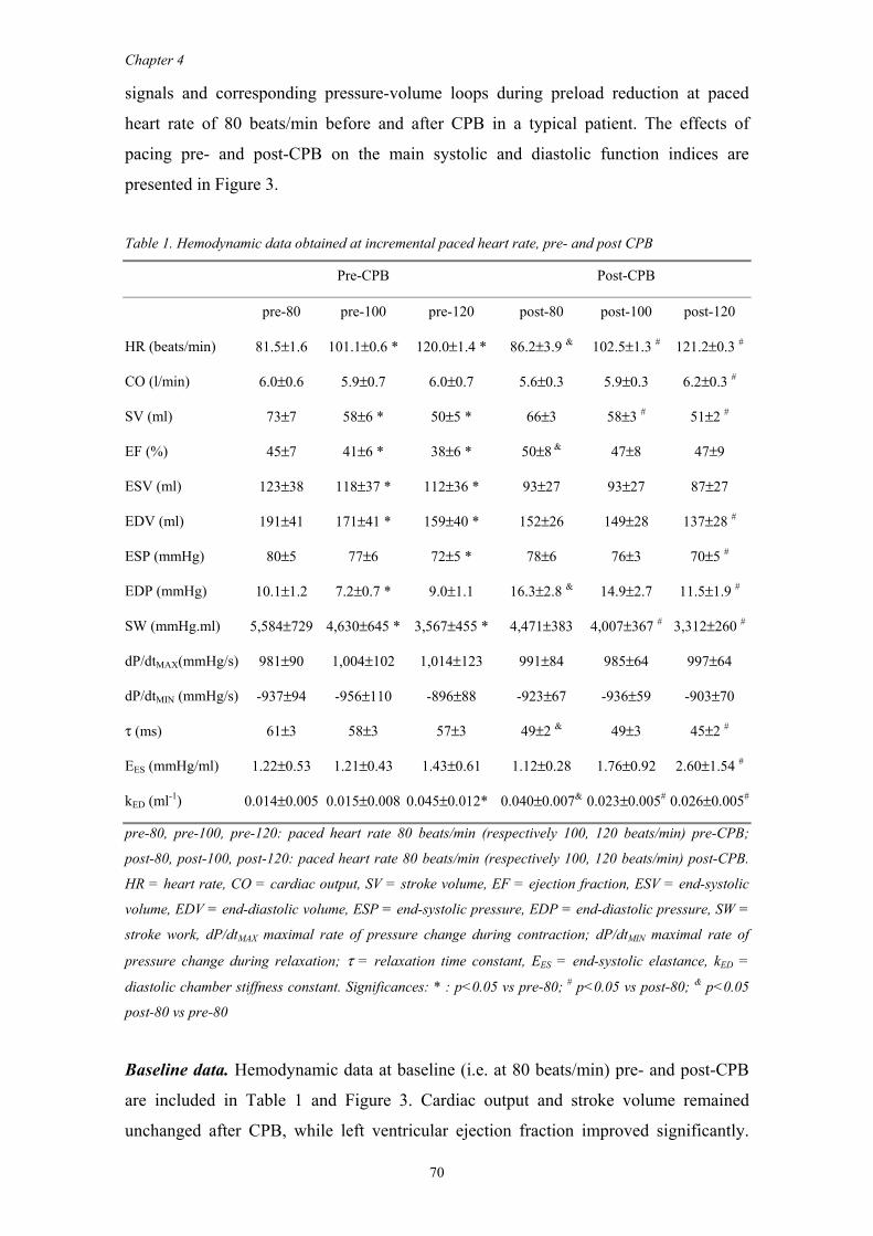
Chapter 4
70
signals and corresponding pressure-volume loops during preload reduction at paced
heart rate of 80 beats/min before and after CPB in a typical patient. The effects of
pacing pre- and post-CPB on the main systolic and diastolic function indices are
presented in Figure 3.
Table 1. Hemodynamic data obtained at incremental paced heart rate, pre- and post CPB
Pre-CPB Post-CPB
pre-80 pre-100 pre-120 post-80 post-100 post-120
HR (beats/min) 81.5±1.6 101.1±0.6 * 120.0±1.4 * 86.2±3.9 & 102.5±1.3 # 121.2±0.3 #
CO (l/min) 6.0±0.6 5.9±0.7 6.0±0.7 5.6±0.3 5.9±0.3 6.2±0.3 #
SV (ml) 73±7 58±6 * 50±5 * 66±3 58±3 # 51±2 #
EF (%) 45±7 41±6 * 38±6 * 50±8 & 47±8 47±9
ESV (ml) 123±38 118±37 * 112±36 * 93±27 93±27 87±27
EDV (ml) 191±41 171±41 * 159±40 * 152±26 149±28 137±28 #
ESP (mmHg) 80±5 77±6 72±5 * 78±6 76±3 70±5 #
EDP (mmHg) 10.1±1.2 7.2±0.7 * 9.0±1.1 16.3±2.8 & 14.9±2.7 11.5±1.9 #
SW (mmHg.ml) 5,584±729 4,630±645 * 3,567±455 * 4,471±383 4,007±367 # 3,312±260 #
dP/dtMAX(mmHg/s) 981±90 1,004±102 1,014±123 991±84 985±64 997±64
dP/dtMIN (mmHg/s) -937±94 -956±110 -896±88 -923±67 -936±59 -903±70
τ (ms) 61±3 58±3 57±3 49±2 & 49±3 45±2 #
EES (mmHg/ml) 1.22±0.53 1.21±0.43 1.43±0.61 1.12±0.28 1.76±0.92 2.60±1.54 #
kED (ml-1) 0.014±0.005 0.015±0.008 0.045±0.012* 0.040±0.007& 0.023±0.005# 0.026±0.005#
pre-80, pre-100, pre-120: paced heart rate 80 beats/min (respectively 100, 120 beats/min) pre-CPB;
post-80, post-100, post-120: paced heart rate 80 beats/min (respectively 100, 120 beats/min) post-CPB.
HR = heart rate, CO = cardiac output, SV = stroke volume, EF = ejection fraction, ESV = end-systolic
volume, EDV = end-diastolic volume, ESP = end-systolic pressure, EDP = end-diastolic pressure, SW =
stroke work, dP/dtMAX maximal rate of pressure change during contraction; dP/dtMIN maximal rate of
pressure change during relaxation; τ = relaxation time constant, EES = end-systolic elastance, kED =
diastolic chamber stiffness constant. Significances: * : p<0.05 vs pre-80; # p<0.05 vs post-80; & p<0.05
post-80 vs pre-80
Baseline data. Hemodynamic data at baseline (i.e. at 80 beats/min) pre- and post-CPB
are included in Table 1 and Figure 3. Cardiac output and stroke volume remained
unchanged after CPB, while left ventricular ejection fraction improved significantly.

Left ventricular function and chronotropic responses after normothermic CABG
71
End-diastolic volume (EDV) and end-systolic volume (ESV) had a clear tendency to
decrease post-CPB (EDV: -39 ml; ESV: -30 ml), but these changes did not reach
statistical significance. Both end-diastolic pressure and diastolic chamber stiffness
increased significantly after CPB, while the relaxation time constant τ decreased
significantly. End-systolic elastance (EES) remained unchanged after CPB. Pre-CPB
LV Volume(mL)
LV Pressure(mmHg)
Post-CPB
LV Volume(mL)
LV Pressure(mmHg)
Time (s) LV Volume (mL)
0
50
100
150
200
0
25
50
75
100
EES =0.74 mmHg/ml
k ED =0.0045 ml-1
0
25
50
75
100
0 50 100 150 200 25
LV P
ress
ure
(mm
Hg)
0
50
100
150
200
0
25
50
75
100
0 4 8
EES =0.80 mmHg/ml
k ED =0.052 ml-1
0
25
50
75
100
0 50 100 150 200 25
LV P
ress
ure
(mm
Hg)
Figure 2. Typical LV pressure and volume signals and pressure-volume loops during preload reduction
by transient vena cava occlusion, pre- and post-CPB, at paced heart rate of 80 beats/min
Effects of pacing. Hemodynamic data pre- and post-CPB at 80, 100, and 120 beats/min
are given in Table 1 and Figure 3. Note that post-CPB the mean baseline heart rate was
86±4 beats/min because in some patients sinus rhythm exceeded the target pacing rate
of 80 beats/min. Cardiac output increased with incremental pacing post-CPB, while pre-
CPB pacing did not affect cardiac output. Stroke volume decreased both before and
after CPB with pacing, but this decrease was less pronounced after CPB (-24 ml pre-
CPB vs -14 ml post-CPB). The smaller reduction in stroke volume with pacing post-
CPB was the result of a less pronounced reduction in end-diastolic volume (pre-CPB: -
33 ml; post-CPB: -15 ml), since end-systolic volume decreased by 11 ml pre-CPB and
by 6 ml post-CPB. Apparently, the capability of the ventricle to fill despite a high heart
rate is relatively improved post-CPB. This is supported by the results for the diastolic
indices. Active relaxation, τ, improved during pacing post-CPB, while it remained
unchanged during pacing pre-CPB. Furthermore, the end-diastolic chamber stiffness
constant increased significantly during pacing pre-CPB, whereas it decreased during
pacing post-CPB. It should be mentioned that baseline diastolic stiffness (i.e. at 80
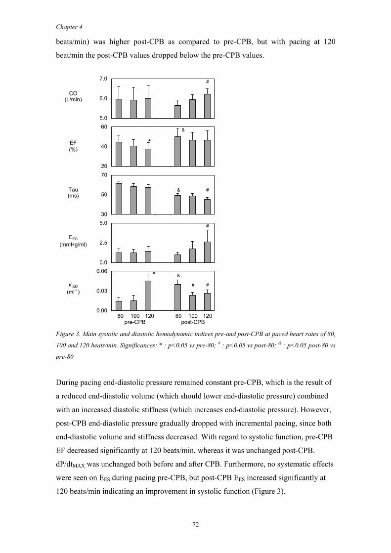
Chapter 4
72
beats/min) was higher post-CPB as compared to pre-CPB, but with pacing at 120
beat/min the post-CPB values dropped below the pre-CPB values.
#
CO(L/min)
&
EF *(%)
Tau & #(ms)
#
EES(mmHg/ml)
* &
k ED # #(ml-1)
80 100 120 80 100 120pre-CPB post-CPB
5.0
6.0
7.0
20
40
60
30
50
70
0.0
2.5
5.0
0.00
0.03
0.06
Figure 3. Main systolic and diastolic hemodynamic indices pre-and post-CPB at paced heart rates of 80,
100 and 120 beats/min. Significances: * : p<0.05 vs pre-80; # : p<0.05 vs post-80; & : p<0.05 post-80 vs
pre-80
During pacing end-diastolic pressure remained constant pre-CPB, which is the result of
a reduced end-diastolic volume (which should lower end-diastolic pressure) combined
with an increased diastolic stiffness (which increases end-diastolic pressure). However,
post-CPB end-diastolic pressure gradually dropped with incremental pacing, since both
end-diastolic volume and stiffness decreased. With regard to systolic function, pre-CPB
EF decreased significantly at 120 beats/min, whereas it was unchanged post-CPB.
dP/dtMAX was unchanged both before and after CPB. Furthermore, no systematic effects
were seen on EES during pacing pre-CPB, but post-CPB EES increased significantly at
120 beats/min indicating an improvement in systolic function (Figure 3).
Left ventricular function and chronotropic responses after normothermic CABG
73
DISCUSSION
CABG is increasingly performed in heart failure patients with concomitant surgical
valvular repair and/or left ventricular restoration. Optimal preservation of myocardial
function is important to facilitate these surgical procedures. Traditional cold heart and
cold cardioplegic arrest may have negative effects on post-operative myocardial
function and currently normothermic procedures are increasingly used as an
alternative.1 Previous studies indicate that normothermic arrest with warm blood
cardioplegia provides metabolic benefits and less cell damage, possibly mediated by a
better protection from from ischemia-reperfusion injury.2,3,9 However, few data are
available on the acute effects on ventricular function. The aim of our study was
therefore to quantify the effects of normothermic on-pump CABG and IAWBC on
systolic and diastolic left ventricular function. In brief, our results show that this
approach has no negative effects on baseline systolic function, whereas it tended to
improve the response of systolic function during incremental pacing. With regard to
diastolic function we found an improved early relaxation, but the end-diastolic stiffness
was increased at baseline. However, incremental pacing revealed improved relaxation
and filling characteristics post-CPB, whereas pre-CPB the diastolic indices remained
constant or worsened during pacing.
Baseline hemodynamic changes
The baseline hemodynamic results (i.e. comparing pre- vs. post-CPB at 80 beats/min)
show a slight but significant increase of ejection fraction after CPB, which is due to a
marked decrease in end-diastolic volume (-39 ml) with a relatively unchanged stroke
volume. Stroke volume remained largely unchanged due to a similar decrease of end-
systolic volume (-30 ml) after CPB. Note that, except for EF, none of these volumetric
changes reached statistical significance. The effect on EDV is the result of impairment
of late passive diastolic function (kED and EDP increased significantly after CPB),
despite the fact that active relaxation (τ) significantly improved after CPB. The
improved ejection fraction and the finding of a reduced end-systolic volume with
maintained end-systolic pressure both point towards an improved systolic function.
However, the load-independent contractility index EES did not change significantly.
Therefore, we would conclude that normothermic CPB with IAWBC at least preserves
systolic function in this patient group. This is in contrast with studies using cold blood
Chapter 4
74
cardioplegia during hypothermia in which a reduced systolic left ventricular function
after CPB was reported.1
With respect to diastolic function we found a somewhat prolonged τ at baseline pre-
CPB, which has already been shown to be representative for patients with coronary
artery disease.16,17 In our study τ decreased significantly after CPB with warm blood
cardioplegia indicating an improved early, active relaxation. This normalization of τ
after revascularization is consistent with previous studies regardless of the use of cold or
warm blood cardioplegia and is most likely related to enhancement of the, highly
oxygen-dependent, calcium re-uptake process by the sarcoplasmic reticulum after
revascularization, and not due to effects of CPB.18 After CPB increased circulating
catecholamines resulting from CPB and ischemia may influence active relaxation.
However, the unchanged systolic pressure and heart rate after CPB indicate that this
effect is unlikely to be very prominent in our study. In contrast to the improvement in τ,
the diastolic chamber stiffness constant, which represents passive late diastolic function
was significantly increased post-CPB. This increased stiffness (thus reduced diastolic
compliance) is likely due to temporary myocardial edema and increased water content
after CPB.19,20 This finding is important when interpreting changes in diastolic function
after surgical interventions such as ventricular restoration and other procedures.21
Apparently, part of the changes in diastolic function, at least in the acute phase, are
related to the cardioplegic arrest and CPB, and should not be attributed to the surgical
procedure per se.
Chronotropic responses
We found a significant improvement of cardiac output during incremental atrial pacing
post-CPB, whereas cardiac output remained constant pre-CPB. This effect reflected a
more pronounced decrease in stroke volume with pacing pre-CPB, compared to post-
CPB. In normal physiology maintained stroke volume (or a limited reduction) during
increased heart rate is obtained by a combination of increased systolic function
(Bowditch effect), which reduces or maintains end-systolic volume, and an improved
relaxation, which limits the reduction in end-diastolic volume resulting from the
reduced diastolic filling time. Our results indicate that neither of these mechanisms is
operative in patients with coronary artery disease (CAD) pre-CPB and consequently
cardiac output did not increase during incremental pacing. Moreover, diastolic stiffness
substantially increased during pacing which further limited filling. The finding that
systolic function does not improve or even decreases with increased heart rate in CAD
Left ventricular function and chronotropic responses after normothermic CABG
75
patients is consistent with previous studies.22 A recent echocardiographic study in
patients undergoing CABG indicates an increased diastolic stiffness during pacing very
similar to our findings.23 Numerous studies have documented increased diastolic
pressure, increased stiffness and upward shifts of the diastolic pressure-volume relation
with pacing angina, however our study shows that more subtle increases in diastolic
stiffness are obtained with a relatively small increase in heart rate in CAD patients with
relatively preserved EF.24
After CPB, diastolic chamber stiffness, end-diastolic pressure and τ all significantly
decreased during pacing which may explain the improvement of cardiac output at higher
heart rates. In addition, EES gradually increased with incremental pacing post-CPB,
whereas it remained constant pre-CPB, indicating that improvement in systolic function
contributed to the increase in cardiac output.
The effects of pacing pre- vs. post-CPB in our study largely mimic the effects of
exercise before and after revascularization surgery as described in a study by Caroll et
al.25 After surgery, but not before, both pacing and exercise induced improvements in
systolic and diastolic function, which enable the required increase in cardiac output.
However, during exercise end-diastolic pressure and volume increased whereas during
pacing in our study these indices decreased. These differences are presumably due to
recruitment of blood volume during exercise leading to increased preload, which does
not occur during pacing.
The impaired chronotropic responses pre-CPB as found in our study are presumably due
to coronary artery disease and the normalization of these responses post-CPB due to
effects of successful revascularization and subsequent relief of ischemia.
Limitations
We did not include a control group with a cold cardioplegic approach. However, this
approach is well documented in the literature and we compared our results against those
reports. Furthermore, our study was performed in patients with relatively normal LV
function, whereas the advantages of IAWBC are presumably most important for
patients with poor LV function. However, in heart failure patients the effects of IAWBC
would be difficult to assess separately because the surgical interventions (CABG and
additional procedures like mitral annuloplasty and/or surgical restoration) may
importantly affect post-operative LV function.
In conclusion, this study shows that intermittent antegrade warm blood cardioplegia
during normothermic cardiopulmonary bypass provides excellent myocardial protection
Chapter 4
76
of systolic properties, whereas improved diastolic and systolic left ventricular
chronotropic responses were found acutely after surgery. This cardioprotective stategy
may be particularly advantageous in patients with heart failure who undergo complex
surgical procedures with long procedure times.
ACKNOWLEDGEMENTS
We gratefully acknowledge Eline F. Bruggemans for statistical review of the
manuscript.
REFERENCES
1. Wallace A, Lam HW, Nose PS, Bellows W, Mangano DT. Changes in systolic and diastolic
ventricular function with cold cardioplegic arrest in man. The Multicenter Study of Peri-operative Ischemia (McSPI) Research Group. J Card Surg. 1994;9:497-502.
2. Jacquet LM, Noirhomme PH, Van Dyck MJ, El Khoury GA, Matta AJ, Goenen MJ, Dion RA. Randomized trial of intermittent antegrade warm blood versus cold crystalloid cardioplegia. Ann Thorac Surg. 1999;67:471-477.
3. Cannon MB, Vine AJ, Kantor HL, Lahorra JA, Nickell SA, Hahn C, Allyn JW, Teplick RS, Titus JS, Torchiana DF, . Warm and cold blood cardioplegia. Comparison of myocardial function and metabolism using 31p magnetic resonance spectroscopy. Circulation. 1994;90:II328-II338.
4. Randomised trial of normothermic versus hypothermic coronary bypass surgery. The Warm Heart Investigators. Lancet. 1994;343:559-563.
5. Yau TM, Ikonomidis JS, Weisel RD, Mickle DA, Ivanov J, Mohabeer MK, Tumiati L, Carson S, Liu P. Ventricular function after normothermic versus hypothermic cardioplegia. J Thorac Cardiovasc Surg. 1993;105:833-843.
6. Calafiore AM, Teodori G, Mezzetti A, Bosco G, Verna AM, Di Giammarco G, Lapenna D. Intermittent antegrade warm blood cardioplegia. Ann Thorac Surg. 1995;59:398-402.
7. Lichtenstein SV, Ashe KA, el Dalati H, Cusimano RJ, Panos A, Slutsky AS. Warm heart surgery. J Thorac Cardiovasc Surg. 1991;101:269-274.
8. Franke UF, Korsch S, Wittwer T, Albes JM, Wippermann J, Kaluza M, Rahmanian PB, Wahlers T. Intermittent antegrade warm myocardial protection compared to intermittent cold blood cardioplegia in elective coronary surgery - do we have to change? Eur J Cardiothorac Surg. 2003;23:341-346.
9. Mezzetti A, Calafiore AM, Lapenna D, Deslauriers R, Tian G, Salerno TA, Verna AM, Bosco G, Pierdomenico SD, Caccurullo F. Intermittent antegrade warm cardioplegia reduces oxidative stress and improves metabolism of the ischemic-reperfused human myocardium. J Thorac Cardiovasc Surg. 1995;109:787-795.
10. Tulner SA, Klautz RJ, Rijk-Zwikker GL, Engbers FH, Bax JJ, Baan J, van der Wall EE, Dion RA, Steendijk P. Peri-operative assessment of left ventricular function by pressure-volume loops using the conductance catheter method. Anesth Analg. 2003;97:950-7, table.
11. Baan J, van der Velde ET, de Bruin HG, Smeenk GJ, Koops J, van Dijk AD, Temmerman D, Senden J, Buis B. Continuous measurement of left ventricular volume in animals and humans by conductance catheter. Circulation. 1984;70:812-823.
12. Steendijk P, Staal E, Jukema JW, Baan J. Hypertonic saline method accurately determines parallel conductance for dual-field conductance catheter. Am J Physiol Heart Circ Physiol. 2001;281:H755-H763.
13. Kass DA, Maughan WL, Guo ZM, Kono A, Sunagawa K, Sagawa K. Comparative influence of load versus inotropic states on indexes of ventricular contractility: experimental and theoretical analysis based on pressure-volume relationships. Circulation. 1987;76:1422-1436.

Left ventricular function and chronotropic responses after normothermic CABG
77
14. Mandinov L, Eberli FR, Seiler C, Hess OM. Diastolic heart failure. Cardiovasc Res. 2000;45:813-825.
15. Slinker BK, Glantz SA. Multiple linear regression is a useful alternative to traditional analyses of variance. Am J Physiol. 1988;255:R353-R367.
16. Bolognesi R, Tsialtas D, Barilli AL, Manca C, Zeppellini R, Javernaro A, Cucchini F. Detection of early abnormalities of left ventricular function by hemodynamic, echo-tissue Doppler imaging, and mitral Doppler flow techniques in patients with coronary artery disease and normal ejection fraction. J Am Soc Echocardiogr. 2001;14:764-772.
17. Ohte N, Narita H, Hashimoto T, Hayano J, Akita S, Kurokawa K. Differentiation of abnormal relaxation pattern with aging from abnormal relaxation pattern with coronary artery disease in transmitral flow with the use of tissue Doppler imaging of the mitral annulus. J Am Soc Echocardiogr. 1999;12:629-635.
18. Humphrey LS, Topol EJ, Rosenfeld GI, Borkon AM, Baumgartner WA, Gardner TJ, Maruschak G, Weiss JL. Immediate enhancement of left ventricular relaxation by coronary artery bypass grafting: intraoperative assessment. Circulation. 1988;77:886-896.
19. Ericsson AB, Takeshima S, Vaage J. Simultaneous antegrade and retrograde delivery of continuous warm blood cardioplegia after global ischemia. J Thorac Cardiovasc Surg. 1998;115:716-722.
20. Wallace AW, Ratcliffe MB, Nose PS, Bellows W, Moores W, McEnany MT, Flachsbart K, Mangano DT. Effect of induction and reperfusion with warm substrate-enriched cardioplegia on ventricular function. Ann Thorac Surg. 2000;70:1301-1307.
21. Dor V, Saab M, Coste P, Kornaszewska M, Montiglio F. Left ventricular aneurysm: a new surgical approach. Thorac Cardiovasc Surg. 1989;37:11-19.
22. Aroesty JM, McKay RG, Heller GV, Royal HD, Als AV, Grossman W. Simultaneous assessment of left ventricular systolic and diastolic dysfunction during pacing-induced ischemia. Circulation. 1985;71:889-900.
23. Royse CF, Royse AG, Wong CT, Soeding PF. The effect of pericardial restraint, atrial pacing, and increased heart rate on left ventricular systolic and diastolic function in patients undergoing cardiac surgery. Anesth Analg. 2003;96:1274-9, table.
24. Bronzwaer JG, de Bruyne B, Ascoop CA, Paulus WJ. Comparative effects of pacing-induced and balloon coronary occlusion ischemia on left ventricular diastolic function in man. Circulation. 1991;84:211-222.
25. Carroll JD, Hess OM, Hirzel HO, Turina M, Krayenbuehl HP. Left ventricular systolic and diastolic function in coronary artery disease: effects of revascularization on exercise-induced ischemia. Circulation. 1985;72:119-129.

CHAPTER 5
Acute-hemodynamic effects of restrictive mitral annuloplasty
in patients with end-stage heart failure
-Analysis by pressure-volume relations-
S.A.F. Tulner
P. Steendijk
R.J.M. Klautz
J.J. Bax
M.I.M. Versteegh
E.E. van der Wall
R.A.E. Dion
J Thorac Cardiovasc Surg 2005; 130: 33-40
Poster presentation at American College of Cardiology, March 2005, Orlando,
Florida, USA
Oral presentation at European Association of Cardiothoracic Surgery, September
2004, Leipzig, Germany
Chapter 5
80
ABSTRACT
Objective. Recent studies show beneficial long-term effects of restrictive mitral
annuloplasty (RMA) in patients with end-stage heart failure (HF). However, concerns
are raised about possible adverse effects on early post-operative systolic and diastolic
function, which may limit application of this approach in HF patients. Therefore, we
evaluated acute effects of RMA on left ventricular (LV) function by load-independent
pressure-volume relations.
Methods. In 23 patients (HF n=10; control n=13) we determined LV systolic and
diastolic function before and after surgery by pressure-volume analysis using the
conductance catheter. All HF patients underwent stringent RMA (two sizes under), and
4 received additional CABG. Transesophageal echocardiography was used for
evaluation of valve repair. Patients with preserved LV function who underwent isolated
CABG served as controls.
Results. RMA (ring size 25±1) restored leaflet coaptation (8.0±0.2mm) with normal
pressure gradients (2.9±1.8mmHg). RMA did not change cardiac output (5.0±1.8 to
5.3±0.9L/min, NS), LV ejection fraction (29±5 to 32±8%, NS) or end-systolic elastance
(0.86±0.50 to 0.99±1.05mmHg/mL, NS). After RMA, end-diastolic volume tended to
decrease (237±89 to 226±52mL, NS), while end-diastolic pressure remained unchanged
(14±6 to 15±5mmHg, NS). Diastolic chamber stiffness tended to increase (0.027±0.035
to 0.041±0.047mL-1, NS), however not significantly. Peak LV wall stress was
unchanged (356±91 to 346±85 mmHg, NS). Baseline values in the control group were
different, but changes in most parameters after surgery showed similar non-significant
trends.
Conclusion. Mitral valve repair by RMA effectively restores mitral valve competence
without inducing significant acute changes in LV systolic or diastolic function in
patients with end-stage heart failure.
INTRODUCTION
Chronic mitral regurgitation is a serious complication in patients with end-stage heart
failure. Patients with mitral regurgitation have a significantly decreased survival at 2
years follow-up versus patients without mitral regurgitation.1
The mechanism of mitral regurgitation in end-stage heart failure is multifactorial.
Acute hemodynamic effects of RMA
81
Briefly, it is related to changes in left ventricular (LV) geometry with a subsequent
displacement of the subvalvular apparatus, annular dilatation2 and restrictive leaflet
motion (class IIIb according to Carpentier's classification3), which results in coaptation
failure.4,5 From a physiological point of view, mitral regurgitation in these patients will
lead to LV overload and reduction of forward stroke volume. This occurs initially in
response to exercise and subsequently at rest, which in turn activates systemic and local
neurohormonal systems and cytokines that worsen cardiac loading conditions and
promote LV remodeling and dysfunction.6 This may create a vicious circle wherein
regurgitation begets more regurgitation.
Previous studies have shown that interrupting this vicious cycle by mitral valve repair is
safe, and improves clinical outcome.7 Several groups advocate the use of a stringent
restrictive ring, two sizes under, to achieve better leaflet coaptation and possibly prevent
recurrence of mitral regurgitation and promote reverse remodeling.8 Mid-term results
(18 months follow-up) with this approach indicate reverse remodeling in 58% of
patients.9 However, the acute effects of restrictive mitral annuloplasty (RMA) on LV
systolic and diastolic function in patients with end-stage heart failure are unknown.
There are concerns that correction of mitral regurgitation may decrease LV systolic
function in the acute phase due to afterload increase caused by closure of a low
resistance runoff into the left atrium. In addition, it has been suggested that undersizing
the mitral annulus may affect LV contractility due to increased mechanical tension on
the base of heart.10 With regard to diastolic function RMA might impair filling. In
contrast, Bolling hypothesized that undersizing the mitral annulus will lead to acute
beneficial geometric changes of the base of the left ventricle, which may reduce LV
volume and wall stress.11 The purpose of this study was therefore to quantify the acute
effects of RMA on global and intrinsic LV systolic and diastolic function in these
patients.
METHODS
A total of 23 patients were studied in the operating room before and after
cardiopulmonary bypass (CPB) by pressure-volume analysis using the conductance
catheter method. We included 10 patients with end-stage heart failure (HF) with co-
existent severe mitral regurgitation who underwent mitral valve repair by stringent
restrictive annuloplasty and 13 control patients with preserved LV function who
Chapter 5
82
underwent elective CABG. The control group was used to distinguish effects of mitral
annuloplasty from effects of CPB and cardioplegic cardiac arrest, per se. In both groups
surgery was performed during normothermic CPB with intermittent antegrade warm
blood cardioplegia. The study protocol was approved by the institutional review board
and all patients provided informed written consent.
Patient selection and echocardiographic criteria
The patients in the RMA group fulfilled the following criteria:
1) NYHA class III or IV
2) LVEF < 30%
3) Mitral regurgitation grade ≥ 2 assessed by transesophageal echocardiography
(TEE) preoperatively (without general anesthesia to avoid underestimation of the
severity of the mitral regurgitation). The severity of mitral regurgitation was graded
semi-quantitatively from color-flow Doppler and characterized as: mild, 1+ (jet area/left
atrial area <10%); moderate, 2+ (jet area/left atrial area 10% to 20%); moderately
severe, 3+ (jet area/left atrial area 20% to 45%); and severe, 4+ (jet area/left atrial area
>45%).12 In patients with mitral regurgitation grade 2 an intra-operative dynamic
loading test was performed as described by Dion et al.5 If this test was positive, that is if
it resulted in a definite worsening of the severity of mitral regurgitation, restrictive
mitral annuloplasty was performed.
4) The mechanism of mitral regurgitation was based on malcoaptation due to
systolic restrictive motion of the mitral leaflets.
5) Maximal medical therapy for heart failure including diuretics, afterload
reduction and beta-blocking agents.
Patients with primary mitral valve dysfunction (mitral valve prolapse, rheumatic valve
disease, mitral valve stenosis) were excluded from the study. Also patients with a
previously implanted biological or mechanical prosthesis in aortic position were not
included in this study.
The control group was recruited from patients with preserved LV function (LVEF >
40%) who underwent elective CABG for multivessel coronary artery disease and who
needed no additional valvular surgery. The patient characteristics of both groups are
summarized in Table 1.
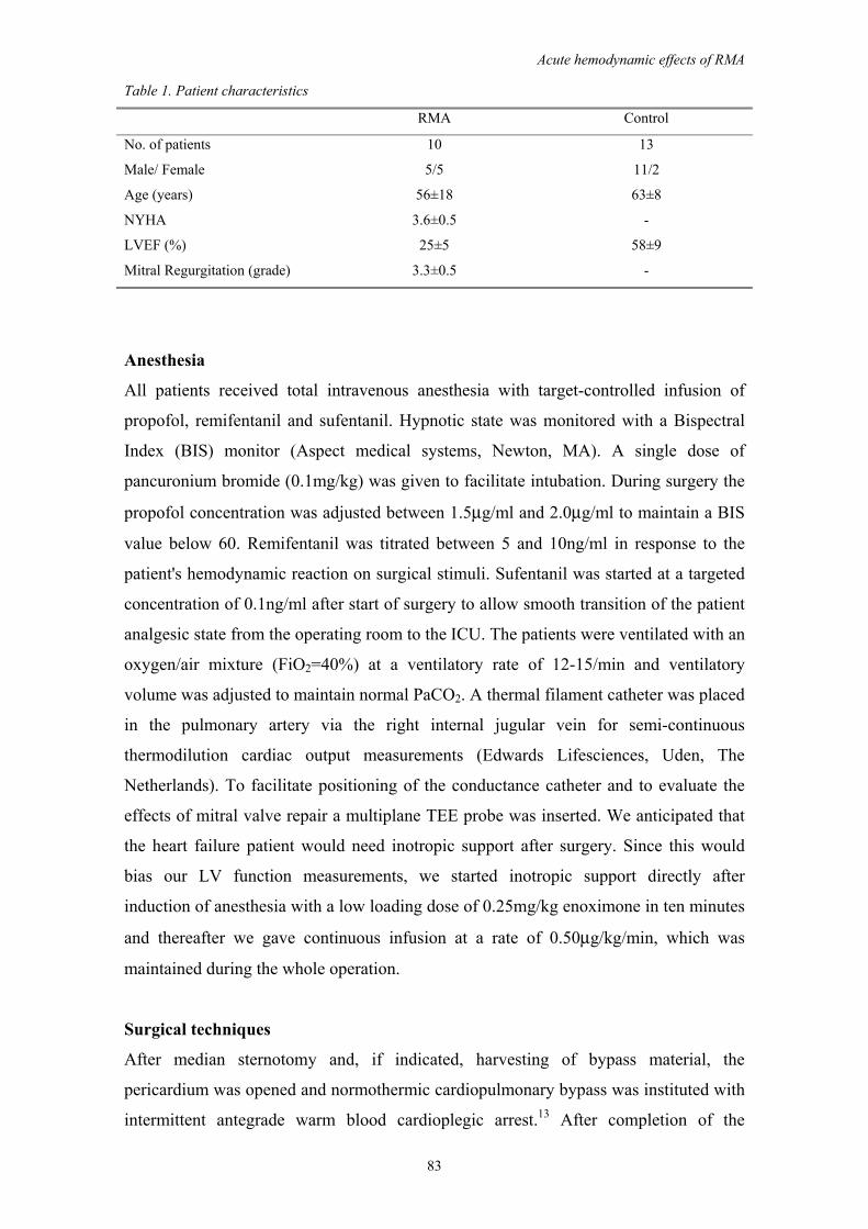
Acute hemodynamic effects of RMA
83
Table 1. Patient characteristics
RMA Control
No. of patients 10 13
Male/ Female 5/5 11/2
Age (years) 56±18 63±8
NYHA 3.6±0.5 -
LVEF (%) 25±5 58±9
Mitral Regurgitation (grade) 3.3±0.5 -
Anesthesia
All patients received total intravenous anesthesia with target-controlled infusion of
propofol, remifentanil and sufentanil. Hypnotic state was monitored with a Bispectral
Index (BIS) monitor (Aspect medical systems, Newton, MA). A single dose of
pancuronium bromide (0.1mg/kg) was given to facilitate intubation. During surgery the
propofol concentration was adjusted between 1.5μg/ml and 2.0μg/ml to maintain a BIS
value below 60. Remifentanil was titrated between 5 and 10ng/ml in response to the
patient's hemodynamic reaction on surgical stimuli. Sufentanil was started at a targeted
concentration of 0.1ng/ml after start of surgery to allow smooth transition of the patient
analgesic state from the operating room to the ICU. The patients were ventilated with an
oxygen/air mixture (FiO2=40%) at a ventilatory rate of 12-15/min and ventilatory
volume was adjusted to maintain normal PaCO2. A thermal filament catheter was placed
in the pulmonary artery via the right internal jugular vein for semi-continuous
thermodilution cardiac output measurements (Edwards Lifesciences, Uden, The
Netherlands). To facilitate positioning of the conductance catheter and to evaluate the
effects of mitral valve repair a multiplane TEE probe was inserted. We anticipated that
the heart failure patient would need inotropic support after surgery. Since this would
bias our LV function measurements, we started inotropic support directly after
induction of anesthesia with a low loading dose of 0.25mg/kg enoximone in ten minutes
and thereafter we gave continuous infusion at a rate of 0.50μg/kg/min, which was
maintained during the whole operation.
Surgical techniques
After median sternotomy and, if indicated, harvesting of bypass material, the
pericardium was opened and normothermic cardiopulmonary bypass was instituted with
intermittent antegrade warm blood cardioplegic arrest.13 After completion of the
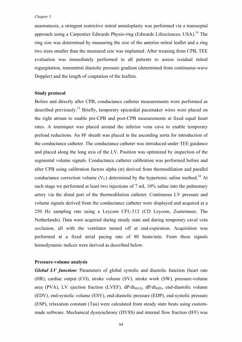
Chapter 5
84
anastomosis, a stringent restrictive mitral annuloplasty was performed via a transseptal
approach using a Carpentier Edwards Physio-ring (Edwards Lifesciences, USA).14 The
ring size was determined by measuring the size of the anterior mitral leaflet and a ring
two sizes smaller than the measured size was implanted. After weaning from CPB, TEE
evaluation was immediately performed in all patients to assess residual mitral
regurgitation, transmitral diastolic pressure gradient (determined from continuous-wave
Doppler) and the length of coaptation of the leaflets.
Study protocol
Before and directly after CPB, conductance catheter measurements were performed as
described previously.15 Briefly, temporary epicardial pacemaker wires were placed on
the right atrium to enable pre-CPB and post-CPB measurements at fixed equal heart
rates. A tourniquet was placed around the inferior vena cava to enable temporary
preload reductions. An 8F sheath was placed in the ascending aorta for introduction of
the conductance catheter. The conductance catheter was introduced under TEE guidance
and placed along the long axis of the LV. Position was optimized by inspection of the
segmental volume signals. Conductance catheter calibration was performed before and
after CPB using calibration factors alpha (α) derived from thermodilution and parallel
conductance correction volume (VC) determined by the hypertonic saline method.16 At
each stage we performed at least two injections of 7 mL 10% saline into the pulmonary
artery via the distal port of the thermodilution catheter. Continuous LV pressure and
volume signals derived from the conductance catheter were displayed and acquired at a
250 Hz sampling rate using a Leycom CFL-512 (CD Leycom, Zoetermeer, The
Netherlands). Data were acquired during steady state and during temporary caval vein
occlusion, all with the ventilator turned off at end-expiration. Acquisition was
performed at a fixed atrial pacing rate of 80 beats/min. From these signals
hemodynamic indices were derived as described below.
Pressure-volume analysis
Global LV function: Parameters of global systolic and diastolic function (heart rate
(HR), cardiac output (CO), stroke volume (SV), stroke work (SW), pressure-volume
area (PVA), LV ejection fraction (LVEF), dP/dtMAX, dP/dtMIN, end-diastolic volume
(EDV), end-systolic volume (ESV), end-diastolic pressure (EDP), end-systolic pressure
(ESP), relaxation constant (Tau) were calculated from steady state beats using custom-
made software. Mechanical dyssynchrony (DYSS) and internal flow fraction (IFF) was
Acute hemodynamic effects of RMA
85
calculated as previously described.17 Effective arterial elastance (Ea), a measure of
afterload, was calculated as ESP/SV. Time-varying wall stress, WS(t), was calculated
from the LV pressure and volume signals (P(t), V(t)) as described by Arts et al.: WS(t)=
P(t)·[1+3·V(t)/VWALL]. LV wall volume (VWALL) was estimated based on the diastolic
posterior wall thickness derived from M-mode echocardiography.18 The gradient across
the LV outflow tract was calculated as the difference between peak LV pressure and
peak aortic pressure.
Systolic and diastolic LV pressure-volume relations: Systolic function was
characterized by the slope of the end-systolic pressure-volume relationship (End-
systolic elastance, EES), the slope of the relation between the dP/dtMAX and EDV (S-dP),
and the slope of the preload recruitable stroke work relation (S-PRSW). The position of
the ESPVR was quantified by calculating the ESV-intercept at a fixed end-systolic
pressure (ESVIND). The positions of the dP/dtMAX - EDV relation and the PRSW relation
were determined by calculating the intercepts at a fixed end-diastolic volume, dP/dtMAX,
IND and SWIND, respectively. As previously described, the fixed end-systolic pressure
and end-diastolic volume levels were set retrospectively as the mean ESP and EDV in
each group.17 Diastolic chamber stiffness (Ked) was quantified by exponential regression
of the end-diastolic pressure-volume relationship.19,20
Statistical analysis
Pre- and post-CPB data were compared with paired t-tests. Statistical significance was
assumed at p<0.05. All data are presented as the mean±SD.
RESULTS
All HF patients were successfully weaned from CPB after successful mitral valve
repair. In six patients the origin of HF was ischemic, in four non-ischemic. In four
ischemic patients additional CABG was performed; the other 2 patients had irreversible
ischemia and did not receive CABG. In three patients with severe tricuspid
regurgitation, a concomitant restrictive tricuspid ring annuloplasty (ring size 26) was
performed. Six (60%) patients needed inotropic support more than 24 hours
postoperatively. However, none of the patients required intra-aortic balloon pump
support. The median stay in the intensive care unit in this group was 4 days (range 2 to
Chapter 5
86
7 days) with a median total hospital stay of 14 days (range 7 to 18 days). All patients
could be discharged in good clinical condition from the hospital. The surgical details of
both groups are summarized in Table 2.
Table 2. Surgical data
RMA (n=10) Control (n=13)
CPB- time (median, minutes) 137 (range 105-287) 104 (range 60-167)
Aox-time (median, minutes) 96 (range 65-196) 75 (range 43-129)
Pre-MR
- AM-size (mm)
- AML-size (mm)
- AM/AML ratio
3.3±0.5
4.1±0.4
2.9±0.3
1.4±0.2
-
-
-
-
Ring-size 25±1 -
CABG No. pts
Anastomosis
4
3.8±1.0
13
3.7±0.9
Length of coaptation 0.8±0.2 -
Transmitral gradient (mmHg) 2.9±1.8 -
ICU-duration (median, days) 4 (range 2-7) 2 (range 1-4)
Hospital stay (median, days) 14 (range 7-18) 9 (range 6-35)
RMA: restrictive mitral annuloplasty, CPB: cardiopulmonary bypass, Aox: aortic cross clamping time,
MR: mitral regurgitation, AM: mitral annulus, AML: anterior mitral leaflet
Echocardiography
Mitral regurgitation quantified before the operation was due to annular dilation and
systolic restrictive motion of the mitral leaflets, and ≥ grade 3 in all patients. After
weaning from CPB intra-operative TEE showed a mean length of coaptation of 8±2mm
without residual mitral regurgitation (Table 2, Figure 1). The mean transmitral diastolic
pressure gradient was 2.9±1.8mmHg (range 1.2 to 7.5mmHg). None of the patients
showed systolic anterior movement of the anterior leaflet.
Hemodynamic indices in RMA and control patients (Table 3)
Cardiac output and LV ejection fraction remained unchanged after RMA. End-systolic
and end-diastolic volume tended to decrease, but these changes did not reach statistical
significance. The active relaxation (Tau) was significantly improved, from 73±18 to
63±15ms (p=0.047). End-diastolic pressure did not increase significantly, and dP/dtMAX,
dP/dtMIN and stroke work were also unchanged. Effective arterial elastance (Ea, a
measure of afterload) was unchanged after RMA. After ring insertion the pressure
Acute hemodynamic effects of RMA
87
gradient across the LV outflow tract was unchanged (from 2.1±3.3 to 2.8±3.3mmHg;
p=0.662). Mechanical dyssynchrony showed a clear tendency to decrease after RMA,
but the changes did not reach statistical significance (p=0.084).
Figure 1. Transesophageal echocardiographic long-axis view before and after restrictive mitral
annuloplasty in a 41 year-old patient with ischemic dilated cardiomyopathy (LVEF: 20%) and severe
mitral regurgitation (grade 4). Mitral annular dilatation was demonstrated as the relative ratio between
the diastolic mitral annular diameter (5.2mm) and the diastolic length of the anterior mitral leaflet
(3.6mm) exceeded 1.3 (1.44). Restrictive mitral annuloplasty (Edwards Physio-ring size 26) was
performed and postoperative mitral leaflet coaptation was 12mm with a normal inflow pressure gradient
(3.5mmHg) and no residual mitral regurgitation. Additional restrictive tricuspidal ring annuloplasty was
performed for severe tricuspidal regurgitation (grade 3)
The mechanical efficiency, calculated as SW/PVA, was unchanged after RMA.
Similarly, peak LV wall stress (PWS: 356±91 to 346±85mmHg, p=0.668) and end-
diastolic wall stress (WSED: 64±30 to 68±17mmHg, p=0.639) were unchanged.
Although baseline values of most parameters in the control patients were substantially
different from those in the RMA patients (consistent with the depressed LV function in
the RMA patients), the changes after surgery were very similar. Like in the RMA
patients, most parameters were unchanged except Tau, which was significantly
improved in both groups, but the change in Tau was approximately the same in both
groups (-10.1±5.0ms in the RMA patients, and -11.1±5.4ms in the control group,
p=0.829). As a difference, LV ejection fraction was significantly improved in the
control patients (46±15% to 52±18%, p=0.025), whereas the increase in the RMA
patients did not reach statistical significance (29±5% to 32±8%, p=0.315).
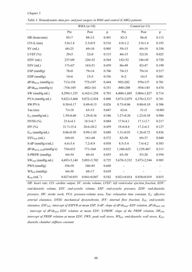
Chapter 5
88
Table 3. Hemodynamic data pre- and post surgery in RMA and control (CABG) patients
RMA (n=10) Control (n=13)
Pre Post p Pre Post p
HR (beats/min) 85±7 88±13 0.491 82±3 86±8 0.113
CO (L/min) 5.0±1.8 5.3±0.9 0.516 4.9±1.2 5.9±1.4 0.193
SV (mL) 68±25 69±10 0.905 59±15 69±19 0.350
LVEF (%) 29±5 32±8 0.315 46±15 52±18 0.025
EDV (mL) 237±89 226±52 0.564 142±52 146±45 0.720
ESV (mL) 171±67 163±51 0.459 86±49 82±47 0.190
ESP (mmHg) 78±8 79±14 0.706 74±13 79±14 0.517
EDP (mmHg) 14±6 15±5 0.356 8±2 14±5 0.001
dP/dtMAX (mmHg/s) 713±154 775±197 0.444 992±282 970±137 0.701
dP/dtMIN (mmHg/s) -754±105 -802±161 0.351 -880±208 -954±185 0.474
SW (mmHg.mL) 4,299±1,335 4,162±1,258 0.703 4,400±1,605 5,004±1,827 0.714
PVA (mmHg.mL) 9,422±3,460 9,072±2,924 0.808 5,873±2,079 6,376±2,517 0.761
SW/PVA 0.50±0.17 0.49±0.15 0.826 0.75±0.06 0.80±0.10 0.306
Tau (ms) 73±18 63±15 0.047 62±6 51±5 <0.001
EA (mmHg/mL) 1.39±0.60 1.29±0.36 0.546 1.27±0.20 1.22±0.38 0.984
DYSS (%) 23.6±4.3 18.5±6.7 0.084 17.8±4.1 17.1±2.7 0.217
IFF (%) 31.7±15.4 24.6±20.2 0.459 19.4±8.6 17.2±6.3 0.127
EES (mmHg/mL) 0.86±0.50 0.99±1.05 0.688 1.31±0.93 1.26±0.72 0.836
ESVIND (mL) 169±81 161±68 0.572 82±50 69±37 0.048
S-dP (mmHg/s/mL) 6.6±5.4 7.2±8.9 0.858 8.5±5.4 7.4±4.2 0.583
dP/dtMAX,,IND(mmHg/s) 734±633 771±264 0.832 1,160±625 1,129±467 0.313
S-PRSW (mmHg) 64±54 60±41 0.855 65±30 55±20 0.594
SWIND (mmHg.mL) 4,693±3,140 5,093±3,702 0.725 5,678±3,532 5,473±2,544 0.985
PWS (mmHg) 356±91 346±85 0.668 - - -
WSED (mmHg) 64±30 68±17 0.639 - - -
KED (mL-1) 0.027±0.035 0.041±0.047 0.542 0.021±0.014 0.038±0.019 0.015
HR: heart rate, CO: cardiac output, SV: stroke volume, LVEF: left ventricular ejection fraction, EDV:
end-diastolic volume, ESV: end-systolic volume, ESP: end-systolic pressure, EDP: end-diastolic
pressure, SW: stroke work, PVA: pressure-volume area, Tau: relaxation time constant, EA: effective
arterial elastance, DYSS: mechanical dyssynchrony, IFF: internal flow fraction, EES: end-systolic
elastance, ESVIND: intercept of ESPVR at mean ESP, S-dP: slope of dP/dtMAX–EDV relation, dP/dtMAX, IND
, intercept of dP/dtMAX–EDV relation at mean EDV, S-PRSW: slope of the PRSW relation, SWIND,
intercept of PRSW relation at mean EDV, PWS: peak wall stress, WSED: end-diastolic wall stress, KED:
diastolic chamber stiffness constant
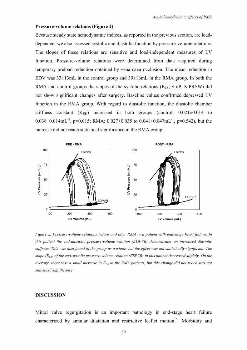
Acute hemodynamic effects of RMA
89
Pressure-volume relations (Figure 2)
Because steady state hemodynamic indices, as reported in the previous section, are load-
dependent we also assessed systolic and diastolic function by pressure-volume relations.
The slopes of these relations are sensitive and load-independent measures of LV
function. Pressure-volume relations were determined from data acquired during
temporary preload reduction obtained by vena cava occlusion. The mean reduction in
EDV was 33±13mL in the control group and 39±16mL in the RMA group. In both the
RMA and control groups the slopes of the systolic relations (EES, S-dP, S-PRSW) did
not show significant changes after surgery. Baseline values confirmed depressed LV
function in the RMA group. With regard to diastolic function, the diastolic chamber
stiffness constant (KED) increased in both groups (control: 0.021±0.014 to
0.038±0.014mL-1, p=0.015; RMA: 0.027±0.035 to 0.041±0.047mL-1, p=0.542), but the
increase did not reach statistical significance in the RMA group.
PRE - RMA POST - RMA
ESPVR ESPVR
EDPVREDPVR
0
25
50
75
100
100 200 300 400
LV Volume (mL)
LV P
ress
ure
(mm
Hg)
0
25
50
75
100
100 200 300 400
LV Volume (mL)
LV P
ress
ure
(mm
Hg)
Figure 2. Pressure-volume relations before and after RMA in a patient with end-stage heart failure. In
this patient the end-diastolic pressure-volume relation (EDPVR) demonstrates an increased diastolic
stiffness. This was also found in the group as a whole, but the effect was not statistically significant. The
slope (EES) of the end-systolic pressure-volume relation (ESPVR) in this patient decreased slightly. On the
average, there was a small increase in EES in the RMA patients, but this change did not reach was not
statistical significance
DISCUSSION
Mitral valve regurgitation is an important pathology in end-stage heart failure
characterized by annular dilatation and restrictive leaflet motion.21 Morbidity and
Chapter 5
90
mortality is high if mitral regurgitation is treated conservatively.22 Grigioni et al. clearly
demonstrated that the severity of mitral regurgitation is directly related to mortality
risk.1
Therefore, it seems reasonable to correct mitral regurgitation in patients with end-stage
heart failure to improve prognosis. Currently, mitral annuloplasty is not routinely
performed in these patients because substantial mortality and high recurrence rates are
reported, and no evidence from randomized studies is available.7,23 However, several
recent studies have shown relatively low operative mortality and suggest improved
long-term survival after stringent restrictive mitral annuloplasty.9,11,24 Unfortunately,
insights in the acute effects of RMA on systolic and diastolic LV performance are still
limited and concerns are raised about possible adverse acute effects on systolic and
diastolic LV function, which would limit application of this approach in patients with
end-stage heart failure. The aim of our study was therefore to quantify these effects by
use of load-independent pressure-volume indices.
We found unchanged systolic function after RMA. This is interesting, because earlier
studies had predicted adverse effects, which would be the result of an afterload
mismatch created by closure of a low-resistance run off into the left atrium. However,
this "pop-off" effect may not exist and the high mortality in earlier studies appears
mainly related to loss of LV function by disruption of the sub-valvular apparatus,
because in that time, valve replacement (rather than repair) was mostly performed.25
Effect on systolic function may also result from acute remodeling of the base of the
heart due to the undersized ring. Bolling et al. argue that this would improve systolic
function, however a study by David et al. implies a negative effect on systolic function
because an undersized ring presumably impairs stretching and shortening of the
proximal part of the basoconstrictor muscles (similar to a rigid ring).26,27 In our study,
we did not find any evidence for an altered, either reduced or improved, systolic
function. In addition, systolic anterior motion of the anterior leaflet leading to LV
outflow tract obstruction was not found in our series.
With regard to diastolic function we found an increase in diastolic chamber stiffness.
This effect was present in both groups, but it was only statistically significant in the
control patients. This increase in diastolic chamber stiffness is probably mainly an effect
of cardioplegic arrest, leading to interstitial myocardial edema.28 LV wall stress was
unchanged after RMA consistent with largely unchanged end-diastolic volume and
pressure.
Acute hemodynamic effects of RMA
91
The number of studies, in which effects of RMA on LV performance are evaluated, is
limited. Several studies show improved LV ejection fraction and reduced end-diastolic
volume.8,11,24,29 Bishay et al. reported improved LV function and reversed remodeling at
two years follow up in patients with severe LV dysfunction.30 However, this group was
heterogeneous and the patients underwent either mitral annuloplasty with various
techniques or mitral valve replacement. Bax et al. studied patients who strictly
underwent restrictive mitral annuloplasty, and showed that reverse remodeling of the
LV is a gradual and time-dependent process.9 These results are consistent with our
findings, which show no acute effects on LV performance after RMA. Interestingly, our
results show a clear tendency for a reduced mechanical dyssynchrony after RMA. This
index has recently been shown a very sensitive marker of LV dysfunction and
potentially this improvement may contribute to beneficial long-term effects.17
Limitations
The sample size in our study was relatively small and potentially positive effects on
systolic function may be demonstrated in a larger group of patients. However, we
performed pre- and post-CPB measurements in each patient, which optimizes the
statistical power to detect possible effects of the surgical intervention. In addition, the
RMA group was heterogeneous since in four patients additional CABG was performed.
This subgroup was too small for meaningful statistical analysis, but the effects on
pressure-volume relations in these patients did not appear to be different compared to
the effects in the whole group. Furthermore, beneficial effects on LV systolic function
in these patients would not be expected early after surgery as effects of
revascularization on hibernating myocardium often occur later after surgery.31
Measurements of global LV function were performed immediately after surgery with
open chest and during inotropic support. The confounding effects of inotropic support
were limited by also performing the measurements before surgery under inotropic
support, but possible altered β-receptor sensitivity cannot be excluded. Assessment of
regional function and of long-term effects under physiological conditions requires
further studies.
In conclusion, mitral valve repair by RMA effectively restores mitral valve leaflet
coaptation in patients with end-stage heart failure and severe mitral regurgitation,
without significant acute changes in baseline hemodynamics and LV systolic and

Chapter 5
92
diastolic function. Our findings support the use of this approach even in patients with
severely depressed LV function in view of the expected beneficial long-term results.
REFERENCES
1. Grigioni F, Enriquez-Sarano M, Zehr KJ, Bailey KR, Tajik AJ. Ischemic mitral regurgitation: long-
term outcome and prognostic implications with quantitative Doppler assessment. Circulation. 2001;103:1759-1764.
2. Hueb AC, Jatene FB, Moreira LF, Pomerantzeff PM, Kallas E, de Oliveira SA. Ventricular remodeling and mitral valve modifications in dilated cardiomyopathy: new insights from anatomic study. J Thorac Cardiovasc Surg. 2002;124:1216-1224.
3. Carpentier A. Cardiac valve surgery--the "French correction". J Thorac Cardiovasc Surg. 1983;86:323-337.
4. Aikawa K, Sheehan FH, Otto CM, Coady K, Bashein G, Bolson EL. The severity of functional mitral regurgitation depends on the shape of the mitral apparatus: A three-dimensional echo analysis. Journal of Heart Valve Disease. 2002;11:627-636.
5. Dion R. Ischemic mitral regurgitation: when and how should it be corrected? J Heart Valve Dis. 1993;2:536-543.
6. Mann DL. Mechanisms and models in heart failure: A combinatorial approach. Circulation. 1999;100:999-1008.
7. Chen FY, Adams DH, Aranki SF, Collins JJ, Jr., Couper GS, Rizzo RJ, Cohn LH. Mitral valve repair in cardiomyopathy. Circulation. 1998;98:II124-II127.
8. Bolling SF, Pagani FD, Deeb GM, Bach DS. Intermediate-term outcome of mitral reconstruction in cardiomyopathy. J Thorac Cardiovasc Surg. 1998;115:381-386.
9. Bax JJ, Braun J, Somer ST, Klautz R, Holman ER, Versteegh MI, Boersma E, Schalij MJ, van der Wall EE, Dion RA. Restrictive annuloplasty and coronary revascularization in ischemic mitral regurgitation results in reverse left ventricular remodeling. Circulation. 2004;110:II103-II108.
10. Dreyfus G, Milaiheanu S. Mitral valve repair in cardiomyopathy. J Heart Lung Transplant. 2000;19:S73-S76.
11. Bolling SF, Deeb GM, Brunsting LA, Bach DS. Early outcome of mitral valve reconstruction in patients with end-stage cardiomyopathy. J Thorac Cardiovasc Surg. 1995;109:676-682.
12. Thomas JD. How leaky is that mitral valve? Simplified Doppler methods to measure regurgitant orifice area. Circulation. 1997;95:548-550.
13. Calafiore AM, Teodori G, Mezzetti A, Bosco G, Verna AM, Di Giammarco G, Lapenna D. Intermittent antegrade warm blood cardioplegia. Ann Thorac Surg. 1995;59:398-402.
14. Carpentier AF, Lessana A, Relland JY, Belli E, Mihaileanu S, Berrebi AJ, Palsky E, Loulmet DF. The "physio-ring": an advanced concept in mitral valve annuloplasty. Ann Thorac Surg. 1995;60:1177-1185.
15. Tulner SA, Klautz RJ, Rijk-Zwikker GL, Engbers FH, Bax JJ, Baan J, van der Wall EE, Dion RA, Steendijk P. Peri-operative assessment of left ventricular function by pressure-volume loops using the conductance catheter method. Anesth Analg. 2003;97:950-7, table.
16. Steendijk P, Staal E, Jukema JW, Baan J. Hypertonic saline method accurately determines parallel conductance for dual-field conductance catheter. Am J Physiol Heart Circ Physiol. 2001;281:H755-H763.
17. Steendijk P, Tulner SA, Schreuder JJ, Bax JJ, Van Erven L, van der Wall EE, Dion RA, Schalij MJ, Baan J. Quantification of left ventricular mechanical dyssynchrony by conductance catheter in heart failure patients. Am J Physiol Heart Circ Physiol. 2004;286:H723-H730.
18. Arts T, Bovendeerd PH, Prinzen FW, Reneman RS. Relation between left ventricular cavity pressure and volume and systolic fiber stress and strain in the wall. Biophys J. 1991;59:93-102.
19. Mandinov L, Eberli FR, Seiler C, Hess OM. Diastolic heart failure. Cardiovasc Res. 2000;45:813-825.
20. Sagawa K. The end-systolic pressure-volume relation of the ventricle: definition, modifications and clinical use. Circulation. 1981;63:1223-1227.
21. Kwan J, Shiota T, Agler DA, Popovic ZB, Qin JX, Gillinov MA, Stewart WJ, Cosgrove DM, McCarthy PM, Thomas JD. Geometric differences of the mitral apparatus between ischemic and dilated cardiomyopathy with significant mitral regurgitation: real-time three-dimensional echocardiography study. Circulation. 2003;107:1135-1140.

Acute hemodynamic effects of RMA
93
22. Trichon BH, Glower DD, Shaw LK, Cabell CH, Anstrom KJ, Felker GM, O'Connor CM. Survival after coronary revascularization, with and without mitral valve surgery, in patients with ischemic mitral regurgitation. Circulation. 2003;108 Suppl 1:II103-II110.
23. Tahta SA, Oury JH, Maxwell JM, Hiro SP, Duran CM. Outcome after mitral valve repair for functional ischemic mitral regurgitation. J Heart Valve Dis. 2002;11:11-18.
24. Gummert JF, Rahmel A, Bucerius J, Onnasch J, Doll N, Walther T, Falk V, Mohr FW. Mitral valve repair in patients with end stage cardiomyopathy: who benefits? Eur J Cardiothorac Surg. 2003;23:1017-1022.
25. Bonchek LI, Olinger GN, Siegel R, Tresch DD, Keelan MH, Jr. Left ventricular performance after mitral reconstruction for mitral regurgitation. J Thorac Cardiovasc Surg. 1984;88:122-127.
26. Bolling SF, Smolens IA, Pagani FD. Surgical alternatives for heart failure. J Heart Lung Transplant. 2001;20:729-733.
27. David TE, Komeda M, Pollick C, Burns RJ. Mitral valve annuloplasty: the effect of the type on left ventricular function. Ann Thorac Surg. 1989;47:524-527.
28. Ericsson AB, Takeshima S, Vaage J. Simultaneous antegrade and retrograde delivery of continuous warm blood cardioplegia after global ischemia. J Thorac Cardiovasc Surg. 1998;115:716-722.
29. Rothenburger M, Rukosujew A, Hammel D, Dorenkamp A, Schmidt C, Schmid C, Wichter T, Scheld HH. Mitral valve surgery in patients with poor left ventricular function. Thorac Cardiovasc Surg. 2002;50:351-354.
30. Bishay ES, McCarthy PM, Cosgrove DM, Hoercher KJ, Smedira NG, Mukherjee D, White J, Blackstone EH. Mitral valve surgery in patients with severe left ventricular dysfunction. Eur J Cardiothorac Surg. 2000;17:213-221.
31. Bax JJ, Visser FC, Poldermans D, Elhendy A, Cornel JH, Boersma E, van Lingen A, Fioretti PM, Visser CA. Time course of functional recovery of stunned and hibernating segments after surgical revascularization. Circulation. 2001;104:I314-I318.

CHAPTER 6
Surgical ventricular restoration in patients with ischemic
dilated cardiomyopathy: Evaluation of systolic and diastolic
ventricular function, wall stress, dyssynchrony, and
mechanical efficiency by pressure-volume loops
S.A.F. Tulner
P. Steendijk
R.J.M. Klautz
J.J. Bax
M.J. Schalij
E.E. van der Wall
R.A.E. Dion
J Thorac Cardiovasc Surg (in press)
Oral presentation at the RESTORE meeting during American Association
Thoracic Surgery, April 2004, Toronto, Canada
Oral presentation at Nederlandse Vereniging Voor Cardiologie, October 2004,
Ermelo, The Netherlands (Awarded as best oral presentation)
Chapter 6
96
ABSTRACT
Objectives. Surgical ventricular restoration (SVR) aims at improving cardiac function
by normalization of left ventricular (LV) shape and size. Recent studies indicate that
SVR is highly effective with an excellent five-year outcome in patients with ischemic
dilated cardiomyopathy. We used pressure-volume analysis to investigate acute changes
in systolic and diastolic LV function, mechanical dyssynchrony and efficiency, and wall
stress.
Methods. In three patient groups (total, n=33), pressure-volume loops were measured
by conductance catheter before and after surgery. The main study group consisted of 10
patients with ischemic dilated cardiomyopathy (NYHA III/IV, LV ejection fraction
<30%) who underwent SVR and CABG. In this group, 7 patients underwent additional
restrictive mitral annuloplasty (RMA). To assess potential confounding effects of RMA
and cardiopulmonary bypass, we included a group of 10 patients (NYHA III/IV, LV
ejection fraction <30%) who underwent isolated RMA and a group of 13 patients with
preserved LV function who underwent isolated CABG.
Results. After SVR, end-diastolic and end-systolic volumes were reduced: 211±54 to
169±34 mL (p=0.03), and 147±41 to 110±59 mL (p=0.04), respectively. LV ejection
fraction (27±7 to 37±13%, p=0.04) and end-systolic elastance (1.12±0.71 to 1.57±0.63
mmHg/mL, p=0.03) improved. Peak wall stress (358±108 to 244±79 mmHg, p<0.01)
and mechanical dyssynchrony (26±4 to 19±6%, p<0.01) were reduced, whereas
mechanical efficiency improved (0.34±13 to 0.49±0.14, p=0.03). End-diastolic pressure
increased (13±6 to 20±5 mmHg, p<0.01), whereas the diastolic chamber stiffness
constant tended to be increased (0.021±0.009 to 0.037±0.021 mL-1, NS).
Conclusions. SVR achieves normalization of LV volumes and improves systolic
function and mechanical efficiency by reducing LV wall stress and mechanical
dyssynchrony.
INTRODUCTION
Surgical ventricular restoration (SVR) by means of endoventricular circular patch plasty
(Dor procedure) is beneficial in patients with left ventricular (LV) post-infarction
aneurysm. Previous studies have shown that this procedure is safe, improves functional
class, long-term survival, and LV ejection fraction.1,2 The exclusion of akinetic or
SVR in patients with ischemic dilated cardiomyopathy
97
dyskinetic segments achieves acute volume reduction, changes in LV shape, and
decreases of LV dyssynchrony.3,4 These acute changes will influence LV global and
intrinsic systolic and diastolic function. The use of pressure-volume analysis to assess
these effects is advantageous because pressure-volume relations accurately reflect
intrinsic LV function, and are relatively independent of loading conditions.5,6 Moreover,
pressure-volume signals can be used to quantify mechanical dyssynchrony and LV wall
stress.7
Theoretical studies predict that volume reduction surgery results in leftward and upward
shifts of the end-systolic and end-diastolic pressure-volume relations in the pressure-
volume diagram, indicating a positive effect on systolic function but an adverse effect
on diastolic function.8,9 However, these effects are likely to be modulated by the
material properties and the size of the resected or excluded region. Artrip et al.
quantified the differential effects of volume reduction on end-systolic and end-diastolic
function in a mathematical model.10 Their findings indicate that an overall negative
effect on LV pump function results if weak but contracting myocardium is resected (like
in the Batista procedure), beneficial effects if the excised region is dyskinetic, and
equivocal effects with akinetic scar resection. However, whether these models are
realistic is unknown since in-vivo data on the effects of SVR and related procedures on
LV pressure-volume relations in humans are very limited. One important aspect, which
is not taken into account by these particular models, is (alterations in) mechanical
dyssynchrony. Recent studies demonstrated that LV mechanical synchrony substantially
improves after SVR resulting in more efficient myocardial pump function.3,4
Furthermore, a recent Special Report from the RESTORE group emphasized the
importance of considering interaction and (re)arrangement of myocardial layers and
fiber orientation, and stressed the need for additional studies to quantify the effects of
SVR and to get a better insight in the underlying mechanisms.11
As SVR reversely remodels ventricular size and shape, this approach may alter systolic
and diastolic function.11,12 Additionally, SVR may decrease LV wall stress and
myocardial oxygen consumption by reducing end-diastolic volume, resulting in
improved functioning of the remote myocardium.13 The aim of this study was to
determine the acute effects of SVR on systolic and diastolic pressure-volume
relationships, LV wall stress, and mechanical dyssynchrony and efficiency in patients
with ischemic dilated cardiomyopathy.
Chapter 6
98
METHODS
Patients
The main study group consisted of 10 patients with ischemic dilated cardiomyopathy
who underwent SVR. SVR is often combined with restrictive mitral annuloplasty
(RMA) and therefore we also included a group of patients with left ventricular
dysfunction in which isolated RMA was performed. To assess confounding effects of
cardiopulmonary bypass and cardioplegic cardiac arrest we also included a control
group of patients with normal LV function who underwent elective coronary artery
bypass grafting (CABG). Thus, the following groups were studied:
1) SVR-group (n=10): Chronic heart failure, New York Heart Association (NYHA)
class III/IV, LV ejection fraction < 30%, LV aneurysm with or without mitral
regurgitation
2) RMA-group (n=10): Chronic heart failure, NYHA class III/IV, LV ejection
fraction < 30%, mitral regurgitation grade ≥ 2
3) CABG-group (n=13): normal LV function (LV ejection fraction > 40%),
elective CABG
Note that some patients in the SVR-group underwent additional RMA, whereas in both
the SVR- and the RMA-group, CABG was performed if indicated. Details are provided
in the Results section. The study was approved by the institutional review committee
and all patients gave informed consent. The patient characteristics of the three groups
are summarized in Table 1.
Anaesthesia and cardioplegic arrest
All patients received total intravenous anesthesia with target-controlled infusion of
propofol, remifentanyl and sufentanyl. A single dose of pancuronium bromide
(0.1mg/kg) was given to facilitate intubation. Subsequently, a thermal filament catheter
was placed in the pulmonary artery via the right internal jugular vein for semi-
continuous cardiac output measurements (Edwards Lifesciences, Uden, The
Netherlands). To facilitate positioning of the conductance catheter and to evaluate the
effects of mitral valve repair, a multiplane transesophageal echo probe was inserted. All
patients underwent normothermic cardiopulmonary bypass and received intermittent
antegrade warm blood cardioplegia as described by Calafiore et al.14
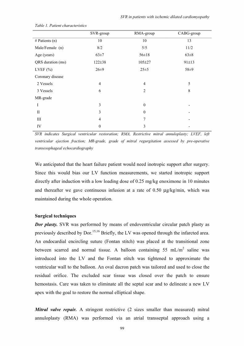
SVR in patients with ischemic dilated cardiomyopathy
99
Table 1. Patient characteristics
SVR-group RMA-group CABG-group
# Patients (n) 10 10 13
Male/Female (n) 8/2 5/5 11/2
Age (years) 63±7 56±18 63±8
QRS duration (ms) 122±38 105±27 91±13
LVEF (%) 26±9 25±5 58±9
Coronary disease
2 Vessels
3 Vessels
4
6
4
2
5
8
MR-grade
I
II
III
IV
3
3
4
0
0
0
7
3
-
-
-
-
SVR indicates Surgical ventricular restoration; RMA, Restrictive mitral annuloplasty; LVEF, left
ventricular ejection fraction; MR-grade, grade of mitral regurgitation assessed by pre-operative
transesophageal echocardiography
We anticipated that the heart failure patient would need inotropic support after surgery.
Since this would bias our LV function measurements, we started inotropic support
directly after induction with a low loading dose of 0.25 mg/kg enoximone in 10 minutes
and thereafter we gave continuous infusion at a rate of 0.50 μg/kg/min, which was
maintained during the whole operation.
Surgical techniques
Dor plasty. SVR was performed by means of endoventricular circular patch plasty as
previously described by Dor.15,16 Briefly, the LV was opened through the infarcted area.
An endocardial encircling suture (Fontan stitch) was placed at the transitional zone
between scarred and normal tissue. A balloon containing 55 mL/m2 saline was
introduced into the LV and the Fontan stitch was tightened to approximate the
ventricular wall to the balloon. An oval dacron patch was tailored and used to close the
residual orifice. The excluded scar tissue was closed over the patch to ensure
hemostasis. Care was taken to eliminate all the septal scar and to delineate a new LV
apex with the goal to restore the normal elliptical shape.
Mitral valve repair. A stringent restrictive (2 sizes smaller than measured) mitral
annuloplasty (RMA) was performed via an atrial transseptal approach using a
Chapter 6
100
Carpentier Edwards Physio ring (Edwards Lifesciences, USA). After weaning from
cardiopulmonary bypass, transesophageal echocardiographic evaluation was performed
in all patients to confirm disappearance of mitral regurgitation and assess the length of
leaflet coaptation (aiming at ≥ 8 mm).
Study protocol
Before and directly after cardiopulmonary bypass, conductance catheter measurements
were performed as described previously.17 Briefly, temporary epicardial pacemaker
wires were placed on the right atrium to enable measurements at fixed heart rates. A
tourniquet was placed around the inferior caval vein to enable temporary preload
reductions. An 8F sheath was placed in the ascending aorta for introduction of the
conductance catheter. The conductance catheter was introduced under transesophageal
echocardiographic guidance and placed along the long axis of the LV. Position was
optimized by inspection of the segmental volume signals. Conductance catheter
calibration was performed using calibration factors alpha (α) derived from
thermodilution and parallel conductance correction volume (Vc) determined by
hypertonic saline injections.5,18 Continuous LV pressure and volume signals derived
from the conductance catheter were displayed and acquired at a 250 Hz sampling rate
using a Leycom CFL-512 (CD Leycom, Zoetermeer, The Netherlands). Data were
acquired during steady state and during temporary caval vein occlusion, all with the
ventilator turned off at end-expiration. Acquisition was performed at a fixed atrial
pacing rate of 80 beats/min. From these signals hemodynamic indexes were derived as
described below.
Data analysis
Global LV function. We determined indexes of global, systolic and diastolic LV
function. Cardiac output was obtained by thermodilution, heart rate, mean arterial
pressure, stroke volume, LV ejection fraction, minimal and maximal rate of LV pressure
change (dP/dtMAX, dP/dtMIN), end-diastolic volume, end-systolic volume, end-diastolic
pressure, end-systolic pressure were obtained from steady state beats using custom-
made software. In addition, we assessed the early, active part of relaxation by the
relaxation time constant (τ), which was determined by fitting LV pressure decay
(starting at the moment of minimal dP/dt) with an exponential curve, as described
previously19: P(t) = A + B·exp(-t/τ). Time-varying wall stress, WS(t), was calculated
from instantaneous LV pressure and volume signals (P(t), V(t) respectively) as
SVR in patients with ischemic dilated cardiomyopathy
101
described by Arts et al.20: WS(t) = P(t)·[1 + 3·V(t) / VWALL]. LV wall volume (VWALL)
was estimated based on the diastolic posterior wall thickness derived from M-mode
echocardiography.
Mechanical work and efficiency. Stroke work (SW) was determined as the area of the
pressure-volume loop, which represents the external work performed by the ventricle.
Pressure-volume area (PVA), a measure of total mechanical work, was calculated as the
sum of stroke work and potential energy. The latter represents mechanical energy loss
converted to heat during the cardiac cycle and is quantified by the triangular area
enclosed by the pressure-volume loop, the end-systolic pressure-volume relation and the
end-diastolic pressure-volume relation. 21,22 Mechanical efficiency (ME) was calculated
as the ratio of stroke work and pressure-volume area: ME = SW / PVA.23
Mechanical dyssynchrony. Nonuniform LV performance (dyssynchrony) was
determined from the segmental LV conductance signals and quantified by calculating
the percentage of time within the cardiac cycle that a specific segment is
dyssynchronous (i.e. opposite in phase with the global LV volume signal). Overall LV
mechanical dyssynchrony was determined as the mean of the segmental dyssynchronies.
In addition, we calculated the internal flow fraction, which quantifies the ineffective,
segment-to-segment shifting of blood volume within the LV due to nonuniform
contraction and filling. This approach was described and validated vs. tissue-Doppler
imaging in a previous study.7
Systolic and diastolic pressure-volume relations. Ventricular function was assessed by
systolic and diastolic pressure-volume relations derived from pressure-volume loops
acquired during gradual preload reduction by vena cava occlusion. The end-systolic
pressure-volume relation (ESPVR) was obtained as a linear fit to the end-systolic
pressure-volume points and characterized by its slope, end-systolic elastance (EES), and
the volume intercept at an end-systolic pressure of 80 mmHg (ESV80). The end-diastolic
pressure-volume points were fitted with an exponential curve: EDP = A + B·exp
(KED·EDV). As illustrated in Figure 1, this relation was quantified by the diastolic
stiffness constant (KED), the pressure intercept at an end-diastolic volume of 0 mL
(EDP0), and the calculated volume intercept at an end-diastolic pressure of 14 mmHg
(EDV14).24,25
Chapter 6
102
ESPVR EES
ESP=80mmHg
EDPVR
EDP=14mmHgEDP0
ESV80 EDV14
0
40
80
120
0 300
LV Volume (mL)
LV P
ress
ure
(mm
Hg)
Figure 1. The end-systolic pressure-volume relation (ESPVR) and the end-diastolic pressure-volume
relation (EDPVR) in the pressure-volume diagram. The linear ESPVR is characterized by its slope, end-
systolic elastance (EES), and its volume intercept at an end-systolic pressure of 80 mmHg (ESV80). The
exponential EDPVR is characterized by the pressure-intercept at an end-diastolic volume of 0 mmHg
(EDP0), the volume intercept at an end-diastolic pressure of 14 mmHg (EDV14), and the diastolic stiffness
constant KED. (See text for further details)
Statistical analysis
Pre- and post-surgery clinical and hemodynamic indexes were compared with paired t-
tests. Changes in systolic and diastolic pressure-volume relations were tested by
multivariate analysis of covariance, using the Wilks’ lambda statistic to test whether
there were differences between conditions for the combination of parameters describing
the relations.26 Statistical significance was assumed at p < 0.05. All data are presented
as the mean ± SD.
RESULTS
Surgical data are summarized in Table 2. In the SVR-group, all patients were treated
with endoventricular circular patch plasty: 7 patients had a dyskinetic scar on pre-
operative echocardiography, the remaining 3 patients had an akinetic scar. All patients

SVR in patients with ischemic dilated cardiomyopathy
103
in the SVR-group had coronary disease and received additional CABG (2.8±1.4 distal
anastomoses per patient). In the SVR-group 7 patients had mitral regurgitation of grade
2 or more and received additional restrictive mitral annuloplasty. In the RMA-group, 4
patients received additional CABG (4.0±0.8 distal anastomoses per patient), while the
other 6 patients in this group underwent isolated restrictive mitral annuloplasty as 2 had
irreversible ischemia and 4 had non-ischemic dilated cardiomyopathy. All patients were
successfully weaned from cardiopulmonary bypass. In the SVR-group, 2 patients
received intra-aortic balloon pump support and 7 patients needed inotropic support for
more than 24 hours.
Table 2: Surgical data
SVR-group
(n=10)
RMA-group
(n=10)
CABG-group
(n=13)
Surgery
SVR + CABG
SVR + CABG + RMA
Isolated RMA
RMA + CABG
CABG
3
7
-
-
-
-
-
6
4
-
-
-
-
-
13
CPB- time (median, minutes) 244 (range 105-287) 137 (range 105-287) 104 (range 60-167)
Aox-time (median, minutes) 172 (range 65-196) 96 (range 65-196) 75 (range 43-129)
# pts with IABP support 2 0 0
# pts with >24 hrs inotropes* 7 5 0
ICU-duration (median, days) 4 (range 3-16) 4 (range 2-7) 2 (range 1-4)
Hospital stay (median, days) 14 (range 9-30) 14 (range 7-18) 9 (range 6-35)
SVR indicates Surgical ventricular restoration; RMA, Restrictive mitral annuloplasty; CPB,
Cardiopulmonary bypass; Aox-time, aortic cross clamping time; IABP, Intra-aortic balloon pump
support; ICU, Intensive care unit; * dobutamine > 2 μg/kg/min
In the RMA-group, 5 patients needed inotropic support for more than 24 hours. None of
the patients had signs of peri-operative myocardial infarction. In patients with mitral
regurgitation, restrictive mitral annuloplasty suppressed regurgitation in all cases and
restored leaflet coaptation (8±2 mm) with normal peak pressure gradients (3.0±2.0
mmHg). All patients were discharged from hospital in good clinical condition.
Figure 2 shows typical pressure-volume relations before and after SVR. After SVR,
end-diastolic and end-systolic volumes were significantly reduced with unchanged
stroke volume indicating improved LV ejection fraction. Before surgery, LV volume
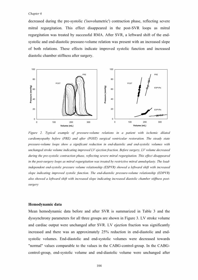
Chapter 6
104
decreased during the pre-systolic ('isovolumetric') contraction phase, reflecting severe
mitral regurgitation. This effect disappeared in the post-SVR loops as mitral
regurgitation was treated by successful RMA. After SVR, a leftward shift of the end-
systolic and end-diastolic pressure-volume relation was present with an increased slope
of both relations. These effects indicate improved systolic function and increased
diastolic chamber stiffness after surgery.
PRE PRE
POST POST
ESPVRs
EDPVRs
0
20
40
60
80
100
0 100 200 300
Volume (mL)
Pres
sure
(mm
Hg)
0
20
40
60
80
100
0 100 200 300Volume (mL)
Pres
sure
(mm
Hg)
Figure 2. Typical example of pressure-volume relations in a patient with ischemic dilated
cardiomyopathy before (PRE) and after (POST) surgical ventricular restoration. The steady state
pressure-volume loops show a significant reduction in end-diastolic and end-systolic volumes with
unchanged stroke volume indicating improved LV ejection fraction. Before surgery, LV volume decreased
during the pre-systolic contraction phase, reflecting severe mitral regurgitation. This effect disappeared
in the post-surgery loops as mitral regurgitation was treated by restrictive mitral annuloplasty. The load-
independent end-systolic pressure volume relationship (ESPVR) showed a leftward shift with increased
slope indicating improved systolic function. The end-diastolic pressure-volume relationship (EDPVR)
also showed a leftward shift with increased slope indicating increased diastolic chamber stiffness post-
surgery
Hemodynamic data
Mean hemodynamic data before and after SVR is summarized in Table 3 and the
dyssynchrony parameters for all three groups are shown in Figure 3. LV stroke volume
and cardiac output were unchanged after SVR. LV ejection fraction was significantly
increased and there was an approximately 25% reduction in end-diastolic and end-
systolic volumes. End-diastolic and end-systolic volumes were decreased towards
"normal" values comparable to the values in the CABG-control-group. In the CABG-
control-group, end-systolic volume and end-diastolic volume were unchanged after

SVR in patients with ischemic dilated cardiomyopathy
105
surgery (86±49 to 82±47 mL (P=0.190) and 142±52 to 146±45 mL (P=0.720),
respectively). After SVR, stroke work was not significantly altered, but potential energy
was substantially reduced (-52%), resulting in a decreased total mechanical work and,
consequently, a significantly increased mechanical efficiency. Peak LV wall stress was
significantly reduced after SVR (from 358±108 to 244±79 mmHg, p<0.01), but
remained unchanged in the RMA-group (356±91 to 346±85 mmHg, p=0.668).
Table 3: Hemodynamic data before (pre) and after (post) SVR
SVR-group (n=10)
Pre Post P-value
HR (beats/min) 81±3 84±7 0.22
CO (L/min) 4.6±1.1 5.4±1.4 0.15
MAP 78±9 63±4 <0.01
ESP 95±18 80±15 0.03
EDV (mL) 211±54 169±34 0.03
ESV (mL) 147±41 110±59 0.04
LVEF (%) 27±7 37±13 0.04
SW (mmHg.L) 4.8±1.5 4.2±1.2 0.32
PE (mmHg.L) 10.6±6.1 5.1±3.5 <0.01
PVA (mmHg.L) 15.4±5.9 9.3±3.5 <0.01
ME 0.34±0.13 0.49±0.14 0.03
dP/dtMAX (mmHg/s) 846±232 819±198 0.64
dP/dtMIN (mmHg/s) -804±191 -750±110 0.25
PWS (mmHg) 358±108 244±79 < 0.01
EDP (mmHg) 13±6 20±5 < 0.01
τ (ms) 85±13 70±12 < 0.01
DYSS (%) 26±4 19±6 < 0.01
IFF (%) 35±14 21±15 0.01
SVR indicates surgical ventricular restoration; HR, heart rate; CO, cardiac output; MAP, mean arterial
pressure; ESP, end-systolic pressure; EDV, end-diastolic volume; ESV, end-systolic volume; LVEF, left
ventricular ejection fraction; SW, stroke work; PE, potential energy; PVA, pressure-volume area; ME,
mechanical efficiency; PWS, peak wall stress; EDP, end-diastolic pressure; τ, relaxation time constant;
DYSS, mechanical dyssynchrony; IFF: internal flow fraction
Active relaxation (τ) was improved, while end-diastolic pressure was significantly
increased. Mechanical dyssynchrony and the internal flow fraction were reduced in all
groups, however these changes were most pronounced and only reached statistical
significance in the SVR-group (Figure 3).
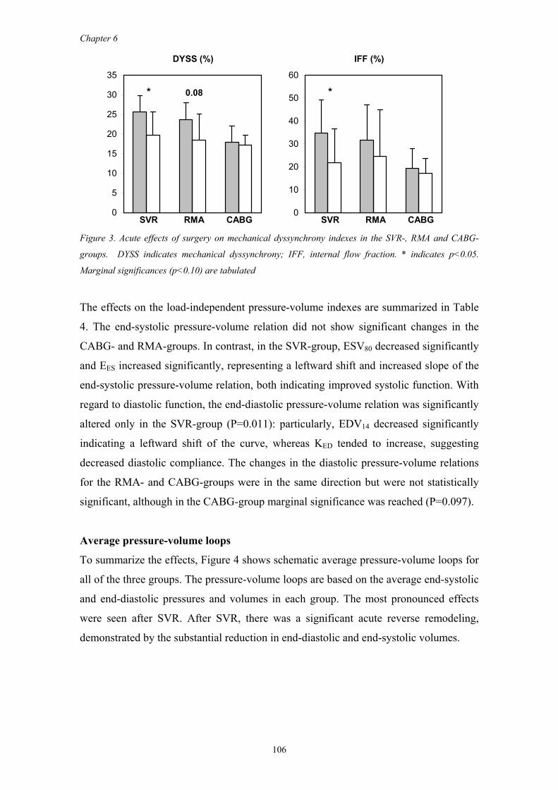
Chapter 6
106
DYSS (%) IFF (%)
* 0.08 *
SVR RMA CABG SVR RMA CABG0
5
10
15
20
25
30
35
0
10
20
30
40
50
60
Figure 3. Acute effects of surgery on mechanical dyssynchrony indexes in the SVR-, RMA and CABG-
groups. DYSS indicates mechanical dyssynchrony; IFF, internal flow fraction. * indicates p<0.05.
Marginal significances (p<0.10) are tabulated
The effects on the load-independent pressure-volume indexes are summarized in Table
4. The end-systolic pressure-volume relation did not show significant changes in the
CABG- and RMA-groups. In contrast, in the SVR-group, ESV80 decreased significantly
and EES increased significantly, representing a leftward shift and increased slope of the
end-systolic pressure-volume relation, both indicating improved systolic function. With
regard to diastolic function, the end-diastolic pressure-volume relation was significantly
altered only in the SVR-group (P=0.011): particularly, EDV14 decreased significantly
indicating a leftward shift of the curve, whereas KED tended to increase, suggesting
decreased diastolic compliance. The changes in the diastolic pressure-volume relations
for the RMA- and CABG-groups were in the same direction but were not statistically
significant, although in the CABG-group marginal significance was reached (P=0.097).
Average pressure-volume loops
To summarize the effects, Figure 4 shows schematic average pressure-volume loops for
all of the three groups. The pressure-volume loops are based on the average end-systolic
and end-diastolic pressures and volumes in each group. The most pronounced effects
were seen after SVR. After SVR, there was a significant acute reverse remodeling,
demonstrated by the substantial reduction in end-diastolic and end-systolic volumes.
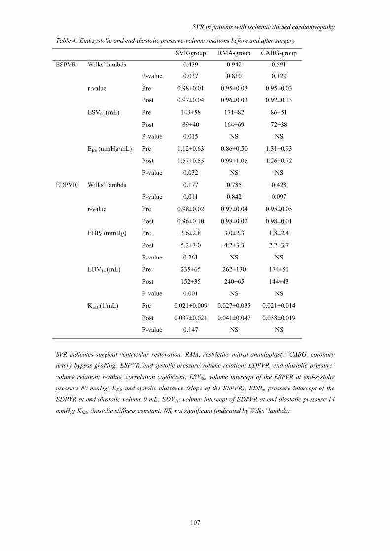
SVR in patients with ischemic dilated cardiomyopathy
107
Table 4: End-systolic and end-diastolic pressure-volume relations before and after surgery
SVR-group RMA-group CABG-group
ESPVR Wilks’ lambda 0.439 0.942 0.591
P-value 0.037 0.810 0.122
r-value Pre 0.98±0.01 0.95±0.03 0.95±0.03
Post 0.97±0.04 0.96±0.03 0.92±0.13
ESV80 (mL) Pre 143±58 171±82 86±51
Post 89±40 164±69 72±38
P-value 0.015 NS NS
EES (mmHg/mL) Pre 1.12±0.63 0.86±0.50 1.31±0.93
Post 1.57±0.55 0.99±1.05 1.26±0.72
P-value 0.032 NS NS
EDPVR Wilks’ lambda 0.177 0.785 0.428
P-value 0.011 0.842 0.097
r-value Pre 0.98±0.02 0.97±0.04 0.95±0.05
Post 0.96±0.10 0.98±0.02 0.98±0.01
EDP0 (mmHg) Pre 3.6±2.8 3.0±2.3 1.8±2.4
Post 5.2±3.0 4.2±3.3 2.2±3.7
P-value 0.261 NS NS
EDV14 (mL) Pre 235±65 262±130 174±51
Post 152±35 240±65 144±43
P-value 0.001 NS NS
KED (1/mL) Pre 0.021±0.009 0.027±0.035 0.021±0.014
Post 0.037±0.021 0.041±0.047 0.038±0.019
P-value 0.147 NS NS
SVR indicates surgical ventricular restoration; RMA, restrictive mitral annuloplasty; CABG, coronary
artery bypass grafting; ESPVR, end-systolic pressure-volume relation; EDPVR, end-diastolic pressure-
volume relation; r-value, correlation coefficient; ESV80, volume intercept of the ESPVR at end-systolic
pressure 80 mmHg; EES, end-systolic elastance (slope of the ESPVR); EDP0, pressure intercept of the
EDPVR at end-diastolic volume 0 mL; EDV14, volume intercept of EDPVR at end-diastolic pressure 14
mmHg; KED, diastolic stiffness constant; NS, not significant (indicated by Wilks’ lambda)
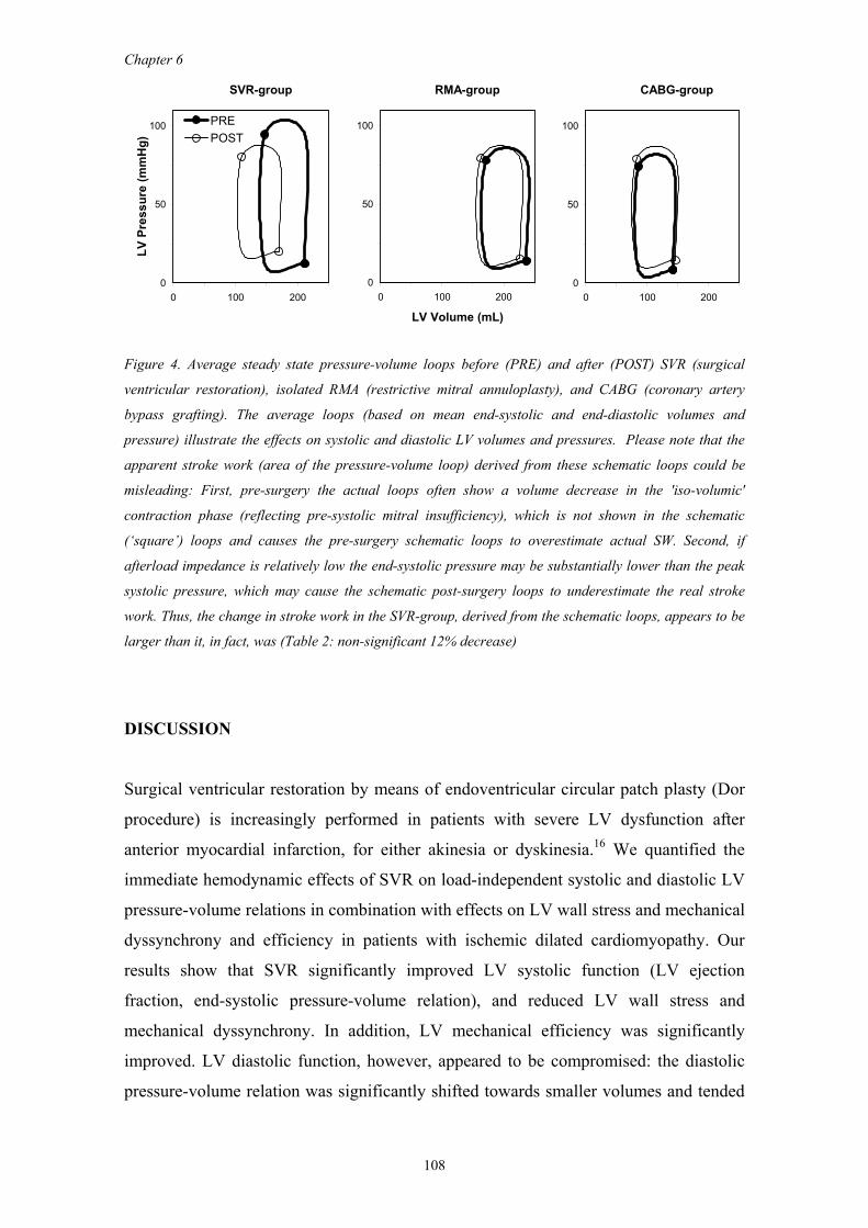
Chapter 6
108
SVR-group RMA-group CABG-group
0
50
100
0 100 200
LV P
ress
ure
(mm
Hg)
PREPOST
0
50
100
0 100 200
LV Volume (mL)
0
50
100
0 100 200
Figure 4. Average steady state pressure-volume loops before (PRE) and after (POST) SVR (surgical
ventricular restoration), isolated RMA (restrictive mitral annuloplasty), and CABG (coronary artery
bypass grafting). The average loops (based on mean end-systolic and end-diastolic volumes and
pressure) illustrate the effects on systolic and diastolic LV volumes and pressures. Please note that the
apparent stroke work (area of the pressure-volume loop) derived from these schematic loops could be
misleading: First, pre-surgery the actual loops often show a volume decrease in the 'iso-volumic'
contraction phase (reflecting pre-systolic mitral insufficiency), which is not shown in the schematic
(‘square’) loops and causes the pre-surgery schematic loops to overestimate actual SW. Second, if
afterload impedance is relatively low the end-systolic pressure may be substantially lower than the peak
systolic pressure, which may cause the schematic post-surgery loops to underestimate the real stroke
work. Thus, the change in stroke work in the SVR-group, derived from the schematic loops, appears to be
larger than it, in fact, was (Table 2: non-significant 12% decrease)
DISCUSSION
Surgical ventricular restoration by means of endoventricular circular patch plasty (Dor
procedure) is increasingly performed in patients with severe LV dysfunction after
anterior myocardial infarction, for either akinesia or dyskinesia.16 We quantified the
immediate hemodynamic effects of SVR on load-independent systolic and diastolic LV
pressure-volume relations in combination with effects on LV wall stress and mechanical
dyssynchrony and efficiency in patients with ischemic dilated cardiomyopathy. Our
results show that SVR significantly improved LV systolic function (LV ejection
fraction, end-systolic pressure-volume relation), and reduced LV wall stress and
mechanical dyssynchrony. In addition, LV mechanical efficiency was significantly
improved. LV diastolic function, however, appeared to be compromised: the diastolic
pressure-volume relation was significantly shifted towards smaller volumes and tended
SVR in patients with ischemic dilated cardiomyopathy
109
to be steeper, evidenced by an increased diastolic stiffness constant, although the latter
effect did not reach statistical significance.
The relatively small changes in systolic function in the patients who underwent isolated
restrictive mitral annuloplasty and in the patients who underwent elective CABG
indicate that the systolic improvements in the SVR group were mainly related to LV
restoration. The increase in LV ejection fraction after SVR was attributed to the surgical
reduction in end-diastolic volume, as LV stroke volume was unchanged. However, LV
ejection fraction may not be an accurate parameter of systolic improvement after SVR
because loading conditions may have changed substantially after surgery. Thus, load-
independent pressure-volume relations are needed to quantify alterations in systolic
function. The slope of the end-systolic pressure-volume relation, end-systolic elastance
EES, is a load-independent parameter of systolic function and EES increased significantly
after SVR. Moreover, the end-systolic pressure-volume relation was significantly
shifted towards smaller volumes, also indicating improved systolic function.26,27 This
improvement may be the result of increased systolic stiffness induced by exclusion of a
large compliant area, as predicted by computational models,10 or due to improved
function of the remote myocardium by reduced LV wall stress, and reduced LV
mechanical dyssynchrony after exclusion of the aneurysm.3,4
Regarding diastolic function, relaxation time constant τ was significantly reduced,
indicating faster relaxation. This time-constant quantifies the speed of LV pressure
decay during isovolumic relation, i.e. between aortic valve closure and mitral valve
opening, which represent the very early, and active, part of relaxation, which is
considered to be importantly co-determined by systolic function.28 This change may
result from coronary revascularization - which may enhance the oxygen dependent re-
uptake process of calcium by the sarcoplasmic reticulum - or from an afterload
reduction as active relaxation is afterload dependent.29 Passive diastolic function was
assessed by the diastolic pressure-volume relationship. Our results show that SVR
induced a substantial leftward shift of the end-diastolic pressure-volume relation as
quantified by the significant decrease in EDV14. In addition, the diastolic stiffness
constant KED tended to increase, indicating by an enhanced steepness of the curve.
Interestingly, diastolic chamber stiffness had a tendency to increase in all groups with a
similar magnitude, although the effects did not reach statistical significance. This
suggests that the increased diastolic stiffness may be contributed largely to the effect of
the cardiopulmonary bypass and cardioplegic arrest leading to interstitial edema.30 In
our center, normothermic cardiopulmonary bypass and intermittent antegrade warm
Chapter 6
110
blood cardioplegia is routinely used31 because this approach may provide metabolic
benefits32,33 and less cell damage,34 possibly mediated by a better protection from
ischemia-reperfusion injury. Our study was not designed to investigate whether
alternative cardioplegic approaches have less effect on post-operative diastolic function,
but previous experimental studies do not appear to show important differences
regarding myocardial edema formation and post-operative diastolic compliance between
warm and cold blood approaches.35
The results in our study are in line with predictions of Artrip et al. which were based on
a composite model of the left ventricle.10 The results of their study emphasize the
importance of the material properties of the region being removed. It was predicted that
resection of weak but contracting muscle such as may occur with the partial left
ventriculectomy (Batista procedure) will lead to a greater leftward shift for the end-
diastolic pressure-volume relation than for the end-systolic pressure-volume relation
resulting in an overall negative effects on cardiac performance. Schreuder et al. studied
the acute effects of partial left ventriculectomy in humans with dilated cardiomyopathy
on LV pressure-volume relations and found significant improvements of systolic
function and mechanical synchrony after surgery.36 The effects on intrinsic diastolic
function like that of the end-diastolic pressure-volume relation were not described in
detail, but the significant increase of end-diastolic pressure two till five days after
surgery suggests diastolic impairment after surgery. Most centers have abandoned the
Batista procedure because of high surgical mortality and late return of heart failure, but
studies by Suma's group indicate that by utilizing intraoperative echocardiography to
select the optimal excision, partial left ventriculectomy may effectively treat severe
heart failure in selected patients with nonischemic dilated cardiomyopathy.37
However, our study focuses on patients with ischemic dilated cardiomyopathy, for
which case Artrip's model would predict improvement of overall cardiac pump function.
Recent studies assessed the acute effects of SVR on pressure-volume relations and
found improved systolic function and reduced mechanical dyssynchrony.4 However, the
effects on diastolic load-independent indices, which may be important after volume
reduction and insertion of an akinetic stiff patch, were not studied. To our best
knowledge, the present study is the first to show the effects of SVR in patients with
ischemic dilated cardiomyopathy on both systolic and diastolic pressure-volume
relations in comparison to other surgical procedures. As expected, the results showed a
leftward shift of both the end-systolic and the end-diastolic pressure-volume relation.
Indexed by ESV80 and EDV14, respectively, the end-systolic pressure-volume relation
SVR in patients with ischemic dilated cardiomyopathy
111
shifted by -55±18 mL, whereas the end-diastolic pressure-volume relation shifted by -
84±17 mL. Consequently, when compared at the same end-diastolic pressure (of 14
mmHg), the hypothetical maximal total work, quantified by the area enclosed by the
end-systolic pressure-volume relation, the end-diastolic pressure-volume relation, and
the end-diastolic volume at 14 mmHg was decreased (from 13.4 to 10.1 mmHg·L). This
finding could be interpreted as a decrease in overall pump function.10 However, in
practice, the LV worked at a higher end-diastolic pressure after SVR, resulting in a
maintained stroke work and cardiac output. Moreover, under physiological conditions
the total work is only partly converted to effective external work (i.e. the area of the
pressure-volume loop, stroke work), the remainder is dissipated as heat (the potential
energy component of the pressure-volume area). Interestingly, our results show that,
whereas stroke work remained fairly constant, the potential energy component was
importantly reduced, indicating an improved mechanical efficiency of the ventricular
contraction. This acute improvement presumably is caused by reduced mechanical
dyssynchrony and reduced wall stress due to the restoration of LV shape. Consistent
with our findings, Di Donato et al. recently demonstrated reduction of mechanical
dyssynchrony after the Dor procedure.3 Usually, LV geometry in patients with chronic
dilated cardiomyopathy is associated with a more transverse orientation of apico-septal
muscle fibers and this orientation results in less efficient contraction and a decrease in
LV pump function.12 SVR achieves restoration of the LV geometry towards a more
elliptical shape,11,38 and the increase in systolic function after SVR, found in our study,
may be partly the result of improvement of geometric rearrangement with restoration of
LV apico-septal fiber orientation.
Our approach involved the use of an intraventricular balloon filled with 55 ml/m2 saline
to standardize the surgery, to avoid creating a too small cavity, and to achieve an
elliptical shape of the left ventricle. Previous studies using a shaper device
recommended a similar residual volume.39 However, at this point it is unknown which
factors determine the optimal residual volume in individual patients. Also, the material
properties of the patch may influence the results. A recent mathematical model study
recommended repair without a patch whenever possible.40 Potentially, the modified
linear closure described by Mickleborough et al. could be advantageous.41 However,
this approach limits options for septal exclusion as compared to the Dor procedure.
Therefore, as pointed out in a recent editorial by Buckberg,42 the linear closure would
only be applicable to a selected patient population. Future studies are required to
investigate these issues.
Chapter 6
112
Limitations
Our study is limited by the fact that the interventions were not randomized and thus
baseline differences between the study groups may have introduced bias. Comparisons
between groups may also be affected by differences in procedure times (Table 2), which
were longer in the SVR-group. The ‘recovery time’ (CPB-time minus the cross-clamp
time) was also longer in the SVR-group than in the RMA-group (72 vs. 41 min).
Although this difference is partly explained by a more extensive echocardiographic
evaluation (which is generally performed still on-pump), it may also indicate that post-
operative function is affected by length of the procedure. A direct comparison between
patients in the SVR group who did or did not receive additional RMA (7 vs. 3 patients)
is not statistically meaningful because the numbers are too small, and any conclusion
would be very speculative and could be misleading.
We anticipated that most of the heart failure patients would need inotropic support after
surgery. Therefore, to avoid bias, in the SVR- and the RMA-groups inotropic support
was started before surgery and, thus, pre- and post-measurement were both done during
inotropic support. In the CABG group none of the patients received inotropic support.
This may have resulted in slightly less pronounced differences between the CABG
group on the one hand and the SVR/RMA groups on the other hand.
A methodological limitation may be present for the calculation of conductance catheter
slope factor α, which corrects underestimation of volume changes, which is due to
electric field inhomogeneity and mismatch of the catheter segments with the LV long
axis. In our study, this factor was calculated by matching the uncalibrated conductance
stroke volume with stroke volume obtained by thermodilution. Because this comparison
with right-sided stroke volume determined by thermodilution would be hampered in
case of mitral insufficiency, we determined uncalibrated conductance catheter stroke
volume as the volume at the moment of dP/dtMAX minus the volume at the moment of
dP/dtMIN. With this approach pre- and post-systolic mitral insufficiency is not included
in the uncalibrated conductance stroke volume. However, some overestimation of actual
forward stroke volume may remain, which theoretically would result in a slight
underestimation of absolute volumes in patients with mitral insufficiency.
In conclusion, SVR by endoventricular circular patch plasty leads to acute
normalization of LV volumes with improved systolic function. At the expense of a
higher diastolic pressure resulting from altered diastolic properties, cardiac pump
SVR in patients with ischemic dilated cardiomyopathy
113
function indexed by stroke work and cardiac output was not importantly altered.
However, mechanical efficiency was significantly improved, presumably resulting from
reduced wall stress and reduced mechanical dyssynchrony. Interestingly, the diastolic
chamber stiffness constant was not more altered after SVR than after the surgical
procedures in the other groups, suggesting that this effect was importantly related to
procedure-induced myocardial edema and may be partially transient. Additional mitral
valve repair is feasible and restores leaflet coaptation, while this procedure in itself does
not importantly affect systolic and diastolic LV function in the acute phase. Future
studies should be directed toward the long-term effects of SVR on systolic and diastolic
pressure-volume relationships.
REFERENCES 1. Athanasuleas CL, Stanley AW, Jr., Buckberg GD, Dor V, DiDonato M, Blackstone EH. Surgical
anterior ventricular endocardial restoration (SAVER) in the dilated remodeled ventricle after anterior myocardial infarction. RESTORE group. Reconstructive Endoventricular Surgery, returning Torsion Original Radius Elliptical Shape to the LV. J Am Coll Cardiol. 2001;37:1199-1209.
2. Athanasuleas CL, Buckberg GD, Stanley AW, Siler W, Dor V, Di Donato M, Menicanti L, Almeida dO, Beyersdorf F, Kron IL, Suma H, Kouchoukos NT, Moore W, McCarthy PM, Oz MC, Fontan F, Scott ML, Accola KA. Surgical ventricular restoration in the treatment of congestive heart failure due to post-infarction ventricular dilation. J Am Coll Cardiol. 2004;44:1439-1445.
3. Di Donato M, Toso A, Dor V, Sabatier M, Barletta G, Menicanti L, Fantini F. Surgical ventricular restoration improves mechanical intraventricular dyssynchrony in ischemic cardiomyopathy. Circulation. 2004;109:2536-2543.
4. Schreuder JJ, Castiglioni A, Maisano F, Steendijk P, Donelli A, Baan J, Alfieri O. Acute decrease of left ventricular mechanical dyssynchrony and improvement of contractile state and energy efficiency after left ventricular restoration. J Thorac Cardiovasc Surg. 2005;129:138-145.
5. Baan J, van der Velde ET, de Bruin HG, Smeenk GJ, Koops J, van Dijk AD, Temmerman D, Senden J, Buis B. Continuous measurement of left ventricular volume in animals and humans by conductance catheter. Circulation. 1984;70:812-823.
6. Kass DA, Maughan WL, Guo ZM, Kono A, Sunagawa K, Sagawa K. Comparative influence of load versus inotropic states on indexes of ventricular contractility: experimental and theoretical analysis based on pressure-volume relationships. Circulation. 1987;76:1422-1436.
7. Steendijk P, Tulner SA, Schreuder JJ, Bax JJ, Van Erven L, van der Wall EE, Dion RA, Schalij MJ, Baan J. Quantification of left ventricular mechanical dyssynchrony by conductance catheter in heart failure patients. Am J Physiol Heart Circ Physiol. 2004;286:H723-H730.
8. Dickstein ML, Spotnitz HM, Rose EA, Burkhoff D. Heart reduction surgery: an analysis of the impact on cardiac function. J Thorac Cardiovasc Surg. 1997;113:1032-1040.
9. Ratcliffe MB, Wallace AW, Salahieh A, Hong J, Ruch S, Hall TS. Ventricular volume, chamber stiffness, and function after anteroapical aneurysm plication in the sheep. J Thorac Cardiovasc Surg. 2000;119:115-124.
10. Artrip JH, Oz MC, Burkhoff D. Left ventricular volume reduction surgery for heart failure: a physiologic perspective. J Thorac Cardiovasc Surg. 2001;122:775-782.
11. Buckberg GD, Weisfeldt ML, Ballester M, Beyar R, Burkhoff D, Coghlan HC, Doyle M, Epstein ND, Gharib M, Ideker RE, Ingels NB, LeWinter MM, McCulloch AD, Pohost GM, Reinlib LJ, Sahn DJ, Sopko G, Spinale FG, Spotnitz HM, Torrent-Guasp F, Shapiro EP. Left ventricular form and function: scientific priorities and strategic planning for development of new views of disease. Circulation. 2004;110:e333-e336.
12. Buckberg GD, Coghlan HC, Torrent-Guasp F. The structure and function of the helical heart and its buttress wrapping. VI. Geometric concepts of heart failure and use for structural correction. Semin Thorac Cardiovasc Surg. 2001;13:386-401.

Chapter 6
114
13. Bogaert J, Bosmans H, Maes A, Suetens P, Marchal G, Rademakers FE. Remote myocardial dysfunction after acute anterior myocardial infarction: impact of left ventricular shape on regional function: a magnetic resonance myocardial tagging study. J Am Coll Cardiol. 2000;35:1525-1534.
14. Calafiore AM, Teodori G, Mezzetti A, Bosco G, Verna AM, Di Giammarco G, Lapenna D. Intermittent antegrade warm blood cardioplegia. Ann Thorac Surg. 1995;59:398-402.
15. Dor V, Saab M, Coste P, Kornaszewska M, Montiglio F. Left ventricular aneurysm: a new surgical approach. Thorac Cardiovasc Surg. 1989;37:11-19.
16. Dor V, Sabatier M, Di Donato M, Montiglio F, Toso A, Maioli M. Efficacy of endoventricular patch plasty in large postinfarction akinetic scar and severe left ventricular dysfunction: comparison with a series of large dyskinetic scars. J Thorac Cardiovasc Surg. 1998;116:50-59.
17. Tulner SA, Klautz RJ, Rijk-Zwikker GL, Engbers FH, Bax JJ, Baan J, van der Wall EE, Dion RA, Steendijk P. Perioperative assessment of left ventricular function by pressure-volume loops using the conductance catheter method. Anesth Analg. 2003;97:950-7.
18. Steendijk P, Staal E, Jukema JW, Baan J. Hypertonic saline method accurately determines parallel conductance for dual-field conductance catheter. Am J Physiol Heart Circ Physiol. 2001;281:H755-H763.
19. Leeuwenburgh BP, Steendijk P, Helbing WA, Baan J. Indexes of diastolic RV function: load dependence and changes after chronic RV pressure overload in lambs. Am J Physiol Heart Circ Physiol. 2002;282:H1350-H1358.
20. Arts T, Bovendeerd PH, Prinzen FW, Reneman RS. Relation between left ventricular cavity pressure and volume and systolic fiber stress and strain in the wall. Biophys J. 1991;59:93-102.
21. Suga H, Yasumura Y, Nozawa T, Futaki S, Igarashi Y, Goto Y. Prospective prediction of O2 consumption from pressure-volume area in dog hearts. Am J Physiol. 1987;252:H1258-H1264.
22. Suga H, Goto Y, Kawaguchi O, Hata K, Takasago T, Saeki A, Taylor TW. Ventricular perspective on efficiency. Basic Res Cardiol. 1993;88 Suppl 2:43-65.
23. Nozawa T, Yasumura Y, Futaki S, Tanaka N, Uenishi M, Suga H. Efficiency of energy transfer from pressure-volume area to external mechanical work increases with contractile state and decreases with afterload in the left ventricle of the anesthetized closed-chest dog. Circulation. 1988;77:1116-1124.
24. Mandinov L, Eberli FR, Seiler C, Hess OM. Diastolic heart failure. Cardiovasc Res. 2000;45:813-825.
25. Sagawa K. The end-systolic pressure-volume relation of the ventricle: definition, modifications and clinical use. Circulation. 1981;63:1223-1227.
26. Burkhoff D, Mirsky I, Suga H. Assessment of systolic and diastolic ventricular properties via pressure-volume analysis: a guide for clinical, translational, and basic researchers. Am J Physiol Heart Circ Physiol. 2005;289:H501-H512.
27. Steendijk P, Baan J, Jr., van der Velde ET, Baan J. Effects of critical coronary stenosis on global systolic left ventricular function quantified by pressure-volume relations during dobutamine stress in the canine heart. J Am Coll Cardiol. 1998;32:816-826.
28. Brutsaert DL, Sys SU. Relaxation and diastole of the heart. Physiol Rev. 1989;69:1228-1315. 29. Leite-Moreira AF, Correia-Pinto J, Gillebert TC. Afterload induced changes in myocardial
relaxation: a mechanism for diastolic dysfunction. Cardiovasc Res. 1999;43:344-353. 30. Ericsson AB, Takeshima S, Vaage J. Simultaneous antegrade and retrograde delivery of
continuous warm blood cardioplegia after global ischemia. J Thorac Cardiovasc Surg. 1998;115:716-722.
31. Tulner SA, Klautz RJ, Engbers FH, Bax JJ, Baan J, van der Wall EE, Dion RA, Steendijk P. Left ventricular function and chronotropic responses after normothermic cardiopulmonary bypass with intermittent antegrade warm blood cardioplegia in patients undergoing coronary artery bypass grafting. Eur J Cardiothorac Surg. 2005;27:599-605.
32. Cannon MB, Vine AJ, Kantor HL, Lahorra JA, Nickell SA, Hahn C, Allyn JW, Teplick RS, Titus JS, Torchiana DF, . Warm and cold blood cardioplegia. Comparison of myocardial function and metabolism using 31p magnetic resonance spectroscopy. Circulation. 1994;90:II328-II338.
33. Mezzetti A, Calafiore AM, Lapenna D, Deslauriers R, Tian G, Salerno TA, Verna AM, Bosco G, Pierdomenico SD, Caccurullo F. Intermittent antegrade warm cardioplegia reduces oxidative stress and improves metabolism of the ischemic-reperfused human myocardium. J Thorac Cardiovasc Surg. 1995;109:787-795.
34. Jacquet LM, Noirhomme PH, Van Dyck MJ, El Khoury GA, Matta AJ, Goenen MJ, Dion RA. Randomized trial of intermittent antegrade warm blood versus cold crystalloid cardioplegia. Ann Thorac Surg. 1999;67:471-477.
35. Ericsson AB, Takeshima S, Vaage J. Warm or cold continuous blood cardioplegia provides similar myocardial protection. Ann Thorac Surg. 1999;68:454-459.
36. Schreuder JJ, Steendijk P, van der Veen FH, Alfieri O, van der NT, Lorusso R, van Dantzig JM, Prenger KB, Baan J, Wellens HJ, Batista RJ. Acute and short-term effects of partial left

SVR in patients with ischemic dilated cardiomyopathy
115
ventriculectomy in dilated cardiomyopathy: assessment by pressure-volume loops. J Am Coll Cardiol. 2000;36:2104-2114.
37. Horii T, Isomura T, Komeda M, Suma H. Left ventriculoplasty for nonischemic dilated cardiomyopathy. J Card Surg. 2003;18:121-124.
38. Menicanti L, Di Donato M. The Dor procedure: what has changed after fifteen years of clinical practice? J Thorac Cardiovasc Surg. 2002;124:886-890.
39. Menicanti L, DiDonato M, Castelvecchio S, Santambrogio C, Montericcio V, Frigiola A, Buckberg G. Functional ischemic mitral regurgitation in anterior ventricular remodeling: results of surgical ventricular restoration with and without mitral repair. Heart Fail Rev. 2004;9:317-327.
40. Dang AB, Guccione JM, Zhang P, Wallace AW, Gorman RC, Gorman JH, III, Ratcliffe MB. Effect of ventricular size and patch stiffness in surgical anterior ventricular restoration: a finite element model study. Ann Thorac Surg. 2005;79:185-193.
41. Mickleborough LL, Merchant N, Ivanov J, Rao V, Carson S. Left ventricular reconstruction: Early and late results. J Thorac Cardiovasc Surg. 2004;128:27-37.
42. Buckberg GD. Early and late results of left ventricular reconstruction in thin-walled chambers: is this our patient population? J Thorac Cardiovasc Surg. 2004;128:21-26.

CHAPTER 7
Pressure-volume measurements by conductance catheter
during cardiac resynchronization therapy
P. Steendijk
S.A.F. Tulner
M. Wiemer
R.A. Bleasdale
J.J. Bax
E.E. van der Wall
J. Vogt
M.J. Schalij
Eur Heart J Suppl 2004; 6: D35-D42
Chapter 7
118
INTRODUCTION
The conductance catheter developed by Baan et al. enables continuous on-line
measurements of left ventricular (LV) volume and pressure.1,2 This method has been
used extensively to assess global systolic and diastolic ventricular function and more
recently the ability of this instrument to pick-up multiple segmental volume signals has
been used to quantify mechanical ventricular dyssynchrony.3-13,14,15 These
characteristics offer interesting possibilities to apply this technique in patients
considered for or treated with cardiac resynchronization therapy (CRT). The aim of the
present review is therefore to give an overview of the (potential) applications of
pressure-volume measurements by conductance catheter in relation to CRT, and discuss
the possibilities and limitations of this approach.
METHODS
The conductance catheter method
The method has been described extensively in previous publications.2,16,17 Briefly, the
conductance methodology is based on the measurement of the electrical conductance of
the blood contained in the LV cavity. To this end the catheter contains multiple
electrodes to generate an intra-cavitary electric field and pick-up the resulting voltage
gradients. In its present form the catheter has 12 electrodes and should be positioned
along the long axis of the LV as depicted in figure 1. The two most distal and two most
proximal electrodes are employed to generate an electrical field. This dual pair of
current electrodes enables the use of a dual excitation mode, which has been shown to
improve the accuracy of the method especially in dilated hearts.16 The remaining 8
electrodes are used pair wise to measure up to 7 segmental conductance signals (Gi)
which represent the instantaneous volumes of corresponding slices (note that the figure
shows only 5 segments).
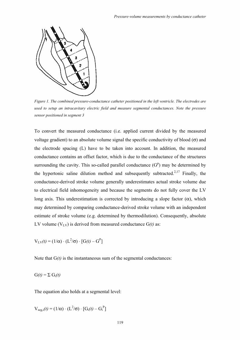
Pressure-volume measurements by conductance catheter
119
Figure 1. The combined pressure-conductance catheter positioned in the left ventricle. The electrodes are
used to setup an intracavitary electric field and measure segmental conductances. Note the pressure
sensor positioned in segment 3
To convert the measured conductance (i.e. applied current divided by the measured
voltage gradient) to an absolute volume signal the specific conductivity of blood (σ) and
the electrode spacing (L) have to be taken into account. In addition, the measured
conductance contains an offset factor, which is due to the conductance of the structures
surrounding the cavity. This so-called parallel conductance (Gp) may be determined by
the hypertonic saline dilution method and subsequently subtracted.2,17 Finally, the
conductance-derived stroke volume generally underestimates actual stroke volume due
to electrical field inhomogeneity and because the segments do not fully cover the LV
long axis. This underestimation is corrected by introducing a slope factor (α), which
may determined by comparing conductance-derived stroke volume with an independent
estimate of stroke volume (e.g. determined by thermodilution). Consequently, absolute
LV volume (VLV) is derived from measured conductance G(t) as:
VLV(t) = (1/α) ⋅ (L2/σ) ⋅ [G(t) – GP]
Note that G(t) is the instantaneous sum of the segmental conductances:
G(t) = Σ Gi(t)
The equation also holds at a segmental level:
Vseg,i(t) = (1/α) ⋅ (L2/σ) ⋅ [Gi(t) – GiP]
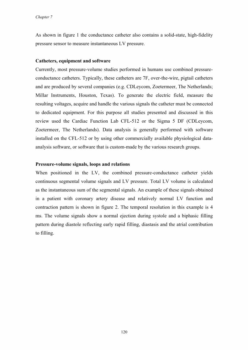
Chapter 7
120
As shown in figure 1 the conductance catheter also contains a solid-state, high-fidelity
pressure sensor to measure instantaneous LV pressure.
Catheters, equipment and software
Currently, most pressure-volume studies performed in humans use combined pressure-
conductance catheters. Typically, these catheters are 7F, over-the-wire, pigtail catheters
and are produced by several companies (e.g. CDLeycom, Zoetermeer, The Netherlands;
Millar Instruments, Houston, Texas). To generate the electric field, measure the
resulting voltages, acquire and handle the various signals the catheter must be connected
to dedicated equipment. For this purpose all studies presented and discussed in this
review used the Cardiac Function Lab CFL-512 or the Sigma 5 DF (CDLeycom,
Zoetermeer, The Netherlands). Data analysis is generally performed with software
installed on the CFL-512 or by using other commercially available physiological data-
analysis software, or software that is custom-made by the various research groups.
Pressure-volume signals, loops and relations
When positioned in the LV, the combined pressure-conductance catheter yields
continuous segmental volume signals and LV pressure. Total LV volume is calculated
as the instantaneous sum of the segmental signals. An example of these signals obtained
in a patient with coronary artery disease and relatively normal LV function and
contraction pattern is shown in figure 2. The temporal resolution in this example is 4
ms. The volume signals show a normal ejection during systole and a biphasic filling
pattern during diastole reflecting early rapid filling, diastasis and the atrial contribution
to filling.
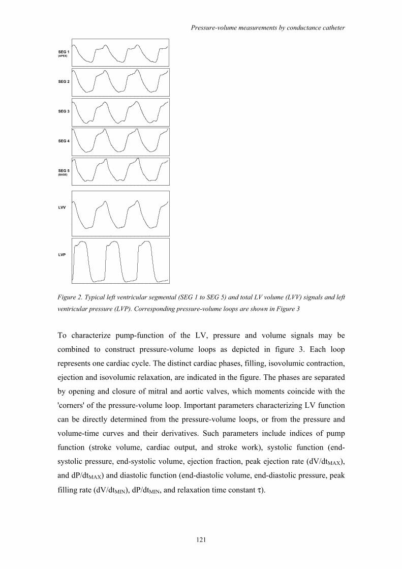
Pressure-volume measurements by conductance catheter
121
SEG 1(APEX)
SEG 2
SEG 3
SEG 4
SEG 5(BASE)
LVV
LVP
Figure 2. Typical left ventricular segmental (SEG 1 to SEG 5) and total LV volume (LVV) signals and left
ventricular pressure (LVP). Corresponding pressure-volume loops are shown in Figure 3
To characterize pump-function of the LV, pressure and volume signals may be
combined to construct pressure-volume loops as depicted in figure 3. Each loop
represents one cardiac cycle. The distinct cardiac phases, filling, isovolumic contraction,
ejection and isovolumic relaxation, are indicated in the figure. The phases are separated
by opening and closure of mitral and aortic valves, which moments coincide with the
'corners' of the pressure-volume loop. Important parameters characterizing LV function
can be directly determined from the pressure-volume loops, or from the pressure and
volume-time curves and their derivatives. Such parameters include indices of pump
function (stroke volume, cardiac output, and stroke work), systolic function (end-
systolic pressure, end-systolic volume, ejection fraction, peak ejection rate (dV/dtMAX),
and dP/dtMAX) and diastolic function (end-diastolic volume, end-diastolic pressure, peak
filling rate (dV/dtMIN), dP/dtMIN, and relaxation time constant τ).
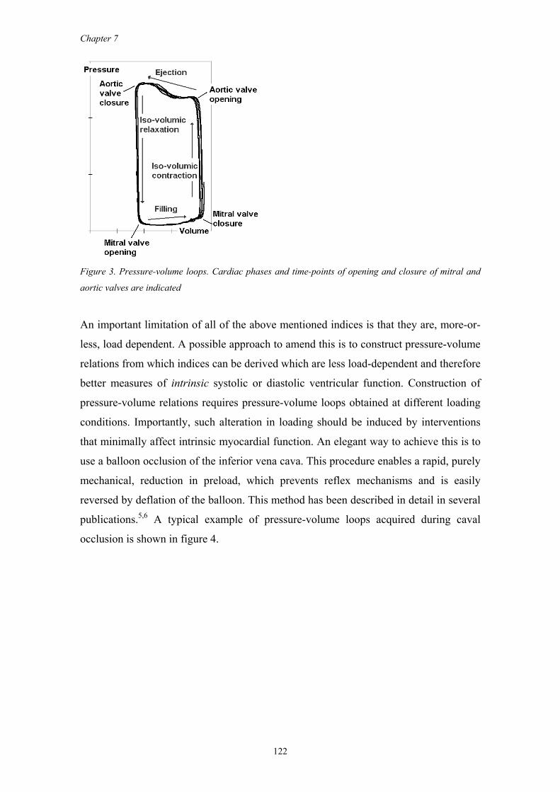
Chapter 7
122
Figure 3. Pressure-volume loops. Cardiac phases and time-points of opening and closure of mitral and
aortic valves are indicated
An important limitation of all of the above mentioned indices is that they are, more-or-
less, load dependent. A possible approach to amend this is to construct pressure-volume
relations from which indices can be derived which are less load-dependent and therefore
better measures of intrinsic systolic or diastolic ventricular function. Construction of
pressure-volume relations requires pressure-volume loops obtained at different loading
conditions. Importantly, such alteration in loading should be induced by interventions
that minimally affect intrinsic myocardial function. An elegant way to achieve this is to
use a balloon occlusion of the inferior vena cava. This procedure enables a rapid, purely
mechanical, reduction in preload, which prevents reflex mechanisms and is easily
reversed by deflation of the balloon. This method has been described in detail in several
publications.5,6 A typical example of pressure-volume loops acquired during caval
occlusion is shown in figure 4.
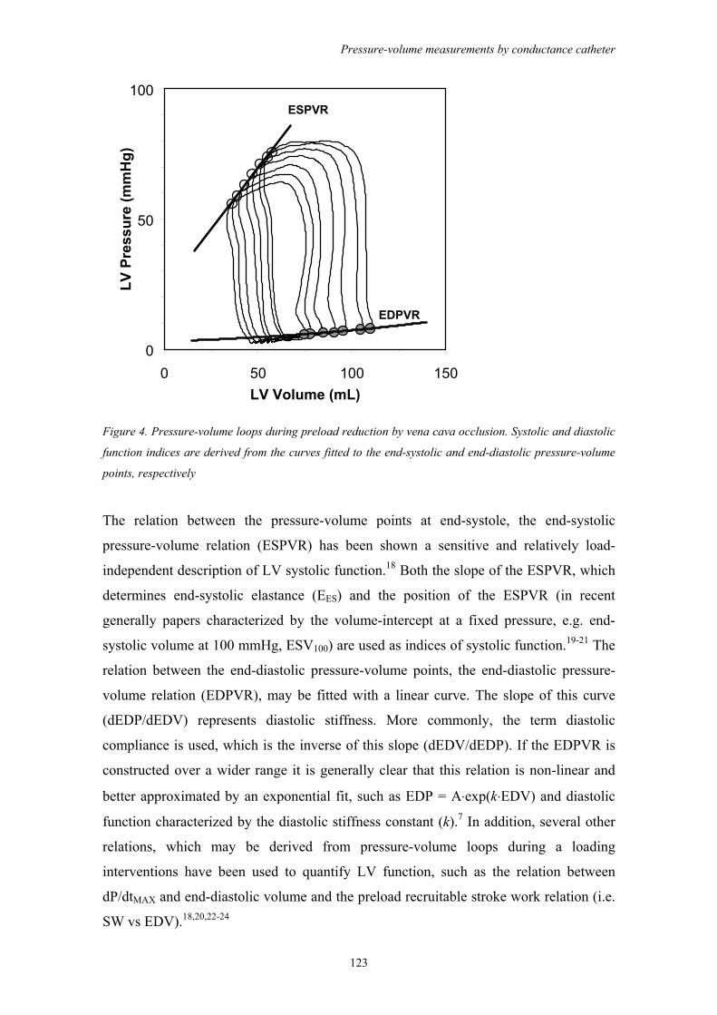
Pressure-volume measurements by conductance catheter
123
ESPVR
EDPVR
0
50
100
0 50 100 150LV Volume (mL)
LV P
ress
ure
(mm
Hg)
Figure 4. Pressure-volume loops during preload reduction by vena cava occlusion. Systolic and diastolic
function indices are derived from the curves fitted to the end-systolic and end-diastolic pressure-volume
points, respectively
The relation between the pressure-volume points at end-systole, the end-systolic
pressure-volume relation (ESPVR) has been shown a sensitive and relatively load-
independent description of LV systolic function.18 Both the slope of the ESPVR, which
determines end-systolic elastance (EES) and the position of the ESPVR (in recent
generally papers characterized by the volume-intercept at a fixed pressure, e.g. end-
systolic volume at 100 mmHg, ESV100) are used as indices of systolic function.19-21 The
relation between the end-diastolic pressure-volume points, the end-diastolic pressure-
volume relation (EDPVR), may be fitted with a linear curve. The slope of this curve
(dEDP/dEDV) represents diastolic stiffness. More commonly, the term diastolic
compliance is used, which is the inverse of this slope (dEDV/dEDP). If the EDPVR is
constructed over a wider range it is generally clear that this relation is non-linear and
better approximated by an exponential fit, such as EDP = A⋅exp(k⋅EDV) and diastolic
function characterized by the diastolic stiffness constant (k).7 In addition, several other
relations, which may be derived from pressure-volume loops during a loading
interventions have been used to quantify LV function, such as the relation between
dP/dtMAX and end-diastolic volume and the preload recruitable stroke work relation (i.e.
SW vs EDV).18,20,22-24
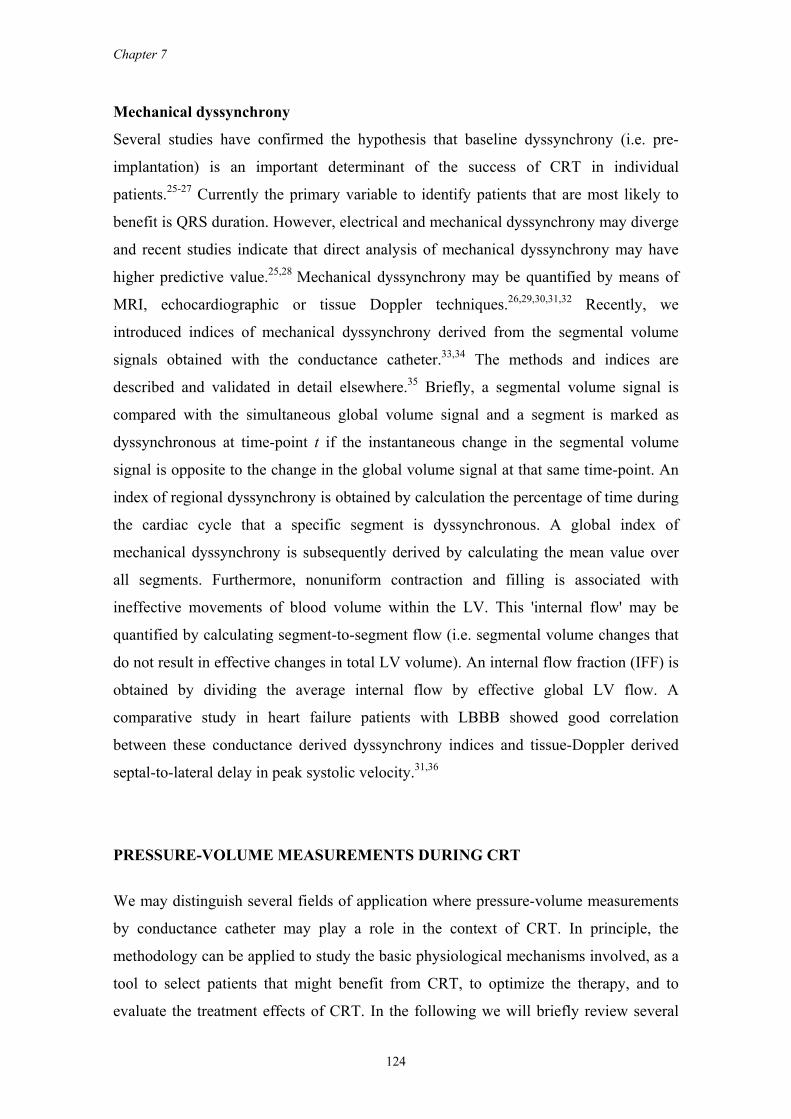
Chapter 7
124
Mechanical dyssynchrony
Several studies have confirmed the hypothesis that baseline dyssynchrony (i.e. pre-
implantation) is an important determinant of the success of CRT in individual
patients.25-27 Currently the primary variable to identify patients that are most likely to
benefit is QRS duration. However, electrical and mechanical dyssynchrony may diverge
and recent studies indicate that direct analysis of mechanical dyssynchrony may have
higher predictive value.25,28 Mechanical dyssynchrony may be quantified by means of
MRI, echocardiographic or tissue Doppler techniques.26,29,30,31,32 Recently, we
introduced indices of mechanical dyssynchrony derived from the segmental volume
signals obtained with the conductance catheter.33,34 The methods and indices are
described and validated in detail elsewhere.35 Briefly, a segmental volume signal is
compared with the simultaneous global volume signal and a segment is marked as
dyssynchronous at time-point t if the instantaneous change in the segmental volume
signal is opposite to the change in the global volume signal at that same time-point. An
index of regional dyssynchrony is obtained by calculation the percentage of time during
the cardiac cycle that a specific segment is dyssynchronous. A global index of
mechanical dyssynchrony is subsequently derived by calculating the mean value over
all segments. Furthermore, nonuniform contraction and filling is associated with
ineffective movements of blood volume within the LV. This 'internal flow' may be
quantified by calculating segment-to-segment flow (i.e. segmental volume changes that
do not result in effective changes in total LV volume). An internal flow fraction (IFF) is
obtained by dividing the average internal flow by effective global LV flow. A
comparative study in heart failure patients with LBBB showed good correlation
between these conductance derived dyssynchrony indices and tissue-Doppler derived
septal-to-lateral delay in peak systolic velocity.31,36
PRESSURE-VOLUME MEASUREMENTS DURING CRT
We may distinguish several fields of application where pressure-volume measurements
by conductance catheter may play a role in the context of CRT. In principle, the
methodology can be applied to study the basic physiological mechanisms involved, as a
tool to select patients that might benefit from CRT, to optimize the therapy, and to
evaluate the treatment effects of CRT. In the following we will briefly review several
Pressure-volume measurements by conductance catheter
125
applications of pressure-volume measurements, and discuss the possibilities and
limitations in these four fields.
Mechanisms
We and several other groups have used pressure-volume analysis to investigate the
physiological mechanisms of CRT. The two primary targets of CRT are normalization
of the pattern of LV activation and optimization of the atrial-ventricular delay.37 In
patients with intraventricular conduction delay mechanical synchrony can be improved
by pre-excitation of the otherwise late-activated region. As shown in figure 5
(unpublished data) this may result in dramatic acute systolic improvements evident from
increased stroke volume and increased stroke work. In this case the improvements are
obtained largely from a reduced end-systolic volume, whereas end-diastolic volume is
unaltered. Similar results were presented by Nelson et al. who very elegantly
demonstrated that the improvement in systolic function is achieved with a minimal
change or even a reduction in myocardial oxygen consumption. 38
0
50
100
150
140 180 220LV Volume (ml)
LV P
ress
ure
(mm
Hg)
BaselineCRT
Figure 5. Acute effects of biventricular pacing on LV pressure-volume loops. Note the increased stroke
volume (width of pressure-volume loops) and stroke work (area of pressure volume loop) during CRT
A nice demonstration of the influence of asynchronous activation on LV function was
presented by Simantirakis et al.39 They determined systolic and diastolic LV function by
pressure-volume loops in patients with long-term right ventricular apical pacing.
Restoration of normal activation was achieved by switching from DDD to AAI pacing
mode. The results indicate an acute improvement in systolic function evident from an
increased end-systolic elastance, preload recruitable stroke work and dP/dtMAX-EDV
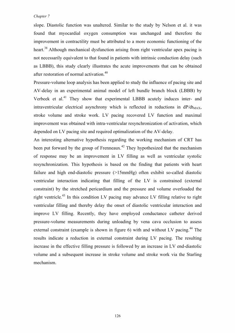
Chapter 7
126
slope. Diastolic function was unaltered. Similar to the study by Nelson et al. it was
found that myocardial oxygen consumption was unchanged and therefore the
improvement in contractility must be attributed to a more economic functioning of the
heart.38 Although mechanical dysfunction arising from right ventricular apex pacing is
not necessarily equivalent to that found in patients with intrinsic conduction delay (such
as LBBB), this study clearly illustrates the acute improvements that can be obtained
after restoration of normal activation.40
Pressure-volume loop analysis has been applied to study the influence of pacing site and
AV-delay in an experimental animal model of left bundle branch block (LBBB) by
Verbeek et al.41 They show that experimental LBBB acutely induces inter- and
intraventricular electrical asynchrony which is reflected in reductions in dP/dtMAX,
stroke volume and stroke work. LV pacing recovered LV function and maximal
improvement was obtained with intra-ventricular resynchronization of activation, which
depended on LV pacing site and required optimalization of the AV-delay.
An interesting alternative hypothesis regarding the working mechanism of CRT has
been put forward by the group of Frenneaux.42 They hypothesized that the mechanism
of response may be an improvement in LV filling as well as ventricular systolic
resynchronization. This hypothesis is based on the finding that patients with heart
failure and high end-diastolic pressure (>15mmHg) often exhibit so-called diastolic
ventricular interaction indicating that filling of the LV is constrained (external
constraint) by the stretched pericardium and the pressure and volume overloaded the
right ventricle.43 In this condition LV pacing may advance LV filling relative to right
ventricular filling and thereby delay the onset of diastolic ventricular interaction and
improve LV filling. Recently, they have employed conductance catheter derived
pressure-volume measurements during unloading by vena cava occlusion to assess
external constraint (example is shown in figure 6) with and without LV pacing.44 The
results indicate a reduction in external constraint during LV pacing. The resulting
increase in the effective filling pressure is followed by an increase in LV end-diastolic
volume and a subsequent increase in stroke volume and stroke work via the Starling
mechanism.
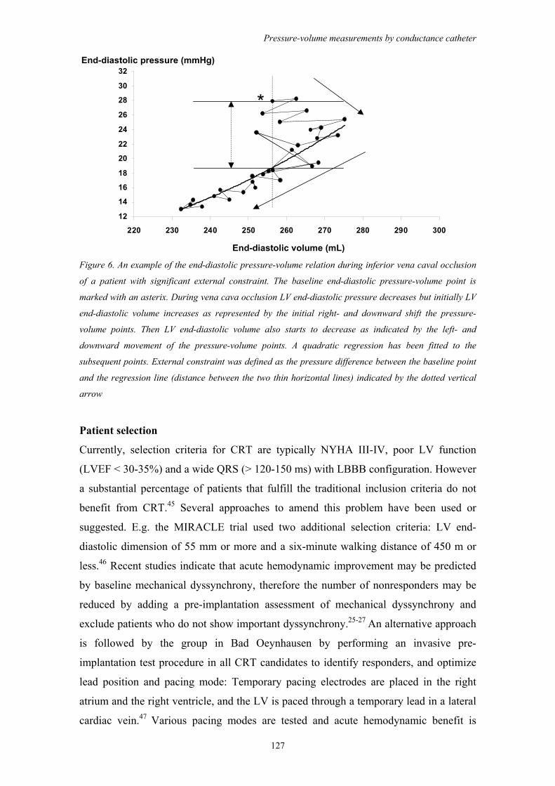
Pressure-volume measurements by conductance catheter
127
End-diastolic pressure (mmHg)
End-diastolic volume (mL)
12
14
16
18
20
22
24
26
28
30
32
220 230 240 250 260 270 280 290 300
*
Figure 6. An example of the end-diastolic pressure-volume relation during inferior vena caval occlusion
of a patient with significant external constraint. The baseline end-diastolic pressure-volume point is
marked with an asterix. During vena cava occlusion LV end-diastolic pressure decreases but initially LV
end-diastolic volume increases as represented by the initial right- and downward shift the pressure-
volume points. Then LV end-diastolic volume also starts to decrease as indicated by the left- and
downward movement of the pressure-volume points. A quadratic regression has been fitted to the
subsequent points. External constraint was defined as the pressure difference between the baseline point
and the regression line (distance between the two thin horizontal lines) indicated by the dotted vertical
arrow
Patient selection
Currently, selection criteria for CRT are typically NYHA III-IV, poor LV function
(LVEF < 30-35%) and a wide QRS (> 120-150 ms) with LBBB configuration. However
a substantial percentage of patients that fulfill the traditional inclusion criteria do not
benefit from CRT.45 Several approaches to amend this problem have been used or
suggested. E.g. the MIRACLE trial used two additional selection criteria: LV end-
diastolic dimension of 55 mm or more and a six-minute walking distance of 450 m or
less.46 Recent studies indicate that acute hemodynamic improvement may be predicted
by baseline mechanical dyssynchrony, therefore the number of nonresponders may be
reduced by adding a pre-implantation assessment of mechanical dyssynchrony and
exclude patients who do not show important dyssynchrony.25-27 An alternative approach
is followed by the group in Bad Oeynhausen by performing an invasive pre-
implantation test procedure in all CRT candidates to identify responders, and optimize
lead position and pacing mode: Temporary pacing electrodes are placed in the right
atrium and the right ventricle, and the LV is paced through a temporary lead in a lateral
cardiac vein.47 Various pacing modes are tested and acute hemodynamic benefit is
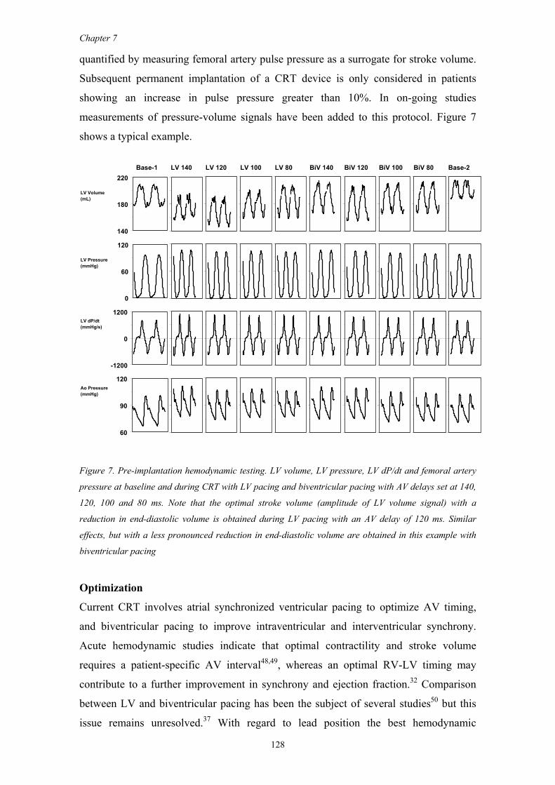
Chapter 7
128
quantified by measuring femoral artery pulse pressure as a surrogate for stroke volume.
Subsequent permanent implantation of a CRT device is only considered in patients
showing an increase in pulse pressure greater than 10%. In on-going studies
measurements of pressure-volume signals have been added to this protocol. Figure 7
shows a typical example.
Base-1 LV 140 LV 120 LV 100 LV 80 BiV 140 BiV 120 BiV 100 BiV 80 Base-2
LV Volume(mL)
LV Pressure(mmHg)
LV dP/dt(mmHg/s)
Ao Pressure(mmHg)
140
180
220
0
60
120
60
90
120
-1200
0
1200
Figure 7. Pre-implantation hemodynamic testing. LV volume, LV pressure, LV dP/dt and femoral artery
pressure at baseline and during CRT with LV pacing and biventricular pacing with AV delays set at 140,
120, 100 and 80 ms. Note that the optimal stroke volume (amplitude of LV volume signal) with a
reduction in end-diastolic volume is obtained during LV pacing with an AV delay of 120 ms. Similar
effects, but with a less pronounced reduction in end-diastolic volume are obtained in this example with
biventricular pacing
Optimization
Current CRT involves atrial synchronized ventricular pacing to optimize AV timing,
and biventricular pacing to improve intraventricular and interventricular synchrony.
Acute hemodynamic studies indicate that optimal contractility and stroke volume
requires a patient-specific AV interval48,49, whereas an optimal RV-LV timing may
contribute to a further improvement in synchrony and ejection fraction.32 Comparison
between LV and biventricular pacing has been the subject of several studies50 but this
issue remains unresolved.37 With regard to lead position the best hemodynamic
Pressure-volume measurements by conductance catheter
129
response of LV pacing is generally obtained through pacing in the mid-lateral or
posterior LV51-53, which is achieved with leads placed in the posterior or lateral
branches of the coronary sinus. Despite advances in percutaneous techniques, special
guiding sheaths and improved lead design47,54, suboptimal lead positioning may still be
an important cause of non-response to CRT. Intraoperative epicardial lead placement is
currently mainly used as rescue in patients with failed endocardial leads, but may
provide an alternative approach with possibilities for optimal lead placement.55,56
Finally some studies suggest that multiple LV sites may be required for optimal
hemodynamic results.57 Despite a large number of studies many questions regarding
optimization of CRT remain disputed. Conceivably studies with the conductance
catheter may resolve some of these issues by providing on-line pressure-volume loops
which may guide optimization of CRT.
Evaluation
Studies have demonstrated acute hemodynamic improvement after CRT, followed by
improvement in symptoms, quality of life and exercise capacity.46,48,58 More recent
studies have provided objective evidence for improved systolic performance and
reversed remodeling during long-term CRT which may provide the basis for the clinical
improvements.27,59 The evaluation of long-term hemodynamic effects is complex
because it involves alterations in both systolic and diastolic function, and in loading
conditions. Although initially the improved systolic function most likely largely reflects
improved contraction synchrony, long-term alternation in intrinsic myocardial function
may be present e.g. due to alterations in wall stress or sympathetic activity.60
Interpretation of traditional diastolic indices is complicated because alterations in filling
time and mitral insufficiency are present and may interact with changes in intrinsic
myocardial function. Analysis in terms of pressure-volume loops and pressure-volume
relations is attractive because it provides relatively load-independent indices of systolic
and diastolic function. In on-going studies we investigate patients before pacemaker
implantation and after 6 months of CRT. The example shown in Figure 8 illustrates
reversed remodeling with improved systolic and diastolic function and reduction in
mitral regurgitation. Analysis of the segmental conductance signals yielded improved
systolic and diastolic mechanical synchrony and reduced internal flow.61
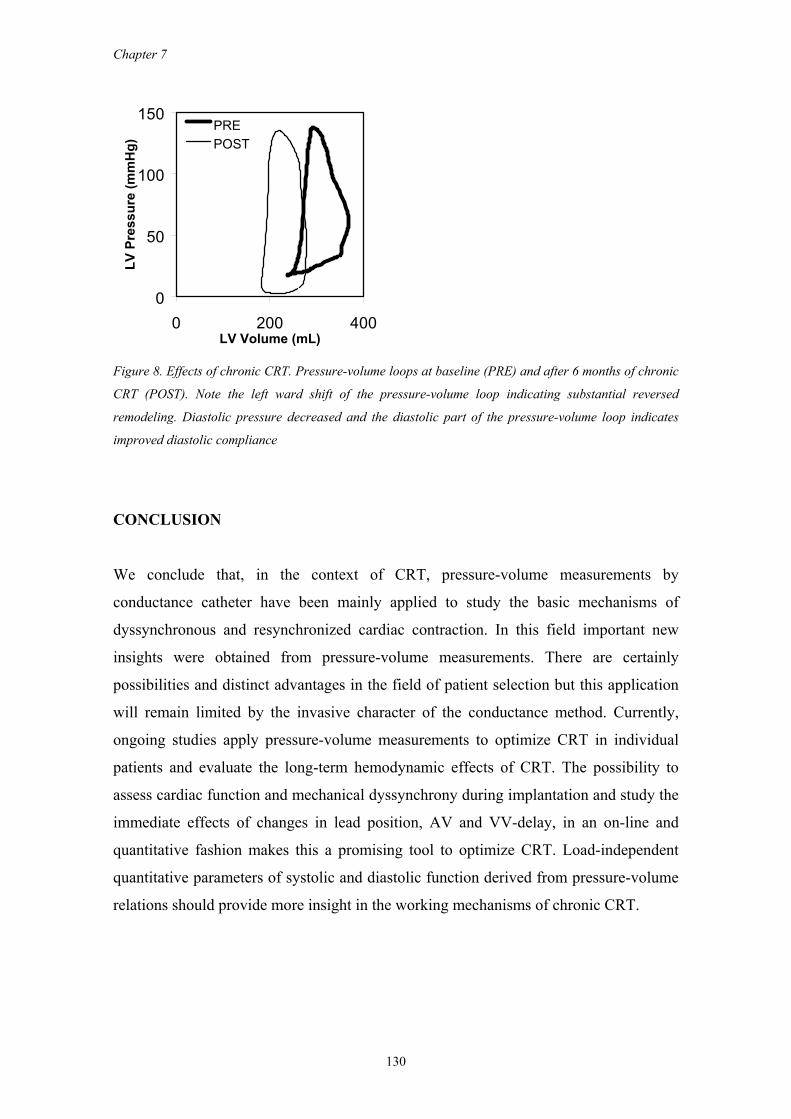
Chapter 7
130
0
50
100
150
0 200 400LV Volume (mL)
LV P
ress
ure
(mm
Hg)
PREPOST
Figure 8. Effects of chronic CRT. Pressure-volume loops at baseline (PRE) and after 6 months of chronic
CRT (POST). Note the left ward shift of the pressure-volume loop indicating substantial reversed
remodeling. Diastolic pressure decreased and the diastolic part of the pressure-volume loop indicates
improved diastolic compliance
CONCLUSION
We conclude that, in the context of CRT, pressure-volume measurements by
conductance catheter have been mainly applied to study the basic mechanisms of
dyssynchronous and resynchronized cardiac contraction. In this field important new
insights were obtained from pressure-volume measurements. There are certainly
possibilities and distinct advantages in the field of patient selection but this application
will remain limited by the invasive character of the conductance method. Currently,
ongoing studies apply pressure-volume measurements to optimize CRT in individual
patients and evaluate the long-term hemodynamic effects of CRT. The possibility to
assess cardiac function and mechanical dyssynchrony during implantation and study the
immediate effects of changes in lead position, AV and VV-delay, in an on-line and
quantitative fashion makes this a promising tool to optimize CRT. Load-independent
quantitative parameters of systolic and diastolic function derived from pressure-volume
relations should provide more insight in the working mechanisms of chronic CRT.
Pressure-volume measurements by conductance catheter
131
REFERENCES 1. Baan J, Aouw Jong TT, Kerkhof PLM et al. Continuous stroke volume and cardiac output from
intra-ventricular dimensions obtained with impedance catheter. Cardiovasc Res. 1981;15:328-334. 2. Baan J, Van Der Velde ET, De Bruin H et al. Continuous measurement of left ventricular volume
in animals and humans by conductance catheter. Circulation. 1984;70:812-823. 3. Baan J, Van Der Velde ET, Steendijk P. Ventricular pressure-volume relations in vivo. Eur Heart
J. 1992;13 (Suppl E):2-6. 4. Caputo M, Schreuder J, Fino C et al. Assessment of myocardial performance with ventricular
pressure-volume relations: clinical applications in cardiac surgery. Ital Heart J. 2000;1:269-274. 5. Kass DA, Midei M, Graves W et al. Use of a conductance (volume) catheter and transient inferior
vena caval occlusion for rapid determination of pressure-volume relationships in man. Cathet Cardiovasc Diagn. 1988;15:192-202.
6. Kass DA. Clinical evaluation of left heart function by conductance catheter technique. Eur Heart J. 1992;13 (Suppl E):57-64.
7. Kass DA. Assessment of diastolic dysfunction. Invasive modalities. Cardiol Clin. 2000;18:571-586.
8. Lang RM, Borow KM, Neumann A et al. Systemic vascular resistance: an unreliable index of left ventricular afterload. Circulation. 1986;74:1114-1123.
9. Leatherman GF, Shook TL, Leatherman SM et al. Use of a conductance catheter to detect increased left ventricular inotropic state by end-systolic pressure-volume analysis. Basic Res Cardiol. 1989;84:247-256.
10. McKay RG, Aroesty JM, Heller GV et al. Left ventricular pressure-volume diagrams and end-systolic pressure-volume relations in human beings. J-Am-Coll-Cardiol. 1984;3:301-12.
11. Schreuder JJ, Biervliet JD, Van Der Velde ET et al. Systolic and diastolic pressure-volume relationships during cardiac surgery. J Cardiothor Vasc Anesth. 1991;5:539-545.
12. Schreuder JJ, Van der Veen FH, Van Der Velde ET et al. Left ventricular pressure volume relationships before and after cardiomyoplasty in patients with heart failure. Circulation. 1997;96:2978-2986.
13. Tulner SA, Klautz RJ, Rijk-Zwikker GL et al. Perioperative assessment of left ventricular function by pressure-volume loops using the conductance catheter method. Anesth Analg. 2003;97:950-957.
14. Steendijk P, Tulner SA, Schreuder JJ et al. Quantification of left ventricular mechanical dyssynchrony by conductance catheter in heart failure patients. Am J Physiol Heart Circ Physiol. 2003;(in press).
15. Strum DP, Pinsky MR. Modeling of asynchronous myocardial contraction by effective stroke volume analysis. Anesthesia & Analgesia. 2000;90:243-51.
16. Steendijk P, Van Der Velde ET, Baan J. Left ventricular stroke volume by single and dual excitation of conductance catheter in dogs. Am J Physiol. 1993;264 (Heart Circ Physiol 33):H2198-H2207.
17. Steendijk P, Staal E, Jukema JW et al. Hypertonic saline method accurately determines parallel conductance for dual-field conductance catheter. Am J Physiol Heart Circ Physiol. 2001;281:H755-H763.
18. Kass DA, Maughan WL, Guo ZM et al. Comparative influence of load versus inotropic states on indexes of ventricular contractility: experimental and theoretical analysis based on pressure-volume relationships [published erratum appears in Circulation 1988 Mar;77(3):559]. Circulation. 1987;76:1422-1436.
19. Little WC, Cheng CP, Peterson T et al. Response of the left ventricular end-systolic pressure-volume relation in conscious dogs to a wide range of contractile states [published erratum appears in Circulation 1989 Jan;79(1):205]. Circulation. 1988;78:736-45.
20. Little WC, Cheng CP, Mumma M et al. Comparison of measures of left ventricular contractile performance derived from pressure-volume loops in conscious dogs. Circulation. 1989;80:1378-87.
21. Van Der Velde ET, Burkhoff D, Steendijk P et al. Nonlinearity and load sensitivity of the end-systolic pressure-volume relation of canine left ventricle in vivo. Circulation. 1991;83:315-327.
22. Glower DD, Spratt JA, Snow ND et al. Linearity of the Frank-Starling relationship in the intact heart: the concept of preload recruitable stroke work. Circulation. 1985;71:994-1009.
23. Kass DA, Yamazaki T, Burkhoff D et al. Determination of left ventricular end-systolic pressure-volume relationships by the conductance catheter. Circulation. 1986;73:586-595.
24. Little WC. The left ventricular dP/dtmax-end-diastolic volume relationship in closed-chest dogs. Circ Res. 1985;56:808-815.
Chapter 7
132
25. Bax JJ, Marwick TH, Molhoek SG et al. Left ventricular dyssynchrony predicts benefit of cardiac resynchronization therapy in patients with end-stage heart failure before pacemaker implantation. Am J Cardiol. 2003;92:1238-1240.
26. Breithardt OA, Stellbrink C, Kramer AP et al. Echocardiographic quantification of left ventricular asynchrony predicts an acute hemodynamic benefit of cardiac resynchronization therapy. J Am Coll Cardiol. 2002;40:536-545.
27. Sogaard P, Egeblad H, Kim WY et al. Tissue Doppler imaging predicts improved systolic performance and reversed left ventricular remodeling during long-term cardiac resynchronization therapy. J Am Coll Cardiol. 2002;723-730.
28. Leclercq C, Faris O, Tunin R et al. Systolic improvement and mechanical resynchronization does not require electrical synchrony in the dilated failing heart with left bundle- branch block. Circulation. 2002;106:1760-1763.
29. Nelson GS, Curry CW, Wyman BT et al. Predictors of systolic augmentation from left ventricular preexcitation in patients with dilated cardiomyopathy and intraventricular conduction delay. Circulation. 2000;101:2703-9.
30. Kawaguchi M, Murabayashi T, Fetics BJ et al. Quantitation of basal dyssynchrony and acute resynchronization from left or biventricular pacing by novel echo-contrast variability imaging. J Am Coll Cardiol. 2002;39:2052-2058.
31. Bax JJ, Molhoek SG, van Erven L et al. Usefulness of myocardial tissue Doppler echocardiography to evaluate left ventricular dyssynchrony before and after biventricular pacing in patients with idiopathic dilated cardiomyopathy. Am J Cardiol. 2003;91:94-97.
32. Sogaard P, Egeblad H, Pedersen AK et al. Sequential versus simultaneous biventricular resynchronization for severe heart failure: evaluation by tissue Doppler imaging. Circulation. 2002;106:2078-2084.
33. Schreuder JJ, Steendijk P, Van der Veen FH et al. Acute and short-term effects of partial left ventriculectomy in dilated cardiomyopathy: assessment by pressure-volume loops.[In Process Citation]. J Am Coll Cardiol. 2000;36:2104-2114.
34. Steendijk P, Tulner SA, Schreuder JJ et al. Quantification of left ventricular mechanical dyssynchrony by conductance catheter in heart failure patients. Am J Physiol Heart Circ Physiol. 2003;(in press).
35. Steendijk P, Tulner SA, Schreuder JJ et al. Quantification of left ventricular mechanical dyssynchrony by conductance catheter in heart failure patients. Am J Physiol Heart Circ Physiol. 2003;(in press).
36. Steendijk P, Tulner SA, Schreuder JJ et al. Quantification of left ventricular mechanical dyssynchrony by conductance catheter in heart failure patients. Am J Physiol Heart Circ Physiol. 2003;(in press).
37. Leclercq C, Kass DA. Retiming the failing heart: principles and current clinical status of cardiac resynchronization. J Am Coll Cardiol. 2002;39:194-201.
38. Nelson GS, Berger RD, Fetics BJ et al. Left ventricular or biventricular pacing improves cardiac function at diminished energy cost in patients with dilated cardiomyopathy and left bundle-branch block. Circulation. 2000;102:3053-3059.
39. Simantirakis EN, Kochiadakis GE, Vardakis KE et al. Left ventricular mechanics and myocardial blood flow following restoration of normal activation sequence in paced patients with long-term right ventricular apical stimulation. Chest. 2003;124:233-241.
40. Xiao HB, Brecker SJ, Gibson DG. Differing effects of right ventricular pacing and left bundle branch block on left ventricular function. Br Heart J. 1993;69:166-173.
41. Verbeek XA, Vernooy K, Peschar M et al. Intra-ventricular resynchronization for optimal left ventricular function during pacing in experimental left bundle branch block. J Am Coll Cardiol. 2003;42:558-567.
42. Morris-Thurgood JA, Turner MS, Nightingale AK et al. Pacing in heart failure: improved ventricular interaction in diastole rather than systolic re-synchronization. Europace. 2000;2:271-275.
43. Atherton JJ, Moore TD, Lele SS et al. Diastolic ventricular interaction in chronic heart failure. Lancet. 1997;349:1720-1724.
44. Bleasdale RA, Turner MS, Mumford CE et al. Left ventricular pacing recruits preload reserve in heart failure patients: a new mechanism. J Am Coll Cardiol. 2003;41:1110-74.
45. Bax JJ, van der Wall EE, Schalij MJ. Cardiac resynchronization therapy for heart failure. N Engl J Med. 2002;347:1803-1804.
46. Abraham WT, Fisher WG, Smith AL et al. Cardiac resynchronization in chronic heart failure. N Engl J Med. 2002;346:1845-1853.
47. Hansky B, Vogt J, Gueldner H et al. Left heart pacing--experience with several types of coronary vein leads. J Interv Card Electrophysiol. 2002;6:71-75.
Pressure-volume measurements by conductance catheter
133
48. Auricchio A, Stellbrink C, Sack S et al. Long-term clinical effect of hemodynamically optimized cardiac resynchronization therapy in patients with heart failure and ventricular conduction delay. J Am Coll Cardiol. 2002;39:2026-2033.
49. Kass DA, Chen CH, Curry C et al. Improved left ventricular mechanics from acute VDD pacing in patients with dilated cardiomyopathy and ventricular conduction delay. Circulation. 1999;99:1567-73.
50. Blanc JJ, Etienne Y, Gilard M et al. Evaluation of different ventricular pacing sites in patients with severe heart failure: results of an acute hemodynamic study. Circulation. 1997;96:3273-3277.
51. Auricchio A, Stellbrink C, Sack S et al. The Pacing Therapies for Congestive Heart Failure (PATH-CHF) study: rationale, design, and endpoints of a prospective randomized multicenter study. American Journal of Cardiology. 1999;83:130D-135D.
52. Ansalone G, Giannantoni P, Ricci R et al. Doppler myocardial imaging to evaluate the effectiveness of pacing sites in patients receiving biventricular pacing. J Am Coll Cardiol. 2002;39:489-499.
53. Butter C, Auricchio A, Stellbrink C et al. Effect of resynchronization therapy stimulation site on the systolic function of heart failure patients. Circulation. 2001;104:3026-3029.
54. Lau CP, Barold S, Tse HF et al. Advances in devices for cardiac resynchronization in heart failure. J Interv Card Electrophysiol. 2003;9:167-181.
55. DeRose JJ, Ashton RC, Belsley S et al. Robotically assisted left ventricular epicardial lead implantation for biventricular pacing. J Am Coll Cardiol. 2003;41:1414-1419.
56. Jansens JL, Jottrand M, Preumont N et al. Robotic-enhanced biventricular resynchronization: an alternative to endovenous cardiac resynchronization therapy in chronic heart failure. Ann Thorac Surg. 2003;76:413-417.
57. Pappone C, Rosanio S, Oreto G et al. Cardiac pacing in heart failure patients with left bundle branch block: impact of pacing site for optimizing left ventricular resynchronization. Ital Heart J. 2000;1:464-469.
58. Cazeau S, Leclercq C, Lavergne T et al. Effects of multisite biventricular pacing in patients with heart failure and intraventricular conduction delay. N Engl J Med. 2001;344:873-880.
59. Yu CM, Chau E, Sanderson JE et al. Tissue Doppler echocardiographic evidence of reverse remodeling and improved synchronicity by simultaneously delaying regional contraction after biventricular pacing therapy in heart failure. Circulation. 2002;105:438-445.
60. Hamdan MH, Zagrodzky JD, Joglar JA et al. Biventricular pacing decreases sympathetic activity compared with right ventricular pacing in patients with depressed ejection fraction. Circulation. 2000;102:1027-1032.
61. Steendijk P, Tulner SA, Bax JJ et al. Improved mechanical synchrony and reversed remodeling after 6 months biventricular pacing assessed by LV pressure-volume loops. Eur J Heart Fail. 2003;2:157.

CHAPTER 8
Hemodynamic effects of long-term cardiac resynchronization
therapy -Analysis by pressure-volume loops-
P. Steendijk
S.A.F. Tulner
J.J. Bax
P.V. Oemrawsingh
G.B. Bleeker
L. van Erven
H. Putter
H.F. Verwey
E.E. van der Wall
M.J. Schalij
Circulation (in press)
Poster presentation at European Society of Heart Failure 2003, Strasbourg,
Luxembourg
Chapter 8
136
ABSTRACT
Background. Acute hemodynamic effects of cardiac resynchronization therapy (CRT)
have been reported previously, but detailed invasive studies showing hemodynamic
consequences of long-term CRT are not available.
Methods and Results. We studied 22 patients scheduled for implantation of a CRT
device based on conventional criteria (NYHA class III or IV, left ventricular (LV)
ejection fraction <35%, left bundle-branch block, QRS duration >120ms). During
diagnostic catheterization prior to CRT we acquired pressure-volume loops using
conductance catheters during right atrial pacing at 80, 100, 120 and 140 beats/min.
These studies were repeated during biventricular pacing at the same heart rates after 6
months of CRT. Our data show significant clinical benefit of CRT (NYHA class:
3.1±0.5 to 2.1±0.8; Quality-of-Life score: 44±12 to 31±16; 6-min hall-walk: 260±149 to
396±129m; all p<0.001), improved LV ejection fraction (29±10 to 40±13%, p<0.01),
decreased diastolic pressure (18±8 to 13±6mmHg, p<0.05), and reverse remodeling
(end-diastolic volume: 257±67 to 205±54mL, p<0.01). Previously reported acute
improvements in LV function remained present at 6 months: dP/dtMAX (+18%, p<0.01),
dP/dtMIN (+13%, p<0.01), stroke work (+34%, p<0.01). Effects of increased heart rate
were improved towards more physiological responses for LV ejection fraction, cardiac
output and dP/dtMAX. Moreover, our study shows improved ventricular-arterial coupling
(+69%, p<0.01) and improved mechanical efficiency (+44%, p<0.01).
Conclusions. Hemodynamic improvements with CRT, which were previously shown in
acute invasive studies, are maintained long-term. In addition, ventricular-arterial
coupling, mechanical efficiency, and chronotropic responses are improved after 6
months of CRT. These findings may help to explain the improved functional status and
exercise tolerance in heart failure patients treated with CRT.
INTRODUCTION
Cardiac resynchronization therapy (CRT) improves quality of life, symptoms, and
exercise capacity in patients with heart failure and intraventricular conduction delay.1 A
recent study confirmed these favorable effects and also demonstrated that CRT
significantly reduced the risk of death.2 Whereas previous randomized controlled trials
have clearly demonstrated beneficial clinical effects over a period of up to 6 months,
Hemodynamic effects of long-term CRT
137
small-scaled studies suggest that these clinical improvements are maintained long-
term.3-5 The primary working mechanism of CRT is the optimization of the mechanical
activation pattern of the left ventricle (LV), which is achieved by pre-excitation of the
region which is otherwise activated late due to delayed intrinsic conduction.6 In addition
to this intraventricular resynchronization, additional benefit may be obtained by
optimizing the delay between atrial and ventricular systole, and the timing of LV and
right ventricle (RV) stimulation. Acute improvements in mechanical dyssynchrony
resulting in enhanced systolic function have been demonstrated by various studies.7-10
Invasive studies have shown increased LV ejection fraction and stroke volume,
accompanied by increased systolic pressure, dP/dtMAX, and stroke work, and reduced
diastolic pressure.9,11 Interestingly, these improvements in cardiac function are obtained
at diminished energy cost.12 In the long-term, CRT is associated with LV reversed
remodeling13 and improved myocardial efficiency.14 However, currently no invasive
studies are available regarding the effects of long-term CRT on systolic and diastolic
hemodynamic parameters. In this study we assessed the long-term hemodynamic effects
of CRT, and investigated the underlying mechanisms. To this end, we acquired
pressure-volume loops prior to CRT during right atrial pacing at 80, 100, 120 and 140
beats/min, and these studies were repeated during biventricular pacing at the same heart
rates after 6 months of CRT.
METHODS
Patients
Twenty-two patients (mean age, 66±11 years; 17 men) with NYHA class III or IV heart
failure despite optimized medical treatment, echocardiographic LV ejection fraction
<35% and QRS duration >120ms scheduled for implantation of a CRT device were
included. The protocol was approved by our institutional review committee and all
patients gave informed consent. The etiology of heart failure was ischemic in 14 and
non-ischemic in 8 patients. All patients received stable medical therapy for chronic
heart failure, including diuretics (n=19), spironolactone (n=8), β-blockers (n=10), ACE
inhibitors (n=20), and amiodarone (n=6). Medication was unchanged and no new
therapies were installed during the 6-months follow-up period. In addition to the
invasive studies described in detail below, we performed echocardiography, 6-minute
Chapter 8
138
hall-walk tests, and quality of life assessments by the Minnesota Living with Heart
Failure Questionnaire at baseline and after 6 months of CRT.
Protocol
Baseline (i.e. pre-CRT) hemodynamic data were obtained during routine diagnostic
right and left heart catheterization, including thermodilution cardiac output, left
ventriculography and coronary angiography. To acquire pressure-volume loops at
incremental heart rates, a 7F combined pressure-conductance catheter (CD Leycom,
Zoetermeer, The Netherlands) was placed in the LV via the femoral artery, and a
temporary pacing lead was placed in the right atrium. Pressure-volume signals were
displayed on-line and digitized at a sample frequency of 250Hz (Leycom CFL, CD
Leycom). LV volume was calibrated using thermodilution and hypertonic saline
dilution as previously described.15,16 Right atrial pacing was performed at 80, 100, 120
and 140 beats/min. Data were acquired consecutively approximately 60s after changing
to a higher rate, and periods of at least 20s were selected for off-line analysis. All
measurements were repeated during recatheterization after at least 6 months of chronic
CRT. During this session biventricular pacing was performed at 80, 100, 120 and 140
beats/min by reprogramming the CRT device. The atrioventricular (AV) delay was kept
fixed at the optimal clinical setting based on Doppler mitral flow velocity recordings
obtained previously at the outpatient clinic.
Data analysis
Analysis of the steady state pressure-volume loops was performed using custom
software as previously described.17 Briefly, for each patient and each pacing rate
hemodynamic indexes were calculated as the mean of all beats during a steady state
period of approximately 20s. LV function was quantified by cardiac output and stroke
volume, end-diastolic and end-systolic volume, LV ejection fraction, end-systolic and
end-diastolic pressure, maximal and minimal rate of LV pressure change (dP/dtMAX,
dP/dtMIN). The time constant of relaxation (τ) was determined using phase-plot
analysis.18 Stroke work was calculated as the area of the pressure-volume loop. LV end-
systolic elastance (EES) was estimated by end-systolic pressure divided by end-systolic
volume, and end-diastolic stiffness (EED) by end-diastolic pressure divided by end-
diastolic volume. Effective arterial elastance (EA) was calculated as end-systolic
pressure divided by stroke volume.19 Ventricular-arterial coupling was quantified as
EES/EA,20 and mechanical efficiency was calculated as the ratio of external stroke work
Hemodynamic effects of long-term CRT
139
and pressure-volume area (a measure of total mechanical work).21 Nonuniform LV
performance was determined from the segmental LV conductance signals and quantified
by calculating the percentage of time within the cardiac cycle that a specific segment is
dyssynchronous (i.e. opposite in phase with the global LV volume signal). Overall LV
mechanical dyssynchrony was determined as the mean of the segmental dyssynchronies.
In addition, we calculated the internal flow fraction, which quantifies the ineffective
shifting of blood volume within the LV due to nonuniform contraction and filling. This
approach was described and validated in a previous study.17 Time-varying wall stress,
WS(t), was calculated from the instantaneous LV pressure and volume signals (P(t) and
V(t), respectively) as described by Arts et al.22: WS(t) = P(t)·(1+3·V(t) / LVM). LV
mass (LVM) was calculated from M-mode echocardiography according to the
conventions proposed by the American Society of Echocardiography.23 Atrioventricular
delay was determined as the time between the right atrial pacing and the start of left
ventricular contraction.24
Statistical analysis
We used a linear mixed-effects model to account for repeated measurements on each
patient. In this model, patients were included as random effects and conditions
(baseline, CRT), pacing (80, 100, 120, and 140 beats/min), and their interaction as fixed
effects.25 To assess statistical significances between pacing levels and conditions,
appropriate contrasts were selected. Data are presented as mean±SD. A p-value <0.05
was considered statistically significant.
RESULTS
Clinical assessment and atrioventricular delay
All patients were successfully implanted with a CRT device (Contac Renewal, Guidant
(n=21), or InSync III, Medtronic (n=1)). All patients received CRT for at least 6 months
(7.2±1.6 months). Table 1 shows the clinical parameters which all improved
significantly, consistent with previous reports1. AV delay was optimized based on
Doppler mitral flow velocity recordings at our outpatient clinic shortly after pacemaker
implantation: The AV delay was set to achieve the longest left ventricular filling time
without premature truncation of the A-wave by mitral valve closure.26 Baseline AV
delay (during right atrial pacing at 80 beats/min) was 184±96 ms and tended to decrease
Chapter 8
140
at higher pacing rates. Mean optimized AV delay with biventricular pacing was 97±15
ms and was unchanged at the higher pacing rates (Table 2).
Table 1. Clinical parameters at baseline (pre-CRT) and after 6 months of CRT
Baseline 6-mo CRT
NYHA class 3.05±0.49 2.05±0.79 ****
Quality of life score 44±12 31±16 ****
6-min hall-walk, m 260±149 396±129 ****
* p<0.05, ** p<0.01, *** p<0.005, **** p<0.001 vs. baseline by paired t-tests
Left ventricular function
Figure 1 shows typical examples of pressure-volume loops at 80 beats/min at baseline
and after 6 months of CRT from two patients. Full hemodynamic data including the
effects of increased pacing rate from all patients are summarized in Table 2.
Comparison of 6 months of CRT vs. baseline at the lowest pacing rate (80 beats/min,
p80) shows that cardiac output and LV ejection fraction improved significantly, whereas
end-diastolic volume and end-systolic volume were significantly reduced. The latter
indicates substantial reversed remodeling consistent with previous reports.13 Improved
systolic function was evidenced by a significantly increased dP/dtMAX, EES and stroke
work. In addition, end-diastolic pressure was significantly reduced. Diastolic stiffness
EED and τ showed a non-significant tendency to reduce. dP/dtMIN was significantly
improved indicating improved active relaxation. The increase in EES combined with a
modest decrease in EA resulted in a significantly improved ventricular-arterial coupling
ratio (EES/EA). The significant increase in external stroke work with unchanged total
mechanical work resulted in a significantly improved mechanical efficiency.
Legends of table 2. p80..p140 indicates paced at 80..140 beats/min; BL, baseline (i.e. pre-CRT) ; CRT, 6-
months cardiac resynchronization therapy; Condition effect, BL vs. CRT; Pacing effect, effect of
incremental paced heart rate; Interaction effect, condition-pacing interaction; HR, heart rate; AVD,
atrioventricular delay; CO, cardiac output; ESV, end-systolic volume; EDV, end-diastolic volume; EF,
ejection fraction; SW, stroke work; ESP, end-systolic pressure; EDP, end-diastolic pressure; τ,
relaxation time constant; PWS, peak wall stress; WSED, end-diastolic wall stress; DYS, mechanical
dyssynchrony; IFF, internal flow fraction; EA, effective arterial elastance; EES, end-systolic elastance;
EED, end-diastolic stiffness; PVA, pressure-volume area; ME = SW/PVA, mechanical efficiency; and
EES/EA, ventricular-arterial coupling. Statistical significances and contrasts (changes vs. p80) were
determined by using a linear mixed-effects model (see text for details). CRT-p80 vs. BL-p80: # p<0.05, ##
p<0.01. Changes vs. p80 at same condition (BL or CRT): * p<0.05, ** p<0.01
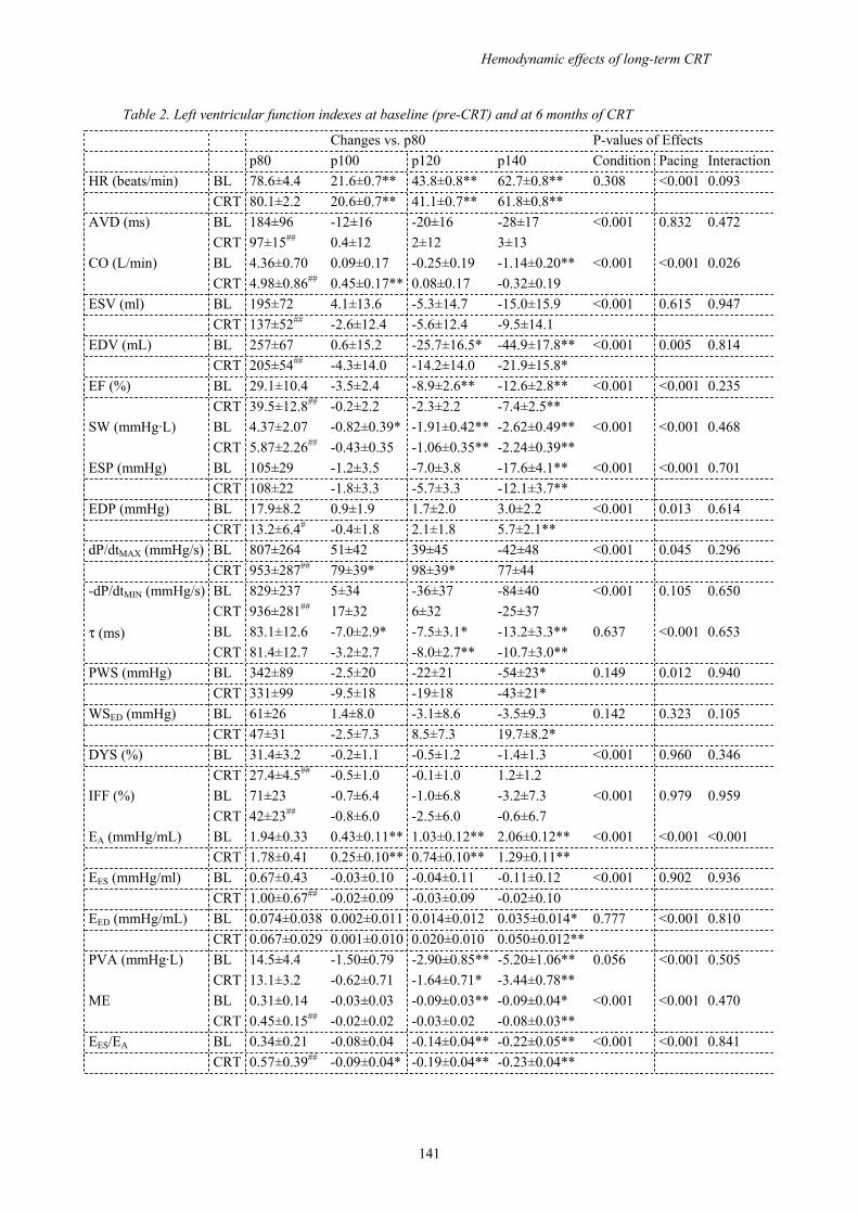
Hemodynamic effects of long-term CRT
141
Table 2. Left ventricular function indexes at baseline (pre-CRT) and at 6 months of CRT
Changes vs. p80 P-values of Effects p80 p100 p120 p140 Condition Pacing InteractionHR (beats/min) BL 78.6±4.4 21.6±0.7** 43.8±0.8** 62.7±0.8** 0.308 <0.001 0.093 CRT 80.1±2.2 20.6±0.7** 41.1±0.7** 61.8±0.8** AVD (ms) BL 184±96 -12±16 -20±16 -28±17 <0.001 0.832 0.472 CRT 97±15## 0.4±12 2±12 3±13 CO (L/min) BL 4.36±0.70 0.09±0.17 -0.25±0.19 -1.14±0.20** <0.001 <0.001 0.026 CRT 4.98±0.86## 0.45±0.17** 0.08±0.17 -0.32±0.19 ESV (ml) BL 195±72 4.1±13.6 -5.3±14.7 -15.0±15.9 <0.001 0.615 0.947 CRT 137±52## -2.6±12.4 -5.6±12.4 -9.5±14.1 EDV (mL) BL 257±67 0.6±15.2 -25.7±16.5* -44.9±17.8** <0.001 0.005 0.814 CRT 205±54## -4.3±14.0 -14.2±14.0 -21.9±15.8* EF (%) BL 29.1±10.4 -3.5±2.4 -8.9±2.6** -12.6±2.8** <0.001 <0.001 0.235 CRT 39.5±12.8## -0.2±2.2 -2.3±2.2 -7.4±2.5** SW (mmHg·L) BL 4.37±2.07 -0.82±0.39* -1.91±0.42** -2.62±0.49** <0.001 <0.001 0.468 CRT 5.87±2.26## -0.43±0.35 -1.06±0.35** -2.24±0.39** ESP (mmHg) BL 105±29 -1.2±3.5 -7.0±3.8 -17.6±4.1** <0.001 <0.001 0.701 CRT 108±22 -1.8±3.3 -5.7±3.3 -12.1±3.7** EDP (mmHg) BL 17.9±8.2 0.9±1.9 1.7±2.0 3.0±2.2 <0.001 0.013 0.614 CRT 13.2±6.4# -0.4±1.8 2.1±1.8 5.7±2.1** dP/dtMAX (mmHg/s) BL 807±264 51±42 39±45 -42±48 <0.001 0.045 0.296 CRT 953±287## 79±39* 98±39* 77±44 -dP/dtMIN (mmHg/s) BL 829±237 5±34 -36±37 -84±40 <0.001 0.105 0.650 CRT 936±281## 17±32 6±32 -25±37 τ (ms) BL 83.1±12.6 -7.0±2.9* -7.5±3.1* -13.2±3.3** 0.637 <0.001 0.653 CRT 81.4±12.7 -3.2±2.7 -8.0±2.7** -10.7±3.0** PWS (mmHg) BL 342±89 -2.5±20 -22±21 -54±23* 0.149 0.012 0.940 CRT 331±99 -9.5±18 -19±18 -43±21* WSED (mmHg) BL 61±26 1.4±8.0 -3.1±8.6 -3.5±9.3 0.142 0.323 0.105 CRT 47±31 -2.5±7.3 8.5±7.3 19.7±8.2* DYS (%) BL 31.4±3.2 -0.2±1.1 -0.5±1.2 -1.4±1.3 <0.001 0.960 0.346 CRT 27.4±4.5## -0.5±1.0 -0.1±1.0 1.2±1.2 IFF (%) BL 71±23 -0.7±6.4 -1.0±6.8 -3.2±7.3 <0.001 0.979 0.959 CRT 42±23## -0.8±6.0 -2.5±6.0 -0.6±6.7 EA (mmHg/mL) BL 1.94±0.33 0.43±0.11** 1.03±0.12** 2.06±0.12** <0.001 <0.001 <0.001 CRT 1.78±0.41 0.25±0.10** 0.74±0.10** 1.29±0.11** EES (mmHg/ml) BL 0.67±0.43 -0.03±0.10 -0.04±0.11 -0.11±0.12 <0.001 0.902 0.936 CRT 1.00±0.67## -0.02±0.09 -0.03±0.09 -0.02±0.10 EED (mmHg/mL) BL 0.074±0.038 0.002±0.011 0.014±0.012 0.035±0.014* 0.777 <0.001 0.810 CRT 0.067±0.029 0.001±0.010 0.020±0.010 0.050±0.012** PVA (mmHg·L) BL 14.5±4.4 -1.50±0.79 -2.90±0.85** -5.20±1.06** 0.056 <0.001 0.505 CRT 13.1±3.2 -0.62±0.71 -1.64±0.71* -3.44±0.78** ME BL 0.31±0.14 -0.03±0.03 -0.09±0.03** -0.09±0.04* <0.001 <0.001 0.470 CRT 0.45±0.15## -0.02±0.02 -0.03±0.02 -0.08±0.03** EES/EA BL 0.34±0.21 -0.08±0.04 -0.14±0.04** -0.22±0.05** <0.001 <0.001 0.841 CRT 0.57±0.39## -0.09±0.04* -0.19±0.04** -0.23±0.04**
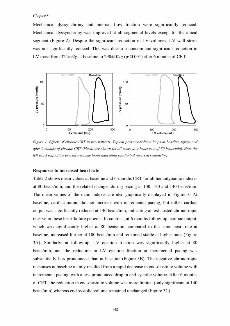
Chapter 8
142
Mechanical dyssynchrony and internal flow fraction were significantly reduced.
Mechanical dyssynchrony was improved at all segmental levels except for the apical
segment (Figure 2). Despite the significant reduction in LV volumes, LV wall stress
was not significantly reduced. This was due to a concomitant significant reduction in
LV mass from 324±92g at baseline to 290±107g (p<0.001) after 6 months of CRT.
6-mo CRT Baseline 6-mo CRT Baseline
0
50
100
0 100 200 300LV volume (mL)
LV p
ress
ure
(mm
Hg)
0
50
100
0 100 200 300LV volume (mL)
LV p
ress
ure
(mm
Hg)
Figure 1. Effects of chronic CRT in two patients. Typical pressure-volume loops at baseline (grey) and
after 6 months of chronic CRT (black) are shown (in all cases at a heart rate of 80 beats/min). Note the
left ward shift of the pressure-volume loops indicating substantial reversed remodeling
Responses to increased heart rate
Table 2 shows mean values at baseline and 6-months CRT for all hemodynamic indexes
at 80 beats/min, and the related changes during pacing at 100, 120 and 140 beats/min.
The mean values of the main indexes are also graphically displayed in Figure 3. At
baseline, cardiac output did not increase with incremental pacing, but rather cardiac
output was significantly reduced at 140 beats/min, indicating an exhausted chronotropic
reserve in these heart failure patients. In contrast, at 6 months follow-up, cardiac output,
which was significantly higher at 80 beats/min compared to the same heart rate at
baseline, increased further at 100 beats/min and remained stable at higher rates (Figure
3A). Similarly, at follow-up, LV ejection fraction was significantly higher at 80
beats/min, and the reduction in LV ejection fraction at incremental pacing was
substantially less pronounced than at baseline (Figure 3B). The negative chronotropic
responses at baseline mainly resulted from a rapid decrease in end-diastolic volume with
incremental pacing, with a less pronounced drop in end-systolic volume. After 6 months
of CRT, the reduction in end-diastolic volume was more limited (only significant at 140
beats/min) whereas end-systolic volume remained unchanged (Figure 3C).
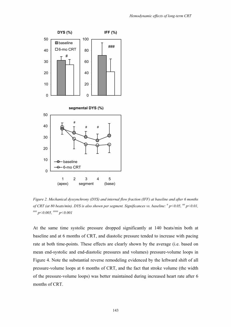
Hemodynamic effects of long-term CRT
143
DYS (%) IFF (%)
###
#
segmental DYS (%)
## #
1 2 3 4 5(apex) segment (base)
0
10
20
30
40
50
baseline6-mo CRT
0
10
20
30
40
50baseline
6-mo CRT
0
20
40
60
80
100
Figure 2. Mechanical dyssynchrony (DYS) and internal flow fraction (IFF) at baseline and after 6 months
of CRT (at 80 beats/min). DYS is also shown per segment. Significances vs. baseline: # p<0.05, ## p<0.01, ### p<0.005, #### p<0.001
At the same time systolic pressure dropped significantly at 140 beats/min both at
baseline and at 6 months of CRT, and diastolic pressure tended to increase with pacing
rate at both time-points. These effects are clearly shown by the average (i.e. based on
mean end-systolic and end-diastolic pressures and volumes) pressure-volume loops in
Figure 4. Note the substantial reverse remodeling evidenced by the leftward shift of all
pressure-volume loops at 6 months of CRT, and the fact that stroke volume (the width
of the pressure-volume loops) was better maintained during increased heart rate after 6
months of CRT.
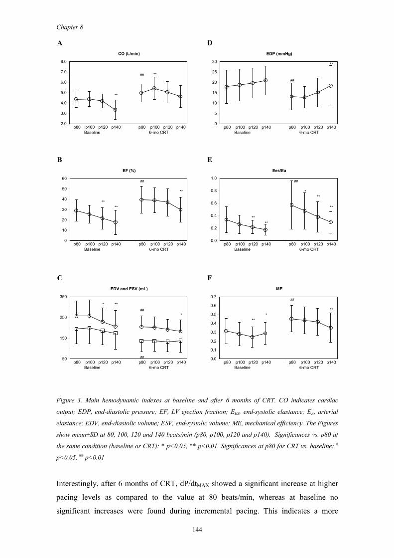
Chapter 8
144
A CO (L/min)
## **
**
p80 p100 p120 p140 p80 p100 p120 p140Baseline 6-mo CRT
2.0
3.0
4.0
5.0
6.0
7.0
8.0
D EDP (mmHg)
**
##
p80 p100 p120 p140 p80 p100 p120 p140Baseline 6-mo CRT
0
5
10
15
20
25
30
B EF (%)
##
**
****
p80 p100 p120 p140 p80 p100 p120 p140Baseline 6-mo CRT
0
10
20
30
40
50
60
E Ees/Ea
##
***
**
****
p80 p100 p120 p140 p80 p100 p120 p140Baseline 6-mo CRT
0.0
0.2
0.4
0.6
0.8
1.0
C EDV and ESV (mL)
* **##
*
##p80 p100 p120 p140 p80 p100 p120 p140
Baseline 6-mo CRT
50
150
250
350
F ME
##
***
**
p80 p100 p120 p140 p80 p100 p120 p140Baseline 6-mo CRT
0.0
0.1
0.2
0.3
0.4
0.5
0.6
0.7
Figure 3. Main hemodynamic indexes at baseline and after 6 months of CRT. CO indicates cardiac
output; EDP, end-diastolic pressure; EF, LV ejection fraction; EES, end-systolic elastance; EA, arterial
elastance; EDV, end-diastolic volume; ESV, end-systolic volume; ME, mechanical efficiency. The Figures
show mean±SD at 80, 100, 120 and 140 beats/min (p80, p100, p120 and p140). Significances vs. p80 at
the same condition (baseline or CRT): * p<0.05, ** p<0.01. Significances at p80 for CRT vs. baseline: #
p<0.05, ## p<0.01
Interestingly, after 6 months of CRT, dP/dtMAX showed a significant increase at higher
pacing levels as compared to the value at 80 beats/min, whereas at baseline no
significant increases were found during incremental pacing. This indicates a more

Hemodynamic effects of long-term CRT
145
physiological response after 6 months of CRT. This is illustrated in Figure 5, which
shows LV pressure and LV dP/dt for the different heart rates at baseline and after 6
months of CRT in a typical patient. Note the higher dP/dtMAX after 6 months of CRT
and the gradual increase in dP/dtMAX with increased pacing rate, which was absent at
baseline. This change towards normalization of chronotropic response was not found for
dP/dtMIN. Ventricular-arterial coupling, quantified by the ratio of ventricular and arterial
elastance, was highly abnormal in the heart failure patients, but improved significantly
after 6 months of CRT. The drop in EES/EA with increased heart rate was still present
after CRT (Figure 3E). Likewise, mechanical efficiency was improved at follow-up, but
dropped significantly at 140 beats/min both at baseline and after 6 months of CRT
(Figure 3F).
Baseline 6-mo CRT
p80p80 baseline
p120 p80p100 p100
p140 p120 p140
0
50
100
100 200
LV Volume (mL)
LV P
ress
ure
(mm
Hg)
0
50
100
100 200
LV Volume (mL)
LV P
ress
ure
(mm
Hg)
Figure 4. Mean pressure-volume loops at baseline and after 6 months of CRT. Mean pressure-volume
loops are based on mean end-systolic and end-diastolic pressures and volumes and are shown at heart
rates 80, 100, 120 and 140 beats/min. At baseline we used right atrial pacing via a temporary pacing
lead; at follow-up biventricular pacing was performed by reprogramming the CRT device
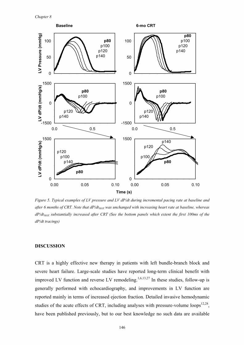
Chapter 8
146
Baseline 6-mo CRT
p80 p80 p100 p100 p120 p120 p140p140
p80 p80 p100 p100
p120 p120 p140 p140
p140 p120
p120 p100 p100 p140 p80
p80
Time (s)
0
50
100LV
Pre
ssur
e (m
mH
g)
-1500
0
1500
0.0 0.5
LV d
P/dt
(mm
Hg/
s)
0
50
100
-1500
0
1500
0.0 0.5
0
1500
0.00 0.05 0.10
LV d
P/dt
(mm
Hg/
s)
0
1500
0.00 0.05 0.10
Figure 5. Typical examples of LV pressure and LV dP/dt during incremental pacing rate at baseline and
after 6 months of CRT. Note that dP/dtMAX was unchanged with increasing heart rate at baseline, whereas
dP/dtMAX substantially increased after CRT (See the bottom panels which extent the first 100ms of the
dP/dt tracings)
DISCUSSION
CRT is a highly effective new therapy in patients with left bundle-branch block and
severe heart failure. Large-scale studies have reported long-term clinical benefit with
improved LV function and reverse LV remodeling.1,6,13,27 In these studies, follow-up is
generally performed with echocardiography, and improvements in LV function are
reported mainly in terms of increased ejection fraction. Detailed invasive hemodynamic
studies of the acute effects of CRT, including analyses with pressure-volume loops12,28,
have been published previously, but to our best knowledge no such data are available
Hemodynamic effects of long-term CRT
147
for chronic CRT. In the present study we obtained invasive hemodynamics by pressure-
volume loops at baseline and after 6 months of CRT. Our data confirm previous
findings regarding clinical benefit, improved LV ejection fraction and reverse
remodeling. In addition, it shows that hemodynamic improvements in terms of
increased dP/dtMAX, dP/dtMIN and stroke work, and reduced end-diastolic pressure,
previously found in acute studies6,11,28,29, are still present at 6 months follow-up.
Moreover, our study shows improved ventricular-arterial coupling and improved
mechanical efficiency. These hemodynamic findings are consistent with the observed
improvements in clinical and functional status. The altered responses to increased heart
rate may partly explain the improved exercise capacity of patients treated with CRT. At
baseline, cardiac output was unchanged when heart rate was increased illustrating the
exhausted LV function reserve of these patients. At follow-up this is converted to a
more physiological response although the capacity to increase cardiac output is still
limited. The latter presumably is partly due to an abnormal relaxation reflected by a
relatively long isovolumic relaxation time (τ), which did not improve after CRT. In the
normal heart, τ substantially shortens at higher heart rate, which enables adequate filling
despite a shortened diastolic period. This response is largely lost in heart failure, and did
not normalize after 6 months of CRT in our patients. Consistent with our findings,
previous studies failed to show improvements in isovolumic relaxation neither with
acute biventricular pacing29,30 nor at long-term.13 A theoretical model by Hay et al.31
shows a close correlation between increased τ and increased diastolic pressure, which is
most evident at high heart rates. Our data are consistent with this prediction and show
that the phenomenon is still present after 6 months of CRT. The improved mechanical
efficiency found in our study is in line with previous studies on acute effects of CRT by
Nelson et al.12 and is consistent with studies by, e.g., Sundell et al.14 in patients treated
long-term. Most likely, the improved mechanical intraventricular synchrony underlies
the more efficient conversion of total mechanical energy to external stroke work. This is
most evident from a highly significant reduction in internal flow fraction from 71 to
42%, which indicates that segmental volume changes are more efficiently used for
effective ejection rather than for energy-wasting shifting of blood volumes between
segments within the ventricle. In addition, ventricular-arterial coupling was significantly
improved which further optimizes production of external work.32,33 However, whereas
in the normal heart optimal ventricular-arterial coupling is maintained with increased
heart rate34, EES/EA significantly dropped in our patients and this abnormal response was
still present after long-term CRT. The baseline values for mechanical efficiency and
Chapter 8
148
ventricular-arterial coupling found in our study (0.31 and 0.34, respectively) were in the
same range but somewhat lower than values reported by Kim et al.35: 0.38 and 0.42,
respectively. However, the patients in their study had less severe heart failure evidenced
by an average NYHA classification of 1.8±0.7 and an LV ejection fraction of 37±13%.
Asanoi et al.36 reported that in the failing heart homeostatic mechanisms maintain
arterial blood pressure within the normal range, but that this blood pressure level causes
a deviation from energetically optimal conditions in hearts with a severely reduced
contractile state. This discrepancy results from worsening of ventricular-arterial
coupling and decreased mechanical efficiency. Conversely, the improved ventricular-
arterial coupling and mechanical efficiency after 6 months of CRT, as found in our
study, constitutes a more optimal energetic condition. Interestingly, despite the
substantial reverse remodeling in our study, wall stress was not significantly reduced
after 6 months of CRT. This was due to a concomitant reduction in LV mass. We would
hypothesize that the regression in LV volumes initially leads to a reduction in wall
stress, which then in turn may cause a reduction in LV hypertrophy. Note however that,
although not statistically significant, diastolic wall stress was reduced by 23% at 80
beats/min and by 30% at 100 beats/min. At higher heart rates, wall stress was virtually
unchanged or even increased compared to baseline (-5% at 120 beat/min, and +24% at
140 beats/min). This finding is explained by the fact that at baseline end-diastolic
volume drops substantially at the high heart rates (which also limits the increase in end-
diastolic pressure), whereas end-diastolic volume is better maintained at 6-months
follow-up. Furthermore, the global model to calculate wall stress does not take into
account spatial dyssynchrony, and conversion to a more uniform contraction pattern at
6-months follow-up may lead to reductions in wall stress at a regional level.
In our study we used simultaneous biventricular pacing in all patients. Sequential
biventricular pacing has been proposed to optimize CRT, and either right ventricular or
left ventricular pre-excitation may optimize hemodynamics in individual patients.8,37
However, Hay et al.28 demonstrated that sequential biventricular stimulation offered
minimal benefit and that, on the average, most systolic and diastolic function parameters
reached a maximum with simultaneous pacing. In addition to improvement of intra- and
interventricular dyssynchrony, the patients may also have benefited from optimization
of the AV delay. In our study the AV delay was reduced from a baseline value of
184±96 ms to a mean value of 97±15 ms during CRT. Studies by Auricchio et al.24
showed that the maximal increases in pulse pressure and dP/dtMAX were obtained at
Hemodynamic effects of long-term CRT
149
45% of the intrinsic AV interval. Consistently, most studies report optimal AV delays of
100-120 ms, but small differences in delay have far less influence than pacing site.29
Because our baseline studies were performed prior to implantation of the CRT device
we could not assess the acute hemodynamic effects of CRT. However, these effects
were documented in previous studies. Acute improvements in CO or SV, in most
studies assessed by changes in aortic pulse pressure, were reported to be in the range of
7 to 15% 3,11,24,28,29,38,39, which is comparable to the 14% increase found at 6-months in
our study. Previous studies show an acute reduction of 10 to 18% in end-systolic
volume, and a relatively smaller reduction in end-diastolic volume of 5 to 9% 8,30,39 ,
which lead to 15 to 33% relative improvement in EF. The reductions in end-systolic and
end-diastolic volume at 6 months in our study were 30 and 20%, respectively.
Apparently, the acute improvement in cardiac output is maintained long-term, but both
end-systolic and end-diastolic volume show a gradual, more or less parallel, further
reversed remodeling, as previously documented over a 3-months period by Yu et al.27
With regard to dP/dtMAX, previous studies fairly consistently showed an acute increase
of 13 to 21% 3,7,11,24,28,29,38, which is close to the 18% increase found in our study at 6-
months follow-up. Yu's study revealed that more than 60% of the gain in dP/dtMAX
obtained after 3 months CRT is lost immediately after turning off the pacemaker,
whereas 4 weeks after cessation of CRT dP/dtMAX had completely returned to pre-CRT
values. Their study also showed that left ventricular volumes increased and other
echocardiographic benefits were gradually lost over the 4-week period. We did not
systematically investigate the effects of turning off the pacemaker, but in a few patients
we registered pressure-volume loops during temporary cessation of pacing in the
follow-up study. Figure 6 shows two typical examples: The pressure-volume loops
show an immediate reduction in stroke volume, whereas dP/dtMAX was decreased by 20
and 7%, respectively. These immediate on-off responses are very similar to those
registered previously in acute studies.29
Limitations
The sample size in our study was too small to justify a meaningful responder/non-
responder analysis. Only 4 patients did not show an improved clinical status: 3 patients
with NYHA class III remained in class III, one class III patient deteriorated to class IV.
All other patients improved by 1 or 2 NYHA classes. In the 'non-responder' group the
baseline end-diastolic volume and end-systolic volume (282±73 and 228±70 mL,
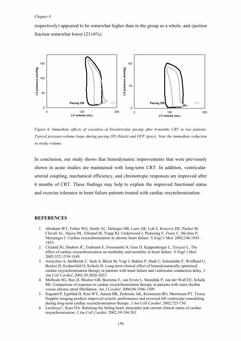
Chapter 8
150
respectively) appeared to be somewhat higher than in the group as a whole, and ejection
fraction somewhat lower (21±6%).
Pacing ON OFF Pacing ON OFF0
50
100
150
0 125 250LV volume (mL)
LV p
ress
ure
(mm
Hg)
0
50
100
0 100 200LV volume (mL)
LV p
ress
ure
(mm
Hg)
Figure 6. Immediate effects of cessation of biventricular pacing after 6-months CRT in two patients.
Typical pressure-volume loops during pacing ON (black) and OFF (grey). Note the immediate reduction
in stroke volume
In conclusion, our study shows that hemodynamic improvements that were previously
shown in acute studies are maintained with long-term CRT. In addition, ventricular-
arterial coupling, mechanical efficiency, and chronotropic responses are improved after
6 months of CRT. These findings may help to explain the improved functional status
and exercise tolerance in heart failure patients treated with cardiac resynchronization.
REFERENCES 1. Abraham WT, Fisher WG, Smith AL, Delurgio DB, Leon AR, Loh E, Kocovic DZ, Packer M,
Clavell AL, Hayes DL, Ellestad M, Trupp RJ, Underwood J, Pickering F, Truex C, McAtee P, Messenger J. Cardiac resynchronization in chronic heart failure. N Engl J Med. 2002;346:1845-1853.
2. Cleland JG, Daubert JC, Erdmann E, Freemantle N, Gras D, Kappenberger L, Tavazzi L. The effect of cardiac resynchronization on morbidity and mortality in heart failure. N Engl J Med. 2005;352:1539-1549.
3. Auricchio A, Stellbrink C, Sack S, Block M, Vogt J, Bakker P, Huth C, Schondube F, Wolfhard U, Bocker D, Krahnefeld O, Kirkels H. Long-term clinical effect of hemodynamically optimized cardiac resynchronization therapy in patients with heart failure and ventricular conduction delay. J Am Coll Cardiol. 2002;39:2026-2033.
4. Molhoek SG, Bax JJ, Bleeker GB, Boersma E, van Erven L, Steendijk P, van der Wall EE, Schalij MJ. Comparison of response to cardiac resynchronization therapy in patients with sinus rhythm versus chronic atrial fibrillation. Am J Cardiol. 2004;94:1506-1509.
5. Sogaard P, Egeblad H, Kim WY, Jensen HK, Pedersen AK, Kristensen BO, Mortensen PT. Tissue Doppler imaging predicts improved systolic performance and reversed left ventricular remodeling during long-term cardiac resynchronization therapy. J Am Coll Cardiol. 2002;723-730.
6. Leclercq C, Kass DA. Retiming the failing heart: principles and current clinical status of cardiac resynchronization. J Am Coll Cardiol. 2002;39:194-201.

Hemodynamic effects of long-term CRT
151
7. Breithardt OA, Stellbrink C, Kramer AP, Sinha AM, Franke A, Salo R, Schiffgens B, Huvelle E, Auricchio A. Echocardiographic quantification of left ventricular asynchrony predicts an acute hemodynamic benefit of cardiac resynchronization therapy. J Am Coll Cardiol. 2002;40:536-545.
8. Sogaard P, Egeblad H, Pedersen AK, Kim WY, Kristensen BO, Hansen PS, Mortensen PT. Sequential versus simultaneous biventricular resynchronization for severe heart failure: evaluation by tissue Doppler imaging. Circulation. 2002;106:2078-2084.
9. Leclercq C, Faris O, Tunin R, Johnson J, Kato R, Evans F, Spinelli J, Halperin H, McVeigh E, Kass DA. Systolic improvement and mechanical resynchronization does not require electrical synchrony in the dilated failing heart with left bundle- branch block. Circulation. 2002;106:1760-1763.
10. Kawaguchi M, Murabayashi T, Fetics BJ, Nelson GS, Samejima H, Nevo E, Kass DA. Quantitation of basal dyssynchrony and acute resynchronization from left or biventricular pacing by novel echo-contrast variability imaging. J Am Coll Cardiol. 2002;39:2052-2058.
11. Auricchio A, Stellbrink C, Block M, Sack S, Vogt J, Bakker P, Klein H, Kramer A, Ding J, Salo R, Tockman B, Pochet T, Spinelli J. Effect of pacing chamber and atrioventricular delay on acute systolic function of paced patients with congestive heart failure. The Pacing Therapies for Congestive Heart Failure Study Group. The Guidant Congestive Heart Failure Research Group. Circulation. 1999;99:2993-3001.
12. Nelson GS, Berger RD, Fetics BJ, Talbot M, Spinelli JC, Hare JM, Kass DA. Left ventricular or biventricular pacing improves cardiac function at diminished energy cost in patients with dilated cardiomyopathy and left bundle-branch block. Circulation. 2000;102:3053-3059.
13. St John Sutton MG, Plappert T, Abraham WT, Smith AL, Delurgio DB, Leon AR, Loh E, Kocovic DZ, Fisher WG, Ellestad M, Messenger J, Kruger K, Hilpisch KE, Hill MR. Effect of cardiac resynchronization therapy on left ventricular size and function in chronic heart failure. Circulation. 2003;107:1985-1990.
14. Sundell J, Engblom E, Koistinen J, Ylitalo A, Naum A, Stolen KQ, Kalliokoski R, Nekolla SG, Airaksinen KE, Bax JJ, Knuuti J. The effects of cardiac resynchronization therapy on left ventricular function, myocardial energetics, and metabolic reserve in patients with dilated cardiomyopathy and heart failure. J Am Coll Cardiol. 2004;43:1027-1033.
15. Baan J, Van Der Velde ET, De Bruin H, Smeenk G, Koops J, Van Dijk AD, Temmerman D, Senden J, Buis B. Continuous measurement of left ventricular volume in animals and humans by conductance catheter. Circulation. 1984;70:812-823.
16. Steendijk P, Staal E, Jukema JW, Baan J. Hypertonic saline method accurately determines parallel conductance for dual-field conductance catheter. Am J Physiol Heart Circ Physiol. 2001;281:H755-H763.
17. Steendijk P, Tulner SA, Schreuder JJ, Bax JJ, van Erven L, van der Wall EE, Dion RA, Schalij MJ, Baan J. Quantification of left ventricular mechanical dyssynchrony by conductance catheter in heart failure patients. Am J Physiol Heart Circ Physiol. 2004;286:H723-H730.
18. Langer SF. Differential laws of left ventricular isovolumic pressure fall. Physiol Res. 2002;51:1-15.
19. Kelly RP, Ting CT, Yang TM, Liu CP, Maughan WL, Chang MS, Kass DA. Effective arterial elastance as index of arterial vascular load in humans. Circulation. 1992;86:513-521.
20. Tachibana H, Cheng HJ, Ukai T, Igawa A, Zhang ZS, Little WC, Cheng CP. Levosimendan Improves Left Ventricular Systolic and Diastolic Performance at Rest and During Exercise after Heart Failure. Am J Physiol Heart Circ Physiol. 2004.
21. Nozawa T, Yasumura Y, Futaki S, Tanaka N, Uenishi M, Suga H. Efficiency of energy transfer from pressure-volume area to external mechanical work increases with contractile state and decreases with afterload in the left ventricle of the anesthetized open-chest dog. Circulation. 1988;77:1116-1124.
22. Arts T, Bovendeerd PH, Prinzen FW, Reneman RS. Relation between left ventricular cavity pressure and volume and systolic fiber stress and strain in the wall. Biophys J. 1991;59:93-102.
23. Deague JA, Wilson CM, Grigg LE, Harrap SB. Discrepancies between echocardiographic measurements of left ventricular mass in a healthy adult population. Clin Sci (Lond). 1999;97:377-383.
24. Auricchio A, Ding J, Spinelli JC, Kramer AP, Salo RW, Hoersch W, KenKnight BH, Klein HU. Cardiac resynchronization therapy restores optimal atrioventricular mechanical timing in heart failure patients with ventricular conduction delay. J Am Coll Cardiol. 2002;39:1163-1169.
25. Laird NM, Ware JH. Random-effects models for longitudinal data. Biometrics. 1982;38:963-974. 26. Kindermann M, Frohlig G, Doerr T, Schieffer H. Optimizing the AV delay in DDD pacemaker
patients with high degree AV block: mitral valve Doppler versus impedance cardiography. Pacing Clin Electrophysiol. 1997;20:2453-2462.
27. Yu CM, Chau E, Sanderson JE, Fan K, Tang MO, Fung WH, Lin H, Kong SL, Lam YM, Hill MR, Lau CP. Tissue Doppler echocardiographic evidence of reverse remodeling and improved
Chapter 8
152
synchronicity by simultaneously delaying regional contraction after biventricular pacing therapy in heart failure. Circulation. 2002;105:438-445.
28. Hay I, Melenovsky V, Fetics BJ, Judge DP, Kramer A, Spinelli J, Reister C, Kass DA, Berger RD. Short-term effects of right-left heart sequential cardiac resynchronization in patients with heart failure, chronic atrial fibrillation, and atrioventricular nodal block. Circulation. 2004;110:3404-3410.
29. Kass DA, Chen CH, Curry C, Talbot M, Berger R, Fetics B, Nevo E. Improved left ventricular mechanics from acute VDD pacing in patients with dilated cardiomyopathy and ventricular conduction delay. Circulation. 1999;99:1567-73.
30. Ansalone G, Giannantoni P, Ricci R, Trambaiolo P, Fedele F, Santini M. Doppler myocardial imaging to evaluate the effectiveness of pacing sites in patients receiving biventricular pacing. J Am Coll Cardiol. 2002;39:489-499.
31. Hay I, Rich J, Ferber P, Burkhoff D, Maurer MS. Role of impaired myocardial relaxation in the production of elevated left ventricular filling pressure. Am J Physiol Heart Circ Physiol. 2005;288:H1203-H1208.
32. Sasayama S, Asanoi H. Coupling between the heart and arterial system in heart failure. Am J Med. 1991;90:14S-18S.
33. Starling MR. Left ventricular-arterial coupling relations in the normal human heart. Am Heart J. 1993;125:1659-1666.
34. Ohte N, Cheng CP, Little WC. Tachycardia exacerbates abnormal left ventricular-arterial coupling in heart failure. Heart Vessels. 2003;18:136-141.
35. Kim IS, Izawa H, Sobue T, Ishihara H, Somura F, Nishizawa T, Nagata K, Iwase M, Yokota M. Prognostic value of mechanical efficiency in ambulatory patients with idiopathic dilated cardiomyopathy in sinus rhythm. J Am Coll Cardiol. 2002;39:1264-1268.
36. Asanoi H, Kameyama T, Ishizaka S, Nozawa T, Inoue H. Energetically optimal left ventricular pressure for the failing human heart. Circulation. 1996;93:67-73.
37. Bordachar P, Lafitte S, Reuter S, Sanders P, Jais P, Haissaguerre M, Roudaut R, Garrigue S, Clementy J. Echocardiographic parameters of ventricular dyssynchrony validation in patients with heart failure using sequential biventricular pacing. J Am Coll Cardiol. 2004;44:2157-2165.
38. Stellbrink C, Breithardt OA, Franke A, Sack S, Bakker P, Auricchio A, Pochet T, Salo R, Kramer A, Spinelli J. Impact of cardiac resynchronization therapy using hemodynamically optimized pacing on left ventricular remodeling in patients with congestive heart failure and ventricular conduction disturbances(1). J Am Coll Cardiol. 2001;38:1957-1965.
39. Ukkonen H, Beanlands RS, Burwash IG, de Kemp RA, Nahmias C, Fallen E, Hill MR, Tang AS. Effect of cardiac resynchronization on myocardial efficiency and regional oxidative metabolism. Circulation. 2003;107:28-31.
CHAPTER 9
Clinical efficacy of surgical ventricular restoration and
restrictive mitral annuloplasty in patients with end-stage
heart failure
S.A.F. Tulner
P. Steendijk
R.J.M. Klautz
J.J. Bax
M.I.M. Versteegh
H.F. Verwey
M. J. Schalij
E.E. van der Wall
R.A.E. Dion
Submitted (J Heart Valve Disease)
Oral presentation at the third biennial meeting of the Society for Heart Valve
Disease, June 17-20, 2005, Vancouver, Canada
Chapter 9
154
ABSTRACT
Background. Surgical ventricular restoration (SVR) and restrictive mitral annuloplasty
(RMA) are increasingly performed for end-stage heart failure. We studied their clinical
efficacy in patients with end-stage heart failure.
Methods. We included 33 patients with NYHA class III/IV and left ventricular ejection
fraction ≤ 35%. In this group, patients with moderate to severe mitral regurgitation
(grade ≥ 2) underwent RMA and patients with anteroseptal aneurysm underwent SVR.
A combined procedure (SVR and RMA) was performed in 12 patients, isolated SVR in
5 patients and isolated RMA in 16 patients. Additional coronary artery bypass grafting
was done in 27 patients. Clinical parameters, including NYHA classification, Minnesota
Quality of Life (QoL) questionnaire, and 6-minute walking distance, were assessed at
baseline and 6 months after surgery.
Results. In the total group, operative mortality was 3% (n=1), in-hospital mortality was
9% (n=3), and there was no late mortality. Four patients (12%) needed post-operative
intra-aortic balloon pump support. The median duration at intensive care was 4 days
(range: 2-28) with a median hospital stay of 13 days (range: 7-49). All clinical
parameters were significantly improved at 6 months follow-up (p<0.001); NYHA
classification was improved from 3.4±0.5 to 1.5±0.5, QoL questionnaire score was
improved from 44±22 to 16±12, and 6-minute walking distance was increased from
248±134 to 422±113 m.
Conclusions. Surgical treatment of end-stage heart failure by SVR and/or RMA was
associated with 12% mortality at 6 months. Surviving patients showed a highly
significant clinical improvement.
INTRODUCTION
Chronic heart failure is one of the major healthcare problems in the world both in terms
of patient numbers, hospitalizations, and economic costs. In the United States, 4 to 5
million people have chronic heart failure, which leads to more than 2 million
hospitalizations each year.1 Despite optimal medical therapy, many patients remain
severely symptomatic. In these patients, cardiac transplantation remains the most
effective surgical therapy with 1-, 5- and 10-year survival rates of 94, 78, and 46%,
Clinical efficacy of SVR and RMA in heart failure patients
155
respectively.2,3 Although effective, heart transplantation is importantly hindered by
donor shortage, chronic rejection, and complications related to medication.
Given the limitations of medical therapy and cardiac transplantation, alternative surgical
therapies such as surgical ventricular restoration (SVR) and restrictive mitral
annuloplasty (RMA) have been introduced and are currently widely performed in
patients with end-stage heart failure.4,5 These therapies aim to correct frequently
observed end-stage complications as left ventricular aneurysm and mitral
regurgitation.6,7 If not treated, these complications usually have important adverse
effects on long-term morbidity and survival.8-10
A long-term study by the RESTORE-group has demonstrated that SVR is safe and
highly effective in the treatment of ischemic cardiomyopathy with a reduction of end-
systolic volume and a five-year survival of 69%.11 Several studies reported promising
results in patients with heart failure treated with RMA with one- and two-year survival
rates of 86% and 84%, respectively.12,13
In the present study, clinical efficacy was evaluated six months after surgery in a cohort
of patients with end-stage heart failure who underwent combined SVR and RMA,
isolated SVR or isolated RMA.
METHODS
Patients
We included 33 patients with end-stage heart failure, NYHA classification III or IV
with left ventricular ejection fraction ≤ 35%. These patients underwent heart failure
surgery for anteroseptal aneurysm and/or moderate to severe mitral regurgitation.
Twelve patients had both anteroseptal aneurysm and moderate to severe mitral
regurgitation (grade ≥ 2) and they underwent combined SVR and RMA; 5 patients had
an anteroseptal aneurysm and underwent isolated SVR (SVR group, n=17). Another 16
patients had severe mitral regurgitation (grade > 2) and no aneurysm and thus
underwent isolated restrictive mitral annuloplasty (RMA group, n=16). All patients
received stable medical therapy for chronic heart failure, including diuretics,
spironolactone, β-blockers, and ACE-inhibitors. The institutional review board
approved the study protocol and all patients provided informed consent. Patient
characteristics are summarized in table 1.
Chapter 9
156
Table 1. Patient characteristics
Variable N=33
Gender (M/F) 20/13
Age, yrs 64±12
Etiology (ischemic vs non-ischemic) 29/4
NYHA class 3.4±0.5
Duration of symptoms (median, months) 8 (2-62)
LVEF, % 27±8
Medication:
- Diuretics/spironolactone
- Nitrates
- ACE-inhibitors/A-II antagonists
- β-blockers
- Anticoagulants/aspirin
25 (76%)
7 (21%)
26 (79%)
21 (64%)
22 (67%)
NYHA, New York Heart Association. LVEF, left ventricular ejection fraction, ACE, Angiotensin
Converting Enzyme; A-II, Angiotensin II
Evaluation of mitral regurgitation
In patients with moderate to severe mitral regurgitation (grade ≥2) on transthoracic
echocardiography (TTE), additional transesophageal echocardiography (TEE) was
performed within 5 days before surgery. The TTE and TEE were performed without
general anesthesia to avoid underestimation of the severity of mitral regurgitation. The
severity of mitral regurgitation was graded semi-quantitatively from color-flow Doppler
in the conventional parasternal long-axis and apical 4-chamber images. Mitral
regurgitation was classified as: mild=1+ (jet area/atrial area <10%), moderate=2+ (jet
area/atrial area 10-20%), moderately severe =3+ (jet area/atrial area 20-45%), and
severe=4+ (jet area/atrial area >45%).14,15 The severity and precise mechanism of mitral
regurgitation was confirmed from the TEE images.
When the severity of mitral regurgitation was grade 2, an intraoperative loading test (as
described previously) was performed.16,17 Briefly, a preload test is performed by rapid
infusion of volume through the aortic cannula until the pulmonary artery capillary
wedge pressure increases by 15 mmHg. During this provocative test, the severity of
mitral regurgitation is continuously monitored, and patients who deteriorated to grade 3
or 4 mitral regurgitation underwent RMA. Immediately after surgery, TEE was repeated
to assess residual mitral regurgitation, transmitral diastolic gradient (determined from
continuous-wave Doppler), and length of coaptation of the mitral leaflets.
Clinical efficacy of SVR and RMA in heart failure patients
157
Surgical procedures
SVR was performed by the endoventricular circular patch plasty as previously described
by Dor.18,19 Briefly, the left ventricle was opened through the infarcted area. An
endocardial encircling suture (Fontan Stitch) was placed at the transitional zone
between scarred and normal tissue. A balloon containing 55mL/m2 body surface area
saline was introduced into the left ventricle and the Fontan stitch was tightened to
approximate the ventricular wall to the balloon. An oval dacron patch was tailored and
used to close the residual orifice. The excluded scar tissue was closed over the patch to
ensure hemostasis. Care was taken to eliminate all septal scar and to delineate a new
apex with the goal to restore the normal elliptical shape.
Stringent restrictive mitral annuloplasty (2 sizes smaller than measured) was performed
via an atrial transseptal approach using a Carpentier Edwards Physio ring (Edwards
Lifesciences, USA) as previously described.20 Additional coronary artery bypass
grafting was performed, if indicated.
Clinical evaluation
Patients were evaluated at the outpatient clinic at baseline and at 6 months after surgery.
Heart failure symptoms were classified using the NYHA score. Quality of Life score
was assessed using the Minnesota Living with Heart Failure questionnaire.21 This
questionnaire contains 21 questions concerning the patient's perception of the effects of
heart failure on daily life activities. Questions are scored from 0 to 5, resulting in a total
score from 0 to 105, with the highest score reflecting the worst quality of life. Exercise
tolerance was evaluated using 6-minute hall-walk tests at both visits.22
Statistics
Baseline and follow-up data were compared with paired t-tests. Statistical significance
was assumed at p < 0.05. All data are presented as the mean value ± SD.
RESULTS
Mean cardiopulmonary bypass (CPB)-time was 192±64 minutes with a mean aortic
cross-clamp time of 132±49 minutes. Weaning from CPB was uneventful in almost all
patients. However, in one case a patient developed an irreversible vasoplegic shock after
weaning from CPB and died during surgery (3% operative mortality) and four patients
Chapter 9
158
(12%) needed post-operative intra-aortic balloon pump support. There were no peri-
operative myocardial infarctions. Three additional patients died in the hospital (9%);
one patient who underwent combined SVR/RMA died 30 days postoperatively due to a
cerebrovascular accident, one patient (isolated SVR) died 5h postoperatively due to left
ventricular failure, and one patient (isolated RMA) died 7 days postoperatively due to
left ventricular failure. Early non-fatal complications consisted of postoperative atrial
fibrillation (3 patients), cerebrovascular accidents (1 patient), and renal failure (1
patient). One patient developed postoperative sepsis with an empyema in the pleural
space, which required surgical evacuation and this patient stayed 54 days at ICU with a
total hospital stay of 66 days. For the remaining patients, the median duration at
intensive care was 4 days (range: 2-28) with a median hospital stay of 13 days (range:
7-49). In the total group, we had no late mortality during the 6 months follow-up period.
Thus, overall mortality in our patient group was 12% at 6 months and complete clinical
assessment was performed in the 29 surviving patients.
Mitral regurgitation
The mean grade of mitral regurgitation at baseline in the patients who underwent RMA
was 3.0±0.6. The length of the anterior mitral leaflet (AML) was 2.88±0.30 cm with a
mean mitral annular diameter (MAD) of 4.08±0.55 cm (mean ratio MAD/AML
1.42±0.18). After surgery, no recurrence of mitral regurgitation was observed in these
patients (0.3±0.4) with restored length of leaflet coaptation of 0.82±0.19 cm and a mean
gradient of 2.9±1.3 mmHg.
NYHA score
In the total group, the mean NYHA score improved from 3.4±0.5 at baseline to 1.5±0.5
at 6 months follow-up (p< 0.001) (Figure 1). In both the RMA and SVR patients the
improvements in NYHA score was similar; in the RMA patients NYHA score improved
from 3.4±0.5 at baseline to 1.5±0.5 at 6 months follow-up (p< 0.001) and in the SVR
patients NYHA score improved from 3.5±0.5 at baseline to 1.5±0.5 at 6 months follow-
up (p< 0.001).
Quality-of-Life Minnesota score
Quality of Life score in the total group at baseline was 44±22 and decreased by 64% to
16±12 at 6 months follow-up (p< 0.001) (Figure 1). The change in the total group was
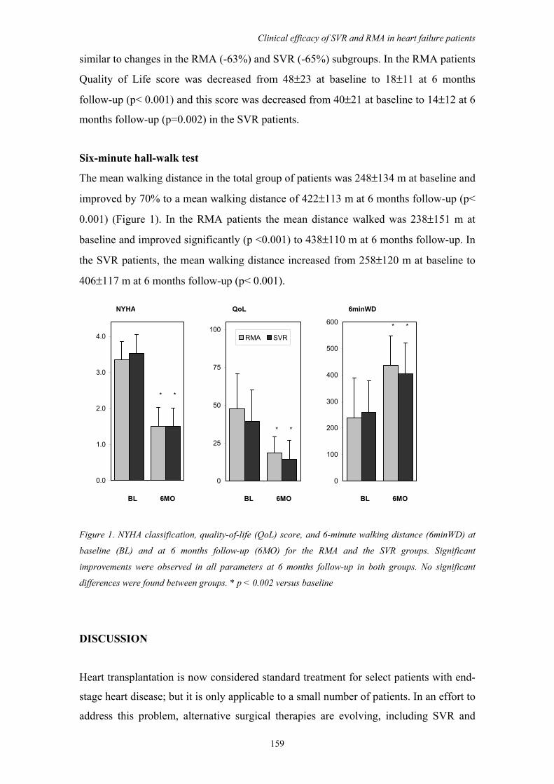
Clinical efficacy of SVR and RMA in heart failure patients
159
similar to changes in the RMA (-63%) and SVR (-65%) subgroups. In the RMA patients
Quality of Life score was decreased from 48±23 at baseline to 18±11 at 6 months
follow-up (p< 0.001) and this score was decreased from 40±21 at baseline to 14±12 at 6
months follow-up (p=0.002) in the SVR patients.
Six-minute hall-walk test
The mean walking distance in the total group of patients was 248±134 m at baseline and
improved by 70% to a mean walking distance of 422±113 m at 6 months follow-up (p<
0.001) (Figure 1). In the RMA patients the mean distance walked was 238±151 m at
baseline and improved significantly (p <0.001) to 438±110 m at 6 months follow-up. In
the SVR patients, the mean walking distance increased from 258±120 m at baseline to
406±117 m at 6 months follow-up (p< 0.001).
NYHA QoL 6minWD
* *
* *
* *
BL 6MO BL 6MO BL 6MO
0.0
1.0
2.0
3.0
4.0
0
25
50
75
100RMA SVR
0
100
200
300
400
500
600
Figure 1. NYHA classification, quality-of-life (QoL) score, and 6-minute walking distance (6minWD) at
baseline (BL) and at 6 months follow-up (6MO) for the RMA and the SVR groups. Significant
improvements were observed in all parameters at 6 months follow-up in both groups. No significant
differences were found between groups. * p < 0.002 versus baseline
DISCUSSION
Heart transplantation is now considered standard treatment for select patients with end-
stage heart disease; but it is only applicable to a small number of patients. In an effort to
address this problem, alternative surgical therapies are evolving, including SVR and
Chapter 9
160
RMA, and other approaches. These operative techniques to alter the shape of the left
ventricle, in combination with optimal medical management for heart failure, may
improve survival. In some patients it may even avoid or postpone transplantation.
The purpose of the present study was to evaluate the clinical efficacy of these treatments
using NYHA classification, Minnesota Living with Heart failure questionnaire, and 6-
minute hall-walk test in a cohort of patients with end-stage heart failure who underwent
SVR and/or RMA at our institution.
We found that the surgical treatment was associated with 12% mortality at 6 months
and resulted in an improvement in symptoms (NYHA class), accompanied by
improvement in 6-minute walking distance and Quality of Life score. Our results
regarding mortality are in line with the results of Dor et al. who reported 12% operative
mortality in 835 patients with end-stage heart failure.23 Earlier studies by Di Donato et
al. indicated a 19% in-hospital mortality and 26% mortality at one-year follow-up.24
However, the mean left ventricular ejection fraction in Di Donato's group was 17±3%,
while the mean ejection fraction in our series was 27±8% and in Dor's group only about
10% of the patients had an left ventricular ejection fraction < 20%. More recently, Qin
et al. reported a lower rate of mortality at six months follow-up of 5% in 60 patients
who underwent SVR with or without mitral valve repair.25 Similar findings were
reported by the RESTORE group with a 30-day mortality after SVR of 5.3% (8.7% with
mitral repair vs. 4.0% without repair).11 However, in this large patient population, also
patients with NYHA classification I/II were included. In all these previous studies, a
significant improvement in NYHA classification has been observed at long-term
follow-up, which was similar to the improvement found in our series.
The aim of alternative surgical interventions (SVR and RMA) in patients with end-stage
heart failure is to reduce left ventricular wall stress leading to reduced oxygen demand,
improved mechanical dyssynchrony and mechanical efficiency. These effects may result
in improvement of global and intrinsic systolic function. These theoretical assumptions
were recently confirmed by hemodynamic studies in patients with end-stage heart
failure.26,27 Operative mortality of both SVR and RMA are acceptable, however long-
term results are limited to survival rates, NYHA classification and hemodynamic
parameters.11,12,28 Therefore, it is still relatively unknown whether these therapies lead
to improvement of clinical status of the patient. Although previous studies indicate
improvement in NYHA classification, to our best knowledge, our study is the first to
show that these interventions lead to clinical improvement using 6-minute walking
distance and Quality of Life score at 6-months follow-up. Our study did not include a
Clinical efficacy of SVR and RMA in heart failure patients
161
control group. However, previous epidemiological studies indicate that 1-year mortality
rate in class III/IV heart failure patients is around 50%. 29 In comparison, the clinical
efficacy of our surgical approach in terms of Quality of Life and 6-minute walking
distance appears to be similar to the outcome after biventricular pacing in patients with
end-stage heart failure.30-32
Limitations
This study represents a single-center experience in a relatively small cohort of patients
with a combined surgical approach of SVR and/or RMA. Subgroup analysis did not
show statistical differences, but the groups were relatively small and treatment
obviously was not randomized. Moreover, this comparison should be taken with caution
because, despite similar baseline clinical parameters, the etiology was different between
groups.
In conclusion, surgical treatment of end-stage heart failure by SVR and RMA seems
relatively safe and surviving patients have clear clinical benefit at six months follow-up.
Long-term prospective clinical randomized trials should be performed to assess benefit
over optimal medical therapy.
REFERENCES
1. Nohria A, Lewis E, Stevenson LW. Medical management of advanced heart failure. JAMA.
2002;287:628-640. 2. Copeland JG, McCarthy M. University of Arizona, Cardiac Transplantation: changing patterns in
selection and outcomes. Clin Transpl. 2001;203-207. 3. Robbins RC, Barlow CW, Oyer PE, Hunt SA, Miller JL, Reitz BA, Stinson EB, Shumway NE.
Thirty years of cardiac transplantation at Stanford university. J Thorac Cardiovasc Surg. 1999;117:939-951.
4. Dor V. The endoventricular circular patch plasty ("Dor procedure") in ischemic akinetic dilated ventricles. Heart Fail Rev. 2001;6:187-193.
5. Bolling SF, Smolens IA, Pagani FD. Surgical alternatives for heart failure. J Heart Lung Transplant. 2001;20:729-733.
6. Gaudron P, Eilles C, Kugler I, Ertl G. Progressive left ventricular dysfunction and remodeling after myocardial infarction. Potential mechanisms and early predictors. Circulation. 1993;87:755-763.
7. Trichon BH, Felker GM, Shaw LK, Cabell CH, O'Connor CM. Relation of frequency and severity of mitral regurgitation to survival among patients with left ventricular systolic dysfunction and heart failure. Am J Cardiol. 2003;91:538-543.
8. Blondheim DS, Jacobs LE, Kotler MN, Costacurta GA, Parry WR. Dilated cardiomyopathy with mitral regurgitation: decreased survival despite a low frequency of left ventricular thrombus. Am Heart J. 1991;122:763-771.
9. Junker A, Thayssen P, Nielsen B, Andersen PE. The hemodynamic and prognostic significance of echo-Doppler-proven mitral regurgitation in patients with dilated cardiomyopathy. Cardiology. 1993;83:14-20.
Chapter 9
162
10. Mangschau A. Akinetic versus dyskinetic left ventricular aneurysms diagnosed by gated scintigraphy: difference in surgical outcome. Ann Thorac Surg. 1989;47:746-751.
11. Athanasuleas CL, Buckberg GD, Stanley AW, Siler W, Dor V, Di Donato M, Menicanti L, Almeida dO, Beyersdorf F, Kron IL, Suma H, Kouchoukos NT, Moore W, McCarthy PM, Oz MC, Fontan F, Scott ML, Accola KA. Surgical ventricular restoration in the treatment of congestive heart failure due to post-infarction ventricular dilation. J Am Coll Cardiol. 2004;44:1439-1445.
12. Bax JJ, Braun J, Somer ST, Klautz R, Holman ER, Versteegh MI, Boersma E, Schalij MJ, van der Wall EE, Dion RA. Restrictive annuloplasty and coronary revascularization in ischemic mitral regurgitation results in reverse left ventricular remodeling. Circulation. 2004;110:II103-II108.
13. Gummert JF, Rahmel A, Bucerius J, Onnasch J, Doll N, Walther T, Falk V, Mohr FW. Mitral valve repair in patients with end stage cardiomyopathy: who benefits? Eur J Cardiothorac Surg. 2003;23:1017-1022.
14. Fisher EA, Goldman ME. Simple, rapid method for quantification of tricuspid regurgitation by two-dimensional echocardiography. Am J Cardiol. 1989;63:1375-1378.
15. Thomas JD. How leaky is that mitral valve? Simplified Doppler methods to measure regurgitant orifice area. Circulation. 1997;95:548-550.
16. Byrne JG, Aklog L, Adams DH. Assessment and management of functional or ischaemic mitral regurgitation. Lancet. 2000;355:1743-1744.
17. Dion R, Benetis R, Elias B, Guennaoui T, Raphael D, Van Dyck M, Noirhomme P, Van Overschelde JL. Mitral valve procedures in ischemic regurgitation. J Heart Valve Dis. 1995;4 Suppl 2:S124-S129.
18. Dor V, Saab M, Coste P, Kornaszewska M, Montiglio F. Left ventricular aneurysm: a new surgical approach. Thorac Cardiovasc Surg. 1989;37:11-19.
19. Dor V, Sabatier M, Di Donato M, Montiglio F, Toso A, Maioli M. Efficacy of endoventricular patch plasty in large postinfarction akinetic scar and severe left ventricular dysfunction: comparison with a series of large dyskinetic scars. J Thorac Cardiovasc Surg. 1998;116:50-59.
20. Braun J, Bax JJ, Versteegh MI, Voigt PG, Holman ER, Klautz RJ, Boersma E, Dion RA. Preoperative left ventricular dimensions predict reverse remodeling following restrictive mitral annuloplasty in ischemic mitral regurgitation. Eur J Cardiothorac Surg. 2005;27:847-853.
21. Rector TS, Kubo SH, Cohn JN. Validity of the Minnesota Living with Heart Failure questionnaire as a measure of therapeutic response to enalapril or placebo. Am J Cardiol. 1993;71:1106-1107.
22. Lipkin DP, Scriven AJ, Crake T, Poole-Wilson PA. Six minute walking test for assessing exercise capacity in chronic heart failure. Br Med J (Clin Res Ed). 1986;292:653-655.
23. Dor V, Sabatier M, Montiglio F, Coste P, Di Donato M. Endoventricular patch reconstruction in large ischemic wall-motion abnormalities. J Card Surg. 1999;14:46-52.
24. Di Donato M, Sabatier M, Montiglio F, Maioli M, Toso A, Fantini F, Dor V. Outcome of left ventricular aneurysmectomy with patch repair in patients with severely depressed pump function. Am J Cardiol. 1995;76:557-561.
25. Qin JX, Shiota T, McCarthy PM, Asher CR, Hail M, Agler DA, Popovic ZB, Greenberg NL, Smedira NG, Starling RC, Young JB, Thomas JD. Importance of mitral valve repair associated with left ventricular reconstruction for patients with ischemic cardiomyopathy: a real-time three-dimensional echocardiographic study. Circulation. 2003;108 Suppl 1:II241-II246.
26. Di Donato M, Toso A, Dor V, Sabatier M, Barletta G, Menicanti L, Fantini F. Surgical ventricular restoration improves mechanical intraventricular dyssynchrony in ischemic cardiomyopathy. Circulation. 2004;109:2536-2543.
27. Schreuder JJ, Castiglioni A, Maisano F, Steendijk P, Donelli A, Baan J, Alfieri O. Acute decrease of left ventricular mechanical dyssynchrony and improvement of contractile state and energy efficiency after left ventricular restoration. J Thorac Cardiovasc Surg. 2005;129:138-145.
28. Bolling SF. Mitral valve reconstruction in the patient with heart failure. Heart Fail Rev. 2001;6:177-185.
29. Cowburn PJ, Cleland JG, Coats AJ, Komajda M. Risk stratification in chronic heart failure. Eur Heart J. 1998;19:696-710.
30. Abraham WT, Fisher WG, Smith AL, Delurgio DB, Leon AR, Loh E, Kocovic DZ, Packer M, Clavell AL, Hayes DL, Ellestad M, Trupp RJ, Underwood J, Pickering F, Truex C, McAtee P, Messenger J. Cardiac resynchronization in chronic heart failure. N Engl J Med. 2002;346:1845-1853.
31. Auricchio A, Stellbrink C, Butter C, Sack S, Vogt J, Misier AR, Bocker D, Block M, Kirkels JH, Kramer A, Huvelle E. Clinical efficacy of cardiac resynchronization therapy using left ventricular pacing in heart failure patients stratified by severity of ventricular conduction delay. J Am Coll Cardiol. 2003;42:2109-2116.
32. Molhoek SG, Bax JJ, Van Erven L, Bootsma M, Boersma E, Steendijk P, van der Wall EE, Schalij MJ. Effectiveness of resynchronization therapy in patients with end-stage heart failure. Am J Cardiol. 2002;90:379-383.
CHAPTER 10
Beneficial mid-term hemodynamic and clinical effects of
surgical ventricular restoration in patients with ischemic
dilated cardiomyopathy
S.A.F. Tulner
J.J. Bax
G.B. Bleeker
P. Steendijk
R.J.M. Klautz
E.R. Holman
M.J. Schalij
R.A.E. Dion
E.E. van der Wall
Submitted (Ann Thorac Surg)
Oral presentation at European Society of Cardiology, August 2005, Stockholm,
Sweden
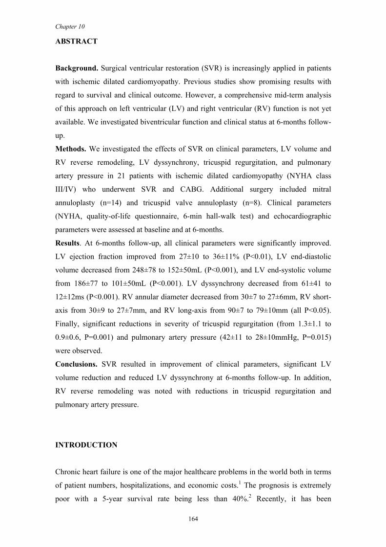
Chapter 10
164
ABSTRACT
Background. Surgical ventricular restoration (SVR) is increasingly applied in patients
with ischemic dilated cardiomyopathy. Previous studies show promising results with
regard to survival and clinical outcome. However, a comprehensive mid-term analysis
of this approach on left ventricular (LV) and right ventricular (RV) function is not yet
available. We investigated biventricular function and clinical status at 6-months follow-
up.
Methods. We investigated the effects of SVR on clinical parameters, LV volume and
RV reverse remodeling, LV dyssynchrony, tricuspid regurgitation, and pulmonary
artery pressure in 21 patients with ischemic dilated cardiomyopathy (NYHA class
III/IV) who underwent SVR and CABG. Additional surgery included mitral
annuloplasty (n=14) and tricuspid valve annuloplasty (n=8). Clinical parameters
(NYHA, quality-of-life questionnaire, 6-min hall-walk test) and echocardiographic
parameters were assessed at baseline and at 6-months.
Results. At 6-months follow-up, all clinical parameters were significantly improved.
LV ejection fraction improved from 27±10 to 36±11% (P<0.01), LV end-diastolic
volume decreased from 248±78 to 152±50mL (P<0.001), and LV end-systolic volume
from 186±77 to 101±50mL (P<0.001). LV dyssynchrony decreased from 61±41 to
12±12ms (P<0.001). RV annular diameter decreased from 30±7 to 27±6mm, RV short-
axis from 30±9 to 27±7mm, and RV long-axis from 90±7 to 79±10mm (all P<0.05).
Finally, significant reductions in severity of tricuspid regurgitation (from 1.3±1.1 to
0.9±0.6, P=0.001) and pulmonary artery pressure (42±11 to 28±10mmHg, P=0.015)
were observed.
Conclusions. SVR resulted in improvement of clinical parameters, significant LV
volume reduction and reduced LV dyssynchrony at 6-months follow-up. In addition,
RV reverse remodeling was noted with reductions in tricuspid regurgitation and
pulmonary artery pressure.
INTRODUCTION
Chronic heart failure is one of the major healthcare problems in the world both in terms
of patient numbers, hospitalizations, and economic costs.1 The prognosis is extremely
poor with a 5-year survival rate being less than 40%.2 Recently, it has been

Beneficial mid-term effects of SVR
165
demonstrated that surgical ventricular restoration (SVR) improves symptoms and long-
term survival in patients with ischemic cardiomyopathy and severe heart failure.3
Several studies have reported on the beneficial effects of SVR, including left ventricular
(LV) volume reduction with an improvement in LV ejection fraction (LVEF),
associated with a reduction in ventricular arrhythmias and reduced mitral
regurgitation.4-6 In addition, recent studies have shown that SVR results in an acute
reduction of LV mechanical dyssynchrony.7,8
In patients with valvular insufficiencies who undergo SVR, additional mitral and or
tricuspid valve repair may be needed to optimize patient outcome.9 Therefore, our
current approach in patients undergoing SVR is to always correct mild to moderate
mitral and tricuspid regurgitation using annuloplasty. Although preliminary data
indicate acceptable survival, larger studies are required to establish survival rate. In
addition, comprehensive data on clinical and hemodynamic status of these patients at
mid-term follow-up are limited. Therefore, we analyzed clinical status and biventricular
function in patients with ischemic dilated cardiomyopathy treated at our institution
before and 6 months after surgery. In particular, we report the effects of our approach
on LV volume, LV dyssynchrony, right ventricular (RV) reverse remodeling and RV
functional parameters (severity of tricuspid regurgitation and pulmonary artery
pressure).
METHODS
Patients and Study Protocol
We studied a group of 21 patients with ischemic dilated cardiomyopathy, who
underwent SVR and who had complete echocardiographic follow-up including tissue
Doppler imaging at 6 months. All patients had severe heart failure symptoms and 13
patients (62%) had accompanying angina pectoris. In particular, 13 (62%) patients were
in New York Heart Association (NYHA) class III and 8 (38%) were in class IV. All 21
patients had a previous anteroseptal infarction and the interval between infarction and
SVR procedure averaged 2.5 years (range 0.25-12 years). All patients had coronary
artery disease (on average 2.4±0.9 stenosed coronary arteries) and were scheduled for
additional CABG. Patients with severe mitral and/or tricuspid regurgitation underwent
additional mitral and/or tricuspid valve repair. Patients who underwent valvular repair
Chapter 10
166
were evaluated before and immediately after surgery by transesophageal
echocardiography (TEE). The baseline characteristics are presented in Table 1.
Table 1. Patient characteristics
Variable N=21
Age, yrs 63±11 (35-76)
Gender (M/F) 14/7
Delay from previous infarction, mo 30±45 (4-144)
QRS duration, ms 107±28 (80-202)
NYHA class
- class III
- class IV
13 (62%)
8 (38%)
Rhythm
- sinus rhythm
- atrial fibrillation
20 (95%)
1 (5%)
Coronary artery disease
- 1-vessel
- 2-vessel
- 3-vessel
4 (19%)
8 (38%)
9 (43%)
Medication:
- Diuretics/spironolactone
- Nitrates
- ACE-inhibitors/A-II antagonists
- β-blockers
- Anticoagulants/aspirin
18 (86%)
7 (33%)
17 (81%)
16 (76%)
15 (71%)
ACE, Angiotensin Converting Enzyme; A-II, Angiotensin II;
NYHA, New York Heart Association
In all patients, before SVR and 6 months after surgery, two-dimensional transthoracic
echocardiography (TTE) at rest was performed to calculate LV volumes and LVEF, and
to assess RV chamber size. Next, tissue Doppler imaging was performed to evaluate LV
dyssynchrony. At the same time points, clinical status was assessed using NYHA
classification, the Minnesota quality-of-life questionnaire, and the 6-minute hall-walk
test. The institutional review board approved the study protocol and all patients
provided informed consent.
Surgical Procedures
The surgical procedures were performed with the use of normothermic cardiopulmonary
bypass with intermittent antegrade warm blood cardioplegia for myocardial protection.
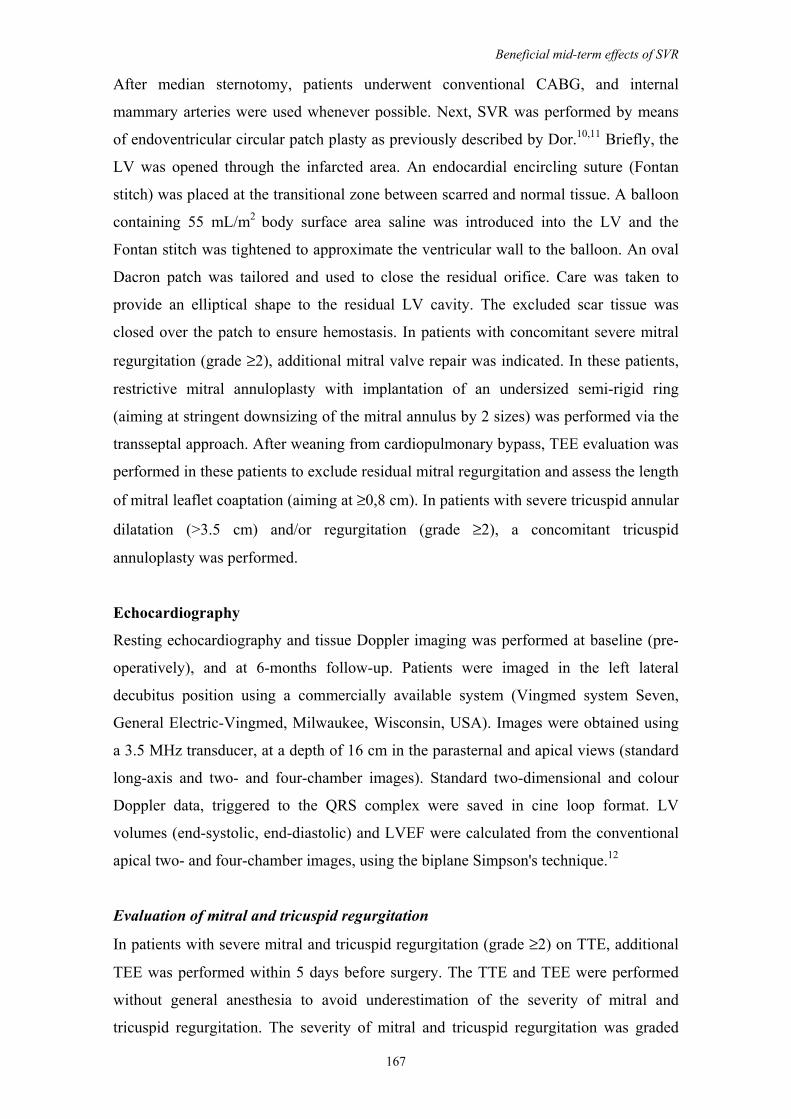
Beneficial mid-term effects of SVR
167
After median sternotomy, patients underwent conventional CABG, and internal
mammary arteries were used whenever possible. Next, SVR was performed by means
of endoventricular circular patch plasty as previously described by Dor.10,11 Briefly, the
LV was opened through the infarcted area. An endocardial encircling suture (Fontan
stitch) was placed at the transitional zone between scarred and normal tissue. A balloon
containing 55 mL/m2 body surface area saline was introduced into the LV and the
Fontan stitch was tightened to approximate the ventricular wall to the balloon. An oval
Dacron patch was tailored and used to close the residual orifice. Care was taken to
provide an elliptical shape to the residual LV cavity. The excluded scar tissue was
closed over the patch to ensure hemostasis. In patients with concomitant severe mitral
regurgitation (grade ≥2), additional mitral valve repair was indicated. In these patients,
restrictive mitral annuloplasty with implantation of an undersized semi-rigid ring
(aiming at stringent downsizing of the mitral annulus by 2 sizes) was performed via the
transseptal approach. After weaning from cardiopulmonary bypass, TEE evaluation was
performed in these patients to exclude residual mitral regurgitation and assess the length
of mitral leaflet coaptation (aiming at ≥0,8 cm). In patients with severe tricuspid annular
dilatation (>3.5 cm) and/or regurgitation (grade ≥2), a concomitant tricuspid
annuloplasty was performed.
Echocardiography
Resting echocardiography and tissue Doppler imaging was performed at baseline (pre-
operatively), and at 6-months follow-up. Patients were imaged in the left lateral
decubitus position using a commercially available system (Vingmed system Seven,
General Electric-Vingmed, Milwaukee, Wisconsin, USA). Images were obtained using
a 3.5 MHz transducer, at a depth of 16 cm in the parasternal and apical views (standard
long-axis and two- and four-chamber images). Standard two-dimensional and colour
Doppler data, triggered to the QRS complex were saved in cine loop format. LV
volumes (end-systolic, end-diastolic) and LVEF were calculated from the conventional
apical two- and four-chamber images, using the biplane Simpson's technique.12
Evaluation of mitral and tricuspid regurgitation
In patients with severe mitral and tricuspid regurgitation (grade ≥2) on TTE, additional
TEE was performed within 5 days before surgery. The TTE and TEE were performed
without general anesthesia to avoid underestimation of the severity of mitral and
tricuspid regurgitation. The severity of mitral and tricuspid regurgitation was graded

Chapter 10
168
semi-quantitatively from color-flow Doppler in the conventional parasternal long-axis
and apical 4-chamber images. Mitral and tricuspid regurgitation were classified as:
mild=1+ (jet area/atrial area <10%), moderate=2+ (jet area/atrial area 10-20%),
moderately severe =3+ (jet area/atrial area 20-45%), and severe=4+ (jet area/atrial area
>45%).13,14 The severity and precise mechanism of mitral regurgitation was confirmed
from the TEE images.
Immediately after surgery, TEE was repeated to assess residual mitral or tricuspid
regurgitation, transmitral diastolic gradient (determined from continuous-wave
Doppler), and length of coaptation of the mitral leaflets. Six months after surgery, TTE
was performed to assess possible recurrence of mitral and tricuspid regurgitation.
Continuous-wave Doppler examination was also performed to estimate pulmonary
artery systolic pressure from the trans-tricuspid maximal regurgitant flow velocity. All
TTE measurements were analyzed in random order by two independent observers
without knowledge of the clinical status of the patient and the timing of the
echocardiogram.
Assessment of RV chamber size
RV end-diastolic chamber size was assessed using three parameters, which were
described previously by Foale et al.15 The first parameter is the diameter of the annulus
of the tricuspid valve (TV ANN), defined as the point of attachment of the septal and
posterior leaflets to the atrioventricular junction. The second measurement is the
maximal dimension of the middle third of the RV, parallel to the tricuspid annulus (RV
SAX). The last measurement is the major axis of the RV (RV LAX) and is defined as
the distance between the RV apex to the mid-point of the tricuspid annulus.
Inter- and intra-observer agreement for assessment of RV chamber size were 98% and
96% for TV ANN, 90% and 92% for RV SAX, and 94% and 95% for RV LAX
respectively.
Tissue Doppler Imaging
In addition to the conventional echocardiographic examination, tissue Doppler imaging
was performed to assess LV dyssynchrony. For tissue Doppler imaging, color Doppler
frame rates varied between 80 and 115 frames/s depending on the sector width of the
range of interest; pulse repetition frequencies were between 500 Hz and 1 KHz,
resulting in aliasing velocities between 16 and 32 cm/s. Tissue Doppler imaging
parameters were measured from color images of three consecutive heart beats by offline
Beneficial mid-term effects of SVR
169
analysis. Data were analyzed using commercial software (Echopac 6.1, General Electric
- Vingmed).
To determine LV dyssynchrony, the sample volume was placed in the basal portions of
the septum and the LV lateral wall; peak systolic velocities and time-to-peak systolic
velocities were obtained and the delay in peak velocity between the septum and the LV
lateral wall was calculated as an indicator of LV dyssynchrony (referred to as the septal-
to-lateral delay).
Inter- and intra-observer agreement for assessment of the septal-to-lateral delay were
90% and 96%, respectively.16
Assessment of Functional Status
Functional status was assessed according to the NYHA classification, quality-of-life
score (using the Minnesota quality-of-life questionnaire) and 6-minute hall-walk test. In
all patients, QRS duration was measured from the surface ECG using the widest QRS
complex from the leads II, V1 and V6. The ECGs were recorded at a speed of 25
mm/sec and were evaluated by two independent observers without knowledge of the
patient’s clinical status. All parameters were assessed within 1 week before surgery and
approximately 6 months post-surgery.
Statistical Analysis
Data are presented as mean ± SD, and compared using the paired or unpaired Student’s
t-test when appropriate. For all tests a P-value <0.05 was considered statistically
significant.
RESULTS
Twenty-one patients were evaluated: 12 patients (57%) had dyskinesia and 9 patients
(43%) had akinesia. (Peri-)operative data and early operative complications (<30 days)
are summarized in Table 2. Note that we only included a selected group of patients with
complete echocardiographic follow-up at 6 months. Therefore, data regarding mortality
are not relevant and clinical findings reflect only patients who survived 6 months
follow-up.
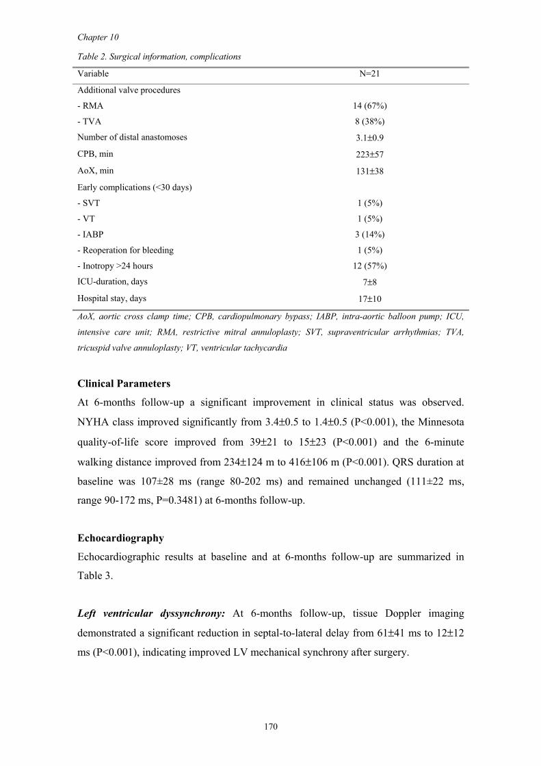
Chapter 10
170
Table 2. Surgical information, complications
Variable N=21
Additional valve procedures
- RMA
- TVA
14 (67%)
8 (38%)
Number of distal anastomoses 3.1±0.9
CPB, min 223±57
AoX, min 131±38
Early complications (<30 days)
- SVT
- VT
- IABP
- Reoperation for bleeding
- Inotropy >24 hours
1 (5%)
1 (5%)
3 (14%)
1 (5%)
12 (57%)
ICU-duration, days 7±8
Hospital stay, days 17±10
AoX, aortic cross clamp time; CPB, cardiopulmonary bypass; IABP, intra-aortic balloon pump; ICU,
intensive care unit; RMA, restrictive mitral annuloplasty; SVT, supraventricular arrhythmias; TVA,
tricuspid valve annuloplasty; VT, ventricular tachycardia
Clinical Parameters
At 6-months follow-up a significant improvement in clinical status was observed.
NYHA class improved significantly from 3.4±0.5 to 1.4±0.5 (P<0.001), the Minnesota
quality-of-life score improved from 39±21 to 15±23 (P<0.001) and the 6-minute
walking distance improved from 234±124 m to 416±106 m (P<0.001). QRS duration at
baseline was 107±28 ms (range 80-202 ms) and remained unchanged (111±22 ms,
range 90-172 ms, P=0.3481) at 6-months follow-up.
Echocardiography
Echocardiographic results at baseline and at 6-months follow-up are summarized in
Table 3.
Left ventricular dyssynchrony: At 6-months follow-up, tissue Doppler imaging
demonstrated a significant reduction in septal-to-lateral delay from 61±41 ms to 12±12
ms (P<0.001), indicating improved LV mechanical synchrony after surgery.
Beneficial mid-term effects of SVR
171
Left ventricular volume reduction: Significant LV volume reduction was shown at 6-
months follow-up. LV end-diastolic volume decreased from 248±78 ml to 152±50 ml
(P<0.001), whereas LV end-systolic volume decreased from 186±77 ml to 101±50 ml
(P<0.001). This resulted in an increase of the LVEF from 27±10 to 36±11%
(P=0.0072).
Right ventricular remodeling: At 6-months follow-up, significant reverse remodeling
of the RV was demonstrated. All three parameters reflecting RV chamber size showed a
significant decrease 6 months after surgery. The TV ANN showed a significant decrease
from 30±7 mm to 27±6 mm (P=0.04), RV SAX decreased from 30±9 mm to 27±7 mm
(P=0.03) and RV LAX showed a reduction from 90±7 mm to 79±10 mm (P<0.001).
Moreover, after surgery, pulmonary artery pressure significantly decreased from 42±11
mmHg to 28±10 mmHg (P=0.02).
Table 3. Echocardiographic data
Baseline TTE 6- months follow-up TTE P-value
LVEF, % 27±10 36±11 0.0072
LVEDV, ml 248±78 152±50 <0.001
LVESV, ml 186±77 101±50 <0.001
Septal-to-lateral delay, ms 61±41 12±12 <0.001
RV chamber size:
- TV ANN, mm
- RV SAX, mm
- RV LAX, mm
30±7
30±9
90±7
27±6
27±7
79±10
0.0430
0.0326
<0.001
Pulmonary artery pressure,
mmHg
42±11 28±10 0.0157
MR, grade 2.0±1.0 1.0±0.7 0.0013
TR, grade 1.3±1.1 0.9±0.6 0.0018
LVEF, left ventricular ejection fraction; LVEDV, left ventricular end-diastolic volume; LVESV, left
ventricular end-systolic volume; RV, right ventricle; TV ANN, tricuspid valve annulus; RV SAX, right
ventricular short axis; RV LAX, right ventricular long axis; MR, mitral regurgitation; TR, tricuspid
regurgitation
Mitral and tricuspid regurgitation: In 14 patients additional restrictive mitral
annuloplasty (median ring size 24; range 24-28) was performed. In 9 (43%) of these
patients, severe (grade 3 to 4+) mitral regurgitation was confirmed by TTE and TEE
performed within 5 days before surgery. In the other 5 (24%) patients, grade 2+ mitral
Chapter 10
172
regurgitation was observed during the TTE and TEE performed within 5 days before
surgery. In these patients, provocative testing was performed in the operating room,
resulting in grade 3 to 4+ mitral regurgitation in all patients. None of the patients had
primary organic valvular disease. The mechanism underlying mitral regurgitation was
systolic restrictive leaflet motion with annular dilatation, which resulted in coaptation
failure (resulting in a central regurgitant jet). The patients who underwent mitral valve
repair (n=14) had grade 2.8±0.7 mitral regurgitation on pre-operative TEE, which
improved to grade 0.1±0.3 immediately after restrictive mitral annuloplasty. TTE after 6
months showed no significant recurrence (grade 0.9±0.7). In these patients, pre-
operative TEE showed a mean length of coaptation of 0.23±0.06 cm, and 0.78±0.12 cm
after restrictive mitral annuloplasty, with a mean transmitral diastolic gradient of
3.1±1.5 mmHg. No systolic anterior movement of the anterior leaflet was observed in
any patient. In the patients who did not receive additional mitral valve repair (n=7),
mitral regurgitation was unchanged at 6-months follow-up (mitral regurgitation grade
1.3±0.9 at baseline versus 1.1±0.8 at follow-up, P=0.604). In the group as a whole,
mitral regurgitation was grade 2.0±1.0 at baseline, and 1.0±0.7 at 6-months follow-up
(P=0.0013).
In 8 patients additional tricuspid annuloplasty (median ring size 28; range 28-32) was
performed for severe tricuspid regurgitation (pre-operative TEE: grade 2.5±0.5). The
tricuspid regurgitation was successfully treated (post-operative TEE: grade 0.1±0.1). In
the group as a whole, tricuspid regurgitation was grade 1.3±1.1 at baseline and 0.9±0.6
at 6-months follow-up (P=0.0018).
DISCUSSION
The number of patients presenting with heart failure is increasing exponentially.17 In
these patients, severe LV dilation and mitral/tricuspid regurgitation are frequently
observed and conservative treatment of both complications is associated with a poor
prognosis.18,19
Therefore, surgical therapies (SVR and if indicated mitral and/or tricuspid annuloplasty)
to correct these complications have evolved with acceptable survival rates. However,
limited data is available about the effects on clinical status and LV and RV
hemodynamics. In the current study, we analyzed clinical status and biventricular
function in a group of patients with ischemic dilated cardiomyopathy undergoing SVR
Beneficial mid-term effects of SVR
173
and revascularization with, if needed, mitral and/or tricuspid valve repair before and 6
months after surgery. In particular, we report the effects of this approach on LV volume,
LV dyssynchrony, right ventricular (RV) reverse remodeling and RV functional
parameters (severity of tricuspid regurgitation and pulmonary artery pressure).
Clinical Status
In the total group of patients, an improvement in heart failure symptoms was observed,
illustrated by a significant reduction of NYHA class from 3.4±0.5 to 1.4±0.5, with all
patients in NYHA class I or II at follow-up. Similar observations were reported by Di
Donato et al. and Suma et al.20,21 In addition, more objective parameters of symptoms
were also evaluated in the present study, including quality-of-life score and 6-minute
walking distance, which improved in parallel to the improvement in NYHA class.
Echocardiographic Evidence of Remodeling
LV function: Besides the improvement in clinical status, previous studies demonstrated
improvement in LVEF and LV volume reduction after SVR. Maxey et al. showed an
acute increase in LVEF from 22±3 to 33±1% in 56 patients who underwent SVR
combined with CABG.22 Qin et al. reported an increase in LVEF from 27±9 to 36±11%
at 6-months follow-up in patients who underwent SVR combined with mitral valve
repair.23 A similar increase in LVEF (from 27±10 to 36±11%, P<0.001) was noted in
the current study. The improvement in LVEF was associated with the decrease in LV
volume, with a mean reduction of 39% in LV end-diastolic volume and 46% in LV end-
systolic volume. Qin et al. showed a comparable reduction in patients undergoing SVR
and mitral valve repair; the LV end-diastolic volume decreased from 235±87 ml at
baseline to 156±73 ml at discharge, whereas the LV end-systolic volume decreased
from 175±80 ml at baseline to 104±63 ml at discharge.23 At 6-months follow-up,
however, LV volume reduction was not fully maintained and re-dilatation occurred with
a final LV end-diastolic and LV end-systolic volume of 177±94 ml (NS vs. baseline)
and 114±66 ml (NS vs. baseline) respectively. The re-dilatation was most outspoken in
patients with recurrent mitral regurgitation, indicating that effective mitral valve repair
is warranted in these patients to prevent re-dilatation. In our series, successful mitral
valve repair without significant recurrence of mitral regurgitation was performed
resulting in significant reduction in LV volumes at 6-months follow-up. Recently, Fujii
et al. demonstrated that LV volume reduction may even be maintained at 3-years
Chapter 10
174
follow-up; in 14 patients undergoing SVR, LV end-systolic volume was significantly
reduced from 165±74 ml at baseline to 94±70 ml at 3-year follow-up.24 Also,
Yamaguchi et al. demonstrated a long-term reduction in LV volumes at 5-year follow-
up after successful SVR with mitral valve repair.25 These preliminary results suggest a
long-term benefit from SVR and mitral annuloplasty, but additional studies with larger
patient populations are needed to confirm these findings. Previous studies indicate an
acute volume reduction after SVR in a range between 33% to 40%, which suggests that
the 39% volume reduction found in our study at 6 moths follow-up is achieved largely
immediate after surgery.7,8,23,24
RV function: The results in the current study illustrate that our surgical approach in
patients with ischemic dilated cardiomyopathy is associated with a significant reduction
in pulmonary artery pressure, with reduction in severity of tricuspid regurgitation and
reverse RV remodeling. Currently, no other data are available regarding the effect of
SVR and mitral and or tricuspid annuloplasty on RV function in patients with heart
failure. One could hypothesize that successful mitral valve repair may lead to a
reduction in pulmonary artery pressure with a recovery in RV function.26 Similarly,
tricuspid annuloplasty would be expected to improve RV function.27 However, in our
series we could not demonstrate significantly different effects on RV function between
the patients who did or did not receive mitral and/or tricuspid annuloplasty. However,
the number of patients in the subgroups is too small for adequate statistical analysis.
The improvement in RV function is clinically important, since decreased RV function
and RV dilatation have been shown to negatively affect hemodynamics, resulting in
deterioration in heart failure symptoms with a worse prognosis.28,29
LV Dyssynchrony
LV dyssynchrony appears to be an important co-determinant of LV dysfunction in
patients with heart failure.30,31 Recently, Di Donato et al. showed an acute reduction of
LV mechanical dyssynchrony after SVR assessed by using centerline analysis of LV
angiographic data.7 Similarly, Schreuder et al. showed that the reduction in LV
dyssynchrony after SVR induced acute improvements in contractile status, energy
efficiency, and LV relaxation.8 In the current study, a significant reduction in LV
dyssynchrony at 6 months after surgery was shown using tissue Doppler imaging.
Recent data suggested that LV dyssynchrony was associated with a worse outcome,
whereas LV resynchronization was associated with a better long-term prognosis.32,33
Beneficial mid-term effects of SVR
175
Additional studies on LV dyssynchrony and subsequent resynchronization in patients
undergoing SVR are needed to determine the clinical value of LV resynchronization.
Limitations of the study
In this study, we evaluated the hemodynamic and clinical status in a group of patients
after SVR with, if indicated, mitral and/or tricuspid annuloplasty, who survived 6
months follow-up. Therefore, this study did not provide data regarding clinical outcome
in terms of mortality and morbidity. Another limitation of this study is the lack of acute
data and therefore we cannot compare mid-term effects of SVR with effects
immediately after surgery.
The effects of additional valve procedures on biventricular function could not be
established as the number of patients in this study was too small and treatment was not
randomized.
In conclusion, SVR with, if indicated, additional mitral and/or tricuspid annuloplasty
resulted in significant improvement of clinical status and heart failure symptoms at 6
months follow-up, combined with an improvement in LV function, reduction in LV
volume, and a reduction in LV dyssynchrony with minimal residual mitral regurgitation.
In addition, a decrease in pulmonary artery pressure, RV reverse remodeling and
reduced tricuspid regurgitation was observed.
REFERENCES
1. Nohria A, Lewis E, Stevenson LW. Medical management of advanced heart failure. JAMA.
2002;287:628-640. 2. Cowburn PJ, Cleland JG, Coats AJ, Komajda M. Risk stratification in chronic heart failure. Eur
Heart J. 1998;19:696-710. 3. Athanasuleas CL, Buckberg GD, Stanley AW, Siler W, Dor V, Di Donato M, Menicanti L,
Almeida dO, Beyersdorf F, Kron IL, Suma H, Kouchoukos NT, Moore W, McCarthy PM, Oz MC, Fontan F, Scott ML, Accola KA. Surgical ventricular restoration in the treatment of congestive heart failure due to post-infarction ventricular dilation. J Am Coll Cardiol. 2004;44:1439-1445.
4. Di Donato M, Sabatier M, Dor V, Gensini GF, Toso A, Maioli M, Stanley AW, Athanasuleas C, Buckberg G. Effects of the Dor procedure on left ventricular dimension and shape and geometric correlates of mitral regurgitation one year after surgery. J Thorac Cardiovasc Surg. 2001;121:91-96.
5. Di Donato M, Sabatier M, Dor V. Surgical ventricular restoration in patients with postinfarction coronary artery disease: effectiveness on spontaneous and inducible ventricular tachycardia. Semin Thorac Cardiovasc Surg. 2001;13:480-485.
6. Kaza AK, Patel MR, Fiser SM, Long SM, Kern JA, Tribble CG, Kron IL. Ventricular reconstruction results in improved left ventricular function and amelioration of mitral insufficiency. Ann Surg. 2002;235:828-832.
Chapter 10
176
7. Di Donato M, Toso A, Dor V, Sabatier M, Barletta G, Menicanti L, Fantini F. Surgical ventricular restoration improves mechanical intraventricular dyssynchrony in ischemic cardiomyopathy. Circulation. 2004;109:2536-2543.
8. Schreuder JJ, Castiglioni A, Maisano F, Steendijk P, Donelli A, Baan J, Alfieri O. Acute decrease of left ventricular mechanical dyssynchrony and improvement of contractile state and energy efficiency after left ventricular restoration. J Thorac Cardiovasc Surg. 2005;129:138-145.
9. Isomura T, Suma H, Yamaguchi A, Kobashi T, Yuda A. Left ventricular restoration for ischemic cardiomyopathy - comparison of presence and absence of mitral valve procedure. Eur J Cardiothorac Surg. 2003;23:614-619.
10. Dor V, Saab M, Coste P, Kornaszewska M, Montiglio F. Left ventricular aneurysm: a new surgical approach. Thorac Cardiovasc Surg. 1989;37:11-19.
11. Dor V, Sabatier M, Di Donato M, Montiglio F, Toso A, Maioli M. Efficacy of endoventricular patch plasty in large postinfarction akinetic scar and severe left ventricular dysfunction: comparison with a series of large dyskinetic scars. J Thorac Cardiovasc Surg. 1998;116:50-59.
12. Schiller NB, Shah PM, Crawford M, DeMaria A, Devereux R, Feigenbaum H, Gutgesell H, Reichek N, Sahn D, Schnittger I, . Recommendations for quantitation of the left ventricle by two-dimensional echocardiography. American Society of Echocardiography Committee on Standards, Subcommittee on Quantitation of Two-Dimensional Echocardiograms. J Am Soc Echocardiogr. 1989;2:358-367.
13. Fisher EA, Goldman ME. Simple, rapid method for quantification of tricuspid regurgitation by two-dimensional echocardiography. Am J Cardiol. 1989;63:1375-1378.
14. Thomas JD. How leaky is that mitral valve? Simplified Doppler methods to measure regurgitant orifice area. Circulation. 1997;95:548-550.
15. Foale R, Nihoyannopoulos P, McKenna W, Kleinebenne A, Nadazdin A, Rowland E, Smith G, Klienebenne A. Echocardiographic measurement of the normal adult right ventricle. Br Heart J. 1986;56:33-44.
16. Bleeker GB, Schalij MJ, Molhoek SG, Verwey HF, Holman ER, Boersma E, Steendijk P, van der Wall EE, Bax JJ. Relationship between QRS duration and left ventricular dyssynchrony in patients with end-stage heart failure. J Cardiovasc Electrophysiol. 2004;15:544-549.
17. Gaudron P, Eilles C, Kugler I, Ertl G. Progressive left ventricular dysfunction and remodeling after myocardial infarction. Potential mechanisms and early predictors. Circulation. 1993;87:755-763.
18. Grigioni F, Enriquez-Sarano M, Zehr KJ, Bailey KR, Tajik AJ. Ischemic mitral regurgitation: long-term outcome and prognostic implications with quantitative Doppler assessment. Circulation. 2001;103:1759-1764.
19. Yamaguchi A, Ino T, Adachi H, Murata S, Kamio H, Okada M, Tsuboi J. Left ventricular volume predicts postoperative course in patients with ischemic cardiomyopathy. Ann Thorac Surg. 1998;65:434-438.
20. Di Donato M, Sabatier M, Montiglio F, Maioli M, Toso A, Fantini F, Dor V. Outcome of left ventricular aneurysmectomy with patch repair in patients with severely depressed pump function. Am J Cardiol. 1995;76:557-561.
21. Suma H, Isomura T, Horii T, Hisatomi K. Left ventriculoplasty for ischemic cardiomyopathy. Eur J Cardiothorac Surg. 2001;20:319-323.
22. Maxey TS, Reece TB, Ellman PI, Butler PD, Kern JA, Tribble CG, Kron IL. Coronary artery bypass with ventricular restoration is superior to coronary artery bypass alone in patients with ischemic cardiomyopathy. J Thorac Cardiovasc Surg. 2004;127:428-434.
23. Qin JX, Shiota T, McCarthy PM, Asher CR, Hail M, Agler DA, Popovic ZB, Greenberg NL, Smedira NG, Starling RC, Young JB, Thomas JD. Importance of mitral valve repair associated with left ventricular reconstruction for patients with ischemic cardiomyopathy: a real-time three-dimensional echocardiographic study. Circulation. 2003;108 Suppl 1:II241-II246.
24. Fujii H, Ohashi H, Tsutsumi Y, Kawai T, Iino K, Onaka M. Radionuclide study of mid-term left ventricular function after endoventricular circular patch plasty. Eur J Cardiothorac Surg. 2004;26:125-128.
25. Yamaguchi A, Adachi H, Kawahito K, Murata S, Ino T. Left ventricular reconstruction benefits patients with dilated ischemic cardiomyopathy. Ann Thorac Surg. 2005;79:456-461.
26. Di Donato M, Frigiola A, Menicanti L, Boghdabi A, Badia T, Neagu A, Montericcio V, Ranucci M. Moderate ischemic mitral regurgitation and coronary artery bypass surgery: effect of mitral repair on clinical outcome. J Heart Valve Dis. 2003;12:272-279.
27. Sugimoto T, Okada M, Ozaki N, Kawahira T, Fukuoka M. Influence of functional tricuspid regurgitation on right ventricular function. Ann Thorac Surg. 1998;66:2044-2050.
28. de Groote P, Millaire A, Foucher-Hossein C, Nugue O, Marchandise X, Ducloux G, Lablanche JM. Right ventricular ejection fraction is an independent predictor of survival in patients with moderate heart failure. J Am Coll Cardiol. 1998;32:948-954.

Beneficial mid-term effects of SVR
177
29. Ghio S, Gavazzi A, Campana C, Inserra C, Klersy C, Sebastiani R, Arbustini E, Recusani F, Tavazzi L. Independent and additive prognostic value of right ventricular systolic function and pulmonary artery pressure in patients with chronic heart failure. J Am Coll Cardiol. 2001;37:183-188.
30. Bax JJ, Molhoek SG, Van Erven L, Voogd PJ, Somer S, Boersma E, Steendijk P, Schalij MJ, van der Wall EE. Usefulness of myocardial tissue Doppler echocardiography to evaluate left ventricular dyssynchrony before and after biventricular pacing in patients with idiopathic dilated cardiomyopathy. Am J Cardiol. 2003;91:94-97.
31. Steendijk P, Tulner SA, Schreuder JJ, Bax JJ, Van Erven L, van der Wall EE, Dion RA, Schalij MJ, Baan J. Quantification of left ventricular mechanical dyssynchrony by conductance catheter in heart failure patients. Am J Physiol Heart Circ Physiol. 2004;286:H723-H730.
32. Bax JJ, Bleeker GB, Marwick TH, Molhoek SG, Boersma E, Steendijk P, van der Wall EE, Schalij MJ. Left ventricular dyssynchrony predicts response and prognosis after cardiac resynchronization therapy. J Am Coll Cardiol. 2004;44:1834-1840.
33. Pitzalis MV, Iacoviello M, Romito R, Guida P, De Tommasi E, Luzzi G, Anaclerio M, Forleo C, Rizzon P. Ventricular asynchrony predicts a better outcome in patients with chronic heart failure receiving cardiac resynchronization therapy. J Am Coll Cardiol. 2005;45:65-69.

CHAPTER 11
Sustained left ventricular reverse remodeling, improved
systolic function and unchanged diastolic function six months
after surgical ventricular restoration
-Analysis by pressure-volume loops-
S.A.F. Tulner
P. Steendijk
J.J. Bax
R.J.M. Klautz
J.W. Jukema
D.E. Atsma
H.F. Verwey
M.J. Schalij
R.A.E. Dion
E.E. van der Wall
Submitted
Poster presentation at scientific meeting of the Dutch Heart Foundation,
October 2005, RAI, Amsterdam, The Netherlands
Chapter 11
180
ABSTRACT
Background. Previous studies have demonstrated that surgical ventricular restoration
(SVR) has acute beneficial effects on mechanical dyssynchrony and left ventricular
(LV) systolic function. However, chronic effects on systolic and diastolic function are
largely unknown.
Methods. We studied 8 patients with ischemic dilated cardiomyopathy who underwent
SVR, restrictive mitral annuloplasty and coronary artery bypass grafting. In all patients,
invasive hemodynamic measurements by the conductance catheter were performed
before and six months after surgery. In addition, NYHA classification, six-minute walk-
test, Minnesota Living with Heart failure questionnaire were assessed at the same time-
points.
Results. At six months follow-up, all patients were alive and clinically improved
significantly (NYHA from 3.6±0.5 to 1.5±0.5, Minnesota-score from 45±23 to 16±16,
and six-minute walking distance from 300±133 to 442±89 m). Hemodynamic data
showed significantly improved LV ejection fraction (from 31±8 to 40±14%), LV
reverse remodeling (end-diastolic volume from 214±57 to 173±46 mL, end-systolic
volume from 146±46 to 100±41 mL), and significantly improved intrinsic systolic
function (end-systolic elastance from 0.98±0.31 to 1.51±0.82 mmHg/mL). In addition,
mechanical efficiency significantly improved (0.40±0.12 to 0.55±0.13) with significant
reduction of mechanical dyssynchrony (30±4 to 26±3%). However, parameters of
diastolic function were unchanged six months after surgery (end-diastolic pressure:
20±9 to 18±7 mmHg; dP/dtMIN: -1149±233 to –1189±307 mmHg/s; tau: 80±14 to 81±17
ms; chamber stiffness constant: 0.097±0.037 to 0.104±0.037 mmHg/mL).
Conclusion. Surgical ventricular restoration leads to clinical improvement with
sustained LV reverse remodeling, improved global and intrinsic systolic function and
unchanged diastolic function. The improved clinical status after six months may be
related to improved mechanical efficiency, intrinsic systolic function and reduced
mechanical dyssynchrony.
Long-term hemodynamic effects of SVR
181
INTRODUCTION
Chronic heart failure is one of the leading causes of morbidity and mortality in the
Western world.1 In the majority of cases, the etiology in these patients is ischemic heart
disease. More specific in patients after anteroseptal infarction, left ventricular (LV)
aneurysm is a frequently observed complication. It leads to ineffective wall motion
during the cardiac cycle and LV geometric shape changes resulting in inefficient LV
pump function and adverse effects on remote myocardium.2 These changes will increase
the systolic workload in the remote myocardium, which may contribute to progressive
heart failure.3 Despite optimal medical treatment, patients with LV aneurysm often
remain symptomatic and surgery may be indicated. Surgical ventricular restoration
(SVR) by endoventricular circular patch plasty is increasingly performed in these
patients. This technique can exclude akinetic or dyskinetic portions of the anterior wall
and septum, reshapes the LV by the use of a patch to re-establishes the ventricular wall
continuity.4 Long-term studies have been demonstrated that SVR is safe and highly
effective in the treatment of ischemic cardiomyopathy with reduction of end-systolic
volume and favorable five-year outcome.5
Currently, little is known about the mechanisms of SVR on long-term LV systolic and
diastolic function. Recent hemodynamic studies have demonstrated that SVR acutely
reduces LV mechanical dyssynchrony with acute improvement of intrinsic LV systolic
function.6,7 In these studies, no data is available about the acute effects on diastolic
function while some studies suggest that SVR may induce diastolic dysfunction.8,9
Furthermore, limited data are available regarding chronic hemodynamic effects of SVR
on LV function. Therefore, the purpose of this study is to evaluate these effects by use
of invasive hemodynamic measurements derived by the conductance catheter before and
six months after surgery.
METHODS
Patients
In this study we included 8 patients with ischemic dilated cardiomyopathy (NYHA class
III/IV, LVEF < 35%) who underwent SVR. All patients underwent restrictive mitral
annuloplasty for at least moderate to severe mitral regurgitation (grade ≥ 2) and all
underwent additional CABG. All patients received stable medical therapy for chronic
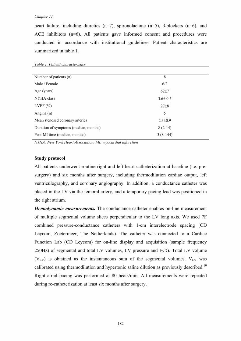
Chapter 11
182
heart failure, including diuretics (n=7), spironolactone (n=5), β-blockers (n=6), and
ACE inhibitors (n=6). All patients gave informed consent and procedures were
conducted in accordance with institutional guidelines. Patient characteristics are
summarized in table 1.
Table 1. Patient characteristics
Number of patients (n) 8
Male / Female 6/2
Age (years) 62±7
NYHA class 3.6± 0.5
LVEF (%) 27±8
Angina (n) 5
Mean stenosed coronary arteries 2.3±0.9
Duration of symptoms (median, months) 8 (2-14)
Post-MI time (median, months) 3 (8-144)
NYHA: New York Heart Association, MI: myocardial infarction
Study protocol
All patients underwent routine right and left heart catheterization at baseline (i.e. pre-
surgery) and six months after surgery, including thermodilution cardiac output, left
ventriculography, and coronary angiography. In addition, a conductance catheter was
placed in the LV via the femoral artery, and a temporary pacing lead was positioned in
the right atrium.
Hemodynamic measurements. The conductance catheter enables on-line measurement
of multiple segmental volume slices perpendicular to the LV long axis. We used 7F
combined pressure-conductance catheters with 1-cm interelectrode spacing (CD
Leycom, Zoetermeer, The Netherlands). The catheter was connected to a Cardiac
Function Lab (CD Leycom) for on-line display and acquisition (sample frequency
250Hz) of segmental and total LV volumes, LV pressure and ECG. Total LV volume
(VLV) is obtained as the instantaneous sum of the segmental volumes. VLV was
calibrated using thermodilution and hypertonic saline dilution as previously described.10
Right atrial pacing was performed at 80 beats/min. All measurements were repeated
during re-catheterization at least six months after surgery.
Long-term hemodynamic effects of SVR
183
Clinical evaluation. Evaluation of clinical status included assessment of NYHA
functional class, quality-of-life score (using the Minnesota quality-of-life questionnaire)
and six-minute hall-walk test.
Echocardiography. In patients with moderate to severe mitral regurgitation (grade ≥2)
on transthoracic echocardiography (TTE), additional transesophageal echocardiography
(TEE) was performed within 5 days before surgery. The TTE and TEE were performed
without general anesthesia to avoid underestimation of the severity of mitral
regurgitation. The severity of mitral regurgitation was graded semi-quantitatively from
color-flow Doppler in the conventional parasternal long-axis and apical 4-chamber
images.11,12 When the severity of mitral regurgitation was less than 3+, a loading test (as
described previously13,14) was performed during anesthesia just before surgery. During
these provocative tests, the severity of mitral regurgitation is followed, and patients who
deteriorate to grade 3 or 4+ mitral regurgitation underwent restrictive mitral
annuloplasty. Immediately after surgery, TEE was repeated to assess residual mitral
regurgitation, transmitral diastolic gradient (determined from continuous-wave
Doppler), and length of coaptation of the mitral leaflets (ideally ≥ 8 mm). Six months
after surgery, TTE was performed to assess the severity of mitral regurgitation. All TTE
measurements were analyzed in random order by two independent observers without
knowledge of the clinical status of the patient and the timing of the echocardiogram.
Surgical procedures
SVR was performed by the endoventricular circular patch plasty as previously described
by Dor.4,15 Briefly, the left ventricle was opened through the infarcted area. An
endocardial encircling suture (Fontan Stitch) was placed at the transitional zone
between scarred and normal tissue. A balloon containing 55mL/m2 body surface area
saline was introduced into the LV and the Fontan stitch was tightened to approximate
the ventricular wall to the balloon. An oval dacron patch was tailored and used to close
the residual orifice. The excluded scar tissue was closed over the patch to ensure
hemostasis. Care was taken to eliminate all septal scar and to delineate a new LV apex
with the goal to restore the normal elliptical shape. After completion of the LV
restoration, a stringent restrictive mitral annuloplasty (2 sizes smaller than measured)
was performed in these patients via an atrial transseptal approach using a Carpentier
Edwards Physio ring (Edwards Lifesciences, USA).
Chapter 11
184
Data-analysis
LV function was quantified by cardiac output (CO), LV ejection fraction (LVEF) and
stroke volume (SV), end-diastolic and end-systolic volume (EDV, ESV), end-systolic
and end-diastolic pressure (ESP, EDP), maximal and minimal rate of LV pressure
change (dP/dtMAX, dP/dtMIN). The time constant of relaxation (τ) was determined using
phase-plot analysis.16 Stroke work (SW) was calculated as the area of the pressure-
volume loop. Mechanical efficiency (ME) was calculated as the ratio of external stroke
work and pressure-volume area (PVA) as a measure of total mechanical work:
ME=SW/PVA17. LV end-systolic elastance (Ees) was estimated by ESP/ESV as
relatively load-independent indexes of systolic function. LV end-diastolic chamber
stiffness (CS) was estimated by EDP/EDV to characterize passive late diastolic
function. Nonuniform LV performance was determined from the segmental LV
conductance signals and quantified by calculating the percentage of time within the
cardiac cycle that a specific segment is dyssynchronous (i.e. opposite in phase with the
global LV volume signal). Overall LV dyssynchrony (DYS) was determined as the
mean of the segmental dyssynchronies. In addition, we calculated the internal flow
fraction (IFF), which quantifies the ineffective shifting of blood volume within the LV
due to nonuniform contraction and filling18.
Statistics
Pre- and post data were compared with paired t-tests. Statistical significance was
assumed at p < 0.05. All data are presented as the mean value ± SD.
RESULTS
There were no peri-operative or hospital deaths and all patients were alive at six months
follow-up. After surgery, two patients needed intra-aortic balloon pump to wean from
cardiopulmonary bypass and six patients needed inotropic support (dobutamine > 2
μg/kg/min) more than 24 hours postoperatively. Immediate after surgery,
transesophageal echocardiography was performed and showed restored leaflet
coaptation with no residual mitral regurgitation. Surgical details are summarized in
table 2.
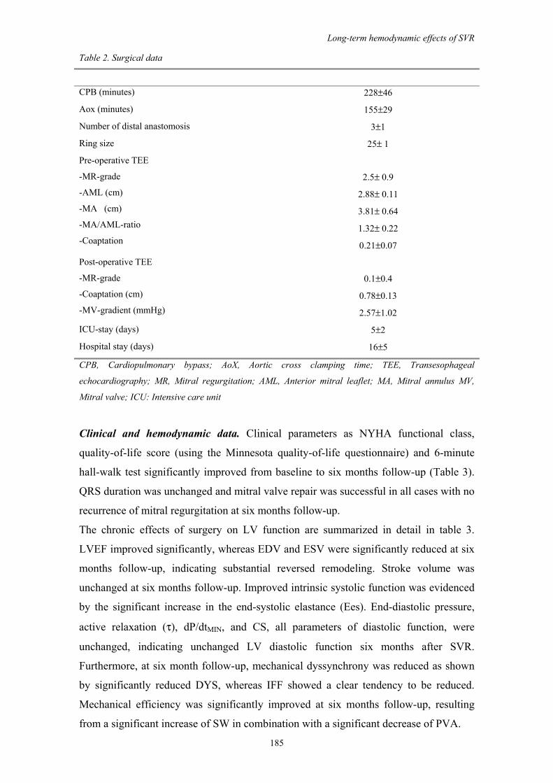
Long-term hemodynamic effects of SVR
185
Table 2. Surgical data
CPB (minutes) 228±46
Aox (minutes) 155±29
Number of distal anastomosis 3±1
Ring size 25± 1
Pre-operative TEE
-MR-grade
-AML (cm)
-MA (cm)
-MA/AML-ratio
-Coaptation
2.5± 0.9
2.88± 0.11
3.81± 0.64
1.32± 0.22
0.21±0.07
Post-operative TEE
-MR-grade
-Coaptation (cm)
-MV-gradient (mmHg)
0.1±0.4
0.78±0.13
2.57±1.02
ICU-stay (days) 5±2
Hospital stay (days) 16±5
CPB, Cardiopulmonary bypass; AoX, Aortic cross clamping time; TEE, Transesophageal
echocardiography; MR, Mitral regurgitation; AML, Anterior mitral leaflet; MA, Mitral annulus MV,
Mitral valve; ICU: Intensive care unit
Clinical and hemodynamic data. Clinical parameters as NYHA functional class,
quality-of-life score (using the Minnesota quality-of-life questionnaire) and 6-minute
hall-walk test significantly improved from baseline to six months follow-up (Table 3).
QRS duration was unchanged and mitral valve repair was successful in all cases with no
recurrence of mitral regurgitation at six months follow-up.
The chronic effects of surgery on LV function are summarized in detail in table 3.
LVEF improved significantly, whereas EDV and ESV were significantly reduced at six
months follow-up, indicating substantial reversed remodeling. Stroke volume was
unchanged at six months follow-up. Improved intrinsic systolic function was evidenced
by the significant increase in the end-systolic elastance (Ees). End-diastolic pressure,
active relaxation (τ), dP/dtMIN, and CS, all parameters of diastolic function, were
unchanged, indicating unchanged LV diastolic function six months after SVR.
Furthermore, at six month follow-up, mechanical dyssynchrony was reduced as shown
by significantly reduced DYS, whereas IFF showed a clear tendency to be reduced.
Mechanical efficiency was significantly improved at six months follow-up, resulting
from a significant increase of SW in combination with a significant decrease of PVA.
Chapter 11
186
Table 3. Clinical and hemodynamic data
Parameter Baseline 6 months follow-up P-value
NYHA class 3.6±0.5 1.5±0.5 <0.001
QoL-test 45±23 16±16 0.028
6-minute HWT (m) 300±133 442±89 0.003
QRS-duration (ms) 103± 19 109± 15 0.348
MR-TTE (grade) 2.1±0.7 0.4±0.5 0.001
HR (bpm) 80±2 83±2 0.059
SV (mL) 64±17 65±12 0.619
CO (L/min) 5±1 5±1 0.270
ESV (mL) 146±46 100±41 0.014
EDV (mL) 214±57 173±46 0.034
EF (%) 31±8 40±14 0.032
ESP (mmHg) 133±33 124±28 0.319
EDP (mmHg) 20±9 18±8 0.435
dPdtMax (mmHg/s) 1270±254 1283±240 0.906
dPdtMin (mmHg/s) -1149±233 -1189±307 0.577
SW (mmHg.mL) 5971±1749 6878±1266 0.062
Tau (ms) 80±15 81±18 0.966
Ees (mmHg/mL) 0.98±0.31 1.51±0.82 0.049
CS (1/mmHg) 0.097±0.037 0.104±0.037 0.667
ME 0.40±0.12 0.55±0.13 0.020
PVA (mmHg.mL) 16000±5160 13047±3407 0.044
DYS (%) 30±5 26±3 0.058
IFF (%) 61±25 41±19 0.169
NYHA, New York Heart Association; QoL, Quality of life; HWT, Hall walk test; MR-TTE, Mitral
regurgitation on transthoracic echocardiography; HR, Heart rate; CO, Cardiac output; ESV,End-systolic
volume; EDV, End-diastolic volume; EF; Ejection fraction; ESP, End systolic pressure; EDP, End-
diastolic pressure; SW, Stroke work; Ees, End-systolic elastance; CS, Chamber stiffness ;ME,
Mechanical efficiency; DYS, Dyssynchrony; IFF, Internal flow fraction
DISCUSSION
SVR by endoventricular circular patch plasty has been applied in patients with ischemic
dilated cardiomyopathy complicated by an apicoseptal LV aneurysm. The short-term
effects of this procedure on LV function are beneficial and consists of acute LV volume
reduction with a decrease of mechanical dyssynchrony and improved LV systolic

Long-term hemodynamic effects of SVR
187
function.6,19 The purpose of the present study was to quantify chronic hemodynamic
effects of SVR and in particular the effects on diastolic function.
The results of this study demonstrate that SVR leads to clinical improvement with
improved LV hemodynamics. At six months follow-up, sustained LV reverse
remodeling and improved global and intrinsic systolic function were observed.
Furthermore, mechanical dyssynchrony was significantly reduced with significantly
improved mechanical efficiency of the LV. Parameters of early (dP/dtmin, tau) and late
diastolic function (EDP, CS) were unchanged six months after surgery, indicating that
volume reduction and patch insertion did not compromise LV diastolic function.
LV reverse remodeling. Our results demonstrated a 20% and 31% reduction of EDV
and ESV respectively at six months follow-up. The observed decrease in EDV and ESV
is consistent with the findings of Qin et al. showing similar reductions of EDV (25%)
and ESV (35%) at six months follow-up in 30 patients who underwent SVR and mitral
valve repair.20
Somewhat larger reductions in LV volumes were reported in studies by Schreuder et al.
and Di Donato et al.6,7 These larger reductions may be due to a more extensive surgical
volume reduction related to larger pre-operative LV volumes. Alternatively, these
differences may reflect redilation, because ours and Qin's studies were done at six
months follow-up, whereas the studies by Schreuder and Di Donato were performed
acutely after surgery. Findings of a later study by Di Donato et al., showing a reduction
in EDV and ESV of 31% and 44% respectively at 12 months follow-up, may suggest
some redilation at late follow-up.21
A possible explanation for redilation in the study of Di Donato is the high recurrence of
mitral regurgitation (38%) at 12 months follow-up. Qin et al. emphasized the
importance of effective mitral valve repair in SVR patients as a pronounced redilation
was occurred in patients with recurrence of mitral regurgitation.20 However, in our study
no recurrence of mitral regurgitation occurred after six months follow-up. Therefore, it
may be suggested that the smaller volume reduction at six months in our study may be
result from less extensive resection in the acute phase due to smaller pre-operative LV
volumes. Long-term sustained volume reduction has been confirmed by a recent study
by Fujii et al. reporting 33% reduction in EDV after isolated SVR at 23 days, which was
unchanged at 32 months follow-up.22
Chapter 11
188
Systolic function. Our data confirm previous findings regarding improved LVEF,
unchanged SV and reduced LV volumes after SVR.23,24 However, to our best
knowledge, the present study is the first study with invasive hemodynamic
measurements before and six months after SVR. At six months follow-up, a significant
increase in Ees (from 0.98±0.31 mmHg/mL to 1.51±0.82 mmHg/mL) was observed in
our study, indicating improved long-term intrinsic systolic function. Furthermore,
mechanical efficiency was significantly improved with a significant reduction in
mechanical dyssynchrony. These results are in line with previous findings by Schreuder
et al. reporting a significant acute increase in Ees (from 1.2±0.6 mmHg/mL to 2.2±1.0
mmHg/mL), reduced mechanical dyssynchrony and improved LV mechanical
efficiency.7 Similarly, Tanoue et al. reported a similar significant increase in Ees (from
1.15±0.6 mmHg/mL to 1.86±0.84 mmHg/mL) and improvement of ventricular
efficiency derived by LV angiography 3 to 4 weeks postoperatively.25 However, these
previous findings merely reflect acute changes of SVR on LV systolic function. Our
study demonstrated that the increase in Ees is sustained after six months follow-up.
Therefore, the improvement of intrinsic systolic function immediate after SVR is
sustained on the long-term. These chronic effects are possibly due to positive acute
effects on the remote myocardium by reduction of mechanical dyssynchrony,
improvement of mechanical efficiency and reduction in LV wall stress. Schreuder et al.
found that changes in Ees are inversely related to parameters of mechanical
dyssynchrony and energy efficiency. Presumably, the chronic beneficial effects on Ees
found in our study are mainly related to acute beneficial effects.
Diastolic function. In this study, both early and late diastolic function were unchanged
at six months follow-up. This is an important finding as endoventricular circular patch
plasty may induce diastolic filling abnormalities with a restrictive pattern.9
Previous acute studies reported altered early diastolic function after SVR with improved
active relaxation (τ).6,7 These acute effects may be related to direct effects of
revascularization26,27 and effects of cardiopulmonary bypass.28,29 Schreuder et al.7 found
an increase in EDP (from 9.4±3 to 13.8±3 mmHg) after SVR, however these changes
may be due to effects of postoperative edema.30 Di Donato et al. recently demonstrated
that EDP was unchanged (from 20±12 to 17±8 mmHg) 10 days after SVR. These
findings are in line with ours as EDP was unchanged (from 20±9 to 18±8 mmHg) after
SVR at six months follow-up. In addition, diastolic chamber stiffness was unchanged
after six months follow-up, indicating that volume reduction and insertion of patch
Long-term hemodynamic effects of SVR
189
plasty does not increase diastolic chamber stiffness. These data imply that SVR does not
alter diastolic function six months after surgery.
In conclusion, surgical ventricular restoration leads to clinical improvement with
sustained LV reverse remodeling, improved global and intrinsic systolic function and
unchanged diastolic function. The improved clinical status after six months may be
related to improved mechanical efficiency, intrinsic systolic function and reduced
mechanical dyssynchrony.
REFERENCES
1. Nohria A, Lewis E, Stevenson LW. Medical management of advanced heart failure. JAMA.
2002;287:628-640. 2. Bogaert J, Bosmans H, Maes A, Suetens P, Marchal G, Rademakers FE. Remote myocardial
dysfunction after acute anterior myocardial infarction: impact of left ventricular shape on regional function: a magnetic resonance myocardial tagging study. J Am Coll Cardiol. 2000;35:1525-1534.
3. Homans DC, Sublett E, Elsperger KJ, Schwartz JS, Bache RJ. Mechanisms of remote myocardial dysfunction during coronary artery occlusion in the presence of multivessel disease. Circulation. 1986;74:588-596.
4. Dor V, Saab M, Coste P, Kornaszewska M, Montiglio F. Left ventricular aneurysm: a new surgical approach. Thorac Cardiovasc Surg. 1989;37:11-19.
5. Athanasuleas CL, Buckberg GD, Stanley AW, Siler W, Dor V, Di Donato M, Menicanti L, Almeida dO, Beyersdorf F, Kron IL, Suma H, Kouchoukos NT, Moore W, McCarthy PM, Oz MC, Fontan F, Scott ML, Accola KA. Surgical ventricular restoration in the treatment of congestive heart failure due to post-infarction ventricular dilation. J Am Coll Cardiol. 2004;44:1439-1445.
6. Di Donato M, Toso A, Dor V, Sabatier M, Barletta G, Menicanti L, Fantini F. Surgical ventricular restoration improves mechanical intraventricular dyssynchrony in ischemic cardiomyopathy. Circulation. 2004;109:2536-2543.
7. Schreuder JJ, Castiglioni A, Maisano F, Steendijk P, Donelli A, Baan J, Alfieri O. Acute decrease of left ventricular mechanical dyssynchrony and improvement of contractile state and energy efficiency after left ventricular restoration. J Thorac Cardiovasc Surg. 2005;129:138-145.
8. Ratcliffe MB, Wallace AW, Salahieh A, Hong J, Ruch S, Hall TS. Ventricular volume, chamber stiffness, and function after anteroapical aneurysm plication in the sheep. J Thorac Cardiovasc Surg. 2000;119:115-124.
9. Salati M, Paje A, Di Biasi P, Fundaro P, Cialfi A, Santoli C. Severe diastolic dysfunction after endoventriculoplasty. J Thorac Cardiovasc Surg. 1995;109:694-701.
10. Baan J, van der Velde ET, de Bruin HG, Smeenk GJ, Koops J, van Dijk AD, Temmerman D, Senden J, Buis B. Continuous measurement of left ventricular volume in animals and humans by conductance catheter. Circulation. 1984;70:812-823.
11. Fisher EA, Goldman ME. Simple, rapid method for quantification of tricuspid regurgitation by two-dimensional echocardiography. Am J Cardiol. 1989;63:1375-1378.
12. Thomas JD. How leaky is that mitral valve? Simplified Doppler methods to measure regurgitant orifice area. Circulation. 1997;95:548-550.
13. Byrne JG, Aklog L, Adams DH. Assessment and management of functional or ischaemic mitral regurgitation. Lancet. 2000;355:1743-1744.
14. Dion R, Benetis R, Elias B, Guennaoui T, Raphael D, Van Dyck M, Noirhomme P, Van Overschelde JL. Mitral valve procedures in ischemic regurgitation. J Heart Valve Dis. 1995;4 Suppl 2:S124-S129.
15. Dor V, Sabatier M, Di Donato M, Montiglio F, Toso A, Maioli M. Efficacy of endoventricular patch plasty in large postinfarction akinetic scar and severe left ventricular dysfunction: comparison with a series of large dyskinetic scars. J Thorac Cardiovasc Surg. 1998;116:50-59.
16. Langer SF. Differential laws of left ventricular isovolumic pressure fall. Physiol Res. 2002;51:1-15.
Chapter 11
190
17. Nozawa T, Yasumura Y, Futaki S, Tanaka N, Uenishi M, Suga H. Efficiency of energy transfer from pressure-volume area to external mechanical work increases with contractile state and decreases with afterload in the left ventricle of the anesthetized closed-chest dog. Circulation. 1988;77:1116-1124.
18. Steendijk P, Tulner SA, Schreuder JJ, Bax JJ, Van Erven L, van der Wall EE, Dion RA, Schalij MJ, Baan J. Quantification of left ventricular mechanical dyssynchrony by conductance catheter in heart failure patients. Am J Physiol Heart Circ Physiol. 2004;286:H723-H730.
19. Schreuder JJ, Steendijk P, van der Veen FH, Alfieri O, van der NT, Lorusso R, van Dantzig JM, Prenger KB, Baan J, Wellens HJ, Batista RJ. Acute and short-term effects of partial left ventriculectomy in dilated cardiomyopathy: assessment by pressure-volume loops. J Am Coll Cardiol. 2000;36:2104-2114.
20. Qin JX, Shiota T, McCarthy PM, Asher CR, Hail M, Agler DA, Popovic ZB, Greenberg NL, Smedira NG, Starling RC, Young JB, Thomas JD. Importance of mitral valve repair associated with left ventricular reconstruction for patients with ischemic cardiomyopathy: a real-time three-dimensional echocardiographic study. Circulation. 2003;108 Suppl 1:II241-II246.
21. Di Donato M, Sabatier M, Dor V, Gensini GF, Toso A, Maioli M, Stanley AW, Athanasuleas C, Buckberg G. Effects of the Dor procedure on left ventricular dimension and shape and geometric correlates of mitral regurgitation one year after surgery. J Thorac Cardiovasc Surg. 2001;121:91-96.
22. Fujii H, Ohashi H, Tsutsumi Y, Kawai T, Iino K, Onaka M. Radionuclide study of mid-term left ventricular function after endoventricular circular patch plasty. Eur J Cardiothorac Surg. 2004;26:125-128.
23. Dor V, Sabatier M, Di Donato M, Maioli M, Toso A, Montiglio F. Late hemodynamic results after left ventricular patch repair associated with coronary grafting in patients with postinfarction akinetic or dyskinetic aneurysm of the left ventricle. J Thorac Cardiovasc Surg. 1995;110:1291-1299.
24. Suma H, Isomura T, Horii T, Sato T, Kikuchi N, Iwahashi K, Hosokawa J. Nontransplant cardiac surgery for end-stage cardiomyopathy. J Thorac Cardiovasc Surg. 2000;119:1233-1244.
25. Tanoue Y, Ando H, Fukumura F, Umesue M, Uchida T, Taniguchi K, Tanaka J. Ventricular energetics in endoventricular circular patch plasty for dyskinetic anterior left ventricular aneurysm. Ann Thorac Surg. 2003;75:1205-1208.
26. Humphrey LS, Topol EJ, Rosenfeld GI, Borkon AM, Baumgartner WA, Gardner TJ, Maruschak G, Weiss JL. Immediate enhancement of left ventricular relaxation by coronary artery bypass grafting: intraoperative assessment. Circulation. 1988;77:886-896.
27. Halow JM, Figueredo VM, Shames DM, Camacho SA, Baker AJ. Role of slowed Ca(2+) transient decline in slowed relaxation during myocardial ischemia. J Mol Cell Cardiol. 1999;31:1739-1748.
28. Brutsaert DL, Sys SU, Gillebert TC. Diastolic dysfunction in post-cardiac surgical management. J Cardiothorac Vasc Anesth. 1993;7:18-20.
29. Zile MR, Brutsaert DL. New concepts in diastolic dysfunction and diastolic heart failure: Part II: causal mechanisms and treatment. Circulation. 2002;105:1503-1508.
30. Tulner SA, Klautz RJ, Engbers FH, Bax JJ, Baan J, van der Wall EE, Dion RA, Steendijk P. Left ventricular function and chronotropic responses after normothermic cardiopulmonary bypass with intermittent antegrade warm blood cardioplegia in patients undergoing coronary artery bypass grafting. Eur J Cardiothorac Surg. 2005;27:599-605.

CHAPTER 12
Summary and conclusions

Chapter 12
192
SHORT INTRODUCTION
The number of patients with chronic heart failure is rapidly increasing as a result of an
aging population and advanced medical therapy leading to a substantial number of
patients who survive a myocardial infarction. Despite improvement of pharmacological
treatment, drug-refractory end-stage heart failure is a challenging problem. Although
successful, cardiac transplantation is limited and consequently alternative invasive
therapies have been developed. Currently, most widely applied are cardiac
resynchronization therapy (CRT), surgical ventricular restoration (SVR) and restrictive
mitral annuloplasty (RMA). Despite promising clinical results of these therapies, the
working mechanisms are still partly unknown. In this thesis, we investigated the acute
and chronic hemodynamic effects of these therapies in patients with end-stage heart
failure. Quantification of these effects may provide further insight in the working
mechanisms of these therapies and may help to explain clinical improvement in heart
failure patients treated with these therapies.
Chapter 2. In this chapter we evaluated the application of the conductance catheter
technique in the operating room. Peri-operative quantification of systolic and diastolic
load independent left ventricular (LV) function by pressure-volume relations is
advantageous since loading conditions are varying during cardiac surgery induced by
anesthesia, cardiopulmonary bypass (CPB) and the surgical procedure itself. In 8
patients undergoing elective CABG, complete hemodynamic data was derived within 15
minutes before and after CPB without any complications. Load-dependent and load-
independent indices of systolic function were unchanged after CPB. However, diastolic
function changed significantly after CPB with a significant increase of end-diastolic
pressure and the diastolic chamber stiffness constant. Active relaxation, quantified by
the active relaxation time constant (tau), decreased significantly. We conclude that the
conductance catheter method provides detailed data on peri-operative LV function.
Therefore, this method may be used to evaluate the acute effects of new surgical
procedures and the data acquired in the group of elective CABG-patients may serve as
reference data.
Chapter 3. In this chapter new parameters of LV mechanical dyssynchrony in patients
with chronic heart failure were introduced, which were derived from online segmental
conductance catheter signals obtained during diagnostic cardiac catheterization. We

Summary and conclusions
193
determined cardiac function and LV mechanical dyssynchrony in heart failure patients
with intraventricular conduction delay and a group of patients with coronary artery
disease and relatively preserved LV function. The heart failure patients showed
depressed systolic and diastolic function. However, the most marked hemodynamic
differences between the groups were found for mechanical dyssynchrony indicating a
high sensitivity and specificity of the new indices. Comparison of conductance catheter
derived indices of mechanical dyssynchrony with septal-to-lateral dyssynchrony derived
by tissue-Doppler velocity imaging showed highly significant correlations. We
concluded that the proposed indices provide additional, new and quantitative
information on temporal and spatial aspects of mechanical dyssynchrony in patients
with heart failure. They may refine diagnosis of cardiac dysfunction and evaluation of
interventions.
Chapter 4. In this chapter, the baseline and chronotropic effects of normothermic
CABG with intermittent antegrade warm blood cardioplegia on LV function were
quantified. This on-pump approach has been applied in all heart failure patients who
underwent SVR and/or RMA in this thesis and thus quantification of these effects in a
control group was warranted. Our findings indicate that on-pump normothermic CABG
with intermittent antegrade warm blood cardioplegia preserves systolic function,
increases diastolic stiffness, and improves systolic and diastolic chronotropic responses.
Normalization of the chronotropic responses after CPB is likely due to the effects of
successful revascularization and subsequent relief of ischemia. The baseline effects on
LV function reported in this chapter are used as reference data to interpret baseline
effects on LV function after SVR and RMA in patients with heart failure.
Chapter 5. In this study we quantified the acute effects of RMA on LV systolic and
diastolic function by pressure-volume analysis using the conductance catheter. In 10
patients with end-stage heart failure and concomitant severe mitral regurgitation,
stringent RMA (two sizes under) effectively restored mitral valve competence (leaflet
coaptation 8.0±0.2mm) without inducing significant acute changes in LV systolic or
diastolic function. This study shows that undersizing the mitral annulus has no adverse
effects on intrinsic systolic and diastolic LV function, and this procedure can be safely
applied in patients with severe heart failure.
Chapter 12
194
Chapter 6. In this chapter, we described the acute effects of SVR on LV systolic and
diastolic function in patients with ischemic dilated cardiomyopathy. The results show
that SVR achieves normalization of LV volumes, improves systolic function and
decreases LV wall stress and mechanical dyssynchrony. At the expense of a higher
diastolic pressure resulting from altered diastolic properties, cardiac pump function
indexed by stroke work and cardiac output was not importantly altered while the
pressure-volume area was significantly reduced after SVR. Therefore, mechanical
efficiency was significantly improved, presumably resulting from reduced wall stress
and reduced mechanical dyssynchrony. Interestingly, the diastolic chamber stiffness
constant was not more altered after SVR than after the surgical procedures in the other
groups, suggesting that this effect was importantly related to procedure-induced
myocardial edema and may be partially transient.
Chapter 7. This chapter gives an overview of the potential applications of pressure-
volume measurements by the conductance catheter during cardiac catheterization. This
review showed that, in the context of CRT, pressure-volume measurements by
conductance catheter have been mainly applied to study the basic mechanisms of
dyssynchronous and resynchronized cardiac contraction. In this field important new
insights were obtained from pressure-volume measurements. There are certainly
possibilities and distinct advantages in the field of patient selection but this application
will remain limited by the invasive character of the conductance method. Currently,
ongoing studies apply pressure-volume measurements to optimize CRT in individual
patients and evaluate the chronic hemodynamic effects of CRT. The possibility to assess
cardiac function and mechanical dyssynchrony during implantation and the possibility
to study the immediate effects of changes in lead position, AV and VV-delay, in an on-
line and quantitative fashion makes this a promising tool to optimize CRT. Load-
independent quantitative parameters of systolic and diastolic function derived from
pressure-volume relations should provide more insight in the working mechanisms of
chronic CRT.
Chapter 8. In this chapter the chronic effects of CRT on LV hemodynamics were
reported. Acute hemodynamic improvements of CRT have been studied previously, but
detailed invasive studies showing hemodynamic consequences of chronic CRT are not
available. We demonstrated that hemodynamic improvements previously shown in
acute studies are maintained mid-term. In addition, ventricular-arterial coupling,
Summary and conclusions
195
mechanical efficiency, and chronotropic responses are improved after 6 months CRT.
These findings may help to explain the improved functional status and exercise
tolerance in heart failure patients treated with cardiac resynchronization.
Chapter 9. In this chapter clinical efficacy was evaluated 6 months after surgery in a
cohort of patients with end-stage heart failure who underwent combined SVR and
RMA, isolated SVR or isolated RMA. Clinical parameters, including NYHA
classification, Minnesota Quality of Life questionnaire, and 6-minute walking distance,
were assessed at baseline and 6 months after surgery. Mortality at 6 months was 12%
and was associated with highly significant improvements in clinical parameters. We
concluded that surgical treatment of end-stage heart failure by SVR and RMA seems
relatively safe with a clear clinical benefit at 6-months follow-up.
Chapter 10. In this chapter, a comprehensive analysis of SVR and if indicated mitral
and/or tricuspid annuloplasty on mid-term hemodynamic and clinical status was
performed. In a selected group of 21 patients who had complete echocardiographic
follow-up including tissue Doppler imaging at 6 months, clinical and echocardiographic
parameters were assessed. In particular, the effects of this surgical approach on LV
volumes, LV dyssynchrony, right ventricular (RV) reverse remodeling and RV
functional parameters (severity of tricuspid regurgitation and pulmonary artery
pressure) were studied. It was demonstrated that this approach resulted in significant
improvements of clinical status and heart failure symptoms, combined with a reduction
in LV volume and LV dyssynchrony with minimal residual mitral regurgitation. In
addition, a decrease in pulmonary artery pressure, RV reverse remodeling and reduced
tricuspid regurgitation was observed. Therefore, this approach proved to have beneficial
mid-term effects in terms of clinical and hemodynamic status.
Chapter 11. In this chapter, chronic effects of SVR on pressure-volume relations were
evaluated. Hemodynamic data showed significantly improved LVEF, sustained LV
volume reduction and significantly improved intrinsic systolic function. In addition,
mechanical efficiency significantly improved with significant reduction of mechanical
dyssynchrony. Interestingly, parameters of diastolic function remain unchanged 6
months after surgery. From these results, we conclude that SVR leads to improved LV
systolic function with unchanged diastolic function at 6 months follow-up. The
Chapter 12
196
improved LV systolic function after 6 months may be related to reduced LV mechanical
dyssynchrony and improved LV efficiency.
CONCLUSIONS
This thesis demonstrates the feasibility and value of LV pressure-volume measurements
by conductance catheter during cardiac surgery. Besides assessment of load-dependent
and load-independent parameters of systolic and diastolic LV function, the conductance
catheter can quantify LV mechanical dyssynchrony, which is an important determinant
of cardiac (dys)function and a new sensitive parameter in patients with chronic heart
failure.
Prior to our studies in heart failure patients, we evaluated our methodology in patients
with relatively normal LV function who underwent elective normothermic CABG. Like
the heart failure patients, these patients were operated during intermittent antegrade
warm blood cardioplegia. Our peri-operative measurements showed no significant
changes in LV systolic function. However, diastolic chamber stiffness significantly
increased, while active relaxation (tau) improved. These effects are probably related to
effects of cardiopulmonary bypass, cardioplegic arrest, and the revascularization. In
general, such effects should be taken into consideration when evaluating peri-operative
hemodynamic measurements in patients with heart failure who undergo surgery with the
same on-pump approach.
With regard to surgical therapies, we focused on SVR and RMA in patients with end-
stage heart failure. Earlier studies have suggested that correction of mitral regurgitation
may decrease LV systolic function in the acute phase. In addition, with regard to
diastolic function, mitral valve repair by RMA might potentially impair filling. These
concerns have caused hesitation to apply these procedures in patients with advanced
heart failure. However, we have demonstrated by pressure-volume analysis that RMA
did not affect systolic and diastolic function in the acute phase. Interestingly, our results
show a clear tendency for a reduced mechanical dyssynchrony after RMA. These
findings support the use of this approach even in patients with severely depressed LV
function in view of beneficial long-term results.
SVR is increasingly applied in patients with ischemic dilated cardiomyopathy and
anteroseptal dyskinesia or akinesia. However, limited data are currently available about
the acute and chronic effects of this therapy on LV function. The present thesis provides
Summary and conclusions
197
measurements of LV systolic and diastolic function and dyssynchrony by use of
pressure-volume relationships. In addition, mid-term (6 months) effects of SVR on
clinical status and biventricular function were provided by clinical parameters (6-min
walk test, Minnesota Living with Heart Failure questionnaire, NYHA classification) and
echocardiography including tissue Doppler imaging. SVR resulted in significant acute
improvement of systolic function, significant acute reduction of LV mechanical
dyssynchrony, and significant reduction of LV wall stress. At the expense of a higher
diastolic pressure resulting from altered diastolic properties, cardiac pump function
indexed by stroke work and cardiac output was not importantly altered while the
pressure-volume area was significantly reduced after SVR. Therefore, mechanical
efficiency was significantly improved, presumably resulting from reduced LV wall
stress and reduced mechanical dyssynchrony. The changes in diastolic stiffness were
relatively limited and were comparable with the changes in patients with preserved LV
function who underwent elective CABG suggesting that this effect was importantly
related to procedure-induced myocardial edema and may be partially transient.
These acute beneficial effects on systolic function are largely maintained chronically
with no significant effects on diastolic function at mid-term follow-up. The pressure-
volume data at 6 months follow-up showed significantly improved LVEF, sustained LV
volume reduction and significantly improved intrinsic systolic function. In addition,
mechanical efficiency was improved with a reduction of mechanical dyssynchrony.
Clinical and echocardiographic data at 6 months follow-up showed improvement of
clinical status and a significant LV volume reduction and reduced LV dyssynchrony. In
addition, RV reverse remodeling after SVR and, if indicated, mitral or tricuspid
annuloplasty was noted with reduction in tricuspid regurgitation and pulmonary artery
pressure. These data indicate that SVR improves LV global and intrinsic systolic
function by reducing mechanical dyssynchrony and LV wall stress without inducing
diastolic dysfunction. In addition, SVR leads to additional beneficial effects at mid-term
follow-up such as reduced pulmonary artery pressure and RV reverse remodeling. The
beneficial hemodynamic effects described in this thesis can explain the improved
clinical status and survival of patients after SVR at long-term follow-up.
A third important new technique to treat patients with end-stage heart failure is CRT,
which is highly effective in patients with left bundle-branch block and severe heart
failure. Previous studies showed long-term clinical benefit, improved LV function and
reverse LV remodeling. Acute hemodynamic effects, previously described, consist of
increased LV ejection fraction and stroke volume, accompanied by increased systolic

Chapter 12
198
pressure, dP/dtMAX, and stroke work, reduced mechanical dyssynchrony and reduced
diastolic pressure. In the present thesis, we obtained invasive hemodynamics by
pressure-volume loops at baseline and after 6 months of CRT and showed that these
improvements are still present at 6 months follow-up. Moreover, our results show
improved ventricular-arterial coupling and improved mechanical efficiency, which
constitute a more optimal energetic condition. In addition, we demonstrated improved
responses to increased heart rate, which may partly explain the enhanced exercise
capacity of patients treated with CRT. However, despite conversion to a more
physiological chronotropic response, the capacity to increase cardiac output remains
limited. The latter presumably is partly due to an abnormal relaxation reflected by a
relatively long isovolumic relaxation time (tau), which did not improve after CRT.
These effects of CRT help to explain the improved functional status and exercise
tolerance in heart failure patients treated with CRT.
In summary, recently several new therapies were introduced to treat patients with end-
stage heart failure. These therapies, SVR, RMA and CRT have all demonstrated to have
clinical benefit. In this thesis, we documented the acute and chronic effects of these
therapies on LV function by pressure-volume analysis. Our findings provide insight in
the underlying mechanisms and help to explain improved functional status achieved
with these therapies.
Samenvatting en conclusies
199
Samenvatting en conclusies
KORTE INLEIDING
Het aantal patiënten met chronisch eindstadium hartfalen neemt snel toe als gevolg van
de vergrijzing en de verbeterde medische behandeling van het acute myocardinfarct.
Ondanks verbeterde medicamenteuze therapie blijft een substantieel deel van deze
groep patiënten symptomatisch. Harttransplantatie, hoewel succesvol, blijft beperkt tot
een kleine groep patiënten. Hierdoor zijn de afgelopen jaren een aantal alternatieve
behandelingen ontwikkeld en toegepast, waaronder cardiale resynchronisatie therapie
(CRT), aneurysmectomie volgens Dor (Dor procedure) en chirurgische mitralisklep-
reparatie door middel van een restrictieve mitralisklep annuloplastiek (RMA). Ondanks
de veelbelovende klinische resultaten van deze behandelingen zijn de
werkingsmechanismen nog grotendeels onbekend. In dit proefschrift worden de acute
en chronische hemodynamische en cardiovasculaire effecten van deze behandelingen
gekwantificeerd o.a. door middel van ventriculaire drukvolume analyse met behulp van
de conductantiecatheter. Het kwantificeren van deze effecten kan meer inzicht
verschaffen in de werkingsmechanismen van deze interventies en daarmee de klinische
verbetering, die doorgaans optreedt na de behandeling in patiënten met chronisch
eindstadium hartfalen, wellicht verklaren. Bovendien geeft het mogelijk
aangrijpingspunten voor het verder verbeteren van deze therapieën.
Hoofdstuk 2. In dit hoofdstuk beschrijven we de toepassing van de
conductantiecatheter in de operatiekamer. Met deze techniek kunnen acute effecten van
hartchirurgische behandelingen worden gekwantificeerd. Met name het meten van
parameters van intrinsieke systolische en diastolische linker kamerfunctie tijdens
hartoperaties is belangrijk, omdat tijdens deze operaties belastingscondities van het hart
sterk kunnen variëren als gevolg van de anesthesie, cardiopulmonale bypass (CPB) en
de chirurgische interventie zelf. Om de toepassing van deze meettechniek te testen en
acute effecten van een controlegroep patiënten met normale hartfunctie te verkrijgen,
zijn metingen verricht in 8 patiënten die een electieve CABG ondergingen. Complete
hemodynamische gegevens zijn verkregen binnen 15 minuten voor en na CPB. Hierbij
traden geen complicaties op. Uit de resultaten blijkt dat zowel de
belastingsonafhankelijke als de belastingsafhankelijke parameters van systolische
Chapter 12
200
functie onveranderd bleven na CPB. De parameters van diastolische functie waren
echter wel veranderd na CPB, waarbij de einddiastolische druk en de diastolische
kamerstijfheidsconstante significant waren toegenomen. Actieve relaxatie,
gekwantificeerd door de relaxatie constante tau, verbeterde significant na CPB.
De resultaten in dit hoofdstuk laten zien dat de conductantiecatheter waardevolle
informatie kan verschaffen over peri-operatieve linker kamerfunctie en dat deze
methode kan worden toegepast om de acute effecten van nieuwe chirurgische
procedures te kwantificeren. De gegevens in dit hoofdstuk zullen verderop in dit
proefschrift worden gebruikt ter vergelijking met de acute hemodynamische effecten
van chirurgische behandelingen in patiënten met ernstig hartfalen.
Hoofdstuk 3. In dit hoofdstuk worden nieuwe parameters van mechanische
dissynchronie van de linker hartkamer bij patiënten met chronisch hartfalen
geïntroduceerd, die gemeten kunnen worden met behulp van segmentale
volumesignalen van de conductantiecatheter. Tijdens diagnostische hartcatheterisaties
werden conductantiemetingen verricht waarbij de hartfunctie en mechanische
dissynchronie werd gemeten in een groep patiënten met chronisch hartfalen. Deze
metingen werden vergeleken met metingen in een groep patiënten met coronairlijden en
een relatief normale linker kamerfunctie. De patiënten met hartfalen hadden een sterk
verminderde systolische en diastolische linker kamerfunctie. De duidelijkste verschillen
tussen de beide groepen werden echter gevonden in de parameters van mechanische
dissynchronie. Vergelijking van deze nieuwe parameters met echografische parameters
van mechanische dissynchronie waaronder septal-to-lateral delay liet een sterke
significante correlatie zien. Hieruit werd geconcludeerd dat deze nieuwe parameters
verkregen met de conductantiecatheter nieuwe additionele en kwantitatieve informatie
kunnen geven over temporele en ruimtelijke aspecten van mechanische dissynchronie
bij patiënten met hartfalen. Deze parameters geven meer inzicht in linker
kamerdysfunctie bij patiënten met hartfalen. Ook kan het effect van interventies op deze
parameters inzicht verschaffen in de werkingsmechanismen van nieuwe behandelingen
voor hartfalen.
Hoofdstuk 4. In dit hoofdstuk worden de acute effecten besproken van normotherme
CABG met intermitterend antegrade warm bloed cardioplegie op de linker
kamerfunctie. Naast de effecten op de hartfunctie bij een frequentie van 80 slagen per
minuut, worden ook de effecten op de hartfunctie tijdens hogere hartfrequenties

Samenvatting en conclusies
201
(chronotrope respons) besproken. Aangezien dezelfde on-pump benadering wordt
toegepast bij patiënten met hartfalen die een Dor procedure en/of een RMA ondergaan
in de volgende hoofdstukken van dit proefschrift, is kwantificatie van de effecten van
deze benadering allereerst zinvol in een controle groep. De bevindingen in deze controle
groep laten zien dat on-pump normotherme CABG met intermitterend antegrade warm
bloed cardioplegie geen systematisch effect heeft op de systolische linker kamerfunctie
maar wel op de diastolische linker kamerfunctie. De diastolische linker kamerstijfheid
neemt toe en de actieve relaxatie verbeterd. Bovendien verbetert zowel de systolische
als diastolische chronotrope respons na CPB. De verbetering van actieve relaxatie en de
normalisatie van de chronotrope respons is waarschijnlijk het gevolg van de succesvolle
revascularisatie.
Hoofdstuk 5. In dit hoofdstuk beschrijven we de acute effecten van chirurgische
mitralisklep reparatie door middel van RMA op de systolische en diastolische linker
kamerfunctie door middel van drukvolume relaties verkregen met de
conductantiecatheter. De resultaten verkregen bij 10 patiënten met eindstadium
hartfalen laten zien dat RMA door middel van het inbrengen van een undersized ring,
d.w.z. twee maten kleinere ring dan de maat die werd gemeten, de coaptatie van de
mitralisklep kan herstellen (8.0±0.2 mm). Hierbij werd de ernstige mitralis-
insufficiëntie opgeheven en de metingen met de conductantiecatheter lieten geen
veranderingen zien in de systolische en diastolische linker kamerfunctie na CPB. Dit
toont aan dat RMA in patiënten met een verminderde linker kamerfunctie geen nadelige
effecten heeft op de intrinsieke diastolische en systolische linker kamerfunctie en dat
deze chirurgische techniek veilig kan worden toegepast bij deze groep ernstig zieke
patiënten
Hoofdstuk 6. In dit hoofdstuk worden de acute effecten op de systolische en
diastolische linker kamerfunctie beschreven van de Dor procedure, uitgevoerd bij
patiënten met ischemische, gedilateerde cardiomyopathie met een anteroseptaal
aneurysma. De gegevens laten zien dat de Dor procedure resulteert in normalisatie van
de linker kamervolumina met een verbetering in systolische functie, een afname van de
linker kamerwandspanning en mechanische dissynchronie. Ten koste van een hogere
einddiastolische druk als gevolg van veranderende diastolische eigenschappen na de
operatie blijft de cardiale pompfunctie weergegeven door de slagarbeid (oppervlakte
van de drukvolume lus) en de cardiale output vrijwel onveranderd. Daarnaast neemt de
Chapter 12
202
mechanische efficiëntie significant toe na de Dor procedure, waarschijnlijk als gevolg
van een vermindering van de wandspanning en mechanische dissynchronie van de
linker kamer. De diastolische kamerstijfheidsconstante neemt echter toe alhoewel deze
toename vergelijkbaar is met die in de controlegroep (zie hoofdstuk 4,5).
Hoofdstuk 7. Dit hoofdstuk geeft een overzicht van de mogelijke toepassingen van de
conductantiecatheter tijdens hartcatheterisaties. Het laat zien dat drukvolume relaties
kunnen worden toegepast in de context van CRT om de basale mechanismen van
dissynchrone en geresynchroniseerde cardiale contractie te bestuderen. Op dit gebied
zijn dan ook belangrijke nieuwe inzichten verkregen met drukvolume metingen. Deze
meettechniek biedt o.a. voordelen op het gebied van patiëntselectie, al blijft de
toepassing van de techniek relatief beperkt vanwege het invasieve karakter van de
conductantiecatheter. Er zijn diverse studies uitgevoerd die drukvolume analyse
toepassen om CRT te optimaliseren in individuele patiënten en de hemodynamische
effecten van CRT te evalueren. De mogelijkheid om de hartfunctie en mechanische
dissynchronie te kwantificeren gedurende implantatie van een biventriculaire pacemaker
en de mogelijkheid om direct de effecten van de positie van de pacemakerdraad, de AV
en VV timing te kunnen meten, maakt deze techniek aantrekkelijk om CRT te
optimaliseren. Belastingsonafhankelijke kwantitatieve parameters van systolische en
diastolische functie verkregen door middel van drukvolume relaties kan meer inzicht
verschaffen in de werkingsmechanismen van CRT op de intrinsieke hartfunctie.
Hoofdstuk 8. In dit hoofdstuk worden de chronische effecten van CRT op de linker
kamerfunctie beschreven. Acute hemodynamische verbeteringen van CRT zijn reeds
bekend, maar gedetailleerde invasieve metingen van hemodynamische effecten op lange
termijn zijn nog niet beschikbaar. De resultaten in dit hoofdstuk laten zien dat de acute
gunstige effecten van CRT op de linker kamerfunctie ook op lange termijn aanwezig
blijven. Daarnaast verbetert de ventriculaire-arteriële koppeling, vermindert de
mechanische dissynchronie en is er sprake van een verbetering in de chronotrope
respons na 6 maanden CRT. Deze bevindingen verklaren mede de verbetering van
klinische parameters en de toename van de inspanningstolerantie in patiënten met
hartfalen die worden behandeld met CRT.
Hoofdstuk 9. In dit hoofdstuk worden klinische parameters gebruikt om het effect van
de gecombineerde Dor/RMA procedure, de geïsoleerde Dor procedure en de geïsoleerde
Samenvatting en conclusies
203
RMA te evalueren in een cohort patiënten met hartfalen. Klinische parameters, inclusief
NYHA classificatie, de kwaliteit-van-leven-test en de 6 minuten looptest zijn bepaald
vóór en 6 maanden na de operatie. De mortaliteit na 6 maanden was in deze selecte
groep patiënten 12% en de klinische parameters verbeterde significant na 6 maanden.
Hieruit werd geconcludeerd dat de chirurgische behandeling van eindstadium hartfalen
door middel van de Dor procedure en/of de RMA relatief veilig is met een duidelijk
klinische verbetering na 6 maanden.
Hoofdstuk 10. In dit hoofdstuk wordt een uitvoerige analyse verricht van de effecten
van de Dor procedure en, indien geïndiceerd, RMA en/of tricuspidalisklepplastiek op de
hemodynamische en klinische status van patiënten met eindstadium hartfalen. In een
selecte groep patiënten met een complete echocardiografische follow-up, inclusief
tissue-Doppler imaging, werden vóór en na 6 maanden klinische en echocardiografische
parameters bepaald. Echocardiografische parameters die werden bepaald waren: het
linker kamervolume, linker kamerdissynchronie, rechter kamerdimensies, de ernst van
de tricuspidalis insufficiëntie en de pulmonale arteriële druk. Na 6 maanden werd een
significante verbetering in klinische conditie van de patiënt gevonden, gecombineerd
met een reductie in linker kamervolume en dissynchronie met een minimale residuele
mitralis/tricuspidalis insufficiëntie. Bovendien werd een significante reductie in het
volume van de rechter kamer gezien met een afname van de pulmonale arteriële druk.
Deze gegevens tonen aan dat deze chirurgische benadering positieve klinische en
hemodynamische effecten heeft op de middellange termijn.
Hoofdstuk 11. In dit hoofdstuk worden de chronische hemodynamische effecten van de
Dor procedure geëvalueerd. De drukvolume relaties, gemeten tijdens hartcatheterisaties,
tonen een verbetering in linker kamerejectiefractie, een reductie van linker
kamervolume en een significante verbetering van de intrinsieke systolische linker
kamerfunctie. Daarnaast neemt de mechanische efficiëntie significant toe na 6 maanden
met een reductie van de linker kamerdissynchronie. Een belangrijk gegeven uit dit
hoofdstuk vormen de onveranderende parameters van de diastolische functie na 6
maanden vergeleken met de parameters voor de operatie. Hieruit kunnen we
concluderen dat de Dor procedure leidt tot een verbetering in globale en intrinsieke
systolische linker kamerfunctie waarbij de diastolische functie na 6 maanden
onveranderd blijft. De verbetering in systolische functie na 6 maanden is mogelijk
Chapter 12
204
gerelateerd aan de reductie van linker kamerdissynchronie en de verbetering in linker
kamerefficiëntie.
CONCLUSIES
In dit proefschrift wordt de haalbaarheid en het belang van drukvolume metingen tijdens
hartchirurgische en cardiologische interventies aangetoond. Naast het verkrijgen van
belastingsafhankelijke en belastingsonafhankelijke parameters van systolische en
diastolische linker kamerfunctie, kan de conductantiecatheter parameters van
mechanische dissynchronie bepalen. Deze parameters zijn sensitief en bepalen mede de
mate van cardiale dysfunctie bij patiënten met chronisch hartfalen.
Voorafgaand aan onze studies in hartfalen patiënten, hebben we de conductantiecatheter
methode geëvalueerd in patiënten met een relatief normale linker kamerfunctie die een
electieve normotherme CABG ondergingen. Evenals bij de hartfalen patiënten, werden
deze patiënten geopereerd met intermitterend antegrade warm bloed cardioplegie. Onze
peri-operatieve metingen toonden geen significante veranderingen in systolische linker
kamerfunctie. De diastolische linker kamerstijfheid in deze patiënten nam echter
significant toe, terwijl de actieve relaxatie constante (tau) significant verbeterde. Deze
effecten zijn hoogst waarschijnlijk het gevolg van de cardiopulmonale bypass, de
cardioplegie en de revascularisatie. In het algemeen, moeten deze effecten in
beschouwing worden genomen wanneer peri-operatieve metingen worden
geïnterpreteerd van patiënten met hartfalen die worden geopereerd onder dezelfde
omstandigheden.
In dit proefschrift hebben we onder andere de effecten chirurgische behandelingen zoals
de Dor procedure en de RMA onderzocht. Op basis van vroegere studies bestaan
aanwijzingen dat RMA in de acute fase na de operatie de linker kamerfunctie in
patiënten met hartfalen verder kan verslechteren. Daarnaast zou RMA mogelijk een
nadelig effect hebben op de diastolische vullingsfase. Deze vermoedens hebben geleid
tot twijfel of deze operatie wel moet worden toegepast bij patiënten met hartfalen. In dit
proefschrift wordt met drukvolume relaties echter aangetoond dat chirurgische
mitralisklep-reparatie door middel van RMA (met een twee maten kleinere ring dan de
ringmaat die werd gemeten) geen nadelig effect heeft op de intrinsieke systolische en
diastolische linker kamerfunctie in patiënten met hartfalen. Daarnaast was er een trend
Samenvatting en conclusies
205
tot reductie van de linker kamerdissynchronie. Deze bevindingen ondersteunen het
gebruik van RMA zelfs in patiënten met een sterk verminderde linker kamerfunctie.
Naast de RMA wordt ook de Dor procedure in toenemende mate toegepast bij patiënten
met eindstadium hartfalen. Deze techniek wordt toegepast in patiënten met ischemisch
gedilateerde cardiomyopathie met anteroseptale dyskinesie of akinesie. Er zijn
momenteel nog weinig gegevens beschikbaar over de acute en chronische effecten van
deze techniek op de linker kamerfunctie bij patiënten met hartfalen. In dit proefschrift
worden met behulp van drukvolume relaties de acute en chronische effecten op de
systolische en diastolische linker kamerfunctie en op de mechanische dissynchronie
beschreven. Daarnaast worden de effecten van de Dor procedure na 6 maanden op de
klinische status van de patiënt (6 minuten looptest, kwaliteit van leven test en NYHA
classificatie), de biventriculaire functie en linker kamerdissynchronie, met behulp van
echocardiografie en tissue Doppler imaging bestudeerd. De acute effecten van de Dor
procedure, zoals beschreven in dit proefschrift, bestaan uit een verbetering in globale en
intrinsieke systolische functie, een reductie van linker kamerdissynchronie en een
significante afname van de linker kamerwandspanning. Ondanks veranderende
diastolische eigenschappen (verhoogde einddiastolische druk en een toegenomen linker
kamerstijfheidsconstante) na de operatie blijft de pompfunctie (cardiale output en
slagarbeid) onveranderd. De pressure-volume area was significant verminderd na de
Dor procedure hetgeen uiteindelijk leidt tot een verbetering in mechanische efficiëntie
van de linker kamer. Dit effect is waarschijnlijk het gevolg van een reductie van linker
kamerwandspanning en een reductie van mechanische dissynchronie. De toename in
diastolische linker kamerstijfheid neemt toe na de Dor procedure, deze toename is
echter vergelijkbaar met de toename in de controle CABG patiënten en waarschijnlijk
het gevolg van tijdelijk oedeem dat ontstaat na de operatie. De positieve effecten op de
systolische linker kamerfunctie, gemeten met drukvolume relaties, zijn na 6 maanden
nog steeds aanwezig. Opmerkelijk is dat de diastolische functie na 6 maanden
onveranderd is ten opzichte van voor de operatie. Kennelijk is de toename in linker
kamerstijfheid, die direct na de operatie aanwezig is, tijdelijk van aard. De drukvolume
gegevens na 6 maanden laten een significante verbetering zien in linker
kamerejectiefractie, een significante reductie van het linker kamervolume en een
significante verbetering in intrinsieke systolische functie. Daarnaast neemt de linker
kamerefficiëntie toe met een reductie in mechanische dissynchronie. Ook de klinische
parameters verbeterden 6 maanden na de Dor procedure. Daarnaast treedt reverse
remodeling op van de rechter kamer en vindt een significante reductie plaats van de
Chapter 12
206
pulmonale arteriële druk na de operatie. Deze resultaten tonen aan dat de Dor
procedure, indien noodzakelijk met een tricuspidalisklepplastiek of een
mitralisklepplastiek, de globale en intrinsieke systolische linker kamerfunctie verbetert
door een reductie in mechanische dissynchronie en linker kamerwandspanning, zonder
verandering in diastolische linker kamerfunctie. Bovendien treedt reverse remodeling
op van de rechter kamer met een afname van de pulmonale arteriële druk. Al deze
verbeteringen in functie verklaren mede de gunstige klinische lange termijn resultaten
na de Dor procedure.
Een derde belangrijke behandelingstechniek voor patiënten met eindstadium hartfalen is
CRT dat effectief is gebleken bij patiënten met een ernstig hartfalen en een linker
bundeltak blok. Eerdere studies laten gunstige lange termijn effecten zien met een
verbetering in overleving en symptomen en een verbetering in linker kamerfunctie met
reverse remodeling van de linker kamer. De reeds bekende acute effecten na CRT zoals
toename in ejectiefractie, slagvolume, systolische druk, dP/dtmax en slagarbeid, en
reductie in mechanische dissynchronie en diastolische druk zijn volgens de resultaten
van dit proefschrift nog steeds aanwezig na 6 maanden behandeling met CRT.
Bovendien laten deze resultaten een verbetering in ventriculaire-arteriële koppeling en
een verbetering in mechanische efficiëntie zien. Verder laten deze resultaten een
verbetering in chronotrope response zien, dat mogelijk de verbetering in
inspanningstolerantie na CRT in deze patiënten mede verklaart. Ondanks conversie naar
een meer fysiologische chronotrope response werd echter een beperkte toename gezien
van de cardiale output na toename van de hartfrequentie. Dit is mogelijk het gevolg van
een abnormale relaxatie door de vertraagde isovolumetrische relaxatie die niet
verbeterde na CRT. Deze hemodynamische verbeteringen na CRT verklaren voor een
deel de toename van de inspanningstolerantie in hartfalen patiënten die worden
behandeld met CRT.
In dit proefschrift zijn de acute en chronische hemodynamische effecten van de Dor
procedure, RMA en CRT onderzocht met behulp van ventriculaire drukvolume analyse
met de conductantiecatheter. De klinische effecten van deze behandelingen bij patiënten
met hartfalen zijn over het algemeen gunstig. De bevindingen in dit proefschrift geven
inzicht in de onderliggende werkingsmechanismen en kunnen de klinische
verbeteringen in deze patiënten mede verklaren.
207
List of publications
1. Tulner SA, Klautz RJ, Rijk-Zwikker GL, Engbers FH, Bax JJ, Baan J, van der Wall
EE, Dion RA, Steendijk P. Peri-operative assessment of left ventricular function by
pressure-volume loops using the conductance catheter method. Anesth Analg 2003;
97: 950-957
2. Steendijk P, Tulner SA, Schreuder JJ, Bax JJ, Van Erven L, van der Wall EE, Dion
RA, Schalij MJ, Baan J. Quantification of left ventricular mechanical dyssynchrony
by conductance catheter in heart failure patients. Am J Physiol Heart Circ Physiol
2004; 286: H723-H730
3. Tulner SA, Schaap GR, Strackee SD, Besselaar PP, Luitse JS, Marti RK. Long-term
results of multiple-stage treatment for posttraumatic osteomyelitis of the tibia. J
Trauma 2004; 56: 633-642
4. Tulner SA, Steendijk P, Klautz RJ, Bax JJ, Versteegh MI, van der Wall EE, Dion
RA. Acute hemodynamic effects of restrictive mitral annuloplasty in patients with
end-stage heart failure: analysis by pressure-volume relations. J Thorac Cardiovasc
Surg 2005; 130: 33-40
5. Tulner SA, Klautz RJ, Engbers FH, Bax JJ, Baan J, van der Wall EE, Dion RA,
Steendijk P. Left ventricular function and chronotropic responses after
normothermic cardiopulmonary bypass with intermittent antegrade warm blood
cardioplegia in patients undergoing coronary artery bypass grafting. Eur J
Cardiothorac Surg 2005; 27: 599-605
6. Steendijk P, Tulner SA, Bax JJ, Oemrawsingh PV, Bleeker GB, van Erven L, Putter
H, Verwey HF, van der Wall EE, Schalij MJ. Hemodynamic effects of long-term
cardiac resynchronization therapy -Analysis by pressure-volume loops-. Circulation
(in press)
7. Tulner SA, Steendijk P, Klautz RJ, Bax JJ, Schalij MJ, van der Wall EE, Dion RA.
Surgical ventricular restoration in patients with ischemic dilated cardiomyopathy.
Evaluation of systolic and diastolic ventricular function, wall stress, dyssynchrony,
and mechanical efficiency by pressure-volume loops. J Thorac Cardiovasc Surg
(in press)

208
209
Acknowledgements
Dit proefschrift is mede tot stand gekomen met de hulp en medewerking van veel
mensen. De metingen met de conductantiecatheter, die we hebben kunnen verrichten op
de afdeling Cardiologie en Thoraxchirurgie, hebben dit proefschrift uiteindelijk tot een
succes gemaakt. Ik wil dan ook in de eerste plaats alle patiënten die bereid waren deel te
nemen aan deze studies bedanken. Dankzij hen zijn we nu meer te weten gekomen over
de werkingsmechanismen van nieuwe behandelingen voor hartfalen. Daarnaast wil ik de
Nederlandse Hartstichting bedanken voor de financiële ondersteuning van mijn
werkzaamheden.
Voorts wil ik alle medewerkers van de afdeling Cardiologie en Thoraxchirurgie
waarmee ik heb samengewerkt en die hebben bijgedragen aan de totstandkoming van dit
proefschrift bedanken. De metingen zal ik blijven herinneren als één van de beste
teamprestaties, aangezien er veel mensen bij betrokken waren.
Mijn (ex) collega's wil ik bedanken voor de gezellige periode in de "tuin". In het
bijzonder mijn paranimfen Bart en Sander, met jullie heb ik vaak in Barrera veel plezier
gehad. De drie musketiers zullen in de toekomst bijeen blijven komen: "All for one, one
for all"!
Een belangrijke bijdrage in mijn promotieperiode speelden mijn ouders, Arne, Jorn en
Mies. Pap en Mam, bedankt dat jullie mij de kans hebben gegeven zover te komen en
dat jullie mij de mogelijkheid hebben geven mezelf te ontplooien. Jullie adviezen en
ondersteuning maken jullie tot goede ouders. Arne, jij bent gedurende deze periode
altijd betrokken geweest bij mijn werk. Ik zal de periode dat wij samengewoond hebben
in Amsterdam nooit vergeten. Jorn, jouw integriteit en goede adviezen heb ik in deze
periode erg gewaardeerd. Lieve Mies, jij bent natuurlijk degene die me de belangrijkste
adviezen gaf en mij enorm heeft gesteund in deze periode. Jouw rustige kijk, goede
adviezen en liefde waren onmisbaar. Veel dank daarvoor.

210
211
Curriculum Vitae
De auteur van dit proefschrift werd geboren op 19 februari 1973 te Gouda. In 1991
behaalde hij zijn Gymnasium diploma aan Het Nieuwe Lyceum te Hilversum, waarna
hij een algemeen oriënterend jaar volgde op de Vrije Hogeschool te Driebergen.
Na een jaar studie Economie (in verband met uitloting), begon hij in 1993 met de studie
Geneeskunde aan de Rijksuniversiteit Groningen. Tijdens de doctoraalfase was hij
student-assistent bij de vakgroep Anatomie en Embryologie (dr. P. Room, Prof. dr. G.
Holstege) en volgde hij diverse stages in buitenlandse ziekenhuizen (Freeman Hospital,
New Castle upon Tyne, Engeland en Hospital de São Paulo, Brazilië). In 1998 behaalde
hij zijn doctoraal diploma aan de Rijksuniversiteit Groningen.
Tussen 1998 en 2000 deed hij zijn co-assistentschappen in het Academisch Medisch
Centrum te Amsterdam. Eind september 2000 behaalde hij het artsexamen aan de
Universiteit van Amsterdam, waarna hij een jaar als arts-assistent Heelkunde heeft
gewerkt in het St. Lucas Andreas ziekenhuis te Amsterdam (Dr. E.Ph. Steller). Tijdens
dit jaar deed hij onderzoek bij de afdeling Orthopedie (dr. G.R. Schaap, Prof. dr. R.K.
Marti) van het Academisch Medisch Centrum te Amsterdam naar de behandeling van
posttraumatische osteomyelitis van de tibia.
In januari 2002 startte hij met promotieonderzoek onder leiding van dr. P. Steendijk op
de afdeling Cardiologie (Prof. dr. E.E. van der Wall, Prof. dr. M.J. Schalij) en
Thoraxchirurgie (Prof. dr. R.A.E. Dion) van het Leids Universitair Medisch Centrum te
Leiden. Het promotieonderzoek werd gesubsidieerd door de Nederlandse Hartstichting
(NHS 2002B133). Tijdens dit onderzoek werkte hij bij Bio Implant Service (onderdeel
van de Nederlandse Transplantatie Stichting) als zelfstandig hartexplanteur. Eind
december 2005 heeft hij het promotieonderzoek afgerond.. Per 1 januari 2006 is hij
gestart met de vooropleiding heelkunde in het VU Medisch Centrum te Amsterdam
(Prof. dr. J.A. Rauwerda) in het kader van de opleiding tot orthopedisch chirurg.

212


