
Nodavirus infection causes mortalities in hatchery … · Nodavirus infection causes mortalities in...
6
DISEASES OF AQUATIC ORGANISMS Dis Aquat Org Vol. 63: 113–118, 2005 Published February 28 INTRODUCTION In 1997 the Central Institute of Brackishwater Aqua- culture succeeded in spawning and larval rearing of captive brood stock of Asian sea bass popularly known in India as Bhetki (Thirunavukkarasu & Kailasam 1999). Lately the demand for the seed of this fish is on the rise and as a result managing fish health is becoming more important. Asian sea bass, like most marine fish, are susceptible to disease during the larval rearing and nursery phases. Susceptibility of the fish to various diseases during these phases has been well documented, par- ticularly for bacterial diseases caused by Vibrio species (Wong & Leong 1989, Anderson & Norton 1991, Azad et al. 2004). Subhasinghe & Shariff (1992) reported on bacteria-associated heavy mortalities in cage-cultured Asian sea bass in Malaysian waters and Flexibacter columnaris disease was described by Soltani et al. (1996). Lymphocystis disease has also been found in Asian sea bass (Chao 1984). The first report of viral infection in Asian sea bass was made by Glazebrook et al. (1990), who described a picorna-like virus associated with mortalities of 15 to 20 d old larvae. This disease was also investigated by Munday et al. (1992) in Asian sea bass and is now recognised as being caused by a piscine nodavirus. Piscine nodavirus belongs to the genus Betanoda- virus of the family Nodaviridae (Mori et al. 1992, Nishizawa et al. 1997, van Regenmortel et al. 2000). It contains 2 single-stranded, positive-sense, non- polyadenylated RNAs, RNA1 and RNA2, the latter of which encodes a structural protein of the virus. The viral particle is 25 to 32 nm in diameter (Mori et al. 1992, Comps et al. 1996). Due to its affinity to nerve cells and its ability to cause marked histopathological alterations in nervous tissue, the disease condition caused by piscine noda- virus is now variously referred to as viral encephalo- pathy and retinopathy (VER), viral nervous necrosis (VNN) or fish encephalitis (Mori et al. 1992, Comps et al. 1994). More than 30 species of fish, from different © Inter-Research 2005 · www.int-res.com *Email: [email protected] Nodavirus infection causes mortalities in hatchery produced larvae of Lates calcarifer : first report from India I. S. Azad 1, 2, *, M. S. Shekhar 1 , A. R. Thirunavukkarasu 1 , M. Poornima 1 , M. Kailasam 1 , J. J. S. Rajan 1 , S. A. Ali 1 , M. Abraham 1 , P. Ravichandran 1 1 Central Institute of Brackishwater Aquaculture, 75-Santhome High Road, R. A. Puram, Chennai 600 028, India 2 Present address: Aquaculture, Fisheries and Marine Environmental Dept, Kuwait Institute for Science Research, Salmiya 22017, Kuwait ABSTRACT: Larvae (15 to 21 d post hatch, dph) of the Asian sea bass Lates calcarifer (Bloch) suffered heavy mortalities (60 to 90%) during the hatchery-rearing phase. Darkened and moribund larvae showed no evidence of bacterial or parasitic infections. Tissue sections of brain and spinal cord showed clear necrotic vacuolation. Electron microscopy revealed membrane-bound viral particles in the cytoplasm of the nerve cells. The viral particles measured 28 to 30 nm in diameter. Primer sets, designed for the amplification of the RNA2 segment of the piscine nodavirus coat protein gene, were used in the RT-PCR analysis of moribund larvae of 20 and 21 dph which produced the amplified prod- uct of 430 bp. The clinical manifestations, pathology and electron microscopy observations supported by the RT-PCR analysis suggest that the nerve necrosis was due to nodavirus infection in the larvae. This is the first report of piscine nodavirus infection from the Indian sub-continent. KEY WORDS: Nodavirus · Lates calcarifer · Larvae · RT-PCR · Histopathology · Nervous necrosis virus Resale or republication not permitted without written consent of the publisher
Transcript of Nodavirus infection causes mortalities in hatchery … · Nodavirus infection causes mortalities in...

DISEASES OF AQUATIC ORGANISMSDis Aquat Org
Vol. 63: 113–118, 2005 Published February 28
INTRODUCTION
In 1997 the Central Institute of Brackishwater Aqua-culture succeeded in spawning and larval rearing ofcaptive brood stock of Asian sea bass popularly knownin India as Bhetki (Thirunavukkarasu & Kailasam1999). Lately the demand for the seed of this fish ison the rise and as a result managing fish health isbecoming more important.
Asian sea bass, like most marine fish, are susceptibleto disease during the larval rearing and nurseryphases. Susceptibility of the fish to various diseasesduring these phases has been well documented, par-ticularly for bacterial diseases caused by Vibrio species(Wong & Leong 1989, Anderson & Norton 1991, Azadet al. 2004). Subhasinghe & Shariff (1992) reported onbacteria-associated heavy mortalities in cage-culturedAsian sea bass in Malaysian waters and Flexibactercolumnaris disease was described by Soltani et al.(1996). Lymphocystis disease has also been found inAsian sea bass (Chao 1984).
The first report of viral infection in Asian sea basswas made by Glazebrook et al. (1990), who describeda picorna-like virus associated with mortalities of 15 to20 d old larvae. This disease was also investigated byMunday et al. (1992) in Asian sea bass and is nowrecognised as being caused by a piscine nodavirus.
Piscine nodavirus belongs to the genus Betanoda-virus of the family Nodaviridae (Mori et al. 1992,Nishizawa et al. 1997, van Regenmortel et al. 2000).It contains 2 single-stranded, positive-sense, non-polyadenylated RNAs, RNA1 and RNA2, the latter ofwhich encodes a structural protein of the virus. Theviral particle is 25 to 32 nm in diameter (Mori et al.1992, Comps et al. 1996).
Due to its affinity to nerve cells and its ability tocause marked histopathological alterations in nervoustissue, the disease condition caused by piscine noda-virus is now variously referred to as viral encephalo-pathy and retinopathy (VER), viral nervous necrosis(VNN) or fish encephalitis (Mori et al. 1992, Comps etal. 1994). More than 30 species of fish, from different
© Inter-Research 2005 · www.int-res.com*Email: [email protected]
Nodavirus infection causes mortalities in hatcheryproduced larvae of Lates calcarifer : first report
from IndiaI. S. Azad1, 2,*, M. S. Shekhar1, A. R. Thirunavukkarasu1, M. Poornima1, M. Kailasam1,
J. J. S. Rajan1, S. A. Ali1, M. Abraham1, P. Ravichandran1
1Central Institute of Brackishwater Aquaculture, 75-Santhome High Road, R. A. Puram, Chennai 600 028, India
2Present address: Aquaculture, Fisheries and Marine Environmental Dept, Kuwait Institute for Science Research,Salmiya 22017, Kuwait
ABSTRACT: Larvae (15 to 21 d post hatch, dph) of the Asian sea bass Lates calcarifer (Bloch) sufferedheavy mortalities (60 to 90%) during the hatchery-rearing phase. Darkened and moribund larvaeshowed no evidence of bacterial or parasitic infections. Tissue sections of brain and spinal cordshowed clear necrotic vacuolation. Electron microscopy revealed membrane-bound viral particles inthe cytoplasm of the nerve cells. The viral particles measured 28 to 30 nm in diameter. Primer sets,designed for the amplification of the RNA2 segment of the piscine nodavirus coat protein gene, wereused in the RT-PCR analysis of moribund larvae of 20 and 21 dph which produced the amplified prod-uct of 430 bp. The clinical manifestations, pathology and electron microscopy observations supportedby the RT-PCR analysis suggest that the nerve necrosis was due to nodavirus infection in the larvae.This is the first report of piscine nodavirus infection from the Indian sub-continent.
KEY WORDS: Nodavirus · Lates calcarifer · Larvae · RT-PCR · Histopathology · Nervous necrosisvirus
Resale or republication not permitted without written consent of the publisher

Dis Aquat Org 63: 113–118, 2005
geographical locations have been reported to be sus-ceptible to the virus (Glazebrook et al. 1990, Yoshi-koshi & Inoue 1990, Mori et al. 1992, Muroga 1995,Comps et al. 1996, Munday & Nakai 1997, Castric et al.2001). The disease has also been reported in freshwa-ter aquarium fish from Singapore (Hegde et al. 2003).
Histology and RT-PCR are the common methodsused for diagnosis of the virus in fish (Mori et al. 1991,Munday et al. 1992, Nishizawa et al. 1994, Grotmol etal. 1995, Lai et al. 2001, Johansen et al. 2002, Hegde etal. 2003). Diagnosis of piscine nodavirus using RT-PCRfor the T4 region (427 bp) of SJNNV coat protein gene(RNA2) has been widely used and many workers havereported positive amplification for the presence of thenodavirus in fish species from different geographicallocations (Nishizawa et al. 1994, 1997, Castric et al.2001, Curtis et al. 2001, Iwamoto et al. 2001, Lai et al.2001). Primer sequences of Nishizawa et al. (1994)have been recommended for the diagnosis of thepiscine nodaviruses (OIE 1997).
A batch of Asian sea bass larvae produced in the fishhatchery facilities of the Central Institute of Brack-ishwater Aquaculture were observed to experiencesudden and unexplained mortality after 15 d of larvalrearing. Subsequent investigations revealed that thecause of the disease was piscine nodavirus. The pre-sent investigation represents the first report of VNNfrom the Indian sub-continent and extends the knownrange of this disease.
MATERIALS AND METHODS
Fish. Broodstock procured from the coastal waters ofChennai (Madras), acclimatized and maintainedin cement tanks are being used for the purposes ofseed production. Since 1997 more than 15 batches ofAsian sea bass larvae have been produced in the insti-tute facility. Though the hatchlings were sampled from0 d post hatch (dph) for routine histology, the larvaefrom 15 to 21 dph, corresponding to the appearance ofclinical signs, were used in the present investigation.
Tissue preparation for light microscopy. Moribundsea bass larvae of 15 to 21 dph in the size range of 5to 8 mm with the clinical signs were fixed in neutralbuffered formalin (NBF). The tissue was processedfollowing standard procedures. Small larvae wereprocessed whole and the larger ones were cut intohead and body portions and processed separately.Thin sections (5 to 6 µm) of the tissue were stained withhaematoxylin and eosin (H&E) and observed.
Tissue preparation for transmission electron micro-scopy. Moribund sea bass larvae (18 to 21 dph) werefixed (as done in the case of light microscopy) in 2.5%glutaraldehyde for 3 to 6 h, depending on the size of
the tissue, rinsed in 0.2 M sodium cacodylate buffer(pH 7.4) and post-fixed in 1% osmium tetroxide. Thetissues were dehydrated in ethanol and embedded inan epoxy resin (Araldite EM embedding resin). Semi-thin sections (0.5 to 1.0 µm) were cut using a Diatomediamond knife and stained with toluidine blue forobservation under a light microscope to mark the areasof interest for later observation by electron microscopy.Ultra-thin sections (80 to 100 nm were cut, contrastedin uranyl acetate/lead citrate and observed under anelectron microscope (Philips, 200C).
Sampling and processing of tissue for RT-PCR. Headportions of the moribund larvae were pooled and halfof the pooled sample was fixed in absolute ethanol.Total RNA was extracted from the tissue using themethodology recommended for VNN diagnosis bythe Office Internationale des Epizooties (OIE 1997).Briefly, 150 mg of fish tissue was homogenised in a0.1% diethyl pyrocarbonate (DEPC) treated and auto-claved plastic tissue homogeniser with 0.5 ml distilledwater (DEPC treated) and centrifuged at 10 000 × gfor 10 min. The resultant supernatant was mixed with40 µl of Proteinase K (1 mg ml–1) and 40 µl of 1%sodium dodecyl sulfate (SDS) and incubated at roomtemperature for 30 min. The suspension was centri-fuged as above and the supernatant was used for totalnucleic acid extraction using the phenol-chloroformextraction procedures.
RT-PCR. Two µl of total RNA was subjected toreverse transcription using murine leukemia virus(MuLV) reverse transcriptase. The cDNA was preparedusing the RT-PCR kit (Bangalore, Genei). Briefly, Thetotal nucleic acids were preheated at 90°C for 5 minand incubated at 42°C for 30 min in 20 µl PCR buffer(10 mM Tris/HCl, pH 8.3, 50 mM KCl) containing 2.5 UMuLV reverse transcriptase (USB), 1.0 U ribonucleaseinhibitor, 0.5 µM reverse primer, 1 mM each of 4deoxynucleotide triphosphates (dNTP), and 5 mMMgCl2. The 2 primers, a reverse primer (5’-CGA-GTC-AAC-ACG-GGT-GAA-GA-3’) and a forward primer(5’-CGT-GTC-AGT-CAT-GTG-TCG-CT-3’), were usedfor amplification of a target sequence (430 bases) of theRNA2 (Nishizawa et al. 1994). The amplified productwas visualised using 2% agarose gel electrophoresis.
RESULTS
History of pathogenesis and symptoms
The sea bass larvae, reared in fibre reinforcedplastic tanks at a density of 30 l–1, were active andnormal in their colouration and swimming behavioruntil 15 dph. Gradually a few of the larvae becameslightly darker than normal and showed lethargy.
114

Azad et al.: Nodavirus infection in hatchery produced larvae of Lates calcarifer
The larvae congregated as clusters on the surfacenear the tank wall. Heavy mortalities in the rangeof 60 to 90% were recorded at this stage. Smallerlarvae were severely affected compared to the largerof their siblings. Anorexia, darkened body coloura-tion and loss of reflexes were the major clinicalsymptoms noticed in the affected fish. Corkscrew orwhirling swimming was not noticed. No bacteria orparasites were recorded from the affected fish inthe study.
Histopathology by light and electron microscopy
Light microscopy using H&E staining revealed vacuo-lation and necrosis in the brain, spinal cord and the eye.The toluidine blue stained sections also confirmed theseobservations. Vacuolation was severe and widespread inthe grey and white matter of the brain compared to thenerve cells of the spinal cord (Fig. 1). Initially (15 dph) thespinal cord (Fig. 1a,b), the brain and retina of the eyeshowed mild necrotic vacuolation (Fig. 1c,d) followed by
115
Fig. 1. Haematoxylin and eosin stained sections of (a,b) spinal cord, (c) brain and (d) eye of the sea bass larvae at 15 dph. (a) Vac-uolation (arrows) of the spinal cord. (b) Pyknostic nerve cells (arrowheads). (c) Vacuolative degeneration (arrows) of the whitematter in the brain tissue. (d) Retina with pyknostic (small arrows) and vacuolative necrosis. Toluidine blue stained semi-thin sec-tions of (e) brain and (f) spinal cord of moribund larvae sampled at 20 d post hatch showing extensive vacuolation (arrows) of bothwhite and grey matter of the brain (e) and spinal cord (f). VC: vertebral column. Magnification: (a) 20×, (b) 100×, (c–f) 40×

Dis Aquat Org 63: 113–118, 2005
heavy and complete necrotic vacuolation of the brainand spinal cord (Fig. 1e,f) by 20 dph.
Electron microscopy of the brain and spinal cordindicated typical membrane bound cytoplasmic inclu-sions (Fig. 2a,b) with densely packed pockets of viri-ons. Virions released into the surroundings of the cellmeasured 28 to 30 nm in diameter (Fig. 2c). Several ofthe nerve cells in the spinal cord showed marginatednucleus and emptied cytoplasm after the viral inclu-sions were shed into the extracellular spaces (Fig. 2d).
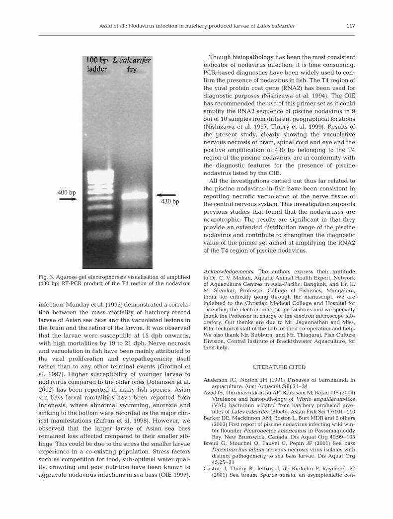
RT-PCR
Amplification with specific primers for RNA2 encod-ing the T4 region of coat protein gene of nodavirus
resulted in the expected PCR product size of 430 bp asshown in Fig. 3.
DISCUSSION
Observations made in the present investigation onabnormalities in body colouration, feeding and swim-ming behaviour were similar to those reported bymany workers investigating nodavirus-associated in-fections in Asian sea bass and other fish species(Glazebrook et al. 1990, Yoshikoshi & Inoue 1990,Munday & Nakai 1997, Breuil et al. 2001, Barker et al.2002). In the present investigation more than 60% ofthe larvae died within 48 h of onset of observed clinicalsymptoms, indicating the severity of the nodavirus
116
Fig. 2. Electron micrographs of the central nervous system. (a) Membrane bound viral particles in the cell organelle (arrows) and(b) viral inclusions (double-ended arrows) depicting intracytoplasmic localisation of the virus. (c) Viral particles measuring 28 to30 nm in the intracellular spaces; (inset) very high magnification of the viral particles. (d) Nerve cell of the spinal cord withemptied cytoplasm and cornered nucleus. Arrows indicate probable exit points of viral particles. VP: viral particles, M: muscle,
N: nucleus. Magnification: (a,b) 70 000×
Azad et al.: Nodavirus infection in hatchery produced larvae of Lates calcarifer
infection. Munday et al. (1992) demonstrated a correla-tion between the mass mortality of hatchery-rearedlarvae of Asian sea bass and the vacuolated lesions inthe brain and the retina of the larvae. It was observedthat the larvae were susceptible at 15 dph onwards,with high mortalities by 19 to 21 dph. Nerve necrosisand vacuolation in fish have been mainly attributed tothe viral proliferation and cytopathogenicity itselfrather than to any other terminal events (Grotmol etal. 1997). Higher susceptibility of younger larvae tonodavirus compared to the older ones (Johansen et al.2002) has been reported in many fish species. Asiansea bass larval mortalities have been reported fromIndonesia, where abnormal swimming, anorexia andsinking to the bottom were recorded as the major clin-ical manifestations (Zafran et al. 1998). However, weobserved that the larger larvae of Asian sea bassremained less affected compared to their smaller sib-lings. This could be due to the stress the smaller larvaeexperience in a co-existing population. Stress factorssuch as competition for food, sub-optimal water qual-ity, crowding and poor nutrition have been known toaggravate nodavirus infections in sea bass (OIE 1997).
Though histopathology has been the most consistentindicator of nodavirus infection, it is time consuming.PCR-based diagnostics have been widely used to con-firm the presence of nodavirus in fish. The T4 region ofthe viral protein coat gene (RNA2) has been used fordiagnostic purposes (Nishizawa et al. 1994). The OIEhas recommended the use of this primer set as it couldamplify the RNA2 sequence of piscine nodavirus in 9out of 10 samples from different geographical locations(Nishizawa et al. 1997, Thiery et al. 1999). Results ofthe present study, clearly showing the vacuolativenervous necrosis of brain, spinal cord and eye and thepositive amplification of 430 bp belonging to the T4region of the piscine nodavirus, are in conformity withthe diagnostic features for the presence of piscinenodavirus listed by the OIE.
All the investigations carried out thus far related tothe piscine nodavirus in fish have been consistent inreporting necrotic vacuolation of the nerve tissue ofthe central nervous system. This investigation supportsprevious studies that found that the nodaviruses areneurotrophic. The results are significant in that theyprovide an extended distribution range of the piscinenodavirus and contribute to strengthen the diagnosticvalue of the primer set aimed at amplifying the RNA2of the T4 region of piscine nodavirus.
Acknowledgements. The authors express their gratitudeto Dr. C. V. Mohan, Aquatic Animal Health Expert, Networkof Aquaculture Centres in Asia-Pacific, Bangkok, and Dr. K.M. Shankar, Professor, College of Fisheries, Mangalore,India, for critically going through the manuscript. We areindebted to the Christian Medical College and Hospital forextending the electron microscope facilities and we speciallythank the Professor in charge of the electron microscope lab-oratory. Our thanks are due to Mr. Jagannathan and Miss.Rita, technical staff of the Lab for their co-operation and help.We also thank Mr. Subburaj and Mr. Thiagaraj, Fish CultureDivision, Central Institute of Brackishwater Aquaculture, fortheir help.
LITERATURE CITED
Anderson IG, Norton JH (1991) Diseases of barramundi inaquaculture. Aust Aquacult 5(8):21–24
Azad IS, Thirunavukkarasu AR, Kailasam M, Rajan JJS (2004)Virulence and histopathology of Vibrio anguillarum-like(VAL) bacterium isolated from hatchery produced juve-niles of Lates calcarifer (Bloch). Asian Fish Sci 17:101–110
Barker DE, Mackinnon AM, Boston L, Burt MDB and 6 others(2002) First report of piscine nodavirus infecting wild win-ter flounder Pleuronectes americanus in PassamaquoddyBay, New Brunswick, Canada. Dis Aquat Org 49:99–105
Breuil G, Mouchel O, Fauvel C, Pepin JF (2001) Sea bassDicentrarchus labrax nervous necrosis virus isolates withdistinct pathogenicity to sea bass larvae. Dis Aquat Org45:25–31
Castric J, Thiéry R, Jeffroy J, de Kinkelin P, Raymond JC(2001) Sea bream Sparus aurata, an asymptomatic con-
117
Fig. 3. Agarose gel electrophoresis visualisation of amplified(430 bp) RT-PCR product of the T4 region of the nodavirus
400 bp430 bp
Dis Aquat Org 63: 113–118, 2005
tagious fish host for nodavirus. Dis Aquat Org 47:33–38Chao TM (1984) Studies on the transmissibility of lymphocysis
disease occurring in sea bass (Lates calcarifer, Bloch).Singap J Pri Ind 12:11–16
Comps M, Pepisn JF Bonami K (1994) Purification and char-acterisation of 2 fish encephalitis viruses (FEV) infectinglates calacrifer and Dicentrarchus labrax. Aquaculture123:1–10
Comps M, Trindade M, Delsert C (1996) Investigations of fishencephalitis virus (FEV) expression in marine fishes usingDIG-labelled probes. Aquaculture 143:113–121
Curtis PA, Drawbridge M, Iwamoto T, Nakai T, Hedrick RP,Gendron AP (2001) Nodavirus infection of juvenile whitesea bass, Atractoscion nobilis, cultured in southern Cali-fornia: first record of viral nervous necrosis (VNN) inNorth America. J Fish Dis 24:263–271
Glazebrook JS, Heasman MP, de Beer SW (1990) Picorna-likeviral particles associated with mass mortalities in larvalbarramundi, Lates calcarifer Bloch. J Fish Dis 13:245–249
Grotmol S, Totland GK, Kvellestad A, Fjell K, Olsen AB (1995)Mass mortality of larval and juvenile hatchery-rearedhalibut (Hippoglossus hippoglossus L.) associated with thepresence of virus-like particles in vacuolated lesions in thecentral nervous system and retina. Bull Eur Assoc FishPathol 15:176–180
Grotmol S, Totland GK, Thorud K, Hjeltnes BK (1997) Vacuo-lating encephalopathy and retinopathy associated with anodavirus-like agent: a probable cause of mass mortalityof cultured larval and juvenile Atlantic halibut Hippo-glossus hippoglossus. Dis Aquat Org 29:85–97
Hegde A, The HC, Lam TJ, Sin YM (2003) Nodavirus infec-tion in freshwater ornamental fish, guppy, Poicelia reti-culata—comparative characterization and pathogenicitystudies. Arch Virol 148:576–586
Iwamoto T, Mori K, Arimoto M, Nakai T (2001) A combinedcell-culture nad RT-PCR method for rapid detection ofpiscine nodaviruses. Dis Aquat Org 24:231–236
Johansen R, Ranheim T, Hansen MK, Taksdal T, Totland GK(2002) Pathological changes in the juvenile Atlantic hal-ibut Hippoglossus hippoglossus persistently infected withnodavirus. Dis Aquat Org 50:161–169
Lai YS, Murali S, Chiu HC, Ju HY and 5 others (2001) Propa-gation of yellow grouper nervous necrosis virus in a newnodavirus susceptible cell line from yellow grouper, Epi-nephelus awoara (Temmink & Schlegel), brain tissue.J Fish Dis 24:299–309
Mori K, Nakai T, Nagahara M, Muroga K, Mekuchi T, KunnoT (1991) A viral disease in hatchery-reared larvae juve-niles of redspotted grouper. Gyobyo Kenkyu 26:209–210
Mori K, Nakai T, Muroga K, Arimoto M, Mushiake K, Furu-sawa I (1992) Properties of a new virus belonging toNodaviridae found in larval striped jack (Pseudocaranxdentex) with nervous necrosis. Virology 187:368–371
Munday BL, Nakai T (1997) Special topic review: nodaviruses
as pathogens in larval and juvenile marine finfish. WorldJ Microbiol Biotechnol 13:375–381
Munday BL, Langdon JS, Kyatt A, Humphrey JD (1992) Massmortality associated with a viral-induced vacuolatingencephalopathy and retinopathy of larvae and juvenilebarramundi, Lates calcarifer Bloch. Aquaculture 103:197–211
Muroga K (1995) Viral and bacterial diseases in larval juve-nile marine fish and shellfish: a review. Fish Pathol 30:71–85
Nishizawa T, Mori K, Nakai T, Furusawa I, Muroga K (1994)Polymerase chain reaction (PCR) amplification of RNA ofstripped jack nervous necrosis virus (SJNNV). Dis AquatOrg 18:103–107
Nishizawa T, Furuhashi M, Nagai T, Nakai T, Muroga K(1997) Genomic classification of fish nodaviruses by mole-cular phylogenetic analysis of the coat protein gene. ApplEnviron Microbiol 63:1633–1636
OIE (1997) Viral encephalopathy and retinopathy or viralnerve necrosis. In: Corne DK, Speare DJ , Griffiths S, CokM, Ritchie R, Olivier G (eds) OIE Diagnostic Manualfor Aquatic Animal Diseases. Office Internationale desEpizooties, Paris, p 99–107
Soltani M, Munday BL, Burke CM (1996) The relative suscep-tibility of fish to infections by Flexibacter columnaris mar-itimus. Aquaculture 140:259–264
Subhasinghe RP, Shariff M (1992) Multiple bacteriosis, withspecial reference to spoilage bacterium Shewanell putre-faciens, in cage culture barramundi in Malaysia. J AquatAnim Health 4:309–311
Thiery R, Arnauld C, Delsert C (1999) Two isolates of seabass, Dicentrarchus labrax L., nervous necrosis virus withdistinct genomes. J Fish Dis 22:201–207
Thirunavukkarasu AR, Kailasam M (1999) Seed productiontechnology for marine fishes. In: Lazarus S, Prakash SG,Vincent SG (eds) Proceedings of the First National Semi-nar on Trends in Marine Biotechnology. ICAS PublicationNo. 2, p 111–114
van Regenmortel MHV, Fauquet CM, Bishop DHL, CarstenseEB and 7 others (eds) (2000) Virus taxonomy, classificationand nomenclature of viruses, 7th edn. Academic Press,San Diego, CA
Wong SY, Leong TS (1989) A comparative study of Vibrioinfections in healthy and diseases marine finfishes cul-tured in floating cages near Penang, Malaysia. SpecialIssue, Asian Fish Sci 3:353–359
Yoshikoshi K, Inoue K (1990) Viral nerve necrosis in hatchery-reared larvae and juveniles of Japanese parrotfish, Opleg-nathus fasciatus (Temmink & Schlegel). J Fish Dis 13:69–77
Zafran, Harada T, Koesharyani I, Yuasa K, Hatai K (1998)Indonesian hatchery reared seabass larvae (Lates cal-carifer), associated with viral nervous necrosis (VNN).Indonesian Fish Res J 4:19–22
118
Editorial responsibility: Jo-Ann Leong, Kaneohe, Hawaii, USA
Submitted: April 2, 2004; Accepted: June 3, 2004Proofs received from author(s): December 17, 2004