
Nobuyuki Nishitani, Martin Sch rmann, Katrin Amunts and...
11
20:60-69, 2005. doi:10.1152/physiol.00043.2004 Physiology Nobuyuki Nishitani, Martin Schürmann, Katrin Amunts and Riitta Hari You might find this additional information useful... 90 articles, 21 of which you can access free at: This article cites http://physiologyonline.physiology.org/cgi/content/full/20/1/60#BIBL 3 other HighWire hosted articles: This article has been cited by [PDF] [Full Text] [Abstract] , September 1, 2005; 94 (3): 2251-2254. J Neurophysiol S. Fecteau, J. L. Armony, Y. Joanette and P. Belin Sensitivity to Voice in Human Prefrontal Cortex [PDF] [Full Text] [Abstract] , December 11, 2006; 0 (2006): bhl141v1-12. Cereb Cortex R. M. Willems, A. Ozyurek and P. Hagoort When Language Meets Action: The Neural Integration of Gesture and Speech [PDF] [Full Text] [Abstract] , January 1, 2007; 17 (1): 230-237. Cereb Cortex M. V. Saarela, Y. Hlushchuk, A. C. d. C. Williams, M. Schurmann, E. Kalso and R. Hari The Compassionate Brain: Humans Detect Intensity of Pain from Another's Face including high-resolution figures, can be found at: Updated information and services http://physiologyonline.physiology.org/cgi/content/full/20/1/60 can be found at: Physiology about Additional material and information http://www.the-aps.org/publications/physiol This information is current as of August 22, 2007 . http://www.the-aps.org/. Copyright © 2005 by the American Physiological Society. ISSN: 1548-9213, ESSN: 1548-9221. Visit our website at 20814-3991. bimonthly in February, April, June, August, October, and December by the American Physiological Society, 9650 Rockville Pike, Bethesda MD ) publishes brief review articles on major physiological developments. It is published News in Physiological Science (formerly published as Physiology on August 22, 2007 physiologyonline.physiology.org Downloaded from
Transcript of Nobuyuki Nishitani, Martin Sch rmann, Katrin Amunts and...

20:60-69, 2005. doi:10.1152/physiol.00043.2004 PhysiologyNobuyuki Nishitani, Martin Schürmann, Katrin Amunts and Riitta Hari
You might find this additional information useful...
90 articles, 21 of which you can access free at: This article cites http://physiologyonline.physiology.org/cgi/content/full/20/1/60#BIBL
3 other HighWire hosted articles: This article has been cited by
[PDF] [Full Text] [Abstract]
, September 1, 2005; 94 (3): 2251-2254. J NeurophysiolS. Fecteau, J. L. Armony, Y. Joanette and P. Belin
Sensitivity to Voice in Human Prefrontal Cortex
[PDF] [Full Text] [Abstract], December 11, 2006; 0 (2006): bhl141v1-12. Cereb Cortex
R. M. Willems, A. Ozyurek and P. Hagoort When Language Meets Action: The Neural Integration of Gesture and Speech
[PDF] [Full Text] [Abstract]
, January 1, 2007; 17 (1): 230-237. Cereb CortexM. V. Saarela, Y. Hlushchuk, A. C. d. C. Williams, M. Schurmann, E. Kalso and R. Hari
The Compassionate Brain: Humans Detect Intensity of Pain from Another's Face
including high-resolution figures, can be found at: Updated information and services http://physiologyonline.physiology.org/cgi/content/full/20/1/60
can be found at: Physiologyabout Additional material and information http://www.the-aps.org/publications/physiol
This information is current as of August 22, 2007 .
http://www.the-aps.org/.Copyright © 2005 by the American Physiological Society. ISSN: 1548-9213, ESSN: 1548-9221. Visit our website at 20814-3991.bimonthly in February, April, June, August, October, and December by the American Physiological Society, 9650 Rockville Pike, Bethesda MD
) publishes brief review articles on major physiological developments. It is publishedNews in Physiological Science (formerly published as Physiology
on August 22, 2007 physiologyonline.physiology.org
Downloaded from
region contains representations of hand actionsand orofacial gestures. In this brief review, we willfocus on the motor functions of Broca’s region. Westart by describing the anatomy and connections ofBroca’s region, and then we discuss the role of thisbrain area in action execution, observation, andunderstanding and the relationship of these func-tions to imitation. Finally, we will speculate aboutwhy Broca’s region is involved in so many appar-ently different functions.
Structure and Connectivity ofBroca’s Region
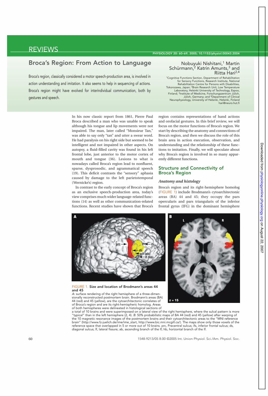
Anatomy and histologyBroca’s region and its right-hemisphere homolog(FIGURE 1) include Brodmann’s cytoarchitectonicareas (BA) 44 and 45; they occupy the parsopercularis and pars triangularis of the inferiorfrontal gyrus (IFG) in the dominant hemisphere
60 1548-9213/05 8.00 ©2005 Int. Union Physiol. Sci./Am. Physiol. Soc.
In his now classic report from 1861, Pierre PaulBroca described a man who was unable to speakalthough his tongue and lip movements were notimpaired. The man, later called “Monsieur Tan,”was able to say only “tan” and utter a swear word.He had paralysis on his right side but seemed to beintelligent and not impaired in other aspects. Onautopsy, a fluid-filled cavity was found in his leftfrontal lobe, just anterior to the motor cortex ofmouth and tongue (36). Lesions to what isnowadays called Broca’s region lead to nonfluent,sparse, dysprosodic, and agrammatical speech(19). This deficit contrasts the “sensory” aphasiacaused by damage to the left parietotemporal(Wernicke’s) region.
In contrast to the early concept of Broca’s regionas an exclusive speech-production area, today’sview comprises much wider language-related func-tions (14) as well as other communication-relatedfunctions. Recent studies have shown that Broca’s
REVIEWS
Broca’s Region: From Action to Language Nobuyuki Nishitani,1 MartinSchürmann,2 Katrin Amunts,3 and
Riitta Hari2,4
1Cognitive Functions Section, Department of Rehabilitationfor Sensory Functions, Research Institute, NationalRehabilitation Centre for Persons with Disabilities,
Tokorozawa, Japan; 2Brain Research Unit, Low TemperatureLaboratory, Helsinki University of Technology, Espoo,
Finland; 3Institute of Medicine, Forschungszentrum Jülich,Jülich, Germany; and 4Department of Clinical
Neurophysiology, University of Helsinki, Helsinki, [email protected]
Broca’s region, classically considered a motor speech-production area, is involved in
action understanding and imitation. It also seems to help in sequencing of actions.
Broca’s region might have evolved for interindividual communication, both by
gestures and speech.
PHYSIOLOGY 20: 60–69, 2005; 10.1152/physiol.00043.2004
A B
prc
L R
z = 15
ifsifs
dslf (ab)
lf (hb)lf (ab)
lf (hb)
FIGURE 1. Size and location of Brodmann’s areas 44and 45A: surface rendering of the right hemisphere of a three-dimen-sionally reconstructed postmortem brain. Brodmann’s areas (BA)44 (red) and 45 (yellow), are the cytoarchitectonic correlates ofof Broca’s region and are its right-hemispheric homolog. Areasof both hemispheres were delineated in histological sections ofa total of 10 brains and were superimposed on a lateral view of the right hemisphere, where the sulcal pattern is more“typical” than in the left hemisphere (2, 4). B: 50% probabilistic maps of BA 44 (red) and 45 (yellow) after warping ofthe 10 magnetic resonance images of the postmortem brains and their cytoarchitectonic areas to the “MNI referencebrain” (http://www.fz-juelich.de/ime/ime_start, http://www.bic.mni.mcgill.ca/). The maps show only those voxels of thereference space that overlapped in 5 or more out of 10 brains. prc, Precentral sulcus; ifs, inferior frontal sulcus; ds,diagonal sulcus; lf, lateral fissure; ab, ascending branch of the lf; hb, horizontal branch of the lf.
on August 22, 2007 physiologyonline.physiology.org
Downloaded from

(the left in 95% of the population). The widely usedBrodmann map (16) represents a simplifieddrawing of only one typical brain, and laterhistological studies have indicated considerableindividual variation in the size and extent of areas44 and 45 with respect to the individual sulcaltopography; for example, area 44 volume maydiffer across individuals even by a factor of ten (2,4). Broca’s region matures later than, for example,the primary sensorimotor cortices, as is evidentfrom both the histological fine structure (3) andfrom cortical thickness maps based on magneticresonance imaging (37).
Although areas 44 and 45 differ in their cytoar-chitecture (2), they share, for example, the pres-ence of very large pyramidal cells in deep layer IIIand in layer V, the lack of a clear border betweenlayers II and III, and the low cell density in layer VI(2). However, whereas area 44 is “dysgranular”(containing a thin layer IV of small granular cellswith pyramidal cells from deep layer III and upperlayer V intermingled with those of layer IV), area 45has densely packed granular cells in layer IV (“gran-ular” area) (2, 4, 65). Although Rizzolatti and Arbib(82) consider area 44 analogous to monkey area F5,the homology between the human area 44 and themonkey F5 has not yet been demonstrated in astrict sense.
Hemispheric asymmetry
Areas 44 and 45 can be found in both hemispheres,but nearly all patients with Broca’s aphasia havelesions in the left inferior frontal cortex. Thisclinical observation raises the question of whetherand how far Broca’s region and its right-hemispheric counterpart differ anatomically andfunctionally.
Anatomic asymmetry. The volume of the histo-logically defined area 44 is larger in the left than inthe right hemisphere, whereas area 45 is more sym-metric (2, 30). Moreover, the cytoarchitecture ofboth areas shows significant interhemispheric dif-ferences (5).
In great apes, the inferior frontal region corre-sponding to human Broca’s region is larger in theleft than in the right hemisphere (18), suggestingthat the neuroanatomic substrates for left-hemi-sphere dominance in vocalization developed asearly as five million years ago, long before speechemerged. It has been suggested that vocalizationswere gradually incorporated into the gestural sys-tem, and in the subsequent switch from manualgesture to vocal language the left hemisphere couldhave taken dominance for both speech and manu-al action (21).
Functional asymmetry. The dominance of theleft-hemispheric area 44/45 in language-relatedfunctions is well established (14). It is far less clear
whether area 44/45 is asymmetric in other commu-nication-related functions (to be reviewed in thesections below). For example, the right IFG is acti-vated during voluntary inhibition of imitative andoverlearned responses (15) as well as during per-ceptual sequencing tasks (97). The right IFG is alsoactivated when people try to make sense ofambiguous emotional expression in face imagesbut not when they view and judge pictures ofambiguous gender (73, 78). Both left and right IFGare activated during detection of errors in musicalsyntax (63). Furthermore, both left and right IFGare essential for imitation (44). Finally, data onimagery of movement suggest a left-hemisphericdominance of area 44 for egocentric movementsbut a right-hemispheric dominance of the samearea for movement characteristics in space (11). Asystematic review of functional asymmetry isbeyond the scope of this article. Below, findingsabout “Broca’s region” refer to the left hemisphere,and activation of the right-hemisphere counterpartwill be mentioned separately, when needed. “Area44/45” will refer to either hemisphere.
Connections of Broca’s region
The available data on brain connectivity derivemainly from tracing and electrophysiologicalexperiments in monkeys, from which they havebeen extrapolated to the human brain. Somerecent studies have applied diffusion tensorimaging to directly analyze connectivity in theliving human brain. The major inputs and outputsof areas 44 and 45 differ to some extent,emphasizing the different functional roles of thesetwo areas.
According to data from monkey F5, the humanIFG (bilaterally) is likely to be connected to theanterior intraparietal cortex, the superior temporalsulcus (STS), the parietal cortex (area PF in mon-keys), the cerebellum, and Wernicke’s area(reviewed in Ref. 6). In contrast to many other brainfunctions, conclusions based on primate researchmust be considered with particular caution whenthe anatomy and physiology of language process-ing are concerned. Electrophysiological experi-ments in primates have implicated both a dorsaland a ventral pathway connecting Wernicke’s areato Broca’s region (54, 89). Such connections in thehuman brain have recently been confirmed byusing diffusion tensor imaging and tractography(80). A dorsal pathway, including the arcuate fasci-culus, was distinguished from a more ventral route,including the external capsule and the uncinatefasciculus. Interestingly, the connections werestronger in the dominant than in the nondominanthemisphere. Although studies on tractography inthe human brain do not demonstrate the existenceof anatomic, synaptic connectivity, they are indica-
61PHYSIOLOGY • Volume 20 • February 2005 • www.physiologyonline.org
REVIEWS
ifs
lf (ab)lf (hb)
on August 22, 2007 physiologyonline.physiology.org
Downloaded from

mation is processed in different frontotemporalnetworks in the left hemisphere (including thetemporo-parieto-occipital junction, parts of theIFG, and the superior temporal lobe). In contrast,the processing of intonation would be supportedby a temporofrontal circuit in the right hemisphere,consisting mainly of the frontal operculum andregions in the superior temporal gyrus. The strictright-hemispheric lateralization of the processingof intonational information can be modulated bystimulus or task demands via the corpus callosum.It was suggested that single regions within thedescribed networks obtain their specific role forthe processing of particular aspects of language viainteraction with other areas.
Perception-action link for communication:mirror neurons
Communication, both verbal and nonverbal,requires that the interacting individuals “staytuned.” Because the conspecifics certainly are verysimilar in their main characteristics, it is then alsomandatory that each subject’s action andperception rely on closely linked neuronalcircuitries—one individual’s output is the other(similar) individual’s input.
Interesting “mirror neurons” were discoveredsome years ago in frontal area F5 of the monkeycortex. These neurons are active during executionof object-related hand actions, but they are alsoactive, importantly, when the monkey is justobserving similar acts (23, 31, 84–86). For example,the mirror neurons are activated when the monkeytakes a raisin from a tray and also when he viewsanother monkey or the human experimenter doingthe same. No information is yet available aboutpossible hemispheric lateralization of the monkeymirror neurons.
Mirror neurons have visuomotor properties,being sensitive to goal-related motor acts (102), butthey can also be activated by sounds that implyactions (55, 57). Importantly, the mirror neurons donot only react to visual input and then project, viasome transformational step, to motor-output-related neurons but are also part of a system thatforms a neuronal representation of the observedmotor acts. Similar to F5, the rostral part of theinferior parietal cortex contains neurons that areactive during action observation and execution(32); this region receives input from the STS, whichis known to contain neurons responding to biolog-ical motion (for review, see Ref. 1).
In search of a human mirror-neuron system(MNS), human counterparts of the monkey mirrorneurons were first looked for with PET, which fol-lows oxygen consumption in the brain (40, 59, 86).Broca’s region was activated when the subjectobserved, imagined, and imitated the examiner
62 PHYSIOLOGY • Volume 20 • February 2005 • www.physiologyonline.org
tors of the existence of anatomic pathwaysbetween brain areas. The functional connectivity ofBroca’s region, evident, for example, in covarianceanalysis of functional magnetic resonance imaging(fMRI), is task specific and much more widelyspread than the anatomic connectivity would pre-dict (42). Of course, covarying activation does notnecessarily imply a network of directly connectednodes.
Broca’s Region with a Mosaic ofFunctions
Below we briefly discuss various functions thathave been ascribed to Broca’s region and/or itsright-hemisphere counterpart. It should be noted,however, that activation of any area in a brainimaging study does not mean that the neuralsubstrate of the mentioned functions is seated(only) there; rather, it indicates that the activatedarea is involved in, or may be an important node in,a widely distributed neuronal network. It is mostlikely that Broca’s region consists of partlyoverlapping subsystems that support variousfunctions, ranging from motor imagery (11, 35) toobject manipulation and grasping (13), to motorpreparation (59, 90), and to planning (25).
We will proceed from the classical functions ofBroca’s region in speech production and languageto more basic functions in perceptual sequencing,action understanding, and imitation.
Language and speech
In her extensive review of fMRI studies of languageareas, Bookheimer (14) showed that areas 44 and 45subserve different functions. The IFG is oftenactivated bilaterally but shows left-hemisphericdominance during tasks requiring naming (91),judgments of phonology (43, 100), semantics (4, 29,101), and syntax (9, 28, 29, 43). Broca’s region is alsoactivated during acquisition of grammatical rules,discrimination of speech sounds, production ofwords, estimation of time intervals, andreproduction of rhythms (14). Thus Broca’s regionseems to be involved in both perception andproduction of speech. We will claim below that thisrole of Broca’s region as an interface of action andperception can be generalized to nonverbalfunctions.
Language production and understanding alsoinvolve prosody, one of the few language-relatedprocesses with right-hemisphere dominance (68,70). The interaction of the two hemispheres, how-ever, seems to be more complex than has beenassumed previously. Integrating evidence fromneuroimaging, psycholinguistics, neurology, andneurophysiology, Friederici and Alter (27) pro-posed that segmental, lexical, and syntactic infor-
REVIEWS
on August 22, 2007 physiologyonline.physiology.org
Downloaded from

using a precision grasp to enclose an object or tomove his/her hand. Thus Broca’s region could con-tain neurons similar to the monkey mirror neu-rons. The activation sequence associated withonline imitation and with observation of anotherperson’s movements also included the STS (76, 88).
The monkey F5 mirror neurons are also activatedby orofacial gestures, and therefore a recent mag-netoencephalography (MEG) study (77) appliedstill pictures of verbal and nonverbal lip forms thatthe subject had to observe, imitate, or make in aself-paced manner (FIGURE 2). In all conditionsand in both hemispheres, the activation spreadfrom occipital cortex (peak activation 120 ms afterthe picture onset) in 20- to 60-ms steps to the STS(the strongest activation), the inferior parietal lob-ule, the inferior frontal lobe (Broca’s region), and,80–100 ms later, to the primary motor cortex.Because the STS is not activated when the subjectmakes movements his- or herself, it can be consid-
ered only as influencing the (motor) MNS.Assuming that the observed MNS activation
sequence would be related to the link between asender and a receiver of an action-related message,some abnormalities could be expected in subjectswho have abnormal imitation skills and difficultiesin understanding motor-act-based intentions ofother subjects. Such deficits are observed in high-functioning autistic (Asperger syndrome) subjects,who in fact displayed delayed and diminished acti-vation in Broca’s region (75) during imitation(FIGURE 3). Moreover, activation was in many sub-jects absent in the right hemisphere.
Within the MNS, the close link between percep-tion and action seems to be realized in functions ofBroca’s region. Such a link may well be important infacilitating communication between an agent andan observer due to shared sensory and motor repre-sentations. Along similar lines, Liberman andMattingly (62) strongly advocated a motor theory of
63PHYSIOLOGY • Volume 20 • February 2005 • www.physiologyonline.org
REVIEWS
Who le-scalp neuromagnetometer N onverbal lip-form stimuli
Activation sequence Responses
–100 0 100 200 300 400 ms
bd
a
ce
a
b
25fT/cm
25fT/cm
c
d
e
ImitationO bservationContro l
FIGURE 2. Brain activation during imita-tion studied with magnetoencephalographyLeft: magnetoencephalography (MEG) recording with a306-channel whole scalp neuromagnetometer at the BrainResearch Unit, Low Temperature Laboratory, Helsinki University of Technology.MEG picks up weak magnetic fields produced by postsynaptic intracellular currents,arising in synchronously activated cortical pyramidal cells. These magnetic fields, mainlygenerated by neural currents in fissural cortex, are measured noninvasively outside thehead. Right: brain activation by imitation and observation of orofacial gestures. Topshows nonverbal lip forms used as stimuli. Bottom illustrates cortical responses duringimitation (red) and observation (green) of nonverbal orofacial gestures. The responsesrecorded from 5 locations are indicated, and the cortical activation, shown on the schematic brain, progresses fromthe occipital visual area to the superior temporal sulcus, to the inferior parietal areas, then to Broca’s region in theinferior frontal cortex, and finally to the primary motor cortex. Similar activation areas and temporal sequences wereseen also in the right hemisphere. The blue traces refer to control stimuli (landscapes that activated only the two firststeps). Modified from Ref. 77.
on August 22, 2007 physiologyonline.physiology.org
Downloaded from

face between perception and action.A role of area 44/45 as an interface between per-
ception and action is also suggested by theinhibitory influence of right IFG on certain imita-tive and overlearned responses (Ref. 15; see Ref. 7for more general inhibitory functions of right IFG).
To sum up, mirror neurons, as important parts oflarger neuronal circuitries, can be considered totransfer action-related information (be it visual orauditory) to knowledge (83). The available informa-tion is in line with the view that the MNS supportscommunicative functions. STS and inferior parietalcortex provide essential input to F5/Broca’s region,where the communicative functions of the MNSbecome manifest.
Action understanding
Rizzolatti and co-workers (83, 87, 88) considerBroca’s region essential for action understanding.Support for such an idea comes from studies inwhich monkey F5 neurons also react when the endpart of the movement is obscured when themonkey only knows what is going to happen (102).Furthermore, a part of the F5 mirror neurons arealso activated by sounds that are related to actualmotor acts and the monkey understands thisrelationship (57).
Observation of different types of mouth actionsactivates several brain areas, including the parsopercularis of the IFG and the adjacent ventral pre-motor cortex, with different patterns and likely viadifferent mechanisms influenced by knowledge ofthe observed action (12, 17). Interestingly, Broca’sregion was not activated when the human subjectswatched a dog barking, i.e., an action that is not inthe observer’s motor repertoire (17). In addition toBroca’s region and premotor cortex, the primarymotor cortex also shows differential activationdependent on action understanding: MEG resultsabout the motor-cortex part of the human MNSsuggest that the motor cortex differentiates naturaland artificially presented movements (52).Moreover, a recent study of observation of chop-stick use demonstrated that the motor cortex isactivated more strongly the more often the(Finnish) subjects had used chopsticks during thelast year. In other words, a dependence on experi-ence was demonstrated in the motor-cortex part ofthe MNS (53).
Humans most likely understand another per-son’s actions, and also their motor-act-based inten-tions, by mapping observed actions, postures, andgaze onto their own motor representations of simi-lar actions. The observed motor sequence mayevoke memories and experiences of motor patternsperformed earlier. If the observed motor sequencecontains recognizable parts that already are includ-ed in the observer’s own motor vocabulary, it is far
64 PHYSIOLOGY • Volume 20 • February 2005 • www.physiologyonline.org
speech, meaning that the listener perceives thespeech sounds in terms of how they are articulatedrather than in terms of their acoustic characteristics.
In line with left-hemisphere control for speech,orofacial gestures show a right hemimouth domi-nance in babies during babbling, as opposed tosmiling (45). Corresponding results have beenobserved in humans (McGurk effect attenuatedwhen the speaker’s right hemimouth is covered;Ref. 74) and in marmosets (right hemimouth dom-inance for social contact calls as opposed toexpressions of negative emotion; Ref. 46).
An action-perception link seems especiallyimportant during language acquisition: when thechild listens to a new word, s/he automatically triesto imitate it, thereby forming a close temporal linkbetween sensing (hearing) and acting (articulat-ing). Language acquisition through imitation ofspeech sounds could well be supported by theacoustic mirror neurons in F5/Broca’s region (57,83). The close connection between speech percep-tion and imitation/production becomes manifestalso in adults when they modify their accent andsyntax according to the speaker with whom theyare interacting.
In a combined transcranial magnetic stimulation(TMS) and PET study, auditory speech activatedthe left IFG, suggesting that this area primes themotor system to respond to heard speech (103),one more hint for a role of Broca’s area as an inter-
REVIEWS
100
150
250
350La
tenc
ies
(ms)
O cc STS IPL IF G M1
300
200
Contro lsub jects
Aspergersub jectsAspergersub jects
**
FIGURE 3. Mean peak latencies in the left hemisphere of control subjects and Aspergersubjects during imitation of lip forms There were no significant differences in the duration ofthe whole activation sequence from the occipital area(Occ) via superior temporal sulcus (STS) to the primarymotor cortex (M1) between both groups. The activationinterval from the inferior parietal lobule (IPL) to Broca’sregion (inferior frontal gyrus; IFG) was statistically signifi-cantly longer for Asperger subjects than for control sub-jects; the statistically significant difference is marked withasterisks. Modified from Ref. 75.
on August 22, 2007 physiologyonline.physiology.org
Downloaded from

easier to both understand and imitate the newsequence.
Imitation
As a part of the human MNS, Broca’s region seemsto have an important role in imitation, a capabilitydifferent from direct copying of the action withoutunderstanding its goal. “True” imitation relies onperception-action coupling and allows the imitatorto perform totally new motor actions, therebyforming the basis for skill learning (67). In trueimitation, the observed motor patterns are directlymatched on the observer’s own internal motorrepresentations; this is a fundamentally differentmechanism from detailed visual analysis, followedby matching of the visual and motor referenceframes.
The role of Broca’s region in imitation is stillunder debate; a recent study claimed that most ofthe previous studies have had too little variabilityin the imitated actions so that the imitator couldhave just kept in mind the limited set of movementpatterns, repeating them as well as if they werecoded with numbers (64). Another possible con-taminating factor in studies reporting activation inBroca’s region could be covert verbalization (“inter-nal speech”) during the motor acts.
In an fMRI study, imitation of action stronglyinvolved the left IFG (49). Imitation of goal-direct-ed actions (as compared with non-goal-directedactions) led to more intense activation of the bilat-eral IFG (58). In an extensive analysis of seven fMRIstudies, Molnar-Szakacs et al. (71) concluded thatBroca’s region is functionally parcellated so thatimitation-related activation occurs at the dorsaland ventral part of the pars opercularis, whereasthe pars triangularis is activated only during obser-vation and not during imitation. Accordingly, MEGrecordings showed stronger responses of Broca’sregion (and of the primary motor cortex) duringimitation than action observation or execution(75–77); the reason may be either facilitation/enhancement of responses by imitation or thecoactivation and summing-up of two differentneuronal populations.
As further support for the importance of the IFGin imitation, fMRI activation was stronger duringimitation than during simple observation of facialexpressions in the IFG, the superior temporal cor-tex, insula, and amygdala (20), and imitation—butnot execution—of finger movements was impairedduring repetitive TMS applied over the left andright pars opercularis (44).
Some action patterns are highly contagious. Forexample, watching another person yawn may trig-ger the viewer to do the same. In an fMRI study inwhich subjects watched videotaped yawns vs. non-nameable, nonyawn facial gestures, no yawn-spe-
cific activation was observed in Broca’s region (98).Thus activation associated with yawn contagious-ness seems not to rely on essential parts of theMNS, in line with the nature of contagious yawnsas automatically released behavioral acts ratherthan a truly imitated motor pattern that wouldrequire detailed action understanding.
Proponents of the ideomotor theory have noted,as early as the 19th century, that an idea leads to anaction, unless it is actively suppressed. Althoughsome of us can view a cold beer on the table with-out drinking it, patients with frontal lobe lesionsmay display echoing behavior so that perceptionleads to an automatic response (61). In healthysubjects, some spinal mechanisms are inhibited atthe same time as facilitation occurs at the corticallevel (8).
Forward and inverse models
Planning an action, for example reaching for anobject, includes expectation of the sensoryconsequences. “Forward models,” considered tounderlie such predictions, are thought to involveefference copies that inform the sensory brainareas about the forthcoming sensory input, whichthen would be compared with the predictions. Forexample, utterances deviating infrequently fromthe frequently produced vowels do not elicitchange-related responses in the human auditorycortex although the same sounds presentedexternally (from tape) do so (22). “Inverse models,”on the other hand, refer to (e.g., visual) feedbackfrom movements that are needed to reach theobject.
Broca’s region has been suggested as an interfacebetween inverse and forward models (48), codingthe goal of an action (in the dorsal part) and alsosending efference copies to the STS (in the ventralpart). Specifically, Broca’s region would receivevisual input from the STS via the parietal cortexand would process it into action plans. A compet-ing hypothesis stresses the role of the posteriorparietal cortex as the interface between inverse andforward models (69). The forward and inversemodels are useful in conceptualizing sequences ofbrain activation during online imitation of anotherperson’s actions.
It is interesting that the inverse and forwardmodels propose activation sequences very similarto those that have already been demonstrated (forthe inverse model case) with MEG; for example,FIGURE 2 pinpointed dynamic activation from theSTS to inferior parietal cortex, Broca’s region, andfinally to the primary motor cortex (77).
Motor and perceptual sequencing
Parsing is essential for understanding any observedactions and for their consequent imitation. Think
65PHYSIOLOGY • Volume 20 • February 2005 • www.physiologyonline.org
REVIEWS
on August 22, 2007 physiologyonline.physiology.org
Downloaded from

speech/language is also evident from the sponta-neous emergence of sign languages in isolatedsocieties of deaf persons (99) and of the brain-imaging findings that sign language activates verysimilar brain regions to those activated by speech(47, 60). Interestingly, Horwitz et al. (47) showed anextensive involvement of area 45 in spoken andsigned language, suggesting representation ofmodality-independent aspects of language genera-tion in the inferior frontal cortex.
Broca’s Region: Conclusions andSpeculations
Broca’s region encompassing Brodmann’scytoarchitectonic areas 44 and 45 in the lefthemisphere, with representations of face, head,and hands—but not of foot—may have evolvedinto a special communication area relying onorofacial gestures and hand movements. Thatfunction requires representation and segmentationof rapidly changing motor and sensory patternsand a close matching of these two to form anaction-perception interface.
Far beyond its classical language functions,Broca’s region contributes to action planning,action observation, action understanding, and imi-tation. Speech production and comprehension canbe considered a highly developed form of actionexecution/observation matching (see also themotor theory of speech; Ref. 62). The new conceptsof “motor cognition” (51) and “sequential cogni-tion” (24) may be useful as first approximations ofthe wide range of functions subserved by Broca’sregion.
The role of Broca’s region in action understand-ing, derived from findings of mirror-neuronresearch, is also supported by the following obser-vations:
1) when subjects view and listen to speaking faces,activation of Broca’s region is stronger duringincongruent than during congruent audiovisualstimuli (79);
2) when dyslexic subjects passively view words,they show stronger Broca’s region activationthan do normal-reading subjects (92); and
3) when patients with cochlear prosthesis listen totheir native language, they show strongerBroca’s region activation than do normal-hearing subjects (72).
In all of these conditions, Broca’s region seems tobe more strongly activated when the task requiresmuch effort for understanding the sensorymessage.
As a likely interface for sensory and motorsequencing, Broca’s region is in a good position to
66 PHYSIOLOGY • Volume 20 • February 2005 • www.physiologyonline.org
for example how while learning a new language wefirst face great difficulties in segmenting themessage into single words. Broca’s region couldhave a role in action segmentation (on the sensoryside) and in action sequencing (on the motor side).In support of such a role in representing sequentialinformation, Broca’s region is activated duringauditory and visual rhythm-monitoring tasks (93)and during attention to timing and speed ofmoving objects, as opposed to attention toproperties of the objects (94–96). Interestingly, IFGis activated by sequences of biological stimuli(such as goal-directed motion) but not duringcompletion of geometric figure sequences (97).Deviation from an expected sequence may explainwhy Broca’s region and its right-hemispherecounterpart are activated when musical syntax isviolated (63).
Brain-damage data suggest that hemispheresmight have different roles in sequencing: Left-hemisphere lesions preferably affect verbalsequencing, and right-hemisphere lesions affectnonverbal sequencing (14, 56).
Hand gestures and their relation to speech
Speech production and speech-related gestures areconnected to such a degree that they have beenconsidered as outlets of the same thought process(39), a view supported by the finding that hand andorofacial gestures are supported by the speechproduction area, i.e., Broca’s region.
Speech-related gestures may occur even whenthe speaker-gesturer knows that others cannot seethe gestures, e.g., during a phone call. Similarly,congenitally blind persons may also gesture whenspeaking with other blind people (38, 50). The closeconnections between speech production and handgestures are also supported by studies of hearingbabies born to deaf parents: the infants’ handactions display a similar rhythm to babbling (81).In stutterers, speech-related hand gestures freezeat the same time as the speech is disturbed; howev-er, non-speech-related hand movements can con-tinue normally (66). Along similar lines, observa-tion of grasping movements can influence theobserver’s simultaneous mouth movements andsyllable pronunciation (33, 34).
All of these findings suggest an intimate connec-tion between speech-related hand and face ges-tures and the production of speech. The corepre-sentation of speech and gestures in Broca’s regioncould reflect shared evolutionary roots.Accordingly, Rizzolatti and Arbib (82) suggestedthat hand and orofacial gestures—rather than pri-mate vocalizations—are the precursors of humanlanguage; their proposal links earlier gestural theo-ries to recent neurophysiological results about theMNS. The close connection between gestures and
REVIEWS
on August 22, 2007 physiologyonline.physiology.org
Downloaded from

support action understanding in general. True imi-tation can follow only when the action is firstparsed and understood. Strong effort for actionunderstanding also recruits top-down influencesbased on the subject’s previous experience, andthus predictive behavior can result (26).
The studies reviewed here converge on a centralrole of Broca’s region as an orchestrator of time-sensitive perceptual and motor functions underly-ing verbal and nonverbal communication.However, several questions still remain open, suchas whether and how specific language functions(e.g., those related to syntax; cf. Refs. 10 and 41)have common evolutionary roots with the percep-tual and motor functions supported by Broca’sregion and to what extent their neuronal correlatesoverlap. Once the basic functions and neuronalsubstrates are identified, information is also need-ed about temporal activation sequences and con-nectivity to fully unravel the multitude of brainfunctions to which Broca’s region contributes. !
References1. Allison T, Puce A, and McCarthy G. Social perception from
visual cues: role of the STS region. Trends Cogn Sci 4:267–278, 2000.
2. Amunts K, Schleicher A, Bürgel U, Mohlberg H, Uylings HB,and Zilles K. Broca’s region revisited: cytoarchitecture andintersubject variability. J Comp Neurol 412: 319–341, 1999.
3. Amunts K, Schleicher A, Ditterich A, and Zilles K. Broca’sregion: cytoarchitectonic asymmetry and developmentalchanges. J Comp Neurol 465: 72–89, 2003.
4. Amunts K, Weiss PH, Mohlberg H, Pieperhoff P, Eickhoff S,Gurd JM, Marshall JC, Shah NJ, Fink GR, and Zilles K.Analysis of neural mechanisms underlying verbal fluency incytoarchitectonically defined stereotaxic space—the roles ofBrodmann areas 44 and 45. Neuroimage 22: 42–56, 2004.
5. Amunts K and Zilles K. Advances in cytoarchitectonic map-ping of the human cerebral cortex. Neuroimaging Clin N Am11: 151–169, 2001.
6. Arbib M and Bota M. Language evolution: neural homologiesand neuroinformatics. Neural Netw 16: 1237–1260, 2003.
7. Aron AR, Robbins TW, and Poldrack RA. Inhibition and theright inferior frontal cortex. Trends Cogn Sci 8: 170–177,2004.
8. Baldissera F, Cavallari P, Craighero L, and Fadiga L.Modulation of spinal excitability during observation of handactions in humans. Eur J Neurosci 13: 190–194, 2001.
9. Ben-Shachar M, Hendler T, Kahn I, Ben-Bashat D, andGrodzinsky Y. The neural reality of syntactic transformations:evidence from functional magnetic resonance imaging.Psychol Sci 14: 433–440, 2003.
10. Ben-Shachar M, Palti D, and Grodzinsky Y. Neural correlatesof syntactic movement: converging evidence from two fMRIexperiments. Neuroimage 21: 1320–1336, 2004.
11. Binkofski F, Amunts K, Stephan KM, Posse S, Schormann T,Freund HJ, Zilles K, and Seitz RJ. Broca’s region subservesimagery of motion: a combined cytoarchitectonic and fMRIstudy. Hum Brain Mapp 11: 273–285, 2000.
12. Binkofski F and Buccino G. Motor functions of the Broca’sregion. Brain Lang 89: 362–369, 2004.
13. Binkofski F, Buccino G, Stephan KM, Rizzolatti G, Seitz RJ,and Freund HJ. A parieto-premotor network for objectmanipulation: evidence from neuroimaging. Exp Brain Res128: 210–213, 1999.
14. Bookheimer S. Functional MRI of language: new approachesto understanding the cortical organization of semantic pro-cessing. Annu Rev Neurosci 25: 151–188, 2002.
15. Brass M, Derrfuss J, and von Cramon DY. The inhibition ofimitative and overlearned responses: a functional double dis-sociation. Neuropsychologia 43: 89–98, 2005.
16. Brodmann K. Vergleichende Lokalisationslehre derGroßhirnrinde in ihren Prinzipien dargestellt auf Grund desZellenbaues. Leipzig: Johann Ambrosius Barth, 1909.
17. Buccino G, Lui F, Canessa N, Patteri I, Lagravinese G, BenuzziF, Porro CA, and Rizzolatti G. Neural circuits involved in therecognition of actions performed by nonconspecifics: anFMRI study. J Cogn Neurosci 16: 114–126, 2004.
18. Cantalupo C and Hopkins WD. Asymmetric Broca’s area ingreat apes. Nature 414: 505, 2001.
19. Caplan D. Language: Structure, Processing, and Disorders.Cambridge, MA: Massachusetts Institute of TechnologyPress, 1996.
20. Carr L, Iacoboni M, Dubeau MC, Mazziotta JC, and Lenzi GL.Neural mechanisms of empathy in humans: a relay from neu-ral systems for imitation to limbic areas. Proc Natl Acad SciUSA 100: 5497–5502, 2003.
21. Corballis MC. From mouth to hand: gesture, speech, and theevolution of right-handedness. Behav Brain Sci 26: 199–208,2003.
22. Curio G, Neuloh G, Numminen J, Jousmäki V, and Hari R.Speaking modifies voice-evoked activity in the human audi-tory cortex. Hum Brain Mapp 9: 183–191, 2000.
23. Di Pellegrino G, Fadiga L, Fogassi L, Gallese V, and RizzolattiG. Understanding motor events: a neurophysiological study.Exp Brain Res 91: 176–180, 1992.
24. Dominey PF, Hoen M, Blanc JM, and Lelekov-Boissard T.Neurological basis of language and sequential cognition: evi-dence from simulation, aphasia, and ERP studies. Brain Lang86: 207–225, 2003.
25. Fincham JM, Carter CS, van Veen V, Stenger VA, andAnderson JR. Neural mechanisms of planning: a computa-tional analysis using event-related fMRI. Proc Natl Acad SciUSA 99: 3346–3351, 2002.
26. Flanagan JR and Johansson RS. Action plans used in actionobservation. Nature 424: 769–771, 2003.
27. Friederici AD and Alter K. Lateralization of auditory languagefunctions: a dynamic dual pathway model. Brain Lang 89:267–276, 2004.
28. Friederici AD and Kotz SA. The brain basis of syntacticprocesses: functional imaging and lesion studies.Neuroimage 20, Suppl 1: S8–S17, 2003.
29. Friederici AD, Rüschemeyer SA, Hahne A, and Fiebach CJ.The role of left inferior frontal and superior temporal cortexin sentence comprehension: localizing syntactic and semanticprocesses. Cereb Cortex 13: 170–177, 2003.
30. Galaburda AM. La région de Broca: observationsanatomiques faites un siècle apres la mort de son découvreur.Rev Neurol (Paris) 136: 609–616, 1980.
31. Gallese V, Fadiga L, Fogassi L, and Rizzolatti G. Action recog-nition in the premotor cortex. Brain 119: 593–609, 1996.
32. Gallese V, Fogassi L, Fadiga L, and Rizzolatti G. Action repre-sentation and the inferior parietal lobule. In: Attention &Performance XIX. Common Mechanisms in Perception andAction, edited by Prinz W and Hommel B. Oxford: OxfordUniversity Press, 2002.
33. Gentilucci M. Grasp observation influences speech produc-tion. Eur J Neurosci 17: 179–184, 2003.
34. Gentilucci M, Benuzzi F, Gangitano M, and Grimaldi S. Graspwith hand and mouth: a kinematic study on healthy subjects.J Neurophysiol 86: 1685–1699, 2001.
35. Gerardin E, Sirigu A, Lehericy S, Poline JB, Gaymard B,Marsault C, Agid Y, and Le Bihan D. Partially overlapping neu-ral networks for real and imagined hand movements. CerebCortex 10: 1093–1104, 2000.
36. Glynn L. An Anatomy of Thought. The Origin and Machineryof the Mind. Oxford: Oxford University Press, 1999.
37. Gogtay N, Giedd JN, Lusk L, Hayashi KM, Greenstein D,Vaituzis AC, Nugent TF 3rd, Herman DH, Clasen LS, Toga AW,Rapoport JL, and Thompson PM. Dynamic mapping ofhuman cortical development during childhood through earlyadulthood. Proc Natl Acad Sci USA 101: 8174–8179, 2004.
67PHYSIOLOGY • Volume 20 • February 2005 • www.physiologyonline.org
REVIEWS
on August 22, 2007 physiologyonline.physiology.org
Downloaded from

57. Kohler E, Keysers C, Umiltà MA, Fogassi L,Gallese V, and Rizzolatti G. Hearing sounds,understanding actions: action representation inmirror neurons. Science 297: 846–848, 2002.
58. Koski L, Wohlschläger A, Bekkering H, Woods RP,Dubeau MC, Mazziotta JC, and Iacoboni M.Modulation of motor and premotor activity dur-ing imitation of target-directed actions. CerebCortex 12: 847–855, 2002.
59. Krams M, Rushworth MF, Deiber MP, FrackowiakRS, and Passingham RE. The preparation, execu-tion and suppression of copied movements in thehuman brain. Exp Brain Res 120: 386–398, 1998.
60. Levänen S, Uutela K, Salenius S, and Hari R.Cortical representation of sign language: com-parison of deaf signers and hearing non-signers.Cereb Cortex 11: 506–512, 2001.
61. Lhermitte F. ‘Utilization behaviour’ and its relationto lesions of the frontal lobes. Brain 106:237–255, 1983.
62. Liberman AM and Mattingly IG. The motor theo-ry of speech perception revised. Cognition 21:1–36, 1985.
63. Maess B, Koelsch S, Gunter TC, and FriedericiAD. Musical syntax is processed in Broca’s area:an MEG study. Nat Neurosci 4: 540–545, 2001.
64. Makuuchi M. Is Broca’s area crucial for imitation?Cereb Cortex. In press.
65. Matelli M and Luppino G. Functional anatomy ofhuman motor cortical areas. In: Handbook ofNeuropsychology, edited by Boller F andGrafman J. Amsterdam: Elsevier, 1997, p. 9–26.
66. Mayberry RI and Jacques J. Gesture productionduring stuttered speech: insights into the natureof gesture-speech integration. In: Language andGesture, edited by McNeill D. Cambridge, UK:Cambridge University Press, 2000, p. 199–214.
67. Meltzoff AN and Decety J. What imitation tells usabout social cognition: a rapprochementbetween developmental psychology and cogni-tive neuroscience. Philos Trans R Soc Lond B BiolSci 358: 491–500, 2003.
68. Meyer M, Alter K, Friederici AD, Lohmann G, andvon Cramon DY. FMRI reveals brain regions medi-ating slow prosodic modulations in spoken sen-tences. Hum Brain Mapp 17: 73–88, 2002.
69. Miall RC. Connecting mirror neurons and forwardmodels. Neuroreport 14: 2135–2137, 2003.
70. Mitchell RL, Elliott R, Barry M, Cruttenden A, andWoodruff PW. The neural response to emotionalprosody, as revealed by functional magnetic reso-nance imaging. Neuropsychologia 41:1410–1421, 2003.
71. Molnar-Szakacs I, Iacoboni M, Koski L, andMazziotta J. Functional segregation within parsopercularis of the inferior frontal gyrus: evidencefrom fMRI studies of imitation and action obser-vation. Cereb Cortex. In press.
72. Naito Y, Okazawa H, Honjo I, Takahashi H,Kawano M, Ishizu K, and Yonekura Y. Cortical acti-vation during sound stimulation in cochlearimplant users demonstrated by positron emissiontomography. Ann Otol Rhinol Laryngol Suppl166: 60–64, 1995.
73. Nakamura K, Kawashima R, Ito K, Sugiura M, KatoT, Nakamura A, Hatano K, Nagumo S, Kubota K,Fukuda H, and Kojima S. Activation of the rightinferior frontal cortex during assessment of facialemotion. J Neurophysiol 82: 1610–1614, 1999.
74. Nicholls ME, Searle DA, and Bradshaw JL. Readmy lips: asymmetries in the visual expression andperception of speech revealed through theMcGurk effect. Psychol Sci 15: 138–141, 2004.
75. Nishitani N, Avikainen S, and Hari R. Abnormalimitation-related cortical activation sequences inAsperger’s syndrome. Ann Neurol 55: 558–562,2004.
38. Goldin-Meadow S. The resilience of language.New York: Psychology Press, 2003.
39. Goldin-Meadow S. The role of gesture in commu-nication and thinking. Trends Cogn Sci 3:419–429, 1999.
40. Grafton ST, Arbib MA, Fadiga L, and Rizzolatti G.Localization of grasp representations in humansby positron emission tomography. 2. Observationcompared with imagination. Exp Brain Res 112:103–111, 1996.
41. Grodzinsky Y. The neurology of syntax: languageuse without Broca’s area. Behav Brain Sci 23:1–71, 2000.
42. He AG, Tan LH, Tang Y, James GA, Wright P,Eckert MA, Fox PT, and Liu Y. Modulation of neu-ral connectivity during tongue movement andreading. Hum Brain Mapp 18: 222–232, 2003.
43. Heim S, Opitz B, and Friederici AD. Distributedcortical networks for syntax processing: Broca’sarea as the common denominator. Brain Lang 85:402–408, 2003.
44. Heiser M, Iacoboni M, Maeda F, Marcus J, andMazziotta JC. The essential role of Broca’s area inimitation. Eur J Neurosci 17: 1123–1128, 2003.
45. Holowka S and Petitto LA. Left hemisphere cere-bral specialization for babies while babbling.Science 297: 1515, 2002.
46. Hook-Costigan MA and Rogers LJ. Lateralizeduse of the mouth in production of vocalizations bymarmosets. Neuropsychologia 36: 1265–1273,1998.
47. Horwitz B, Amunts K, Bhattacharyya R, Patkin D,Jeffries K, Zilles K, and Braun AR. Activation ofBroca’s area during the production of spoken andsigned language: a combined cytoarchitectonicmapping and PET analysis. Neuropsychologia 41:1868–1876, 2003.
48. Iacoboni M, Kaplan J, and Wilson S. A neuralarchitecture for imitation and intentional rela-tions. In: Imitation and Social Learning in Robots,Humans, and Animals: Behavioural, Social andCommunicative Dimensions, edited by Nehaniv Cand Dautenhahn K. Cambridge, UK: CambridgeUniversity Press. In press.
49. Iacoboni M, Woods RP, Brass M, Bekkering H,Mazziotta JC, and Rizzolatti G. Cortical mecha-nisms of human imitation. Science 286:2526–2528, 1999.
50. Iverson JM and Goldin-Meadow S. Why peoplegesture when they speak. Nature 396: 228, 1998.
51. Jackson PL and Decety J. Motor cognition: a newparadigm to study self-other interactions. CurrOpin Neurobiol 14: 259–263, 2004.
52. Järveläinen J, Schürmann M, Avikainen S, andHari R. Stronger reactivity of the human primarymotor cortex during observation of live ratherthan video motor acts. Neuroreport 12:3493–3495, 2001.
53. Järveläinen J, Schürmann M, and Hari R.Activation of the human primary motor cortexduring observation of tool use. Neuroimage 23:187–192, 2004.
54. Kaas JH and Hackett TA. ‘What’ and ‘where’ pro-cessing in auditory cortex. Nat Neurosci 2:1045–1047, 1999.
55. Keysers C, Kohler E, Umiltà MA, Nanetti L,Fogassi L, and Gallese V. Audiovisual mirror neu-rons and action recognition. Exp Brain Res 153:628–636, 2003.
56. Kim Y, Royer F, Bonstelle C, and Boller F. Temporalsequencing of verbal and nonverbal materials:the effect of laterality of lesion. Cortex 16:135–143, 1980.
76. Nishitani N and Hari R. Temporal dynamics of cor-tical representation for action. Proc Natl Acad SciUSA 97: 913–918, 2000.
77. Nishitani N and Hari R. Viewing lip forms: corticaldynamics. Neuron 36: 1211–1220, 2002.
78. Nomura M, Iidaka T, Kakehi K, Tsukiura T,Hasegawa T, Maeda Y, and Matsue Y. Frontal lobenetworks for effective processing of ambiguouslyexpressed emotions in humans. Neurosci Lett348: 113–116, 2003.
79. Ojanen V, Möttönen R, Pekkola J, Jääskeläinen IP,and Sams M. Processing of audiovisual speech inthe Broca’s area. Under revision.
80. Parker GJM, Luzzi S, Alexander DC, Wheeler-Kingshott CAM, Ciccarelli O, and Lambon RalphMA. Lateralization of ventral and dorsal auditory-language pathways in the human brain.Neuroimage. In press.
81. Petitto LA, Holowka S, Sergio LE, and Ostry D.Language rhythms in baby hand movements.Nature 413: 35–36, 2001.
82. Rizzolatti G and Arbib MA. Language within ourgrasp. Trends Neurosci 21: 188–194, 1998.
83. Rizzolatti G and Craighero L. The mirror-neuronsystem. Annu Rev Neurosci 27: 169–192, 2004.
84. Rizzolatti G, Fadiga L, Fogassi L, and Gallese V.Resonance behaviors and mirror neurons. ArchItal Biol 137: 85–100, 1999.
85. Rizzolatti G, Fadiga L, Gallese V, and Fogassi L.Premotor cortex and the recognition of motoractions. Brain Res Cogn Brain Res 3: 131–141,1996.
86. Rizzolatti G, Fadiga L, Matelli M, Bettinardi V,Paulesu E, Perani D, and Fazio F. Localization ofgrasp representations in humans by PET: 1.Observation versus execution. Exp Brain Res 111:246–252, 1996.
87. Rizzolatti G, Fogassi L, and Gallese V. Motor andcognitive functions of the ventral premotor cor-tex. Curr Opin Neurobiol 12: 149–154, 2002.
88. Rizzolatti G, Fogassi L, and Gallese V.Neurophysiological mechanisms underlying theunderstanding and imitation of action. Nat RevNeurosci 2: 661–670, 2001.
89. Romanski LM, Tian B, Fritz JB, Mishkin M,Goldman-Rakic PS, and Rauschecker JP. Reply to“‘What’, ‘where’ and ‘how’ in auditory cortex’.”Nat Neurosci 3: 966, 2000.
90. Rushworth MF, Krams M, and Passingham RE. Theattentional role of the left parietal cortex: the dis-tinct lateralization and localization of motorattention in the human brain. J Cogn Neurosci 13:698–710, 2001.
91. Salmelin R, Hari R, Lounasmaa OV, and Sams M.Dynamics of brain activation during picture nam-ing. Nature 368: 463–465, 1994.
92. Salmelin R, Service E, Kiesilä P, Uutela K, andSalonen O. Impaired visual word processing indyslexia revealed with magnetoencephalography.Ann Neurol 40: 157–162, 1996.
93. Schubotz RI, Friederici AD, and von Cramon DY.Time perception and motor timing: a commoncortical and subcortical basis revealed by fMRI.Neuroimage 11: 1–12, 2000.
94. Schubotz RI and von Cramon DY. A blueprint fortarget motion: fMRI reveals perceived sequentialcomplexity to modulate premotor cortex.Neuroimage 16: 920–935, 2002.
95. Schubotz RI and von Cramon DY. Functionalorganization of the lateral premotor cortex: fMRIreveals different regions activated by anticipationof object properties, location and speed. BrainRes Cogn Brain Res 11: 97–112, 2001.
REVIEWS
68 PHYSIOLOGY • Volume 20 • February 2005 • www.physiologyonline.org
on August 22, 2007 physiologyonline.physiology.org
Downloaded from

REVIEWS
99. Senghas A, Kita S, and Ozyurek A. Children cre-ating core properties of language: evidence froman emerging sign language in Nicaragua. Science305: 1779–1782, 2004.
100. Thierry G, Boulanouar K, Kherif F, Ranjeva JP, andDémonet JF. Temporal sorting of neural compo-nents underlying phonological processing.Neuroreport 10: 2599–2603, 1999.
96. Schubotz RI and von Cramon DY. Predicting per-ceptual events activates corresponding motorschemes in lateral premotor cortex: an fMRIstudy. Neuroimage 15: 787–796, 2002.
97. Schubotz RI and von Cramon DY. Sequences ofabstract nonbiological stimuli share ventral pre-motor cortex with action observation andimagery. J Neurosci 24: 5467–5474, 2004.
98. Schürmann M, Hesse MD, Stephan KE, Saarela M,Zilles K, Hari R, and Fink GR. Yearning to yawn:the neural basis of contagious yawning.Neuroimage. In press.
101. Thompson-Schill SL, D’Esposito M, Aguirre GK,and Farah MJ. Role of left inferior prefrontal cor-tex in retrieval of semantic knowledge: a reevalu-ation. Proc Natl Acad Sci USA 94: 14792–14797,1997.
102. Umiltà MA, Kohler E, Gallese V, Fogassi L, FadigaL, Keysers C, and Rizzolatti G. I know what youare doing. A neurophysiological study. Neuron31: 155–165, 2001.
103. Watkins K and Paus T. Modulation of motorexcitability during speech perception: the role ofBroca’s area. J Cogn Neurosci 16: 978–987, 2004.
69PHYSIOLOGY • Volume 20 • February 2005 • www.physiologyonline.org
In the Forthcoming IssueThe Protein Tyrosine Kinase-Dependent Pathway Mediates the Effect of K+ Intake on
Renal K+ SecretionDao-Hong Lin, Hyacinth Sterling, and Wen-Hui Wang
How Does the Kidney Filter Plasma?Karl Tryggvason and Jorma Wartiovaara
Transgenic and Knockout Mice in Diabetes Research: Novel Insights intoPathophysiology, Limitations, and Perspectives
L. Plum, T.Wunderlich, S. Baudler,W. Krone, and J. C. Brüning
Molecular Physiology of Urate TransportMatthias A. Hediger, Richard J. Johnson, Hiroki Miyazaki, and Hitoshi Endou
on August 22, 2007 physiologyonline.physiology.org
Downloaded from


![A Ridiculously Simple and Explicit Implicit Function Theorem · as the Lagrange (or Lagrange–Bu¨rmann) inversion formula [3,7,16,18,34,42,47,48]. What seems to be less well known](https://static.fdocuments.in/doc/165x107/5f0451987e708231d40d632e/a-ridiculously-simple-and-explicit-implicit-function-theorem-as-the-lagrange-or.jpg)


