No. 26
29
No. 26 No. 26 Sensory Pathways (1) Sensory Pathways (1)
-
Upload
carson-stanton -
Category
Documents
-
view
29 -
download
0
description
No. 26. Sensory Pathways (1). Section 3 The Nervous Pathways. Introduction: As known, the basic way of activity of nervous system is reflex. The morphological foundation of completing the reflex is called reflex arc. - PowerPoint PPT Presentation
Transcript of No. 26

No. 26No. 26
Sensory Pathways (1)Sensory Pathways (1)

Section 3 The Nervous PathwaysSection 3 The Nervous Pathways
Introduction:Introduction: As known, the basic way of activity of As known, the basic way of activity of
nervous system is reflex. The nervous system is reflex. The morphological foundation of completing morphological foundation of completing the reflex is called reflex arc.the reflex is called reflex arc.
A simple reflex only consists of two A simple reflex only consists of two neurons, i.e. afferent (sensory) and neurons, i.e. afferent (sensory) and efferent (motor) neurons. For example, the efferent (motor) neurons. For example, the reflex arc of knee jerk is merely composed reflex arc of knee jerk is merely composed of two neurons.of two neurons.

But a complicated reflex arc includes many But a complicated reflex arc includes many neurons. In view of this, the complicated reflex neurons. In view of this, the complicated reflex activity is accomplished by the linkage consisting activity is accomplished by the linkage consisting of afferent, intermediate and efferent neurons. of afferent, intermediate and efferent neurons. We named the neuronal linkage as the nervous We named the neuronal linkage as the nervous pathway. It has characteristic with long pathway. It has characteristic with long connected course, including afferent and efferent connected course, including afferent and efferent portions.portions.
The The nervous pathwaysnervous pathways are the routes formed are the routes formed by chains of neurons, through which sensory by chains of neurons, through which sensory awareness reaches the cerebral cortex and a awareness reaches the cerebral cortex and a motor response is initiated.motor response is initiated.
For convenience of study, the nervous For convenience of study, the nervous pathways are commonly classified into:pathways are commonly classified into:
sensory (ascending) pathwayssensory (ascending) pathways,, motor (descending) pathwaysmotor (descending) pathways..

Ⅰ Ⅰ. The . The Sensory (ascending) PathwaysSensory (ascending) Pathways In most sensory pathways there are three orders In most sensory pathways there are three orders
of neurons involved:of neurons involved: ① ① The lower (the first order) sensory neurons, The lower (the first order) sensory neurons,
located in the ganglia, and their peripheral and located in the ganglia, and their peripheral and central processes;central processes;
② ② The intermediate (the second order) neurons in The intermediate (the second order) neurons in the spinal cord or brain stem and their processes;the spinal cord or brain stem and their processes;
③ ③ The upper (the third order) sensory neurons, The upper (the third order) sensory neurons, which are the cells of the thalamus and the fibers which are the cells of the thalamus and the fibers passing from them to the cerebral cortex.passing from them to the cerebral cortex.
The sensory pathways mainly include the deep The sensory pathways mainly include the deep and and superficial sensorysuperficial sensory, , visualvisual and and acoustic acoustic pathwayspathways..
ⅠⅠ) The ) The Proprioceptive Proprioceptive (or (or DeepDeep)) Sensory Sensory PatPathwayshways
It mediate the It mediate the proprioceptiveproprioceptive ( (deepdeep)) sensati sensationsons (including the sensations of the body post (including the sensations of the body posture, movement, vibration) and ure, movement, vibration) and fine touch senfine touch sensationsation (discriminatory sensation). (discriminatory sensation).

1. The 1. The conscious proprioceptiveconscious proprioceptive and and fine toufine touch sensory pathway of trunk and limbsch sensory pathway of trunk and limbs
It consists of three orders of neurons.It consists of three orders of neurons. (1) The first neurons and their fibers(1) The first neurons and their fibers The The first neuronsfirst neurons ( (pseudounipolar neuronspseudounipolar neurons))
are located in the spinal ganglia.are located in the spinal ganglia. Their Their peripheral processes peripheral processes accompany the coraccompany the cor
responding spinal nerve to the responding spinal nerve to the proprioceptivproprioceptive receptorse receptors of the muscle, tendon, periosteu of the muscle, tendon, periosteum and joint, and the m and joint, and the fine touch sensory recepfine touch sensory receptorstors..


Their Their central processescentral processes enter the spinal cord as a med enter the spinal cord as a medial bundle (thick myelinated fibers) through the dorsal ial bundle (thick myelinated fibers) through the dorsal roots of the spinal nerve. The longer ascending fibers rroots of the spinal nerve. The longer ascending fibers run upward in the ipsilateral posterior funiculus, where un upward in the ipsilateral posterior funiculus, where they form the they form the fasciculus gracilisfasciculus gracilis (fibers derived from (fibers derived from below the T5 spinal segments) and below the T5 spinal segments) and fasciculus cuneatfasciculus cuneatusus (fibers derived from above the T4 spinal segments). (fibers derived from above the T4 spinal segments).
Both fasciculi end separately in the gracile and cuneatBoth fasciculi end separately in the gracile and cuneate nuclei, in which the second neurons of this pathway e nuclei, in which the second neurons of this pathway begin.begin.

(2) The second neurons and their fibers(2) The second neurons and their fibers The bodies of the The bodies of the second neuronssecond neurons are in the are in the gracilegracile a a
nd nd cuneate nucleicuneate nuclei.. The majority of the fibers of the second neurons sweeThe majority of the fibers of the second neurons swee
p ventrally round the central gray matter as the p ventrally round the central gray matter as the interninternal arcuate fibersal arcuate fibers, and then bend medially to reach th, and then bend medially to reach the median plane, where they decussate with the correse median plane, where they decussate with the corresponding fibers of the opposite side, forming the ponding fibers of the opposite side, forming the decusdecussation of medial lemniscussation of medial lemniscus. After decussation, the fib. After decussation, the fibers ascend along the two side of median line, constituers ascend along the two side of median line, constituting the ting the medial lemniscusmedial lemniscus..

After that, as the medial lemnisci, they ascend After that, as the medial lemnisci, they ascend on each side through the medulla oblongata, on each side through the medulla oblongata, pons (at the anterior border of tegmentum) anpons (at the anterior border of tegmentum) and midbrain (lateral to the red nucleus) to the vd midbrain (lateral to the red nucleus) to the ventroposterior lateral nucleus of the thalamus entroposterior lateral nucleus of the thalamus which contains the third neurons.which contains the third neurons.

(3) The third neurons and their fibers(3) The third neurons and their fibers The The third neuronsthird neurons locating in the locating in the ventropostventropost
erior lateral nucleuserior lateral nucleus of the thalamus send ou of the thalamus send out axons to form the t axons to form the thalamocortical tractthalamocortical tract, run, running through the posterior limb of the internal ning through the posterior limb of the internal capsule to the cortex of the superior and middcapsule to the cortex of the superior and middle parts of the postcentral gyrus and to the posle parts of the postcentral gyrus and to the posterior part of the paracentral lobule.terior part of the paracentral lobule.

Interruption of pathways by lesions below the Interruption of pathways by lesions below the level of the decussation of medial lemniscus clevel of the decussation of medial lemniscus causes hemiplegia on the affected side of the bauses hemiplegia on the affected side of the body, including the loss of all the sensations coody, including the loss of all the sensations conducted by the path. An interruption by a lesionducted by the path. An interruption by a lesion above the level of the decussation produces n above the level of the decussation produces hemiplegia on the opposite side of the body, ihemiplegia on the opposite side of the body, including the loss of all the sensations conductncluding the loss of all the sensations conducted by the path.ed by the path.

2. The 2. The unconscious proprioceptive sensory pathway unconscious proprioceptive sensory pathway of trunk and limbsof trunk and limbs
This pathway is only composed of two order neurons.This pathway is only composed of two order neurons. (1) First order neurons(1) First order neurons The cell bodies of the The cell bodies of the first order neuronsfirst order neurons are located are located
in the in the spinal gangliaspinal ganglia.. Their peripheral fibers end in the proprioceptive sensTheir peripheral fibers end in the proprioceptive sens
ory receptors of the muscles, tendon, periosteum and ory receptors of the muscles, tendon, periosteum and joint, and their central processes enter the spinal cord joint, and their central processes enter the spinal cord and terminate in the thoracic nucleus of segments C8-and terminate in the thoracic nucleus of segments C8-L2 and the lateral part of layers Ⅴ--Ⅶ in the cervical aL2 and the lateral part of layers Ⅴ--Ⅶ in the cervical and lumbosacral enlargements of the spinal cord. nd lumbosacral enlargements of the spinal cord.

(2) Second order neurons(2) Second order neurons The second order neurons are in the thoracic nThe second order neurons are in the thoracic n
ucleus and Ⅴ--Ⅶ in the cervical and lumbosacucleus and Ⅴ--Ⅶ in the cervical and lumbosacral enlargements.ral enlargements.
The axons of the secondary neurons ascend in The axons of the secondary neurons ascend in the lateral funiculi as the anterior or posterior the lateral funiculi as the anterior or posterior spinocerebellar tracts which enter the cerebellspinocerebellar tracts which enter the cerebellum by way of the superior and inferior cerebellum by way of the superior and inferior cerebellar peduncles respectively.ar peduncles respectively.

The connections are made with neurons of the The connections are made with neurons of the cortex of the cerebellum, where motor impulscortex of the cerebellum, where motor impulses are returned to the segmental levels of the ses are returned to the segmental levels of the spinal cord through part of the extrapyramidal tpinal cord through part of the extrapyramidal tracts to maintain the coordination movement racts to maintain the coordination movement and equilibrium and appropriate posture of thand equilibrium and appropriate posture of the body.e body.

ⅡⅡ) The ) The Pain, Thermal and Rude tactilePain, Thermal and Rude tactile and and PressurePressure (or (or SuperficialSuperficial)) Sensory Sensory PathwaysPathways
They include three orders of neurons and They include three orders of neurons and convey the sensation of pain, temperature convey the sensation of pain, temperature and rude tactility and pressure from the and rude tactility and pressure from the skin and mucosa to the centers. skin and mucosa to the centers.


1. The pain, thermal and rude tactile and press1. The pain, thermal and rude tactile and pressure (superficial) sensory pathway of trunk and ure (superficial) sensory pathway of trunk and limbslimbs
(1) First neurons(1) First neurons The The first neuronsfirst neurons of the route are pseudounipof the route are pseudounip
olar and located in the olar and located in the spinal gangliaspinal ganglia.. Their Their peripheral processesperipheral processes join the spinal nerv join the spinal nerv
e to the cutaneous exteroceptors of the trunk e to the cutaneous exteroceptors of the trunk and limbs.and limbs.

Their Their central processescentral processes enter the spinal cord. enter the spinal cord. The fine fibers (in the lateral part of posterior rThe fine fibers (in the lateral part of posterior r
oot) conducting pain and thermal sensation eoot) conducting pain and thermal sensation enter the spinal cord through the nter the spinal cord through the dorsolateral fdorsolateral fasciculusasciculus and terminate in the second neuronand terminate in the second neurons.s.
The rude fibers (in the medial part of posterior The rude fibers (in the medial part of posterior root) conducting rude tactile and pressure entroot) conducting rude tactile and pressure enter the posterior funiculus and then terminate ier the posterior funiculus and then terminate in the second neurons.n the second neurons.

(2) Second neurons(2) Second neurons The cell bodies of the The cell bodies of the second neuronssecond neurons are mainly in t are mainly in t
he he layersⅠand fromlayersⅠand from Ⅳ to ⅦⅣ to Ⅶ of gray matter of spinal of gray matter of spinal cord.cord.
The second order fibers from the second neurons run The second order fibers from the second neurons run upward within 1–2 segments of spinal cord and cross tupward within 1–2 segments of spinal cord and cross to the lateral funiculus and anterior funiculus of the opo the lateral funiculus and anterior funiculus of the opposite side through the anterior white commissure, foposite side through the anterior white commissure, forming the lateral spinothalamic tract (conducting the rming the lateral spinothalamic tract (conducting the pain and thermal sensations) and anterior spinothalapain and thermal sensations) and anterior spinothalamic tract (conducting the tactile and pressure sensatimic tract (conducting the tactile and pressure sensations). ons).

The spinothalamic tracts go upward lying dorsThe spinothalamic tracts go upward lying dorsolaterally to the inferior olivary nucleus in the olaterally to the inferior olivary nucleus in the medulla oblongata, and, dorsolaterally to the medulla oblongata, and, dorsolaterally to the medial lemniscus in the pons and midbrain, amedial lemniscus in the pons and midbrain, and terminate in the ventral posterolateral nuclnd terminate in the ventral posterolateral nucleus.eus.

(3) Third neurons(3) Third neurons The cell bodies of the third neurons are locateThe cell bodies of the third neurons are locate
d in the ventral posterolateral nucleus.d in the ventral posterolateral nucleus. The third neurons send out axons to join the The third neurons send out axons to join the cc
entral thalamic radiationentral thalamic radiation (or (or thalamocorticthalamocortical tractal tract) and pass through the posterior limb o) and pass through the posterior limb of the internal capsule to thef the internal capsule to the upperupper and and middle middle parts of the postcentral gyrusparts of the postcentral gyrus, and the , and the posteposterior part of the paracentral lobulerior part of the paracentral lobule..
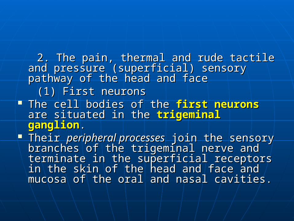
2. The pain, thermal and rude tactile and 2. The pain, thermal and rude tactile and pressure (superficial) sensory pathway of pressure (superficial) sensory pathway of the head and facethe head and face
(1) First neurons(1) First neurons The cell bodies of the The cell bodies of the first neuronsfirst neurons are are
situated in the situated in the trigeminal gangliontrigeminal ganglion.. Their Their peripheral processesperipheral processes join the sensory join the sensory
branches of the trigeminal nerve and branches of the trigeminal nerve and terminate in the superficial receptors in terminate in the superficial receptors in the skin of the head and face and mucosa the skin of the head and face and mucosa of the oral and nasal cavities.of the oral and nasal cavities.


Their central processes enter the pons via the Their central processes enter the pons via the sensory root of the trigeminal nerve.sensory root of the trigeminal nerve.
The fibers conducting the impulses associated The fibers conducting the impulses associated with pain and thermal sensation descends to fwith pain and thermal sensation descends to form the orm the spinal tract of trigeminal nervespinal tract of trigeminal nerve and and terminate in the terminate in the spinal nucleus of trigeminal spinal nucleus of trigeminal nervenerve..
The fibers being concerned with tactile and prThe fibers being concerned with tactile and pressure sensation terminate in the essure sensation terminate in the pontine nucpontine nucleus of trigeminal nerveleus of trigeminal nerve..
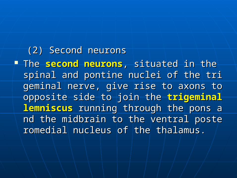
(2) Second neurons(2) Second neurons The The second neuronssecond neurons, situated in the spinal an, situated in the spinal an
d pontine nuclei of the trigeminal nerve, give rid pontine nuclei of the trigeminal nerve, give rise to axons to opposite side to join the se to axons to opposite side to join the trigemitrigeminal lemniscusnal lemniscus running through the pons and t running through the pons and the midbrain to the ventral posteromedial nuclhe midbrain to the ventral posteromedial nucleus of the thalamus.eus of the thalamus.

(3) Third neurons(3) Third neurons The cell bodies of the The cell bodies of the third neuronsthird neurons are in the are in the
ventral posteromedial nucleusventral posteromedial nucleus of the thalam of the thalamus. us.
They send out fibers to form the They send out fibers to form the central thalacentral thalamic radiation mic radiation ((thalamocortical tractthalamocortical tract)) coursin coursing through the posterior limb of the internal cag through the posterior limb of the internal capsule to thepsule to the inferiorinferior part of the postcentral gpart of the postcentral gyrusyrus..
here.here.