nn - Enlighten: Publicationseprints.gla.ac.uk/100250/1/100250.pdf · molecular level (Ahmad and...
31
nn Christie, J. M., Blackwood, L., Petersen, J., and Sullivan, S. (2014) Plant flavoprotein photoreceptors. Plant and Cell Physiology . ISSN 0032-0781 Copyright © 2014 The Authors http://eprints.gla.ac.uk/100250/ Deposited on: 17 December 2014 Enlighten – Research publications by members of the University of Glasgow http://eprints.gla.ac.uk
-
Upload
hoangthuan -
Category
Documents
-
view
215 -
download
0
Transcript of nn - Enlighten: Publicationseprints.gla.ac.uk/100250/1/100250.pdf · molecular level (Ahmad and...

nn
Christie, J. M., Blackwood, L., Petersen, J., and Sullivan, S. (2014) Plant flavoprotein photoreceptors. Plant and Cell Physiology . ISSN 0032-0781 Copyright © 2014 The Authors http://eprints.gla.ac.uk/100250/ Deposited on: 17 December 2014
Enlighten – Research publications by members of the University of Glasgow
http://eprints.gla.ac.uk

1
Plant Cell Physiology as Review
Plant Flavoprotein Photoreceptors
John M. Christie*, Lisa Blackwood, Jan Petersen, Stuart Sullivan Institute of Molecular Cell and Systems Biology, College of Medical, Veterinary and Life Sciences, University of Glasgow, G12 8QQ, UK
*To whom correspondence should be addressed: e-mail [email protected], tel. +44 141 330 2392, fax +44 141 330 4447 Words: 6,815 Figures: 4
© The Author(s) 2014. Published by Oxford University Press on behalf of Japanese Society of Plant Physiologists. This is an Open Access article distributed under the terms of the Creative Commons Attribution License (http://creativecommons.org/licenses/by/4.0/), which permits unrestricted reuse, distribution, and reproduction in any medium, provided the original work is properly cited.
Plant and Cell Physiology Advance Access published December 15, 2014 at Periodicals D
ept on Decem
ber 17, 2014http://pcp.oxfordjournals.org/
Dow
nloaded from

2
Abstract Plants depend on the surrounding light environment to direct their growth. Blue light (390-500 nm) in particular acts to promote a wide variety of photomorphogenic responses including seedling establishment, phototropism and circadian clock regulation. Several different classes of flavin-based photoreceptors have been identified that mediate the effects of blue light in the dicotyledonous genetic model Arabidopsis thaliana. These include the cryptochromes, the phototropins and members of the Zeitlupe family. In this review, we discuss recent advances, which contribute to our understanding of how these photosensory systems are activated by blue light and how they initiate signaling to regulate diverse aspects of plant development. Key words: blue light; chromophore; cryptochrome; flavin; phototropin; Zeitlupe family Abbreviations: ABCB19, ABC transporter B family member 19; bHLH, basic helix-loop-helix; BLUS1, blue light signaling 1; CCT, cryptochrome C-terminus; CDF, cycling DOF factor; CIB1, cry-interacting bHLH1; CO, Constans; COP1, constitutive photomorphogenic 1; CPD, cyclobutane pyrimidine dimer; cry, cryptochrome; DASH, Drosophila-Arabidopsis-Synechocystis-Human; FADH�, neutral FAD radical; fkf1, flavin-binding kelch repeat F-box 1; FT, flowering locus T; FTIR, Fourier transform infrared; GI, gigantea; HY5, long hypocotyl 5; KD, kinase domain; lkp2, LOV kelch protein 2; LOV, Light, oxygen or voltage sensing; MTHF, 5.10-methyltetrahydrofolate; NPH3, non-phototropic hypocotyl 3; PAS Per-ARNT-Sim; phot, phototropin; PHR, photolyase homology region; PHY, phytochrome; PKS4, phytochrome kinase substrate 4; RPT2, root phototropism protein 2; SAXS, small angle x-ray scattering; SCF, skp cullin F-box; SPA1, suppressor of phyA 1; TOC1, timing of CAB expression 1; UVR8, UV Resistance locus 8; ztl, Zeitlupe
at Periodicals Dept on D
ecember 17, 2014
http://pcp.oxfordjournals.org/D
ownloaded from

3
Introduction Light is recognized ubiquitously throughout nature as a source of energy. For many organisms, including plants and algae, light is an important environmental stimulus that directs their development, morphogenesis and physiology. This is achieved by specialized photoreceptors that detect and respond to changes in light intensity, quality, direction, and duration. These light-responsive proteins typically contain a prosthetic cofactor or chromophore that enables them to perceive and respond to specific wavelengths of light. To date, five photosensory systems have been identified in higher plants. The phytochromes (phys) respond largely to red (600-700 nm) and far-red (700-750 nm) regions of the solar spectrum (Chen and Chory 2011), whereas UV Resistance locus 8 (UVR8) monitors ultra-violet B (UV-B) wavelengths (280-315 nm) to regulate both developmental and UV-protective processes (Jenkins 2014). Plant responses to blue light (390-500 nm) are extensive and are mediated by three different classes of photoreceptor: the cryptochromes (crys) (Chaves et al. 2011), phototropins (phots) (Christie 2007) and members of the Zeitlupe family (ztl, fkf1 and lkp2) (Suetsugu and Wada 2013).
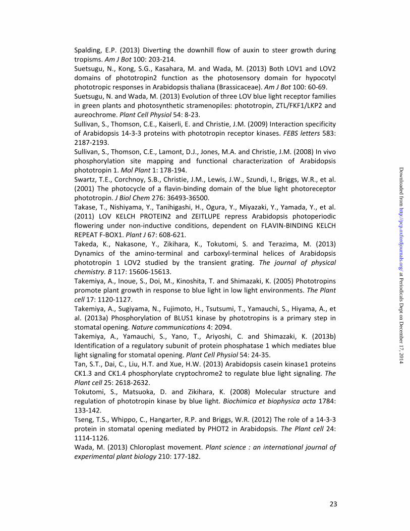
Genetic analyses in the model flowering plant Arabidopsis thaliana have been instrumental in defining the molecular basis of plant blue-light receptors and their mechanism of action. However, action spectra for a number of blue light-initiated responses were described prior to the advent of Arabidopsis genetics and provided important information regarding the photochemical properties of the photoreceptors involved. For instance, the action spectrum for phototropism resembles the absorption spectrum of a flavoprotein; it shows maximal activity between 400 and 500 nm and reveals a degree of fine structure with a major peak at 450 nm and subsidiary shoulders at 430 and 470 nm (Fig. 1A). An additional broad, less effective peak is typically observed in the UV-A region of the spectrum at 380 nm. Such properties are common to all plant blue light-receptors as each class binds oxidized flavin as a light-absorbing chromophore (Conrad et al. 2014).
Here, we summarize how light activates these different classes of flavoprotein photoreceptors to regulate a variety of blue light responses in Arabidopsis. Throughout this review we will adopt the nomenclature first introduced for Arabidopsis phys (Quail et al. 1994) and phots (Briggs et al. 2001) where the holoprotein with its chromophore photoreceptor is designated in lower case (cry, phot, ztl) and in uppercase when the apoprotein without its chromophore is described (CRY, PHOT, ZTL). By convention, we also use upper case italics for the genes encoding the photoreceptor apoproteins (CRY, PHOT, ZTL) and lower case italics to refer to mutant alleles (cry, phot, ztl). Cryptochromes Cryptochromes were the first plant blue-light receptors to be characterized at the molecular level (Ahmad and Cashmore 1993). They are major regulators of plant photomorphogenic development (also referred to as de-etiolation) and function to control the photoperiodic control of flowering in addition to entrainment of the circadian clock (Liu et al. 2011). Cryptochromes exhibit significant homology to photolyases, which are blue light-activated enzymes found in both prokaryotes and eukaryotes that catalyze the light-dependent repair of damaged DNA produced from exposure to UV-B irradiation (Ahmad and Cashmore 1993). Cryptochromes together
at Periodicals Dept on D
ecember 17, 2014
http://pcp.oxfordjournals.org/D
ownloaded from

4
with the photolyases comprise a superfamily of proteins that can be subdivided into five groups: cyclobutane pyrimidine dimer (CPD) photolyases, (6–4) pyrimidine-pyrimidone adduct [(6-4) photoproduct] photolyases, cry-DASH proteins, animal cryptochromes and plant cryptochromes (Chaves et al. 2011).
Three cryptochromes have been identified in Arabidopsis (cry1-3). Arabidopsis cry1 and cry2 are localized predominantly in the nucleus (Cashmore et al. 1999; Kleiner et al. 1999). They do not exhibit DNA repair activity but function to control various aspects of plant growth and development; cry1 has a major role in regulating seedling de-etiolation under blue light, whereas cry2 is involved regulating flowering in response to day-length (Liu et al. 2011). Cry2 differs from cry1 in that it undergoes blue-light–dependent ubiquitination and degradation in the nucleus (Zuo et al. 2012). Hence, cry2 functions preferentially under low light conditions or where light is limiting. Cry3 is a cry-DASH (Drosophila-Arabidopsis-Synechocystis-Human) protein that localizes to mitochondria and chloroplasts (Kleine et al. 2003) and is reported to repair UV-induced lesions in single-stranded DNA (Selby and Sancar 2006) as well as in loop structures of double-stranded DNA (Pokorny et al. 2008). Cryptochrome structure and light sensing
Arabidopsis cryptochromes, like photolyases bind two chromophores: flavin adenine dinucleotide (FAD) and a pterin derivative 5,10-methenyltetrahydrofolate (MTHF). FAD is bound non-covalently within the photolyase homology region (PHR) and functions as the primary light sensor (Fig.1B), whereas the role of MTHF, as is the case for photolyases, is to harvest and transfer additional light energy to the FAD chromophore from the near UV-region (370-390 nm) (Hoang et al. 2008). In addition to the PHR domain (~500 amino acids) cry1 and cry2 also contain a distinctive cryptochrome C-terminus (CCT) that is absent from cry3, cry-DASH proteins and other photolyases (Fig. 1B). While photosensing is mediated by the PHR domain, the CCT (~100-200 amino acids) is important for cryptochrome signaling (Yang et al. 2000; Yu et al. 2009).
Light sensing by the PHR domain is proposed to promote conformational changes within the CCT that are necessary for interacting with downstream signaling components. The crystal structure of the PHR domain of Arabidopsis cry1 was solved a decade ago (Brautigam et al. 2004). Structural information for the CCT is still lacking at present. The PHR domain of cry1 shows strong structural similarity to photolyases (Fig. 2A). The PHR fold comprises an N-terminal α/β domain (residues 13-139) and a C-terminal α domain (residues 217-495). A connecting region of 77 amino acids links these two domains together. The FAD chromophore is buried within a hydrophobic U-shaped pocket inside the α domain. The binding site for MTHF has been solved at the structural level for cry3 (Klar et al. 2007), but not for cry1 or cry2 since this accessory chromophore is typically lost upon their purification from heterologous expression systems. In cry3, MTHF is bound at a distance of 15.2 Å from the FAD chromophore. The MTHF binding region is highly conserved in DASH-type cryptochromes. However, a structural comparison between cry3 and cry1 suggests some differences in MTHF binding within this region (Klar et al. 2007). Spectroscopic analysis of recombinant cry1 shows that it binds fully oxidized FAD in the inactive or resting state, which strongly absorbs blue light (λmax ~450 nm). Irradiation of the PHR domain results in the formation of a semi-reduced
at Periodicals Dept on D
ecember 17, 2014
http://pcp.oxfordjournals.org/D
ownloaded from

5
(semiquinone) neutral FAD radical (FADH�). FADH� is generated by photoreduction and subsequent protonation of the FAD chromophore (Fig. 3A). Light-driven generation of FADH� occurs within microseconds (Langenbacher et al. 2009) and is considered to represent the signaling state that triggers photoreceptor activation. In the absence of light, FADH� is reportedly unstable and can revert back to FAD within minutes (Chaves et al. 2011). Inhibitory effects of green light have been observed on cryptochrome function presumably by antagonizing FADH� formation, which absorbs in this spectral region (Fig. 3A). Recombinant cryptochromes therefore undergo a reversible photocycle between FAD and FADH� states.
Photoreduction of the FAD cofactor is proposed to involve a conserved flavin-reducing triad of tryptophan residues (Chaves et al. 2011) within the α domain (W400, W377 and W324). W400 is the predicted electron donor proximal to the flavin whereby W324 is exposed to the protein surface (Fig. 2A). This photoreduction mechanism is still under investigation, but is reported to involve transfer of electrons between the FAD and the tryptophan cascade as well as protonation of the flavin by the putative aspartic acid proton donor D396 (Cailliez et al. 2014). Mutation of the residues within the tryptophan triad to alanine impairs flavin photoreduction in recombinant cry2 providing support for this reaction mechanism, at least in vitro (Li et al. 2011). Similar mutations have also been used effectively to impact the photoreduction of cry1 (Bouly et al. 2007; Zeugner et al. 2005).
While mutation of the tryptophan triad impairs flavin photoreduction in vitro, structure-function studies in Arabidopsis are conflicted regarding its biological role in cryptochrome photoactivation. Arabidopsis mutants lacking cry1 and cry2 are impaired in de-etiolation responses and exhibit a long hypocotyl and small, unopened cotyledons when grown in continuous blue light (Liu et al. 2011). Triad mutations W400F and W324F failed to restore cry1 function when expressed in cry-deficient mutants, consistent with a role in photoreduction and light sensing (Zeugner et al. 2005). Comparable mutations have also been reported to impact the blue light-dependent degradation of cry2 (Li et al. 2011). However, mutations in the tryptophan triad of cry2 have been shown to elicit different functional effects compared to those observed for cry1. Mutation of the surface exposed tryptophan in cry2 (W321) weakened its activity, while side chain replacements of the remaining tryptophans (W397 and W374) resulted in constitutive cry2 activity (Li et al. 2011). It therefore seems likely that mutation of W397 and W394 cause structural alterations within the CRY2 apoprotein such that it adopts an activate conformation in that absence of light. Hence, the biological role of the tryptophan triad in cryptochrome photoactivation is still under debate (Liu et al. 2010). Indeed, recent studies indicate that alternative electron transport pathways, independent of the tryptophan triad, are also involved in flavin photoreduction (Engelhard et al. 2014). Cryptochrome activation Despite the lack of structural information on the CCT, its role in initiating crytochrome signaling is well established (Chaves et al. 2011; Liu et al. 2011). The PHR domain and the CCT are proposed to form a closed or inactive conformation in darkness (Fig. 4A). Blue light sensing by the PHR domain would then trigger a conformational change in the CCT to produce an open or active conformation that
at Periodicals Dept on D
ecember 17, 2014
http://pcp.oxfordjournals.org/D
ownloaded from
6
initiates signaling. Consistent with this mode of action, light-induced conformational changes associated with the CCT of cry1 have been reported using a variety of techniques, including limited proteolysis (Partch et al. 2005), pulsed laser-induced transient grating (Kondoh et al. 2011) and Fourier transform infrared (FTIR) difference spectroscopy (Kottke et al. 2006). Furthermore, overexpression of the CCT in Arabidopsis confers constitutive cryptochrome signaling (Yang et al. 2000; Yu et al. 2009), suggesting that this region can adopt an active conformation in the absence of the PHR domain. Indeed, mutation of residues G380 and L470 within Arabidopsis cry1 appears to impact inter-domain association between the PHR domain and the CCT, leading to constitutive activation in the absence of light (Exner et al. 2010; Gu et al. 2012).
The mechanism(s) by which the FAD to FADH� transition within the PHR domain is propagated to bring about the proposed conformational change within the CCT is still not fully understood. However, recent studies suggest that a contributing factor might involve the ability of cryptochrome to bind ATP. The PHR domain of Arabidopsis cry1 was co-crystallized with a non-hydrolysable ATP analogue (AMP-PNP), which binds close to the flavin cofactor (Fig. 2A). ATP binding is reported to both increase the yield and prolong the lifetime of the FADH� signaling state (Burney et al. 2009; Cailliez et al. 2014; Immeln et al. 2007; Muller et al. 2014). Specifically, Muller et al. (2014) suggest that ATP binding increases the pKa of the potential aspartic acid proton donor D396 to facilitate its protonation (D396H) under physiological pH conditions and ensure that the FAD remains in close contact with W400 to promote electron transfer (Cailliez et al. 2014). The deprotonation of D396 (D396−) that occurs upon light-driven FADH� formation would be expected to introduce strong electrostatic changes within its hydrophobic environment that could trigger conformational changes necessary for cryptochrome activation. Phosphorylation of the CCT has also been shown to correlate closely with the photoactivation and biological function of plant cryptochromes (Liu et al. 2011). Both Arabidopsis cry1 and cry2 become rapidly phosphorylated in etiolated seedlings upon exposure to blue light (Shalitin et al. 2002; Shalitin et al. 2003). Phosphorylation on multiple serine residues could be expected to promote electrostatic repulsion of the CCT from the PHR domain upon exposure to light (Liu et al. 2011). Several different protein kinases are likely to be involved in CCT phosphorylation. In the case of cry2, CCT phosphorylation by Casein Kinase 1 appears to induce ubiquitination and subsequent degradation of the protein (Tan et al. 2013). The observation that Arabidopsis cry1 purified from insect cells is phosphorylated upon photoexcitation, combined with its ability to bind ATP has led to the proposal that cry1 itself exhibits autophosphorylation activity (Bouly et al. 2003; Shalitin et al. 2003). However, given that cry1 shows no homology to protein kinases, phosphorylation of the CCT by a co-purifying kinase following its exposure upon irradiation cannot be excluded entirely from these studies. Cryptochrome signaling
Nuclear-localized cry1 and cry2 largely mediate their effects on plant development through changes in gene expression (Chaves et al. 2011; Liu et al. 2011). Approximately 5–25% of the gene expression changes that occur during seedling de-etiolation under blue light can be attributed to the action of cry1 and cry2 in
at Periodicals Dept on D
ecember 17, 2014
http://pcp.oxfordjournals.org/D
ownloaded from
7
Arabidopsis (Folta and Spalding 2001; Ma et al. 2001; Ohgishi et al. 2004). Two mechanisms of transcriptional control have been elucidated for plant cryptochromes. The first mechanism involves modulating the abundance of positive regulators, including the bZIP transcription factor Long Hypocotyl 5 (HY5) by suppressing their degradation in response to light (Osterlund et al. 2000). In etiolated seedlings, HY5 is targeted for proteolysis by the Constitutive Photomorphogenic 1 (COP1)/Suppressor of phyA 1 (SPA1) complex. COP1/SPA1 act as a substrate receptor for the CUL4-DDB1 E3 ubiquitin ligase complex, which is responsible for degrading proteins that promote photomorphogenic development (Lau and Deng 2012). Upon photoactivation, cry1 and cry2 can bind SPA1 and suppress the action of the COP1-SPA1 complex (Fig. 4A). This COP1-SPA1-cry1 interaction leads to an accumulation of HY5 and other factors, which in turn, regulate the transcription of genes required for de-etiolation. Expression of constitutively active forms of plant cryptochromes, including the truncated CCT region can impair this mode of regulation and permanently sequester the COP1-SPA1 complex from degrading HY5, giving rise to a light-grown or constitutive photomorphogenic (cop) phenotype (short hypocotyl and opened cotyledons) when seedlings are grown in darkness (Yang et al. 2000; Yu et al. 2009). Similarly, light-driven interactions between COP1-SPA1 and cry2 have been shown to suppress proteolysis of the zinc finger transcriptional regulator Constans (CO), a key regulator of flowering in response to long days (Zuo et al. 2011). Light-dependent degradation of cry2 may also involve the action of SPA1 in concert with phyA (Weidler et al. 2012). In addition to controlling the abundance of transcription factors such as HY5 through the COP1-SPA1 pathway, cry2 has been reported to interact directly with and modulate the activity of transcription factors associated with the photoperiodic control of flowering (Fig. 4A). Upon photoactivation, cry2 binds the basic helix-loop-helix (bHLH) transcription factor cry-interacting bHLH 1 (CIB1) via its PHR domain (Kennedy et al. 2010) to promote expression of Flowering Locus T (FT) (Liu et al. 2008), a mobile transcriptional regulator of floral initiation. Other CIB family members, including CIB2 and CIB5 bind to cry2 and function redundantly in regulating CRY2-dependent flowering (Liu et al. 2013). However, CIB1 does not appear to directly bind the FT promoter (Fig. 4A), but instead heterodimerizes with other CIB homologues to fulfill this function (Liu et al. 2013).
Although predominantly localized to the nucleus, evidence suggests that cry1 can also influence signaling events at the plasma membrane. Blue light induces a transient depolarization of the plasma membrane in Arabidopsis (Spalding 2000) that is linked to hypocotyl growth inhibition (Cho and Spalding 1996). Nuclear-localized cry1 is sufficient to promote this response (Wu and Spalding 2007). Moreover, membrane depolarization is rapid (occurring within 30 seconds) implying that the nucleus-plasma membrane communication mechanism involved is too rapid to include changes in gene expression. Yet, the signaling events that couple photoactivation of cry1 in the nucleus to plasma membrane depolarization remain largely unexplored. As discussed below, a separate class of flavoprotein photoreceptor known as the phototropins largely mediates blue light sensing at the plasma membrane.
at Periodicals Dept on D
ecember 17, 2014
http://pcp.oxfordjournals.org/D
ownloaded from

8
Phototropins
Phototropin blue-light receptors were named after their role in mediating higher plant phototropism (Christie et al. 1999). The absorption properties of these photoreceptors correspond closely to the action spectra for this response (Fig. 1A; Fig. 3B). Phototropins are light-activated serine/threonine kinases that undergo autophosphorylation in response to blue light irradiation. Arabidopsis contains two phototropins (phot1 and phot2), which regulate a range of photoresponses that serve to optimize photosynthetic efficiency and promote growth particularly under weak light conditions (Takemiya et al. 2005). Both phot1 and phot2 are predominantly localized to the plasma membrane (Kong et al. 2006; Sakamoto and Briggs 2002), but are not integral membrane proteins. Instead, they are attached to the intracellular side of the plasma membrane and can be released by treatment with non-ionic detergents (Knieb et al. 2004; Kong et al. 2013a). Their mode of membrane attachment is still not clearly defined, but may involve peptide sequences residing within the extreme C-terminal region of the protein (Kong et al. 2013b). Recent reports suggest that phot1 in addition to phot2 can localise to the outer membrane of the chloroplast consistent with their role in regulating chloroplast photorelocation movements (Kong et al. 2013b).
Blue light irradiation has been shown to elicit different impacts on the subcellular localization of phot1 and phot2. A fraction of phot1 re-localizes from the plasma membrane to the cytosol upon irradiation (Han et al. 2008; Kaiserli et al. 2009; Sakamoto and Briggs 2002; Wan et al. 2008), whereas phot2 is targeted to the Golgi apparatus (Aggarwal et al. 2014; Aihara et al. 2008; Kong et al. 2007; Kong et al. 2006). Translocation from the plasma membrane is inhibited when the kinase activities of phot1 (Kaiserli et al. 2009) and phot2 (Aggarwal et al. 2014; Kong et al. 2007; Kong et al. 2006) are impaired. Although the biological significance of these translocation processes is still not known, internalization of phot1 has been linked to modulating receptor signaling, at least for phototropism (Wan et al. 2008).
Genetic analysis in Arabidopsis has shown that phot1 and phot2 overlap in function to regulate hypocotyl and root phototropism (Sakai et al. 2001), chloroplast accumulation movement (Kagawa et al. 2001), stomatal opening (Kinoshita et al. 2001), leaf positioning and leaf flattening (Inoue et al. 2008b; Sakamoto and Briggs 2002). In the case of hypocotyl phototropism, phot2 functions predominantly at high light intensities, whereas phot1 acts over a broad range of fluence rates (Sakai et al. 2001). Phot2, on the other hand, is solely responsible for eliciting chloroplast and nuclear avoidance movements in response to high intensity blue light (Higa et al. 2014; Kagawa et al. 2001). An additional role for phot2 in regulating resistance protein-mediated viral defense has been reported (Jeong et al. 2010). Phot1-specific responses have also been identified and include the rapid inhibition of hypocotyl growth upon transfer of etiolated seedlings to light (Folta et al. 2003), transcript destabilization (Folta and Kaufman 2003) and more recently the suppression of lateral root growth (Moni et al. 2014).
at Periodicals Dept on D
ecember 17, 2014
http://pcp.oxfordjournals.org/D
ownloaded from

9
Phototropin structure and light sensing Phototropins comprise a serine/threonine kinase domain at their C-terminus and two specialized Light, Oxygen or Voltage sensing (LOV) domains, designated LOV1 and LOV2, at their N-terminus (Fig. 1B). LOV1 and LOV2 function as blue light sensors and bind oxidized flavin mononucleotide (FMN) non-covalently as chromophore (Christie et al. 1999). The structural and biophysical properties of the LOV domain have been studied extensively and provide a valuable framework to understand how the phototropin light switch operates (Christie et al. 2012). LOV domains (~110 amino acids) form a subset of the Per-ARNT-Sim (PAS) superfamily and consist primarily of five antiparallel β-sheets (Aβ, Bβ, Gβ, Hβ, and Iβ) and several α-helices (Cα, Dα, Eα and Fα) (Freddolino et al. 2013). The FMN chromophore is bound tightly inside these structural elements, which comprise the LOV/PAS core (Fig. 2B). Spectroscopic analyses of recombinant LOV domains obtained from a variety of phototropin proteins show that they bind oxidized FMN in the inactive state, absorbing strongly in the blue region of the spectrum (λmax ~450 nm). Like the PHR domain of plant cryptochromes, purified LOV domains are photochemically active in solution (Fig. 3B). Irradiation of the LOV domain results in the formation a covalent bond between the C(4a) carbon of the FMN and the sulfur atom of a nearby, conserved cysteine residue (Salomon et al. 2000; Swartz et al. 2001). FMN-cysteinyl adduct formation occurs within microseconds of illumination and produces a spectral species that no longer absorbs blue light (Fig. 3B). FMN-cysteinyl adduct formation represents the active signaling state that leads to photoreceptor activation. This photochemical reaction is fully reversible in darkness. Thermal decay of the covalent adduct back to the dark state occurs within tens to thousands of seconds depending on the LOV domain (Christie et al. 2012).
Recombinant protein fragments of Arabidopsis phot1 and phot2 containing both LOV domains (LOV1+2) exhibit more complex photochemistry than either domain alone. For instance, adduct-decay kinetics for LOV1+2 proteins is noticeably slower compared to individual LOV domains taking on the order of tens of minutes to recover (Kasahara et al. 2002). This slower rate of adduct decay is proposed to enhance receptor photosensitivity by prolonging the life-time of the photoproduct (Christie et al. 2002). Conversely, modest acceleration of the LOV photocycle can be achieved, at least in vitro, by simultaneous or subsequent irradiation with UV-A light (Kennis et al. 2004), which antagonizes FMN-cysteinyl adduct formation which absorbs in this region (λmax ~390 nm; Fig. 2B). Moreover, specific amino acid changes within the FMN-binding pocket have been identified that significantly accelerate or slow down the rate of adduct decay of the LOV photocycle (Christie et al. 2007; Circolone et al. 2012; Kawano et al. 2013; Zayner and Sosnick 2014; Zoltowski et al. 2009). These findings therefore open up new possibilities to fine-tune photoproduct life time and modulate photoreceptor activity. Mutation of the conserved cysteine involved in adduct formation (Fig. 2B) abolishes the photochemical reactivity of the LOV domain (Salomon et al. 2000; Swartz et al. 2001) and has been used to functionally dissect the roles of LOV1 and LOV2 in regulating phot1 activity. The C512A mutation in LOV2 fails to restore phot1 function when expressed in phot1-deficient mutants of Arabidopsis (Christie et al. 2002). These and other structure-function studies highlight the importance of LOV2
at Periodicals Dept on D
ecember 17, 2014
http://pcp.oxfordjournals.org/D
ownloaded from

10
in regulating light-dependent autophosphorylation, which is essential for phototropin function (Cho et al. 2007; Jones et al. 2007). Yet, similar mutational analyses on phot2 suggests that LOV1 can still have a residual role in light sensing when LOV2 is photochemically inactivated, at least for phototropism (Cho et al. 2007; Suetsugu et al. 2013). Further structure-function analyses in Arabidopsis provide additional evidence for a photoactive role for phot1 LOV1 in arresting chloroplast accumulation movement at high light intensities (Kaiserli et al. 2009). Such a role could serve to promote an efficient transition from chloroplast accumulation to avoidance movement under excess light. Additional work is now needed to clarify the roles of LOV1 and LOV2 and whether these differ between phot1 and phot2. Although, the functional significance of LOV1 still remains unclear, its presence is proposed to modulate LOV2 photoreactivity (Christie et al. 2002; Okajima et al. 2014; Sullivan et al. 2008) and play a role in receptor dimerization (Nakasako et al. 2008).
Phototropin activation
There is presently no crystal structure for the full-length phototropin. However, recent FTIR difference spectroscopy (Pfeifer et al. 2010) and small-angle x-ray scattering (SAXS) combined with low resolution molecular modeling (Okajima et al. 2014) have provided insights into the domain architecture of the photoreceptor, as well as the global structural changes that occur upon photoexcitation. SAXS analyses suggest that LOV1, LOV2 and the kinase domain (KD) are arranged sequentially within the molecule, with LOV1 and the KD being positioned on either side of LOV2 (Fig. 4B). Such an arrangement is consistent with a model whereby LOV2 functions closely with the KD to act as the main regulator of phototropin activity (Christie 2007; Tokutomi et al. 2008). In a mechanism comparable to that depicted for cryptochrome activation (Fig. 4A), the N-terminal photosensory region of phototropin (comprising LOV1 and LOV2) is hypothesized to form a closed or inactive conformation with the C-terminal KD in darkness, in which LOV2 together with adjacent regulatory sequences act to repress phototropin kinase activity. Blue light sensing by LOV2 results in a conformational change that causes the N-terminus to move and alleviate this repression (Fig. 4B), which leads to an opening of the catalytic cleft between the N- and C-terminal lobes of the KD to promote ATP binding and initiate receptor autophosphorylation (Pfeifer et al. 2010). The mechanism by which FMN-cysteinyl adduct formation in LOV2 is propagated to bring about a global light-driven conformational change initially involves a conserved glutamine residue within LOV2 which hydrogen bonds with the O4 oxygen of the FMN chromophore in darkness (Fig. 3B). FMN-cysteinyl adduct formation results in side chain rotation of this glutamine to temporarily alter its hydrogen bonding with the FMN chromophore (Crosson and Moffat 2001). Q575 resides within the β-scaffold of Arabidopsis phot1 LOV2, which acts as a docking site for a conserved α-helix (Jα) located outside the LOV core (Fig. 2B). Side chain rotation of this glutamine is proposed to invoke structural alterations with the β-sheet surface that lead to displacement and unfolding of the Jα-helix flanking the C-terminus of the LOV2-core (Freddolino et al. 2013; Harper et al. 2003). Mutation of this glutamine in the LOV2 domain of Arabidopsis phot1 attenuates light activated
at Periodicals Dept on D
ecember 17, 2014
http://pcp.oxfordjournals.org/D
ownloaded from

11
disordering of the Jα-helix (Nash et al. 2008) and consequently impacts receptor autophosphorylation (Jones et al. 2007). Jα-helix displacement therefore represents an integral component of the phototropin light switch. Indeed, artificial disruption of the Jα-helix from the LOV2-core through targeted mutagenesis uncouples this mode of regulation and leads to phot1 activation in the absence of light (Harper et al. 2004; Jones et al. 2007; Kaiserli et al. 2009). Recent crystallographic studies (Halavaty and Moffat 2013) have shown that sequences flanking the N-terminus of LOV2 also comprise an α-helix (Fig. 2B). Several lines of evidence suggest that this α-helix (A’α) plays a key role in regulating LOV2 activity in concert with Jα (Freddolino et al. 2013; Takeda et al. 2013; Zayner et al. 2012). Mutations within this region appear to disrupt the repressive action of LOV2 on phototropin kinase activity (Aihara et al. 2012). Consequently, mutations in A’α have adverse effects on phototropin signaling (Sharma et al. 2014). Together, these findings highlight the importance of A′α together with Jα-helix in activating the LOV2 photoswitch. Phototropin autophosphorylation and signaling
A primary consequence of phototropin activation is receptor autophoshorylation (Christie et al. 1998). Phosphorylation occurs predominantly on multiple serine residues and several studies have been successful in mapping the sites involved. So far at least 21 phosphorylation sites have been identified in Arabidopsis phot1 (Boex-Fontvieille et al. 2014; Deng et al. 2014; Inoue et al. 2008a; Salomon et al. 2003; Sullivan et al. 2008) and 29 in Arabidopsis phot2 (Inoue et al. 2011). These sites are listed in Table 1 for convenience. Structure-function studies carried out to date indicate that autophosphorylation at two conserved serine residues within the kinase activation loop or T-loop (S849 and S851 in phot1; S761 or S763 in phot2) is essential for receptor signaling in Arabidopsis (Inoue et al. 2008a; Inoue et al. 2011). The majority of the remaining phosphorylation sites reside within either the N-terminal region upstream of LOV1 or the linker sequence between LOV1 and LOV2 (Fig. 1B). Although the biological significance of these upstream phosphorylation sites remains largely unexplored, the occurrence of some of these phosphoserines is fluence-rate dependent (Salomon et al. 2003), suggesting that they may have different biochemical consequences.
At least for phot1, phosphorylation at S350, S376 and S410 located within the linker region between LOV1 and LOV2 is required to bind members of the non-epsilon group of Arabidopsis 14-3-3 regulatory proteins (Inoue et al. 2008a; Sullivan et al. 2009). Mutation of the 14-3-3 binding site does not appear to affect the functionality of phot1 in Arabidopsis (Inoue et al. 2008a). By contrast, binding of 14-3-3λ to phot2 is reported to involve Ser747 within the KD. Mutation of this site impairs phot2-mediated stomatal opening, but fails to impact phot2-meidated phototropism, as well as leaf positioning and flattening (Tseng et al. 2012). These findings suggest that 14-3-3-binding to phot2 is an important signaling step for stomatal opening. Whether an equivalent serine mediates a similar function for phot1 awaits further investigation. The mechanisms underlying blue light-induced stomatal opening have been extensively studied and therefore represent one of the best-characterized phototropin signaling pathways (Inoue et al. 2010). The plasma membrane
at Periodicals Dept on D
ecember 17, 2014
http://pcp.oxfordjournals.org/D
ownloaded from
12
H+ATPase, together with inward-rectifying K+ channels are key components of this pathway. Phototropin-mediated activation of the H+ATPase induces hyperpolarization of the guard cell plasma membrane, which allows K+ uptake through inward-rectifying K+ channels (Shimazaki et al. 2007). Accumulation of K+ induces the swelling of the guard cells, which promotes stomatal opening. Activation of the guard cell H+-ATPase involves phosphorylation and subsequent 14-3-3 binding at its C-terminus (Kinoshita and Shimazaki 1999). The kinase responsible for phosphorylating the H+-ATPase has not been identified. However, recent work has shown that phototropin-activation of the H+-ATPase is initiated by the guard cell-specific kinase known as Blue Light Signaling 1 (BLUS1). BLUS1 is directly phosphorylated by both phot1 and phot2 (Fig. 4B) at S348 located within its C-terminus (Takemiya et al. 2013a). Phosphorylation of S348 and BLUS1 kinase activity are essential for activation of the H+-ATPase. As a result, Arabidopsis mutants lacking BLUS1 show no blue-light induced stomatal opening, but exhibit normal phototropism, chloroplast relocation movement and leaf flattening (Takemiya et al. 2013a). Phototropin-mediated phosphorylation of BLUS1 therefore represents a key primary step in the signal cascade leading to phosphorylation and activation of the H+-ATPase. The signaling events coupling BLUS1 phosphorylation to activation of the H+-ATPase are still not well defined, but are known to involve protein phosphatase 1 activity (Takemiya et al. 2013b). Two other phosphorylation targets have been reported for Arabidopsis phot1 besides BLUS1. The auxin efflux carrier ABCB19 is a member of the ATP-binding cassette B family of transporter proteins (Spalding 2013). Phot1 can phosphorylate ABCB19 in vitro and has been shown to inhibit ABCB19 transporter activity when co-expressed in HeLa cells (Christie et al. 2011). Nevertheless, this process is not required to establish the lateral auxin gradient that drives phototropic growth. Instead, phot1-mediated inhibition of ABCB19 appears to function in enhancing phototropic responsiveness (Nagashima et al. 2008; Noh et al. 2003). In addition to ABCB19, members of the Phytochrome Kinase Substrate (PKS) family have also been shown to play a role in phototropism. PKS4 is rapidly phosphorylated by phot1 in response to blue light and is proposed to positively or negatively impact phototropism depending on its phosphorylation status (Demarsy et al. 2012; Kami et al. 2014). The biochemical function of PKS proteins is presently unknown, but likely impact auxin signaling or transport (Kami et al. 2014). For a more detailed description of the signaling events associated with phototropism, readers are directed to several recently published reviews for more information regarding phototropic signaling (Christie and Murphy 2013; Goyal et al. 2013; Hohm et al. 2013; Liscum et al. 2014; Sakai and Haga 2012), as well as comprehensive overviews covering the mechanisms underlying phototropin-mediated chloroplast photorelocation movement (Kong and Wada 2014; Wada 2013). The search for additional phototropin substrate targets is undoubtedly ongoing. In addition to autophosphorylation, Arabidopsis phot1 becomes ubiquitinated following blue light irradiation (Roberts et al. 2011). Mass spectrometry analysis has identified K526 within Fα of the LOV2-core as a site of ubiquitination (Deng et al. 2014). Ubiquitination requires the phot1 interacting protein Non-Phototropic Hypocotyl 3 (NPH3) (Roberts et al. 2011), which appears to function as part of a Cullin 3-based E3 ubiquitin ligase complex that regulates protein
at Periodicals Dept on D
ecember 17, 2014
http://pcp.oxfordjournals.org/D
ownloaded from

13
degradation and subcellular trafficking (Roberts et al. 2011; Wan et al. 2012). Further elucidation of the biochemical action of NPH3 and related proteins such as RPT2 (Sakai and Haga 2012) is now essential to understanding their role in phototropin signaling, particularly phototropism.
Zeitlupe and related blue-light receptors
Several other LOV domain-containing photoreceptors exist in Arabidopsis besides the phototropins. Members of the ztl family play important roles in controlling the degradation and stability of components associated with circadian clock regulation and the photoperiodic control of flowering. Ztl members localize to the cytosol or the nucleus (Takase et al. 2011) and comprise three members: Zeitlupe (ztl), Flavin-binding kelch repeat F-box 1 (fkf1) and LOV kelch protein 2 (lkp2) (Ito et al. 2012; Suetsugu and Wada 2013). Genetic analysis in Arabidopsis indicates that Ztl, fkf1 and lkp2 partially overlap in function (Baudry et al. 2010; Fornara et al. 2009; Takase et al. 2011). Arabidopsis mutants lacking ztl are primarily impaired in circadian clock function (Somers et al. 2000), whereas fkf1 mutants mostly show alterations in flowering time (Imaizumi et al. 2003). While mutants lacking lkp2 show minimal alterations in circadian regulation and flowering (Baudry et al. 2010), overexpression of LPK2 in Arabidopsis compromises both these processes (Schultz et al. 2001).
Members of the ztl family contain a LOV domain at their N-terminus followed by an F-box and six kelch repeats at their C-terminus (Fig. 1B). F-boxes are associated with Skp Cullin F-box (SCF)-type E3 ubiquitin ligases, which target proteins for degradation via the ubiquitin-proteosome system (Ito et al. 2012), whereas kelch repeats serve to mediate protein-protein interactions and heterodimerization between lkp2 and the other two family members (Takase et al. 2011). In principle, the primary protein architecture of the ztl family is similar to cryptochrome and phototropin where the photosensory module is located upstream from a C-terminal effector region. Phototropin is however the only blue-light receptor identified to date that contains two LOV domains. Recombinant LOV domains derived from ztl, fkf1 and lkp2 bind oxidized FMN as chromophore and show photochemical reactivity analogous to that of phototropin LOV domains, whereby blue light irradiation drives the formation of a FMN-cysteinyl adduct within the protein (Imaizumi et al. 2003). The photocycle of the LOV domain of fkf1 differs substantially to that of phototropin LOV domains in that adduct decay is extremely slow occurring in the order of days (Zikihara et al. 2006). These slow photocycle properties have been attributed to the presence of a 9 amino acid insertion between Eα and Fα that is absent from phototropin LOV domains (Zikihara et al. 2006). This failure to rapidly photocycle does not appear to impact the ability of fkf1 to respond to day length since both fkf1 and ztl are reported to exhibit high rates of protein turnover, which facilitates their recycling to measure the day-night transition (Kim et al. 2007; Sawa et al. 2007). A recent study however has shown that the LOV domain of ztl undergoes adduct decay on a time scale of hours (Pudasaini and Zoltowski 2013). These findings therefore suggest that fkf1 and ztl possess distinct photochemical properties, which may contribute to their abilities to accurately measure day length.
Although no structural information is available for ztl, fkf1 and lkp2, it is well established that these proteins function to control biological timing in Arabidopsis by
at Periodicals Dept on D
ecember 17, 2014
http://pcp.oxfordjournals.org/D
ownloaded from

14
regulating the stabilization of key regulatory targets. The expression of core circadian genes is controlled by factors such as the transcriptional repressor Timing of CAB expression 1 (TOC1) (Gendron et al. 2012; Huang et al. 2012; Pokhilko et al. 2012). Several studies indicate that the degradation of TOC1 is mediated by ztl and this process is inhibited by blue light (Fujiwara et al. 2008; Kiba et al. 2007; Mas et al. 2003). Light-mediated binding of the flowering- and circadian clock-associated protein Gigantea (GI) to ztl restricts its SCF activity causing TOC1 to accumulate (Sawa et al. 2007). The interactions between ztl and GI are mediated by the ztl LOV domain (Fig. 4C), which results in the reciprocal stabilization of these proteins (Kim et al. 2013; Kim et al. 2007). GI binding therefore appears to limit the action of SCFztl on its regulatory targets.
A similar LOV-based stabilization mechanism has been reported for fkf1 and GI (Fornara et al. 2009; Sawa et al. 2007). However, in contrast to ztl, GI binding appears to promote its SCFfkf1 activity. The onset of flowering by the fkf1 regulatory pathway involves targeted degradation of key transcriptional repressors of FT and CO expression. Transcriptional regulation of CO ensures that FT transcription is activated under long days (Andres and Coupland 2012). The kelch repeats of fkf1 are known to interact with Cycling DOF factors (CDFs) that bind to the promoter of CO and repress its transcription (Fornara et al. 2009; Imaizumi et al. 2005; Sawa et al. 2007). CO and FT expression is promoted under long days by fkf1-mediated degradation of these repressors, including CDF1 (Fig. 4C). The light-driven increase in SCFfkf1 activity involves GI-binding to the fkf1 LOV domain (Sawa et al. 2007). Thus, GI binding to fkf1 and ztl can produce different effects on their SCF action. In addition to stabilizing GI, fkf1 also interacts with and stabilizes CO protein via its LOV domain to promote flowering under long days by activating FT expression (Song et al. 2012). The action of fkf1 on CO function is therefore mediated at both the transcriptional and post-translational level. A detailed description of the molecular mechanisms associated with circadian clock regulation and the photoperiodic control of flowering and how these different light input pathways are integrated is beyond the scope of this review, but can be found in several recently published articles (Andres and Coupland 2012; Ito et al. 2012; Lau and Deng 2012; Suetsugu and Wada 2013).
Conclusions and future work
Since the cloning of the CRY1 locus over 20 years ago (Ahmad and Cashmore 1993), a great deal of progress has been made in characterizing the molecular basis of blue light perception in Arabidopsis. Three different classes of photoreceptors have been identified, all of which utilize flavin as a light-absorbing chromophore. However, many questions remain unanswered with respect to their mode of activation and signaling, and whether these photoreceptors operate as dimers. In the case of cryptochrome, structural information of the C-terminus will provide a clearer understanding as to how this region is important for receptor signaling. More work is needed to distinguish why the PHR domain initiates some aspects of cryptochrome signaling (CIB1) while others involve binding to the CCT (COP1-SPA1). With respect to phototropin, the function of receptor ubiquitination has yet to be resolved, as is the role of LOV1 photoactivation and the significance of receptor re-localization from the plasma membrane. Whether the mode of activation of ztl family members
at Periodicals Dept on D
ecember 17, 2014
http://pcp.oxfordjournals.org/D
ownloaded from

15
involves LOV-driven conformational changes within their C-terminus, as proposed for phototropins, requires further investigation, as will how GI-binding to ztl and fkf1 can elicit different effects on their SCF activity. Plants also contain a unique LOV-containing protein that is unrelated to the phototropins or members of the ztl family (Kasahara et al. 2010), yet the function of this flavoprotein still remains unknown. Answering these unresolved aspects of blue light perception in plants would undoubtedly yield exciting advances in the years to come. Acknowledgements
We acknowledge project support from UK Biotechnology and Biological Sciences Research Council (BB/J016047/1 to J.M.C). L.B. was supported by a UK Biotechnology and Biological Science Research Council PhD Studentship.
References
Aggarwal, C., Banas, A.K., Kasprowicz-Maluski, A., Borghetti, C., Labuz, J., Dobrucki, J., et al. (2014) Blue-light-activated phototropin2 trafficking from the cytoplasm to Golgi/post-Golgi vesicles. Journal of experimental botany 65: 3263-3276. Ahmad, M. and Cashmore, A.R. (1993) HY4 gene of A. thaliana encodes a protein with characteristics of a blue-light photoreceptor. Nature 366: 162-166. Aihara, Y., Tabata, R., Suzuki, T., Shimazaki, K. and Nagatani, A. (2008) Molecular basis of the functional specificities of phototropin 1 and 2. Plant J 56: 364-375. Aihara, Y., Yamamoto, T., Okajima, K., Yamamoto, K., Suzuki, T., Tokutomi, S., et al. (2012) Mutations in N-terminal flanking region of blue light-sensing light-oxygen and voltage 2 (LOV2) domain disrupt its repressive activity on kinase domain in the Chlamydomonas phototropin. J Biol Chem 287: 9901-9909. Andres, F. and Coupland, G. (2012) The genetic basis of flowering responses to seasonal cues. Nature reviews. Genetics 13: 627-639. Banerjee, R., Schleicher, E., Meier, S., Viana, R.M., Pokorny, R., Ahmad, M., et al. (2007) The signaling state of Arabidopsis cryptochrome 2 contains flavin semiquinone. J Biol Chem 282: 14916-14922. Baudry, A., Ito, S., Song, Y.H., Strait, A.A., Kiba, T., Lu, S., et al. (2010) F-box proteins FKF1 and LKP2 act in concert with ZEITLUPE to control Arabidopsis clock progression. The Plant cell 22: 606-622. Boex-Fontvieille, E., Davanture, M., Jossier, M., Zivy, M., Hodges, M. and Tcherkez, G. (2014) Photosynthetic activity influences cellulose biosynthesis and phosphorylation of proteins involved therein in Arabidopsis leaves. Journal of experimental botany 65: 4997-5010. Bouly, J.P., Giovani, B., Djamei, A., Mueller, M., Zeugner, A., Dudkin, E.A., et al. (2003) Novel ATP-binding and autophosphorylation activity associated with Arabidopsis and human cryptochrome-1. European journal of biochemistry / FEBS 270: 2921-2928. Bouly, J.P., Schleicher, E., Dionisio-Sese, M., Vandenbussche, F., Van Der Straeten, D., Bakrim, N., et al. (2007) Cryptochrome blue light photoreceptors are activated through interconversion of flavin redox states. J Biol Chem 282: 9383-9391. Brautigam, C.A., Smith, B.S., Ma, Z., Palnitkar, M., Tomchick, D.R., Machius, M., et al. (2004) Structure of the photolyase-like domain of cryptochrome 1 from Arabidopsis thaliana. Proc Natl Acad Sci U S A 101: 12142-12147.
at Periodicals Dept on D
ecember 17, 2014
http://pcp.oxfordjournals.org/D
ownloaded from

16
Briggs, W.R., Beck, C.F., Cashmore, A.R., Christie, J.M., Hughes, J., Jarillo, J.A., et al. (2001) The phototropin family of photoreceptors. The Plant cell 13: 993-997. Burney, S., Hoang, N., Caruso, M., Dudkin, E.A., Ahmad, M. and Bouly, J.P. (2009) Conformational change induced by ATP binding correlates with enhanced biological function of Arabidopsis cryptochrome. FEBS letters 583: 1427-1433. Cailliez, F., Muller, P., Gallois, M. and de la Lande, A. (2014) ATP binding and aspartate protonation enhance photoinduced electron transfer in plant cryptochrome. J Am Chem Soc 136: 12974-12986. Cashmore, A.R., Jarillo, J.A., Wu, Y.J. and Liu, D. (1999) Cryptochromes: blue light receptors for plants and animals. Science 284: 760-765. Chaves, I., Pokorny, R., Byrdin, M., Hoang, N., Ritz, T., Brettel, K., et al. (2011) The cryptochromes: blue light photoreceptors in plants and animals. Annual review of
plant biology 62: 335-364. Chen, M. and Chory, J. (2011) Phytochrome signaling mechanisms and the control of plant development. Trends in cell biology 21: 664-671. Cho, H.Y., Tseng, T.S., Kaiserli, E., Sullivan, S., Christie, J.M. and Briggs, W.R. (2007) Physiological roles of the light, oxygen, or voltage domains of phototropin 1 and phototropin 2 in Arabidopsis. Plant Physiol 143: 517-529. Cho, M.H. and Spalding, E.P. (1996) An anion channel in Arabidopsis hypocotyls activated by blue light. Proc Natl Acad Sci U S A 93: 8134-8138. Christie, J.M. (2007) Phototropin blue-light receptors. Annual review of plant biology 58: 21-45. Christie, J.M., Corchnoy, S.B., Swartz, T.E., Hokenson, M., Han, I.S., Briggs, W.R., et al. (2007) Steric interactions stabilize the signaling state of the LOV2 domain of phototropin 1. Biochemistry-Us 46: 9310-9319. Christie, J.M., Gawthorne, J., Young, G., Fraser, N.J. and Roe, A.J. (2012) LOV to BLUF: flavoprotein contributions to the optogenetic toolkit. Mol Plant 5: 533-544. Christie, J.M. and Murphy, A.S. (2013) Shoot phototropism in higher plants: new light through old concepts. Am J Bot 100: 35-46. Christie, J.M., Reymond, P., Powell, G.K., Bernasconi, P., Raibekas, A.A., Liscum, E., et al. (1998) Arabidopsis NPH1: a flavoprotein with the properties of a photoreceptor for phototropism. Science 282: 1698-1701. Christie, J.M., Salomon, M., Nozue, K., Wada, M. and Briggs, W.R. (1999) LOV (light, oxygen, or voltage) domains of the blue-light photoreceptor phototropin (nph1): binding sites for the chromophore flavin mononucleotide. Proc Natl Acad Sci U S A 96: 8779-8783. Christie, J.M., Swartz, T.E., Bogomolni, R.A. and Briggs, W.R. (2002) Phototropin LOV domains exhibit distinct roles in regulating photoreceptor function. Plant J 32: 205-219. Christie, J.M., Yang, H., Richter, G.L., Sullivan, S., Thomson, C.E., Lin, J., et al. (2011) phot1 inhibition of ABCB19 primes lateral auxin fluxes in the shoot apex required for phototropism. PLoS biology 9: e1001076. Circolone, F., Granzin, J., Jentzsch, K., Drepper, T., Jaeger, K.E., Willbold, D., et al. (2012) Structural basis for the slow dark recovery of a full-length LOV protein from Pseudomonas putida. J Mol Biol 417: 362-374. Conrad, K.S., Manahan, C.C. and Crane, B.R. (2014) Photochemistry of flavoprotein light sensors. Nature chemical biology 10: 801-809.
at Periodicals Dept on D
ecember 17, 2014
http://pcp.oxfordjournals.org/D
ownloaded from
17
Crosson, S. and Moffat, K. (2001) Structure of a flavin-binding plant photoreceptor domain: insights into light-mediated signal transduction. Proc Natl Acad Sci U S A 98: 2995-3000. Demarsy, E., Schepens, I., Okajima, K., Hersch, M., Bergmann, S., Christie, J., et al. (2012) Phytochrome Kinase Substrate 4 is phosphorylated by the phototropin 1 photoreceptor. The EMBO journal 31: 3457-3467. Deng, Z., Oses-Prieto, J.A., Kutschera, U., Tseng, T.S., Hao, L., Burlingame, A.L., et al. (2014) Blue light-induced proteomic changes in etiolated Arabidopsis seedlings. Journal of proteome research 13: 2524-2533. Engelhard, C., Wang, X., Robles, D., Moldt, J., Essen, L.O., Batschauer, A., et al. (2014) Cellular Metabolites Enhance the Light Sensitivity of Arabidopsis Cryptochrome through Alternate Electron Transfer Pathways. The Plant cell. Exner, V., Alexandre, C., Rosenfeldt, G., Alfarano, P., Nater, M., Caflisch, A., et al. (2010) A gain-of-function mutation of Arabidopsis cryptochrome1 promotes flowering. Plant Physiol 154: 1633-1645. Folta, K.M. and Kaufman, L.S. (2003) Phototropin 1 is required for high-fluence blue-light-mediated mRNA destabilization. Plant molecular biology 51: 609-618. Folta, K.M., Lieg, E.J., Durham, T. and Spalding, E.P. (2003) Primary inhibition of hypocotyl growth and phototropism depend differently on phototropin-mediated increases in cytoplasmic calcium induced by blue light. Plant Physiol 133: 1464-1470. Folta, K.M. and Spalding, E.P. (2001) Unexpected roles for cryptochrome 2 and phototropin revealed by high-resolution analysis of blue light-mediated hypocotyl growth inhibition. Plant J 26: 471-478. Fornara, F., Panigrahi, K.C., Gissot, L., Sauerbrunn, N., Ruhl, M., Jarillo, J.A., et al. (2009) Arabidopsis DOF transcription factors act redundantly to reduce CONSTANS expression and are essential for a photoperiodic flowering response. Developmental
cell 17: 75-86. Freddolino, P.L., Gardner, K.H. and Schulten, K. (2013) Signaling mechanisms of LOV domains: new insights from molecular dynamics studies. Photochemical &
photobiological sciences : Official journal of the European Photochemistry
Association and the European Society for Photobiology 12: 1158-1170. Fujiwara, S., Wang, L., Han, L., Suh, S.S., Salome, P.A., McClung, C.R., et al. (2008) Post-translational regulation of the Arabidopsis circadian clock through selective proteolysis and phosphorylation of pseudo-response regulator proteins. J Biol Chem 283: 23073-23083. Gendron, J.M., Pruneda-Paz, J.L., Doherty, C.J., Gross, A.M., Kang, S.E. and Kay, S.A. (2012) Arabidopsis circadian clock protein, TOC1, is a DNA-binding transcription factor. Proc Natl Acad Sci U S A 109: 3167-3172. Goyal, A., Szarzynska, B. and Fankhauser, C. (2013) Phototropism: at the crossroads of light-signaling pathways. Trends Plant Sci 18: 393-401. Gu, N.N., Zhang, Y.C. and Yang, H.Q. (2012) Substitution of a conserved glycine in the PHR domain of Arabidopsis cryptochrome 1 confers a constitutive light response. Mol Plant 5: 85-97. Halavaty, A.S. and Moffat, K. (2013) Coiled-coil dimerization of the LOV2 domain of the blue-light photoreceptor phototropin 1 from Arabidopsis thaliana. Acta
crystallographica. Section F, Structural biology and crystallization communications 69: 1316-1321.
at Periodicals Dept on D
ecember 17, 2014
http://pcp.oxfordjournals.org/D
ownloaded from

18
Han, I.S., Tseng, T.S., Eisinger, W. and Briggs, W.R. (2008) Phytochrome A regulates the intracellular distribution of phototropin 1-green fluorescent protein in Arabidopsis thaliana. The Plant cell 20: 2835-2847. Harper, S.M., Christie, J.M. and Gardner, K.H. (2004) Disruption of the LOV-Jalpha helix interaction activates phototropin kinase activity. Biochemistry-Us 43: 16184-16192. Harper, S.M., Neil, L.C. and Gardner, K.H. (2003) Structural basis of a phototropin light switch. Science 301: 1541-1544. Higa, T., Suetsugu, N. and Wada, M. (2014) Plant nuclear photorelocation movement. Journal of experimental botany 65: 2873-2881. Hoang, N., Bouly, J.P. and Ahmad, M. (2008) Evidence of a light-sensing role for folate in Arabidopsis cryptochrome blue-light receptors. Mol Plant 1: 68-74. Hohm, T., Preuten, T. and Fankhauser, C. (2013) Phototropism: translating light into directional growth. Am J Bot 100: 47-59. Huang, W., Perez-Garcia, P., Pokhilko, A., Millar, A.J., Antoshechkin, I., Riechmann, J.L., et al. (2012) Mapping the core of the Arabidopsis circadian clock defines the network structure of the oscillator. Science 336: 75-79. Imaizumi, T., Schultz, T.F., Harmon, F.G., Ho, L.A. and Kay, S.A. (2005) FKF1 F-box protein mediates cyclic degradation of a repressor of CONSTANS in Arabidopsis. Science 309: 293-297. Imaizumi, T., Tran, H.G., Swartz, T.E., Briggs, W.R. and Kay, S.A. (2003) FKF1 is essential for photoperiodic-specific light signalling in Arabidopsis. Nature 426: 302-306. Immeln, D., Schlesinger, R., Heberle, J. and Kottke, T. (2007) Blue light induces radical formation and autophosphorylation in the light-sensitive domain of Chlamydomonas cryptochrome. J Biol Chem 282: 21720-21728. Inoue, S., Kinoshita, T., Matsumoto, M., Nakayama, K.I., Doi, M. and Shimazaki, K. (2008a) Blue light-induced autophosphorylation of phototropin is a primary step for signaling. Proc Natl Acad Sci U S A 105: 5626-5631. Inoue, S., Kinoshita, T., Takemiya, A., Doi, M. and Shimazaki, K. (2008b) Leaf positioning of Arabidopsis in response to blue light. Mol Plant 1: 15-26. Inoue, S., Matsushita, T., Tomokiyo, Y., Matsumoto, M., Nakayama, K.I., Kinoshita, T., et al. (2011) Functional analyses of the activation loop of phototropin2 in Arabidopsis. Plant Physiol 156: 117-128. Inoue, S., Takemiya, A. and Shimazaki, K. (2010) Phototropin signaling and stomatal opening as a model case. Curr Opin Plant Biol 13: 587-593. Ito, S., Song, Y.H. and Imaizumi, T. (2012) LOV domain-containing F-box proteins: light-dependent protein degradation modules in Arabidopsis. Mol Plant 5: 573-582. Jenkins, G.I. (2014) The UV-B photoreceptor UVR8: from structure to physiology. The
Plant cell 26: 21-37. Jeong, R.D., Chandra-Shekara, A.C., Barman, S.R., Navarre, D., Klessig, D.F., Kachroo, A., et al. (2010) Cryptochrome 2 and phototropin 2 regulate resistance protein-mediated viral defense by negatively regulating an E3 ubiquitin ligase. Proc Natl Acad
Sci U S A 107: 13538-13543. Jones, M.A., Feeney, K.A., Kelly, S.M. and Christie, J.M. (2007) Mutational analysis of phototropin 1 provides insights into the mechanism underlying LOV2 signal transmission. J Biol Chem 282: 6405-6414.
at Periodicals Dept on D
ecember 17, 2014
http://pcp.oxfordjournals.org/D
ownloaded from

19
Kagawa, T., Sakai, T., Suetsugu, N., Oikawa, K., Ishiguro, S., Kato, T., et al. (2001) Arabidopsis NPL1: a phototropin homolog controlling the chloroplast high-light avoidance response. Science 291: 2138-2141. Kaiserli, E., Sullivan, S., Jones, M.A., Feeney, K.A. and Christie, J.M. (2009) Domain swapping to assess the mechanistic basis of Arabidopsis phototropin 1 receptor kinase activation and endocytosis by blue light. The Plant cell 21: 3226-3244. Kami, C., Allenbach, L., Zourelidou, M., Ljung, K., Schutz, F., Isono, E., et al. (2014) Reduced phototropism in pks mutants may be due to altered auxin-regulated gene expression or reduced lateral auxin transport. Plant J 77: 393-403. Kasahara, M., Swartz, T.E., Olney, M.A., Onodera, A., Mochizuki, N., Fukuzawa, H., et al. (2002) Photochemical properties of the flavin mononucleotide-binding domains of the phototropins from Arabidopsis, rice, and Chlamydomonas reinhardtii. Plant
Physiol 129: 762-773. Kasahara, M., Torii, M., Fujita, A. and Tainaka, K. (2010) FMN binding and photochemical properties of plant putative photoreceptors containing two LOV domains, LOV/LOV proteins. J Biol Chem 285: 34765-34772. Kawano, F., Aono, Y., Suzuki, H. and Sato, M. (2013) Fluorescence imaging-based high-throughput screening of fast- and slow-cycling LOV proteins. PloS one 8: e82693. Kennedy, M.J., Hughes, R.M., Peteya, L.A., Schwartz, J.W., Ehlers, M.D. and Tucker, C.L. (2010) Rapid blue-light-mediated induction of protein interactions in living cells. Nature methods 7: 973-975. Kennis, J.T., van Stokkum, I.H., Crosson, S., Gauden, M., Moffat, K. and van Grondelle, R. (2004) The LOV2 domain of phototropin: a reversible photochromic switch. J Am Chem Soc 126: 4512-4513. Kiba, T., Henriques, R., Sakakibara, H. and Chua, N.H. (2007) Targeted degradation of PSEUDO-RESPONSE REGULATOR5 by an SCFZTL complex regulates clock function and photomorphogenesis in Arabidopsis thaliana. The Plant cell 19: 2516-2530. Kim, J., Geng, R., Gallenstein, R.A. and Somers, D.E. (2013) The F-box protein ZEITLUPE controls stability and nucleocytoplasmic partitioning of GIGANTEA. Development 140: 4060-4069. Kim, W.Y., Fujiwara, S., Suh, S.S., Kim, J., Kim, Y., Han, L., et al. (2007) ZEITLUPE is a circadian photoreceptor stabilized by GIGANTEA in blue light. Nature 449: 356-360. Kinoshita, T., Doi, M., Suetsugu, N., Kagawa, T., Wada, M. and Shimazaki, K. (2001) Phot1 and phot2 mediate blue light regulation of stomatal opening. Nature 414: 656-660. Kinoshita, T. and Shimazaki, K. (1999) Blue light activates the plasma membrane H(+)-ATPase by phosphorylation of the C-terminus in stomatal guard cells. The EMBO
journal 18: 5548-5558. Klar, T., Pokorny, R., Moldt, J., Batschauer, A. and Essen, L.O. (2007) Cryptochrome 3 from Arabidopsis thaliana: structural and functional analysis of its complex with a folate light antenna. J Mol Biol 366: 954-964. Kleine, T., Lockhart, P. and Batschauer, A. (2003) An Arabidopsis protein closely related to Synechocystis cryptochrome is targeted to organelles. The Plant Journal 35: 93-103. Kleiner, O., Kircher, S., Harter, K. and Batschauer, A. (1999) Nuclear localization of the Arabidopsis blue light receptor cryptochrome 2. Plant J 19: 289-296.
at Periodicals Dept on D
ecember 17, 2014
http://pcp.oxfordjournals.org/D
ownloaded from

20
Knieb, E., Salomon, M. and Rudiger, W. (2004) Tissue-specific and subcellular localization of phototropin determined by immuno-blotting. Planta 218: 843-851. Kondoh, M., Shiraishi, C., Muller, P., Ahmad, M., Hitomi, K., Getzoff, E.D., et al. (2011) Light-induced conformational changes in full-length Arabidopsis thaliana cryptochrome. J Mol Biol 413: 128-137. Kong, S.G., Kagawa, T., Wada, M. and Nagatani, A. (2013a) A C-terminal membrane association domain of phototropin 2 is necessary for chloroplast movement. Plant
Cell Physiol 54: 57-68. Kong, S.G., Kinoshita, T., Shimazaki, K., Mochizuki, N., Suzuki, T. and Nagatani, A. (2007) The C-terminal kinase fragment of Arabidopsis phototropin 2 triggers constitutive phototropin responses. Plant J 51: 862-873. Kong, S.G., Suetsugu, N., Kikuchi, S., Nakai, M., Nagatani, A. and Wada, M. (2013b) Both phototropin 1 and 2 localize on the chloroplast outer membrane with distinct localization activity. Plant Cell Physiol 54: 80-92. Kong, S.G., Suzuki, T., Tamura, K., Mochizuki, N., Hara-Nishimura, I. and Nagatani, A. (2006) Blue light-induced association of phototropin 2 with the Golgi apparatus. Plant J 45: 994-1005. Kong, S.G. and Wada, M. (2014) Recent advances in understanding the molecular mechanism of chloroplast photorelocation movement. Biochimica et biophysica acta 1837: 522-530. Kottke, T., Batschauer, A., Ahmad, M. and Heberle, J. (2006) Blue-light-induced changes in Arabidopsis cryptochrome 1 probed by FTIR difference spectroscopy. Biochemistry-Us 45: 2472-2479. Langenbacher, T., Immeln, D., Dick, B. and Kottke, T. (2009) Microsecond light-induced proton transfer to flavin in the blue light sensor plant cryptochrome. J Am
Chem Soc 131: 14274-14280. Lau, O.S. and Deng, X.W. (2012) The photomorphogenic repressors COP1 and DET1: 20 years later. Trends Plant Sci 17: 584-593. Li, X., Wang, Q., Yu, X., Liu, H., Yang, H., Zhao, C., et al. (2011) Arabidopsis cryptochrome 2 (CRY2) functions by the photoactivation mechanism distinct from the tryptophan (trp) triad-dependent photoreduction. Proc Natl Acad Sci U S A 108: 20844-20849. Liscum, E., Askinosie, S.K., Leuchtman, D.L., Morrow, J., Willenburg, K.T. and Coats, D.R. (2014) Phototropism: growing towards an understanding of plant movement. The Plant cell 26: 38-55. Liu, B., Liu, H., Zhong, D. and Lin, C. (2010) Searching for a photocycle of the cryptochrome photoreceptors. Curr Opin Plant Biol 13: 578-586. Liu, H., Liu, B., Zhao, C., Pepper, M. and Lin, C. (2011) The action mechanisms of plant cryptochromes. Trends Plant Sci 16: 684-691. Liu, H., Yu, X., Li, K., Klejnot, J., Yang, H., Lisiero, D., et al. (2008) Photoexcited CRY2 interacts with CIB1 to regulate transcription and floral initiation in Arabidopsis. Science 322: 1535-1539. Liu, Y., Li, X., Li, K., Liu, H. and Lin, C. (2013) Multiple bHLH proteins form heterodimers to mediate CRY2-dependent regulation of flowering-time in Arabidopsis. PLoS genetics 9: e1003861.
at Periodicals Dept on D
ecember 17, 2014
http://pcp.oxfordjournals.org/D
ownloaded from

21
Ma, L., Li, J., Qu, L., Hager, J., Chen, Z., Zhao, H., et al. (2001) Light control of Arabidopsis development entails coordinated regulation of genome expression and cellular pathways. The Plant cell 13: 2589-2607. Mas, P., Kim, W.Y., Somers, D.E. and Kay, S.A. (2003) Targeted degradation of TOC1 by ZTL modulates circadian function in Arabidopsis thaliana. Nature 426: 567-570. Moni, A., Lee, A.Y., Briggs, W.R. and Han, I.S. (2014) The blue light receptor Phototropin 1 suppresses lateral root growth by controlling cell elongation. Plant
biology. Muller, P., Bouly, J.P., Hitomi, K., Balland, V., Getzoff, E.D., Ritz, T., et al. (2014) ATP binding turns plant cryptochrome into an efficient natural photoswitch. Scientific
reports 4: 5175. Nagashima, A., Uehara, Y. and Sakai, T. (2008) The ABC subfamily B auxin transporter AtABCB19 is involved in the inhibitory effects of N-1-naphthyphthalamic acid on the phototropic and gravitropic responses of Arabidopsis hypocotyls. Plant Cell Physiol 49: 1250-1255. Nakasako, M., Zikihara, K., Matsuoka, D., Katsura, H. and Tokutomi, S. (2008) Structural basis of the LOV1 dimerization of Arabidopsis phototropins 1 and 2. J Mol
Biol 381: 718-733. Nash, A.I., Ko, W.H., Harper, S.M. and Gardner, K.H. (2008) A conserved glutamine plays a central role in LOV domain signal transmission and its duration. Biochemistry-
Us 47: 13842-13849. Noh, B., Bandyopadhyay, A., Peer, W.A., Spalding, E.P. and Murphy, A.S. (2003) Enhanced gravi- and phototropism in plant mdr mutants mislocalizing the auxin efflux protein PIN1. Nature 423: 999-1002. Ohgishi, M., Saji, K., Okada, K. and Sakai, T. (2004) Functional analysis of each blue light receptor, cry1, cry2, phot1, and phot2, by using combinatorial multiple mutants in Arabidopsis. Proc. Natl Acad. Sci. USA 101: 2223-2228. Okajima, K., Aihara, Y., Takayama, Y., Nakajima, M., Kashojiya, S., Hikima, T., et al. (2014) Light-induced conformational changes of LOV1 (light oxygen voltage-sensing domain 1) and LOV2 relative to the kinase domain and regulation of kinase activity in Chlamydomonas phototropin. J Biol Chem 289: 413-422. Osterlund, M.T., Hardtke, C.S., Wei, N. and Deng, X.W. (2000) Targeted destabilization of HY5 during light-regulated development of Arabidopsis. Nature 405: 462-466. Partch, C.L., Clarkson, M.W., Ozgur, S., Lee, A.L. and Sancar, A. (2005) Role of structural plasticity in signal transduction by the cryptochrome blue-light photoreceptor. Biochemistry-Us 44: 3795-3805. Pfeifer, A., Mathes, T., Lu, Y., Hegemann, P. and Kottke, T. (2010) Blue light induces global and localized conformational changes in the kinase domain of full-length phototropin. Biochemistry-Us 49: 1024-1032. Pokhilko, A., Fernandez, A.P., Edwards, K.D., Southern, M.M., Halliday, K.J. and Millar, A.J. (2012) The clock gene circuit in Arabidopsis includes a repressilator with additional feedback loops. Molecular systems biology 8: 574. Pokorny, R., Klar, T., Hennecke, U., Carell, T., Batschauer, A. and Essen, L.O. (2008) Recognition and repair of UV lesions in loop structures of duplex DNA by DASH-type cryptochrome. Proc Natl Acad Sci U S A 105: 21023-21027.
at Periodicals Dept on D
ecember 17, 2014
http://pcp.oxfordjournals.org/D
ownloaded from

22
Pudasaini, A. and Zoltowski, B.D. (2013) Zeitlupe senses blue-light fluence to mediate circadian timing in Arabidopsis thaliana. Biochemistry-Us 52: 7150-7158. Quail, P.H., Briggs, W.R., Chory, J., Hangarter, R.P., Harberd, N.P., Kendrick, R.E., et al. (1994) Spotlight on Phytochrome Nomenclature. The Plant cell 6: 468-471. Roberts, D., Pedmale, U.V., Morrow, J., Sachdev, S., Lechner, E., Tang, X., et al. (2011) Modulation of phototropic responsiveness in Arabidopsis through ubiquitination of phototropin 1 by the CUL3-Ring E3 ubiquitin ligase CRL3(NPH3). The Plant cell 23: 3627-3640. Sakai, T. and Haga, K. (2012) Molecular genetic analysis of phototropism in Arabidopsis. Plant Cell Physiol 53: 1517-1534. Sakai, T., Kagawa, T., Kasahara, M., Swartz, T.E., Christie, J.M., Briggs, W.R., et al. (2001) Arabidopsis nph1 and npl1: blue light receptors that mediate both phototropism and chloroplast relocation. Proc Natl Acad Sci U S A 98: 6969-6974. Sakamoto, K. and Briggs, W.R. (2002) Cellular and subcellular localization of phototropin 1. The Plant cell 14: 1723-1735. Salomon, M., Christie, J.M., Knieb, E., Lempert, U. and Briggs, W.R. (2000) Photochemical and mutational analysis of the FMN-binding domains of the plant blue light receptor, phototropin. Biochemistry-Us 39: 9401-9410. Salomon, M., Knieb, E., von Zeppelin, T. and Rudiger, W. (2003) Mapping of low- and high-fluence autophosphorylation sites in phototropin 1. Biochemistry-Us 42: 4217-4225. Sawa, M., Nusinow, D.A., Kay, S.A. and Imaizumi, T. (2007) FKF1 and GIGANTEA complex formation is required for day-length measurement in Arabidopsis. Science 318: 261-265. Schultz, T.F., Kiyosue, T., Yanovsky, M., Wada, M. and Kay, S.A. (2001) A role for LKP2 in the circadian clock of Arabidopsis. The Plant cell 13: 2659-2670. Selby, C.P. and Sancar, A. (2006) A cryptochrome/photolyase class of enzymes with single-stranded DNA-specific photolyase activity. Proc Natl Acad Sci U S A 103: 17696-17700. Shalitin, D., Yang, H., Mockler, T.C., Maymon, M., Guo, H., Whitelam, G.C., et al. (2002) Regulation of Arabidopsis cryptochrome 2 by blue-light-dependent phosphorylation. Nature 417: 763-767. Shalitin, D., Yu, X., Maymon, M., Mockler, T. and Lin, C. (2003) Blue light-dependent in vivo and in vitro phosphorylation of Arabidopsis cryptochrome 1. The Plant cell 15: 2421-2429. Sharma, S., Kharshiing, E., Srinivas, A., Zikihara, K., Tokutomi, S., Nagatani, A., et al. (2014) A dominant mutation in the light-oxygen and voltage2 domain vicinity impairs phototropin1 signaling in tomato. Plant Physiol 164: 2030-2044. Shimazaki, K., Doi, M., Assmann, S.M. and Kinoshita, T. (2007) Light regulation of stomatal movement. Annual review of plant biology 58: 219-247. Somers, D.E., Schultz, T.F., Milnamow, M. and Kay, S.A. (2000) ZEITLUPE encodes a novel clock-associated PAS protein from Arabidopsis. Cell 101: 319-329. Song, Y.H., Smith, R.W., To, B.J., Millar, A.J. and Imaizumi, T. (2012) FKF1 conveys timing information for CONSTANS stabilization in photoperiodic flowering. Science 336: 1045-1049. Spalding, E.P. (2000) Ion channels and the transduction of light signals. Plant, cell &
environment 23: 665-674.
at Periodicals Dept on D
ecember 17, 2014
http://pcp.oxfordjournals.org/D
ownloaded from
23
Spalding, E.P. (2013) Diverting the downhill flow of auxin to steer growth during tropisms. Am J Bot 100: 203-214. Suetsugu, N., Kong, S.G., Kasahara, M. and Wada, M. (2013) Both LOV1 and LOV2 domains of phototropin2 function as the photosensory domain for hypocotyl phototropic responses in Arabidopsis thaliana (Brassicaceae). Am J Bot 100: 60-69. Suetsugu, N. and Wada, M. (2013) Evolution of three LOV blue light receptor families in green plants and photosynthetic stramenopiles: phototropin, ZTL/FKF1/LKP2 and aureochrome. Plant Cell Physiol 54: 8-23. Sullivan, S., Thomson, C.E., Kaiserli, E. and Christie, J.M. (2009) Interaction specificity of Arabidopsis 14-3-3 proteins with phototropin receptor kinases. FEBS letters 583: 2187-2193. Sullivan, S., Thomson, C.E., Lamont, D.J., Jones, M.A. and Christie, J.M. (2008) In vivo phosphorylation site mapping and functional characterization of Arabidopsis phototropin 1. Mol Plant 1: 178-194. Swartz, T.E., Corchnoy, S.B., Christie, J.M., Lewis, J.W., Szundi, I., Briggs, W.R., et al. (2001) The photocycle of a flavin-binding domain of the blue light photoreceptor phototropin. J Biol Chem 276: 36493-36500. Takase, T., Nishiyama, Y., Tanihigashi, H., Ogura, Y., Miyazaki, Y., Yamada, Y., et al. (2011) LOV KELCH PROTEIN2 and ZEITLUPE repress Arabidopsis photoperiodic flowering under non-inductive conditions, dependent on FLAVIN-BINDING KELCH REPEAT F-BOX1. Plant J 67: 608-621. Takeda, K., Nakasone, Y., Zikihara, K., Tokutomi, S. and Terazima, M. (2013) Dynamics of the amino-terminal and carboxyl-terminal helices of Arabidopsis phototropin 1 LOV2 studied by the transient grating. The journal of physical
chemistry. B 117: 15606-15613. Takemiya, A., Inoue, S., Doi, M., Kinoshita, T. and Shimazaki, K. (2005) Phototropins promote plant growth in response to blue light in low light environments. The Plant
cell 17: 1120-1127. Takemiya, A., Sugiyama, N., Fujimoto, H., Tsutsumi, T., Yamauchi, S., Hiyama, A., et al. (2013a) Phosphorylation of BLUS1 kinase by phototropins is a primary step in stomatal opening. Nature communications 4: 2094. Takemiya, A., Yamauchi, S., Yano, T., Ariyoshi, C. and Shimazaki, K. (2013b) Identification of a regulatory subunit of protein phosphatase 1 which mediates blue light signaling for stomatal opening. Plant Cell Physiol 54: 24-35. Tan, S.T., Dai, C., Liu, H.T. and Xue, H.W. (2013) Arabidopsis casein kinase1 proteins CK1.3 and CK1.4 phosphorylate cryptochrome2 to regulate blue light signaling. The
Plant cell 25: 2618-2632. Tokutomi, S., Matsuoka, D. and Zikihara, K. (2008) Molecular structure and regulation of phototropin kinase by blue light. Biochimica et biophysica acta 1784: 133-142. Tseng, T.S., Whippo, C., Hangarter, R.P. and Briggs, W.R. (2012) The role of a 14-3-3 protein in stomatal opening mediated by PHOT2 in Arabidopsis. The Plant cell 24: 1114-1126. Wada, M. (2013) Chloroplast movement. Plant science : an international journal of
experimental plant biology 210: 177-182.
at Periodicals Dept on D
ecember 17, 2014
http://pcp.oxfordjournals.org/D
ownloaded from

24
Wan, Y., Jasik, J., Wang, L., Hao, H., Volkmann, D., Menzel, D., et al. (2012) The signal transducer NPH3 integrates the phototropin1 photosensor with PIN2-based polar auxin transport in Arabidopsis root phototropism. The Plant cell 24: 551-565. Wan, Y.L., Eisinger, W., Ehrhardt, D., Kubitscheck, U., Baluska, F. and Briggs, W. (2008) The subcellular localization and blue-light-induced movement of phototropin 1-GFP in etiolated seedlings of Arabidopsis thaliana. Mol Plant 1: 103-117. Weidler, G., Zur Oven-Krockhaus, S., Heunemann, M., Orth, C., Schleifenbaum, F., Harter, K., et al. (2012) Degradation of Arabidopsis CRY2 is regulated by SPA proteins and phytochrome A. The Plant cell 24: 2610-2623. Wu, G. and Spalding, E.P. (2007) Separate functions for nuclear and cytoplasmic cryptochrome 1 during photomorphogenesis of Arabidopsis seedlings. Proc Natl
Acad Sci U S A 104: 18813-18818. Yang, H.Q., Wu, Y.J., Tang, R.H., Liu, D., Liu, Y. and Cashmore, A.R. (2000) The C termini of Arabidopsis cryptochromes mediate a constitutive light response. Cell 103: 815-827. Yu, X., Sayegh, R., Maymon, M., Warpeha, K., Klejnot, J., Yang, H., et al. (2009) Formation of nuclear bodies of Arabidopsis CRY2 in response to blue light is associated with its blue light-dependent degradation. The Plant cell 21: 118-130. Zayner, J.P., Antoniou, C. and Sosnick, T.R. (2012) The amino-terminal helix modulates light-activated conformational changes in AsLOV2. J Mol Biol 419: 61-74. Zayner, J.P. and Sosnick, T.R. (2014) Factors that control the chemistry of the LOV domain photocycle. PloS one 9: e87074. Zeugner, A., Byrdin, M., Bouly, J.P., Bakrim, N., Giovani, B., Brettel, K., et al. (2005) Light-induced electron transfer in Arabidopsis cryptochrome-1 correlates with in vivo function. J Biol Chem 280: 19437-19440. Zikihara, K., Iwata, T., Matsuoka, D., Kandori, H., Todo, T. and Tokutomi, S. (2006) Photoreaction cycle of the light, oxygen, and voltage domain in FKF1 determined by low-temperature absorption spectroscopy. Biochemistry-Us 45: 10828-10837. Zoltowski, B.D., Vaccaro, B. and Crane, B.R. (2009) Mechanism-based tuning of a LOV domain photoreceptor. Nature chemical biology 5: 827-834. Zuo, Z., Liu, H., Liu, B., Liu, X. and Lin, C. (2011) Blue light-dependent interaction of CRY2 with SPA1 regulates COP1 activity and floral initiation in Arabidopsis. Current
biology : CB 21: 841-847. Zuo, Z.C., Meng, Y.Y., Yu, X.H., Zhang, Z.L., Feng, D.S., Sun, S.F., et al. (2012) A study of the blue-light-dependent phosphorylation, degradation, and photobody formation of Arabidopsis CRY2. Mol Plant 5: 726-733.
Figure Legends
Figure 1. Action and domain structure of plant flavoprotein blue-light receptors
(A) Stylized representation of a typical action spectrum for phototropism. (B) Domain structures of cryptochrome 1, phototropin 1 and zeitlupe: PHR, photolyase homology region; CCT, cryptochrome C-terminus; LOV; light, oxygen or voltage sensing; KD, kinase domain; F, F-box; Kelch; Kelch repeats.
Figure 2. Structural features of the chromophore-binding regions of cry1 and phot1
(A) Structure of the photolyase homology region of Arabidopsis cry1 in the dark state with the non-hydrolyzable ATP analogue adenylyl-imidodiphosphate (AMP-PNP)
at Periodicals Dept on D
ecember 17, 2014
http://pcp.oxfordjournals.org/D
ownloaded from

25
bound (Brautigam et al. 2004). N-terminal α/β and C-terminal α domains are indicated along with the FAD chromophore. Residues proposed to be important for photoactivation are shown as spheres. (B) Structure of the LOV2 domain of Arabidopsis phot1 in the dark state (Halavaty and Moffat 2013). Positions of the FMN chromophore, the A’α- and Jα-helix are indicated as are residues important for photoactivation. Figure 3. Photochemical reactivity of the PHR domain and the LOV domain
(A) Graph illustrates the light-induced absorbance changes observed for the PHR domain of Arabidopsis cry2 (Banerjee et al. 2007). The black line represents the absorption spectrum of the dark state. The blue line represents the absorption spectrum following blue light irradiation. Blue light results in the formation of a semi-reduced neutral FAD radical (FADH�), which is generated by photoreduction and protonation of the FAD chromophore (inset). (B) Graph illustrates the light-induced absorbance changes observed for Arabidopsis phot1 LOV2 (Jones et al. 2007). The black line represents the absorption spectrum of the dark state. The blue line represents the absorption spectrum following blue light irradiation. Blue light induces formation of covalent adduct between the FMN chromophore and a conserved cysteine residue within LOV2 (inset). Adduct formation results in side chain rotation of a nearby glutamine residue and altered hydrogen bonding with the FMN chromophore. Figure 4. Primary signaling events associated with plant blue-light receptors
(A) Cryptochrome photoactivation and indirect regulation of transcription by cry1 (left). In darkness, HY5 is degraded by the action of the COP1-SPA1 complex. In blue light, cry1 alters it structure to bind to and sequester COP1-SPA1 resulting in the accumulation of HY5 and the promotion of gene expression. Direct regulation of transcription by cry2 (right). Upon photoactivation, cry2 binds to CIB1 and other CIB proteins via its PHR domain to promote flowering by inducing gene expression including FT. (B) Light-induced autophosphorylation and substrate phosphorylation by phot1. In the dark state, phot1 kinase activity is inhibited. Blue light drives a conformational change in the protein that results in autophosphorylation within the kinase domain, the linker region between the LOV domains and sequences upstream of LOV1. Concomitantly, phot1 can phosphorylate substrate targets including BLUS1, PKS4 and ABCB19. (C) Photoactivation of ztl and stabilization of TOC1 (left). In darkness, ztl functions to degrade TOC1. GI binding to ztl in blue light results in TOC1 accumulation and the transcriptional repression of circadian genes by inhibiting ztl SCF activity. Blue light induced binding of GI to fkf1 (right) results in the degradation of the transcriptional repressor CDF1 to promote CO expression and flowering under long days. Table 1. List of the in vivo phosphorylation sites identified for Arabidopsis phot1
and phot2. Their relative positions within the domain structure of the protein are indicated on the left.
at Periodicals Dept on D
ecember 17, 2014
http://pcp.oxfordjournals.org/D
ownloaded from

phot1 phot2
N-terminus S12, S58, S92, S141, T144,
S165, S166, S170, S185
S9, S22, S30, T34, S37,
T38, S39, S53, S54, T67,
S88, S105, S106, S111,
S112, S114, S121
LOV1-LOV2 S350, T353, T360, S364,
S376, S406, S409, S410,
S442, S450
S284, S289, S291, T294,
S300, S301, T302, T303,
T305, S319, T328, S364
Kinase domain S849 and/or S851
C-terminus T993
at Periodicals Dept on D
ecember 17, 2014
http://pcp.oxfordjournals.org/D
ownloaded from
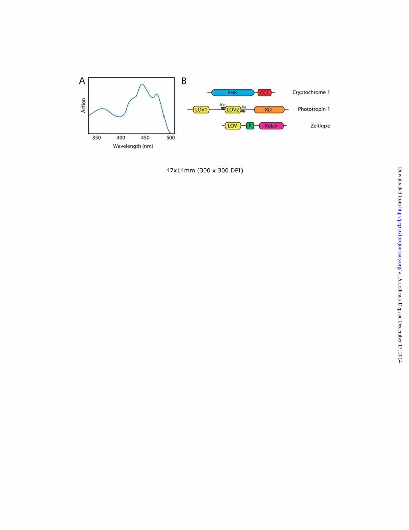
47x14mm (300 x 300 DPI)
at Periodicals Dept on D
ecember 17, 2014
http://pcp.oxfordjournals.org/D
ownloaded from

86x45mm (300 x 300 DPI)
at Periodicals Dept on D
ecember 17, 2014
http://pcp.oxfordjournals.org/D
ownloaded from
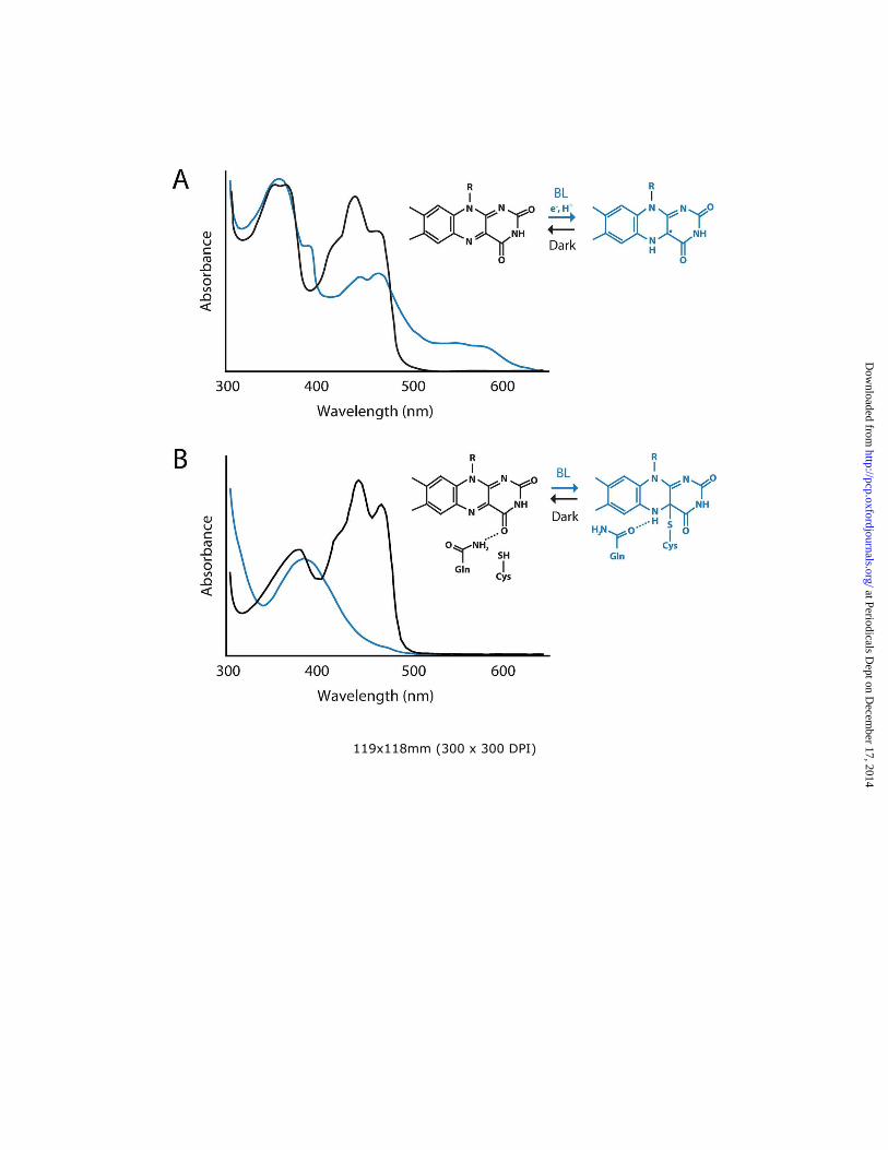
119x118mm (300 x 300 DPI)
at Periodicals Dept on D
ecember 17, 2014
http://pcp.oxfordjournals.org/D
ownloaded from

94x113mm (300 x 300 DPI)
at Periodicals Dept on D
ecember 17, 2014
http://pcp.oxfordjournals.org/D
ownloaded from