
Nitrosomonas eutropha: A Study of the Effects of ... · Nitrosomonas eutropha, as well as how...
27
JCCC Honors Journal Volume 7 Issue 1 Fall 2015 Article 3 Nitrosomonas eutropha: A Study of the Effects of Nitrosomonas on Pathogenic Bacterium and the Effects of Current Hygiene Habits on the Colonization of Nitrosomonas Within Our Normal Flora Shelli Kramer Johnson County Community College, [email protected] Follow this and additional works at: hp://scholarspace.jccc.edu/honors_journal is Article is brought to you for free and open access by the Honors Program at ScholarSpace @ JCCC. It has been accepted for inclusion in JCCC Honors Journal by an authorized administrator of ScholarSpace @ JCCC. For more information, please contact [email protected]. Recommended Citation Kramer, Shelli () "Nitrosomonas eutropha: A Study of the Effects of Nitrosomonas on Pathogenic Bacterium and the Effects of Current Hygiene Habits on the Colonization of Nitrosomonas Within Our Normal Flora," JCCC Honors Journal: Vol. 7: Iss. 1, Article 3. Available at: hp://scholarspace.jccc.edu/honors_journal/vol7/iss1/3
Transcript of Nitrosomonas eutropha: A Study of the Effects of ... · Nitrosomonas eutropha, as well as how...

JCCC Honors JournalVolume 7Issue 1 Fall 2015 Article 3
Nitrosomonas eutropha: A Study of the Effects ofNitrosomonas on Pathogenic Bacterium and theEffects of Current Hygiene Habits on theColonization of Nitrosomonas Within OurNormal FloraShelli KramerJohnson County Community College, [email protected]
Follow this and additional works at: http://scholarspace.jccc.edu/honors_journal
This Article is brought to you for free and open access by the Honors Program at ScholarSpace @ JCCC. It has been accepted for inclusion in JCCCHonors Journal by an authorized administrator of ScholarSpace @ JCCC. For more information, please contact [email protected].
Recommended CitationKramer, Shelli () "Nitrosomonas eutropha: A Study of the Effects of Nitrosomonas on Pathogenic Bacterium and the Effects ofCurrent Hygiene Habits on the Colonization of Nitrosomonas Within Our Normal Flora," JCCC Honors Journal: Vol. 7: Iss. 1, Article3.Available at: http://scholarspace.jccc.edu/honors_journal/vol7/iss1/3

Nitrosomonas eutropha: A Study of the Effects of Nitrosomonas onPathogenic Bacterium and the Effects of Current Hygiene Habits on theColonization of Nitrosomonas Within Our Normal Flora
AbstractNitrosomonas eutropha is a motile, gram-negative, bacillus that metabolizes ammonia as its energy source.Nitrosomonas eutropha was isolated from a probiotic skin product and used to determine how it wouldfunction in the normal flora of the integumentary system, and how it potentially benefits the human body. Theeffects of current hygiene products and practices on the ability of the Nitrosomonas eutropha to form andmaintain healthy colonies were also examined.
Cover Page FootnoteDr. Ellyn R. Mulcahy, PhD, MPH was the Faculty Advisor for this Honors contract. The research of formerstudents Heather Pointdexer, Araceli Hernandez Guerrero, Keith Kennedy, and Sarah Tonnies was utilized bythe author to complete the research contained in this paper.
This article is available in JCCC Honors Journal: http://scholarspace.jccc.edu/honors_journal/vol7/iss1/3

Introduction
The goals for the research included determining the effectiveness of the probiotic
skin product. It was determined through research that the bacterium present in the
solution was once a valuable part of the naturally existing colonization of beneficial
bacteria found on our skin. Due to modern hygienic practices, including more frequent
bathing, the use of harsher hygienic products, and antibacterial cleansers normal flora,
such as Nitrosomonas eutropha is not present in beneficial amounts within the flora of
the integumentary system. Additionally, reintroduction of Nitrosomonas eutropha would
reduce the need for frequent bathing and harsh hygienic products. Additional research
goals included determining how potentially pathogenic bacteria interacted with
Nitrosomonas eutropha, as well as how various hygienic products available today effect
the growth of Nitrosomonas eutropha and its colonization of the normal flora within the
integumentary system (1, 2, 5 ,7).
Testing revealed that Nitrosomonas eutropha is a highly motile bacterium. It
grows optimally in an acidic and dark environment at 230C. Nitrosomonas eutropha
metabolizes urea, as well as citrate and is a facultative anaerobe, growing both
aerobically and anaerobically. Nitrosomonas eutropha is a chemolithoautotroph, which
is a classification of microbes meaning they use CO2 for their primary carbon source
and power their metabolism using inorganic compounds. In the case of Nitrosomonas
eutropha, the inorganic compound is ammonia. Nitrosomonas eutropha also has the
capability to change pigmentation depending on the environment. This is due to the
release of chromophores when stressors are imposed upon the bacterium. Stressors
can be sub-optimal temperatures, which produced a different pigmentation than
Nitrosomonas eutropha grown in its optimal temperatures. The pH within the medium
also influenced the pigmentation of the growing Nitrosomonas eutropha as
demonstrated when grown while exposed to various soaps. When testing the
susceptibility of various pathogenic bacteria, it was determined that many of them had
significantly inhibited growth when Nitrosomonas eutropha are present. To explore why
Nitrosomonas eutropha have become extinct from the normal flora of our skin, we
examined the effects of various soaps, scrubs, and cleansers on Nitrosomonas
eutropha bacteria. It was found that many of the more natural cleansers such as pure
glycerin soaps, scrubs made with oatmeal, carrot, or other natural products either did
not inhibit the growth at all or did so minimally, allowing for regrowth in a matter of a
couple days. On the other hand, manufactured soaps such dial and dove more
significantly inhibited their growth and did not allow for regrowth for longer periods (3,
6).
1
Kramer: Nitrosomonas eutropha
Published by ScholarSpace @ JCCC,

Materials and Methods
To begin the project it was required that first the bacterium, Nitrosomonas
eutropha, found to be in the AO+ Biome samples, be isolated in order to determine that
it contains a bacterium of which is beneficial to the integumentary system, and once a
part of the normal skin flora. The method that allowed the bacterium to be isolated and
grown used 1000 microliters of the original sample, which had been growing in an
unknown nutrient solution incubated at 370C for ten days with no observable growth.
Samples from this concentration were then from this original sample and used to
inoculate incremental pH broth tubes of pH values of 2,4,6,8, and 10. Since the
bacterium grows on human skin, as indicated by research, then it would prefer a more
acidic environment. After two days, observable growth within the pH 6 broth tubes was
seen. At this point, two more pH 6 broth tubes were inoculated from the original 1000-
microliter tube. At the same time, growth in salinity environment was tested. Tubes
with salinities of 0%, 5%, 10%, 15%, and 20% were then inoculated using growth from
the initial pH 6 broth containing visible growth and incubated for five days at 370C (2, 7).
Once the optimal growing environment had been determined and several healthy
colonies were available in pH 6 broth, the process of getting the Nitrosomonas eutropha
to grow on Nutrient Agar plates began. The first growth of Nitrosomonas eutropha on a
Nutrient Agar plate was established by using four loops of samples from the established
growth in the pH 6 broth and incubated in the incubator at 370C for three days. Once
healthy lawns of the Nitrosomonas eutropha were established, the inoculation of streak
plates was begun in order to obtain and observe isolated colonies. At the same time,
each week new pH 6 broth tubes and Nutrient Agar plates were inoculated utilizing
samples from the growth established on the previously inoculated Nutrient Agar plate
showing good growth. The inoculation of fresh pH 6 broth tubes and Nutrient Agar
plates each week from previous week’s growth continued throughout the time in the lab.
It was found that the best way to obtain and view individual colonies on a streak plate
was to use a sterile loop to inoculate streaks one, two, and three then use a sterile
needle to inoculate streak four. A new sterile loop was used for each streak on the
streak plate.
Due to time restrictions and need to preserve the organisms, some of the
samples were placed in the refrigerator to slow down the metabolism and growth of the
Nitrosomonas eutropha. It was after refrigerating the samples that pigmentation
changes were noticed in the growth and determination was made that the Nitrosomonas
eutropha produces a chromophore. Further research on the Nitrosomonas eutropha
was conducted to determine why chromophores were being produced and what other
environmental conditions could cause this effect. This research also yielded information
regarding the Nitrosomonas eutropha’s preference for lower growing temperatures that
was put in place for future incubations (3, 6, 7).
2
JCCC Honors Journal, Vol. 7 [], Iss. 1, Art. 3
http://scholarspace.jccc.edu/honors_journal/vol7/iss1/3

After observable growth of the Nitrosomonas eutropha had been established
both in broth media and on Nutrient Agar, slides were prepared with the best results in
staining methods resulting from adjusting the traditional staining procedures. The ideal
staining procedures to obtain the best views of the Nitrosomonas eutropha and its
morphology as shown on Slide 1E (Figure 6), was using a sterile loop full of sample
from a well-colonized lawn that was incubated at room temperature in the dark and
transferring a heavy inoculation to the single drop of distilled H2O on the slide and heat
fixing it to the slide. Once the sample is heat fixed to the slide the stains were applied
as follows: crystal violet for one minute, rinse with distilled H2O, iodine for one minute,
rinse with distilled H2O, and then safranin for one minute, and then a final rinse with
distilled H2O before drying for viewing. Slide 2E (Figure 7) was prepared in the exact
same manner as slide 1E with the only difference being the time each of the stains was
allowed to remain on the slide. Instead of one minute per stain, two minutes per stain
was used in order to create a more distinct image of the bacterial cells. The staining
procedure for slide 2A (Figure 8) was as follows: used one loopful of sample from pH 6
broth tubes that had been in the refrigerator heat fixed to the slide with one drop of
distilled H2O; on top of that two drops of the same pH 6 broth containing the cultured
Nitrosomonas eutropha was placed on top of the heat fixed sample using a sterile pipet
and heat fixed on top of the first sample on the slide. Once heat fixed, the crystal violet
was placed on the slide for one minute then rinsed with distilled H2O, next was iodine
for one minute then rinsed with distilled H2O, and finally safranin for one minute then
final rinse with distilled H2O and air dried for viewing under the microscope.
Additional testing to determine metabolism, motility, and respiration of the
Nitrosomonas eutropha were conducted. All testing was done using either a sterile loop
or needle, taking samples from fresh healthy growth, and incubated at 230C in a dark
environment. A control of the exact same media/test was used as the inoculated
media/test and was incubated un-inoculated at the same time and in the same
environment as the inoculated test to use for comparison of results. The Citrate test
was inoculated using a sterile needle and sample from healthy growth on a lawn
inoculated seven days prior, and then the Citrate tubes were allowed to incubate at
room temperature in a darkened environment for five days. The same procedure that
was used to inoculate the Citrate test was used to inoculate the Sulfide-Indole-Motility
(SIM) tubes to test for motility, and utilization of sulfur, in addition the sample used for
the SIMs test came from the same lawn that was used to provide the sample for the
Citrate test. The rapid urea test was also inoculated in the exact same manner as the
aforementioned tests. Tryptic Soy Agar (TSA) slant was inoculated using a sterile
needle containing a sample of Nitrosomonas eutropha from a healthy lawn inoculated
fourteen days prior to stab the center of the slant agar to just above the bottom of the
slant tube and inoculating the upper part of the slant using a zig zag pattern. Phenol
Red with Dextrose and Phenol Red with Lactose tests were inoculated using sterile
3
Kramer: Nitrosomonas eutropha
Published by ScholarSpace @ JCCC,

loops and healthy samples from lawn inoculated seven days prior. The Enterotube II
was inoculated using healthy samples from a lawn of Nitrosomonas eutropha inoculated
five days prior. Once the initial readings on the Enterotube II were made without the
regents nine days after the inoculation of the Enterotube II, then Kovac’s regents were
added to the indicated space for the H2S/Indole and additional readings taken. The
Vogues-Proskauer (VP) testing was performed using a sterile loop to inoculate the VP
broth tubes and the Nitrosomonas was allowed to grow in the VP broth for nine days.
With good visible growth observed in the VP broth a sterile pipet was used to transfer 1
mL of the gently agitated VP broth into a new test tube and added five drops of Methyl
Red regent. Observations were immediately made and recorded. A second new test
tube was obtained and 1 mL of solution containing Nitrosomonas eutropha growth from
the VP broth was placed into this tube using a sterile pipet. To this tube of 1 mL of VP
broth containing Nitrosomonas eutropha growth fifteen drops of regent A (α-napthol)
and regent B (KOH 40%) were added. Observations were taken and recorded on this
tube every ten minutes for one hour. A Nitrate reduction test was conducted to show if
and how Nitrosomonas eutropha utilized or reduced nitrate.
Since the product stated that the Nitrosomonas eutropha was beneficial
bacterium for humans, present in the normal flora of the skin, the next step was to test
how it reacted with various available possibly pathogenic strains of bacteria. The
testing of the Nitrosomonas eutropha’s ability to inhibit potentially pathogenic bacteria
was tested using the following protocol: sterile Kirby Bauer disc were saturated in broth
containing Nitrosomonas eutropha and then placed using sterile tweezers onto the
center of a Nutrient Agar (NA) plate which was freshly inoculated in a with a pathogenic
bacteria in a lawn pattern. Once the NA plate was inoculated with a pathogenic bacteria,
and had a Nitrosomonas eutropha saturated disc placed in the center the NA plates
were then incubated at 370C in the incubator, because this temperature is the
pathogenic bacteria’s optimal growing temperature. The possibly pathogenic bacteria
species used for this experiment are Staphylococcus aureus, Micrococcus luteus,
Escherichia coli, and Staphylococcus epidermidis. A blank un-inoculated disc was used
for a control. For a comparison between the inhibition of the pathogenic bacterium with
Nitrosomonas eutropha and inhibition of pathogenic bacterium with antiseptics, an
experiment using disc soaked in bleach for one set of lawns innoculated with
pathogenic bacterium, 70% isopropol alcohol for another set, and water on a third for
control was conducted.
The next stage of this research project involves testing how various soaps and
hygienic products effect the growth of Nitrosomonas eutropha and the healthy
colonization of the normal flora of the human skin. It was determined that the best
method was an adaptation of procedures used in another experiment by Jen Ruble by
modifying it for sterile technique, the soaps being tested, as well as the use of Kirby
4
JCCC Honors Journal, Vol. 7 [], Iss. 1, Art. 3
http://scholarspace.jccc.edu/honors_journal/vol7/iss1/3

Bauer disc allowing for testing of the hygienic products to determine how much they
inhibited the growth of the Nitrosomonas eutropha. The procedure followed when
testing the soaps involved saturating a sterile Kirby Bauer disc in the liquid soap to be
tested, using sterile tweezers to place a bank, sterile Kirby Bauer disc into a paper cup
containing the liquid soap to be tested. Then using a newly sterilized pair of tweezers to
pick up the saturated Kirby Bauer disc and placing it in the center of a newly inoculated
lawn of Nitrosomonas eutropha, sealing the NA plate and incubating it at 230C in a dark
environment. For the two soap bars that were tested, the procedure was to use a sterile
scalpel to cut off a small sliver measuring 17 mm by 10 mm of each soap to be tested.
Then using sterile tweezers or the sterile scalpel to transfer it to the center of a newly
inoculated lawn of Nitrosomonas eutropha, seal the plate, and incubate it at 230C in a
dark environment. Observations made, recorded, and pictures taken of results at
seventy-two hours post inoculation and at one-hundred and twenty hours post
inoculation. The soaps that tested are, with brand name first then product name, are as
follows: Yes! To Carrots Daily Facial Cleaner, Dove Deep Moisture Liquid Body Wash,
Yardley London Oatmeal and Almond Bar Soap, Nirvana Spa NSPA Real Fruit
Goodness Exotically Creamy Coconut Rich Body Butter, Clearly Natural Pure Glycerin
Soap (unscented), Freeman Charcoal and Black Sugar Polishing Mask, Yes to
Tomatoes Clear Skin Acne Daily Pore Scrub, Tree Hut SHEA Sugar Scrub with Almond
and Honey, SoftSoap Liquid Milk and Honey Hand Soap, and Dial Liquid Springwater
Antibacterial Soap (4).
Results
When testing the optimal environmental pH for the growth of the Nitrosomonas
eutropha, the pH that yielded the best growth of Nitrosomonas eutropha was the broth
with a pH level of 6 as seen in Figure 1.
5
Kramer: Nitrosomonas eutropha
Published by ScholarSpace @ JCCC,

The results of testing the optimal salinity of
the environment for the growth of Nitrosomonas
eutropha concluded that the ideal salinity range
was 0%-5% salinity as shown in Figure 2 below.
When working on establishing growth of the Nitrosomonas eutropha on a
Nutrient Agar plate adequate growth was observed at 370C. However, it was later
determined that optimal growth occurred at 230C in a dark environment. Growth on the
Nutrient Agar plate can be described as a clear mucoid growth that changes the
pigmentation of the media to a darker auburn color shown in Figure 3 below.
Nitrosomonas eutropha in pH 6
broth on the left and control on the
right. Note the turbid, pigmented
broth in the tube on the left
indicating the growth of the
Nitrosomonas eutropha.
Figure 1: Growth of Nitrosomonas
eutropha in a pH 6 broth
Figure 2: Optimal salinity for Nitrosomonas
eutropha growth.
Note the pigmentation and turbidity changes in
the 5% and 0% salinity tubes on the far right
compared to the other salinity tubes indicating
growth of the Nitrosomonas eutropha.
6
JCCC Honors Journal, Vol. 7 [], Iss. 1, Art. 3
http://scholarspace.jccc.edu/honors_journal/vol7/iss1/3
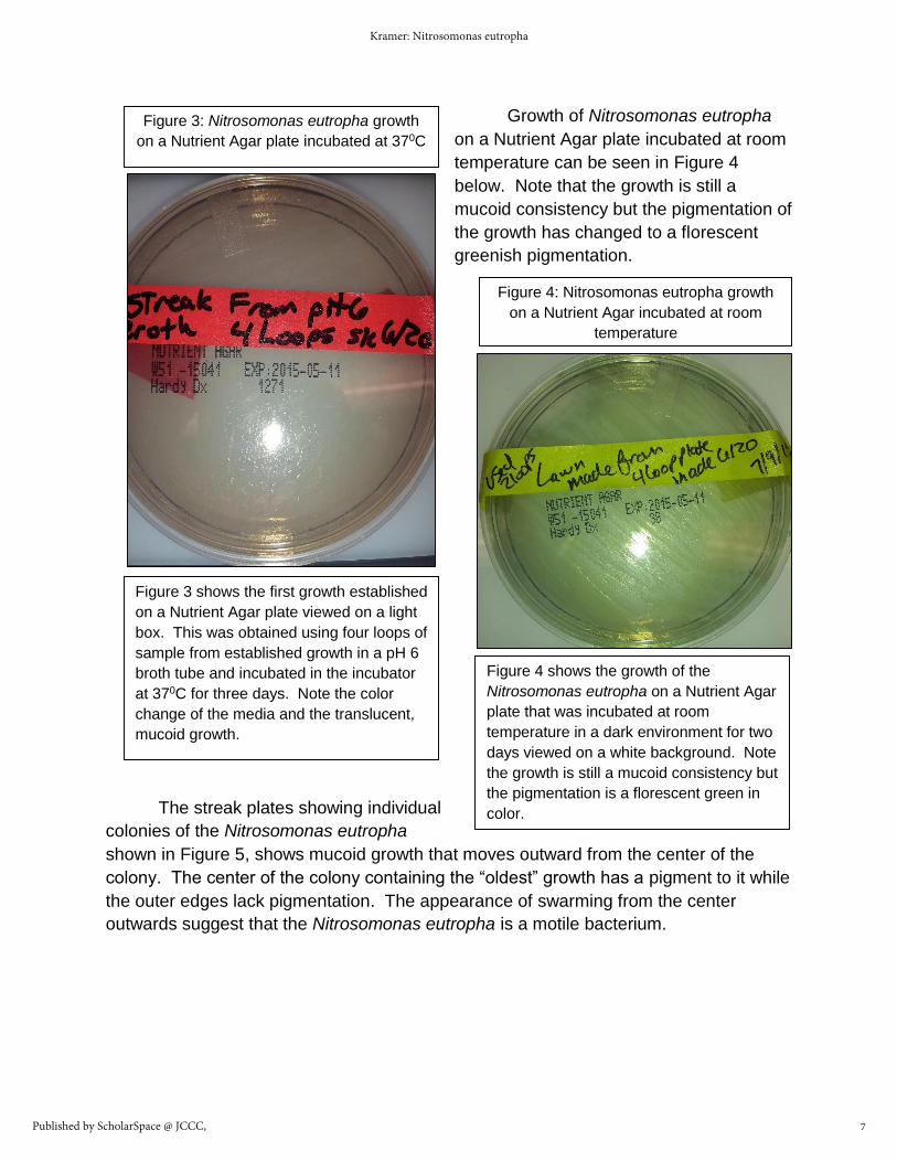
Growth of Nitrosomonas eutropha
on a Nutrient Agar plate incubated at room
temperature can be seen in Figure 4
below. Note that the growth is still a
mucoid consistency but the pigmentation of
the growth has changed to a florescent
greenish pigmentation.
The streak plates showing individual
colonies of the Nitrosomonas eutropha
shown in Figure 5, shows mucoid growth that moves outward from the center of the
colony. The center of the colony containing the “oldest” growth has a pigment to it while
the outer edges lack pigmentation. The appearance of swarming from the center
outwards suggest that the Nitrosomonas eutropha is a motile bacterium.
Figure 3: Nitrosomonas eutropha growth
on a Nutrient Agar plate incubated at 370C
Figure 3 shows the first growth established
on a Nutrient Agar plate viewed on a light
box. This was obtained using four loops of
sample from established growth in a pH 6
broth tube and incubated in the incubator
at 370C for three days. Note the color
change of the media and the translucent,
mucoid growth.
Figure 4: Nitrosomonas eutropha growth
on a Nutrient Agar incubated at room
temperature
Figure 4 shows the growth of the
Nitrosomonas eutropha on a Nutrient Agar
plate that was incubated at room
temperature in a dark environment for two
days viewed on a white background. Note
the growth is still a mucoid consistency but
the pigmentation is a florescent green in
color.
7
Kramer: Nitrosomonas eutropha
Published by ScholarSpace @ JCCC,

The slides that produced the
best images are labeled as Slide 1E,
2E, and 2A. Each slide shows some
unique features of the Nitrosomonas
eutropha. Slide 1E shows basic
morphology of the Nitrosomonas
eutropha as being a Gram-negative,
short rod bacterium seen in Figure 6.
Slide 2E shows an improved view and
contrast of the cell wall of the
Nitrosomonas eutropha as shown in
Figure 7 including the tendency of the
bacteria to clump together. Slide 2A
shows the changes that occur with the
Nitrosomonas eutropha when the
bacterium is exposed to a different
temperature shown in Figure 8. The
differences in appearance of the
bacterial cells between slides 1 and 2
E and slide 2A can be directly
attributed to the temperature and
media that the bacteria grew in.
Figure 5: Streak Plate Containing Isolated
Colonies of Nitrosomonas eutropha
Figure 5 shows the streak plate as viewed with
room lighting. Note how the growth in the
individual colonies is mucoid in appearance,
spreads out from the center, and the center of the
isolated colony is pigmented while the outer
edges remain clear.
Figure 6: Slide 1E Nitrosomonas eutropha
Slide 1E showing Nitrosomonas eutropha as a
short rod, gram-negative bacillus at 100X and no
zoom on the camera.
Figure 7: Slide 2E Nitrosomonas eutropha
Slide 2E showing an improved contrast of
the cell walls of the Nitrosomonas
eutropha as well as the clumping
behavior of the bacteria. Seen at 100X
magnification.
8
JCCC Honors Journal, Vol. 7 [], Iss. 1, Art. 3
http://scholarspace.jccc.edu/honors_journal/vol7/iss1/3

The Citrate test results show that
Nitrosomonas eutropha does utilize citrate for a
nutrient source as shown by the slight color
change from the blue of the control tube to a
more greenish color in the tube inoculated with
Nitrosomonas eutropha in Figure 9.
The results of the Sulfide-Indole Motility or SIMs testing shown in Figure 10,
indicates that Nitrosomonas eutropha is highly mobile and does not utilize sulfur. It was
also determined after the Kovac’s regent was added that Nitrosomonas eutropha does
not break down into indole and pyruvate as seen in Figure 11.
Figure 8: Slide 2A Nitrosomonas
eutropha
Slide 2A showing in the middle far left of
the picture what appears as a
florescence to the cell wall of the
Nitrosomonas eutropha when viewing
the cells that were obtained from a
sample that was refrigerated. This
change illustrates the chromophore
production capabilities of the
Nitrosomonas eutropha. Seen at 100X
Figure 9: Images of the Citrate Test
Citrate test shows a positive result in the
Nitrosomonas eutropha’s use of Citrate as a
nutrient source. The inoculated tube on the
right shows a change of color from the control
tube on the left indicating the positive test
result.
9
Kramer: Nitrosomonas eutropha
Published by ScholarSpace @ JCCC,

Results of the rapid urea testing determined that the
Nitrosomonas eutropha rapidly hydrolyzes urea and has a
strong production of urease in the presence of urea. This is
shown by the color change in the inoculated media compared
to the control as seen in Figure 12.
The Tryptic Soy Agar (TSA)
testing for motility and respiration
confirms that Nitrosomonas eutropha is
highly motile, capable of respiring both
anaerobically and aerobically as shown
in Figure 13. The TSA test also confirms
that the Nitrosomonas eutropha is not in
the Enterobacteriaceae family.
Figure 10: Sulfide-Indole
Motility Test Prior to
Kovac’s Regent
Figure 10 to the left shows the
inoculated SIMs tube on the left of the
image demonstrating Nitrosomonas
eutropha to be highly motile. The
control, un-inoculated tube on the
right compared to the inoculated tube
shows that no sulfur is utilized due to
no color change in the media.
Figure 11: Sulfide-
Indole Motility Test
After Addition of
Kovac’s Regent
Figure 11 to the right shows the
inoculated tube on the left and the un-
inoculated control tube on the right.
The SIMs test after the addition of
Kovac’s regent shows that
Nitrosomonas eutropha does not break
down Indole into pyruvate due to the
lack of color change in the media.
Figure 12: Rapid
Urea Test
Positive result on the
Rapid Urea test as
shown by the color
change in the
inoculated media on
the right compared to
the control on the left.
Figure 13: Tryptic
Soy Agar Test
Figure 13 to the right shows the control for
the TSA test on the left and the inoculated
TSA tube is on the right as evidenced by
the growth into the media and outward
from the stab inoculation with
Nitrosomonas eutropha. This growth
demonstrates the bacterium’s ability
respire anaerobically and the growth on
the upper portion of the slant demonstrates
the ability to respire aerobically.
10
JCCC Honors Journal, Vol. 7 [], Iss. 1, Art. 3
http://scholarspace.jccc.edu/honors_journal/vol7/iss1/3

Phenol Red with Dextrose testing, shown in Figure 14 resulted in an orange color
and tiny bubbles in the Durham tube giving a slightly positive result for dextrose
fermentation and gas production. The Phenol Red with Lactose test, shown in Figure
15, is orange in color with no bubble
indicating again a very slight positive
result for lactose fermentation and no
gas production during the metabolism
and fermentation of lactose.
The results of the Enterotube II after nine days of incubation are shown in Table
1 below. Images for the Enterotube II results prior to the addition of the Kovac’s regent
and after the addition of the regent are shown in Figures 16, 17, and 18 on the following
page.
Table 1: Results of Enterotube II Inoculated with Nitrosomonas eutropha
Test Positive or Negative What Result Means
Glucose Negative Does not ferment glucose
Lysine Negative Does not decarboxylate lysine
Ornithine Negative Does not decarboxylate ornithine
H2S/Indole Positive/Negative Reduces sulfur and does not produce Indole
Adonitol Negative Does not ferment Adonitol
Lactose Negative Does not ferment Lactose
Arabinose Negative Does not ferment Arabinose
Sorbitol Negative Does not ferment Sorbitol
VP Negative result Did have a greenish beige color which was different than expected but NOT a positive result
Figure 14: Phenol
Red with Dextrose
Figure 14 to the left shows the color change
in the tube on the left inoculated with
Nitrosomonas eutropha to an orange color
compared to the control on the right, which
remains red.
Figure 15: Phenol
Red with Lactose
Figure 15 to the right shows the color
change in the tube to the right inoculated
with Nitrosomonas eutropha to an orange
color while the control is on the left remains
red.
11
Kramer: Nitrosomonas eutropha
Published by ScholarSpace @ JCCC,

The Vogues-
Proskauer broth that is used
for the Methyl Red test and
the Vogues-Proskauer (VP)
test shows good growth of
Nitrosomonas eutropha as
seen in Figure 19. No color
change as seen in Figure 20
on the following page, once
the Methyl Red was added to the tube containing 1 mL of VP broth colonized with
Nitrosomonas eutropha demonstrates no mixed acid fermentation took place. The
Vogues-Proskauer (VP) testing revealed that there is no mixed acid fermentation during
the metabolic processes of Nitrosomonas eutropha. This is evidenced by the lack of
notable color change once fifteen drops of α-napthol regent and fifteen drops of KOH
40% regent were added to the VP broth containing Nitrosomonas eutropha shown in
Figure 21, and there continued to be no color change checked every ten minutes over a
Figure 16: Enterotube II Inoculated with Nitrosomonas eutropha Prior to Addition of Kovac’s
Regent with Labels Visible (below)
Figure 17: Enterotube II Inoculated with Nitrosomonas eutropha Prior to Addition of Kovac’s
Regent Showing the Backside of the Individual Cells with Room Light in Background (below)
Note: Image is the reverse of Figure 16
Figure 18: Enterotube II H2S/Indole Cell After Kovac’s
Regent is Added (below)
12
JCCC Honors Journal, Vol. 7 [], Iss. 1, Art. 3
http://scholarspace.jccc.edu/honors_journal/vol7/iss1/3
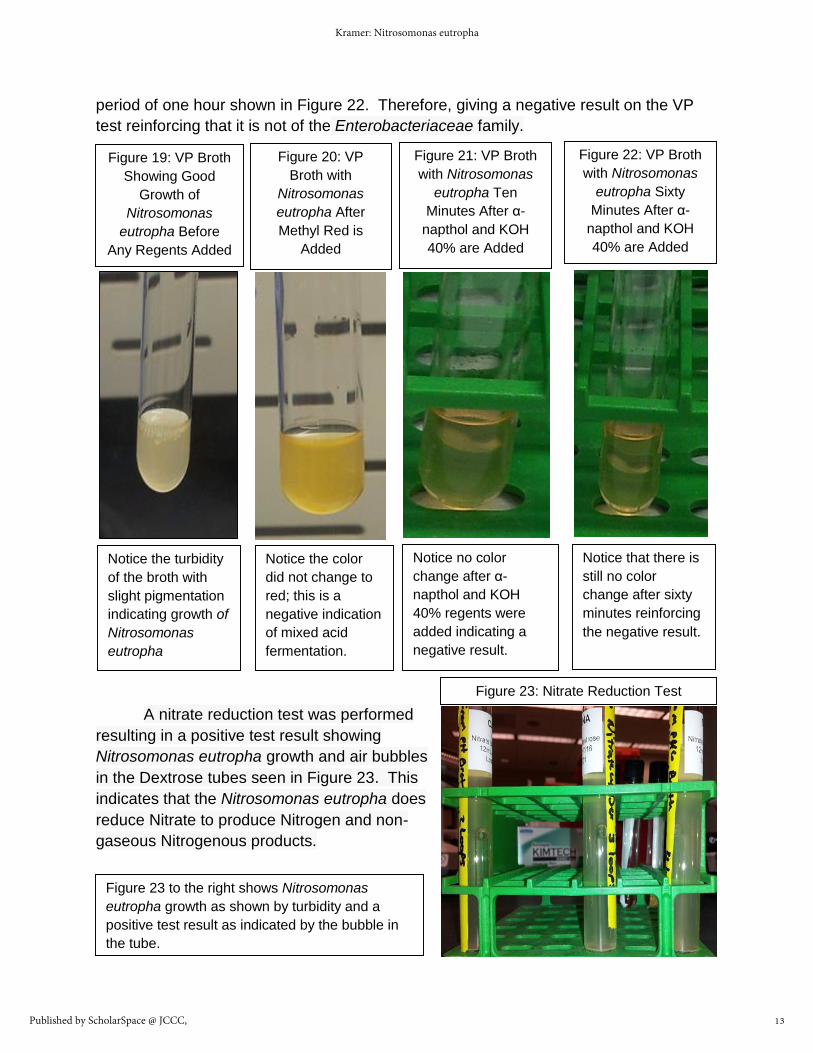
period of one hour shown in Figure 22. Therefore, giving a negative result on the VP
test reinforcing that it is not of the Enterobacteriaceae family.
A nitrate reduction test was performed
resulting in a positive test result showing
Nitrosomonas eutropha growth and air bubbles
in the Dextrose tubes seen in Figure 23. This
indicates that the Nitrosomonas eutropha does
reduce Nitrate to produce Nitrogen and non-
gaseous Nitrogenous products.
Figure 19: VP Broth
Showing Good
Growth of
Nitrosomonas
eutropha Before
Any Regents Added
Notice the turbidity
of the broth with
slight pigmentation
indicating growth of
Nitrosomonas
eutropha
Figure 20: VP
Broth with
Nitrosomonas
eutropha After
Methyl Red is
Added
Notice the color
did not change to
red; this is a
negative indication
of mixed acid
fermentation.
Figure 21: VP Broth
with Nitrosomonas
eutropha Ten
Minutes After α-
napthol and KOH
40% are Added
Notice no color
change after α-
napthol and KOH
40% regents were
added indicating a
negative result.
Figure 22: VP Broth
with Nitrosomonas
eutropha Sixty
Minutes After α-
napthol and KOH
40% are Added
Notice that there is
still no color
change after sixty
minutes reinforcing
the negative result.
Figure 23: Nitrate Reduction Test
Figure 23 to the right shows Nitrosomonas
eutropha growth as shown by turbidity and a
positive test result as indicated by the bubble in
the tube.
13
Kramer: Nitrosomonas eutropha
Published by ScholarSpace @ JCCC,

The results of the testing of whether or not the Nitrosomonas eutropha inhibited
the growth of potentially pathogenic bacteria showed that the disc soaked in the
Nitrosomonas eutropha did indeed inhibit the growth of all of the species of bacterium
tested as seen in Figures 24-27. The inhibition zones are shown in Table 2.
Figure 24: Staphylococcus
aureus Lawn with
Nitrosomonas eutropha
Saturated Disc
The Zone of Inhibition
measures 26 mm and a
florescent green
pigmentation is observed
where it appears the
Nitrosomonas eutropha is
growing into the
Staphylococcus aureus.
Figure 25: Micrococcus
luteus Lawn with
Nitrosomonas eutropha
Saturated Disc
The Zone of Inhibition
measures 24 mm and an
auburn pigmentation is
noted. There appears to be
a area of no growth of
Micrococcus luteus with
regrowth around the disk.
Figure 26: Escherichia coli
Lawn with Nitrosomonas
eutropha Saturated Disc
The Zone of Inhibition
measures 30 mm and a
florescent green
pigmentation is noted.
The Nitrosomonas
eutropha appears to be
growing into the
Escherichia coli.
Figure 27: Staphylococcus epidermidis Lawn
with Nitrosomonas eutropha Saturated Disc
The Zone of Inhibition for the Figure to the
left measures 36 mm and a florescent green
pigmentation is observed. It appears that the
Nitrosomonas eutropha is growing into the
Staphylococcus epidermidis.
14
JCCC Honors Journal, Vol. 7 [], Iss. 1, Art. 3
http://scholarspace.jccc.edu/honors_journal/vol7/iss1/3

Organism ZOI in mm Pigmentation
Staphylococcus aureus
26 mm Florescent Green
Micrococcus luteus 24 mm Auburn
Escherichia coli 30 mm Florescent Green
Staphylococcus epidermidis
36 mm Florescent Green
Table 2: Zones of Inhibition for
Pathogenic Bacteria
Figure 28: Comparison of the Inhibition of Pathogenic Bacterial Species between the
Use of Antiseptics and Nitrosomonas eutropha with a Control Shown
In Figure 28 above, a comparison is shown in how the potentially pathogenic bacteria is
inhibited in one of three ways with the control on the bottom row. The top row is the
pathogenic bacterial lawn with the Nitrosomonas eutropha saturated Kirby Bauer disc. The
second row from the top is the pathogenic bacterial lawn with a bleach soaked Kirby Bauer
disc. The third row down is pathogenic bacterial lawn with a 70% isopropyl alcohol saturated
Kirby Bauer disc. Again, the very bottom row is the control, a pathogenic bacterial lawn with
a sterile Kirby Bauer disc in the center. The pathogenic bacterial laws are from right to left:
Escherichia coli, Micrococcus luteus, Staphylococcus aureus, and Staphylococcus
epidermidis.
15
Kramer: Nitrosomonas eutropha
Published by ScholarSpace @ JCCC,

The results for the comparison between how Nitrosomonas eutropha inhibits
pathogenic bacterica and how antiseptics such as bleach and isopropol alcohol inhibit
the growth of the same pathogenic bacterium is shown in Figure 28 at the bottom of the
previous page.
The pigmentation changes that are seen in the pictures throughout the paper are
the result of the fact that Nitrosomonas eurtropha produce a chromophore and exhibit
pigmentation changes when subjected to tempurature, pH, and other enviornmental
stressors. Pigmentation can range from a greenish color to a yellow, or even deep
auburn.
The results of the different soaps ability to inhibit the growth of the Nitrosomonas
eutropha are shown by a Zone of Inhibition Table at seventy-two hours out from
inoculation that includes descriptions and active ingredients for each soap shown as
Table 3, and a second Table showing Zones of Inhibition and descriptions at one-
hundred twenty hours post inoculation shown in Table 4. In addition Figures 29–38
show the interaction between the soaps and the Nitrosomonas eutropha at seventy-two
hours and Figures 39-48 show the interaction between the soaps and the Nitrosomonas
eutropha at one-hundered twenty hours post inoculation.
The results of the testing of the soaps revealed additional chemolithoautotrophic
characteristics in the form of a wider range of pigmentation even though the
temperature and other environmental conditions remained constant. This leads to the
hypothesis that the environmental stressors of the Nitrosomonas eutropha are due to
the pH of the soaps as well as the ingredients found in the soaps and their chemical
reactions with the Nitrosomonas eutropha. This was evidenced by the most drastic color
changes taking place with soaps that were high in sugars or other carbohydrates such
as the Sugar scrubs and Oatmeal scrubs which yielded a more auburn pigment in the
growth around the disc saturated with soap, while other soaps resulted in a white to a
yellowish-green pigmentation. The fewer the ingredients and the more natural the
ingredients overall the less inhabitation of the Nitrosomonas eutropha was observed
and if inhibited at all the faster it grew back into the inhibition zone. In addition, the
more natural and fewer the ingredients in the soaps the lighter and less pigmentation
was observed in the Nitrosomonas eutropha growth.
16
JCCC Honors Journal, Vol. 7 [], Iss. 1, Art. 3
http://scholarspace.jccc.edu/honors_journal/vol7/iss1/3

Table 3: Inhibition of Nitrosomonas eutropha growth at 72 hours with soaps
Soap Zone of Inhibition
In mm
Pigmentation Top Five Ingredients
Softsoap Milk and Honey hand soap
3-5 mm None Water, Sodium C14-16 Olefin Sulfonate, Laureth-3, Cocamidopropyl Betaine, Glycol Stearate…
Dial Liquid Springwater Antibacterial soap
2 mm Solid white-auburn ring
Active: Benzethonium Chloride 0.10% Inactive Top Five: Water, Cetrimonium Chloride, Glycerin, Lauramine Oxide, Sodium Chloride…
Dove Deep Moisture Liquid Body Wash
0 mm Cream colored Water, Cocamidopropyl Betaine, Sodium Hydroxpropl Starch Phosphate, Lauric acid, Sodium Lauroyl Glycinate…
Yes! To Tomatoes Clear Skin Acne Daily Pore Scrub
0 mm None Active: Salicylic acid 2% Inactive Top Five: Water, Stearic Acid, Propanediol, Sodium Stearate, Glycerin…
Charcoal and Black Sugar Polishing Mask
0 mm Florescent Green Sucrose, Propylene Glycol, Carbon (activated charcoal), Kaolin, musa sapientum (banana)…
SHEA Sugar Scrub with Almond and Honey
2-5 mm Auburn pigment furthur from the disc
Sucrose, glycerin, polysorbate 20, silica, honey…
Exotically Creamy Coconut Rich Body Butter
0 mm None Water, cetearyl alcohol, Paraffinum liquidum (mineral oil), Cyclopentasiloxane, Glyceryl stearate SE…
Yes! To Carrots Daily Facial Cleanser
0 mm None to cream colored
Water, Glycerin, Disodium Cocomphodipropionate, stearic acid, glyceryl…
17
Kramer: Nitrosomonas eutropha
Published by ScholarSpace @ JCCC,

Clearly Natural Pure Glycerin Bar Soap (unscented)
Irregular 1-5 mm Translucent green to an auburn pigment
Glycerin, propylene glycol, sodium stearate, Pecyl Glucoside, Sorbitol…
Yardley London Oatmeal and Almond Bar Soap
Irregular 1-5 mm Translucent green to auburn pigment
Sodium Tallowate, water, sodium cocoate, glycerin, Fragrance…
Table 4: Inhibition of Nitrosomonas eutropha growth or evidence of regrowth with
soaps at 120 hours
Soap Zone of Inhibition in mm
Pigment Regrowth observed
Softsoap Milk and Honey hand soap
4-6 mm Cream to Auburn colored
Small specs of what appears as individual colonies around a disc of white in the form of tiny white specs
Dial Liquid Springwater Antibacterial soap
2 mm Solid white to auburn
None
Dove Deep Moisture Liquid Body Wash
Up to 5 mm auburn Shows new growth up to 5 mm from disc with a greenish pigment
Yes! To Tomatoes Clear Skin Acne Daily Pore Scrub
0 mm Cream to light auburn
None
Charcoal and Black Sugar Polishing Mask
0 mm Translucent to deep auburn
None
SHEA Sugar Scrub with Almond and Honey
0 mm Deep auburn to purplish color
Growth has moved into the ring observed at 72 hours
Exotically Creamy Coconut Rich Body Butter
0 mm Slight auburn to translucent
None
Yes! To Carrots Daily Facial Cleanser
0 mm Cream colored to translucent
None
18
JCCC Honors Journal, Vol. 7 [], Iss. 1, Art. 3
http://scholarspace.jccc.edu/honors_journal/vol7/iss1/3

Clearly Natural Pure Glycerin Bar Soap (unscented)
2-4 mm Darker auburn pigmentation then at 72 hours.
Appears to have a ring of concentrated growth around the sliver of soap measuring 2-4 mm out
Yardley London Oatmeal and Almond Bar Soap
1-2 mm Translucent to greenish pigmentation
There appears to be white mucoid growth on the sliver of soap and 1-2 mm of heavy growth around the soap.
Figure 29: Nitrosomonas
eutropha Lawn with a
Softsoap Milk and Honey
Kirby Bauer Disc at 72 Hours
Good growth is observed up
to approximately 3 mm from
the disc when the growth is
more sporadic or absent.
Notice the auburn pigment of
the growth.
Figure 30: Nitrosomonas
eutropha Lawn with a
Liquid Dial Kirby Bauer
Disc at 72 Hours
Good growth is observed
up to the 2 mm wide
solid white-auburn ring
around the disc
Figure 31: Nitrosomonas
eutropha Lawn with a Kirby
Bauer Disc saturated with
Dove Liquid soap at 72 Hours
Good growth is observed
throughout the plate having a
cream-colored pigmentation
with a thick area of growth
around the disc
19
Kramer: Nitrosomonas eutropha
Published by ScholarSpace @ JCCC,

Figure 32: Nitrosomonas
eutropha Lawn with a Yes to
Tomatoes Kirby Bauer disc
at 72 hours
Notice the excellent auburn
tinted growth throughout the
plate, even right up to the
disc saturated in Tomato
scurb
Figure 33: Nitrosomonas
eutropha Lawn with a
Charcoal and Black Sugar disc
at 72 hours
Notice the heavy pigmentation
ranging from deep green to a
deep auburn in the growth on
the media and heavy growth
up to the disc
Figure 34: Nitrosomonas
eutropha Lawn with a SHEA
Sugar Scrub disc at 72
Hours
Notice the good growth
throughout the plate with
auburn pigmentation of
growth further from the disc
and greenish pigmentation
closer to the disc.
Figure 35: Nitrosomonas
eutropha Lawn with a
Coconut Body Butter disc at
72 Hours
Notice the excellent growth
with no pigmentation to a
slight cream color even right
up to the disc
Figure 36: Nitrosomonas
eutropha Lawn with a Yes to
Carrots disc at 72 Hours
Notice the uninterrupted
growth right up to the disc that
has no pigmentation to a
slight cream color
Figure 37: Nitrosomonas
eutropha Lawn with a sliver
of Pure Glycerin Bar soap
at 72 Hours
Notice the thick healthy
green to auburn growth
extending from the sliver
of soap
20
JCCC Honors Journal, Vol. 7 [], Iss. 1, Art. 3
http://scholarspace.jccc.edu/honors_journal/vol7/iss1/3

Figure 38: Nitrosomonas
eutropha Lawn with a sliver
of Oatmeal and Almond Bar
soap at 72Hours
Notice the irregular heavy
growth around the sliver of
soap, which has a green to
auburn pigmentation
depending on the distance
from the soap sliver.
Figure 39: Nitrosomonas
eutropha Lawn with
Softsoap Milk and Honey
disc at 120 Hours
Notice continued good
growth throughout the plate
with auburn pigmentation
and new growth forming in
a ring around the disc
Figure 40: Nitrosomonas
eutropha Lawn with Liquid
Dial disc at 120 Hours
Notice solid white to auburn
tinted ring around the disc,
continued growth at outer
edges of the plate with no
pigmentation in the growth
beyond and no new growth
observed
Figure 41: Nitrosomonas
eutropha Lawn with Liquid
Dove disc at 120 Hours
Notice the green
pigmentation in the new
growth around the disc and
the auburn pigmentation in
the growth further out
Figure 42: Nitrosomonas
eutropha Lawn with Tomato
Scrub disc at 120 Hours
Notice the healthy mucoid
growth with no
pigmentation changes even
against the disc containing
the tomato scrub
Figure 43: Nitrosomonas
eutropha Lawn with Charcoal
and Black sugar disc at 120
Hours
Notice the health growth of
the throughout the plate,
even right up to the disc also
note the darker auburn
pigmentation
21
Kramer: Nitrosomonas eutropha
Published by ScholarSpace @ JCCC,

Figure 44: Nitrosomonas
eutropha Lawn with SHEA
Sugar scrub disc at 120
Hours
Notice the thick growth and
dark auburn pigmentation.
There appears to be regrowth
into the area that was
considered the ZOI at 72
hours.
Figure 45: Nitrosomonas
eutropha Lawn with Coconut
Body Butter disc at 120
Hours
Notice the continued healthy
growth even up to the disc.
The pigmentation has
changed slightly in some
areas to a slight auburn
pigmentation
Figure 46: Nitrosomonas
eutropha Lawn with Carrot
scrub disc at 120 Hours
Notice the continued healthy
growth even up to the disc
and no pigmentation
changes.
Figure 47: Nitrosomonas eutropha
Lawn with sliver of Glycerin Bar
Soap at 120 Hours
Notice the darker auburn pigment
and loss of the green pigment in
the growth compared to 72 hours.
Also, notice the heavier growth
seen around the sliver of soap
extending out 2-4 mm from the
sliver of soap
Figure 48: Nitrosomonas eutropha
Lawn with sliver of Oatmeal and
Almond soap at 120 Hours
Notice that the growth further
from the sliver of soap is a slight
auburn pigmentation while closer
to the sliver it is a greenish
pigment. Note increased growth
1-2 mm out from the sliver.
22
JCCC Honors Journal, Vol. 7 [], Iss. 1, Art. 3
http://scholarspace.jccc.edu/honors_journal/vol7/iss1/3

Discussion
All testing, research, and experimentation leads to the conclusion that the AO+
Biome product does indeed contain the bacteria Nitrosomonas eutropha, which are a
highly motile, gram-negative, short rod-shaped, ammonia oxidizing bacteria.
Nitrosomonas eutropha have chromolitoautophic characteristics and is able to respire in
both anaerobic and aerobic environments. Nitrosomonas eutropha prefers a slightly
acidic environment with a pH of 6, it grows well in salinities of 0-5%, and grows the best
in temperatures between 200C and 240C. Nitrosomonas eutropha is a nitrate oxidizing
bacteria producing nitrogen and non-gaseous nitrogenic byproducts. Nitrosomonas
eutropha hydrolyzes urea and produces urease in the presence of urea. Nitrosomonas
eutropha does utilize citrate as a nutrient source, and will ferment arabinose, a naturally
occurring monosaccharide found in the products of the hydrolysis of various wood-
based materials or other biomasses. This is fitting as Nitrosomonas eutropha naturally
lives in the soil as well as within the normal flora of our skin.
Nitrosomonas eutropha significantly inhibits the growth of the potentially
pathogenic bacterium Staphylococcus epidermidis, and Staphylococcus aureus, and a
moderate inhabitation of Escherichia coli and Micrococcus luteus. Further research and
experimentation with additional species of bacteria could expand on the list of
potentially pathogenic bacteria that Nitrosomonas eutropha inhibits. This leads to the
conclusion that the Nitrosomonas eutropha is indeed beneficial to the natural flora of the
human skin.
When testing some of the soaps available on the market today, including some of
the more natural glycerin soaps used in previous generations, it was found that the
soaps with the most ingredients, that were more chemically based, and had
antibacterial ingredients inhibited the growth of the Nitrosomonas eutropha the greatest
with the slowest regrowth rate of the Nitrosomonas eutropha. Soaps that contained the
fewest and most natural ingredients such as the tomato scrub, Dove, and Oatmeal
scrub, etc., had little to no effect on the growth of the Nitrosomonas eutropha and if any
initial interruption of growth did occur, the regrowth of the Nitrosomonas eutropha was
rapid and abundant. When looking at the results of the soap testing and looking at
Tables 3 and 4, one must keep in mind that people wash their hands throughout the day
ad nauseum, and most people bathe daily or at the least every other day. The
experiment done on this small list of available soaps shows that those soaps that do
inhibit the growth of Nitrosomonas eutropha initially generally require an average of 2-4
days to regrow and repopulate the areas that were initially inhibited due to the soap
used. The idea of reintroducing the Nitrosomonas eutropha after bathing or washing
one’s hands through an artificial method such as a spray, cream, or other vehicle as
23
Kramer: Nitrosomonas eutropha
Published by ScholarSpace @ JCCC,

being beneficial over having to wait for the Nitrosomonas eutropha to repopulate the
skin naturally, is a very sound one. Most likely, there will not be enough time for the
Nitrosomonas eutropha to grow back on its own between bathing and handwashing to
provide a protective benefit as was shown when testing how the soaps inhibit the
growth of Nitrosomonas eutropha and regrowth was observed in period of 72 to 120
hours depending on the soap.
It would be advantageous to encourage further study on the benefits of
Nitrosomonas eutropha to humans via the repopulation and reintroduction of
Nitrosomonas eutropha to the normal flora of human skin. Researching the possible
significant benefits in recolonization of the human flora with the Nitrosomonas eutropha
such as the reduction of infections within the general population, infections caused by
drug resistant pathogens, and infections in immunocompromised individuals, such as
those who have cancer, immature or weakened immune systems due to autoimmune
disease, medication or other pathophysiological conditions that render the immune
system less than fully functional. Additionally, the reduction in infections among
agricultural workers both crop and livestock farmers because of their constant contact
with Escherichia coli and other pathogenic bacteria within their occupation, with a
possibility of also reducing the spread of disease within a heard on a farm where
individuals utilize products containing Nitrosomonas eutropha before, after, and in
between handling individual animals, could yield some important breakthroughs in
public health.
24
JCCC Honors Journal, Vol. 7 [], Iss. 1, Art. 3
http://scholarspace.jccc.edu/honors_journal/vol7/iss1/3
References
1. “Nitric Oxide Functions in Humans.” AOBiome. Web. March 2015.
http://aobiome.com/nitric-oxide-function-humans
2. “The Human Microbiome.” AOBiome. Web. March 2015.
http://aobiome.com/human-microbiome
3. Hommes, Norman G., Luis A. Sayavedra-Soto, and Daniel J. Arp.
“Chemolithooganotrophic Growth of Nitrosonomas europaea on Fructose.”
Journal of Bacteriology 185.23 (2003): 6809-6814. Web. 18 June 2015.
http://jb.asm.org/content/185/23/6809.full
4. Rubel, Jan. “Comparing the Effects of Commercial Antibacterial Soaps on
Bacteria.” 24 Feb 1997. Web. 26 Oct 2015.
http://web.horacemann.org/academics/science/expbio/webpages/rubel/bacteria.h
tml
5. Schmidt, Ingo, Rob J. M. van Spanning, and Mike S. M. Jetten. “Denitrification
and Ammonia Oxidation by Nitrosonomas eruopaea Wild Type, and NirK-and
NorB Deficient Mutants.” Microbiology 150.12 (2004): 4107-4114. Web. 18 June
2015. http://mic.sgmjournals.org/content/150/12/4107.long
6. Schmidt, Ingo. “Chemooranoheterotrophic Growth of Nitrosomons europaea and
Nitrosomonas eutropha”. Current Microbiology 59.2 (2009): 130-138. ProQuest.
Web. August 2015. http://dx.doi.org/10.1007/s00284-009-9409-8
7. Stein, L.Y., et al. “Whole-Genome Analysis of the Ammonia-Oxidizing Bacterium,
Nitrosonomas europha C91: Implications for Niche Adaptation.” Environmental
Microbiology 12 (2007): 2993-3007. PubMed. Web. 18 June 2015.
http://www.ncbi.nlm.nih.gov/pubmed/17991028
25
Kramer: Nitrosomonas eutropha
Published by ScholarSpace @ JCCC,