
Nitrapyrin: Comments to Proposition 65 CIC Regarding · PDF filePublic Comments Submitter ....
69
Public Comments Submitter Dow AgroSciences LLC Study Title NITRAPYRIN: COMMENTS TO PROPOSITION 65 CIC REGARDING CIC REVEIW FOR POSSIBLE DELISTING Test Guidelines This study is not designed to fulfill any testing guidelines Authors J.L. LaRocca, Ph.D. R.J. Rasoulpour, Ph.D. Comments Completion Date October12, 2015 Submitter Address Dow AgroSciences LLC 9330 Zionsville Road Indianapolis, Indiana 46268 Contact Phone 317-337-5404 Pages 69
Transcript of Nitrapyrin: Comments to Proposition 65 CIC Regarding · PDF filePublic Comments Submitter ....

Public Comments Submitter
Dow AgroSciences LLC
Study Title
NITRAPYRIN: COMMENTS TO PROPOSITION 65 CIC
REGARDING CIC REVEIW FOR POSSIBLE DELISTING
Test Guidelines This study is not designed to fulfill any testing guidelines
Authors J.L. LaRocca, Ph.D.
R.J. Rasoulpour, Ph.D.
Comments Completion Date
October12, 2015
Submitter Address
Dow AgroSciences LLC 9330 Zionsville Road
Indianapolis, Indiana 46268
Contact Phone 317-337-5404
Pages
69

Page 2 of 69
TABLE OF CONTENTS
1. Executive Summary ...........................................................................................3
2. Introduction and Cancer Classification History ...............................................12
3. Discussion and Mechanisms of Relevant Carcinogenicity Endpoints .............17
A. Genotoxicity ...............................................................................................17
B. Forestomach ...............................................................................................24
C. Epididymal Tumors ...................................................................................27
D. Harderian Gland Tumors ...........................................................................31
E. Kidney Tumors ..........................................................................................35
F. Liver Tumors .............................................................................................38
4. Conclusions ......................................................................................................61
5. References ........................................................................................................62

Page 3 of 69
1. Executive Summary
Dow AgroSciences greatly appreciates the opportunity to provide our comments to the Cancer Identification Committee (CIC) on their consideration of nitrapyrin for delisting as a carcinogen under California’s Proposition 65. We hope the members find our comments helpful to their deliberations.
Introduction of Nitrapyrin
On November 4, 2015, the CIC is scheduled to deliberate on whether the delisting of nitrapyrin should proceed under Proposition 65 and, as part of this deliberation, will assess the carcinogenic potential of nitrapyrin in humans. The CIC will determine if nitrapyrin has been “clearly shown through scientifically valid testing according to generally accepted principles to cause cancer,” which in turn determines whether nitrapyrin will remain on the Proposition 65 list or be removed. In preparation for this meeting, The Office of Environmental Health Hazard Assessment (OEHHA) compiled a review and interpretation of the relevant scientific studies on nitrapyrin. This document comments on the resulting OEHHA Hazard Identification Document (HID) and represents Dow AgroSciences’ review of these studies supporting nitrapyrin.
A complete database of toxicological studies has supported the registration of nitrapyrin in the United States for many years. Included in this database are three relevant cancer bioassays, one in the rat and two in the mouse. In the rat, following exposure to 0, 5, 20, or 60 mg/kg/day nitrapyrin, there was no indication of treatment-related tumors except for an increase in male rat kidney tumors related to the α2µ-globlin mechanism at the highest dose. In the original mouse study, mice were exposed to nitrapyrin at doses of 0, 5, 25, or 75 mg/kg/day, and no treatment-related increase in tumor induction at any site in either sex was observed. In a subsequent study, mice were exposure to nitrapyrin at doses of 0, 125, or 250 mg/kg/day, and an increase in forestomach neoplasms, Harderian gland adenomas, histiocytic sarcomas of the epididymides, and hepatocellular adenomas and carcinomas were observed.
To further understand the nature of these tumors, additional research was conducted, independent Pathology Working Groups were convened to interpret pathological findings, and the wealth of publically available literature on the underlying biology leading to these tumors was considered. Integrating all available data including apical, molecular and biological endpoints into a rigorous line of evidence approach using well-defined criteria demonstrates that nitrapyrin-induced tumors are not relevant for human carcinogenic risk. The specific justifications for each relevant endpoint are listed below:
A. Genotoxicity: Integration of robust, relevant data, including two negative in vivo assays and several negative in vitro results, demonstrates that nitrapyrin is

Page 4 of 69
nongenotoxic and that there should be no concern for a mutagenic mode of action.
B. Forestomach: Due to structural and physiological differences between mice and humans and the results of several nitrapyrin-specific studies, nitrapyrin-induced forestomach lesions that occur are secondary to local irritation are not considered relevant to humans.
C. Epididymal Tumors: When both carcinogenicity studies are taken together, histiocytic sarcomas in the epididymis were observed with a similar incidence in control and treated male mice and are considered to be incidental.
D. Harderian Gland Tumors: Due to a lack of a clear dose-response and incidence just outside of historical control range, the observed Harderian gland tumors are considered spurious and not related to treatment.
E. Kidney Tumors: Nitrapyrin-induced rat kidney tumors occur via α2µ-globulin nephropathy, which is a mechanism considered not to be relevant to carcinogenic risk to humans.
F. Liver Tumors: Nitrapyrin-induced mouse liver tumors are mediated by CAR activation and subsequent hepatocellular proliferation. Accordingly, due to qualitative differences between mice and humans, nitrapyrin is not likely to be carcinogenic to humans.
We are providing the following comments in a format that is intended to be helpful to the members of the CIC during their evaluation process. As an active ingredient approved by the US EPA for use under the Federal Insecticide, Fungicide and Rodenticide Act (FIFRA), nitrapyrin is supported by an abundance of thorough, US EPA Guideline studies. Accordingly, the resulting data base is very informative for determining the carcinogenic potential of nitrapyrin in humans.
As our comments illustrate, Dow AgroSciences believes that nitrapyrin clearly does not meet the criteria for a compound that is clearly shown through scientifically valid testing according to generally accepted principles to cause cancer. We conclude that nitrapyrin should be removed from the Proposition 65 list of carcinogens. Summaries of the six areas that were considered by the Authoritative Body and discussed in the OEHHA HID are included below:
Page 5 of 69
A. Genotoxicity
Several US EPA and NTP Guideline genotoxicity studies have been completed with nitrapyrin. These include 3 independent Salmonella mutagenicity tests, in vitro gene mutation in mammalian cells and liver cell UDS studies, an in vivo bone marrow micronucleus test in mice and an in vivo/in vitro unscheduled DNA synthesis test in mouse liver. Integration of a relevant data from several different studies demonstrates that nitrapyrin is nongenotoxic and that there is no concern for a mutagenic mode of action. This conclusion is in agreement with the US EPA’s most recent evaluation of nitrapyrin carcinogenic potential (USEPA, 2012).
Nitrapyrin was judged to be not mutagenic in Salmonella when tested using the standard plate (Kennelly, 1985) or pre-incubation (Mecchi, 2007) protocol with 10% Aroclor-induced rat liver S9. While the mutagenic responses in the absence of S9 and with the standard 10% rat liver S9 were consistent among all three tests (Kennelly, 1985; Mecchi, 2007; Zeiger et al., 1988), the NTP study (Zeiger et al., 1988) judged nitrapyrin to be weakly mutagenic in the preincubation protocol when tested using 10% and 30% rat liver S9 or hamster liver S9. The different conclusions by the authors in the tests with S9 resulted from the criteria used to determine a positive response. Both Kennelly (1985) and Mecchi (2007) used the “two-fold” rule for determining a positive response; i.e., the response had to be concentration-related and reaching at least two-fold greater than background for Salmonella strain TA100, and at least three-fold over background for strains TA98, TA1535, and TA1537. In contrast, Zeiger et al. (1988) required only a reproducible, concentration-related response, with no requirement for a two- or three-fold increase over the solvent control background value.
The HID by OEHHA (2015) discusses the different methods that can be used to judge the significance of the results derived from mutagenicity studies, as well as concludes that a single positive result cannot be out-ruled by negative results. This is in contrast to the US EPA (2012a), where upon considering the collective data, concluded that the criteria used by the authors of the studies and the resulting weight of evidence led to the conclusion that “in the absence of a mutagenic effect in at least two in vivo mutagenicity studies, there is no concern for a mutagenic mode of action” (US EPA 2012a).
Included in the weight of evidence for the genotoxicity potential for nitrapyrin is the most recent bacterial mutagenicity assay, which was reviewed by the expert genetic toxicologist, Dr. Errol Zeiger (author of the 1988 NTP mutagenicity study). Importantly, he concluded that nitrapyrin was negative in this microbial assay (Zeiger, 2010). Dr. Zeiger also completed an independent review of the genetic toxicity of nitrapyrin, which “provides an integrated evaluation of the totality of the data in regard to the genotoxicity of nitrapyrin and also provides some perspectives on the relevance of the data in the concert with nitrapyrin’s in vivo effects in the context of the EPA Guidelines for Carcinogen Risk Assessment (USEPA, 2005a)” (Zeiger, 2010). Additional data analyzed in this review was from a range a genotoxicity assays. Nitrapyrin did not induce hypoxanthine-guanine-phosphoribosyl transferase (HGPRT) gene mutations in Chinese hamster ovary (CHO) cells in culture
Page 6 of 69
(Linscombe and Gollapudi, 1986) or an increase in unscheduled DNA synthesis (UDS) in rat hepatocytes exposed in vitro (Mendrala and Schumann, 1982). The in vivo mouse micronucleus test was negative at 800 mg/kg (above carcinogenic dose nitrapyrin) and the in vivo UDS study in mice at 125 and 250 mg/kg (at and above carcinogenic dose nitrapyrin, respectively) was negative as well. Taking all of the relevant data from a range of genotoxicity assays into account, the conclusions of this review support the conclusion that the single weak positive finding is not sufficient to ascribe a mutagenic MoA to nitrapyrin (Zeiger, 2010).
B. Forestomach Tumors
The carcinogenic effect of high concentrations of nitrapyrin on the mucosa of the forestomach of mice is considered to have little human health relevance. This is in agreement with US EPA CARC’s conclusions that “tumors in the forestomach of mice were treatment-related, but are not relevant for human risk assessment based on differences in the structural/physiological function of the forestomach.” (USEPA, 2005a)
Male and female mice administered 125 and 250 mg/kg/day had a treatment-related increase in the number of animals with focal or multifocal hyperplasia of the mucosa of the forestomach as well as in increase in animals with one or more neoplasms (papillomas or squamous cell carcinomas) of the mucosa of the forestomach. The probable mode of action for these effects is local irritancy in the forestomach following chronic exposure to high doses of nitrapyrin. There was no evidence of compound-induced irritation, hyperplasia, or neoplasia in the oral cavity or esophagus of mice following nitrapyrin exposure. This is not surprising given that the exposure time would be much shorter in these areas due to gastroesophageal transit time compared to the forestomach, which is a storage organ in rodents. Additionally, nitrapyrin exposure has demonstrated irritancy effects based on dermal and eye irritation studies in rabbits (US EPA 2005c). Nongenotoxic carcinogens of the mucosa of the forestomach are not likely to be hazardous to humans under conditions that do not produce irritation or hyperplasia (Kroes and Wester, 1986) . In contrast to mice, the human stomach does not have a nonglandular (squamous) portion and is lined by columnar epithelial cells.
The HID by OEHHA (2015) uses a quote from the IARC to note that findings of forestomach neoplasia, in and of themselves, should not automatically be considered irrelevant to humans. We agree. Accordingly, the findings for nitrapyrin, and the supporting weight of evidence as summarized above, lead to the conclusion that the carcinogenic effect of high concentrations of nitrapyrin on the mucosa of the forestomach of mice are not relevant for human health risk assessment.

Page 7 of 69
C. Epididymal Tumors
With both carcinogenicity studies taken together, epididymal histiocytic sarcomas were observed with a similar incidence in control and treated male mice. The Authoritative Body concluded that “these lesions were incidental and not attributable to nitrapyrin.” (USEPA, 2012)
In 2005 the CARC noted that male mice had an increase in undifferentiated sarcomas of the epididymis in the repeat nitrapyrin mouse carcinogenicity study (Stebbins and Cosse, 1997). A Scientific Advisory Group (SAG; (Yano et al., 2008; Hardisty, 2004)), consisting of independent, expert pathologists, examined the spectrum of proliferative changes that were reported in the epididymis from both carcinogenicity studies. The SAG considered the tumors to be histiocytic tumors (tissue macrophages), in contrast to the original study pathologist's diagnosis of Leydig cell tumors (Quast et al., 1990) or undifferentiated sarcomas (Stebbins and Cosse, 1997). Subsequently, an independent Pathology Working Group (PWG) was convened with the purpose to reexamine hematoxylin and eosin (H&E) stained slides containing proliferative lesions in the epididymis from male mice, to classify each lesion following current nomenclature and diagnostic criteria, and to determine the histogenesis of the neoplastic cells. The PWG (Hardisty, 2010) confirmed the epididymal tumors in both mouse carcinogenicity studies as histiocytic sarcomas. A significant finding was that the PWG identified an additional control mouse from the Quast et al. (1990) study that had a histiocytic sarcoma of the epididymis and this increased the number of control mice with this tumor to three as compared to the incidence of two controls reported by Quast et al. (1990) and the earlier SAG review (Yano et al., 2008; Hardisty, 2004). CARC’s consulting pathologist reviewed the PWG report, and confirmed these results noting “results of the immunohistochemical characterization of these tumors leave no doubt.”
The HID by OEHHA (OEHHA, 2015) discussion of the epididymal tumors acknowledges the similar incidence of histiocytic sarcomas in control and treated mice, as borne out through the PWG (Hardisty, 2010) and the US EPA (USEPA, 2012) but appears to have missed this conclusion in the HID’s Summary and Conclusion section. These data support the CARC’s conclusion that “these lesions were incidental and not attributable to nitrapyrin.” (USEPA, 2012)
D. Harderian Gland Tumors
The Authoritative Body has concluded that the Harderian gland tumors are not treatment-related (USEPA, 2005a). Female mice administered 125 and 250 mg/kg/day nitrapyrin had a significant increase in the incidence of Harderian gland adenomas. Harderian gland adenomas are a commonly occurring tumor in mice, and the morphology of the neoplasms was identical in control and treated animals. The data support that the increased incidence in adenomas in the Harderian gland were due to the unusually low incidence of this commonly occurring tumor in mice in the concurrent female control group. When considering
Page 8 of 69
the entire range of doses used in the two mouse oncogenicity studies, there is no dose response with increasing doses. Additionally, the incidences in both treatment groups were similar to the historical control range for B6C3F1 mice in studies conducted by both The Dow Chemical Company and by the National Toxicology Program. These data are consistent with CARC’s conclusions that
“although the incidence of Harderian gland tumors in female mice is slightly outside of the historical control range (2nd study), there is a lack of clear dose response between 125 (16%) and 250 (18%) mg/kg/day and the concurrent control for the second study is considered low relative to the first. Therefore, it is difficult to interpret the significance of this lesion due to the variation in control incidence between the first and second study. The CARC concluded that the Harderian gland tumors were not treatment-related” (USEPA, 2005a).
The HID by OEHHA (OEHHA, 2015) provides an alternate approach to the selection and use of historical control data to the approach used by the US EPA (USEPA, 2005a) for nitrapyrin. As the US EPA evaluation indicates (including the summary quote in the above paragraph), the Authoritative Body does acknowledge that the assessment of the historical control data, in and of themselves, does not bring the US EPA to their conclusion regarding Harderian gland adenomas. Rather, the US EPA took an appropriate data integration approach and considered all factors, including dose response and cross-study comparison of the findings.
E. Kidney Tumors
The Authoritative Body has concluded that the kidney tumors and neoplasia observed in male rats at the highest dose level are not appropriate for human risk assessment due to the evidence that they were induced through α2µ-globulin nephropathy (USEPA, 1992; USEPA, 2012). In the two-year rat cancer bioassay, there was no evidence of carcinogenicity other than an increase in male-specific kidney tumors at the highest dose administered, 60 mg/kg/day. Data support that the mechanism through which these tumors were formed was via α2µ-globulin nephropathy. Chemically induced α2µ-globulin nephropathy leading to an increase in male-specific kidney tumors is a mechanism not considered to be relevant to carcinogenic risk to humans by both IARC and the US EPA. Immunoperoxidase staining for α2µ-globulin of kidney tissue from male rats administered 60 mg/kg/day (12-month sacrifice) demonstrated a marked retention of α2µ-globulin within tubules containing protein droplets and mineralization. The only renal lesion in female rats attributable to nitrapyrin administration was the presence of renal tubules dilated with proteinaceous casts (very slight) following nitrapyrin exposure at 60 mg/kg/day, suggesting an exacerbation of naturally occurring renal disease in this sex. Female rats did not demonstrate protein droplet accumulation in segments of the proximal convoluted tubules or α2µ-globulin accumulation.
Page 9 of 69
Integration of data from relevant endpoints, including the weight of evidence supporting that nitrapyrin is non-genotoxic, demonstrate that these data meet several of the criteria for both the US EPA and IARC for establishing the role of α2µ-globulin nephropathy, and therefore the nitrapyrin-mediated rat kidney tumors are not considered relevant for human health. These data support the CPRC’s 1992 decision that “renal toxicity and neoplasia induced through this mechanism are not appropriate for human risk assessment” (USEPA, 1992; USEPA, 2012).
The HID by OEHHA (OEHHA, 2015) lists out 7 observations that are stated to be IARC’s criteria for determining whether observations of kidney tumors in male rats are relevant to humans and goes on to conclude that 5 of these factors were not met. Unfortunately, the HID assessment bases its opinion on conclusions that are stated as facts where they are actually contrary to the US EPA and author conclusions – including that nitrapyrin has been found to be genotoxic and that effects observed in females were also significant. The conclusion that the kidney tumors in male rats are not appropriate to human risk assessment is consistent with both US EPA and IARC framework approaches.
F. Liver Tumors
The liver tumors observed in mice upon exposure to nitrapyrin are the result of a specific mode of action, CAR nuclear receptor activation, and accordingly would not occur in humans. Extensive mode of action data has been generated to enable this conclusion to be made with sufficient certainty.
Nitrapyrin exposure at 125 and 250 mg/kg/day produced a significant increase in hepatocellular adenomas and carcinomas in male and female mice. The mode-of-action (MoA) for the observed nitrapyrin-induced liver tumors is characterized by the following key events: 1) constitutive androstane receptor (CAR) nuclear receptor (NR) activation, and 2) increased hepatocellular proliferation, which leads to increased hepatocellular foci and tumor formation (apical endpoint). Nitrapyrin exposure induced a robust, dose-related increase in the Cyp2b10/CAR-associated transcript and protein, which is consistent with activation of CAR. Furthermore, the Cyp2b10/CAR-associated transcript and Cyp2b10 protein data define a very specific nitrapyrin MoA while, at the same time, gene expression data rule out several other potential nuclear receptor-mediated MoAs for rodent hepatic carcinogens. While a lack of Cyp2b10-associated 7-pentoxyresorufin-O-dealkylase (PROD) enzyme activity was observed, subsequent in vitro experiments clearly demonstrated that nitrapyrin resulted in mechanism-based (suicide) inhibition of the enzyme in vivo. Similar to the temporal and dose-response relationships within key event #1, nitrapyrin exposure also elicited a clear dose-responsive increase in hepatocellular proliferation (key event #2). To determine if CAR was necessary for end points associated with the nitrapyrin liver tumor MoA and eliminate alternative MoAs, CAR knockout (KO) mice were evaluated for their hepatic response to nitrapyrin. Importantly, in contrast to nitrapyrin-treated wild type (WT) mice, there was no indication of a proliferative response in CAR KO mice, supporting that CAR activation is necessary for the critical key

Page 10 of 69
event (proliferation) in the pathogenesis of rodent hepatocellular tumors resulting from nitrapyrin exposure. The key events show clear, threshold-based, dose-responsive alterations and provide informative, temporal-specific characterization of nitrapyrin-induced liver effects. The data generated by previous studies in conjunction with the CAR KO mouse experiments clearly indicate CAR activation as the MoA for nitrapyrin-induced liver tumors in mice, which is generally not considered relevant for human health risk assessment (Holsapple et al., 2006; Elcombe et al., 2014). However, in order to definitively evaluate human relevance for the nitrapyrin-specific response, in vitro studies evaluating the proliferative response of primary mouse and human hepatocytes to nitrapyrin were conducted. While nitrapyrin exposure induced a clear, dose-responsive increase in DNA synthesis in mouse hepatocytes, no change in DNA synthesis in human hepatocytes was observed at any dose. Therefore, it can be concluded that the critical key event for nitrapyrin-mediated mouse liver tumors (increased proliferation) would not occur in humans.
The HID by OEHHA (2015) discusses the key events that support the CAR-activation mechanism and questions the specificity of the mechanism for nitrapyrin. In the details provided in a latter section of our comments, we provide the CIC with clarifications for why the OEHHA issues and concerns, in fact, have been addressed by the extensive mechanistic data that has been developed to exclude alternates to this MoA.
Conclusions
Several tumor sites (forestomach, epididymal, Harderian gland, kidney, and liver) have been reported in rodent bioassays following chronic dietary administration of nitrapyrin. Each of these tumor responses have been addressed based on a close examination of all relevant data (apical and mechanistic), including an understanding of the tumor biology and pathology underlying these responses. Nitrapyrin does not operate through a mutagenic mode of action. It was tested for genotoxicity in vitro and in vivo in a range of assays and is not genotoxic in mammalian or microbial test systems. Mode of action data shows the male rat kidney tumor response is a result α2µ-globulin nephropathy which has been long accepted as not relevant to humans. Mechanistic studies also indicate that the mouse liver tumors are a result of CAR activation. Given the marked species differences in hepatocellular proliferation in response to CAR activation, induced liver tumors in rats are not relevant to humans. The mouse forestomach tumors are of low relevance to humans since the anatomic and functional differences between the species would make it is unlikely that humans would achieve a local deposition of high doses directly in contact with epithelium cells for long periods of time to result in the carcinogenic response. The Harderian gland tumors in female mice and the epididymal tumors in male mice are not treatment-related. The incidence of the Harderian gland tumors was only slightly outside of the historical control range, lacked a dose response, and the treatment control had an unusually low tumor incidence. A PWG confirmed that the

Page 11 of 69
epididymal tumors were histiocytic sarcomas with a similar incidence in control and treated male mice.
It can be concluded that nitrapyrin is unlikely to pose a carcinogenic risk to humans on the basis of mechanistic data supporting the absence of genotoxicity, the lack of treatment-related response for Harderian gland and epididymal tumors, and the lack of human relevance for the kidney, liver, and forestomach tumor responses. Hence, nitrapyrin does not meet the criteria of clearly shown through scientifically valid testing according to generally accepted principles to cause cancer in humans.
Page 12 of 69
2. Introduction and Cancer Classification History
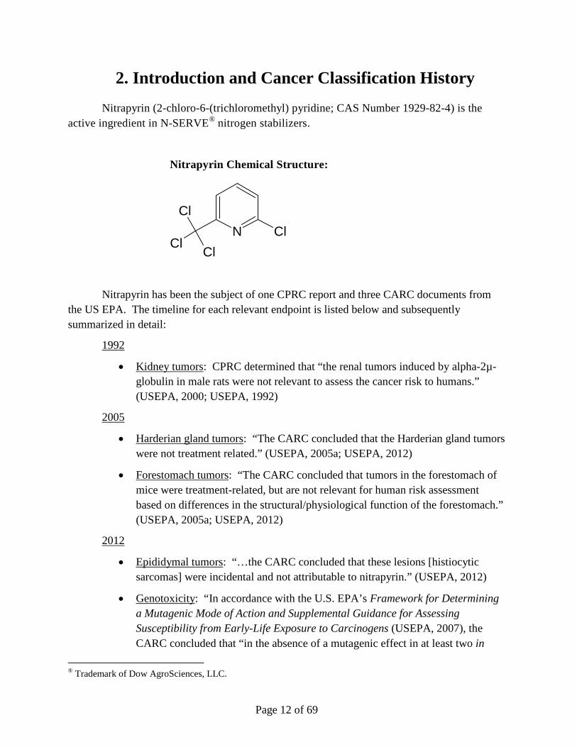
Nitrapyrin (2-chloro-6-(trichloromethyl) pyridine; CAS Number 1929-82-4) is the active ingredient in N-SERVE® nitrogen stabilizers.
Nitrapyrin Chemical Structure:
N ClCl
ClCl
Nitrapyrin has been the subject of one CPRC report and three CARC documents from the US EPA. The timeline for each relevant endpoint is listed below and subsequently summarized in detail:
1992
• Kidney tumors: CPRC determined that “the renal tumors induced by alpha-2µ-globulin in male rats were not relevant to assess the cancer risk to humans.” (USEPA, 2000; USEPA, 1992)
2005
• Harderian gland tumors: “The CARC concluded that the Harderian gland tumors were not treatment related.” (USEPA, 2005a; USEPA, 2012)
• Forestomach tumors: “The CARC concluded that tumors in the forestomach of mice were treatment-related, but are not relevant for human risk assessment based on differences in the structural/physiological function of the forestomach.” (USEPA, 2005a; USEPA, 2012)
2012
• Epididymal tumors: “…the CARC concluded that these lesions [histiocytic sarcomas] were incidental and not attributable to nitrapyrin.” (USEPA, 2012)
• Genotoxicity: “In accordance with the U.S. EPA’s Framework for Determining a Mutagenic Mode of Action and Supplemental Guidance for Assessing Susceptibility from Early-Life Exposure to Carcinogens (USEPA, 2007), the CARC concluded that “in the absence of a mutagenic effect in at least two in
® Trademark of Dow AgroSciences, LLC.
Page 13 of 69
vivo mutagenicity studies, there is no concern for a mutagenic mode of action.” (USEPA, 2012)
• Liver Tumors: The CARC determined that the data were not sufficient to support the proposed MoA for liver tumors, and identified specific uncertainties including lack of PROD activity and burst of mitotic activity. The most recent decision regarding nitrapyrin by the US EPA (2012) was “In accordance with the EPA’s Final Guidelines for Carcinogen Risk Assessment (March 2005), the CARC re-classified nitrapyrin as “Suggestive Evidence of Carcinogenic Potential.” Subsequent to this US EPA CARC decision in 2012, Dow AgroSciences has completed additional mechanistic studies that addressed uncertainties regarding the liver tumor MoA, and has submitted these studies and an associated update to the MoA and Human Relevance Framework analysis (LaRocca et al., 2015) to the US EPA in 2015 for their evaluation and, ultimately, final downgrading of the cancer classification of nitrapyrin.
In 1992 the Cancer Peer Review Committee (USEPA, 1992) classified nitrapyrin as category Group D- not classifiable as to human carcinogenicity. There was no indication of treatment-related tumors in a two-year chronic bioassay in male and female rats except for renal tumors in male rats, which were induced by α2µ-globulin mechanism and considered not relevant to assess cancer risk in humans. A two-year oncogenicity study in B6C3F1 mice administered 0, 5, 25, and 75 mg/kg/day (Quast et al. 1990) also was reviewed and the Committee concluded that there was no indication of treatment-related tumors. However, the Committee was of the opinion that the dosing in the mouse study was not adequate for assessing the carcinogenic potential and therefore, requested a new dose-range finding study and an additional two-year study at higher dose levels. A repeat two-year oncogenicity study was conducted with B6C3F1 mice that were administered 0, 125, or 250 mg/kg/day nitrapyrin (Stebbins and Cosse, 1997). The dose levels for the repeat oncogenicity study were selected based on discussions between the registrant and the US EPA. Study results indicated an increased incidence of mice with hepatocellular tumors at 125 (females only), or 250 mg/kg/day nitrapyrin (both sexes) in the diet. In addition, an increased number of male and female mice had papillomas and/or squamous cell carcinomas of the forestomach (non-glandular stomach), and there was an increased number of female mice with Harderian gland adenomas and an increased number of male mice with epididymal tumors. Based on the results of the repeat oncogenicity study, an US EPA Cancer Assessment Review Committee (USEPA, 2000) classified nitrapyrin as "Likely to be carcinogenic in humans" according to the EPA's July, 1999 Draft Guidelines for Carcinogen Risk. The Committee also indicated that, although nitrapyrin was not mutagenic in standard guideline studies, a National Toxicology Program (NTP) study (Zeiger et al., 1988) reported that the compound was mutagenic in a Salmonella typhimurium assay in the presence of S9 activation and also indicated that there was support from structure-

Page 14 of 69
activity relationship (SAR) for nitrapyrin having genotoxic potential. However, the CARC further commented that 6-chloropicolinic acid, a major metabolite of nitrapyrin, did not have evidence of carcinogenicity in B6C3Fl mice. The Committee concluded that the weight-of-the-evidence analysis was not a sufficient basis to ascribe a mutagenic mode of action to the carcinogenic response of nitrapyrin but that the issue of whether the potential mutagenicity contributes to the mode of action (MoA) for nitrapyrin-induced carcinogenesis was unresolved.
Dow AgroSciences sponsored a Scientific Advisory Group (SAG) in 2004 which was asked to provide an independent scientific review of the histopathology of the proliferative lesions in the repeat mouse carcinogenicity study and also to evaluate relevant mechanistic data including genotoxicity. The report of this evaluation (Hardisty, 2004) was submitted to the US EPA and also published in a peer-reviewed scientific journal (Yano et al., 2008). The SAG emphasized that the repeat mouse study (Stebbins and Cosse, 1997) at higher dose levels (0, 125 or 250 mg/kg/day) was designed to be complementary to the previous study (Quast et al., 1990) which was conducted at lower dose levels (0, 5,25 or 75 mg/kg/day) of nitrapyrin and that the results from the two studies should be evaluated together. The SAG concluded that the mouse forestomach tumors had little relevance to humans because of anatomic and functional differences between the species. The SAG indicated that there was an unusually low incidence of Harderian gland tumors in the concurrent control female mice and that there was no treatment-related increase in these tumors. The SAG also concluded that the cell of origin for epididymal tumors in the male mice was a fixed tissue macrophage (histiocytic tumors) and not undifferentiated sarcomas or Leydig cell tumors as diagnosed by the original study pathologists. The incidence of epididymal tumors in control and treated males from both mouse oncogenicity studies together was attributed to biological variation and not to treatment with nitrapyrin. The SAG concluded that the mouse liver tumors were a result of a nongenotoxic mode of action (prolonged hepatocellular cytotoxicity and hepatocellular proliferation) and that doses that do not produce toxic effects in the liver would not be expected to increase the risk of cancer. The SAG considered it unlikely that nitrapyrin was acting through a genotoxic mode of action.
A subsequent US EPA Cancer Assessment Review Committee (USEPA, 2005a) determined that the increased incidence of Harderian gland tumors in the female mice were no longer considered a response to treatment and that the forestomach tumors in the mice were not relevant to human health risk assessment. On the other hand, the CARC retained concern for the epididymal tumors in the male mice and continued to refer to these tumors as undifferentiated sarcomas. The CARC did not accept prolonged hepatocellular cytotoxicity and cell proliferation as a proposed liver tumor mode of action. The CARC also had continued concerns about the NTP report that nitrapyrin was mutagenic in Salmonella and that structure-activity relationship (SAR) factors predict nitrapyrin could have mutagenic potential. Although the CARC concluded that the weight-of-the-evidence analysis was not a sufficient basis to ascribe a mutagenic mode of action to the carcinogenic response of nitrapyrin, the issue of whether the potential mutagenicity contributes to the mode of action (MoA) for nitrapyrin-

Page 15 of 69
induced carcinogenesis was considered to be unresolved. The CARC reaffirmed the cancer classification of "Likely to be carcinogenic to humans".
In order the address the uncertainties surrounding nitrapyrin-induced tumor formation in mice, an in vivo liver proliferation MoA study, a repeat Ames test, and an in vivo unscheduled DNA synthesis (UDS) test with mouse liver cells were performed. These data were subsequently evaluated within the original MoA/HRF (Mode of Action/Human Relevance Framework document) that assessed the key events for the MoA which ultimately result in the formation of mouse liver tumors after lifetime exposures to relatively large dose levels of nitrapyrin (Eisenbrandt et al., 2010). A Pathology Working Group (PWG) provided immunohistochemical identification and independent expert review and perspective on the male mouse epididymal tumors (Hardisty, 2010). A new study evaluated the MoA and key events which ultimately result in the formation of mouse liver tumors after lifetime exposures to relatively large dose levels of nitrapyrin. In addition, a Human Relevance Framework evaluation had been completed to provide further perspective on the mouse liver tumors (Eisenbrandt et al., 2010). Within this assessment, CARC concluded that the histiocytic sarcomas in male epididymides were incidental and not attributable to nitrapyrin. Importantly,
“in accordance with the U.S. EPA’s Framework for Determining a Mutagenic Mode of Action and Supplemental Guidance for Assessing Susceptibility from Early-Life Exposure to Carcinogens (U.S. 2007), the CARC concluded that in the absence of a mutagenic effect in at least two in vivo mutagenicity studies, there is no concern for a mutagenic mode of action” (USEPA, 2012).
Based on these data, the US EPA CARC re-classified nitrapyrin as “Suggestive Evidence of Carcinogenic Potential” according with the US EPA’s Final Guidelines for Carcinogenic Risk Assessment in 2011 (USEPA, 2012).
Regarding liver tumors, CARC determined additional data uncertainties were raised that precluded inclusion of nitrapyrin as “Not likely to be carcinogenic to humans.” In order to address these uncertainties, additional studies were performed that support the conclusion of CAR activation as the MoA for nitrapyrin-induced liver tumors. These recently conducted studies include an in vitro assay for suicide inhibition, a CAR KO (knock-out) mouse study, and a comparison of human and mouse primary hepatocyte culture DNA synthesis (i.e., proliferation) responses to nitrapyrin exposure. Collectively, these studies demonstrate that the MoA for nitrapyrin-mediated liver tumor formation is CAR activation and satisfy the previously identified uncertainties. These studies are included in an updated MoA/HRF (LaRocca et al., 2015), which integrates the previous MoA/HRF analysis (Eisenbrandt et al., 2010) and has submitted to the US EPA as of August, 2015. The data presented in the revised HRF support the conclusion that nitrapyrin-induced mouse liver tumors are mediated by CAR activation, and due to qualitative differences between humans and rodents these tumors should be classified as “Not likely to be carcinogenic to humans” (Boobis et al., 2007; Cohen et al., 2003; Holsapple et al., 2006; Meek et al., 2003; Sonich-Mullin et al., 2001). Details supporting this statement for

Page 16 of 69
all relevant endpoints (a. genotoxicity, b. forestomach tumors, c. epididymal tumors, d. Harderian gland tumors, e. kidney tumors, and f. liver tumors) are comprehensively summarized in subsequent section of these comments.

Page 17 of 69
3. Discussion and Mechanisms of Relevant Carcinogenicity Endpoints
A. Genotoxicity
Summary of Results
Data are available from 3 independent Salmonella mutagenicity tests with nitrapyrin (Kennelly, 1985; Mecchi, 2007; Zeiger et al., 1988). The Mecchi (2007) report also contains data from a test using E. coli WP2uvrA. Nitrapyrin was judged to be not mutagenic in Salmonella when tested using the standard plate (Kennelly, 1985) or preincubation (Mecchi, 2007) protocol with 10% Aroclor-induced rat liver S9. In contrast, an NTP study (Zeiger et al., 1988) judged nitrapyrin to be weakly mutagenic in the preincubation protocol when tested using 10% and 30% rat liver S9 or hamster liver S9.
The mutagenic responses in the absence of S9 and with the standard 10% rat liver S9 were consistent among all three tests. The different conclusions by the authors in the tests with S9 resulted from the criteria used to determine a positive response. Both Kennelly (1985) and Mecchi (2007) used the “two-fold” rule for determining a positive response; i.e., the response had to be concentration-related and reaching at least two-fold greater than background for Salmonella strain TA100, and at least three-fold over background for strains TA98, TA1535, and TA1537. In contrast, Zeiger et al. (1988) required only a reproducible, concentration-related response, with no requirement for a two- or three-fold increase over the solvent control background value. The most recent bacterial mutagenicity assay was reviewed by the expert genetic toxicologist, Dr. Errol Zeiger (author of the weak positive NTP mutagenicity study), and he concluded that nitrapyrin was negative in this microbial assay. Importantly, Dr. Errol Zeiger completed an independent review of the genetic toxicity of nitrapyrin, which “provides an integrated evaluation of the totality of the data in regard to the genotoxicity of nitrapyrin and also provides some perspectives on the relevance of the data in the concert with nitrapyrin’s in vivo effects in the context of the EPA Guidelines for Carcinogen Risk Assessment (USEPA, 2005b; Zeiger, 2010). This review clearly supports the conclusion that the single weak positive finding is not sufficient to ascribe a mutagenic MoA to nitrapyrin.
Nitrapyrin did not induce hypoxanthine-guanine-phosphoribosyl transferase (HGPRT) gene mutations in Chinese hamster ovary (CHO) cells in culture (Linscombe and Gollapudi, 1986) or an increase in unscheduled DNA synthesis (UDS) in rat hepatocytes exposed in vitro (Mendrala and Schumann, 1982).
An in vivo bone marrow micronucleus (MN) test with nitrapyrin was conducted in male and female CD-1 mice (Kirkland, 1985). The test utilized a single, oral, gavage dose of 800 mg nitrapyrin/kg and the mice were sacrificed at 24, 48, or 72 hrs after administration. The 800

Page 18 of 69
mg/kg dose was significantly higher than the dose levels utilized in the short-term and chronic toxicity studies with nitrapyrin (Yano et al., 2008). There were no early deaths from the nitrapyrin, but bone marrow toxicity was present as evidenced by lower polychromatic to normochromatic erythrocyte (PCE/NCE) ratios in both males and females at the 48-hr and 72-hr sacrifice times. There were no increases in micronuclei at any sacrifice time in male or female mice.
An in vivo/in vitro liver UDS test with nitrapyrin was conducted in male B6C3F1 mice (Pant and Celestin, 2009). Three pilot toxicity assays prior to the in vivo UDS test indicated that the mice would not survive single, oral, gavage doses of 500 mg/kg or greater based on a 3-day observation period. Therefore, male mice were administered single, oral, gavage doses of 125 or 250 mg nitrapyrin/kg. These dose levels were equivalent to the repeated dietary dose levels utilized in the Stebbins and Cosse (1997) mouse carcinogenicity study. Piloerection, but no other clinical signs, was seen following the 250 mg/kg dose. Animals were sacrificed 2-4 hours and 12-16 hours after dosing for removal of the liver and culturing of hepatocytes. There were no increases in UDS at either dose at either time point. A summary of genotoxicity data is listed in Table 1.
Table 1. Summary of Genotoxicity Data for Nitrapyrin
System Assay Result Ref.
In Vitro 1. Ames (plate incorporation)
2. Ames (pre-incubation)
3. Ames (pre-incubation)
4. CHO/HGPRT
5. Rat hepatocyte UDS
1. Negative
2. Negative
3. Weak Pos.
4. Negative
5. Negative
1. (Kennelly, 1985)
2. (Mecchi, 2007)
3. (Zeiger et al., 1988)
4. (Linscombe and Gollapudi, 1986)
5. (Mendrala and Schumann, 1982)
In Vivo 1. Mouse BM Micronucleus
2. Mouse Liver UDS
1. Negative
2. Negative
1. (Kirkland, 1985)
2. (Pant and Celestin, 2009)
Evaluation and Interpretation of the Totality of Genotoxicity Data for Nitrapyrin.
Nitrapyrin has not been shown to be activated to a DNA-reactive intermediate and the structure of nitrapyrin does not indicate that such an intermediate would be formed. The metabolic pathway of nitrapyrin in rodents (Domoradzki and Brzak, 1998) is presented in Figure 1.

Page 19 of 69
Figure 1. Metabolic Pathway of Nitrapyrin in Rodents
The primary metabolite of nitrapyrin, 6-chloropicolinic acid, has no structural alerts for DNA reactivity. Dietary administration of 6-chloropicolinic acid to B6C3F1 mice for two years at dose levels of 0, 100, 300, and 900 mg/kg/day was not carcinogenic (USEPA, 2000; Zimmer et al., 1986) and, specifically, there was no evidence of treatment-related hepatocellular hyperplasia or tumors.
The Cancer Assessment Review Committee (USEPA, 2005a) indicated that there is support from SAR for nitrapyrin having genotoxic potential. The US EPA’s assessment on SAR factors was primarily based on the mutagenicity studies on 2-chloropyridine reported in the literature (Claxton et al., 1987). N-Oxidation in the presence of S9 was hypothesized to be responsible for making the chlorine in the 2-position more susceptible for nucleophilic attack and a likely mechanism for the mutagenicity of 2-chloropyridine. However, there is no in vivo evidence for N-oxidation of nitrapyrin (a substituted 2-chloropyridine) in the mouse or rat, especially in the presence of a sterically hindering trichloromethyl blocking group in the 6 position. The lack of DNA reactivity is evident from the relatively weak and/or inconsistent responses seen in the Salmonella mutagenicity assays for nitrapyrin and a similar lack of genotoxic reactivity of other halogenated pyridines (Claxton et al., 1987). Nitrapyrin also did not induce HGPRT mutations in CHO cells when tested with S9 (Linscombe and Gollapudi,

Page 20 of 69
1986) or UDS in metabolically competent primary rat hepatocytes (Mendrala and Schumann, 1982). Additional evidence for the lack of activation to a reactive intermediate is the absence of in vivo liver UDS (Pant and Celestin, 2009) and in vivo bone marrow micronucleus responses (Kirkland, 1985) in mice at doses equal to or greater than the repeated dose levels that produced hepatocellular hyperplasia and hepatocellular tumors in the mouse studies. Based on the carcinogenicity and mutagenicity databases developed by the U.S. National Toxicology Program, approximately 70% of the chemicals that produce mouse tumors only in the liver are not mutagenic, and a large proportion of these chemicals are chlorinated aliphatics and aromatics (Ashby and Tennant, 1988; Huff et al., 1991).
Increased cell proliferation, with or without accompanying necrosis, can lead to an increased mutation frequency and subsequent tumor formation (Ames and Gold, 1990b; Ames and Gold, 1990a; Cohen and Ellwein, 1991; USEPA, 2005b). In such a situation, the resulting mutations are not consequences of a direct reaction of the chemical with DNA, but a secondary effect of the increased cell proliferation. Supporting data that indicate nitrapyrin operates by the latter mechanism for liver tumors are the lack of mouse liver UDS (Pant and Celestin, 2009) at the same doses that produce liver hyperplasia in the short-term studies as well as hyperplasia and hepatocellular tumors in the chronic mouse study (Stebbins and Cosse, 1997). Other support for an increased cell proliferation mechanism is the absence of genetic toxicity (as evidenced by no increased micronuclei in the bone marrow) at 800 mg nitrapyrin/kg body weight (Kirkland, 1985).
The OEHHA HID included a comprehensive review of relevant guidelines, which are included and expanded upon in the following paragraphs (OEHHA, 2015). As noted in the HID, the US EPA draft Framework for Determining a Mutagenic Mode of Action for Carcinogenicity (USEPA, 2007) distinguishes cancer modes of action where the mutagenic event is an initial or early event from the situation when mutations are acquired subsequent to other events such as increased proliferation. This guidance states that
“The determination that a chemical carcinogen can induce mutation in one of a number of mutation assays is not sufficient to conclude that it causes specific tumors by a mutagenic MoA or that mutation is the only key event in the pathway to tumor induction.”
Similarly, the US EPA Guidelines for Carcinogen Risk Assessment (USEPA, 2005b) specifically recognizes regenerative cell proliferation as an early event in cancer induction and consider it to be a possible non-genotoxic mode of action. Cell proliferation can be the result of an enhancement of mitosis in the tissue or a proliferative regeneration following toxicity. The weak and/or inconsistent positive finding for nitrapyrin in the Zeiger et al. (1988) Ames test is not sufficient to conclude that nitrapyrin causes tumors by a mutagenic MoA.
While there is no clear cut-off (2-fold, 3-fold, etc.) defined in OECD 471 guideline for the bacterial reverse mutation test, “biological relevance of the results must be considered first.”

Page 21 of 69
(OECD, 1997) Additional guidance for the interpretation of in vitro and in vivo genotoxicity studies has been outlined in Thybaud et al. (2007), which includes the statement:
“In some cases a clear and reproducible positive in vitro result is seen, yet the other assays in the initial battery, including any required in vivo test, are negative. The in vitro result is not automatically overruled by the negative in vivo result, and some follow-up testing or investigation is generally necessary to determine the relevance of the in vitro positive result. For example, the ICH scheme and suggests follow-up testing with a second in vivo test in addition to the in vivo cytogenetics test in the initial regulatory battery. It might be assumed that the concern about the positive in vitro result lessens as the number and types of negative in vivo assay results increase. However, this assumption may not be valid since the in vivo assays may have different sensitivities and /or evaluate different genotoxic endpoints. It is important that relevant endpoints are examined in the most relevant tissues in vivo.” (Thybaud et al., 2007)
This excerpt clearly states that follow-up testing is recommended in certain cases, particularly in determining the relevance of an in vitro positive result. In the case of nitrapyrin, follow up testing was conducted, which included both in vitro and in vivo studies. Importantly, liver was the tissue tested with the in vivo UDS test, which is the relevant tissue to assess genotoxicity for the observed nitrapyrin-mediated liver tumors. Additionally, the article also notes that:
“When a non-reproducible or marginal in vitro positive result is obtained, and results from other assays with a similar endpoint are negative, the weight of evidence should be considered to determine if further testing is necessary or whether, based on the available data, the evidence suggests a low level of potential risk that does not require further testing. Factors that may suggest lower concern include: (a) weak effects without a strong dose relationship and values within or close to a range that could occur by chance variability (negative control historical data), (b) effects that occur only at very high levels of cytotoxicity, but not at moderate levels, in the chromosomal aberration or mouse lymphoma tk+/− assays (e.g., approaching 50% or greater cytotoxicity in the chromosome aberration test, or >80% in the mouse lymphoma assay), (c) results that are not consistently repeatable, and (d) the absence of structural alerts or any other cause of concern. In most cases, the result is not of concern and no testing beyond the standard battery for that type of substance will be required.” (Thybaud et al., 2007)

Page 22 of 69
Indeed, this is consistent with the guidance from the International Programme on Chemical Safety (IPCS) Harmonized Scheme for Mutagenicity Testing, which states both:
“At all stages of the outlined testing strategy, a weight of evidence approach and scientific judgment should be used. Multiple negative results may not be sufficient to remove concern for mutagenicity raised by a clear positive result in a single mutagenicity assay. Most short-term tests in bacteria and mammalian cell cultures have been designed primarily for hazard identification and thus can represent only the starting point in the process of risk assessment. Whether or not the observed effects are relevant for humans under anticipated exposure conditions depends on pharmacokinetic, pharmacodynamic and other factors that require investigation in vivo.” (Eastmond et al., 2009)
As well as:
“Further in vivo testing is recommended in the case of positive in vitro studies. Again, the second in vivo test is chosen on a case-by-case basis... If the test is negative, it is concluded that there is no evidence for in vivo mutagenicity.” (Eastmond et al., 2009)
In contrast to the OEHHA HID statements, Dow AgroSciences supports that the Weight of Evidence demonstrates nitrapyrin is not acting through a mutagenic or genotoxic MoA, which is in agreement with the aforementioned guidelines (see Table 2 below). This conclusion is supported by the clear lack of genotoxicity and mutagenicity in several in vitro tests, including a newer repeat Ames test, and subsequent negative results for nitrapyrin in two follow-up in vivo (MN and UDS) tests. This conclusion is in agreement with the conclusions of the US EPA.

Page 23 of 69
Table 2. Comparison of OEHHA HID Statements and Dow AgroSciences Position on Genotoxicity of Nitrapyrin
Endpoint OEHHA HID
Statements
OEHHA HID Rationale Dow AgroSciences
Position
Dow AgroScience Justification
Genotoxicity Nitrapyrin appears to be genotoxic
One weak positive Salmonella mutagenicity test result (out of 7 tests total) along with different criteria used for other mutagenicity tests suggests nitrapyrin may be genotoxic. HID implies that negative results cannot outweigh even a single weak positive result.
Nitrapyrin is nongenotoxic
The integration of several relevant endpoints, including two negative in vivo assays and several negative in vitro results (4 of 5), demonstrates that nitrapyrin is nongenotoxic, and there is no concern for a mutagenic mode of action. Regarding the single weak positive result, Dr. Zeiger (external expert and author of NTP report) concluded it is not sufficient to ascribe a mutagenic MoA.
Page 24 of 69
B. Forestomach
Summary of Results
Following 12 months of administration of nitrapyrin at dose levels of 125 and 250 mg/kg/day, treatment-related focal or multifocal hyperplasia of the forestomach was observed. The foci of hyperplasia of the forestomach were frequently accompanied by treatment-related focal or multifocal hyperkeratosis. Male and female mice administered 125 and 250 mg/kg/day had a treatment-related increase in the number of animals with focal or multifocal hyperplasia of the mucosa of the forestomach. The foci of hyperplasia (increased number of cells) of the forestomach were frequently accompanied by treatment-related focal or multifocal hyperkeratosis (thickening the keratin layer which lines the forestomach mucosa). Male and female mice administered 125 or 250 mg/kg/day for two years also had a treatment-related increase in the number of animals with one or more neoplasms (papillomas or squamous cell carcinomas) of the mucosa of the forestomach.
Discussion of Results
As noted in the IARC technical publication on predictive value of rodent forestomach in evaluating carcinogenic risk to humans, discrete foci of mucosal hyperplasia, papilloma, and squamous cell carcinoma of the mucosa of the forestomach probably represent a continuum in the development of treatment-related hyperplasia (IARC, 2003). Studies on rabbits have shown that nitrapyrin results in dermal irritancy (Cosse et al., 1992) and transient irritation of the eyes (Carreon et al., 1986). This potential for local irritancy following repeated daily ingestion of nitrapyrin for up to two years is the probable mode of action for the development of hyperplasia, hyperkeratosis, papilloma and squamous cell carcinomas of the mucosa of the forestomach. Proliferative alterations of the mucosa of the forestomach of rodents have been induced by numerous nongenotoxic compounds via oral administration (IARC, 2003). The carcinogenic activity of nongenotoxic substances such as diallyl phthalate, allyl chloride, propionic acid and sodium saccharin was most likely due to their strong irritating and hyperplastogenic properties, which lead to the eventual development of papillomas and/or squamous cell carcinomas of the mucosa of the forestomach (Kroes and Wester, 1986). Similar effects have been seen in short-term and chronic studies with the nongenotoxic compound, 3-tert-butyl-4-hydroxyanisole (BHA). Short-term dietary exposure to BHA resulted in irritation and hyperplasia of the mucosa of the forestomach (Altmann et al., 1985), and chronic exposure resulted in hyperplasia, papillomas and squamous cell carcinomas of the forestomach (Ito et al., 1983; Whysner and Williams, 1996).
As noted in the IARC publication (IARC, 2003) and referenced in the OEHHA HID (OEHHA, 2015):
Page 25 of 69
''for some carcinogens not known to be genotoxic in the forestomach, irritation leading to enhanced and sustained cell proliferation may be essential for tumor development."
However, the IARC publication also states:
“While humans do not have a forestomach, they do have comparable squamous epithelial tissues in the oral cavity and the upper two-thirds of the oesophagus. Thus, in principle, carcinogens targeting the forestomach squamous epithelium in rodents are relevant for humans.”
The WoE for nitrapyrin demonstrates that the forestomach tumors in mice are not relevant for human risk assessment for several scientific reasons. There was no evidence of compound-induced irritation, hyperplasia, or neoplasia in the oral cavity or esophagus of mice given 125 or 250 mg/kg/day nitrapyrin for two years. The function of the forestomach in the rodent is a storage site to aid in the digestive process and thus allows prolonged exposure to ingested substances, and prolonged exposure to extremely high levels of nitrapyrin could lead to local irritation. The carcinogenic effect of high concentrations of nitrapyrin on the mucosa of the forestomach of mice is considered to have little relevance to man. Nongenotoxic carcinogens of the mucosa of the forestomach are not likely to be hazardous to man under conditions that do not produce irritation or hyperplasia (Kroes and Wester, 1986). In contrast to mice, the human stomach does not have a nonglandular (squamous) portion. The nearest equivalent in humans to the forestomach of rodents is the esophagus, which is unlikely to be altered by compounds known to cause hyperplasia in the stomach (Betton and Salmon, 1984). Since the exposure time of the mouth, pharynx and esophagus to ingested compounds is much shorter than in the forestomach, which is a storage organ, it is unlikely that nitrapyrin would induce irritation and subsequent hyperplasia in the esophagus. This, in addition to the fact that there was no evidence of irritation in the oral cavity or esophagus in mice following nitrapyrin-exposure, demonstrates that the forestomach tumors are not relevant for human health risk assessment in comparable squamous epithelial tissues.
The body of scientific evidence indicates that the forestomach tumors are likely being induced by a nongenotoxic mode of action and no increased risk of cancer would be expected at doses that do not produce irritation and hyperplasia. This is in agreement with the 2005 US EPA CARC decision, which reevaluated the mouse forestomach tumors and determined that these treatment-related tumors were ". . .not relevant for human risk assessment based on differences [between rodents and humans] in the structural/physiological function of the forestomach” (USEPA, 2005a). Since the forestomach tumors in mice are considered not relevant for human risk assessment, they do not factor into the cancer classification for nitrapyrin. A comparison of OEHHA HID statements and Dow AgroSciences positions is summarized in Table 3 below.

Page 26 of 69
Table 3. Comparison of OEHHA HID Statements and Dow AgroSciences Position on Forestomach Tumors
Endpoint OEHHA HID
Statements
OEHHA HID Rationale Dow AgroSciences
Position
Dow AgroSciences Justification
Forestomach Forestomach tumors add to the WoE that nitrapyrin is carcinogenic
IARC criteria:
“While humans do not have a forestomach, they do have a comparable epithelial tissue in the oral cavity and the upper two-third of the esophagus. Thus, in principle, carcinogens targeting the forestomach squamous epithelium in rodents are relevant for humans. Also, the target tissue for carcinogens may differ between experimental animals and humans, and a forestomach carcinogen in rodents may target a different tissue in humans”
Forestomach tumors are not relevant to humans
There was no evidence of compound-induced irritation, hyperplasia, or neoplasia in the oral cavity or esophagus of mice given 125 or 250 mg/kg/day nitrapyrin for two years. Since the exposure time of the mouth, pharynx and esophagus to ingested compounds is much shorter than in the forestomach, which is a storage organ, it is unlikely that nitrapyrin would induce irritation and subsequent hyperplasia in the esophagus. Due to structural and physiological differences between mice and humans, nitrapyrin-induced forestomach lesions that occur secondary to local irritation are not considered relevant to humans. Hence, a local disposition of nitrapyrin needed to result in cancer response in the epithelium would not be achievable in humans compared to the rat.

Page 27 of 69
C. Epididymal Tumors
Summary of Results
An increase in undifferentiated sarcomas of the epididymis was observed in male mice in the repeat nitrapyrin mouse carcinogenicity study (Stebbins and Cosse, 1997). In 2005, the EPA CARC determined that the epididymal tumors were treatment-related and evidence of the carcinogenic potential of nitrapyrin in mice.
A Scientific Advisory Group (Yano et al., 2008; Hardisty, 2004), consisting of independent, expert pathologists, examined the spectrum of proliferative changes that were reported in the epididymis for the two nitrapyrin mouse oncogenicity studies. These tumors were observed in the epididymis of male mice killed only at the terminal sacrifice, indicating that they most likely occurred late in the study. The SAG considered the tumors to be histiocytic tumors (tissue macrophages) in contrast to the original study pathologist's diagnosis of Leydig cell tumors (Quast et al., 1990) or undifferentiated sarcomas (Stebbins and Cosse, 1997). Two epididymal tumors were present in the control group from the first mouse oncogenicity study (Quast et al., 1990), but no epididymal tumors were observed in the controls from the repeat study (Stebbins and Cosse, 1997). The presence of two histiocytic tumors in the low-dose group and four in the high-dose group in the repeat study was considered to be an incidental finding as the result of biological variation and unrelated to treatment with nitrapyrin. The SAG concluded that the results of these two studies clearly indicate that the epididymal neoplasms were not related to treatment with nitrapyrin.
A subsequent Cancer Assessment Review Committee (USEPA, 2005a) indicated that they did not agree that the epididymal tumors in the high-dose group of the Stebbins and Cosse (1997) study were spontaneous and the result of natural variation. The CARC stated that the epididymal undifferentiated sarcomas were biologically significant and cannot be dismissed from the carcinogenicity evaluation
In contrast to the 2005 CARC report, the SAG report (Yano et al., 2008; Hardisty, 2004) actually stated that the epididymal tumors reported by the study pathologist (Stebbins and Cosse, 1997) as undifferentiated sarcomas were considered to be histiocytic tumors by the SAG reviewing pathologist. The SAG report also indicated that the epididymal tumors in controls from the Quast et al. (1990) study that were recorded by the study pathologist as Leydig cell tumors were considered to be identical to the epididymal tumors in the Stebbins and Cosse (1997) study. The SAG was trying to indicate that all of these tumors (both Leydig cell and undifferentiated sarcomas) actually were histiocytic tumors, however the wording in the SAG report was not clear and, as a result, the CARC (USEPA, 2005a) misinterpreted the statements of the SAG by considering all of the epididymal tumors as undifferentiated sarcomas since they are a malignant and rare tumor in mice. However, the CARC stated that the "...diagnosis provided by the SAG (undifferentiated sarcoma) is appropriate for the evaluation of the epididymal tumors, therefore, superseding the original diagnosis (Leydig cell tumor)." The

Page 28 of 69
CARC tabulated the incidence of all epididymal tumors from both the Quast et al. (1990) study and the Stebbins and Cosse (1997) study as undifferentiated sarcomas.
A PWG (Hardisty, 2010) was asked to improve the clarity of the tumor nomenclature and incidence of the epididymal tumors identified in both the original Quast et al. (1990) and the repeat Stebbins and Cosse (1997) nitrapyrin mouse carcinogenicity studies. The PWG procedures were in compliance with all aspects of U.S. EPA's Pesticide Regulation (PR) 94-5, August 24, 1994. The group of independent, expert pathologists reexamined hematoxylin and eosin (H&E) stained slides containing proliferative lesions in the epididymis from the male mice and classified each lesion following current nomenclature and diagnostic criteria and determined the histogenesis of the neoplastic cells. Importantly, immunohistochemical characterization of the tumors also was completed in conjunction with the PWG. The PWG (Hardisty, 2010) diagnosed the epididymal tumors in both mouse carcinogenicity studies as histiocytic sarcomas (with one exception which was diagnosed as malignant lymphoma and thus not relevant to the evaluation). A significant finding was that the PWG identified an additional control mouse from the Quast et al. (1990) study that had a histiocytic sarcoma of the epididymis and this increased the number of control mice with this tumor to three as compared to the incidence of two controls reported by Quast et al. (1990) and the earlier SAG review (Yano et al., 2008; Hardisty, 2004). The additional animal (87A1610) was diagnosed with histiocytic sarcoma in the liver and this mouse also had the histiocytic sarcoma in the epididymis.
Discussion of Results
Overall, the histiocytic sarcomas were observed with a similar incidence in control and treated male mice when both nitrapyrin studies were considered together. The incidence of histiocytic sarcomas in the epididymis as determined by PWG consensus for both nitrapyrin mouse bioassays is listed in Table 4:
Table 4. Incidence of Epididymal Histiocytic Sarcomas in Male Mice
Quast et al. (1990) Stebbins and Cosse (1997)
Dose (mg/kg/day) 0 5 25 75 0 125 250
Number Animals Examined 50 50 50 50 50 50 50
Number of Animals with Epididymis Examined
50 8 10 50 50 50 50
Epididymis – Histiocytic Sarcoma
3 0 0 1 0 2 4

Page 29 of 69
There were three control animals with histiocytic sarcoma of the epididymis in the Quast et al. (1990) study compared to two low-dose and four high-dose animals with histiocytic sarcoma of the epididymis in the Stebbins and Cosse (1997) study. The PWG report (Hardisty, 2010; Hardisty, 2004) includes photomicrographs of the lesions that demonstrate the immunohistochemical confirmation of the tumors as histiocytic sarcomas (i.e., not undifferentiated sarcomas or Leydig cell tumors) and also show that the tumors in control and treated animals were identical in morphology. The PWG concluded that the incidence and distribution of the epididymal tumors clearly indicated that they were spontaneous, incidental findings and unrelated to treatment with nitrapyrin.
As noted in the nitrapyrin OEHHA HID (OEHHA, 2015), US EPA Guidelines state that
“The most relevant historical control data come from the same laboratory and the same supplied and are gathered within 2 or 3 years one way or the other of the study under review; other data should be used only with extreme caution.” (USEPA, 2005b).
Indeed, historical control data for this timespan is not available for the performing laboratory for histiocytic sarcomas. Therefore, during the nitrapyrin PWG, survey results from the National Toxicology Program (NTP) database (NTP, 2009)were compiled by the PWG Reviewing Pathologist and compared with the results from the nitrapyrin mouse carcinogenicity studies. The PWG was an independent review, which followed procedures consistent with NTP. The procedures followed during the PWG were in compliance with all aspects of the US EPA’s Pesticide Regulation (PR) 94-5, August 24, 1994. Epididymal histiocytic sarcomas were identified in approximately 51 cases from the NTP B6C3Fl database. Nine cases were identified in control animals. The remaining cases were identified from a variety of study types (feed, dermal, gavage, etc.) and across treatment groups. Histologically, the histiocytic sarcomas from the NTP studies were similar to those examined at the nitrapyrin PWG. Furthermore, the NTP survey indicated that epididymal histiocytic sarcomas can be either primary neoplasms in the epididymis or associated with other organ involvement. The NTP report indicated a historical control range of 0-4% for histiocytic sarcoma (all sites). Importantly, while the incidence of histiocytic sarcomas in the epididymis in nitrapyrin treated animals (125 or 250 mg/kg/day) does slightly exceed the historical control range of 0-4% reported by the NTP, so does the incidence of histiocytic sarcomas in the epididymis in control animals (6%) in the original oncogenicity study. Although the two oncogenicity studies were performed 7 years apart, they were conducted in the same performing laboratory, and the studies should be reviewed in parallel in order to obtain a full view of dose-response data for nitrapyrin. Therefore, the tumors cannot be defined as being treatment related due to their incidence just outside of historical controls.
Taking a WoE approach, the data suggest that the observed epididymal histiocytic tumors in mice are not related to treatment and therefore they do not factor into the cancer classification for nitrapyrin. The determination that the tumors are incidental and not treatment

Page 30 of 69
related is in agreement with the independent PWG review, as well as the CARC’s consulting pathologist, Dr. John Pletcher (Hardisty, 2010; USEPA, 2012). A comparison of the OEHHA HID statements and Dow AgroSciences position is listed in Table 5 below.
Table 5. Comparison of OEHHA HID Statements and Dow AgroSciences Position on Epididymal Tumors
Endpoint OEHHA HID
Statements
OEHHA HID Rationale Dow AgroSciences
Position
Dow AgroSciences Justification
Epididymal Tumors
Epididymal tumors could be considered to be treatment-related
Tumors appear treatment related when compared to controls with each study. HID suggests the two carcinogenicity studies cannot be compared and historical control data used are not appropriate.
Epididymal tumors are not treatment-related
When both carcinogenicity studies are taken together, which is appropriate given the unusually low percent of control tumor incidence, histiocytic sarcomas in the epididymis were observed with a similar incidence in control and treated male mice and are considered to be incidental. This is in agreement with the independent expert PWG review, which was in compliance with all aspects of the US EPA’s Pesticide Regulation (PR) 94-5, August 24, 1994
Page 31 of 69
D. Harderian Gland Tumors
Summary and Discussion of Results
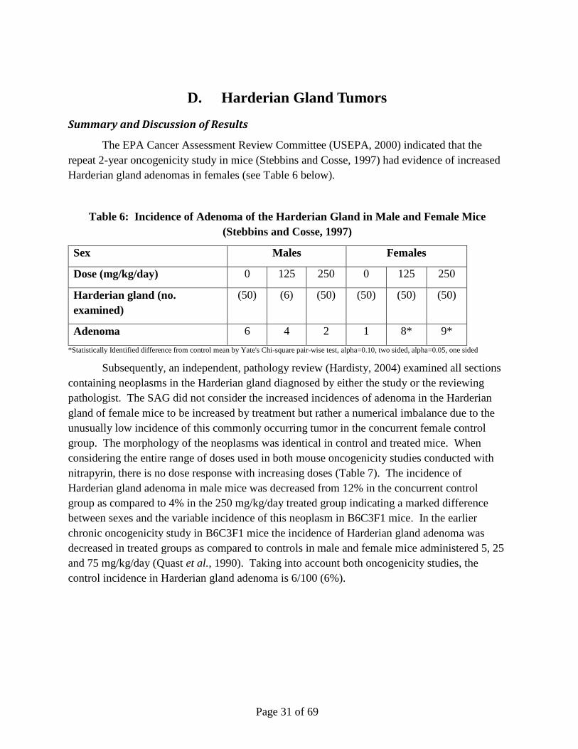
The EPA Cancer Assessment Review Committee (USEPA, 2000) indicated that the repeat 2-year oncogenicity study in mice (Stebbins and Cosse, 1997) had evidence of increased Harderian gland adenomas in females (see Table 6 below).
Table 6: Incidence of Adenoma of the Harderian Gland in Male and Female Mice (Stebbins and Cosse, 1997)
Sex Males Females
Dose (mg/kg/day) 0 125 250 0 125 250
Harderian gland (no. examined)
(50) (6) (50) (50) (50) (50)
Adenoma 6 4 2 1 8* 9*
*Statistically Identified difference from control mean by Yate's Chi-square pair-wise test, alpha=0.10, two sided, alpha=0.05, one sided
Subsequently, an independent, pathology review (Hardisty, 2004) examined all sections containing neoplasms in the Harderian gland diagnosed by either the study or the reviewing pathologist. The SAG did not consider the increased incidences of adenoma in the Harderian gland of female mice to be increased by treatment but rather a numerical imbalance due to the unusually low incidence of this commonly occurring tumor in the concurrent female control group. The morphology of the neoplasms was identical in control and treated mice. When considering the entire range of doses used in both mouse oncogenicity studies conducted with nitrapyrin, there is no dose response with increasing doses (Table 7). The incidence of Harderian gland adenoma in male mice was decreased from 12% in the concurrent control group as compared to 4% in the 250 mg/kg/day treated group indicating a marked difference between sexes and the variable incidence of this neoplasm in B6C3F1 mice. In the earlier chronic oncogenicity study in B6C3F1 mice the incidence of Harderian gland adenoma was decreased in treated groups as compared to controls in male and female mice administered 5, 25 and 75 mg/kg/day (Quast et al., 1990). Taking into account both oncogenicity studies, the control incidence in Harderian gland adenoma is 6/100 (6%).
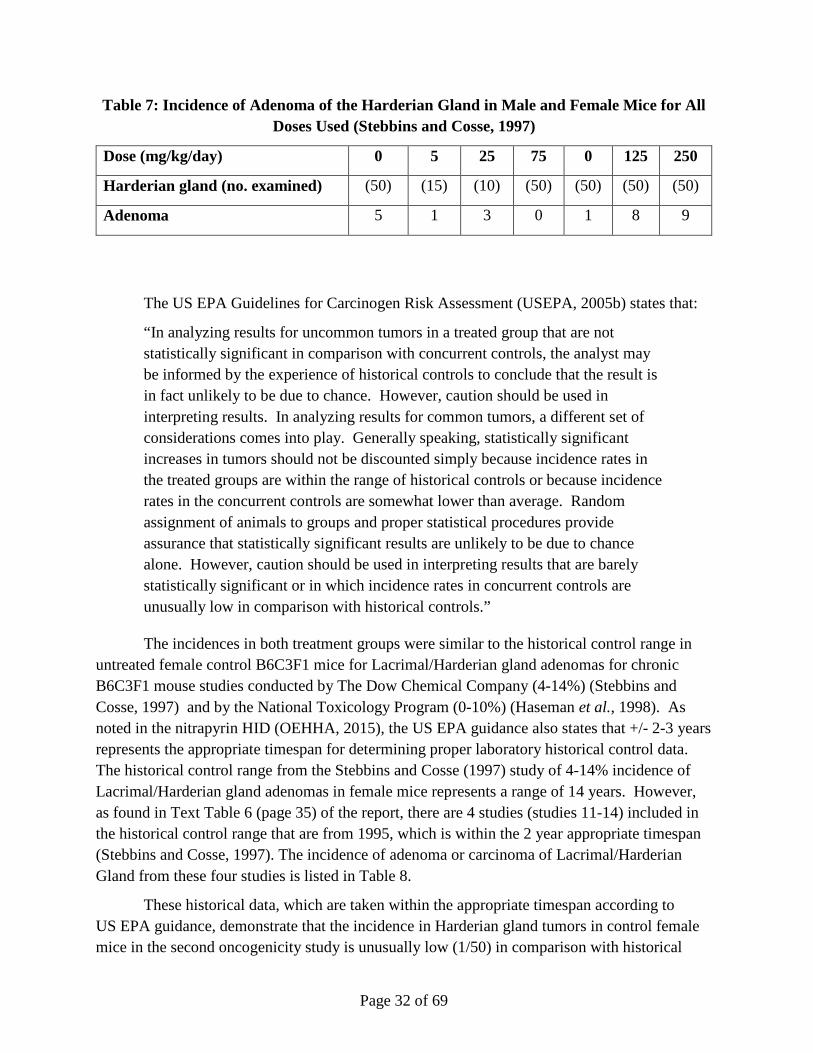
Page 32 of 69
Table 7: Incidence of Adenoma of the Harderian Gland in Male and Female Mice for All Doses Used (Stebbins and Cosse, 1997)
Dose (mg/kg/day) 0 5 25 75 0 125 250
Harderian gland (no. examined) (50) (15) (10) (50) (50) (50) (50)
Adenoma 5 1 3 0 1 8 9
The US EPA Guidelines for Carcinogen Risk Assessment (USEPA, 2005b) states that:
“In analyzing results for uncommon tumors in a treated group that are not statistically significant in comparison with concurrent controls, the analyst may be informed by the experience of historical controls to conclude that the result is in fact unlikely to be due to chance. However, caution should be used in interpreting results. In analyzing results for common tumors, a different set of considerations comes into play. Generally speaking, statistically significant increases in tumors should not be discounted simply because incidence rates in the treated groups are within the range of historical controls or because incidence rates in the concurrent controls are somewhat lower than average. Random assignment of animals to groups and proper statistical procedures provide assurance that statistically significant results are unlikely to be due to chance alone. However, caution should be used in interpreting results that are barely statistically significant or in which incidence rates in concurrent controls are unusually low in comparison with historical controls.”
The incidences in both treatment groups were similar to the historical control range in untreated female control B6C3F1 mice for Lacrimal/Harderian gland adenomas for chronic B6C3F1 mouse studies conducted by The Dow Chemical Company (4-14%) (Stebbins and Cosse, 1997) and by the National Toxicology Program (0-10%) (Haseman et al., 1998). As noted in the nitrapyrin HID (OEHHA, 2015), the US EPA guidance also states that +/- 2-3 years represents the appropriate timespan for determining proper laboratory historical control data. The historical control range from the Stebbins and Cosse (1997) study of 4-14% incidence of Lacrimal/Harderian gland adenomas in female mice represents a range of 14 years. However, as found in Text Table 6 (page 35) of the report, there are 4 studies (studies 11-14) included in the historical control range that are from 1995, which is within the 2 year appropriate timespan (Stebbins and Cosse, 1997). The incidence of adenoma or carcinoma of Lacrimal/Harderian Gland from these four studies is listed in Table 8.
These historical data, which are taken within the appropriate timespan according to US EPA guidance, demonstrate that the incidence in Harderian gland tumors in control female mice in the second oncogenicity study is unusually low (1/50) in comparison with historical

Page 33 of 69
controls. Taking the referenced guidelines into account, it can be considered appropriate to compare the incidence rates of treated animals with historical control data as well as evaluate the two oncogenicity studies together. Using a WoE approach, the data demonstrate that the incidence of Harderian gland adenomas in treated groups falls slightly out of the historical control range for studies completed at Dow within a 2 year timespan, and there is no clear dose response in the incidence of these tumors.
Table 8. Historical Control Incidence of Tumors of the Lacrimal/Harderian Gland; Excerpt from Text Table 6, Page 35 (Stebbins and Cosse, 1997)
Sex
Dose
Number of mice examined
Male
0 mg/kg/day
50 mice examined/study
Female
0 mg/kg/day
50 mice examined/study
Study Number of mice with adenoma or
carcinoma of the lacrimal/ Harderian Gland (%)
Study 11, Route - Dietary, Report Date - 1995
3 (6%) 4 (8%)
Study 12, Route - Dietary, Report Date - 1995
9 (18%) 3 (6%)
Study 13, Route - Dietary, Report Date - 1995
6 (12%) 3 (6%)
Study 14, Route - Dietary, Report Date - 1995
5 (10%) 5 (10%)
These data for nitrapyrin are in agreement with the US EPA CARC decision in 2005, which reevaluated the Harderian gland tumor data and concurred that:
"Although the incidence of Harderian gland tumors in female mice is slightly outside of the historical control range (2nd study), there is a lack of a clear dose response between 125 (16%) and 250 (18%) mg/kg/day and the concurrent control for the second study is considered low relative to the first." (USEPA, 2005a).
The CARC concluded that the Harderian gland tumors "were not considered to be treatment-related." Since the Harderian gland tumors in mice are not related to treatment, they do not

Page 34 of 69
factor into the cancer classification for nitrapyrin. A comparison of OEHHA HID statements and Dow AgroSciences positions are listed in Table 9.
Table 9. Comparison of OEHHA HID Statements and Dow AgroSciences Position on Harderian Gland Tumors
Endpoint OEHHA HID
Statements
OEHHA HID Rationale Dow AgroSciences
Position
Dow AgroSciences Justification
Harderian Gland Tumors
Harderian Gland tumors are treatment-related
Tumors appear treatment related when compared to in study controls. HID suggests that historical control data cannot be used because it spans farther than +/- 2-3 years. HID indicates the two carcinogenicity studies cannot be compared.
Harderian Gland tumors are not treatment-related
There are 4 studies included in the historical control range that is within the 2 year appropriate timespan, which demonstrate incidence in Harderian gland tumors in control female mice in the second oncogenicity study is unusually low. When both carcinogenicity studies are taken together, due to a lack of a clear dose-response and incidence just outside of historical control range, the observed Harderian gland tumors are not considered to be nitrapyrin treatment-related tumors.
Page 35 of 69
E. Kidney Tumors
Summary of Results
In the two-year rat cancer bioassay, there was no evidence of carcinogenicity other than an increase in male-specific kidney tumors at the highest dose administered, 60 mg/kg/day. Data support that the mechanism through which these tumors were formed was via α2µ-globulin nephropathy. The major excretory pathway of nitrapyrin has been shown to be renal in the male Fischer 344 rat (Timchalk et al., 1987). The increase in hyaline droplets, in the proximal tubule epithelial cell cytoplasm, is postulated to result in crowding and eventual cell death (through increase in necrobiosis). The increase in necrobiosis of renal tubule epithelial cells, histologically evidenced by degenerative/-regenerative tubules lined by areas of basophilic, flat epithelial cells (as occurs in male rats administered 60 mg/kg/day) are considered by Alden et al. (Alden et al., 1985) to be areas of tubule cell regeneration distinct from the basophilic, atrophic tubules, often surrounded by thickened basement membranes, associated with ageing rat nephropathy (Gray et al., 1982). It is this increase in cellular turnover, with its resultant proliferative activity, that may in nitrapyrin toxicity, as with volatile hydrocarbon toxicity, result in the weak carcinogenic response (Charbonneau et al., 1989; Lipsky and Trump, 1987; Short et al., 1987), especially in compounds which show no genotoxicity such as nitrapyrin. The sex prevalence of hyaline droplet nephropathy is related to a sex-specific α2µ-globulin (Vandoren et al., 1983) produced in the male rat which passes into the glomerular filtrate. Immunoperoxidase staining for α2µ-globulin of kidney tissue from male rats (but not female rats) administered 60 mg/kg/day (12-month sacrifice) demonstrated a marked retention of α2µ-globulin within tubules containing protein droplets and mineralization. The only renal lesion in female rats attributable to nitrapyrin administration was the presence of renal tubules dilated with proteinaceous casts (very slight) following nitrapyrin exposure at 60 mg/kg/day, suggesting an exacerbation of naturally occurring renal disease in this sex. Female rats did not demonstrate protein droplet accumulation in segments of the proximal convoluted tubules or α2µ-globulin accumulation.
Discussion of Results
Chemically induced α2µ-globulin nephropathy leading to an increase in male-specific kidney tumors is a mechanism not considered to be relevant to carcinogenic risk to humans by both IARC and the US EPA. α2µ-globulin mechanism is not relevant for the evaluation of carcinogenic risk due to the absence of production of an analogous protein in humans (Doi et al., 2007). The US EPA and IARC both have criteria to establish the role of α2µ-globulin nephropathy leading to an increase in male-specific kidney tumors, which are itemized in Table 10 (Doi et al., 2007). Table 10 also itemizes nitrapyrin-specific data that helps meet the identified criteria.

Page 36 of 69
Table 10. Evidence of α2µ-globulin Nephropathy in Nitrapyrin-Treated Rats
Criteria Evidence in Nitrapyrin-Treated Rats
US EPA
1. Increased number and size of hyaline droplets in renal proximal tubule cells of treated male rats
2. Protein in the hyaline droplets is α2µ-globulin
3. Additional pathological sequence of lesions
1. Evidence of renal histomorphologic alterations (i.e. protein droplet nephropathy)
2. Marked retention of α2µ-globulin within tubules containing protein droplets and mineralization
3. Mineralization, confined primarily to the thin loops of Henle deep in the renal papilla observed at 12 months. Increase in the severity of chronic progressive glomeru1onephropathy (CPG-severe) and an increase in primary renal tumors observed at 24 months.
IARC (Essential Evidence)
1. Tumors occur only in male rats
2. Acute exposure exacerbates hyaline droplet formation
3. α2µ-Globulin accumulates in hyaline droplets
4. Subchronic lesions include granular casts and linear papillary mineralization
5. Absence of hyaline droplets and other histopathological changes in female rats and mice
6. Negative for genotoxicity
1. Renal tumors found only in male rats
2. No data for nitrapyrin
3. Marked retention of α2µ-globulin within tubules
containing protein droplets and mineralization
4. No evidence for nitrapyrin for subchronic lesions, but mineralization, confined primarily to the thin loops of Henle deep in the renal papilla was observed at 12 months.
5. No retention of α2µ-globulin observed in female rats. The only renal lesion in female rats attributable to nitrapyrin administration was the presence of renal tubules dilated with proteinaceous casts (very slight) following nitrapyrin exposure at 60 mg/kg/day, suggesting an exacerbation of naturally occurring renal disease in this sex
6. Data from a range of assays supports that conclusion that nitrapyrin is non-genotoxic
The data for nitrapyrin meet many of the criteria for both the EPA and IARC for establishing the role of α2µ-globulin nephropathy. While not every criteria is specifically met (i.e. acute exposure exacerbates hyaline droplet formation), the determination that α2µ-globulin nephropathy is the mechanism for nitrapyrin-induced kidney tumors in male rats is based on integration of data with well-defined criteria to evaluate this particular MoA. The data support that this mechanism for nitrapyrin is biologically plausible and coherent for nitrapyrin, which
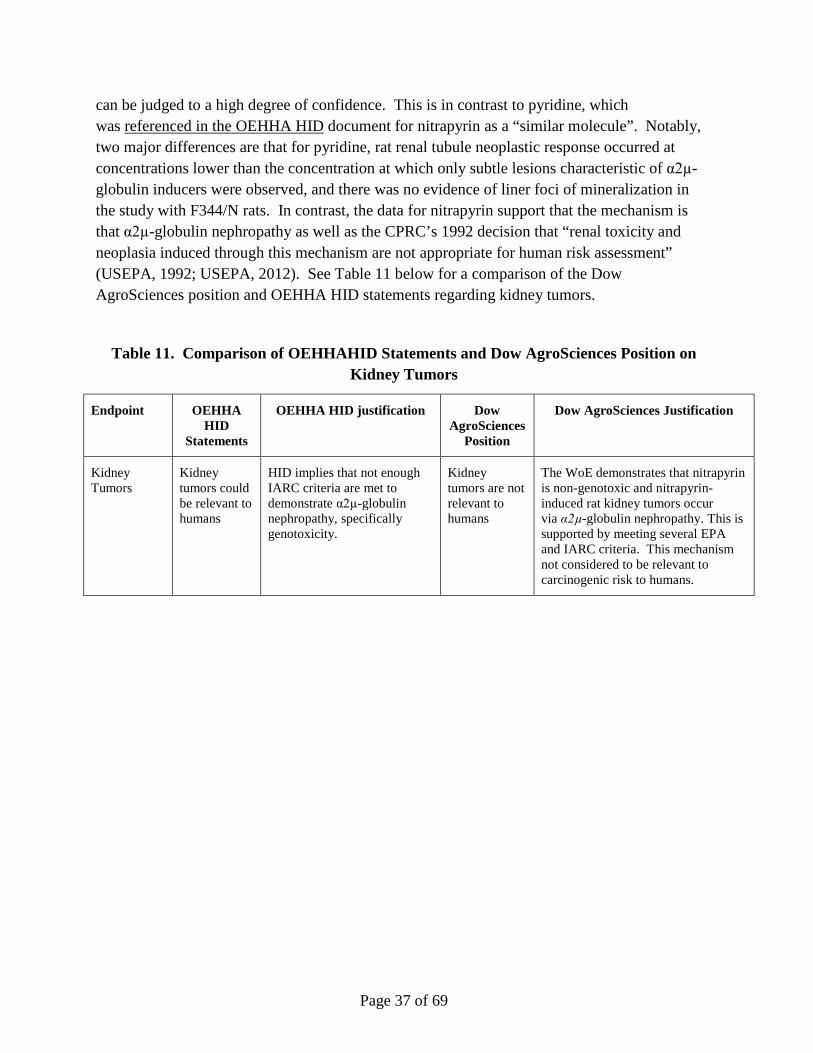
Page 37 of 69
can be judged to a high degree of confidence. This is in contrast to pyridine, which was referenced in the OEHHA HID document for nitrapyrin as a “similar molecule”. Notably, two major differences are that for pyridine, rat renal tubule neoplastic response occurred at concentrations lower than the concentration at which only subtle lesions characteristic of α2µ-globulin inducers were observed, and there was no evidence of liner foci of mineralization in the study with F344/N rats. In contrast, the data for nitrapyrin support that the mechanism is that α2µ-globulin nephropathy as well as the CPRC’s 1992 decision that “renal toxicity and neoplasia induced through this mechanism are not appropriate for human risk assessment” (USEPA, 1992; USEPA, 2012). See Table 11 below for a comparison of the Dow AgroSciences position and OEHHA HID statements regarding kidney tumors.
Table 11. Comparison of OEHHAHID Statements and Dow AgroSciences Position on Kidney Tumors
Endpoint OEHHA HID
Statements
OEHHA HID justification Dow AgroSciences
Position
Dow AgroSciences Justification
Kidney Tumors
Kidney tumors could be relevant to humans
HID implies that not enough IARC criteria are met to demonstrate α2µ-globulin nephropathy, specifically genotoxicity.
Kidney tumors are not relevant to humans
The WoE demonstrates that nitrapyrin is non-genotoxic and nitrapyrin-induced rat kidney tumors occur via α2µ-globulin nephropathy. This is supported by meeting several EPA and IARC criteria. This mechanism not considered to be relevant to carcinogenic risk to humans.
Page 38 of 69
F. Liver Tumors
Introduction to Nitrapyrin-Induced Liver Tumors
In 1992 the Cancer Peer Review Committee (USEPA, 1992) classified nitrapyrin as category Group D- not classifiable as to human carcinogenicity. There was no indication of treatment-related tumors in a two-year chronic bioassay in male and female rats except for renal tumors in male rats which were induced by α-2μ-globulin mechanism and considered not relevant to assess cancer risk in humans. A two-year oncogenicity study in B6C3F1 mice administered 0, 5, 25, or 75 mg/kg/day also was reviewed (Quast et al., 1990). The Committee was of the opinion that the dosing in the mouse study was not adequate for assessing the carcinogenic potential and requested a new dose-range finding study and an additional two-year study.
A repeat two-year oncogenicity study was conducted with B6C3F1 mice that were administered 0, 125, or 250 mg/kg/day nitrapyrin (Stebbins and Cosse, 1997). Study results indicated an increased incidence of mice with hepatocellular tumors at 125 (females only), or 250 mg/kg/day (males and females) nitrapyrin in the diet. Based in part on these results, the US EPA Cancer Assessment Review Committees (USEPA, 2000; USEPA, 2005a) classified nitrapyrin as “Likely to be carcinogenic in humans” according to the US EPA’s July, 1999 Draft Guidelines for Carcinogen Risk. The Committee also indicated that, although nitrapyrin was not mutagenic in standard guideline studies, a National Toxicology Program (NTP) study (Zeiger et al., 1988) reported that the compound was mutagenic in a Salmonella typhimurium assay in the presence of S9 activation and also indicated that there was support from structure-activity relationship (SAR) for nitrapyrin having genotoxic potential. The Committee concluded that the weight-of-the-evidence analysis was not a sufficient basis to ascribe a mutagenic mode of action to the carcinogenic response of nitrapyrin and that the issue of whether the potential mutagenicity contributes to the mode of action (MoA) for nitrapyrin-induced carcinogenesis was unresolved.
In order the address the uncertainties surrounding nitrapyrin-induced tumor formation in mice, an in vivo liver proliferation MoA study, a repeat Ames test, and an in vivo unscheduled DNA synthesis (UDS) test with mouse liver cells were performed. These data were subsequently evaluated within the original Mode of Action (MoA)/Human Relevance Framework (HRF) that assessed the key events for the MoA, which ultimately result in the formation of mouse liver tumors after lifetime exposures to relatively large dose levels of nitrapyrin (Eisenbrandt et al., 2010). Based on these results, the US EPA CARC re-classified nitrapyrin as “Suggestive Evidence of Carcinogenic Potential” according with US EPA’s Final Guidelines for Carcinogenic Risk Assessment in 2011 (USEPA, 2012). Within this assessment,
Page 39 of 69
additional data uncertainties were raised that precluded inclusion of nitrapyrin as “Not likely to be carcinogenic to humans.” In order to address these uncertainties, additional studies were performed that support the conclusion of CAR activation as the MoA for nitrapyrin-induced liver tumors. These recently conducted studies include an in vitro assay for suicide inhibition, a CAR KO mouse study, and a comparison of human and mouse primary hepatocyte culture DNA synthesis (i.e., proliferation) responses to nitrapyrin exposure. Collectively, these studies demonstrate that the MoA for nitrapyrin-mediated liver tumor formation is CAR activation. These studies are included in an updated MoA/HRF (LaRocca et al., 2015), which integrates the previous MoA/HRF analysis (Eisenbrandt et al., 2010) with the new studies and follows the guidance established for this process. The data presented in this revised HRF support the conclusion that nitrapyrin-induced mouse liver tumors are mediated by CAR activation, and due to qualitative differences between humans and rodents these tumors should be classified as “Not likely to be carcinogenic to humans” (Boobis et al., 2007; Cohen et al., 2003; Holsapple et al., 2006; Meek et al., 2003; Sonich-Mullin et al., 2001).
Background of the Proposed MoA for Nitrapyrin-Induced Liver Tumors
The MoA for nitrapyrin-induced liver tumors is via CAR (nuclear receptor) activation, and thus will be described in detail. Induction of cytochrome P450 (CYP) enzyme activity is a well-known MoA for rodent hepatocarcinogenesis, with phenobarbital (PB) as the standard example (Holsapple et al., 2006; Whysner et al., 1996). PB is non-genotoxic and is considered a tumor promoter in rodents, but is not considered relevant for human health risk assessment. The key events for this MoA include activation of the CAR receptor leading an increase in hepatocellular proliferation, and subsequent induction of proliferative lesions in the liver including hepatocellular foci, adenomas and carcinomas (Cohen, 2010). Activation of CAR in rodents results in activation of genes which leads to hepatocellular proliferation that is critical for development of liver tumors (Cohen, 2010; Whysner et al., 1996; Elcombe et al., 2014). On the other hand, PB exposure in humans results in activation of CAR/PXR receptors and induction of CYP enzymes, but different enzymes are induced in humans compared to rodents (Lambert et al., 2009) and, more importantly, there is no evidence that this results in an increase in cell proliferation in the human liver. Extensive epidemiologic studies at exposure levels in humans that are comparable to those in rodent bioassays do not result in increased cancer risks (Lamminpaa et al., 2002; Whysner et al., 1996). Based on this assessment, PB is not a hepatocarcinogen in humans due to qualitative differences between humans and rodents. Overall, human epidemiologic and laboratory mouse studies support the well-established conclusion that a non-genotoxic hepatocarcinogenic response, such as that observed with PB, in rodents due to the CAR activation MoA is not relevant to humans (Holsapple et al., 2006; Elcombe et al., 2014).
The essential role of CAR in PB-induced liver tumor formation in rodents has been demonstrated in genetically engineered mice lacking this nuclear receptor (Lake, 2009). PB

Page 40 of 69
exposure in CAR knockout mice did not increase liver weight, nor induce Cyp2b forms or stimulate replicative DNA synthesis. Also, no liver tumors were formed in CAR knockout mice following initiation with diethylnitrosamine (DEN) and promotion with PB. Further support for the critical role of rodent CAR activity in the pathogenesis of rodent liver tumors has been demonstrated with the use of humanized mouse models, where the murine CAR (and PXR) have been replaced with the human receptors (Ross et al., 2010; Braeuning et al., 2014; Luisier et al., 2014). In this model, replacing rodent receptors (CAR/PXR) with human orthologs resulted in a lack of a proliferative effect (Ross et al., 2010). However, recent work has demonstrated a transcriptional signature of proliferation in humanized mice following administration of sufficiently high doses of PB such that presence of the human receptor in murine hepatocytes may not completely recapitulate the human response (Braeuning et al., 2014). Based on the MoA assessment, CAR activations (e.g. PB) are not hepatocarcinogens in humans, presumably as a result of the divergent biological response with respect to hepatocellular proliferation.
Summary of Nitrapyrin Mouse Liver Tumor Mode of Action Data
The relevant experimental data for evaluation of the nitrapyrin liver tumor MoA and human relevance include the standard, repeated-dose, mouse studies for nitrapyrin (2-week, 13-week, 12-month, and two-year studies) as well as several mechanistic MoA studies. The data from these studies demonstrate that nitrapyrin-induced liver tumors are mediated by a MoA characterized by the following key events: 1) CAR activation, and 2) increased hepatocellular proliferation, leading to increased hepatocellular foci and tumors (apical endpoint). The data also support that other MoAs can be excluded, and that this MoA is not relevant for human health risk assessment.
The key events, along with supporting, associated events, for the nitrapyrin-induced mouse liver tumors are listed in Table 12, and the data that support these key events are described in subsequent sections of this document (Cohen, 2010; Elcombe et al., 2014; Holsapple et al., 2006).

Page 41 of 69
Table 12. Key Events for Nitrapyrin Mode of Action (MoA)
(1) CAR Activation
i. Induction of specific liver CYP enzyme Cyp2b10
ii. Irreversible inhibition of Cyp2b10-mediated PROD activity via suicide inhibition
iii. Increased liver weight
iv. Microscopic hepatocellular hypertrophy
v. Reversibility upon discontinuance of treatment
(2) Increased Hepatocellular Proliferation
i. Reversibility upon discontinuance of treatment
(Apical Endpoint) Increased hepatocellular foci and tumors
Nuclear Receptor (CAR) Activation.
The activation of CAR is the initial key event. Binding to CAR has been occasionally identified as an independent key event, but activation of CAR has been shown to occur by two independent mechanisms: 1) direct agonism by a ligand such as TCPOBOP (1,4-bis [2-(3,5-dichloropyridyloxy)]benzene), or 2) indirect by compounds such as PB that activate CAR through a phosphorylation-dependent mechanism (Rencurel et al., 2006). Regardless of the means of CAR activation, the key events related to the MoA are identical. Therefore NR activation, and not necessarily direct binding, is the most appropriate and measureable first key event. Therefore, RT-PCR analysis was utilized to investigate the following genes to investigate nuclear receptor-mediated pathways related to rodent hepatocarcinogenesis in hepatic tissue: Cyp1a1 (AhR-associated), Cyp2b10 (CAR-associated), Cyp3a11 (PXR-associated), and Cyp4a10 (PPAR-α-associated). Gene expression was measured in livers from mice from the MoA study and the mouse strain comparison study. Following 7 days of nitrapyrin treatment, there was a clear, threshold-based biological signature of CAR activation when measured at the gene expression level. Specifically, Cyp2b10 showed a clear, dose-related increase in transcript level compared to control of 4.1-, 351.0-, or 716.0-fold at 75, 250, or 400 mg/kg/day, respectively. This magnitude of fold induction for Cyp2b10 was similar to a contemporary, but unrelated, internal study with PB at a carcinogenic concentration (150 mg/kg/day in the diet for 7 days), where PB induced an 807.4-fold induction. The AhR-responsive Cyp1a1 and PXR-responsive Cyp3a11 transcripts were essentially unchanged at all dose levels, indicating that nitrapyrin is not a direct activator for AhR or PXR. The PPAR-α-inducible transcript Cyp4a10 was essentially unchanged at the 75

Page 42 of 69
mg/kg/day dose level, but was increased 6.8-fold at 250 mg/kg/day and 5.2-fold at 400 mg/kg/day. This minor, non-dose-responsive increase of Cyp4a10 at the highest doses suggests a potential non-specific or indirect result of general increased cytochrome induction.
The results of the targeted gene expression analysis revealed CAR-related changes after 14 days of nitrapyrin administration when compared to those seen with 7 days of treatment. For Cyp2b10, when compared to control, there was a 4.5-, 389.6-, 1092.3-fold induction for 75, 250, or 400 mg/kg/day nitrapyrin, respectively. Cyp1a1 and Cyp3a11 were also consistent with the 7-day time point in that there was no biologically significant alteration in the transcript levels. The Cyp4a10 transcript was increased to a lesser degree in the animals treated for 14 days (4.2- and 2.9-fold for 250 and 400 mg/kg/day, respectively) compared to 7 days, and the lack of a dose-response and minor fold-induction suggest the effects may be caused by a non-specific or indirect mechanism.
Data from the 21-day recovery groups indicated recovery from the gene expression changes that were induced following 14 days of nitrapyrin administration. Although the Cyp2b10 levels in the recovery groups were elevated slightly when compared to controls, the values were minimal compared to the significant increase after 14 days of treatment and before removal of the test material. Consistent with this decrement in gene expression, the slight increase in PPAR-α-associated Cyp4a10 transcript that was seen at 14 days was diminished in the recovery group animals treated with 250 or 400 mg/kg/day nitrapyrin.
In the strain comparison study (Murphy et al., 2014a), following 4- and 7-day exposures to 250 mg/kg/day nitrapyrin, there were robust increases in the levels of Cyp2b10 mRNA transcript levels in livers from both B6C3F1 and C57BL/6NTac strains, indicating CAR activation. Cyp2b10 transcript levels were 370.7-fold higher than controls in B6C3F1 mice after 4 days and were subsequently increased to 562.5-fold after 7 days of exposure. The results at 4 days were consistent with the previous MoA study in which Cyp2b10 levels were 351-fold higher than controls after 7 days of exposure to 250 mg/kg/day nitrapyrin (LeBaron et al., 2010). A similar increase in Cyp2b10 induction was also observed in the C57BL/6NTac mice following exposure to nitrapyrin. Of note, the Cyp2b10 transcript levels were similar at 4 days (240.7-fold) and 7 days (168.7-fold) of exposure for the C57BL/6NTac strain.
The dose response and temporality of the nitrapyrin-induced CAR activation, as evidenced by Cyp2b10 gene expression in male mice, are presented in Table 13. This initial key event for the MoA of nitrapyrin-induced mouse liver tumors, as well as the reversibility of this key event upon discontinuance of treatment, are essential data that are necessary to support this MoA (Cohen, 2010).
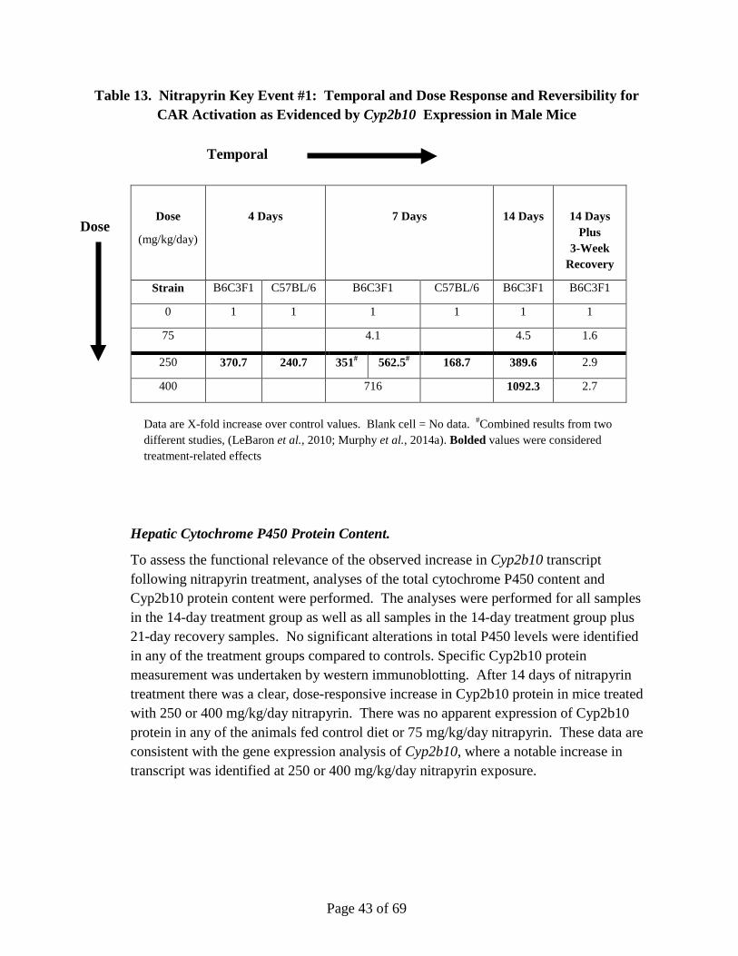
Page 43 of 69
Table 13. Nitrapyrin Key Event #1: Temporal and Dose Response and Reversibility for CAR Activation as Evidenced by Cyp2b10 Expression in Male Mice
Dose
(mg/kg/day)
4 Days
7 Days
14 Days
14 Days Plus
3-Week Recovery
Strain B6C3F1 C57BL/6 B6C3F1 C57BL/6 B6C3F1 B6C3F1
0 1 1 1 1 1 1
75 4.1 4.5 1.6
250 370.7 240.7 351# 562.5# 168.7 389.6 2.9
400 716 1092.3 2.7
Data are X-fold increase over control values. Blank cell = No data. #Combined results from two different studies, (LeBaron et al., 2010; Murphy et al., 2014a). Bolded values were considered treatment-related effects
Hepatic Cytochrome P450 Protein Content.
To assess the functional relevance of the observed increase in Cyp2b10 transcript following nitrapyrin treatment, analyses of the total cytochrome P450 content and Cyp2b10 protein content were performed. The analyses were performed for all samples in the 14-day treatment group as well as all samples in the 14-day treatment group plus 21-day recovery samples. No significant alterations in total P450 levels were identified in any of the treatment groups compared to controls. Specific Cyp2b10 protein measurement was undertaken by western immunoblotting. After 14 days of nitrapyrin treatment there was a clear, dose-responsive increase in Cyp2b10 protein in mice treated with 250 or 400 mg/kg/day nitrapyrin. There was no apparent expression of Cyp2b10 protein in any of the animals fed control diet or 75 mg/kg/day nitrapyrin. These data are consistent with the gene expression analysis of Cyp2b10, where a notable increase in transcript was identified at 250 or 400 mg/kg/day nitrapyrin exposure.
Temporal
Dose

Page 44 of 69
Hepatic Metabolic Enzyme Activity.
Following the results of the gene expression and protein analyses, which indicated a significant elevation of Cyp2b10 at the gene and protein levels, a functional analysis of the Cyp2b10 enzyme was undertaken using 7-Pentoxy-Resorufin O-Deethylation (PROD) activity as the metric. Increased PROD enzyme activity is one of the primary alterations observed following CAR activation (Lubet et al., 1985). The analysis was performed for all samples in the 14-day treatment group as well as all samples in the 14-day treatment group plus recovery. There were no significant alterations in the PROD activities for any of the treatment groups compared to control after 14 days of nitrapyrin treatment or after 14 days of treatment and 21 days of recovery.
Suicide Inhibition of P450 Enzyme Activity.
A number of studies using structurally diverse compounds have previously identified inhibitors of specific cytochrome activity while increasing the expression of the relevant gene as well as the protein levels. Inhibition of cytochrome activity may occur as a result of substrate competition, metabolic intermediate complexation, or mechanism-based (suicide) inhibition (Halpert, 1995; Murray and Reidy, 1990). For example, in the rat and dog the antibiotic chloramphenicol has been shown to be a mechanism-based inactivator of PB-induced (i.e., Cyp2b family) cytochrome activity (Halpert et al., 1985; Ciaccio et al., 1987) .
Given the paradoxical finding of robust increases in Cyp2b10 with no associated change in the activity of the enzyme as measured by PROD, in vitro experiments were conducted with phenobarbital (PB)-induced liver microsomes to investigate the role for suicide inhibition. In this system, PB-induces PROD activity in microsomes in vivo, therefore, as PB is known not to be a suicide inhibitor of Cyp2b activity, exposure to additional PB to these isolated microsome in vitro should not affect PROD activity in either direction. PROD activity is only reduced following in vitro exposure to a suicide inhibitor of PROD, such as curcumin. In this experiment, PB-induced microsomes were analyzed for PROD activity following treatment with PB (negative control), curcumin (positive control, (Thapliyal and Maru, 2001)), or nitrapyrin (Figure 2). PB had no effect on PROD activity. Curcumin had a dose-related inhibition of PROD activity of up to 63% at 40 μM, also consistent as a positive control. Finally, in this system, nitrapyrin inhibited PROD activity in a dose-related manner and up to 96% at 500 μM. These results indicate that nitrapyrin and/or its metabolites irreversibly inhibited Cyp2b10-mediated PROD activity of PB-induced microsomes. As noted in the HID, the lack of PROD activity was listed as a major uncertainty to the nitrapyrin liver tumor MoA. These data elucidate the apparent inconsistency between protein levels and enzyme activity of nitrapyrin-treated livers. Hence, the lack of PROD activity in nitrapyrin-treated liver does not indicate a lack of CAR-mediated activity. In fact,
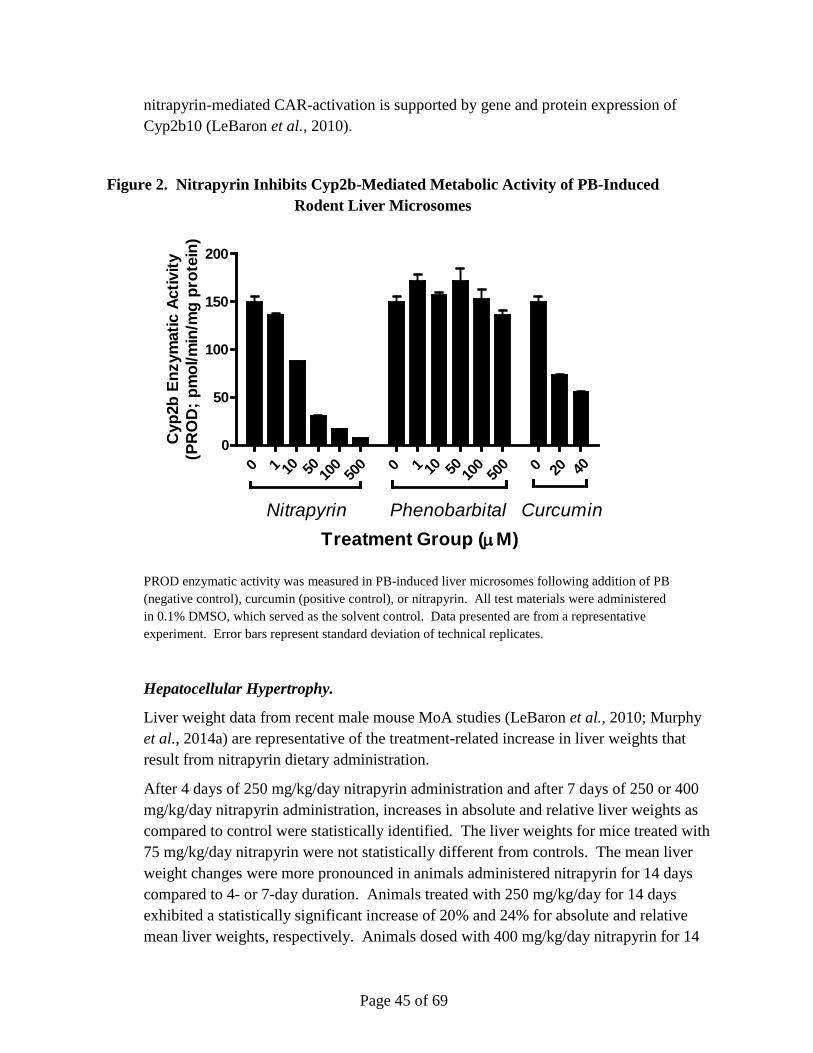
Page 45 of 69
nitrapyrin-mediated CAR-activation is supported by gene and protein expression of Cyp2b10 (LeBaron et al., 2010).
Figure 2. Nitrapyrin Inhibits Cyp2b-Mediated Metabolic Activity of PB-Induced Rodent Liver Microsomes
PROD enzymatic activity was measured in PB-induced liver microsomes following addition of PB (negative control), curcumin (positive control), or nitrapyrin. All test materials were administered in 0.1% DMSO, which served as the solvent control. Data presented are from a representative experiment. Error bars represent standard deviation of technical replicates.
Hepatocellular Hypertrophy.
Liver weight data from recent male mouse MoA studies (LeBaron et al., 2010; Murphy et al., 2014a) are representative of the treatment-related increase in liver weights that result from nitrapyrin dietary administration.
After 4 days of 250 mg/kg/day nitrapyrin administration and after 7 days of 250 or 400 mg/kg/day nitrapyrin administration, increases in absolute and relative liver weights as compared to control were statistically identified. The liver weights for mice treated with 75 mg/kg/day nitrapyrin were not statistically different from controls. The mean liver weight changes were more pronounced in animals administered nitrapyrin for 14 days compared to 4- or 7-day duration. Animals treated with 250 mg/kg/day for 14 days exhibited a statistically significant increase of 20% and 24% for absolute and relative mean liver weights, respectively. Animals dosed with 400 mg/kg/day nitrapyrin for 14
0 0 00
50
100
150
200
PhenobarbitalNitrapyrin Curcumin
Cyp
2b E
nzym
atic
Act
ivity
(PR
OD
; pm
ol/m
in/m
g pr
otei
n)
1 10 50 100
500 1 10 50 10
050
0 20 40
Treatment Group (µM)
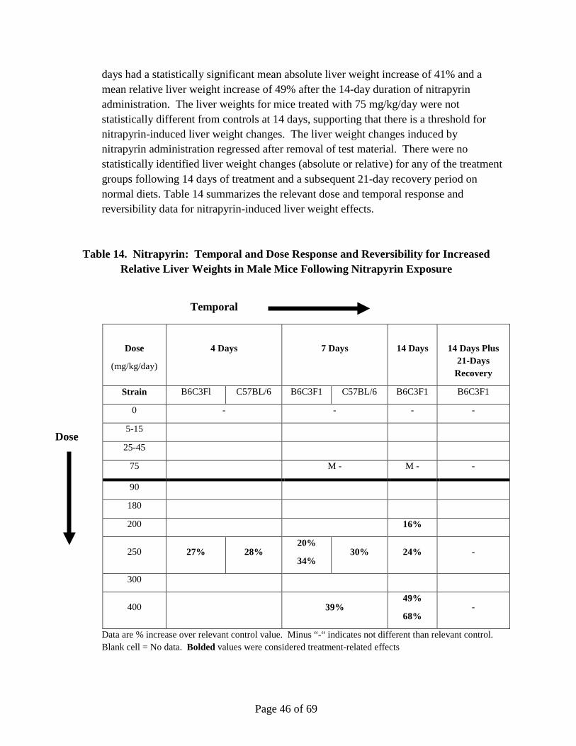
Page 46 of 69
days had a statistically significant mean absolute liver weight increase of 41% and a mean relative liver weight increase of 49% after the 14-day duration of nitrapyrin administration. The liver weights for mice treated with 75 mg/kg/day were not statistically different from controls at 14 days, supporting that there is a threshold for nitrapyrin-induced liver weight changes. The liver weight changes induced by nitrapyrin administration regressed after removal of test material. There were no statistically identified liver weight changes (absolute or relative) for any of the treatment groups following 14 days of treatment and a subsequent 21-day recovery period on normal diets. Table 14 summarizes the relevant dose and temporal response and reversibility data for nitrapyrin-induced liver weight effects.
Table 14. Nitrapyrin: Temporal and Dose Response and Reversibility for Increased Relative Liver Weights in Male Mice Following Nitrapyrin Exposure
Dose
(mg/kg/day)
4 Days
7 Days
14 Days
14 Days Plus 21-Days
Recovery
Strain B6C3Fl C57BL/6 B6C3F1 C57BL/6 B6C3F1 B6C3F1
0 - - - -
5-15
25-45
75 M - M - -
90
180
200 16%
250 27% 28% 20%
34% 30% 24% -
300
400 39% 49%
68% -
Data are % increase over relevant control value. Minus “-“ indicates not different than relevant control. Blank cell = No data. Bolded values were considered treatment-related effects
Temporal
Dose

Page 47 of 69
Microscopic Hepatocellular Hypertrophy.
Hepatocellular hypertrophy associated with nitrapyrin administration in the mouse is also reflected in histopathological evaluation of liver tissue. The data from two studies (LeBaron et al., 2010; Murphy et al., 2014a) are representative of the treatment-related increase in hepatocellular hypertrophy that result from nitrapyrin dietary administration. Mice treated with 75 mg/kg/day nitrapyrin did not have evidence of hepatocellular hypertrophy at any time point. B6C3F1 mice exposed to 250 or 400 mg/kg/day nitrapyrin for 7 or 14 days had very slight, slight, or moderate centrilobular/midzonal hepatocyte hypertrophy with altered tinctorial properties of the cytoplasm (increased eosinophilia). These effects were no longer present at these dose levels after 3 weeks of recovery on standard diet. B6C3F1 mice and C57Bl/6NTac mice exposed to 250 mg/kg/day nitrapyrin for 4 days also exhibited increased in slight centrilobular/midzonal hepatocyte hypertrophy with increased cytoplasmic eosinophilia. Notably, these effects were consistent with observed liver weight increases.
As would be expected, similar to the liver weight data, there is an increased incidence in microscopic hepatocellular hypertrophy at dose levels ≥90 mg/kg/day and essentially no increase in incidence at dose levels ≤75 mg/kg/day nitrapyrin. These results correlated with the Cyp2b10 gene expression data for nitrapyrin which demonstrated significant increases at 250 and 400 mg/kg/day after 4, 7, or 14 days, but not at 75 mg/kg/day. The microscopic hepatocellular hypertrophy data also correlate with the hepatocellular tumor results for the combined nitrapyrin mouse oncogenicity studies (Quast et al., 1990; Stebbins and Cosse, 1997) in that there was no increase in hepatocellular tumors at 5, 25, or 75 mg/kg/day, while an increased incidence of tumors was detected at 125 (females) and 250 (males and females) mg/kg/day.
Hepatocellular Proliferation.
Data from two studies (LeBaron et al., 2010; Murphy et al., 2014a) are representative of the treatment-related increase in hepatocellular proliferation that result from nitrapyrin dietary administration. Immunohistochemical staining for BrdU-labeled nuclei as a measure of hepatocellular proliferation was based on interpretation of the nuclear staining intensity and an approximation of the location within three hepatolobular zones. Following 4 and 7 days of nitrapyrin exposure (Murphy et al., 2014a), B6C3F1 mice had a mild to moderate statistically identified, treatment-related increase in hepatocellular proliferation as measured by an increase in BrdU labeling indices (LI) in the midzonal and periportal regions compared to controls. Although the magnitude of the liver weight increase was slightly higher at 7 days, the hepatocellular proliferation was similar at both timepoints for both the midzonal (2.3-fold vs. 2.5-fold) and

Page 48 of 69
periportal (5.9-fold vs. 6.2-fold) regions. Hepatocellular proliferation in the centrilobular zone was similar to controls following exposure to nitrapyrin at 4 days and was slightly increased at 7 days (1.8-fold higher compared to controls). In (LeBaron et al., 2010), similar increases in hepatocellular proliferation were observed following 250 mg/kg/day nitrapyrin exposure for 7 days. Similar to the B6C3F1 mice, the C57BL/6NTac mice had statistically identified treatment-related increases in hepatocellular proliferation at 4 days in both the midzonal and periportal regions. This increase was evident at 7 days, although the % BrdU labeling indices (LI) in the midzonal and periportal zones were slightly lower than the response observed at 4 days.
Hepatocellular proliferation analysis after 7 days of 400 mg/kg/day nitrapyrin administration in mice resulted in a statistically significant increased percentage of stained cells, when compared to those of the control group, in the midzonal (3.0-fold) and periportal (8.0-fold) regions. Furthermore, the totaled panlobular counts also were statistically identified at the 400 mg/kg/day dose level after 7 days of nitrapyrin administration. Although the periportal region was the only statistically significant increased zone in animals treated with 250 mg/kg/day nitrapyrin, there was a notable increase in proliferation in the midzonal region and panlobular counts. Therefore, in male mice given 250 mg/kg/day nitrapyrin for 4 or 7 days, or 400 mg/kg/day nitrapyrin for 7 days, there was a general treatment-related increase in hepatocellular proliferation for the periportal and midzonal regions, as well as the total panlobular count.
The trend of hepatocellular proliferation was similar, but more robust, in animals treated with 14 days of nitrapyrin by dietary administration (with a BrdU osmotic pump present for the last 7 days of treatment) when compared to 4 or 7 days of treatment. Specifically, in animals treated with 400 mg/kg/day there was a statistically identified increase in the proliferation index in midzonal, periportal, and total (panlobular) counts of 3.8-, 11.2-, and 4.9-fold, respectively, when compared to control animals. In addition, animals treated with 250 mg/kg/day nitrapyrin had statistically identified increases in hepatocellular proliferation of 3.8-fold in the periportal region and 2.4-fold in the summed, panlobular count. Although not statistically significant, there was a 2.1- and 1.7-fold increase in labeled hepatocytes in the centrilobular and midzonal regions, respectively.
The induction of hepatocellular proliferation was completely reversed in animals allowed to recover for a total of 21 days (with a BrdU osmotic pump present for the last 7 days of recovery) following treatment with nitrapyrin for 14 days. There were no statistically significant increases in hepatocellular proliferation at any dose level or in any of the hepatolobular regions analyzed. Interestingly, there was a statistically identified decrease in proliferation of -3.1-fold in the periportal region for the animals allowed to recover after being treated with 400 mg/kg/day nitrapyrin. Furthermore, there was also a notable, albeit statistically non-significant, decrease in proliferation in
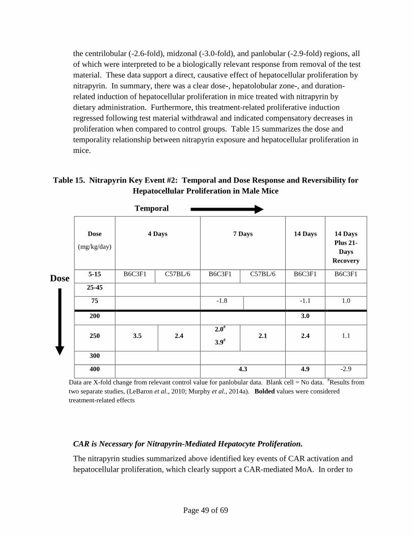
Page 49 of 69
the centrilobular (-2.6-fold), midzonal (-3.0-fold), and panlobular (-2.9-fold) regions, all of which were interpreted to be a biologically relevant response from removal of the test material. These data support a direct, causative effect of hepatocellular proliferation by nitrapyrin. In summary, there was a clear dose-, hepatolobular zone-, and duration-related induction of hepatocellular proliferation in mice treated with nitrapyrin by dietary administration. Furthermore, this treatment-related proliferative induction regressed following test material withdrawal and indicated compensatory decreases in proliferation when compared to control groups. Table 15 summarizes the dose and temporality relationship between nitrapyrin exposure and hepatocellular proliferation in mice.
Table 15. Nitrapyrin Key Event #2: Temporal and Dose Response and Reversibility for Hepatocellular Proliferation in Male Mice
CAR is Necessary for Nitrapyrin-Mediated Hepatocyte Proliferation.
The nitrapyrin studies summarized above identified key events of CAR activation and hepatocellular proliferation, which clearly support a CAR-mediated MoA. In order to
Dose
(mg/kg/day)
4 Days
7 Days
14 Days
14 Days Plus 21-
Days Recovery
5-15 B6C3F1 C57BL/6 B6C3F1 C57BL/6 B6C3F1 B6C3F1
25-45
75 -1.8 -1.1 1.0
200 3.0
250 3.5 2.4 2.0#
3.9# 2.1 2.4 1.1
300
400 4.3 4.9 -2.9
Data are X-fold change from relevant control value for panlobular data. Blank cell = No data. #Results from two separate studies, (LeBaron et al., 2010; Murphy et al., 2014a). Bolded values were considered treatment-related effects
Temporal
Dose

Page 50 of 69
confirm this MoA, and eliminate alternative MoAs, CAR KO and WT (knock-out and wild-type) mice were compared for their hepatic response to nitrapyrin.
As discussed in previous sections, nitrapyrin exposure resulted in similar responses (liver weight increases, liver hypertrophy, and hepatocellular proliferation) in both B6C3F1 and C57BL/6NTac strains. Notably, these responses were also comparable to previous data obtained from a nitrapyrin MoA study (LeBaron et al., 2010). Overall responsiveness to nitrapyrin-mediated hepatic effects was largely similar between B6C3F1 and C57BL/6NTac mice at both timepoints analyzed (4 or 7 days of exposure). Gene expression responses and increased hepatocellular proliferation for C57BL/6NTac mice were greater at 4 days compared to 7 days exposure and indicated that a shorter exposure to nitrapyrin was sufficient for induction of hepatic responses. Based on similar responses of liver weight, hepatocellular proliferation, Cyp2b10 gene expression, liver hypertrophy, B6C3F1 and C57BL/6NTac were considered qualitatively similar in their hepatic responses to nitrapyrin.
Consistent with previous studies, administration of the carcinogenic dose level of 250 mg/kg/day nitrapyrin to WT mice resulted in treatment-related increases in relative (20%) and absolute (24%) liver weights compared to untreated control WT animals. These liver weight changes were consistent with observed treatment-related histopathological alterations of: 1) a very slight increase in mitotic figures (hepatocytes in mitoses), 2) slight centrilobular/midzonal hepatocellular hypertrophy with increased cytoplasmic eosinophilia, and 3) a very slight vacuolization of centrilobular/midzonal hepatocytes consistent with a minimal fatty change. Gene expression analysis of the liver indicated a robust increase in the Cyp2b10/CAR-associated transcript (493.7-fold higher than unexposed WT animals) consistent with direct activation of this nuclear receptor but not AhR, PXR, or PPAR-α. Consistent with CAR activation, nitrapyrin-treated WT mice had a treatment-related increase in BrdU labeling index (2.5-fold) in hepatocytes in the periportal lobular region of the liver as compared to untreated WT controls.
While the liver weight changes in nitrapyrin-exposed CAR KO mice were similar to treated WT mice, the histopathological and molecular responses were markedly different. In CAR KO mice given 250 mg/kg/day nitrapyrin, the histopathological findings were limited to very slight hepatocellular hypertrophy with increased eosinophilia and very slight vacuolization, consistent with fatty change in centrilobular/midzonal hepatocytes. In contrast to nitrapyrin-treated WT mice, there was no indication of a proliferative response in CAR KO mice, as indicated by the absence of mitotic figures.
Furthermore, the gene expression response of the liver following nitrapyrin exposure in CAR KO mice was distinctly different than treated WT mice. Specifically, there was no biologically significant induction of the Cyp2b10/CAR-associated transcript (2.2-

Page 51 of 69
fold vs. 493.7-fold in WT mice). Similar to the response to nitrapyrin in WT mice, the Cyp3a11/PXR- and Cyp4a10/PPAR-α-associated transcripts were unchanged, indicating no clear activation of those pathways. However, the Cyp1a1/AhR-associated transcript was induced to a greater degree in treated CAR KO mice compared to treated WT mice. It has been established that there is extensive crosstalk between nuclear receptors and transcription factors (Moreau et al., 2008; Gao and Xie, 2012; Pascussi et al., 2003). The presence of various nuclear receptors that have different yet overlapping specificity for ligands allows for an organism to respond to exposure to xenobiotics more effectively. Competition for cofactors and other cellular transcription resources may account for differences in CYP induction following exposure to xenobiotics. In fact, for AhR specifically, reduction in the amount of cofactor and increasing the amount of competing non-AhR binding proteins reduces Cyp1a1 gene expression (Simon et al., 2015). For nitrapyrin, in the absence of a functional CAR signaling pathway, the AhR pathway is weakly activated potentially as an adaptive response due to the lack of competition for cofactors and co-activators. Importantly, the magnitude of induction (104.4-fold) in the CAR-KO mouse was substantially less than that seen with a prototypical AhR activator (up to 5000-fold for a similar exposure period) (Boverhof et al., 2006). This supports that AhR activation observed in the CAR KO animals was likely an adaptive liver enzyme response due to the absence of a functional CAR signaling pathway. The observed changes in liver histopathology in CAR KO mice also likely reflect activation of an adaptive enzyme response.
Finally, hepatocellular proliferation was evaluated in WT and CAR KO mice given is it the critical event in nitrapyrin-induced liver tumor formation (Table 16). Similar to what was observed in the strain comparison study, there was a clear increase in hepatocellular proliferation as measured by BrdU incorporation in WT mice following nitrapyrin administration. However, no increase in proliferation was observed in CAR KO animals. This is consistent with the previously discussed differences (increase in mitotic figures) between CAR KO and WT responses to nitrapyrin exposure. These results, combined with the observed liver weight and histopathologic changes in CAR KO mice, suggest that while nitrapyrin exposure still confers some form of hypertrophic response in the absence of CAR, it does not confer the critical hyperplastic response (proliferation) that is necessary for tumor formation in this MoA.

Page 52 of 69
Table 16. Summary of Hepatocellular Proliferation by Lobular Zones in Livers of WT and CAR KO Male C57BL/6NTac Mice
WT Male C57BL/6NTac Mice
Nitrapyrin Dose (mg/kg/day) Centrilobular Midzonal Periportal Total
0 1.0 1.0 1.0 1.0
250 0.8 1.4 2.4* 1.5*
CAR KO Male C57BL/6NTac Mice
Nitrapyrin Dose (mg/kg/day) Centrilobular Midzonal Periportal Total
0 1.0 1.0 1.0 1.0
250 0.6 0.8 0.9 0.8
Data are fold change relative to control labeling indices. Bold values were considered treatment-related effects. * = Significantly different from control mean by t-test, alpha=0.05.
The data described herein using CAR KO and WT mice exposed to nitrapyrin support that CAR activation is necessary for the key events (CAR activation and proliferation) in the pathogenesis of nitrapyrin-induced rodent hepatocellular tumors. Despite having a similar response to WT mice in liver weight increase, the histopathological and molecular signatures overwhelming indicate the response to nitrapyrin treatment was markedly different in CAR KO mice. Specifically, no CAR activation, hepatocellular proliferation, or an increase in mitotic figures was observed in CAR KO mice. Given the absence of increased hepatocellular proliferation in CAR KO mice following exposure to a tumorigenic dose of nitrapyrin, the results from this study confirm the specificity of CAR pathway activation in the MoA for nitrapyrin-induced liver tumors.
Nitrapyrin Elicits Increased Proliferation in Mouse but Not Human Hepatocytes.
Considering CAR activation causes liver tumors and hepatocyte proliferation in mice but not in humans (Elcombe et al., 2014), and our data support that CAR is necessary for nitrapyrin-mediated liver proliferation in mice, we tested the hypothesis that nitrapyrin increases proliferation of primary mouse hepatocytes but not primary human hepatocytes. Exposing primary human hepatocytes to chemicals represents a useful tool to investigate CAR-mediated proliferation response, which is a critical key event that ultimately leads to liver tumor formation. This has been specifically established with
Page 53 of 69
PB. As noted in the nitrapyrin OEHHA HID, there are qualitative differences among rodents and humans regarding CAR-mediated induction of CYP genes. In rodents, activation of CAR (such as from PB exposure) induces expression of cyp2b10. In humans, PB has been shown to induce several hepatic CYP genes, including CYP2B6, CYP2A6, CYP2C9, CYP2C19, and CYP3A4 (Elcombe et al., 2014). While PB exposure can induce hepatocyte hypertrophy in both rodents and humans, hyperplastic effects are only observed in rodents.
While the OEHHA HID (OEHHA, 2015) notes that there is variability in the expression of CYP2B6 induced by human CAR and that two human donors may not represent the human population, it is the proliferative response, not CYP gene expression, that is the critical endpoint to examine. Additionally, cyp gene expression serves as a biomarker of CAR activation. CAR activation, not cyp gene expression, induces hyperplastic effects in rodents. Again, utilizing cultured human hepatocytes has previously been valuable to determine human relevance for PB-mediated liver effects in rodents, and is a useful system to determine the relevance of nitrapyrin-mediated hepatocellular proliferation in rodents.
A primary hepatocyte culture system was used to evaluate proliferation of mouse and human hepatocytes in response to nitrapyrin. The adequacy of the experimental conditions for induction of cell proliferation in mouse and human hepatocytes was evaluated by employing a positive control chemical (25 ng/µl epidermal growth factor (EGF)) that is known to increase primary hepatocyte proliferation (Bowen et al., 2014). Primary CD-1 mouse hepatocytes and primary human hepatocytes were cultured on glass coverslips and exposed to test material for 48 hours. A concentration gradient of nitrapyrin was studied up to a maximal concentration of 100 µM. Hepatocyte cytotoxicity was examined by monitoring mitochondrial metabolic activity using a MTT reagent, thiazolyl blue tetrazolium bromide. Proliferative DNA synthesis (i.e., cell proliferation) was examined by adding the thymidine analog 5-ethynyl-2′-deoxyuridine (EdU) for 24 hours at the end of the culture period, detecting EdU and nuclei with fluorescence techniques, and counting the proportion of hepatocytes positive for nuclear EdU incorporation using an epifluorescence microscope.
Cytotoxicity was used to determine appropriate test material concentrations for examination of hepatocyte proliferation. Hepatocyte proliferation was scored at test material concentrations causing less than 30% cytotoxicity. EGF did not alter the viability of either mouse or human primary hepatocytes. Cytotoxicity above the 30% threshold level for examination of cell proliferation was observed in mouse primary hepatocytes exposed to nitrapyrin concentrations ≥30 µM. Nitrapyrin did not cause cytotoxicity of human hepatocytes above the 30% threshold level for examination of cell proliferation at concentrations up to and including 100 µM.

Page 54 of 69
Nitrapyrin caused a treatment-related increase in proliferation of mouse primary hepatocytes but not human primary hepatocytes (Figure 3) (Johnson and Kan, 2015). EGF increased cell proliferation approximately four to five fold in mouse and human primary hepatocytes, demonstrating the similar responsiveness of mouse and human primary hepatocyte cultures to a proliferative stimulus. A treatment-related three-fold increase in proliferation of mouse hepatocytes was observed after exposure to 10 µM nitrapyrin. Nitrapyrin did not increase the proliferation of human hepatocytes at concentrations up to and including 100 µM. Therefore, under the conditions of this study, nitrapyrin induced murine hepatocyte proliferation consistent with the in vivo response in mice; however, human hepatocytes did not proliferate in response to nitrapyrin administration.
Figure 3. Mouse and Human Primary Hepatocyte Response to EGF or Nitrapyrin Exposure
Mouse Human
Compared to vehicle control (PBS), 25 ng/ml EGF exposure caused a statistically-identified approximately five fold increase in human hepatocyte proliferation and approximately four fold increase in mouse hepatocyte proliferation. Compared to vehicle control (DMSO), nitrapyrin exposure increased mouse hepatocyte proliferation, which was approximately three fold and statistically-identified at 10 μM. Compared to vehicle control (DMSO), nitrapyrin exposure did not alter human hepatocyte proliferation at any concentration tested. Data shown are the mean ± standard deviation. *p-value < 0.05. (Johnson and Kan, 2015)

Page 55 of 69
Increased Hepatocellular Foci:
Increased hepatocellular proliferation is a key event for a NR/Cyp MoA for hepatocellular carcinogens. The increased hepatocellular proliferation leads to the induction of proliferative lesions in the liver, including foci, adenomas, and carcinomas (Cohen, 2010). Nitrapyrin administration was associated with an increase in the number of mice with one or more eosinophilic and/or basophilic foci of altered hepatocytes in male and female mice administered 250 mg/kg/day. The number of male and female mice with eosinophilic foci was greater than the number of mice with basophilic foci at 250 mg/kg/day. Also, the number of mice with multiple eosinophilic foci (0, 1, 9 males and 0, 0, 11 females at 0, 125, 250 mg/kg/day, respectively) was greater than the number of mice with multiple basophilic foci (0, 0, 6 males and 0, 0, 2 females at 0, 125, 250 mg/kg/day, respectively) (data not shown; see (Stebbins and Cosse, 1997)). The increased number of mice with foci and the increased number of foci per mouse (especially eosinophilic foci) correlate with the increased number of mice with hepatocellular adenomas and/or carcinomas attributed to nitrapyrin treatment at 250 mg/kg/day. The number of mice with foci of altered cells was not increased at 125 mg/kg/day, but this dose approximates a threshold dose for hepatocellular tumor formation as evidenced by the absence of an increase in male mice with hepatocellular adenomas and/or carcinomas at 125 mg/kg/day and no increases in hepatocellular tumors in either sex at ≤75 mg/kg/day.
Summary of Nitrapyrin Liver Tumor MoA
The MoA for nitrapyrin-induced mouse liver tumors is nuclear receptor-mediated CAR CYP enzyme induction, which is similar to the MoA for PB-induced rodent liver tumors. The relevant molecular and pathological endpoints for nitrapyrin-induced liver effects in mice are summarized in Table 17. The table is organized such that the metrics are consistent with the established key events of nuclear receptor-mediated rodent hepatocarcinogenesis (Cohen et al., 2003; Lake, 2009).
Key event #1 for the nitrapyrin liver tumor MoA is defined as activation of the nuclear receptor (i.e., CAR), which was surrogately measured by induction of the Cyp2b10 transcript as well as the protein. Experimental evidence support the hypothesis that the lack of elevated PROD metabolic activity typically observed with CAR activation was attributed to mechanism-based (suicide) inhibition of the enzyme. Supportive, associative key events to #1 include increased liver weight and microscopic hepatocellular hypertrophy, which were increased in a dose-dependent manor following nitrapyrin exposure. Key event #2 is characterized as an increase in hepatocellular proliferation. Exposure at or above the tumorigenic dose of nitrapyrin for males (250 mg/kg/day) consistently produced characteristic causal and associative events for this MoA in several different experiments and in multiple strains of mice. Studies utilizing
Page 56 of 69
CAR KO and WT mice corroborated that increased hepatocellular proliferation, a key event for nitrapyrin-induced liver tumors, was dependent on CAR activation, thereby confirming the proposed MoA. The key events demonstrate reversibility upon discontinuance of treatment, a fundamental element typical for the nuclear receptor mediated liver tumor MoA. Furthermore, in vitro experiments conducted with mouse and human primary hepatocytes demonstrated that nitrapyrin exposure induced cellular proliferation (key event #2) in rodent cells but not humans, consistent with the fact that this MoA is not relevant for human health assessment given qualitative differences between species.
The key events for nitrapyrin show clear, threshold, dose-responsive alterations and provide informative, temporal-specific characterization of nitrapyrin-induced liver effects, which are consistent with a CAR-mediated MoA. The temporality, dose response, and reversibility of the key events for the nitrapyrin mouse liver tumor MoA are summarized in Table 17 for the dose levels utilized on both nitrapyrin mouse carcinogenicity studies. These data provide convincing evidence that the MoA key events as well as the hepatocellular tumors do not occur at or below a defined dose level of 75 mg/kg/day. These data definitely support that CAR nuclear receptor activation is the MoA for nitrapyrin-mediated liver tumors, as well as clearly exclude alternative MoAs. These data have been incorporated into a human relevance framework (LaRocca et al., 2015), which is summarized below:

Page 57 of 69
Table 17. Nitrapyrin: Temporality, Dose Response, and Reversibility for MoA Key Events Related to Male Mouse Liver Tumors at Dose Levels Used for Cancer Studies
Dose for 2-Yr
Studies (mg/kg/
day)
Key Event 1 Key Event 2
Key Events After
Recovery
Apical Endpoints:
Increased Hepatocellular
Tumors and Altered Foci
Causal: CAR Activation
(Cyp2b10 Transcript &
Protein)
Associated: Increased Liver
Weights/ Hypertrophy
Hepatocellular Proliferation
4-14 Days 4-14 Days 4-14 Days 14 Days Plus
21 Days Recovery
2 Yrs
5 - -
25 - -
75 - - - - -
125 +,+# +,+# -
250 + + + - +
250 CAR KO - +@ -
+ Indicates effect present, - indicates effect absent at indicated duration of treatment. Blank cell = No data. #Data only from 1-year interim sacrifice (Stebbins and Cosse, 1997). @Suggestive of compensatory changes in CAR KO
NITRAPYRIN MOUSE LIVER TUMOR HUMAN RELEVANCE FRAMEWORK
Question 1. Is the weight of evidence sufficient to establish the mode of action in animals?
The answer is yes. The data presented herein support that the MoA for nitrapyrin-induced mouse liver tumors is congruent with the well-established MoA of CAR nuclear receptor activation, similar to the PB-like MoA (Elcombe et al., 2014; Cohen, 2010; Holsapple et al., 2006; Lake, 2009; Whysner et al., 1996). The relevant molecular and
Temporal
Dose

Page 58 of 69
pathological endpoints for nitrapyrin-induced liver effects in mice via this MoA are consistently supported across several repeat-dose studies and carcinogenicity studies (LeBaron, 2010; LeBaron et al., 2010; LeBaron et al., 2014; Murphy et al., 2014b; Murphy et al., 2014a; Yano et al., 2008), and there is clear correlation of the dose responses between the MoA data and the hepatocellular tumors.
Key event #1 for the nitrapyrin liver tumor MoA is defined as activation of the CAR nuclear receptor which is surrogately measured by induction of the Cyp2b10 transcript as well as the protein. Key event #1 is accompanied by mechanism-based (suicide) inhibition of the enzyme PROD, which elucidates the unexpected lack of elevated of PROD metabolic activity. Supportive, associative key events to key event #1 include increased liver weight and microscopic hepatocellular hypertrophy.
Key event #2 is an increase in hepatocellular proliferation at tumorigenic dose levels. Importantly, both of these key events demonstrate reversibility upon discontinuance of treatment and show consistency across multiple studies. The key events for nitrapyrin show clear, thresholded, dose-responsive alterations and are consistent with a CAR-mediated MoA. The absence of effects associated with these key events in CAR KO mice exposed to a tumorigenic dose of nitrapyrin confirmed that CAR is necessary for nitrapyrin-mediated liver effects that lead to tumorigenesis. Furthermore, other possible MoAs are considered unlikely based on an analysis of all relevant data for nitrapyrin.
Question 2. Can human relevance of the MoA be reasonably excluded based on fundamental qualitative differences in key events between experimental animals and humans?
The answer is yes. The key events for this MoA in rodents include activation of CAR and leads to an increase in hepatocellular proliferation and subsequent induction of proliferative lesions in the liver including foci, adenomas, and carcinomas. Activation of CAR in rodents leads to activation of genes that leads to hepatocellular proliferation, which is critical for development of liver tumors (Cohen, 2010; Whysner et al., 1996; Elcombe et al., 2014). While exposure to PB, a prototypical CAR activator, in humans results in activation of the CAR/PXR receptors and induction of CYP enzymes, different enzymes are induced in humans compared to rodents and, more importantly, there is no evidence that this results in an increase in cell proliferation in the human liver (Lambert et al., 2009). Consistent with this prior evidence, while nitrapyrin exposure to primary mouse hepatocytes led to increased proliferation, this effect was not observed in primary human hepatocytes, thereby substantiating that the MoA for nitrapyrin-mediated liver tumors in mice is not relevant for humans (Elcombe et al., 2014).

Page 59 of 69
Regarding cancer risk for humans, extensive epidemiologic studies in humans with PB at exposure levels that are comparable to those in rodent bioassays conclude that PB exposure at these levels does not result in increased cancer risks (Lamminpaa et al., 2002; Whysner et al., 1996). Based on the MoA assessment, PB is not a hepatocarcinogen in humans. As noted in the HID by OEHHA (2015), there is however limited evidence showing similar responses to PB-exposure in CAR/PXR humanized mice as compared to control mice. However, these responses do not correlate with human in vitro studies (summarized in (Elcombe et al., 2014)), and the relevance of humanized mice for nongenotoxic rodent carcinogens remains questionable (Scheer and Wilson, 2015; Elcombe et al., 2014). There are several disadvantages to using genetically humanized mice, which include but are not limited to: the expression of selected human genes only (the rest of genome remains mouse) and potential compensatory gene expression changes. The accepted consensus with several regulatory agencies across the globe is that a hepatocarcinogenic response in rodents for compounds which have data to support a CAR-mediated (i.e., PB-like) MoA is not relevant to humans (Holsapple et al., 2006; Elcombe et al., 2014). While there was one epidemiologic study noted in the HID that demonstrated an increased risk of cancer in several sites among patients taking antiepileptic drugs, this study was unable to assess cancer risk with PB specifically (Lamminpaa et al., 2002). Indeed, there is a wealth of epidemiologic data which demonstrate that patients have no evidence of increased liver tumor risk following extended treatment with PB (summarized in (Elcombe et al., 2014)).
On these bases, the mouse liver tumors associated with administration of higher dose levels of nitrapyrin would not pose a cancer hazard to humans. See Table 18 below for a comparison of the OEHHA HID statements and Dow AgroSciences positions on nitrapyrin-induced mouse liver tumors.
Question 3. Can human relevance of the MoA be reasonably excluded based on quantitative differences in either kinetic or dynamic factors between experimental animals and humans?
Given that human relevance of the experimental animal MoA can be reasonably excluded on the basis of qualitative differences in key events (Question 2), a quantitative assessment of kinetic or dynamic factors is not necessary.

Page 60 of 69
Table 18. Comparison of the OEHHA HID Statements and Dow AgroSciences Position on Nitrapyrin-Induced Mouse Liver Tumors
Endpoint OEHHA HID
Statements
OEHHA HID Justification Dow AgroSciences
Position
Dow AgroSciences Justification
Liver Tumors
Liver tumors could be relevant to humans
Some of the key events for CAR activation were not observed with nitrapyrin treatment (PROD activity), so data not sufficient to ascribe this MoA. Additionally, HID is of position that PB, which has a CAR-mediated MoA, is possibly carcinogenic to human. The HID also noted two studies with CAR/PXR humanized mice that demonstrated similar responses to PB as control mice. HID also asserts the two human hepatocyte donors do not represent the entire human population.
Liver tumors are not relevant to humans
The wealth of data support that nitrapyrin-induced mouse liver tumors are mediated by CAR activation and subsequent hepatocellular proliferation, and due to qualitative differences between mice and humans, nitrapyrin is not likely to be carcinogenic to humans. This includes demonstration of suicide inhibition of PROD, and absence of proliferative response in CAR-KO mice and primary human hepatocytes. The human hepatocyte model is also a useful tool to investigate CAR-mediated proliferation response. Currently, the predictability of PXR/CAR humanized mice is questionable for rodent non-genotoxic carcinogenics given limitations of this model. The CAR (PB-like) MoA is well-established to be non-relevant to humans.
Page 61 of 69
4. Conclusions
Dow AgroSciences greatly appreciates the opportunity to provide the Cancer Identification Committee with our comments on nitrapyrin. Using relevant information from several studies in robust data integration and MoA/HRF approaches, it can be reasonably concluded that nitrapyrin is both non-genotoxic and that the observed nitrapyrin-induced tumors in rodents are not relevant for carcinogenic risk to humans. The specific justifications for each of the endpoints that have been considered by the Authoritative Body and are detailed the previous sections of these comments are:
• Genotoxicity: Integration of robust, relevant data, including two negative in vivo assays and several negative in vitro results, demonstrates that nitrapyrin is nongenotoxic and that there should be no concern for a mutagenic mode of action.
• Forestomach: Due to structural and physiological differences between mice and humans and the results of several nitrapyrin-specific studies, nitrapyrin-induced forestomach lesions that occur are secondary to local irritation are not considered relevant to humans.
• Epididymal Tumors: When both carcinogenicity studies are taken together, histiocytic sarcomas in the epididymis were observed with a similar incidence in control and treated male mice and are considered to be incidental.
• Harderian Gland Tumors: Due to a lack of a clear dose-response and incidence just outside of historical control range, the observed Harderian gland tumors are considered spurious and not related to treatment.
• Kidney Tumors: Nitrapyrin-induced rat kidney tumors occur via α2µ-globulin nephropathy, which is a mechanism considered not to be relevant to carcinogenic risk to humans.
• Liver Tumors: Nitrapyrin-induced mouse liver tumors are mediated by CAR activation and subsequent hepatocellular proliferation. Accordingly, due to qualitative differences between mice and humans, nitrapyrin is not likely to be carcinogenic to humans.
The evaluation of the majority of these data by the US EPA led to the most recent 2012 CARC decision that re-classified nitrapyrin as “Suggestive Evidence of Carcinogenic Potential.” Subsequent to the CARC decision in 2012, Dow AgroSciences has completed additional mechanistic studies that addressed uncertainties regarding the liver tumor MoA, and has submitted an updated human relevance framework to the US EPA in 2015 (LaRocca et al., 2015). These data support that nitrapyrin should be further re-classified as “Not Likely to be Relevant to Humans,” and that nitrapyrin does not pose a carcinogenic risk to humans.
Under California’s Proposition 65, nitrapyrin does not meet the criteria of clearly shown through scientifically valid testing according to generally accepted principles to cause cancer in humans, consistent with the proposed delisting of nitrapyrin under Proposition 65.

Page 62 of 69
5. References Alden CL, Ridder G, Stone lC and Kanerva RL (1985). Pathology of petrochemical fuels in male rats. Acute toxicity. In Renal Heterogeneity and Target Cell Toxicity. In P. H. a. l. Bach, E.A., (Eds). New York: John Wiley and Sons. 461. Altmann HJ, Wester PW, Matthiaschk G, Grunow W and van der Heijden CA (1985). Induction of early lesions in the forestomach of rats by 3-tert-butyl-4-hydroxyanisole (BHA). Food Chem Toxicol 23 (8): 723-731. Ames BN and Gold LS (1990a). Too many rodent carcinogens: mitogenesis increases mutagenesis. Science 249 (4972): 970-971. Ames BN and Gold LS (1990b). Chemical carcinogenesis: too many rodent carcinogens. Proc Natl Acad Sci U S A 87 (19): 7772-7776. Ashby J and Tennant RW (1988). Chemical structure, Salmonella mutagenicity and extent of carcinogenicity as indicators of genotoxic carcinogenesis among 222 chemicals tested in rodents by the U.S. NCI/NTP. Mutat Res 204 (1): 17-115. Betton GR and Salmon GK (1984). Pathology of the forestomach in rats treated for 1 year with a new histamine H2-receptor antagonist, SK&F 93479 trihydrochloride. Scand J Gastroenterol Suppl 101 103-108. Boobis AR, Cohen SM, Dellarco V, McGregor D, Meek ME, Vickers C, Willcocks D and Farland W (2007). IPCS Framework for analysing the relevance of a cancer mode of action for humans. World Health Organization, Geneva. Harmonization Project Document No. 4, pp 10-29. Boverhof DR, Burgoon LD, Tashiro C, Sharratt B, Chittim B, Harkema JR, Mendrick DL and Zacharewski TR (2006). Comparative toxicogenomic analysis of the hepatotoxic effects of TCDD in Sprague Dawley rats and C57BL/6 mice. Toxicol Sci 94 (2): 398-416. Bowen WC, Michalopoulos AW, Orr A, Ding MQ, Stolz DB and Michalopoulos GK (2014). Development of a chemically defined medium and discovery of new mitogenic growth factors for mouse hepatocytes: mitogenic effects of FGF1/2 and PDGF. PLoS One 9 (4): e95487. Braeuning A, Gavrilov A, Brown S, Wolf CR, Henderson CJ and Schwarz M (2014). Phenobarbital-mediated tumor promotion in transgenic mice with humanized CAR and PXR. Toxicol Sci 140 (2): 259-270. Carreon RE, Battjes JE and Zimmer MA (1986). 2-CHLORO-6-(TRICHLOROMETHYL)PYRIDINE: ACUTE DERMAL TOXICITY STUDY IN NEW ZEALAND WHITE RABBITS. . Report of the Dow Chemical Company, Midland, MI.
Page 63 of 69
Charbonneau M, Strasser J, Jr., Lock EA, Turner MJ, Jr. and Swenberg JA (1989). Involvement of reversible binding to alpha 2u-globulin in 1,4-dichlorobenzene-induced nephrotoxicity. Toxicol Appl Pharmacol 99 (1): 122-132. Ciaccio PJ, Duignan DB and Halpert JR (1987). Selective inactivation by chloramphenicol of the major phenobarbital-inducible isozyme of dog liver cytochrome P-450. Drug Metab Dispos 15 (6): 852-856. Claxton LD, Dearfield KL, Spanggord RJ, Riccio ES and Mortelmans K (1987). Comparative mutagenicity of halogenated pyridines in the Salmonella typhimurium/mammalian microsome test. Mutat Res 176 (2): 185-198. Cohen SM and Ellwein LB (1991). Genetic errors, cell proliferation, and carcinogenesis. Cancer Res 51 (24): 6493-6505. Cohen SM, Meek ME, Klaunig JE, Patton DE and Fenner-Crisp PA (2003). The human relevance of information on carcinogenic modes of action: overview. Crit Rev Toxicol 33 (6): 581-589. Cohen SM (2010). Evaluation of possible carcinogenic risk to humans based on liver tumors in rodent assays: the two-year bioassay is no longer necessary. Toxicol Pathol 38 (3): 487-501. Cosse PF, Stebbins KE and Stewart HS (1992). NITRAPYRIN: PROBE AND 21-DAY REPEATED DOSE DERMAL TOXICITY STUDY IN NEW ZEALAND WHITE RABBITS. . Report of the Dow Chemical Company, Midland, MI. Doi AM, Hill G, Seely J, Hailey JR, Kissling G and Bucher JR (2007). alpha 2u-globulin nephropathy and renal tumors in national toxicology program studies. Toxicol Pathol 35 (4): 533-540. Domoradzki JY and Brzak KA (1998). Nitrapyrin: Metabolism and tissue distribution of 14C-labeled nitrapyrin in B6C3F1 mice. Laboratory Project Study ID: 0971119 Report of the Dow Chemical Company Midland, MI. MRID 44679301. September 30, 1998. Eastmond DA, Hartwig A, Anderson D, Anwar WA, Cimino MC, Dobrev I, Douglas GR, Nohmi T, Phillips DH and Vickers C (2009). Mutagenicity testing for chemical risk assessment: update of the WHO/IPCS Harmonized Scheme. Mutagenesis 24 (4): 341-349. Eisenbrandt DL, LeBaron MJ and Gallapudi BB (2010). Mode of action and human relevance framework for nitrapyrin-induced mouse liver tumors. MRID: 48210505. July 20, 2010. Elcombe CR, Peffer RC, Wolf DC, Bailey J, Bars R, Bell D, Cattley RC, Ferguson SS, Geter D, Goetz A, Goodman JI, Hester S, Jacobs A, Omiecinski CJ, Schoeny R, Xie W and Lake BG (2014). Mode of action and human relevance analysis for nuclear receptor-mediated liver toxicity: A case study with phenobarbital as a model constitutive androstane receptor (CAR) activator. Crit Rev Toxicol 44 (1): 64-82.

Page 64 of 69
Gao J and Xie W (2012). Targeting xenobiotic receptors PXR and CAR for metabolic diseases. Trends Pharmacol Sci 33 (10): 552-558. Gray JE, van ZWieten MJ and Hollander CF (1982). Early light microscopic changes of chronic progressive nephrosis in several strains of aging laboratory rats. J Geront 37 142-150. Halpert J, Balfour C, Miller NE, Morgan ET, Dunbar D and Kaminsky LS (1985). Isozyme selectivity of the inhibition of rat liver cytochromes P-450 by chloramphenicol in vivo. Mol Pharmacol 28 (3): 290-296. Halpert JR (1995). Structural basis of selective cytochrome P450 inhibition. Annu Rev Pharmacol Toxicol 35 29-53. Hardisty JF (2004). Two-year dietary oncogenicity study in B6C3F1 mice: Nitrapyrin (N-Serve* nitrogen stabilizer) – A histopathology peer review and scientific advisory group (SAG) review of potential target organs (liver, forestomach, Harderian gland and epididymis) and mode of carcinogenic action. Report of The Dow Chemical Company, Research Triangle Park, NC. Hardisty JF (2010). Pathology working group (PWG) review of epididymal tumors from two-year dietary oncogenicity studies with nitrayrin in B6C3F1 mice. Report of The Dow Chemical Company, Experimental Pathology Laboratories, Research Triangle Park, NC. Haseman JK, Hailey JR and Morris RW (1998). Spontaneous neoplasm incidences in Fischer 344 rats and B6C3F1 mice in two-year carcinogenicity studies: a National Toxicology Program update. Toxicol Pathol 26 (3): 428-441. Holsapple MP, Pitot HC, Cohen SM, Boobis AR, Klaunig JE, Pastoor T, Dellarco VL and Dragan YP (2006). Mode of action in relevance of rodent liver tumors to human cancer risk. Toxicol Sci 89 (1): 51-56. Huff J, Cirvello J, Haseman J and Bucher J (1991). Chemicals associated with site-specific neoplasia in 1394 long-term carcinogenesis experiments in laboratory rodents. Environ Health Perspect 93 247-270. IARC (2003). International Agency for Research on Cancer. Predictive value of rodent forestomach and gastric neuroendocrine tumours in evaluating carcinogenic risk to humans (Views and expert opinions of an IARC Working Group, Lyon, 29 November -1 December 1999). Technical Publication No. 39. World Health Organization, Lyon, France. Ito N, Fukushima S, Hagiwara A, Shibata M and Ogiso T (1983). Carcinogenicity of butylated hydroxyanisole in F344 rats. J Natl Cancer Inst 70 (2): 343-352.

Page 65 of 69
Johnson KJ and Kan HL (2015). Nitrapyrin: In vitro analysis of mouse and human primary hepatocyte cell proliferation (unpublished). Dow Agrosciences LLC. Toxicology and Environmental Research and Consulting, Indianapolis, IN. Kennelly JC (1985). Study to determine the ability of nitrapyrin to induce mutation in four histidine-requiring of Salmonella typhimurium. Study No. DCE 3/S/SR/AF2. . Technical Report, Dow Chemical Europe, Unpublished study prepared by Microtest Research Limited, 39pp. Kirkland DJ (1985). Nitrapyrin: micronucleus test in mice. Study No. DCE 3/MNT/AR/KF11. Unpublished report of study conducted by Microtest Research Limited, 26pp. Kroes R and Wester PW (1986). Forestomach carcinogens: possible mechanisms of action. Food Chem Toxicol 24 (10-11): 1083-1089. Lake BG (2009). Species differences in the hepatic effects of inducers of CYP2B and CYP4A subfamily forms: relationship to rodent liver tumour formation. Xenobiotica 39 (8): 582-596. Lambert CB, Spire C, Claude N and Guillouzo A (2009). Dose- and time-dependent effects of phenobarbital on gene expression profiling in human hepatoma HepaRG cells. Toxicol Appl Pharmacol 234 (3): 345-360. Lamminpaa A, Pukkala E, Teppo L and Neuvonen PJ (2002). Cancer incidence among patients using antiepileptic drugs: a long-term follow-up of 28,000 patients. Eur J Clin Pharmacol 58 (2): 137-141. LaRocca JL, LeBaron MJ and Rasoulpour RJ (2015). Draft updated mode of action and human relevance framework for nitrapyrin-induced mouse liver tumors (unpublished). Dow AgroSciences LLC, Indianapolis, IN. LeBaron MJ (2010). Nitrapyrin: Cell proliferation analysis of liver from a subchronic (3-month) oral toxicity study in B6C3F1 mice – a retrospective study. Report of the Dow Chemical Company, Midland, MI. Laboratory Project Study ID: 080427. LeBaron MJ, Schisler MR, Kan HL and Thomas J (2010). Analysis of molecular, cellular, and biochemical changes in the livers of male B6C3F1/CRL mice treated with nitrapyrin after 1- or 2-week treatment and 3-week recovery. Report of the Dow Chemical Company, Midland, MI. Laboratory Project Study ID: 090346. LeBaron MJ, Schisler MR and Kan HL (2014). Nitrapyrin: In vitro investigation of suicide inhibition of hepatic CYP2B enzyme activity. Laboratory Project Study ID 130185. Linscombe VA and Gollapudi BB (1986). Evaluation of nitrapyrin in the Chinese hamster ovary cell/hypoxanthine-guanine-phosphoribosyl transferase (CHO/HGPRT) forward mutation assay. R & D Report. Health & Environmental Sciences, The Dow Chemical Company, Lake Jackson, TX.

Page 66 of 69
Lipsky MM and Trump BF (1987). Light hydrocarbon nephropathy and renal carcinoma. In Nephrotoxicity in the Experimental and Clinical Situation. P. H. a. l. Bach, E.A., (Eds). Boston: Martinus Nijhoff Publishers. 463. Lubet RA, Mayer RT, Cameron JW, Nims RW, Burke MD, Wolff T and Guengerich FP (1985). Dealkylation of pentoxyresorufin: a rapid and sensitive assay for measuring induction of cytochrome(s) P-450 by phenobarbital and other xenobiotics in the rat. Arch Biochem Biophys 238 (1): 43-48. Luisier R, Lempiainen H, Scherbichler N, Braeuning A, Geissler M, Dubost V, Muller A, Scheer N, Chibout SD, Hara H, Picard F, Theil D, Couttet P, Vitobello A, Grenet O, Grasl-Kraupp B, Ellinger-Ziegelbauer H, Thomson JP, Meehan RR, Elcombe CR, Henderson CJ, Wolf CR, Schwarz M, Moulin P, Terranova R and Moggs JG (2014). Phenobarbital induces cell cycle transcriptional responses in mouse liver humanized for constitutive androstane and pregnane x receptors. Toxicol Sci 139 (2): 501-511. Mecchi MS (2007). Salmonella-Escherichia coli/mammalian-microsome reverse mutation assay preincubation method with a confirmatory assay with nitrapyrin TGAI. Report of the Dow Chemical Company, Midland, MI. Covance Laboratories, Inc. Laboratory Project Study ID: 061171. Meek ME, Bucher JR, Cohen SM, Dellarco V, Hill RN, Lehman-McKeeman LD, Longfellow DG, Pastoor T, Seed J and Patton DE (2003). A framework for human relevance analysis of information on carcinogenic modes of action. Crit Rev Toxicol 33 (6): 591-653. Mendrala AL and Schumann AM (1982). N-SERVE TG (nitrapyrin: 2-chloro-6-(trichloromethyl)pyridine): Evaluation of N-SERVE TG in the rat hepatocyte unscheduled DNA synthesis assay. R & D Report. Health & Environmental Sciences, The Dow Chemical Company, Midland, MI. Moreau A, Vilarem MJ, Maurel P and Pascussi JM (2008). Xenoreceptors CAR and PXR activation and consequences on lipid metabolism, glucose homeostasis, and inflammatory response. Mol Pharm 5 (1): 35-41. Murphy LA, Marshall VA and Thomas J (2014a). Nitrapyrin: Evaluation of hepatic responses in male B6C3f1 and C57Bl/6ntac mice treated for 4- or 7- days. Laboratory Project Study ID 141012. Murphy LA, Marshall VA and Thomas J (2014b). Nitrapyrin: Evaluation of hepatic responses in male C57BL/6NTac wild type and CAR knockout mice. Laboratory Project Study ID 141015. Murray M and Reidy GF (1990). Selectivity in the inhibition of mammalian cytochromes P-450 by chemical agents. Pharmacol Rev 42 (2): 85-101.
Page 67 of 69
NTP (2009). National Toxicology Program: http://ntp.niehs.nih.gov. OECD (1997). Organisation for Economic Co-operation and Development. OECD guidelines for testing of chemicals. Test No. 471: Bacterial Reverse Mutation Test. Available from http://www.oecd.org/chemicalsafety/testing/oecdguidelinesforthetestingofchemicals.htm. OEHHA (2015). Nitrapyrin. A chemical listed “as causing cancer” by the authoritative bodies mechanism and under review by the Carcinogen Identification Committee. Pant K and Celestin N (2009). Nitrapyrin technical grade active ingredient: Unscheduled DNA synthesis (UDS) test with mouse liver cells in vivo. Report of the Dow Chemical Company, Midland, MI. BioReliance Laboratory Project Study ID: 081146. Pascussi JM, Gerbal-Chaloin S, Drocourt L, Maurel P and Vilarem MJ (2003). The expression of CYP2B6, CYP2C9 and CYP3A4 genes: a tangle of networks of nuclear and steroid receptors. Biochim Biophys Acta 1619 (3): 243-253. Quast JF, Cosse PF and Corley RA (1990). Nitrapyrin (N-SERVE): two-year dietary oncogenicity study in B6C3F1 mice. Report of the Dow Chemical Company, Midland, MI. Laboratory Project Study ID: K-031304-027. Rencurel F, Foretz M, Kaufmann MR, Stroka D, Looser R, Leclerc I, da Silva Xavier G, Rutter GA, Viollet B and Meyer UA (2006). Stimulation of AMP-activated protein kinase is essential for the induction of drug metabolizing enzymes by phenobarbital in human and mouse liver. Mol Pharmacol 70 (6): 1925-1934. Ross J, Plummer SM, Rode A, Scheer N, Bower CC, Vogel O, Henderson CJ, Wolf CR and Elcombe CR (2010). Human constitutive androstane receptor (CAR) and pregnane X receptor (PXR) support the hypertrophic but not the hyperplastic response to the murine nongenotoxic hepatocarcinogens phenobarbital and chlordane in vivo. Toxicol Sci 116 (2): 452-466. Scheer N and Wilson ID (2015). A comparison between genetically humanized and chimeric liver humanized mouse models for studies in drug metabolism and toxicity. Drug Discov Today (in press) Short BG, Burnett VL, Cox MG, Bus JS and Swenberg JA (1987). Site-specific renal cytotoxicity and cell proliferation in male rats exposed to petroleum hydrocarbons. Lab Invest 57 (5): 564-577. Simon TW, Budinsky RA and Rowlands JC (2015). A model for aryl hydrocarbon receptor-activated gene expression shows potency and efficacy changes and predicts squelching due to competition for transcription co-activators. PLoS One 10 (6): e0127952. Sonich-Mullin C, Fielder R, Wiltse J, Baetcke K, Dempsey J, Fenner-Crisp P, Grant D, Hartley M, Knaap A, Kroese D, Mangelsdorf I, Meek E, Rice JM, Younes M and International
Page 68 of 69
Programme on Chemical S (2001). IPCS conceptual framework for evaluating a mode of action for chemical carcinogenesis. Regul Toxicol Pharmacol 34 (2): 146-152. Stebbins K and Cosse P (1997). Nitrapyrin (N-Serve™ nitrogen stabilizer): Two-year dietary oncogenicity study in B6C3F1 mice. Report of The Dow Chemical Company, Toxicology Research Laboratory, Midland, MI. Laboratory Project Study ID: K-031304-036, February 27, 1997. Thapliyal R and Maru GB (2001). Inhibition of cytochrome P450 isozymes by curcumins in vitro and in vivo. Food Chem Toxicol 39 (6): 541-547. Thybaud V, Aardema M, Clements J, Dearfield K, Galloway S, Hayashi M, Jacobson-Kram D, Kirkland D, MacGregor JT, Marzin D, Ohyama W, Schuler M, Suzuki H and Zeiger E (2007). Strategy for genotoxicity testing: hazard identification and risk assessment in relation to in vitro testing. Mutat Res 627 (1): 41-58. Timchalk C, Dryzga MD and Campbell RA (1987). The metabolism and tissue distribution of orally administered 14C-nitrapyrin in Fischer 344 rats. Unpublished report, Mammalian & Environmental Toxicology Research Laboratory, The Dow Chemical Company, Midland, MI. HET K-031304-026. August 11, 1987. USEPA (1992). Carcinogenicity peer review of nitrapyrin. PC Code 069203. Document number 009760. September 14, 1992. USEPA (2000). Evaluation of the carcinogenic potential of nitrapyrin P.C. Code: 069203. May 5, 2000. USEPA (2005a). Nitrapyrin: Report of the cancer assessment review committee. PC Code: 069203. March 26, 2005. USEPA (2005b). Guidelines for carcinogen risk assessment. EPA/630/P-03/001B. USEPA (2007). Framework for determining a mutagenic mode of action for carcinogenicity: Using EPA’s 2005 cancer guidelines and supplemental guidance for assessing susceptibility from early-life exposure to carcinogens. EPA 120/R-07/002-A. External Peer Review Draft. September 2007. USEPA (2012). Nitrapyrin: Fourth Report of the Cancer Assessment Review Committee. PC Code 06923. March 1, 2012. Vandoren G, Mertens B, Heyns W, Van Baelen H, Rombauts W and Verhoeven G (1983). Different forms of alpha 2u-globulin in male and female rat urine. Eur J Biochem 134 (1): 175-181.

Page 69 of 69
Whysner J, Ross PM and Williams GM (1996). Phenobarbital mechanistic data and risk assessment: enzyme induction, enhanced cell proliferation, and tumor promotion. Pharmacol Ther 71 (1-2): 153-191. Whysner J and Williams GM (1996). Butylated hydroxyanisole mechanistic data and risk assessment: conditional species-specific cytotoxicity, enhanced cell proliferation, and tumor promotion. Pharmacol Ther 71 (1-2): 137-151. Yano BL, Hardisty JF, Seely JC, Butterworth BE, McConnell EE, Swenberg JA, Williams GM, Stebbins KE, Golllapudi BB and Eisenbrandt DL (2008). Nitrapyrin: a scientific advisory group review of the mode of action and carcinogenicity in B6C3F1 mice. Regul Toxicol Pharmacol 51 (1): 53-65. Zeiger E, Anderson B, Haworth S, Lawlor T and Mortelmans K (1988). Salmonella mutagenicity tests: IV. Results from the testing of 300 chemicals. Environ Mol Mutagen 11 Suppl 12 1-158. Zeiger E (2010). Evaluation of the genetic toxicity of nitrapyrin. Report of Errol Zeiger Consulting, Chapel Hill, NC. July, 2010. Zimmer MA, Eisenbrandt DL, Cieszlak FS, Hannah MA and Stott WT (1986). 6-Chloropicolinic acid: 2-Year dietary chronic toxicity-oncogenicity study in B6C3F1 mice. Report of Mammalian and Environmental Toxicology Research Laboratory, The Dow Chemical Company, Midland, MI.


