NICU- Neonatal Intensive Care Unit Equipment Manufacturers in India
Upload
hoanghuongCategory
view
220download
1
Neonatal clinical management guidelinesNinth edition

The guideline advisory committee
The guideline advisory committee is a group of respected practicing neonatologists who collaborate on the regular revision and
validation of evidence-based clinical management guidelines.
David Adamkin, MD University of Louisville, Louisville, KY
Gary Dreyer, MD Mercy Hospital, St. Louis, MO
Jeffrey B. Gould, MD, MPH Stanford University Medical Center, Palo Alto, CA
Linda L. Gratny, MD Children’s Mercy Hospitals and Clinics, Kansas City, MO
Kenneth L. Harkavy, MD Reston Hospital Center, Reston, VA
Victor C. Herson, MD Connecticut Children’s Medical Center, Hartford, CT
Andrew Hopper, MD Loma Linda University School of Medicine, Loma Linda, CA
Mark L. Hudak, MD University of Florida College of Medicine, Jacksonville, Fl
Anthony J. Orsini, DO Winnie Palmer Hospital for Women and Babies, Orlando, FL
Steven A. Ringer, MD, PhD Brigham and Women’s Hospital, Boston, MA
Robert Stavis, MD, PhD Bryn Mawr Hospital, Bryn Mawr, PA
Thomas E. Wiswell, MD Florida Children’s Hospital,Orlando, FL
Contributors Maria C.
Antigua, MD Alison
Cook, DO Michael
Kornhauser, MD Roy
Schneiderman, MD
Introduction
We are pleased to present the ninth edition of Neonatal clinical
management guidelines. These guidelines address specific areas of clinical practice and can be beneficial to the outcome of an infant in the neonatal intensive care unit (NICU).
They address issues including:
• Feeding
• Apnea, bradycardiaand desaturation
• Thermoregulation
• Sepsis
• Phototherapy
• Neonatal drugexposure/withdrawal
• Discharge
These guidelines are aimed at providing a reasonable and literature/ peer-based approach to care for each of the areas discussed. The goal is continuity, efficiency and quality of care.
We developed its first edition of neonatal clinical management
guidelines in 1998 with the help of a distinguished group of neonatologists representing both academic and clinical practice. Revisions to these guidelines have occurred on an ongoing basis with input from neonatologists around the country. This edition was initially reviewed by neonatologists and staff and was subsequently reviewed, edited, and ratified by The guideline advisory committee of practicing neonatologists. Panel members are listed on the previous page.
The appropriate use of these guidelines requires sound medical judgment. Care should be based on patient assessments and evidence-based medicine. These guidelines are designed to address common clinical situations that arise within each area of care. That said, individual patient circumstances must always be considered, and may lead to modification in the interpretation and use.
As you become familiar with these guidelines, you may have comments about them that you wish to share. Please feel free to contact ?
Neoonatal clinical management guidelines were developed to address common clinical situations that arise in the NICU.
Individual patient circumstances should be considered which may lead to modification in the interpretation of these guidelines. PROPRIETARY INFORMATION Neoonatal clinical management guidelines | Ninth edition

Table of contents
Feeding guideline ....................................................................1
Apnea, bradycardia and desaturation guideline ......................21
Thermoregulation guideline ...................................................34
Sepsis guideline .....................................................................39
Phototherapy guideline .........................................................48
Neonatal drug exposure/withdrawal guideline ........................54
Discharge guideline ...............................................................65
Neonatal clinical management guidelines were developed to address common clinical situations that arise in the NICU.
Individual patient circumstances should be considered which may lead to modification in the interpretation of these guidelines. PROPRIETARY INFORMATION Neonatal clinical management guidelines | Ninth edition

1Neonatal clinical management guidelines were developed to address common clinical situations that arise in the NICU.
Individual patient circumstances should be considered which may lead to modification in the interpretation of these guidelines. PROPRIETARY INFORMATION Neonatal clinical management guidelines | Ninth edition
Feeding Guideline
Applies to all infants in the neonatal intensive care unit (NICU) or special care nursery
Key points
• Human milk provides unique short- and long-term benefits.
• Cue based is the physiologically sound approach to oral feedings.
• Feedings consisting of solely human milk with multidisciplinaryteam support provide the greatest chance of success of exclusivebreastfeeding.
General considerations
• Human milk is the preferred primary source of infant nutrition. TheWorld Health Organization recommends exclusive breastfeeding until6 months of age to enhance overall health.
• Breastfeeding has significant positive effects on maternal health.
• Adequate nutrition is important to prevent growth failure and topromote normal neurodevelopmental outcome. Cumulative energy/protein deficit is predictive of poor head growth. In conjunction withadvancing enteral feeding, early initiation of parenteral nutrition(within the first 24 hours of birth, particularly in the very low birthweight [VLBW] infant) is recommended to preserve protein storesand to deliver adequate caloric intake. For larger infants (e.g. > 2 kg),who are anticipated to be advancing enteral nutrition within a fewdays, parenteral nutrition may be deferred.
• Early initiation of nutrition support in extremely low birth weight(ELBW) infants minimizes initial weight loss, improves weight gain,and enhances earlier achievement of full enteral feedings. Absenceof enteral feedings prior to diagnosis is associated with severity ofnecrotizing enterocolitis (NEC).

2Neonatal clinical management guidelines were developed to address common clinical situations that arise in the NICU.
Individual patient circumstances should be considered which may lead to modification in the interpretation of these guidelines. PROPRIETARY INFORMATIO Neonatal clinical management guidelines | Ninth edition
• Growth velocity approaching in utero accretion rates during an ELBW infant’s hospitalization helps minimize extrauterine growth restriction and may exert a significant positive effect on neurodevelopment and growth outcomes at 18 to 22 months corrected age. Goals include the following catch up: weight 18 gm/kg/day, head circumference 0.9 cm/week after return to birth weight to discharge. Obesity should be avoided.
• Early parenteral nutrition with appropriate intake of amino acids and lipids is typically well tolerated immediately after birth by VLBW infants. It significantly increases positive nitrogen balance and caloric intake without increasing the risk of metabolic acidosis, hyperuricemia or hypertriglyceridemia.
• Standardized feeding regimens provide the single most important tool to decrease variability in feeding practices. This can result in decreasing the severity of extrauterine growth restriction, improve nutrient intake and growth, minimize the duration of total parenteral nutrition and central line placement, and decrease the incidence of NEC in preterm neonates.
• Delays in initiating enteral feedings beyond day two in growth-restricted infants does not necessarily decrease the incidence of NEC and may lead to increased risks of cholestatic jaundice.
• A multidisciplinary team (including a neonatal nutritionist and a lactation consultant) and ongoing assessment of feeding progression are helpful in optimizing the approach to enteral nutrition, particularly for infants < 1500 grams birth weight.
• Fortified human milk or preterm formula is recommended for infants ≤ 34 weeks gestation. Liquid human milk fortifiers should be encouraged to enhance protein content and minimize risk of bacterial infection, particularly Enterobacter sakazakii. High protein preterm formula is recommended for the preterm infant not receiving human milk. Some current formulations of liquid human milk fortifier may contribute to significant renal tubular acidosis and affect weight gain and bone mineralization.
Feeding guideline
3Neonatal clinical management guidelines were developed to address common clinical situations that arise in the NICU.
Individual patient circumstances should be considered which may lead to modification in the interpretation of these guidelines. PROPRIETARY INFORMATION Neonatal clinical management guidelines | Ninth edition
• Preterm babies, particularly those ≤ 1800 grams birth weight, have increased needs for minerals and proteins for optimal growth and bone mineralization. Therefore, nutrient- and mineral-enriched human milk or formula is recommended post-discharge to maintain adequate growth velocity and to prevent metabolic bone disease of prematurity.
• American Academy of Pediatrics (AAP) recommendations regarding bone mineral status evaluation and calcium, phosphorous and vitamin D intake in preterm infants should be followed.
• Iron fortification may reduce the need for blood transfusions and prevent iron deficiency anemia in VLBW infants.
• Enteral feedings are sometimes held because of concern over the presence of NEC. Isolated positive stool test for occult blood in babies with indwelling nasogastric (NG) tubes is typically not a sign of NEC unless accompanied with clinical signs of feeding intolerance and/or abdominal distension and radiological signs of bowel pathology. In addition, gastric residuals (green or otherwise) in asymptomatic infants are not necessarily predictive of NEC.
• Caution should be taken regarding using liquid-thickening products in preterm infants as they are associated with NEC.
• Weight should be measured daily unless medically contraindicated. Head circumference and length should be measured weekly and all measurements compared to growth curves through 52 weeks (e.g. Fenton).
• Discharged infants, particularly those that are preterm, should have careful follow-up of growth velocity and nutritional status in the outpatient setting.
• Breastfeeding mothers should receive continued support from health care providers after discharge to help maximize breastfeeding goals.
Feeding guideline
4Neonatal clinical management guidelines were developed to address common clinical situations that arise in the NICU.
Individual patient circumstances should be considered which may lead to modification in the interpretation of these guidelines. PROPRIETARY INFORMATIO Neonatal clinical management guidelines | Ninth edition
Human milk
• Human milk from the infant’s mother cannot be replicated by commercial formula or donor human milk.
• Actions to increase breastfeeding rates can minimize preventable illnesses and deaths
• Breastfeeding is associated with a decrease in sudden infant death syndrome (SIDS).
• Breastfeeding may decrease the risk of obesity.
• Human milk feedings greatest benefits are linked to higher doses. There is a positive dose-dependent response on neurocognitive and developmental tests with a reduced risk of rehospitalization.
• Colostrum promotes intestinal growth with anti-inflammatory and anti-infective components. Replacing colostrum with artificial feedings as the initial postnatal nutrition may have a detrimental effect on the physical and biochemical integrity of the gastrointestinal tract.
• There is a dose-response relationship in the first month of life for reduction of feeding intolerance, nosocomial infection, NEC, chronic lung disease and retinopathy of prematurity. An exclusive human milk diet, including human milk-based fortifier, can decrease the incidence of medical and surgical NEC and its associated mortality. It is therefore advisable to promote human milk.
• Despite potential for slower growth compared to formula, infants fed higher doses of exclusive human milk allow discharge at lower weight/post menstrual age (PMA) via attenuating the incidence and severity of morbidities. In addition, infants fed human milk match their formula-fed counterparts in overall growth later in infancy.
• Health care providers should fully inform families prenatally and postnatal of the benefits of human milk. This should be discussed in a culturally sensitive manner and may include handouts and other educational resources. Healthy People 2020 objectives continue to strive for increased breastfeeding rates. Population-wide promotion of exclusive breastfeeding will be necessary to achieve these targets.
Feeding guideline

5Neonatal clinical management guidelines were developed to address common clinical situations that arise in the NICU.
Individual patient circumstances should be considered which may lead to modification in the interpretation of these guidelines. PROPRIETARY INFORMATION Neonatal clinical management guidelines | Ninth edition
• Nursing education can support the mother with initiating and maintaining successful breastfeeding in her preterm infant.
• Quality improvement initiatives can lead to improved rates of human milk use.
• Women who don’t want to breastfeed may be willing to use a breast pump to supply expressed human milk. Concerns that promotion of human milk feeding may make women feel guilty or pressured into changing their decision from using formula is not supported by literature.
• The addition of manual breast compression and hands-on pumping to electric pumping improves human milk production and fat content.
• Formula supplementation following birth has the detrimental effect of diminishing the number of mothers who ultimately exclusively breastfeed their infant.
• Directly putting an infant to breast during the hospital stay is associated with a greater percentage of human milk feedings at discharge.
• Stimulation with human milk odor enhances infant sucking and transition to full oral feeding.
• The most common reason for cessation of breastfeeding is maternal perception of inadequate milk production. This may be prevented by providing education and adequate resources. Lactation consultants may be helpful in improving the success of long term breastfeeding.
• Maternal stress reduction and therapeutic interventions are options to improve milk volume in pump-dependent women.
• Breastfeeding peer counselors (BPC) are new additions to NICUs and can be incorporated into the lactation team. A BPC should be familiar with the unique nutritional needs of premature infants and the cultural and ethnic background of the family. In addition, participation in parent groups in which peers are breastfeeding can have a positive impact on the continuation of breastfeeding.
Feeding guideline
6Neonatal clinical management guidelines were developed to address common clinical situations that arise in the NICU.
Individual patient circumstances should be considered which may lead to modification in the interpretation of these guidelines. PROPRIETARY INFORMATIO Neonatal clinical management guidelines | Ninth edition
• For successful breastfeeding, mothers may need access to a hospital-grade electric breast pump. Provisions should be made for maternal visits to facilitate breastfeeding. Breast pumps should be supplied to the mother as early as possible following delivery of the infant. Single family rooms may also help promote breastfeeding.
• Emphasis should be placed on collecting milk in excess of current needs to minimize risk of suboptimal production in later weeks. Maternal milk volume records will facilitate this process.
• Creamatocrit measurement is a proven technique for approximating human milk lipid and caloric content though, at times, may overestimate fat content and therefore calories. Near-infrared spectrophotometry is a newer technique to evaluate carbohydrate, fat and protein content in human milk with subsequent tailored nutrient modifications but is currently considered a research tool.
• Slow weight gain in the infant fed human milk can be attenuated by separating pumped foremilk from hindmilk and preferentially feeding the latter. In addition, large volumes of pumped milk obtained in the morning following a pump-free night will typically have lower caloric content and should be saved for future use.
• Pre- and post-feeding weighing may provide a reliable measurement of milk intake (1 mL = 1 gram).
• Pasteurized donor human milk is a feeding alternative for infants whose mothers are reluctant or unable to provide their own milk or as a supplement for women whose pumping volumes are insufficient to meet their infants’ feeding volume. The United States Food and Drug Administration (FDA) recommends only using milk from a source that has taken the appropriate precautions to ensure safety. Allocation schema should be developed to ensure that this milk is used for the most appropriate recipients, if the demand outweighs supply.
Feeding guideline

7Neonatal clinical management guidelines were developed to address common clinical situations that arise in the NICU.
Individual patient circumstances should be considered which may lead to modification in the interpretation of these guidelines. PROPRIETARY INFORMATION Neonatal clinical management guidelines | Ninth edition
Non-nutritive suck (NNS) (Applies to all birth weights and gestational ages)
• Non-nutritive sucking can be performed at either the breast or via a pacifier. NNS during gavage feeding improves digestion of enteral feedings. It facilitates the development of sucking behavior and hence the transition from gavage to breast/bottle-feeding in preterm infants.
• NNS is encouraged in all newborns once an infant’s medical status is stabilized. NNS may be used for infants during or after feeding by naso/orogastric tube, before or after PO feeding or outside of feeding times.
• In clinically stable VLBW infants who have achieved full volume gavage feeds, sensory-motor-oral stimulation, together with early NNS facilitate earlier initiation of oral feedings. Oral feeding experience may result in more rapid transition to full oral feedings regardless of severity of illness. These practices are associated with a shorter length of stay.
• Use of pacifiers for NNS does not appear to interfere with successful breastfeeding.
Initiating enteral feeding for infants < 34 weeks gestational age
Initiate enteral feeding within 2 days of birth if no exclusion criteria are present (see ‘Relative exclusion criteria to enteral feeding’ on page 8), or within 3 days after exclusion criteria have subsided (timing dependent on severity of exclusion criteria).
Feeding guideline

8Neonatal clinical management guidelines were developed to address common clinical situations that arise in the NICU.
Individual patient circumstances should be considered which may lead to modification in the interpretation of these guidelines. PROPRIETARY INFORMATIO Neonatal clinical management guidelines | Ninth edition
Minimal enteral nutrition (Trophic feeding)
Trophic feeding has significant benefits for the preterm infant. The immature intestinal tract responds to the first enteral feed with rapid increases in gut mass and surface area, blood flow, motility, digestive capacity and nutrient absorption. Trophic feeding facilitates feeding tolerance, faster attainment of full feedings and better growth. This should be considered for infants not ready for advancing nutritional feeding, and not meeting exclusion criteria (see ‘Relative exclusion criteria to enteral feeding’ below), using the following regimen.
• Start within hours to 3 days of birth.
• Human milk (preferably) or formula should be used.
• Administer 10-20 mL/kg/day.
Relative exclusion criteria to enteral feeding
• Signs of “gut” dysfunction/not tolerating feeding (i.e., distended or non-soft abdomen, discolored abdomen, significant gastric drainage or bilious aspirates, vomiting, absence of bowel sounds or GI bleeding).
• Recent events that may produce gut ischemia.
• Hemodynamic instability requiring vasopressors.
• Anomalies preventing enteral nutrition.
• Neither placement of an umbilical artery catheter nor intrauterine growth restriction are contraindications to initiating or advancing feeds.
Feeding guideline

9Neonatal clinical management guidelines were developed to address common clinical situations that arise in the NICU.
Individual patient circumstances should be considered which may lead to modification in the interpretation of these guidelines. PROPRIETARY INFORMATION Neonatal clinical management guidelines | Ninth edition
Advancing enteral feeding for infants < 34 weeks gestational age
Rate of advance (if no exclusion criteria are present)
▸ For infants < 1000 grams birth weight in the absence of exclusion factors, feed with an average daily advance of up to 20 mL/kg/day.
▸ For infants ≥ 1000 grams birth weight in the absence of exclusion factors, feed with an average daily advance of up to 30 mL/kg/day.
• Standardizing enteral feeding advancement in VLBW infants has the potential to achieve full volume quicker with accompanying lower rates of parenteral nutrition use, infection and extrauterine growth restriction.
• To meet protein requirements, preterm infants < 34 weeks gestation should have high protein formula or fortified human milk ideally providing 4 gm/kg/day of protein at 120 kcal/kg/day. This will typically result in weight gain that approaches in utero accretion rates.
• If an infant is not gaining weight at an average of 15-20 gm/kg/day over 48-72 hours, evaluate human milk lipid/caloric content or formula caloric makeup and consider increasing protein and caloric intake and/or evaluating /treating possible underlying conditions.
• For gavage-fed infants, continuous and intermittent bolus feedings each have unique advantages. Intermittent bolus feedings may be given by gravity or, for infants not tolerating gravity feeding, by pump over a longer period to improve intestinal tolerance. Feeding VLBW infants every two hours as opposed to every three hours may result in less feeding intolerance and shorter duration to achieve full volume. Continuous feeds may be better tolerated in some infants, particularly those with poor gut motility or malabsorption. Bolus feedings are associated with faster transition to oral feeds.
Feeding guideline
10Neonatal clinical management guidelines were developed to address common clinical situations that arise in the NICU.
Individual patient circumstances should be considered which may lead to modification in the interpretation of these guidelines. PROPRIETARY INFORMATIO Neonatal clinical management guidelines | Ninth edition
• Continuous transpyloric feedings are not recommended for routine use in preterm infants as they are associated with a greater incidence of complications.
• A switch from continuous to bolus feeding should occur prior to anticipated time of oral feeding so as not to delay oral feeding attempts.
• Transition from gavage feeding to breastfeeding may be facilitated by kangaroo care, NNS and consistent staff support.
• The presence of any exclusion criteria may lead to holding of a feeding(s), which should be reviewed prior to the next feeding(s).
Starting PO feeding for infants < 34 weeks gestational age
• Behavioral cues should be part of an ongoing assessment of the infant by 32 weeks PMA. Oral stimulation procedures prior to beginning PO feedings may facilitate better PO attempts. Employing this strategy, studies have shown that preterm infants have the ability to PO feed prior to 34 weeks PMA. A developmentally supportive cue-based approach encouraging parental involvement will help guide transition from gavage to oral feeding. Consequently, PO feeding attempts should be based on physiology and feeding cues, not arbitrarily chosen based on PMA.
• The NNS score should be determined daily to identify readiness for oral feeding as this system has been shown to reliably indicate feeding readiness of preterm infants.
• With optimal support, VLBW infants have the capacity for early devel-opment of oral motor competence that is sufficient for establishment of full breastfeeding as early as 32 weeks PMA.
Criteria for starting PO feeding (Behavioral cues are paramount, not PMA)
▸ Infant shows hunger cues such as sucking on fingers/pacifier, hand to mouth behavior, etc.
Feeding guideline

11Neonatal clinical management guidelines were developed to address common clinical situations that arise in the NICU.
Individual patient circumstances should be considered which may lead to modification in the interpretation of these guidelines. PROPRIETARY INFORMATION Neonatal clinical management guidelines | Ninth edition
▸ Infant has periods of wakefulness, particularly before feeding times.
▸ Rooting occurs (i.e., turning of the head/opening of the mouth in response to touching of the cheek or the smell of milk).
Criteria for withholding PO feeding
▸ Respiratory or cardiopulmonary instability is present.
▸ Infant displays poor suck/swallow coordination.
▸ Infant does not display behavioral cues.
▸ An anomaly preventing oral intake is present.
Advancing PO feeding < 34 weeks gestational age
Increase PO feeding commensurate with the infant’s behavioral cues. PO feeding is a learned behavior in addition to a maturational process.
Criteria for increasing PO feeding
▸ “Semi-demand” is the most physiologically sound method for advancing PO feedings. Allow the infant to PO each feeding with gavage supplement if necessary, with full gavage feeding given only if the infant is not arousable at feeding times. This will facilitate more PO feeding attempts, improve nipple feeding performance and hasten earlier attainment of full PO feeding. Inability to complete an oral feeding is not a contraindication to offering more frequent opportunities.
▸ If maternal availability is limited, bottle feeding or other oral intake methods (cup feeding, etc.) should be substituted so that PO feeding can be initiated/advanced.
▸ A trial of ad-lib PO feedings may be reasonable in select infants who have demonstrated the ability to orally complete the majority of their feedings.
Feeding guideline
12Neonatal clinical management guidelines were developed to address common clinical situations that arise in the NICU.
Individual patient circumstances should be considered which may lead to modification in the interpretation of these guidelines. PROPRIETARY INFORMATIO Neonatal clinical management guidelines | Ninth edition
Criteria for not increasing PO feeding
▸ Infant does not wake for PO feedings.
▸ Infant has an adverse response to suck/swallow (demonstrates significant cyanosis, bradycardia, oxygen desaturations, coughing/choking with feedings, etc.) that is not corrected by a position change or a brief interruption of feeding.
▸ Evidence of delayed gastric emptying.
▸ Infant shows fatigue or decreased tone during PO feeding attempts.
Feeding for infants ≥ 34 weeks gestational age
• If no exclusion criteria exist (see ‘Relative exclusion criteria to enteral feeding’ on page 8), PO feeding ad-lib should be initiated. If exclusion criteria exist, initiate enteral feeding within 3 days after exclusion criteria have subsided (timing dependent on severity of exclusion criteria).
• Infants are considered at “full” PO feeding when they are nippling all feedings in a progressive fashion with volumes appropriate for their age.
Special clinical situations
Hypoglycemia
The American Academy of Pediatrics Committee on Fetus and Newborn provides recommendations for the screening and management of postnatal hypoglycemia in infants born at 34 weeks gestation and above. This includes early feedings and glucose screening.
Use of prokinetic agents/H2 blockers
There is insufficient evidence to recommend the use of prokinetic agents, such as erythromycin or metoclopramide, for infants with or
Feeding guideline

13Neonatal clinical management guidelines were developed to address common clinical situations that arise in the NICU.
Individual patient circumstances should be considered which may lead to modification in the interpretation of these guidelines. PROPRIETARY INFORMATION Neonatal clinical management guidelines | Ninth edition
at risk for feeding intolerance. H2 blocker therapy should be used with caution. It has the potential to alter gut pH with subsequent changes in colonization and increased risks of infection and NEC.
Surgical Patient
Infants who undergo surgery are at particular risk of delayed initiation and advancement of enteral nutrition and suboptimal growth. Infants who undergo bowel resection are at risk of short gut syndrome and secondary growth failure. These patients necessitate careful monitoring of their caloric intake and growth. Nipple feeding difficulties are common in surgical patients and contribute to prolonged hospitalization. These babies may benefit from a multi-disciplinary team approach including gastroenterology/pediatric surgery, nutrition and speech/occupational therapy involvement to optimize transition from parenteral to enteral nutrition and decrease the length of postoperative hospitalization.
G-Tube/PEG Timing
There are no definitive standards regarding surgical intervention for infants not showing reasonable oral feeding progress as PMA advances. Infants deserve a formal and extensive evaluation to assess oromotor feeding skills with transfer to another facility if the current resources are inadequate. If home gavage feedings are not an option, early family discussions regarding potential need for surgical intervention should ensue. For long-term oral feeding success, surgical intervention allowing removal of an indwelling gavage tube may help mitigate oral aversion.
Late Preterm Infant
The late preterm infant born between 34 and 36 6/7 weeks gesta-tion deserves special consideration in light of reports documenting a potential for increased morbidity. This group of infants is at increased risk of poor feeding with secondary poor weight gain and subsequent hyperbilirubinemia that might require treatment. This particularly applies to the exclusively breast-fed late preterm infant. As such, late preterm infants should have appropriate follow-up for feeding prob-lems, hyperbilirubinemia, significant weight loss and dehydration.
Feeding guideline
14Neonatal clinical management guidelines were developed to address common clinical situations that arise in the NICU.
Individual patient circumstances should be considered which may lead to modification in the interpretation of these guidelines. PROPRIETARY INFORMATIO Neonatal clinical management guidelines | Ninth edition
References: Feeding
Abrams SA; Committee on Nutrition. Calcium and vitamin D requirements of enterally fed preterm infants. Pediatrics. 2013;131(5):e1676-1683.
Adamkin DH. Feeding problems in the late preterm infant. Clinics in Perinatology. 2006;33(4):831-837.
American Academy of Pediatrics: Section on Breastfeeding. Breastfeeding and the use of human milk. Pediatrics. 2012;129;e827-e841.
Arslanoglu S, Moro GE, Ziegler EE. Preterm infants fed fortified human milk receive less protein than they need. Journal of Perinatology. 2009;29(7):489–492.
Bartick M, Reinhold A. The burden of suboptimal breastfeeding in the United States: a pediatric cost analysis. Pediatrics. 2010;125(5):e1048-e1056.
Bartick M, Stuebe A, Shealy KR, et al. Closing the quality gap: promoting evidence-based breast feeding care in the hospital. Pediatrics. 2009;124(4);e793-e802.
Berglund S, Westrup B, Domellöf M. Iron supplements reduce the risk of iron deficiency anemia in marginally low birth weight infants. Pediatrics. 2010;126(4):e874-e883.
Berseth CL. Feeding strategies and necrotizing enterocolitis. Current Opinion in Pediatrics. 2005; 17(2):170–173.
Berseth CL, Bisquera J, Paje V. Prolonging small feeding volumes early in life decreases the incidence of necrotizing enterocolitis in very low birth weight infants. Pediatrics. 2003;111(3):529-534.
Berseth CL, Van Aerde JE, Gross S, et al. Growth, efficacy, and safety of feeding an iron-fortified human milk fortifier. Pediatrics. 2004;114(6):e699-e706.
Bhatia J. Human milk and the premature infant. Journal of Perinatology. 2007;27(Supplement 1s):S71-S74.
Black A. Breastfeeding the premature infant and nursing implications. Advances in Neonatal Care. 2012;12(1):10-14.
Brumberg HL, Kowalski L, Troxell-Dorgan A, et al. Randomized trial of enteral protein and energy supplementation in infants less than or equal to 1250 g at birth. Journal of Perinatology. 2010. 30(8):517-521.
Cameron AJ, Hesketh K, Ball K, et al. Influence of peers on breastfeeding discontinuation among new parents: the Melbourne InFANT Program. Pediatrics. 2010;126(3):e601-e607.
Campbell-Yeo ML, Allen AC, Joseph KS, et al. Effect of domperidone on the composition of preterm human breast milk. Pediatrics. 2010;125(1):e107-e114.
Caple J, Armentrout D, Huseby V, et al. Randomized, controlled trial of slow versus rapid feeding volume advancement in preterm infants. Pediatrics. 2004;114(6):1597-1600.
Clark RH, Wagner CL, Merritt RJ, et al. Nutrition in the neonatal intensive care unit: how do we reduce the incidence of extrauterine growth restriction? Journal of Perinatology. 2003;23(4):337-344.
Cobb BA, Carlo WA, Ambalavanan N. Gastric residuals and their relationship to necrotizing enterocolitis in very low birth weight infants. Pediatrics. 2004;113(1 Pt 1):50-53.
Cole CR, Hansen NI, Higgins RD, et al; Eunice Kennedy Shriver NICHD Neonatal Research Network. Very low birth weight preterm infants with surgical short bowel syndrome: incidence, morbidity and mortality, and growth outcomes at 18 to 22 months. Pediatrics. 2008;122(3):e573-e582.
Feeding guideline

15Neonatal clinical management guidelines were developed to address common clinical situations that arise in the NICU.
Individual patient circumstances should be considered which may lead to modification in the interpretation of these guidelines. PROPRIETARY INFORMATION Neonatal clinical management guidelines | Ninth edition
Committee on Fetus and Newborn, Adamkin DH. Postnatal glucose homeostasis in late-preterm and term infants. Pediatrics. 2011;127(3):575-579.
Cooke RJ. Postdischarge nutrition of preterm infants: more questions than answers. Nestle Nutrition Workshop Series Pediatric Program. 2007;59:213-24; discussion 224-228.
Davey A, Wagner C, Cox C. Feeding premature infants while low umbilical artery catheters are in place: a prospective randomized trial. Journal of Pediatrics. 1994;124(5 Pt 1):795-798.
DeMauro SB, Abbasi S, Lorch S. The impact of feeding interval on feeding outcomes in very low birth-weight infants. Journal of Perinatology. 2011;31(7):481-486.
DesRobert C, Lane R, Li N, Neu J. Neonatal nutrition and consequences on adult health. NeoReviews. 2005;6(5):e211-e219.
Domanico R, Davis DK, Coleman F, Davis BO. Documenting the NICU design dilemma: comparative patient progress in open-ward and single family room units. Journal of Perinatology. 2011;31(4):281-288.
Donovan R, Puppala B, Angst D, Coyle BW. Outcomes of early nutrition support in extremely low-birth weight infants. Nutrition in Clinical Practice. 2006;21(4):395-400.
Duijts L, Jaddoe VW, Hofman A, Moll HA. Prolonged and exclusive breastfeeding reduces the risk of infectious diseases in infancy. Pediatrics. 2010;126(1):e18-e25.
Ehrenkranz RA, Das A, Wrage LA, et al; Eunice Kennedy Shriver National Institute of Child Health and Human Development Neonatal Research Network. Early nutrition mediates the influence of severity of illness on extremely LBW infants. Pediatric Research. 2011;69(6):522-529.
Ehrenkranz RA, Dusick AM, Vohr BR, et al. Growth in the neonatal intensive care unit influences neurodevelopmental and growth outcomes of extremely low birth weight infants. Pediatrics. 2006; 117(4):1253-1261.
Embleton ND. Optimal protein and energy intakes in preterm infants. Early human development. 2007; 83(12):831-837.
Fenton TR, A new growth chart for preterm babies: Babson and Benda’s chart updated with recent data and a new format. BioMed Central Pediatrics; 2003 [Accessed 2013 October]. Available from: http://www.biomedcentral.com/1471-2431/3/13.
Fucile S, Gisel E, Lau C. Effect of an oral stimulation program on sucking skill maturation of preterm infants. Developmental Medicine and Child Neurology. 2005;47(3):158-162.
Furman L, Minich N. Efficiency of breastfeeding as compared to bottle-feeding in very low birth weight infants. Journal of Perinatology. 2004;24(11):706-713.
Giannì ML, Roggero P, Taroni F, et al. Adiposity in small for gestational age preterm infants assessed at term equivalent age. Archives of Disease in Childhood Fetal and Neonatal Edition. 2009;94(5):F368-F372.
Gillespie M, Kuijpers M, Van Rossem M, et al. Determinants of intensive care unit length of stay for infants undergoing cardiac surgery. Congenital Heart Disease. 2006;1(4):152-160.
Groh-Wargo S, Sapsford A. Enteral nutrition support of the preterm infant in the neonatal intensive care unit. Nutrition in Clinical Practice. 2009;24:363-376.
Groh-Wargo S, Thompson M, Cox J, eds. Nutritional Care of the High-Risk Infant. Precept Press; 2000.
Guillet R, Stoll BJ, Cotten CM, et al; National Institute of Child Health and Human Development Neonatal Research Network. Association of H2-blocker therapy and higher incidence of necrotizing enterocolitis in very low birth weight infants. Pediatrics. 2006;117(2):e137-e142.
Feeding guideline
16Neonatal clinical management guidelines were developed to address common clinical situations that arise in the NICU.
Individual patient circumstances should be considered which may lead to modification in the interpretation of these guidelines. PROPRIETARY INFORMATIO Neonatal clinical management guidelines | Ninth edition
Hauck FR, Thompson JM, Tanabe KO, et al. Breastfeeding and reduced risk of sudden infant death syndrome: a meta-analysis. Pediatrics. 2011;128(1):103-110.
Horta BL, Victora CG. Long-term effects of breastfeeding. A systematic review. 2013 [Accessed 2013 October] Available from: http://apps.who.int/iris/bitstream/10665/79198/1/9789241505307_eng.pdf.
Human Milk Banking Association of North America. Guidelines for the Establishment and Operation of a Donor Human Milk Bank. 6th edition. Human Milk Bank Association of North America, Inc; 2009.
Ibrahim HM, Jeroudi MA, Baier RJ, et al. Aggressive early total parental nutrition in low-birth-weight infants. Journal of Perinatology. 2004;24(8):482-486.
Jones JR, Kogan MD, Singh GK, et al . Factors associated with exclusive breastfeeding in the United States. Pediatrics. 2011;128(6):1117-1125.
Jones LR. Oral feeding readiness in the neonatal intensive care unit. Neonatal Network. 2012;31(3):148-155.
Kamitsuka MD, Horton MK, Williams MA. The incidence of necrotizing enterocolitis after introducing standardized feeding schedules for infants between 1250 and 2500 grams and less than 35 weeks of gestation. Pediatrics. 2000;105(2):379-384.
Kirk A, Alder S, King J. Cue-based oral feeding clinical pathway results in earlier attainment of full oral feeding in premature infants. Journal of Perinatology. 2007;27(9):572–578.
Kliegman R. The relationship of neonatal feeding practices and the pathogenesis and prevention of necrotizing enterocolitis. Pediatrics. 2003;111(3):671-672.
Kogon BE, Ramaswamy V, Todd K, et al. Feeding difficulty in newborns following congenital heart surgery. Congenital Heart Disease. 2007;2(5):332-337.
Kuschel CA, Evans N, Askie L, et al. A randomized trial of enteral feeding volumes in infants born before 30 weeks gestation. Journal of Paediatrics and Child Health. 2000;36(6):581-586.
Kuzma-O’Reilly B, Duenas M, Greecher C, et al. Evaluation, development, and implementation of potentially better practices in neonatal intensive care nutrition. Pediatrics. 2003;111(4 Pt 2):e461-e470.
Lapillonne A, Salle Bl, Glorieux FH, Claris O. Bone mineralization and growth are enhanced in preterm infants fed an isocaloric, nutrient-enriched preterm formula through term. American Journal of Clinical Nutrition. 2004;80(6):1595-1603.
Lau C. Oral feeding in the preterm infant. NeoReviews. 2006;7(1):e19-e26.
Leaf A, Dorling J, Kempley S, et al; Abnormal Doppler Enteral Prescription Trial Collaborative Group. Early or delayed enteral feeding for preterm growth-restricted infants: a randomized trial. Pediatrics. 2012;129(5):e1260-e1268.
Lee HC, Kurtin PS, Wight NE, et al. A quality improvement project to increase breast milk use in very low birth weight infants. Pediatrics. 2012;130(6):e1679-e1687.
Lucas A, Fewtrell M, Morley R, et al. Randomized trial of nutrient-enriched formula versus standard formula for post-discharge preterm infants. Pediatrics. 2001;108(3):703-711.
Lucas A, Morley R, Cole T. Randomized trial of early diet in preterm babies and later intelligence quotient. British Medical Journal. 1998;317(7171):1481-1487.
Martin CR, Brown YF, Ehrenkranz RA, et al. Nutritional practices and growth velocity in the first month of life in extremely premature infants. Pediatrics. 2009;124(2):649-657.
McCain G. An evidence-based guideline for introducing oral feeding to healthy preterm infants. Neonatal Network. Journal of Neonatal Nursing. 2003;22(5):45-50.
Feeding guideline

17Neonatal clinical management guidelines were developed to address common clinical situations that arise in the NICU.
Individual patient circumstances should be considered which may lead to modification in the interpretation of these guidelines. PROPRIETARY INFORMATION Neonatal clinical management guidelines | Ninth edition
McCain G, Gartside P, Greenberg J, Lott J. A feeding protocol for healthy preterm infants that shortens time to oral feeding. Journal of Pediatrics. 2001;139(3):374-379.
McCallie KR, Lee HC, Mayer O, et al. Improved outcomes with a standardized feeding protocol for very low birth weight infants. Journal of Perinatology. 2011;31(Supplement 1s):S61-S67.
McGrath J, Braescu A. State of the science: feeding readiness in the preterm infant. Journal of Perinatal and Neonatal Nursing. 2004;18(4):353-368.
McGuire W, McEwan P. Systematic review of transpyloric versus gastric tube feeding for preterm infants. Archives of Disease in Childhood Fetal and Neonatal Edition. 2004;89(3):F245-F248.
Meier PP, Engstrom JL. Evidence-based practices to promote exclusive feeding of human milk in very low-birthweight infants. NeoReviews. 2007;8(11):e467-e477.
Meier PP, Engstrom JL, Patel AL, et al. Improving the use of human milk during and after the NICU stay. Clinics in Perinatology. 2010;37(1):217-245.
Mihatsch WA, von Schoenaich P, Fahnenstich H, et al. The significance of gastric residuals in the early enteral feeding advancement of extremely low birth weight infants. Pediatrics. 2002;109(3):457-459.
Miracle DJ, Szucs KA, Torke AM, Helft PR. Contemporary ethical issues in human milk-banking in the United States. Pediatrics. 2011;128(6):1186-1191.
Morgan J, Young L, McGuire W. Slow advancement of enteral feed volumes to prevent necrotising enterocolitis in very low birth weight infants. Cochrane Database of Systematic Reviews. 2013, Issue 3. Art. No.: CD001241. DOI: 10.1002/14651858.CD001241.pub4.
Morton J, Wong RJ, Hall JY, et al. Combining hand techniques with electric pumping increases the caloric content of milk in mothers of preterm infants. Journal of Perinatology. 2012;32(10):791-796.
Moss RL, Kalish LA, Duggan C, et al. Clinical parameters do not adequately predict outcome in necrotizing enterocolitis: a multi-institutional study. Journal of Perinatology. 2008;28:665-674.
Moya F, Sisk PM, Walsh KR, Berseth CL. A new liquid human milk fortifier and linear growth in preterm infants. Pediatrics. 2012;130(4):e928-e935.
National Association of Neonatal Nurses. The use of human milk and breastfeeding in the neonatal intensive care unit: NANN position statement #3052. Advances in Neonatal Care. 2012;12(1):56-60.
Neiva FC, Leone C, Leone CR. Non-nutritive sucking scoring system for preterm newborns. Acta Paediatrica. 2008;1997(10):1370-1375.
Neu J. Myths and dogmas in neonatal gastroenterology and nutrition. NeoReviews. 2007; 8(4):e485-e489.
Ng E, Shah VS. Erythromycin for the prevention and treatment of feeding intolerance in preterm infants. Cochrane Database of Systematic Reviews. 2008, Issue 3. Art. No.: CD001815. DOI: 10.1002/14651858.CD001815.pub2.
Nye C. Transitioning premature infants from gavage to breast. Neonatal Network. 2008;27(1):7-13.
Nyqvist KH. Early attainment of breast feeding competence in very preterm infants. Acta Paediatrica. 2008;97(6):776-781.
O’Connor DL, Khan S, Weishuhn K, et al. Postdischarge Feeding Study Group. Growth and nutrient intakes of human milk-fed preterm infants provided with extra energy and nutrients after hospital discharge. Pediatrics. 2008;121(4):766-776.
Oddy WH, Li J, Whitehouse AJ, et al. Breastfeeding duration and academic achievement at 10 years. Pediatrics. 2011;127(1):e137-e145.
Feeding guideline
18Neonatal clinical management guidelines were developed to address common clinical situations that arise in the NICU.
Individual patient circumstances should be considered which may lead to modification in the interpretation of these guidelines. PROPRIETARY INFORMATIO Neonatal clinical management guidelines | Ninth edition
Odom EC, Li R, Scanlon KS, Perrine CG, Grummer-Strawn L. Reasons for earlier than desired cessation of breastfeeding. Pediatrics. 2013;131(3):e726-e732.
O’Neill EF, Radmacher PG, Sparks B, Adamkin DH. Creamatocrit analysis of human milk overestimates fat and energy content when compared to a human milk analyzer using mid-infrared spectroscopy. Journal of Pediatric Gastroenterology and Nutrition. 2013;56(5):569-572.
Parker LA, Neu J, Torrazza RM, Li Y. Scientifically Based Strategies for Enteral Feeding in Premature Infants. NeoReviews. 2013;14;e350-e359.
Patel AL, Johnson TJ, Engstrom JL, Fogg LFet al. Impact of early human milk on sepsis and health-care costs in very low birth weight infants. Journal of Perinatology. 2013;33(7):514-519.
Patole S, de Klerk N. Impact of standardised feeding regimens on incidence of neonatal necrotising enterocolitis: a systematic review and meta analysis of observational studies. Archives of Disease and Childhood Fetal and Neonatal Edition. 2005;90(2):F147-F151.
Perrine CG, Scanlon KS, Li R, et al. Baby-Friendly hospital practices and meeting exclusive breastfeeding intention. Pediatrics. 2012;130(1):54-60.
Phillips RM, Goldstein M, Hougland K, et al. Multidisciplinary guidelines for the care of late preterm infants. Journal of Perinatology. 2013;33 Supplement 2:S5-S22.
Picaud JC, Decullier E, Plan O, et al. Growth and bone mineralization in preterm infants fed preterm formula or standard term formula after discharge. Journal of Pediatrics. 2008;153(5):616-621.
Pickler RH, Best A, Crosson D. The effect of feeding experience on clinical outcomes in preterm infants. Journal of Perinatology. 2009;29(2):124-129.
Pineda R. Direct breast-feeding in the neonatal intensive care unit: is it important? Journal of Perinatology. 2011;31(8):540-545.
Pinelli J, Symington AJ. Non-nutritive sucking for promoting physiologic stability and nutrition in preterm infants. Cochrane Database of Systematic Reviews. 2005, Issue 4. Art. No.: CD001071. DOI: 10.1002/14651858.CD001071.pub2.
Pinheiro JM, Clark DA, Benjamin KG. A critical analysis of the routine testing of newborn stools for occult blood and reducing substances. Advances in Neonatal Care. 2003;3(3):133-138.
Premji SS, Chessell L. Continuous nasogastric milk feeding versus intermittent bolus milk feeding for premature infants less than 1500 grams. Cochrane Database of Systematic Reviews. 2011, Issue 11. Art. No.: CD001819. DOI: 10.1002/14651858.CD001819.pub2.
Puckett B, Grover VK, Holt T, et al. Cue-based feeding for preterm infants: a prospective trial. American Journal of Perinatology. 2008;25(10):623-628.
Rocha A, Moreira M, Pimenta H, et al. A randomized study of the efficacy of sensory-motor-oral stimulation and non-nutritive sucking in very low birthweight infants. Early Human Development. 2007;83(6):385-388.
Rochow N, Jochum F, Redlich A, et al. Fortification of breast milk in VLBW infants: metabolic acidosis is linked to the composition of fortifiers and alters weight gain and bone mineralization. Clinical Nutrition. 2011;30(1):99-105.
Ronnestad A, Abrahamsen TG, Medbo S, et al. Late-onset septicemia in a Norwegian national cohort of extremely premature infants receiving very early full human milk feeding. Pediatrics. 2005; 115(3):e269-e276.
Sangild PT. Gut responses to enteral nutrition in preterm infants and animals. Experimental Biology and Medicine. 2006;231(11):1695-1711.
Feeding guideline

19Neonatal clinical management guidelines were developed to address common clinical situations that arise in the NICU.
Individual patient circumstances should be considered which may lead to modification in the interpretation of these guidelines. PROPRIETARY INFORMATION Neonatal clinical management guidelines | Ninth edition
Sauer CW, Kim JH. Human milk macronutrient analysis using point-of-care near-infrared spectrophotometry. Journal of Perinatology. 2011;31(5):339-343.
Schanler R. Evaluation of the evidence to support current recommendations to meet the needs of premature infants: the role of human milk. American Journal of Clinical Nutrition. 2007; 85(2):625S-628S.
Schanler R, Shulman R, Lau C. Feeding strategies for premature infants: beneficial outcomes of feeding fortified human milk versus preterm formula. Pediatrics. 1999;103(6):1150-1157.
Schanler R, Shulman R, Lau C, et al. Feeding strategies for premature infants: randomized trial of gastrointestinal priming and tube-feeding method. Pediatrics. 1999;103(2):434-439.
Schwalbe-Terilli CR, Hartman DH, Nagle ML, et al. Enteral feeding and caloric intake in neonates after cardiac surgery. American Journal of Critical Care. 2009;18(1):52-57.
Shaker C, Woida A. An evidence-based approach to nipple feeding in a level III NICU: nurse autonomy, developmental care, and teamwork. Neonatal Network. 2007;26(2):77-83.
Simpson C, Schanler R, Lau C. Early introduction of oral feeding in preterm infants. Pediatrics. 2002; 110(3):517-522.
Sisk PM, Lovelady CA, Dillard RG, et al. Early human milk feeding is associated with a lower risk of necrotizing enterocolitis in very low birth weight infants. Journal of Perinatology. 2007;7(7):428-433.
Sneve J, Kattelmann K, Ren C, Stevens DC. Implementation of a multidisciplinary team that includes a registered dietitian in a neonatal intensive care unit improved nutrition outcomes. Nutrition Clinical Practice. 2008;23(6):630-634.
Street JL, Montgomery D, Alder SC, et al. Implementing feeding guidelines for NICU patients <2000 g results in less variability in nutrition outcomes. Journal of Parenteral and Enteral Nutrition. 2006; 30(6):515-518.
Stuebe AM, Schwarz EB. The risks and benefits of infant feeding practices for women and their children. Journal of Perinatolology. 2010;30(3):155-162.
Sullivan S, Schanler RJ, Kim JH, et al. An exclusively human milk-based diet is associated with a lower rate of necrotizing enterocolitis than a diet of human milk and bovine milk-based products. The Journal of Pediatrics. 2010;156(4):562-567.
Tan MJ, Cooke RW. Improving head growth in very preterm infants--a randomised controlled trial I: neonatal outcomes. Archives of Diseases of Childhood, Fetal and Neonatal Edition. 2008; 93(5):F337-F341.
Terrin G, Passariello A, De Curtis M, et al. Ranitidine is associated with infections, necrotizing enterocolitis, and fatal outcome in newborns. Pediatrics. 2012;129(1):e40-e45.
Thoyre SM. Developmental transition from gavage to oral feeding in the preterm infant. Annual Review of Nursing Research. 2003;21:61-92.
Thoyre SM, Shaker CS, Pridham KF. The early feeding skills assessment for preterm infants. Neonatal Network. 2005;24(3):7-16.
Torrazza RM, Neu J. Evidence-based guidelines for optimization of nutrition for the very low birthweight infant. NeoReviews. 2013;14;e350-e359.
Uhing MR, Das UG. Optimizing growth in the preterm infant. Clinics in Perinatology. 2009; 36(1):165-176.
Feeding guideline

20Neonatal clinical management guidelines were developed to address common clinical situations that arise in the NICU.
Individual patient circumstances should be considered which may lead to modification in the interpretation of these guidelines. PROPRIETARY INFORMATIO Neonatal clinical management guidelines | Ninth edition
United States Department of Health and Human Services. Healthy People 2020: Maternal, infant, and child health: objectives. [Accessed 2013 October]. Available from: http://www.healthypeople.gov/2020/topicsobjectives2020/objectiveslist.aspx?topicId=26.
United States Food and Drug Administration, Use of donor human milk. [Accessed 2013 October]. Available from: http://www.fda.gov/scienceresearch/specialtopics/pediatrictherapeuticsresearch/ucm235203.htm.
United States Food and Drug Administration, Recall – Firm Press Release. [Accessed 2013 October]. Available from: http://www.fda.gov/Safety/Recalls/ucm257841.htm.
Vohr BR, Poindexter BB, Dusick AM, et al. Beneficial effects of breast milk in the neonatal intensive care unit on the developmental outcome of extremely low birth weight infants at 18 months of age. Pediatrics. 2006;118(1):e115-e123.
Watson J, McGuire W. Transpyloric versus gastric tube feeding for preterm infants. Cochrane Database of Systematic Reviews. 2013, Issue 2. Art. No.: CD003487. DOI: 10.1002/14651858.CD003487.pub3.
Weiler HA, Fitzpatrick-Wong SC, Schellenberg JM, et al. Minimal enteral feeding within 3 d of birth in prematurely born infants with birth weight < or = 1200 g improves bone mass by term age. American Journal of Clinical Nutrition. 2006;83(1):155-162.
Woods CW, Oliver T, Lewis K, Yang Q. Development of necrotizing enterocolitis in premature infants receiving thickened feeds using SimplyThick®. Journal of Perinatology. 2012;32(2):150-152.
Yildiz A, Arikan D, Gözüm S, et al. The effect of the odor of breast milk on the time needed for transition from gavage to total oral feeding in preterm infants. Journal of Nursing Scholarship. 2011; 43(3):265-273.
_____________________________________________________________
_____________________________________________________________
_____________________________________________________________
_____________________________________________________________
_____________________________________________________________
_____________________________________________________________
_____________________________________________________________
_____________________________________________________________
_____________________________________________________________
_____________________________________________________________
Notes
Feeding guideline

Neonatal clinical management guidelines were developed to address common clinical situations that arise in the NICU.
Individual patient circumstances should be considered which may lead to modification in the interpretation of these guidelines. PROPRIETARY INFORMATION Neonatal clinical management guidelines | Ninth edition
Key points
• Due to a paucity of available evidence, there is a variable approach to the management of infants with suspected apnea, bradycardia and desaturation events.
• Apnea, bradycardia and desaturation may persist following hospital discharge in clinically asymptomatic, maturing preterm infants.
• American Academy of Pediatrics (AAP) supports home monitoring for early detection of events.
General considerations
Healthcare providers have a heterogeneous approach to apnea, bradycardia and oxygen desaturation events. This may be due to site differences in monitoring, recording and definition of events. Apnea can occur in both term and preterm infants. Most premature infants have occasional apnea of prematurity (AOP) as defined by pauses in breathing with or without cardiovascular changes. AOP is a diagnosis of exclusion. Other etiologies should be considered prior to making a diagnosis, including infection (with evaluation for respiratory syncytial virus during the prevalent season), congenital heart disease, central nervous system disorders, metabolic/inherited genetic disorders and medication/iatrogenic processes. Apnea of infancy, which occurs less frequently, refers to infants with a gestational age of 37 weeks or more at the onset of apnea and is more likely to be associated with an underlying etiology.
The precise point at which apnea becomes pathologic remains unclear as healthy preterm and term infants can have normal periods of short respiratory pauses, heart rate decelerations and oxygen desaturations. Pathologic apnea is commonly defined as occurring when breathing
21
Apnea, bradycardia and desaturation guideline
Applies to all infants in the neonatal intensive care unit (NICU) or special care nursery

Neonatal clinical management guidelines were developed to address common clinical situations that arise in the NICU.
Individual patient circumstances should be considered which may lead to modification in the interpretation of these guidelines. PROPRIETARY INFORMATION Neonatal clinical management guidelines | Ninth edition22
is interrupted for 20 seconds duration or longer or for less than 20 seconds when accompanied by a significant decrease in heart rate or oxygen saturation. Apnea, bradycardia and oxygen desaturation may persist following hospital discharge in clinically asymptomatic, maturing preterm infants.
Accurate characterization of AOP is essential, but difficult. In many cases, nursing observations correlate poorly with data from cardiorespiratory and oxygen saturation monitors. Review of heart rate, respiratory rate and oxygen saturation data from bedside monitors may be more reliable in documenting apnea. Both the complications and possible adverse effects of non-pharmacologic and pharmacologic treatments must be considered when deciding on an AOP clinical management strategy.
Definition of a clinically significant cardiopulmonary event (CSCPE)
Any one of the following is considered a CSCPE:
• Apnea ≥ 20 seconds.
• Apnea < 20 seconds with heart rate fall to < 80 bpm (more mature infants or those with a lower resting heart rates may consider using a heart rate fall > 33.3% below baseline in definition).
• Apnea < 20 seconds with oxygen saturation < 85% (excludes transient oxygen desaturation < 85% unless requiring supplemental oxygen to resolve).
Isolated bradycardia or oxygen desaturation without associated apnea is not related to AOP and may be related to obstructive apnea, vagal phenomenon or other underlying processes.
Transient self-limited events may be benign and physiologic in nature not indicating pathology. Since criteria for stimulation varies by institution and from nurse to nurse, characterizing an event as self-limiting does not in itself suggest the event is not significant. The above definition of CSCPE should be used when charting and making clinical decisions.
Apnea, bradycardia and desaturation guideline
Neonatal clinical management guidelines were developed to address common clinical situations that arise in the NICU.
Individual patient circumstances should be considered which may lead to modification in the interpretation of these guidelines. PROPRIETARY INFORMATION Neonatal clinical management guidelines | Ninth edition 23
Criteria for care interventions should be based on the clinical appearance of the infant within context of accurate interpretation of the bedside monitor.
Monitor tracings may be helpful in accurately assessing CSCPEs. Ensuring appropriate monitor alarm settings as well as eliminating the need for continued pulse oximetry in convalescing infants no longer requiring oxygen will help differentiate pathologic from physiologic events.
Apnea/bradycardia/oxygen desaturation induced by care interventions
Events that are preceded by a medical or nursing intervention known to induce events (i.e. placement of gavage tube, nipple feeding, suctioning, eye exam) are not counted as CSCPEs, as they are triggered by care provider interventions. However, the severity of these events should be evaluated when making a decision to send an infant home or to use a home apnea monitor, as parents may trigger events at home similar to those precipitated by nurses at the bedside.
Methylxanthine therapy
The use of methylxanthines in the United States varies across geographic regions. Short-term side effects including a decrease in lower esophageal sphincter tone and lower weight gain may be offset by decreasing the risk of developing chronic lung disease. Short-term improvement in survival and neurodevelopmental disability are attenuated at five year follow up without an increase in adverse events. It remains prudent to use methylxanthines judiciously and discontinue their use when no longer clinically indicated.
Clinical studies have confirmed the advantages of caffeine citrate over aminophylline/theophylline in the treatment of AOP. Compared to theophylline, caffeine has a longer half-life, has a wider therapeutic index (5-25 mcg/mL), fewer GI side effects and can be given in a once-daily regimen. The development of a commercially available intravenous preparation has made it readily available and easy to administer. Routine monitoring of caffeine drug levels is not indicated.
Apnea, bradycardia and desaturation guideline

Neonatal clinical management guidelines were developed to address common clinical situations that arise in the NICU.
Individual patient circumstances should be considered which may lead to modification in the interpretation of these guidelines. PROPRIETARY INFORMATION Neonatal clinical management guidelines | Ninth edition
If treatment with methylxanthines is being considered, caffeine is the drug of choice.
Discontinuation of methylxanthines
To minimize potential complications and avoid unnecessary delays in discharge, a trial off of methylxanthines should be initiated at the following times:
• As soon as feasible when (1) CSCPEs are no longer of concern (generally 3-7 days after the most recent event) and (2) when the infant reaches a post menstrual age (PMA) of ≥ 32 weeks.
• In infants who remain methylxanthine dependent to prevent CSCPEs, an alternative practice is to continue treatment on an outpatient basis until 43 weeks PMA in conjunction with a home monitor. By this time, AOP is resolved in the majority of infants and methylxanthines can be safely discontinued while on documented monitoring.
Feeding related events
Events that occur during feeding are generally not reflective of an underlying airway or breathing abnormality. These events most commonly reflect immature suck/swallow coordination and are not pathophysiologically related to AOP. The significance of more severe feeding related events should be assessed by the degree of apnea and bradycardia, associated color change and the extent of intervention needed. As such, these aforementioned events typically do not justify a traditional apnea countdown, but may warrant actions such as pacing and adjusting nipple flow for bottle fed infants. Thickened feedings for preterm infants should be instituted with caution and with a non-xanthum gum-based product. Parental/caregiver involvement for teaching at this stage is imperative.
24
Apnea, bradycardia and desaturation guideline
Neonatal clinical management guidelines were developed to address common clinical situations that arise in the NICU.
Individual patient circumstances should be considered which may lead to modification in the interpretation of these guidelines. PROPRIETARY INFORMATION Neonatal clinical management guidelines | Ninth edition 25
Gastroesophageal reflux (GER)
GER is a physiologic process typically associated with minimal clinical consequences and should be distinguished from pathologic gastro- esophageal reflux disease (GERD). Numerous studies have demonstrated an absence of a causal relationship between gastro-esophageal reflux and AOP and bradycardia.
This is reinforced by data utilizing both multiple intraluminal impedance and pH studies. The incidence of CSCPEs is unchanged pre- and post-feeding. Furthermore, there is a lack of documented efficacy and potential treatment complications inherent in the use of GERD medications. Thus, the use of anti-reflux medications (e.g. antacids, prokinetic agents, proton-pump inhibitors) to treat AOP and bradycardia cannot be recommended. Any medication use should take into account risks and benefits with cessation of therapy if clinical outcomes are not achieved. If continued, a trial off medication following a set period should be planned given the naturally expected improvement over time. This includes caffeine used to treat AOP due to its effect of decreasing lower esophageal sphincter pressure.
Continuous-type feedings may alleviate reflux symptoms, but the benefits need to be balanced against the risk that a nasogastric catheter located in the lower esophageal sphincter potentially precipitating GER. Advancing oral feeds in a timely manner may attenuate these issues.
Infant positioning
Infants positioned prone sleep longer, have more central apnea and experience fewer arousals than infants positioned in the supine position. Arousal from sleep is an important survival response to an acute life threatening event (ALTE) and any impairment may contribute to sudden infant death syndrome (SIDS).
Recent data from the Collaborative Home Infant Monitoring Evaluation (CHIME) demonstrates that preterm infants are no more likely to experience an extreme cardiorespiratory event when placed in the prone or the side position when compared to infants placed
Apnea, bradycardia and desaturation guideline

Neonatal clinical management guidelines were developed to address common clinical situations that arise in the NICU.
Individual patient circumstances should be considered which may lead to modification in the interpretation of these guidelines. PROPRIETARY INFORMATION Neonatal clinical management guidelines | Ninth edition26
in the supine position. NICU nurses often identify prone position as advantageous for patients requiring intensive care during the initial stages of their illness. Yet infant supine positioning has clearly been shown to decrease the incidence of SIDS, which peaks at an age when most NICU graduates are already discharged home. Specific hospital policies should be developed regarding transitioning to a supine position taking into consideration the parents’ modeling of nursing care, which may have greater impact than written or spoken instructions. The transition to supine positioning should take place in a time frame that allows adequate hospital observation prior to discharge. It is reasonable to establish a policy that, unless contraindicated, all infants managed in a crib be supine. Parents should be informed of the potential risks of prone sleeping prior to discharge consistent with the “Back to Sleep” campaign. In addition, close observation during the initial period of mother-infant bonding may help attenuate ALTEs.
Temperature
An increase in environmental temperature can attenuate the maturational gain in respiratory responses to hypoxia as evidenced by clusters of apnea observed following a rapid rise in incubator air temperature. Infants should not be overwrapped to avoid overheating which may precipitate apnea. Parents should be informed about the association between overheating and SIDS prior to discharge consistent with the “Back to Sleep” campaign.
Term infants
Diagnostic evaluation is warranted for any term infant with apnea. By definition, these patients do not have apnea of prematurity. Apnea of Infancy is a diagnosis of exclusion that should only be made after a thorough investigation of all other causes of apnea. Appropriate hospital stay should be based on the underlying diagnosis and associated co-morbidities with an appropriate time frame for resolution or monitoring following therapeutic interventions.
Apnea, bradycardia and desaturation guideline

Neonatal clinical management guidelines were developed to address common clinical situations that arise in the NICU.
Individual patient circumstances should be considered which may lead to modification in the interpretation of these guidelines. PROPRIETARY INFORMATION Neonatal clinical management guidelines | Ninth edition 27
Pneumocardiogram (PCG)
PCG should not be used as a screening tool in asymptomatic infants. PCGs have a high false positive rate, cannot predict with accuracy the occurrence of severe apnea or death and are not beneficial in identifying which patients should be discharged with a home monitor. Thus PCGs are not recommended in the management of AOP.
• Given the lack of validity of PCGs, hospital discharge criteria should be based on a reasonable time period after discontinuing caffeine or the last CSCPE, and not timing of PCG. (Refer to ‘Discharge of a premature infant with a history of CSCPEs’ on page 28.)
• The practice of performing repeat PCGs following care interventions is not indicated given lack of value of the study itself.
• Prolonged hospitalization following an abnormal PCG is not indicated given lack of efficacy of the study itself. Length of stay should be predicated on the infant’s clinical status.
• Periodic breathing is typically a normal physiologic event (can represent 2-6% of breathing time in full term and 19-25% in preterm infants) and should not be considered pathologic in interpretation of PCG unless associated with hypoxemia, bradycardia or apnea.
Home monitoring
The use of home monitors varies by physician practice and regional preferences. In the high-risk population, cardiorespiratory events are common but are not likely to be immediate precursors specifically related to SIDS. These events can be present up to 43 weeks PMA. The use of home apnea monitors is still a common practice for infants discharged from a NICU with a recent history of apnea although there is no evidence that the use of home monitors prevents the occurrence of SIDS. Home monitoring may be appropriate for infants who have occasional apnea and are otherwise ready for discharge. Documented monitoring shortens the duration of home monitoring and is recommended when a home monitor is prescribed.
Apnea, bradycardia and desaturation guideline

Neonatal clinical management guidelines were developed to address common clinical situations that arise in the NICU.
Individual patient circumstances should be considered which may lead to modification in the interpretation of these guidelines. PROPRIETARY INFORMATION Neonatal clinical management guidelines | Ninth edition28
According to AAP policy, home monitoring may be considered for the following patients:
• Infants who continue to have documented apnea, bradycardia, and cyanosis when all other criteria for discharge have been met. Home cardio-respiratory monitoring may be justified to recognize such events. Home monitoring in this population should be limited to approximately 43 weeks PMA or after the cessation of episodes, whichever comes later.
• Infants who have unstable airways, rare medical conditions affecting regulation of breathing, symptomatic chronic lung disease or are technology dependent (tracheostomy, continuous positive airway pressure).
Discharge of a premature infant with a history of CSCPEs (Applies to preterm infants with a history of CSCPEs at or near discharge)
• Educate parents on the safe sleeping environment, including proper bedding and sleep position, avoidance of soft crib toys and a smoke-free environment.
• A five-to-seven day “CSCPE-free” period is typical for preterm infants discharged home without a monitor, with up to an eight-day “CSCPE-free” period for those preterm infants born at < 32 weeks gestation. This “countdown” period should begin following the event and not the ensuing calendar day. Hospital stay should not be extended for non-medical indications in units that choose shorter observation periods.
• It is important to discontinue methylxanthines when the infant is ≥ 32 weeks PMA and 3-7 days after the last CSCPE in order to avoid confounding the analysis of an apnea-free period prior to discharge. If caffeine levels are not readily available, the clinician must consider the half-life of caffeine when determining an appropriate apnea free period. A 3-7 day observation period is typical. If serum caffeine levels are available, regularly scheduled drug testing with rapid laboratory
Apnea, bradycardia and desaturation guideline

Neonatal clinical management guidelines were developed to address common clinical situations that arise in the NICU.
Individual patient circumstances should be considered which may lead to modification in the interpretation of these guidelines. PROPRIETARY INFORMATION Neonatal clinical management guidelines | Ninth edition 29
analysis to document sub-therapeutic level (caffeine level < 5 mcg/mL) may be obtained. A home apnea monitor should be considered if additional observation is desired in lieu of a sub-therapeutic caffeine level or a delay in discontinuing caffeine.
• If an infant is still receiving methylxanthines (or has recently had methylxanthines discontinued) consider discharge on a home monitor.
• For infants being discharged home on a monitor (with or without methylxanthines), a 72 hour CSCPE-free period is a reasonable observation time frame in the hospital prior to discharge. Apnea, bradycardia and oxygen desaturation events can persist past discharge in preterm infants. AAP policy statement supports using home cardiorespiratory monitoring to recognize such events. ALTEs defined as apnea, cyanosis, and marked hypotonia requiring significant intervention may justify a longer in-hospital observation period followed by discharge home on a monitor.
• For infants having breakthrough CSCPEs during an apnea “countdown,” consideration should be given to discharging the infant home on a monitor. While AOP has no association with SIDS and monitoring has no impact on SIDS incidence, a home monitor may help reassure both the family and medical team via early detection of potential life-threatening events. Given the lack of association between AOP and SIDS plus the known persistence of apnea, bradycardia and oxygen desaturation in maturing preterm infants following hospital discharge, the practice of “repeat countdowns” in general should be reserved for infants with events requiring significant intervention (e.g. vigorous stimulation, supplemental oxygen, positive pressure ventilation).
• Documented monitoring (event recording) is the preferred method for home monitoring.
• When a decision to send an infant home on a monitor is made, family members should be instructed about home monitor use as soon as possible. Parents should be specifically instructed that home monitors are not known to prevent the occurrence of SIDS. An
Apnea, bradycardia and desaturation guideline

Neonatal clinical management guidelines were developed to address common clinical situations that arise in the NICU.
Individual patient circumstances should be considered which may lead to modification in the interpretation of these guidelines. PROPRIETARY INFORMATION Neonatal clinical management guidelines | Ninth edition30
appropriately trained individual should provide infant CPR education to the caregivers of an infant prior to discharge on a home monitor.
• Consideration whether to discharge home on a monitor must also be given to families with limited care support or resources at home, such as lack of home care availability and/or limited access for home monitor follow up.
• Outpatient follow-up for an infant discharged on a home monitor should be arranged with an apnea program or with a physician capable of caring for such a patient. The parents, durable medical equipment (DME) provider and primary care physician should be given the name and telephone number prior to discharge of the apnea program responsible for management of the apnea monitor.
• Continued CSCPEs which are not resolving may require further evaluation and/or treatment. This, at minimum, should include review of event(s) recording(s) and clinical correlation.
Discharge of a term infant with a history of CSCPEs (Applies to term infants with a history of CSCPEs at or near discharge)
• Discharge should be based on the underlying diagnosis and subsequent care interventions. Two to three days following cessation of events is an appropriate observation period. Parental teaching including decision to utilize a home monitor should be completed during this observation period.
• Continuous hospital cardiorespiratory monitoring cannot identify infants who are at risk of SIDS.
Discharge of an infant with a history of non-CSCPEs (Applies to all infants with a history of non-CSCPEs at or near discharge)
By definition, these events are not classified as CSCPEs or ALTEs, but remain difficult to differentiate as physiologic vs. pathologic. Evaluation, as indicated, should be performed to determine any underlying etiology.
Apnea, bradycardia and desaturation guideline
Neonatal clinical management guidelines were developed to address common clinical situations that arise in the NICU.
Individual patient circumstances should be considered which may lead to modification in the interpretation of these guidelines. PROPRIETARY INFORMATION Neonatal clinical management guidelines | Ninth edition 31
• Self-limited events are typically benign and do not warrant additional evaluation or observation.
• Observation for feeding-related events should be based on the severity of the event and response to interventions. An observation period of two to three days is appropriate for infants who require more than just pacing. Observation of feeding related events should include documented observation of the parents/caregivers ability to feed their baby successfully prior to discharge home.
• Isolated non-CSCPE events which are not self-limited, feeding related or care-induced merit an observation period of two to three days. Discharge with a home monitor may be beneficial for early recognition of event recurrence or if there are concerns of event repetition and family ability to respond timely.
References: Apnea, bradycardia and desaturation
American Academy of Pediatrics Committee on Fetus and Newborn. Hospital discharge of the high-risk neonate. Pediatrics. 2008;122(5):1119-1126.
American Academy of Pediatrics Task Force on Sudden Infant Death Syndrome. SIDS and other sleep-related infant deaths: expansion of recommendations for a safe infant sleeping environment. Pediatrics. 2011;128(5):1030-1039.
American Academy of Pediatrics Task Force on Sudden Infant Death Syndrome. SIDS and other sleep-related infant deaths: expansion of recommendations for a safe infant sleeping environment. Pediatrics. 2011;128(5): e1341-e1367.
Aris C, Stevens T, Lemura C. NICU nurses’ knowledge and discharge teaching related to infant sleep position and risk of SIDS. Advances in Neonatal Care. 2006;6(5):281-294.
Barrington K, Finer N, Dejuan L. Predischarge respiratory recording in very low birth weight infants. Journal of Pediatrics. 1996;129(5):934–940.
Barrington K, Tan K, Rich W. Apnea at discharge and gastro-esophageal reflux in the preterm infant. Journal of Perinatology. 2002;22(1):8-11.
Bhat R, Hannam S, Pressler R, et al. Effect of prone and supine position on sleep, apneas, and arousal in preterm infants. Pediatrics. 2006;118(1):101-107.
Brandon GD, Adeniyi-Jones S, Kirkby S, et al. Are outcomes and care processes for preterm neonates influenced by health insurance status? Pediatrics. 2009;124(1):122-127.
Carbone T, Ostfeld B, Gutter D, et al. Parental compliance with home cardiorespiratory monitoring. Archives of Disease in Childhood Fetal and Neonatal Edition. 2001;84(3):270-272.
Committee on Fetus and Newborn. American Academy of Pediatrics. Apnea, sudden infant death syndrome, and home monitoring. Pediatrics. 2003;111(4 Pt 1):914-917.
Apnea, bradycardia and desaturation guideline

Neonatal clinical management guidelines were developed to address common clinical situations that arise in the NICU.
Individual patient circumstances should be considered which may lead to modification in the interpretation of these guidelines. PROPRIETARY INFORMATION Neonatal clinical management guidelines | Ninth edition32
Côté A, Hum C, Brouillette RT, et al. Frequency and timing of recurrent events in infants using home cardiorespiratory monitors. Journal of Pediatrics. 1998;132(5):783-789.
Darnall R, Ariagno R, Kinney H. The late preterm infant and the control of breathing, sleep, and brainstem development: A review. Clinics in Perinatology. 2006;33(4):883-914.
Darnall R, Kattwinkel J, Nattie C, et al. Margin of safety for discharge after apnea in preterm infants. Pediatrics. 1997;100(5):795-801.
Di Fiore J. Neonatal cardiorespiratory monitoring techniques. Seminars in Neonatology. 2004;9(3):195-203.
Di Fiore J, Arko M, Herynk B, et al. Characterization of cardiorespiratory events following gastroesophageal reflux in preterm infants. Journal of Perinatology. 2010;30(10):683-687.
Di Fiore J, Arko M, Miller M, et al. Cardiorespiratory events in preterm infants referred for apnea monitoring studies. Pediatrics. 2001;108(6):1304-1308.
Di Fiore J, Arko M, Whitehouse M, et al. Apnea is not prolonged by acid gastroesophageal reflux in preterm infants. Pediatrics. 2005;116(5):1059-1063.
Di Fiore T. Use of sleep studies in the neonatal intensive care unit. Neonatal Network. 2005;24(1):23-30.
Eichenwald EC, Blackwell M, Lloyd JS, et al. Inter-Neonatal Intensive Care Unit variation in discharge timing: influence of apnea and feeding management. Pediatrics. 2001;108(4):928-933.
Eichenwald EC, Zupancic JA, Mao WY, et al. Variation in diagnosis of apnea in moderately preterm infants predicts length of stay. Pediatrics. 2011;127(1):e53-e58.
Finer N, Higgins R, Kattwinkel J, Martin R. Summary proceedings from the apnea-of-prematurity group. Pediatrics. 2006;117(3):47-51.
Finer N, Leone T. Oxygen saturation monitoring for the preterm infant: the evidence basis for current practice. Pediatric Research. 2009;65(4):375-380.
Goldsmith JP, Karotkin EH, eds. Assisted Ventilation of the Neonate. 5th edition. Elsevier Saunders; 2011.
Henderson-Smart DJ, De Paoli AG. Methylxanthine treatment for apnoea in preterm infants. Cochrane Database of Systematic Reviews. 2010, Issue 12. Art. No.: CD000140. DOI: 10.1002/14651858.CD000140.pub2.
Henderson-Smart DJ, Steer PA. Caffeine versus theophylline for apnea in preterm infants. Cochrane Database of Systematic Reviews. 2010, Issue 1. Art. No.: CD000273. DOI: 10.1002/14651858.CD000273.pub2.
Hibbs A, Lorch S. Metoclopramide for the treatment of gastroesophageal reflux disease in infants: a systematic review. Pediatrics. 2006;118(2):746-752.
Lister G, Rybin DV, Colton T, Heeren TC, et al. Collaborative Home Infant Monitoring Evaluation Study Group. Relationship between sleep position and risk of extreme cardiorespiratory events. Journal of Pediatrics. 2012;161(1):22-25.
Martin R, Abu-Shaweesh J, Baird T. Apnoea of prematurity. Paediatric Respiratory Review. 2004; 5(Supplement A):S377-S382.
Millar D, Schmidt B. Controversies surrounding xanthine therapy. Seminars in Neonatology. 2004; 9(3):239-244.
Apnea, bradycardia and desaturation guideline

Neonatal clinical management guidelines were developed to address common clinical situations that arise in the NICU.
Individual patient circumstances should be considered which may lead to modification in the interpretation of these guidelines. PROPRIETARY INFORMATION Neonatal clinical management guidelines | Ninth edition 33
Miller M, Kiatchoosakun P. Relationship between respiratory control and feeding in the developing infant. Seminars in Neonatology. 2004;9(3):221-227.
Natarajan G, Lulic-Botica M, Aranda JV. Clinical pharmacology of caffeine in the newborn. NeoReviews. 2007;8(5):e214-e220.
Natarajan G, Botica ML, Thomas R, Aranda JV. Therapeutic drug monitoring for caffeine in preterm neonates: an unnecessary exercise? Pediatrics. 2007;119(5):936-940.
National Institutes of Health Consensus Development Conference on Infantile Apnea and Home Monitoring, Sept 29 to Oct 1, 1986. Pediatrics. 1987;79(2):292-299.
Poets C. Gastroesophageal reflux: a critical review of its role in preterm infants. Pediatrics. 2004; 113(2):e128-e132.
Poets A, Steinfeldt R, Poets CF. Sudden deaths and severe apparent life-threatening events in term infants within 24 hours of birth. Pediatrics. 2011;127(4):e869-873.
Ramanathan R, Corwin MJ, Hunt CE, et al. Cardiorespiratory events recorded on home monitors: comparison of healthy infants with those at increased risk for SIDS. Journal of American Medical Association. 2001;285(17):2199-2207.
Schmidt B, Anderson PJ, Doyle LW, et al. Caffeine for Apnea of Prematurity (CAP) Trial Investigators. Survival without disability to age 5 years after neonatal caffeine therapy for apnea of prematurity. The Journal of the American Medical Association. 2012;307(3):275-282.
Schmidt B, Roberts R, Davis P, et al. Caffeine therapy for apnea of prematurity. New England Journal of Medicine. 2006;354(20):2112-2121.
Schmidt B, Roberts R, Davis P, et al. Long-term effects of caffeine therapy for apnea of prematurnity. New England Journal of Medicine. 2007;357(19):1893-1902.
Silvestri J. Indications for home apnea monitoring (or not). Clinics in Perinatology. 2009;36(1):87-99.
Silvestri J, Lister G, Corwin M, et al. Collaborative home infant monitoring evaluation study group. Factors that influence use of a home cardiorespiratory monitor for infants: the collaborative home infant monitoring evaluation. Archives of Pediatric and Adolescent Medicine. 2005;159(1):18-24.
Slocum C, Arko M, Di Fiore J, et al. Apnea, bradycardia and desaturation in preterm infants before and after feeding. Journal of Perinatology. 2009;29(3):209-212.
Tipnis NA, Tipnis SM. Controversies in the treatment of gastroesophageal reflux disease in preterm infants. Clinics in Perinatology. 2009;36(1):153-164.
Truven Health Analytics Inc. Neofax. Available from: http://truvenhealth.com.
United States Food and Drug Administration, Recall -- Firm Press Release. [Accessed 2013 October]. Available from: http://www.fda.gov/Safety/Recalls/ucm257841.htm.
Wheatley E, Kennedy KA. Cross-over trial of treatment for bradycardia attributed to gastroesophageal reflux in preterm infants. Journal of Pediatrics. 2009;155(4):516-21.
Woods CW, Oliver T, Lewis K, Yang Q. Development of necrotizing enterocolitis in premature infants receiving thickened feeds using SimplyThick®. Journal of Perinatology. 2012;32(2):150-152.
Zupancic J, Richardson D, O’Brien B, et al. Cost-effectiveness analysis of predischarge monitoring for apnea of prematurity. Pediatrics. 2003;111(1):146-152.
Apnea, bradycardia and desaturation guideline

Neonatal clinical management guidelines were developed to address common clinical situations that arise in the NICU.
Individual patient circumstances should be considered which may lead to modification in the interpretation of these guidelines. PROPRIETARY INFORMATION Neonatal clinical management guidelines | Ninth edition
Key points
• Physiologic status, as opposed to arbitrary weight or post menstrual age, should be used to determine crib readiness.
• Incubator weaning to an open crib can proceed safely without adverse effects on oral feeding or weight gain.
General considerations
Preterm infants receive care in a neutral thermal environment to prevent cold stress and thus minimize oxygen requirements and energy consumption. Before discharge they must be weaned to an open crib and demonstrate the ability to maintain their temperature. Normal axillary temperature in an open crib with appropriate clothing is defined as 36.5-37.4°C (97.7-99.3°F).
Arbitrary criteria such as weight, post menstrual age or feeding status are too often used as a threshold for weaning from incubator to an open crib based on the belief that these criteria will allow for more rapid weight gain. This belief and practice pattern is not supported by prospective trials utilizing current neonatal care techniques. These trials demonstrate that infants have the ability to begin successful incubator weaning as early as 1500 grams, when they are clinically stable, without sacrificing weight gain.
More recent data continues to support successful placement of moderately preterm infants in open cribs at weights as low as 1600 grams without adverse outcomes. Delays in weaning from incubator to an open crib may have the detrimental effect of delaying the attainment of full oral feedings, decreasing growth velocity and prolonging length of stay. Furthermore, excessive incubator temperature can precipitate iatrogenic apnea.
34
Thermoregulation Guideline
Applies to infants requiring an incubator for thermal stability

Neonatal clinical management guidelines were developed to address common clinical situations that arise in the NICU.
Individual patient circumstances should be considered which may lead to modification in the interpretation of these guidelines. PROPRIETARY INFORMATION Neonatal clinical management guidelines | Ninth edition
There is a growing body of evidence to suggest that increased maternal infant interactions result in better neurodevelopmental outcomes. Maternal perception may be more positive when the infant is cared for in an open crib and contribute to increased breastfeeding rates. Nurses caring for infants in an open crib may also perceive that progressive care is possible due to improved access. Delaying incubator weaning in preterm infants can have the undesirable effect of decreasing parental interaction, bonding and delay discharge planning.
Weaning based on this guideline will allow the premature infant to make a timely transition to an open crib without experiencing thermal stress and its attendant problems. These steps will also assist the infant in achieving a safe and timely discharge.
Criteria for beginning to wean infant from incubator
• Infant is ≥ 32 weeks post menstrual age or weighs approximately 1500 grams. Growth restricted infants of advanced gestational age and similar weight may be more likely to successfully wean than appropriate for gestation age (AGA) infants at the same weight. Sufficient chronological age is an additional factor to indicate ability to begin weaning.
• Infant is medically stable and in a condition that permits swaddling.
• Infant is gaining weight adequately, at least 10-15 gm/kg/day on average, if this is expected based on infant’s chronological age and gestation.
• Infant is tolerating feeding but does not need to achieve full PO feeds before incubator weaning is accomplished since progress to these goals can proceed in parallel.
• For air mode weaning, ambient temp is ≤ 32°C (89.6°F) for 24 hours, and the infant maintains normal temperature with a t-shirt/blanket/hat during this time.
• Environmental temperature should be 22-26°C (72-78°F) to facilitate weaning.
35
Thermoregulation guideline

Neonatal clinical management guidelines were developed to address common clinical situations that arise in the NICU.
Individual patient circumstances should be considered which may lead to modification in the interpretation of these guidelines. PROPRIETARY INFORMATION Neonatal clinical management guidelines | Ninth edition36
Process for air mode manual weaning
• Swaddle the infant in one or two blankets and cover the head prior to decreasing incubator temperature.
• Decrease ambient temperature of incubator by 0.5-1°C every 4-8 hours to maintain axillary temperature in the normal range. Larger or more mature (post menstrual age) infants are expected to wean faster.
• If the axillary temperature is above normal at any time, wean ambient temperature by an additional 0.5°C. Measure axillary temperature every 3-4 hours until the infant is euthermic or in a crib.
• The infant should be moved to an open crib when the ambient incubator temperature of 28°C (82.4°F) has been maintained and tolerated for 8-24 hours.
• If the infant’s temperature falls below normal while in the crib (axillary temperature < 36.5°C [97.7°F]), add extra blankets as needed to assist the infant in maintaining his/her temperature.
• Stop weaning or place infant back in incubator if infant’s temperature falls below normal in spite of hat/t-shirt/extra blanket or if infant displays signs of cold stress including mottling, irritability, lethargy, poor feeding or tachycardia.
Process for servo control weaning (Servo controlled “self-weaning”)
The servo control system adjusts the environmental temperature to keep the skin temperature constant. Changes in incubator temperature must be observed since the infant’s skin temperature will not change. Manufacturer’s instructions should be followed.
• The infant should be undressed or clothed in a t-shirt only. Set the temperature control to maintain the infant’s temperature within the normal range. Usually a set point of 36.5°C (97.7°F) skin temperature will maintain a normal temperature.
Thermoregulation guideline

Neonatal clinical management guidelines were developed to address common clinical situations that arise in the NICU.
Individual patient circumstances should be considered which may lead to modification in the interpretation of these guidelines. PROPRIETARY INFORMATION Neonatal clinical management guidelines | Ninth edition 37
• Care should be taken to prevent the probe from coming off the skin. If this should occur, the unit will sense a lower temperature and increase the environmental temperature, possibly overheating the infant.
• Both the ambient temperature and the infant’s axillary temperature should be recorded every 3-4 hours and compared to avoid masking the infant’s true condition.
• The infant should be moved to an open crib when an ambient temperature of 28°C (82.4°F) has been maintained and tolerated for 8-24 hours. Dress and swaddle the infant in one or two blankets and cover the head prior to removing from the incubator.
• If the infant’s temperature falls below normal while in the crib (axillary temperature < 36.5°C [97.7°F]), add extra blankets as needed to assist the infant in maintaining his/her normal temperature.
• Place infant back in incubator if infant’s temperature falls below normal in spite of hat/t-shirt/extra blanket or if infant displays signs of cold stress including mottling, irritability, lethargy, poor feeding or tachycardia.
Incubator weaning failure
Crib failure for hypothermia should be based on axillary temperature measurements less than 36.5°C (97.7°F) in an open crib with appropriate clothing and bundling. Isolated weight loss is not an indication to place an infant back in an incubator.
If an infant is placed back into the incubator, a repeat trial of weaning to an open crib should be considered within 24-48 hours if criteria for weaning continue to be met. An evaluation of the neonatal intensive care unit or specialty care nursery environment (temperature, location near window or vent, etc.) and/or other medical reasons why the infant may have failed to wean properly should be considered.
Thermoregulation guideline
Neonatal clinical management guidelines were developed to address common clinical situations that arise in the NICU.
Individual patient circumstances should be considered which may lead to modification in the interpretation of these guidelines. PROPRIETARY INFORMATION Neonatal clinical management guidelines | Ninth edition38
References: Thermoregulation
American Academy of Pediatrics. Committee on Fetus and Newborn. Hospital discharge of the high-risk neonate. Pediatrics. 2008;122(5):1119-1126.
Forcada-Guex M, Pierrehumbert B, Borghini A, et al. Early dyadic patterns of mother-infant interactions and outcomes of prematurity at 18 months. Pediatrics. 2006;118(1):e107-e114.
Gelhar D, Miserendino C, O’Sullivan P. Research from the research utilization project: environmental temperatures. Journal of Obstetric, Gynecologic, and Neonatal Nursing. 1994;23(4):341-344.
Glass L, Silverman WA, Sinclair JC. Effect of the thermal environment on cold resistance and growth of small infants after the first week of life. Pediatrics. 1968;41(6):1033-1046.
Medoff-Cooper B. Transition of the preterm infant to an open crib. Journal of Obstetric, Gynecologic, and Neonatal Nursing. 1994;23(4):329-335.
Meier PP. Transition of the preterm infant to an open crib: process of the project group. Journal of Obstetric, Gynecologic, and Neonatal Nursing. 1994;23(4), 321-326.
Meyer EC, Garcia Coll CT, Lester BM, et al. Family-based intervention improves maternal psychological well-being and feeding interaction of preterm infants. Pediatrics. 1994;93(2):241-246.
New K, Flenady V, Davies MW. Transfer of preterm infants from incubator to open cot at lower versus higher body weight. Cochrane Database of Systematic Reviews. 2011, Issue 9. Art. No.: CD004214. DOI: 10.1002/14651858.CD004214.pub4.
Polin RA, Fox WW, Abman SH, eds. Fetal and Neonatal Physiology. 4th edition. Elsevier Saunders; 2011.
Riley LE, Stark AR, eds. Guidelines for Perinatal Care. 7th Edition. American Academy of Pediatrics and The American College of Obstetricians and Gynecologists; 2012.
Schneiderman R, Kirkby S, Turenne W, Greenspan J. Incubator weaning in preterm infants and associated practice variation. Journal of Perinatology. 2009;29(8):570-574.
West CR, Williams M, Weston PJ. Feasibility and safety of early transfer of premature infants from incubators to cots: a pilot study. Journal of Paediatrics and Child Health. 2005;41(12):659-662.
Zecca E, Corsello M, Priolo F, et al. Early weaning from incubator and early discharge of preterm infants: randomized clinical trial. Pediatrics. 2010;126(3):e651-e656.
Thermoregulation guideline

Neonatal clinical management guidelines were developed to address common clinical situations that arise in the NICU.
Individual patient circumstances should be considered which may lead to modification in the interpretation of these guidelines. PROPRIETARY INFORMATION Neonatal clinical management guidelines | Ninth edition
Key points
• Early onset sepsis (EOS) is defined by the identification of a pathogen from a sterilely obtained blood culture acquired in the first 3 to 7 days of life.
• Risk factors that increase the likelihood of sepsis include maternal chorioamnionitis, inadequately treated maternal group B streptococcal colonization, prematurity, and prolonged rupture of membranes (≥ 18 hours).
• This guideline draws on 2010 recommendations from the Centers for Disease Control and Prevention (CDC) and a series of guidelines authored by the American Academy of Pediatrics (AAP) Committee on Fetus and Newborn (COFN) or committee members between 2012 and 2014 (Polin 2012; Polin et al 2012; Brady and Polin 2013, Polin, Watterberg, Benitz, et al 2014). That the current CDC and AAP guidelines are not perfectly congruent reflects a continuing evolution of approach based on changing consensus opinion in response to emerging evidence.
• CDC and AAP guidelines do agree in two important areas.
1. An infant with clinical signs consistent with sepsis should undergo a full diagnostic evaluation and receive antibiotic treatment with antimicrobials targeted to likely neonatal pathogens. If an infant has no risk factors for sepsis, a clinician may choose to withhold antibiotics while observing the infant for a limited period of time to determine whether the infant will demonstrate transitional improvement.
2. When antibiotics are initiated in a well-appearing infant due to maternal chorioamnioniitis (treated or untreated) and the blood culture obtained at birth is negative, antibiotics should be
39
Sepsis Guideline
Applies to infants ≥ 35 weeks gestation being treated for possible sepsis/pneumonia

Neonatal clinical management guidelines were developed to address common clinical situations that arise in the NICU.
Individual patient circumstances should be considered which may lead to modification in the interpretation of these guidelines. PROPRIETARY INFORMATION Neonatal clinical management guidelines | Ninth edition40
discontinued at 48 hours in a term infant and no later than 72 hours in a preterm infant if the infant remains clinically well, even if one or more diagnostic laboratory studies were abnormal.
• Common laboratory tests, including complete blood counts with differentials and acute phase reactants such as C-reactive protein, have a low positive predictive value for sepsis. One or more of these laboratories is likely to be abnormal in well-appearing infants born to mothers with chorioamnionitis.
General considerations
The evaluation of a newborn infant who is at increased risk for early onset sepsis is a frequent occurrence.
The goals of the published guidelines are to provide effective antibiotic treatment when needed but to eliminate unnecessary use or excessive duration of antibiotic treatment.
Judicious use of antibiotic treatment in newborn infants is recommended to minimize the continuing emergence of resistant organisms.
In preterm infants, studies have correlated increasing duration of initial antibiotic treatment with increased risk of later infection, necrotizing enterocolitis, and death (Cotton 2009, Alexander 2011, Kuppala 2011).
Early onset group B streptococcal (GBS) disease prophylaxis
Intrapartum antibiotic prophylaxis (IAP) for GBS disease has decreased the incidence of early onset GBS sepsis in the newborn by up to a factor of 10. Indications for IAP prophylaxis include the following:
• Positive maternal GBS culture under any circumstances other than a setting of a cesarean section without labor with intact membranes.
Sepsis guideline

Neonatal clinical management guidelines were developed to address common clinical situations that arise in the NICU.
Individual patient circumstances should be considered which may lead to modification in the interpretation of these guidelines. PROPRIETARY INFORMATION Neonatal clinical management guidelines | Ninth edition
• Unknown maternal GBS status when 1 or more intrapartum risk factors (e.g., chorioamnionitis, maternal temperature ≥100.4°F=38.0°C), rupture of membranes for ≥18 hours, or < 37 weeks’ gestation.
• History of GBS bacteriuria during current pregnancy.
• History of a previous infant with GBS disease.
Adequate IAP for early onset GBS disease is defined as initiation of maternal antibiotic treatment at least 4 hours before delivery.
Evaluation
• Neonates with clinical signs consistent with sepsis warrant a full diagnostic workup (blood culture, complete blood count with differential, chest x-ray in the presence of respiratory signs, and lumbar puncture if the infant is stable) and initiation of antibiotic therapy. In an infant without risk factors for sepsis, a clinician may choose to initiate a limited workup but defer treatment for a short time to observe whether the infant demonstrates clinical improvement consistent with a postnatal transition.
• In well-appearing term and preterm infants, a diagnosis of maternal chorioamnionitis increases the risk of early onset sepsis and hence warrants a diagnostic evaluation (blood culture at birth and complete blood count with differential at birth and/or at 6-12 hours of age) and initiation of antibiotics.
• In well-appearing infants ≥35 weeks’ gestation born to mothers without chorioamnionitis, the following recommendations apply:
1. Neonates born to mothers who received adequate IAP for early onset GBS disease do not require laboratory studies but should be observed for 48 hours. A term infant can be discharged as early as 24 hours if the infant is clinically well, has met other discharge criteria, has ready access to medical care, and has a caregiver who can reliably comply with instructions for home observation.
41
Sepsis guideline

Neonatal clinical management guidelines were developed to address common clinical situations that arise in the NICU.
Individual patient circumstances should be considered which may lead to modification in the interpretation of these guidelines. PROPRIETARY INFORMATION Neonatal clinical management guidelines | Ninth edition
2. For term neonates born to mothers who received inadequate IAP: The CDC guideline called for a limited diagnostic evaluation (blood culture, CBC with differential at 6-12 hours of age) only if membranes had been ruptured ≥ 18 hours before delivery. Observation for 48 hours was recommended for all infants. The AAP guideline called for observation for 48 hours and no routine laboratory tests unless careful observation is not possible.
3. For preterm infants born to mothers who received inadequate IAP: The CDC guideline called for a limited diagnostic evaluation (blood culture, CBC with differential at 6-12 hours of age) and 48 hours observation. The AAP guideline (Brady and Polin 2013) called for a CBC and differential ± a CRP but no blood culture unless antibiotics are started due to abnormal laboratory data.
Diagnosis
• Differentiating sepsis from systemic inflammation caused by a non-infectious etiology, using a CBC and differential or CRP is not reliable. A positive blood culture has a sensitivity of 90% if a minimum of 0.75 mL of blood is collected.
• The following results in a newborn without clinical signs of sepsis have a low positive predictive value for sepsis:
▸ WBC < 5000
▸ ANC < 1750
▸ I:T ratio > 0.2
▸ CRP > 1.0 mg/dL (10 mg/liter) on serial samples (preferably tested at 12 hours and 24 hours of life)
• CRP levels increase during the first 4 to 6 hours of infection and usually exceed normal limits within 24 hours of the onset of infection. Levels generally peak at two to three days after the onset of infection and remain elevated until the infection is controlled and the inflammation begins to resolve. As the inflammation subsides, CRP declines rapidly.the inflammation
42
Sepsis guideline
Neonatal clinical management guidelines were developed to address common clinical situations that arise in the NICU.
Individual patient circumstances should be considered which may lead to modification in the interpretation of these guidelines. PROPRIETARY INFORMATION Neonatal clinical management guidelines | Ninth edition
begins to resolve. As the inflammation subsides, CRP declines rapidly.
• A single abnormal CRP level should not be used to diagnose infection; the positive predictive value for one abnormal CRP level (> 1 mg/dL) is only 7% for culture positive EOS. However, two sequential CRP levels in the normal range have a > 99% negative predictive value.
• CRP is an unreliable test when the newborn is neutropenic. .
History, physical examination and the overall clinical evaluation have great value in informing decision making, even in an era of molecular diagnostics and therapeutics. A single normal laboratory test should not sway a clinician against empirical therapy for a newborn if a newborn exhibits signs of sepsis. An abnormal CBC with differential and/or abnormal acute phase reactant results in the absence of clinical findings are not sufficient to demand continuation of antibiotic therapy beyond 48 hours in term and 72 hours in preterm infants.
43
Sepsis guideline
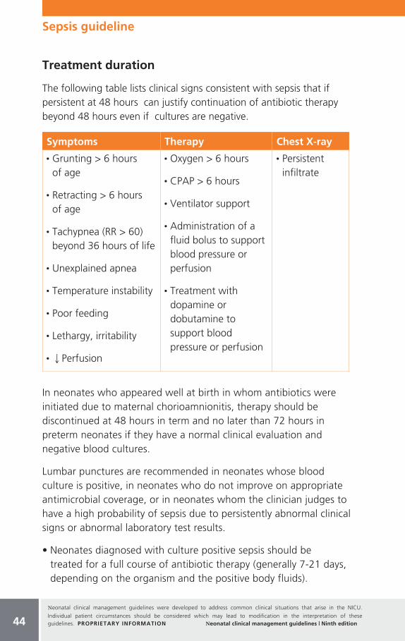
Neonatal clinical management guidelines were developed to address common clinical situations that arise in the NICU.
Individual patient circumstances should be considered which may lead to modification in the interpretation of these guidelines. PROPRIETARY INFORMATION Neonatal clinical management guidelines | Ninth edition
Treatment duration
The following table lists clinical signs consistent with sepsis that if persistent at 48 hours can justify continuation of antibiotic therapy beyond 48 hours even if cultures are negative.
Symptoms Therapy Chest X-ray
• Grunting > 6 hours of age
• Retracting > 6 hours of age
• Tachypnea (RR > 60) beyond 36 hours of life
• Unexplained apnea
• Temperature instability
• Poor feeding
• Lethargy, irritability
• QPerfusion
• Oxygen > 6 hours
• CPAP > 6 hours
• Ventilator support
• Administration of a fluid bolus to support blood pressure or perfusion
• Treatment with dopamine or dobutamine to support blood pressure or perfusion
• Persistent infiltrate
In neonates who appeared well at birth in whom antibiotics were initiated due to maternal chorioamnionitis, therapy should be discontinued at 48 hours in term and no later than 72 hours in preterm neonates if they have a normal clinical evaluation and negative blood cultures.
Lumbar punctures are recommended in neonates whose blood culture is positive, in neonates who do not improve on appropriate antimicrobial coverage, or in neonates whom the clinician judges to have a high probability of sepsis due to persistently abnormal clinical signs or abnormal laboratory test results.
• Neonates diagnosed with culture positive sepsis should be treated for a full course of antibiotic therapy (generally 7-21 days, depending on the organism and the positive body fluids).
44
Sepsis guideline

Neonatal clinical management guidelines were developed to address common clinical situations that arise in the NICU.
Individual patient circumstances should be considered which may lead to modification in the interpretation of these guidelines. PROPRIETARY INFORMATION Neonatal clinical management guidelines | Ninth edition
• Treatment for pneumonia beyond 7 days is generally reserved for neonates with persistent infiltrate(s) on chest x-ray infiltrate and prolonged clinical signs of sepsis.
• No data support a prolonged course of antibiotic treatment in neonates without signs of sepsis who are born to women with chorioamnionitis and a positive placental culture.
• Recent data demonstrate that intravenous immune globulin does not change the outcome of neonatal sepsis, hence its use is not recommended.
• For neonates who require antibiotic therapy beyond 7 days but who are otherwise ready for discharge, home/outpatient antibiotic therapy may be considered if the neonate is clinically well and support can be provided by community resources.
Post treatment care
• Once a neonate has completed a full course of antibiotic therapy and meets all discharge criteria, available evidence does not support the need for additional monitoring off antibiotic therapy.
• A hearing assessment (e.g., an auditory brainstem response) should be performed at the conclusion of treatment with aminoglycoside antibiotics. This test may be performed in the outpatient setting if necessary.
References: Sepsis
Alexander VN, Northrup V, Bizzarro MJ. Antibiotic exposure in the newborn intensive care unit and the risk of necrotizing enterocolitis. Journal of Pediatrics. 2011;159;392-7.
American Academy of Pediatrics Committee on Infectious Diseases and Committee on Fetus and Newborn. Recommendations for the Prevention of Perinatal Group B Streptococcal (GBS) Disease. Pediatrics. 2011;128(3):611-616.
Benitz W, Han M, Madan A, et al. Serial serum C-reactive protein levels in the diagnosis of neonatal infection. Pediatrics. 1998;102(4):e41.
Benitz WB, Wynn JL, Polin RA, Journal of Pediatrics. (article in press)
Brady MT, Polin RA. Prevention and management of infants with suspected or proven neonatal sepsis. Pediatrics. 2013;132(1):166-168.
45
Sepsis guideline

Neonatal clinical management guidelines were developed to address common clinical situations that arise in the NICU.
Individual patient circumstances should be considered which may lead to modification in the interpretation of these guidelines. PROPRIETARY INFORMATION Neonatal clinical management guidelines | Ninth edition46
Cotton CM, Taylor S, Stoll B, et al. Prolonged duration of initial empirical antibiotic treatment is associated with increased rates of necrotizing enterocolitis and death for extremely low birth weight infants. Pediatrics. 2009;123:58-66.
Cotton CM. Antibiotic Stewardship. Clinics in Perinatology. 2015;42(1):195-206.
Franz A, Bauer K, Schalk A, et al. Measurement of interleukin 8 in combination with C-reactive protein reduced unnecessary antibiotic therapy in newborn infants: a multicenter, randomized, controlled trial. Pediatrics. 2004;114(1):1-8.
Gagliardi L, Rusconi F, Bellu R, and Zanini R. Association of maternal hypertension and chorioamnionitis with preterm outcomes. Pediatrics. 2014;134:e154-e161.
Garcia-Munoz RF, Galan HG, Figueras AJ, Garcia-Alix PA. Outcomes of very-low-birth-weight infants exposed to maternal clinical chorioamnionitis: a multicentre study. Neonatology 2014;106:229-234.
Garcia-Munoz RF, Galan HGM, Ospina CG. Morbidity and mortality among very-low-birth-weight infants born to mothers with clinical chorioamnionitis. Pediatrics and Neonatology. 2014;55:381-386.
Gerdes J. Diagnosis and management of bacterial infections in the neonate. Pediatric Clinics of North America. 2004;21(4):939-959.
Higgins RD, Baker CJ, Raju TN. Executive summary of the workshop on infection in the high-risk infant. Journal of Perinatology. 2010;30(6):379-383.
INIS Collaborative Group, Brocklehurst P, Farrell B, King A, et al. Treatment of neonatal sepsis with intravenous immune globulin. New England Journal of Medicine. 2011;29: 365(13):1201-1211.
Institute of Medicine of the National Academies, Antibiotic resistance: implications for global health and novel intervention strategies - workshop summary (2010). [Accessed 2013 October]. Available from: http://iom.edu/Reports/2010/Antibiotic-Resistance-Implications-for-Global-Health-and-Novel-Intervention-Strategies.aspx.
Jackson G, Engle W, Sendelbach D, et al. Are complete blood cell counts useful in the evaluation of asymptomatic neonates exposed to suspected chorioamnionitis? Pediatrics. 2004;113(5):1173-1180.
Kumar Y, Qunibi M, Neal N, et al. Time to positivity of neonatal blood cultures. Archives of Disease in Childhood Fetal and Neonatal Edition. 2001;85(3):F182-F186.
Kuppula VS, Meinzen-Derr J, Morrow AL et al. Prolonged initial empirical antibiotic treatment is associated with adverse outcomes in premature infants. Journal of Pediatrics. 2011;159:720-5.
Mukhopadhyay S, Eichenwald EC, Puopolo KM. Neonatal early-onset sepsis evaluations among well-appearing infants: projected impact of changes in CDC GBS guidelines. Journal of Perinatology. 2013; 33(3):198-205.
Newman TB, Puopolo KM, Wi S, et al. Interpreting complete blood counts soon after birth in newborns at risk for sepsis. Pediatrics. 2010;126(5):903-909.
Pappas A, Kendrick DE, Shankaran S., Stoll BJ, Bell EF, Laptook AR, et al. Chorioamnionitis and early childhood outcomes among extremely low-gestational-age neonates. JAMA Pediatrics. 2014;168: 137-147.
Pickering LK, Baker CJ, Kimberlin DW, Long SS, eds. Red Book: 2012 Report of the Committee on Infectious Diseases. 29th Edition. American Academy of Pediatrics; 2012.
Polin RA; Committee on Fetus and Newborn. Management of neonates with suspected or proven early-onset bacterial sepsis. Pediatrics. 2012;129(5):1006-1015.
Polin RA, Watterberg K, Benitz W, et al. The conundrum of early-onset sepsis. Pediatrics; 2014;133:1122-1123.
Sepsis guideline
Neonatal clinical management guidelines were developed to address common clinical situations that arise in the NICU.
Individual patient circumstances should be considered which may lead to modification in the interpretation of these guidelines. PROPRIETARY INFORMATION Neonatal clinical management guidelines | Ninth edition 47
Remington JS, Klein JO, Wilson CB, et al, eds. Infectious Diseases of the Fetus and Newborn. 7th edition. Elsevier Saunders; 2011.
Santos RP, Tristam D. Pediatric Clinics of North America. 2015:62(2):491-508.
Verani JR, McGee L, Schrag SJ; Division of Bacterial Diseases, National Center for Immunization and Respiratory Diseases, Centers for Disease Control and Prevention (CDC). Prevention of perinatal group B streptococcal disease--revised guidelines from CDC, 2010. Morbidity and Mortality Weekly Report-Recommendations and Reports. 2010;59(RR-10):1-36.
Weinberg G, D’Angio CT. A practical guide to the diagnosis,treatment, and prevention of neonatal infections. Remington and Klein’s Infectious Diseases of the Fetus and Newborn Infant. 8th Edition, 36,1132-1146.
Weitkamp J, Aschner J. Diagnostic use of C-reactive protein (CRP) in assessment of neonatal sepsis. NeoReviews. 2005;6(11):e508-e515.
_____________________________________________________________
_____________________________________________________________
_____________________________________________________________
_____________________________________________________________
_____________________________________________________________
_____________________________________________________________
_____________________________________________________________
_____________________________________________________________
_____________________________________________________________
_____________________________________________________________
_____________________________________________________________
_____________________________________________________________
_____________________________________________________________
Notes
Sepsis guideline
Neonatal clinical management guidelines were developed to address common clinical situations that arise in the NICU.
Individual patient circumstances should be considered which may lead to modification in the interpretation of these guidelines. PROPRIETARY INFORMATION Neonatal clinical management guidelines | Ninth edition48
Key points
• American Academy of Pediatrics (AAP) presents recommendations for initiation of phototherapy based on gestation, age and risk factors.
• Outpatient monitoring of bilirubin level(s) is supported once phototherapy is discontinued.
General considerations
This guideline incorporates the latest recommendations from the AAP, emphasizing an approach that will reduce the frequency of severe neonatal hyperbilirubinemia and bilirubin encephalopathy.
• Inadequate breastfeeding may lead to dehydration and contribute to the development of hyperbilirubinemia. Increasing the frequency of nursing minimizes the risk of developing significant hyperbilirubinemia. Encourage the mother to provide 8 to 12 breastfeedings per day. According to the AAP, routine water/dextrose supplementation for non-dehydrated breast-fed infants does not prevent hyperbilirubinemia nor decrease total serum bilirubin (TSB) levels.
• Frequent oral feedings may decrease enterohepatic circulation, enhance bilirubin elimination and attenuate a rise in bilirubin levels.
• Routine bilirubin screening prior to discharge has the potential to reduce the incidence of severe hyperbilirubinemia levels that may lead to bilirubin encephalopathy.
• Prior to discharge, all infants should be assessed for their risk of hyperbilirubinemia. Two clinical options can either be used individually or in combination for the systematic assessment of risk: measurement of the bilirubin level using TSB or transcutaneous bilirubin (TcB) and/or assessment of clinical risk factors.
Phototherapy guideline
Applies to infants ≥ 35 weeks gestation

Neonatal clinical management guidelines were developed to address common clinical situations that arise in the NICU.
Individual patient circumstances should be considered which may lead to modification in the interpretation of these guidelines. PROPRIETARY INFORMATION Neonatal clinical management guidelines | Ninth edition
• Bilirubin measurement should be obtained (TcB and/or TSB bilirubin measurement) if there is known antenatal sensitization, jaundice in the first 24 hours of life, or jaundice excessive for the infant’s age. Elevated TcB levels should be verified by TSB. Bilirubin levels should be interpreted based on the infant’s age in hours.
• Visual estimation of the degree of jaundice can be erroneous, particularly in darkly pigmented infants. TcB or TSB should be measured if question exists regarding the degree of jaundice.
• Phototherapy or exchange transfusion should be initiated per current AAP criteria.
• Prior to discharge, perform a thorough history, physical examination and, if indicated, laboratory evaluation.
• Educate families about the significance and management of jaundice.
• Follow up bilirubin levels and exams should be based on the age of the infant, bilirubin level at time of discharge, presence of breastfeeding and other risk factors for the development of hyperbilirubinemia.
• Infants with lower gestational age, blood incompatibilities, glucose-6-phosphate dehydrogenase deficiency, increased weight loss and who are exclusively breastfed have an increased risk of developing elevated TcB in a time dependent manner.
• Infants discharged with a screening bilirubin level categorized as low risk still must be vigilantly observed for subsequent hyperbilirubinemia.
• Any infant discharged ≤ 72 hours of life should be evaluated by a healthcare professional within two days of discharge.
• Home phototherapy may be considered for infants without risk factors for hyperbilirubinemia and otherwise meeting discharge parameters.
• Infants with breast milk jaundice who are asymptomatic require no treatment if the total serum bilirubin level remains below the threshold to initiate phototherapy.
49
Phototherapy guideline

Neonatal clinical management guidelines were developed to address common clinical situations that arise in the NICU.
Individual patient circumstances should be considered which may lead to modification in the interpretation of these guidelines. PROPRIETARY INFORMATION Neonatal clinical management guidelines | Ninth edition50
General criteria for hospital phototherapy Based on recommendations from the AAP.
Tota
l Ser
um B
iliru
bin
(mg/
dL)
umol
/L
Age
25
20
15
10
5
0
428
342
257
171
85
0
Infants at lower risk (≥ 38 wk and well)
Infants at medium risk (≥ 38 wk + risk factors or 35-37 6/7 wk and well)
Infants at higher risk (35-37 6/7 wk + risk factors)
Birth 24h 48h 72h 96h 5 Days 6 Days 7 Days
• Use total bilirubin. Do not subtract direct reacting or conjugated bilirubin.
• Risk factors = isoimmune hemolytic disease, G6PD deficiency, asphyxia, significant lethargy, temperature instability, sepsis, acidosis, or albumin < 3.0g/dL (if measured).
• For well infants 35-37 6/7 week gestation, one can adjust TSB levels for intervention around the medium risk line. It is an option to intervene at lower TSB levels for infants closer to 35 weeks and higher or TSB levels for those closer to 37 6/7 wk.
• It is an option to provide conventional phototherapy in hospital or at home at TSB levels 2-3 mg/dL (35-50 mmol/L) below those shown but home phototherapy should not be used in any infant with risk factors.
Figure reproduced with permission from Pediatrics, Vol. 114 (1), Pages 297-316. Copyright © 2004 by the AAP.
Phototherapy guideline

Neonatal clinical management guidelines were developed to address common clinical situations that arise in the NICU.
Individual patient circumstances should be considered which may lead to modification in the interpretation of these guidelines. PROPRIETARY INFORMATION Neonatal clinical management guidelines | Ninth edition 51
The figure on the previous page indicates the appropriate bilirubin level to initiate “intensive” phototherapy based on gestation, age in hours when bilirubin was obtained and risk factors.
• The three curves differ based on gestation and risk factors.
• Intensive phototherapy implies high irradiance covering a large surface area (i.e. multiple phototherapy sources (as close as 10 cm to the infant). The threshold for initiating intensive phototherapy is based on the TSB and age in hours for the appropriate curve. Intensive phototherapy implies the use of high levels of irradiance in the 430-490 nm wavelength of at least 30 μW/cm2 per nm (measured at the infant’s skin directly below the center of the phototherapy unit) and delivered to as much of the infant’s surface area as possible.
• Conventional phototherapy refers to the use of fiberoptic or single overhead phototherapy. The threshold for initiating conventional phototherapy is TSB 2-3 mg/dL below the appropriate curve, based on age in hours.
An internet-based application indicating the appropriate bilirubin level to initiate phototherapy is available taking into account the latest recommendations from the American Academy of Pediatrics.*
*Alere does not control the content and makes no representations with respect to that content.
Discontinuing phototherapy
The AAP has not issued a standard for discontinuing either conventional or intensive phototherapy in non-readmitted babies. Given that it is an option to initiate conventional phototherapy 2-3 mg/dL below the curve based on age in hours, a reasonable stop point for most infants would be 5 mg/dL below the appropriate curve, based on age in hours (see ‘General criteria for hospital phototherapy’).
• There is no medical evidence supporting the practice of stepwise weaning from intensive to non-intensive phototherapy (triple to
Phototherapy guideline
Neonatal clinical management guidelines were developed to address common clinical situations that arise in the NICU.
Individual patient circumstances should be considered which may lead to modification in the interpretation of these guidelines. PROPRIETARY INFORMATION Neonatal clinical management guidelines | Ninth edition52
double to single phototherapy). Stepwise weaning, though, may be warranted based on degree of hyperbilirubinemia and age in hours.
• Observation for rebound bilirubin should not delay hospital discharge. For those infants with hemolytic disease treated with phototherapy or in cases when phototherapy is initiated and discontinued prior to day of life 3 or 4, a follow-up bilirubin level within 24 hours after discharge is recommended. Although significant rebound is rare in infants readmitted with hyperbilirubinemia and then discharged, a repeat TSB measurement or clinical follow-up 24 hours after discharge is a feasible option.
Criteria for readmitted infants
Infants should be readmitted to the hospital for TSB levels requiring intensive phototherapy. Phototherapy may be discontinued when the serum bilirubin level falls 5 mg/dL below the appropriate curve based on age in hours (see ‘General criteria for hospital phototherapy’). Significant rebound once phototherapy is discontinued is a rare phenomenon. Follow up TSB levels 24 hours after discharge is optional, though all babies readmitted for phototherapy should be followed closely by their primary care physician after discharge.
References: Phototherapy
American Academy of Pediatrics Subcommittee on Hyperbilirubinemia. Management of hyperbilirubinemia in the newborn infant 35 or more weeks of gestation. Pediatrics. 2004; 114(1):297-316.
Bhutani V, Johnson L, Keren R. Diagnosis and management of hyperbilirubinemia in the term neonate: for a safer first week. Pediatric Clinics of North America. 2004;51(4):843-861.
Bhutani VK, Maisels MJ, Stark AR, Buonocore G. Management of jaundice and prevention of severe neonatal hyperbilirubinemia in infants >or=35 weeks gestation. Expert Committee for Severe Neonatal Hyperbilirubinemia; European Society for Pediatric Research; American Academy of Pediatrics. Neonatology. 2008;94(1):63-67.
BiliToolTM. [Accessed 2013 October]. Available from: http://bilitool.org.
Bromiker R, Bin-Nun A, Schimmel MS, Hammerman C, Kaplan M. Neonatal hyperbilirubinemia in the low-intermediate-risk category on the bilirubin nomogram. Pediatrics. 2012; 130(3):e470-e475.
Committee on Fetus and Newborn. Phototherapy to prevent severe neonatal hyperbilirubinemia in the newborn infant 35 or more weeks of gestation. Pediatrics. 2011;128(4):e1046-e1052.
Phototherapy guideline

Neonatal clinical management guidelines were developed to address common clinical situations that arise in the NICU.
Individual patient circumstances should be considered which may lead to modification in the interpretation of these guidelines. PROPRIETARY INFORMATION Neonatal clinical management guidelines | Ninth edition 53
Eggert LD, Wiedmeier SE, Wilson J, Christensen RD. The effect of instituting a prehospital-discharge newborn bilirubin screening program in an 18-hospital health system. Pediatrics. 2006; 117(5):e855-e862.
Kaplan M, Kaplan E, Hammerman C, et al. Post-phototherapy neonatal bilirubin rebound: a potential cause of significant hyperbilirubinaemia. Archives of Disease in Childhood. 2006; 91(1):31–34.
Keren R, Bhutani VK, Luan X, et al. Identifying newborns at risk of significant hyperbilirubinaemia: a comparison of two recommended approaches. Archives of Disease in Childhood. 2005;90(4):415-421.
Kuzniewicz MW, Escobar GJ, Newman TB. Impact of universal bilirubin screening on severe hyperbilirubinemia and phototherapy use. Pediatrics. 2009;124(4):1031-1039.
Lazarus C, Avchen RN. Neonatal hyperbilirubinemia management: a model for change. Journal of Perinatology. 2009;29 (Supplement 1s):S58-S60.
Mah MP, Clark SL, Akhigbe E, et al. Reduction of severe hyperbilirubinemia after institution of predischarge bilirubin screening. Pediatrics. 2010;125(5):e1143-e1148.
Maisels MJ, Bhutani VK, Bogen D, et al. Hyperbilirubinemia in the newborn infant ≥ 35 weeks’ gestation: an update with clarifications. Pediatrics. 2009;124(4):1193-1198.
Maisels MJ, DeRidder JM, Kring EA, Balasubramaniam M. Routine transcutaneous bilirubin measurements combined with clinical risk factors improve the prediction of subsequent hyperbilirubinemia. Journal of Perinatology. 2009;9(29):612-617.
Maisels MJ, Kring E. Rebound in serum bilirubin level following intensive phototherapy. Archives of Pediatric and Adolescent Medicine. 2002;156(7):669-672.
Mantagou L, Fouzas S, Skylogianni E, et al. Trends of transcutaneous bilirubin in neonates who develop significant hyperbilirubinemia. Pediatrics. 2012;130(4):e898-e904.
Nanjundaswamy S, Petrova A, Mehta R, et al. The accuracy of transcutaneous bilirubin measurements in neonates: a correlation study. Biology of the Neonate. 2004;85(1):21-25.
Preer GL, Philipp BL. Understanding and managing breast milk jaundice. Archives of Disease in Childhood - Fetal and Neonatal Edition. 2011;96(6):F461-F466.
Profit J, Cambric-Hargrove AJ, Tittle KO, et al. Delayed pediatric office follow-up of newborns after birth hospitalization. Pediatrics. 2009;124(2):548-554.
Stokowski LA. Fundamentals of phototherapy for neonatal jaundice. Advances in Neonatal Care. 2006;6(6):303-312.
Phototherapy Guideline
Neonatal clinical management guidelines were developed to address common clinical situations that arise in the NICU.
Individual patient circumstances should be considered which may lead to modification in the interpretation of these guidelines. PROPRIETARY INFORMATION Neonatal clinical management guidelines | Ninth edition
Key points
• Neonatal abstinence syndrome (NAS) is a constellation of neonatal signs of withdrawal consequent to exposure to certain drugs.
• Most infants who develop NAS have been antenatally exposed to opioids. Concomitant antenatal exposure to maternal SSRIs, benzodiazepines, and smoking exacerbate signs of NAS.*
• Postnatal exposure to opioids and/or benzodiazepine due to treatment of critical illness can also result in NAS.
• Instruments that evaluate the severity of NAS typically assist in treatment decisions. These tools were developed specifically for signs of opioid withdrawal. Initial therapy should focus on effective non-pharmacologic treatments including optimization of the environment of care, involvement of family caregivers as feasible and prudent, and breast-feeding as indicated.
• If drug therapy for withdrawal becomes clinically indicated, first line therapy in general should employ a drug from the same class as the principal drug contributing to the signs of withdrawal.
• The evidence-based literature does not identify the optimal pharmacologic treatment regimen for infants with opioid withdrawal. Typically oral morphine or methadone are the first line treatment choices. If needed, second-line therapy choices include phenobarbital and clonidine. The use of buprenorphine continues to be evaluated (Clinics in Perinatology. 2013; 40(3):509-524).
• New research has demonstrated that adherence to a standardized protocol of evaluation, treatment, and weaning of medication reduces length of hospital stay.
Neonatal drug exposure/withdrawal guideline
Applies to infants who have been prenatally exposed to substances that cause signs of toxicity or withdrawal
54

Neonatal clinical management guidelines were developed to address common clinical situations that arise in the NICU.
Individual patient circumstances should be considered which may lead to modification in the interpretation of these guidelines. PROPRIETARY INFORMATION Neonatal clinical management guidelines | Ninth edition 55
General considerations
Fetal exposure to specific drugs can be teratogenic, cause acute toxicity, or produce signs of neonatal withdrawal. NAS is a condition that results from the abrupt cessation of in utero exposure to certain drugs (primarily opioids). Maternal histories of drug use (legal or illegal) during pregnancy are frequently inaccurate as may be demonstrated by positive drug testing when the mother has not admitted to drug use. Thus, a negative maternal history should not be considered to be definitive when an infant displays signs of withdrawal. For a number of reasons, maternal or infant testing may not detect drugs that the mother has admitted to using or from which the infant is showing signs of withdrawal.
In the past decade, the number of mothers dependent on opioids has increased nearly five-fold with a corresponding three-fold increase in NAS in their offspring. Part of this increase is due to more frequent use of prescription pain medication during pregnancy. No consistent relationship has been documented between maternal methadone or buprenorphine dose and severity of NAS. While up to 90% of infants exposed to narcotics during fetal life have some signs, only 50-75% will require treatment. Compared to methadone, fetal exposure to buprenorphine appears to lessen the likelihood and severity of NAS and to reduce the durations of medical treatment and hospital stay.
Infants at risk for NAS should be carefully monitored in the hospital for the development of signs consistent with withdrawal. The appropriate duration of hospital observation depends on recent maternal drug use. An infant born to a mother on a low-dose prescription opioid with a short half-life(e.g., hydrocodone) may be safely discharged if there are no signs of withdrawal by age 3 days, whereas an infant born to a mother on an opioid with a prolonged half-life (e.g., methadone) should be observed for a minimum of 5-7 days (Hudak ML, Tan RC. Neonatal drug withdrawa. Pediatrics. 2012; 129(2):e540-560). Affected infants may exhibit the onset of signs of drug withdrawal as early as 24-48 hours or as late as 7-10 days of age depending on the timing of the mother’s last dose and the drug’s half-life. Subacute signs from opioid withdrawal may last up to 6 months.
Neonatal drug exposure/withdrawal guideline

Neonatal clinical management guidelines were developed to address common clinical situations that arise in the NICU.
Individual patient circumstances should be considered which may lead to modification in the interpretation of these guidelines. PROPRIETARY INFORMATION Neonatal clinical management guidelines | Ninth edition56
Selective serotonin re-uptake inhibitor (SSRI) use among pregnant women has become more prevalent. Transient signs are typically observed during the first week of life and are in most cases due to a drug effect. When an infant exposed to SSRIs shows signs of opioid withdrawal, the caretaker should suspect concomitant maternal opioid use. In infants exposed to SSRIs, 30% are reported to demonstrate some abnormal clinical signs. Infants exposed antenatally to opioids and SSRIs develop more severe and prolonged signs of NAS compared to opioid exposure alone.
Fetal amphetamine exposure does not result in signs of withdrawal seen following opioid exposure. The most common presentation in infants is lethargy, somnolence and poor feeding, though some present with agitation and tachypnea. This typically requires minimal short term supportive treatment, possibly including gavage feeding.
Cocaine exposure by itself does not cause signs of withdrawal.
Though marijuana is the most common illicit drug used by pregnant women, it does not cause clinically significant neonatal withdrawal.
Phencyclidine exposure can result in hypertonicity, agitation and gastrointestinal symptoms which can persist up to 2-3 weeks.
NAS presents with a constellation of clinical signs including CNS abnormalities (hypertonicity, excessive motor activity, irritability), gastrointestinal dysfunction (vomiting and diarrhea), respiratory distress, (tachypnea), and vague autonomic signs (mottling, congestion, sneezing). In severe or untreated cases, seizures may occur. The prevalence of maternal multiple drug use complicates attempts to analyze any single drug’s contribution to the symptom complex.
Signs of NAS may mimic manifestations of neonatal illnesses such as hypoglycemia, hypocalcemia, hematologic disturbances, sepsis, and neurological illnesses.
Neonatal drug exposure/withdrawal guideline
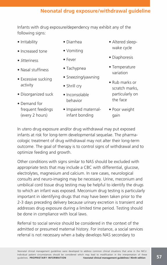
Neonatal clinical management guidelines were developed to address common clinical situations that arise in the NICU.
Individual patient circumstances should be considered which may lead to modification in the interpretation of these guidelines. PROPRIETARY INFORMATION Neonatal clinical management guidelines | Ninth edition 57
Infants with drug exposure/dependency may exhibit any of the following signs:
• Irritability
• Increased tone
• Jitteriness
• Nasal stuffiness
• Excessive sucking activity
• Disorganized suck
• Demand for frequent feedings (every 2 hours)
• Diarrhea
• Vomiting
• Fever
• Tachypnea
• Sneezing/yawning
• Shrill cry
• Inconsolable behavior
• Impaired maternal-infant bonding
• Altered sleep- wake cycle
• Diaphoresis
• Temperature variation
• Rub marks or scratch marks, particularly on the face
• Poor weight gain
In utero drug exposure and/or drug withdrawal may put exposed infants at risk for long-term developmental sequelae. The pharma- cologic treatment of drug withdrawal may not alter their long-term outcome. The goal of therapy is to control signs of withdrawal and to optimize feeding and growth.
Other conditions with signs similar to NAS should be excluded with appropriate tests that may include a CBC with differential, glucose, electrolytes, magnesium and calcium. In rare cases, neurological consults and neuro-imaging may be necessary. Urine, meconium and umbilical cord tissue drug testing may be helpful to identify the drugs to which an infant was exposed. Meconium drug testing is particularly important in identifying drugs that may have been taken prior to the 2-3 days preceding delivery because urinary excretion is transient and addresses drug exposure during a limited time period. Testing should be done in compliance with local laws.
Referral to social service should be considered in the context of the admitted or presumed maternal history. For instance, a social services referral is not necessary when a baby develops NAS secondary to
Neonatal drug exposure/withdrawal guideline

Neonatal clinical management guidelines were developed to address common clinical situations that arise in the NICU.
Individual patient circumstances should be considered which may lead to modification in the interpretation of these guidelines. PROPRIETARY INFORMATION Neonatal clinical management guidelines | Ninth edition58
maternal opioid treatment for a legitimate medical condition. Local legal requirements for reporting must be met.
Finnegan abstinence scoring system
• The Finnegan abstinence scoring system is the usual instrument used in the United States to quantitate the severity of NAS and to trend the infant’s response to supportive non-pharmacologic interventions or pharmacotherapy. Another widely used evaluation tool is the Lipsitz scale, which uses a simpler numeric scoring system to evaluate the need for therapy. This guideline refers to Finnegan scores for treatment protocols. Though the Finnegan abstinence scoring system was originally validated for full term infants, it is reasonable to apply it to late preterm infants. Nursing educational programs can improve accuracy and minimize interrater variability of the assessments.
• Infants who are at risk for NAS should be cared for in a physical environment that minimizes external stimulation and provides necessary non-pharmacologic support (see ‘Treatment Considerations’). A skilled caretaker should initiate Finnegan scoring only if he/she has concerns that the infant is developing signs of withdrawal, for these are not subtle. Scores should be performed about an hour after feeding and repeated every 2-4 hours
• Most protocols specify that 3 consecutive Finnegan scores ≥ 8 or a single score ≥ 12 triggers the need for pharmacologic treatment.
• The goals of initiating pharmacotherapy are to stabilize but not necessarily eliminate clinical manifestations of withdrawal so that the infant can feed adequately, be consolable, achieve sufficient sleep, and begin to develop socialization. (Update on the pharmacologic management of neonatal abstinence syndrome. Journal of Perinatology. 2011;31:692-701)
• A Finnegan score < 8 typically allows appropriate drug weaning.
Neonatal drug exposure/withdrawal guideline

Neonatal clinical management guidelines were developed to address common clinical situations that arise in the NICU.
Individual patient circumstances should be considered which may lead to modification in the interpretation of these guidelines. PROPRIETARY INFORMATION Neonatal clinical management guidelines | Ninth edition 59
Treatment considerations
• The initial treatment of a neonate at risk for or with early signs of withdrawal should be supportive. Breastfeeding should be allowed unless there are concurrent contraindications (e.g., maternal HIV or continuing illicit drug use). Breast milk feedings in mothers who are on opioid maintenance therapy (e.g., methadone and/or buprenorphine) delay the onset of NAS, reduce its severity and minimize the need for pharmacologic treatment regardless of the infant’s gestation and type of drug exposure. Drug therapy may not be necessary for infants with mild symptoms and is rarely needed for babies exposed to SSRIs alone. In addition to breastfeeding, supportive therapy should include swaddling, holding, vertical rocking with the infant faced away from the caretaker, minimizing environmental stimuli (including light and noise), and infant massage. These actions can lessen signs and improve the ability to deliver appropriate care for these infants.
• Infants may require frequent feeding. An intake up to 150 Kcal/kg/day may be needed to achieve consistent weight gain. Hypercaloric formulas may be needed to meet increased metabolic demand.
• A pacifier for excessive sucking can be provided.
• Skin excoriation of pressure points can be minimized by application of protective material such as opsite. Mittens on hands and filing of fingernails may help prevent facial scratching.
• Perianal barrier protection is important to minimize dermatitis.
• Infants should be monitored for sleeping habits, temperature instability, weight gain or loss and skin excoriation (especially in the diaper area).
• Daily weight and I&O should be recorded.
Neonatal drug exposure/withdrawal guideline

Neonatal clinical management guidelines were developed to address common clinical situations that arise in the NICU.
Individual patient circumstances should be considered which may lead to modification in the interpretation of these guidelines. PROPRIETARY INFORMATION Neonatal clinical management guidelines | Ninth edition60
Pharmacotherapy considerations
The decision to use pharmacologic agents should be individualized in the context of a standard protocol that assesses the benefits and risks of treatment in light of the severity of signs of withdrawal. Current AAP guidelines call for using a drug from the same class as the drug causing the signs of withdrawal. Length of treatment will vary depending on the type(s) of drug exposure and severity of signs.
Primary drugs:
Oral morphine sulfate
Oral morphine solution is a first-line therapy for opioid withdrawal.
Dilutions of oral morphine to concentrations of 0.02-0.04 mg/ml are safe and stable.
Initial dosage of oral morphine for mild to moderate NAS is of 0.2-0.3 mg/kg/day divided every 3-4 hours. With severe NAS, 0.4-0.5 mg/kg/day divided every 3-4 hours may be used. The dose may be increased gradually to a maximum of 1.2 mg/kg/day until signs are controlled.
Steady-state levels of morphine may require 12-24 hours to achieve. If an infant has not demonstrated adequate clinical improvement in this time period, it is preferable to treat with an additional dose of morphine (0.03-0.06 mg/kg, depending on the initial severity) rather than to increase the total daily dose or to add a second drug.
The maximum therapeutic effect is usually seen in 24-48 hours.
Methadone
Oral methadone is a first-line therapy for opioid withdrawal. Initial dosage of oral methadone for mild to moderate NAS is 0.2-0.3 mg/kg/day divided every 6-12 hours. With severe NAS, an additional loading dose of 0.2 mg/kg may be given to hasten achievement of steady state levels. The total daily dose may be increased to 0.5-0.6 mg/kg/day divided every 6-12 hours to control signs of NAS.
Neonatal drug exposure/withdrawal guideline
Neonatal clinical management guidelines were developed to address common clinical situations that arise in the NICU.
Individual patient circumstances should be considered which may lead to modification in the interpretation of these guidelines. PROPRIETARY INFORMATION Neonatal clinical management guidelines | Ninth edition 61
Due to the long half-life of methadone in infants (about 24 hours on average), 2-3 days may be needed to achieve steady-state levels and realize full therapeutic effect. If a loading dose of methadone was not given, infants who are not responding to treatment can be given an additional dose of 0.2 mg/kg to accelerate attainment of steady state levels.
Methadone may be used alone or in combination with other agents for weaning from in utero drug exposure. The long half-life of methadone compared to morphine results in lower variability of serum levels at steady-state and requires less frequent dosing. Injudicious dosing can result in higher than therapeutic levels and QT interval prolongation. (Update on the pharmacologic management of Neonatal Abstinence Syndrome, Journal of Perinatology. 2011;31:692-701). Consider screening with electrocardiogram prior to treatment and during treatment due to prolonged QRS.
In mothers on methadone maintenance, feeding human milk may minimize or eliminate NAS symptoms.
Both opioids
Once signs of NAS are adequately controlled, weaning may commence in as early as 24 hours. Dosage should be weaned every 24-48 hours by 10-20% of the amount of the therapeutic total daily dose. Weaning should be guided by serial clinical assessments (weight gain/loss; feeding performance; activity level; sleep) in conjunction with Finnegan scores. As an infant matures, Finnegan scores increase in normal babies. After 21 days, the threshold Finnegan score may be increased by 2.
When the total daily dose is 0.05-0.1 mg/kg/day, it is appropriate to trial a discontinuation of treatment. Infants should be observed for 24-48 hours to monitor for recurrence of signs of NAS.
Neonatal drug exposure/withdrawal guideline

Neonatal clinical management guidelines were developed to address common clinical situations that arise in the NICU.
Individual patient circumstances should be considered which may lead to modification in the interpretation of these guidelines. PROPRIETARY INFORMATION Neonatal clinical management guidelines | Ninth edition62
Adjunctive medications:
Oral phenobarbital
Phenobarbital may be considered as adjunctive therapy when infants have not shown adequate clinical improvement on the maximum recommended dose of a first-line medication. In these cases, average NAS scores generally remain high despite pharmacotherapy. Phenobarbital may be a drug of choice in infants undergoing withdrawal from non-opioid drugs, such as benzodiazepines. Phenobarbital is a sedative that will reduce signs of excessive CNS activity but will not affect gastrointestinal hyperactivity. The blood level necessary to control narcotic withdrawal signs is unknown.
• A loading dose of 10-20 mg/kg (in combination with opioid treatment) is typically effective in attenuating clinical signs of opioid withdrawal.
• If signs of NAS persist, additional doses can be given to achieve a serum level of 20-30mcg/mL.
• Once the infant is adequately treated, a maintenance dose of 3-5 mg/kg/day divided every 12 to 24 hours is appropriate.
• Phenobarbital levels should be monitored if clinically indicated.
In infants on opioids and phenobarbital, the opioid should be weaned and discontinued before decreasing the dose of phenobarbital.
• When the infant has been stable without opioids for 24 hours, taper the phenobarbital dose by 10-20% per day or as tolerated.
• Discontinue phenobarbital when the infant is stable at a low dose (2-3 mg/kg/day) or when the serum level is low (</= 10 mcg/mL).
Clonidine
Clonidine is an alpha 2-adrenergic receptor agonist that has been used to blunt signs and symptoms of opioid withdrawal in adults and older children. Clonidine reduces sympathetic outflow by decreasing central catecholamine release which is thought to palliate symptoms of
Neonatal drug exposure/withdrawal guideline
Neonatal clinical management guidelines were developed to address common clinical situations that arise in the NICU.
Individual patient circumstances should be considered which may lead to modification in the interpretation of these guidelines. PROPRIETARY INFORMATION Neonatal clinical management guidelines | Ninth edition 63
autonomic hyperactivity. The total morphine dose was approximately 60% lower when combined with clonidine, resulting in fewer days of drug therapy and hospitalization.* Some clinicians have used clonidine as a primary treatment for NAS. (Pediatric Clinics of North America. 2015;62(2):525-544) but more research seems prudent before this approach can be routinely recommended.
Pharmacotherapy agents not recommended:
Tincture of opium is not recommended due to a high concentration of opioids that risks medication errors. In addition, this medication has a 19% alcohol content.
Paregoric is not recommended because it contains a plethora of compounds, including anise oil, benzoic acid and camphor and because it has a high alcohol content.
Diazepam is not recommended due to possible cerebral and hepatic dysfunction, its lack of efficacy and its adverse effects on feeding (Clinics in Perinatology. 2013;40(3):509-524).
Chlorpromazine has limited use in neonates due to adverse effects such as cerebellar dysfunction, decreased seizure threshold and hematologic abnormalities.
Naloxone is contraindicated for infants exposed to in utero opiates.
Discharge planning for infants at risk for drug withdrawal
Given the natural history of withdrawal, it is important to monitor the asymptomatic drug-exposed infant closely during the hospital stay. Infants who are exposed in utero to low doses of opioids with short half-lives may be safely discharged in 48 hours. Infants exposed to opioids or benzodiazepines may be ready for discharge as early as the 5th day of life if they have not demonstrated clinical signs.
Uncommonly, signs of withdrawal in infants exposed to methadone or buprenorphine may be delayed until as late as 7 days of age. In
Neonatal drug exposure/withdrawal guideline

Neonatal clinical management guidelines were developed to address common clinical situations that arise in the NICU.
Individual patient circumstances should be considered which may lead to modification in the interpretation of these guidelines. PROPRIETARY INFORMATION Neonatal clinical management guidelines | Ninth edition64
general, if such an infant is completely normal by 5-7 days of age, discharge may proceed once caregivers understand educational instructions and follow-up appointments are in place (see ‘Discharge Instructions’ below).
Discharge criteria for infants treated with medication(s)
• The infant is physiologically stable.
• The infant is taking oral feeds well, sleeping well, (Neonatal abstinence syndrome. Pediatrics 2014;134:e547), is consolable and gaining weight appropriately. Scoring helps to achieve consistency of care but functional characteristics are the best determinants of readiness for discharge (Pediatric Clinics of North America. 2015; 62(2):525-544).
• The infant shows neurobehavioral recovery (e.g., reaches full alert state, responds to social stimuli and can be consoled with routine measures).
• Optimal treatment for the infant with NAS is not necessarily achieved by minimizing the duration of hospitalization in the neonatal period (Journal of Opioid Management. 2009;5:47-55).
• All necessary screenings (e.g., hearing) and other assessments have been completed.
• A social service consult has been performed. Referral to the state’s child protection agency can be made, if deemed appropriate by the social worker and/or other health care provider(s). The infant’s actual caregivers should be involved in the infant’s care in the hospital as early as possible. When appropriate, foster parents may facilitate safe discharge.
Neonatal drug exposure/withdrawal guideline

65Neonatal clinical management guidelines were developed to address common clinical situations that arise in the NICU.
Individual patient circumstances should be considered which may lead to modification in the interpretation of these guidelines. PROPRIETARY INFORMATION Neonatal clinical management guidelines | Ninth edition
Discharge instructions for infants with NAS
• Explain the signs of withdrawal to caregivers.
• Provide the caregivers full instructions on how to comfort and console a baby.
• Arrange a follow-up appointment with the infant’s pediatrician within 24-48 hours after discharge.
• Schedule home care visit(s) by a nurse and/or social worker as may be required following the initial pediatrician’s office visit.
• Consider neurodevelopmental follow-up for the infant if appropriate.
• If necessary, refer the infant’s parents to a drug treatment program while the infant is in the hospital.
• In carefully selected situations, infants have been safely discharged home on medication with weaning completed in the outpatient setting. Typically, this plan of care has succeeded if the parents adhere to a regular schedule of physician visits and allow home visitations as needed so that the infant can be carefully and continually assessed and medication adjusted appropriately. This may involve provision of only the required number of pre-measured doses until the next assessment to minimize the risk of drug overdosing. On the other hand, the literature suggests that duration of outpatient treatment may be prolonged compared to inpatient therapy, especially if an infant is discharged home on an adjunctive medication. This may represent an unfavorable risk to benefit practice; further refinement and testing of outpatient treatment protocols seem prudent to develop an evidence-based recommendation.
65
Neonatal drug exposure/withdrawal guideline

66Neonatal clinical management guidelines were developed to address common clinical situations that arise in the NICU.
Individual patient circumstances should be considered which may lead to modification in the interpretation of these guidelines. PROPRIETARY INFORMATIO Neonatal clinical management guidelines | Ninth edition
References: Neonatal drug exposure/withdrawal
Abdel-Latif M, Pinner J, Clews S, et al. Effects of breast milk on the severity and outcome of neonatal abstinence syndrome among infants of drug-dependent mothers. Pediatrics. 2006; 117(6):e1163-e1169.
Agthe AG, Kim GR, Mathias KB, et al. Clonidine as an adjunct therapy to opioids for neonatal abstinence syndrome: a randomized, controlled trial. Pediatrics. 2009;123(5):e849-e856.
Alto WA, O’Connor AB. Management of women treated with buprenorphine during pregnancy. American Journal of Obstetrics and Gynecology. 2011;205(4):302-308.
American Academy of Pediatrics Committee on Drugs. The transfer of drugs and other chemicals into human milk. Pediatrics. 2001;108(3):776-789.
Backes CH, Backes CR, Gardner D, et al. Neonatal abstinence syndrome: transitioning methadone-treated infants from an inpatient to an outpatient setting. Journal of Perinatology. 2012;32(6):425-430.
Bio LL, Siu A, Poon CY. Update on the pharmacologic management of neonatal abstinence syndrome. Journal of Perinatology. 2011;31(11):692-701.
Brown MS, Hayes MJ, Thorton LM. Journal of Perinatal Medicine. 2015;35:278-83.
Burgos AE, Burke BL. Neonatal Abstinence Syndrome. NeoReviews. 2009;10(5);e222-e229.
Clinics in Perinatology. 2013;40(3):509-524.
Coyle M, Ferguson A, Lagasse L, et al. Diluted tincture of opium (DTO) and phenobarbital versus DTO alone for neonatal opiate withdrawal in term infants. Journal of Pediatrics. 2002;140(5):561-564.
Coyle M, Ferguson A, Lagasse L, et al. Neurobehavioral effects of treatment for opiate withdrawal. Archives of Disease in Childhood Fetal and Neonatal Edition. 2005; 90(1):F73-F74.
Ferreira E, Carceller AM, Agogué C, et al. Effects of selective serotonin reuptake inhibitors and venlafaxine during pregnancy in term and preterm neonates. Pediatrics. 2007;119(1):52-59.
Finnegan L, Connaughton J, Kron R, et al. Neonatal abstinence syndrome: assessment and management. Addictive Diseases International Journal. 1975;2(1-2):141-158.
Finnegan L, Kaltenbach K. Primary Pediatric Care. 2nd edition. Mosby. 1992;1367-1378.
Hall ES, Wexelblatt SL, Crowley M, Grow JL, Jasin LR, Klebanoff MA, McClead RE, Meinzen-Derr J, Mohan VK, Stein H, Walsh MC, OCHNAS Consortium. A multicenter cohort study of treatments and hospital outcomes in neonatal abstinence syndrome. Pediatrics. 2014;134:e527-34.
Hall ES, Wexelblatt SL, Crowley M., Grow JL, Jasin LR, Klebanoff MA, McClead RE, Meinzen-Derr J, Mohan VK, Stein H, Walsh MC; OCHNAS Consortium. Implementation of a neonatal abstinence syndrome weaning protocol; a multicenter cohort study. Pediatrics. 2014;136:e803-810.
Hudak ML, Tan RC; Committee on Drugs; Committee on Fetus and Newborn; American Academy of Pediatrics. Neonatal drug withdrawal. Pediatrics. 2012;129(2):e540-e560.
Hussain T, Ewer AK. Maternal methadone may cause arrhythmias in neonates. Acta Paediatrica. 2007;96(5):768-769.
Isemann B, Meinzen-Derr J, Akinbi H. Maternal and neonatal factors impacting response to methadone therapy in infants treated for neonatal abstinence syndrome. Journal of Perinatology. 2011;31(1):25-29.
66
Neonatal drug exposure/withdrawal guideline

67Neonatal clinical management guidelines were developed to address common clinical situations that arise in the NICU.
Individual patient circumstances should be considered which may lead to modification in the interpretation of these guidelines. PROPRIETARY INFORMATION Neonatal clinical management guidelines | Ninth edition
Jackson L, Ting A, McKay S, et al. A randomized controlled trial of morphine versus phenobarbitone for neonatal abstinence syndrome. Archives of Disease in Childhood Fetal and Neonatal Edition. 2004;89(4):F300-F304.
Jansson L, Choo R, Melez M, et al. Methadone maintenance and breast feeding in the neonatal period. Pediatrics. 2008;121(1):106-114.
Johnson K, Gerada C, Greenough A. Treatment of neonatal abstinence syndrome. Archives of Disease in Childhood Fetal and Neonatal Edition. 2003;88(1):F2-F5.
Johnson K, Greenough A, Gerada C. Maternal drug use and length of neonatal unit stay. Addiction. 2003; 98(6):785-789.
Jones HE, Kaltenbach K, Heil SH, et al. Neonatal abstinence syndrome after methadone or buprenorphine exposure. New England Journal of Medicine. 2010;363(24):2320-2331.
Journal of Perinatology. 2011;31,692-701
Kuschel C. Managing drug withdrawal in the newborn infant. Seminars in Fetal and Neonatal Medicine. 2007;12(2):e127-e133.
Lainwala S. A retrospective study of length of hospital stay in infants treated for neonatal abstinence syndrome with methadone versus oral morphine preparations. Advances in Neonatal Care. 2005; 5(5):265-272.
Levinson-Castiel R, Merlob P, Linder N, et al. Neonatal abstinence syndrome after in utero exposure to selective serotonin reuptake inhibitors in term infants. Archives of Pediatrics & Adolescent Medicine. 2006;160(2):173-176.
Lipsitz P. A proposed narcotic withdrawal score for use with newborn infants: A pragmatic evaluation of its efficacy. Clinical Pediatrics. 1975;14(6):592-594.
Lucas K, Knobel RB. Implementing practice guidelines and education to improve care of infants with neonatal abstinence syndrome. Advances in Neonatal Care. 2012;12(1):40-45.
Maichuk G, Zahorodny W, Marshall R. Use of positioning to reduce the severity of neonatal narcotic withdrawal syndrome. Journal of Perinatology. 1999;19(7):510-513.
McQueen KA, Murphy-Oikonen J, Gerlach K, Montelpare W. The impact of infant feeding method on neonatal abstinence scores of methadone-exposed infants. Advances in Neonatal Care. 2011; 11(4):282-290.
Montgomery D, Plate C, Alder SC, et al. Testing for fetal exposure to illicit drugs using umbilical cord tissue vs meconium. Journal of Perinatology. 2006;26(1):11-14.
Moses-Kolko EL, Bogen D, Perel J, et al. Neonatal signs after late in utero exposure to serotonin reuptake inhibitors: literature review and implications for clinical applications. Journal of the American Medical Association. 2005;293(19):2372-2383.
O’Donnell M, Nassar N, Leonard H et al. Increasing prevalence of neonatal withdrawal syndrome: population study of maternal factors and child protection involvement. Pediatrics. 2009;123: e614-e621.
Oei JL, Kingsbury A, Dhawan A, et al. Amphetamines, the pregnant woman and her children: a review. Journal of Perinatology. 2012;32(10):737-747.
Patrick SW, Schumacher RE, Benneyworth BD, et al. Neonatal abstinence syndrome and associated health care expenditures: United States, 2000-2009. The Journal of the American Medical Association. 2012;307(18):1934-1940.
67
Neonatal drug exposure/withdrawal guideline

68Neonatal clinical management guidelines were developed to address common clinical situations that arise in the NICU.
Individual patient circumstances should be considered which may lead to modification in the interpretation of these guidelines. PROPRIETARY INFORMATIO Neonatal clinical management guidelines | Ninth edition
Patrick SW, Davis MM, Lehman CU, Cooper WO. Increasing incidence and geographic distribution of neonatal abstinence syndrome: United States 2009 to 2012. Journal of Perinatology. 2015; 35:650-5.
Payot A, Berner M. Hospital stay and short-term follow up of children of drug-abusing mothers born in an urban community hospital–a retrospective review. European Journal of Pediatrics. 2000; 159(9):679-683.
Pediatric Clinics of North America. 2015;62(2):525-544
Pediatrics. 2014;134:e547
Thajam D, Atkinson DE, Sibley CP, Lavender T. Is neonatal abstinence syndrome related to the amount of opiate used? Journal of Obstetric, Gynecologic, and Neonatal Nursing. 2010;39(5):503-509.
Tolia VN, Patrick SW, Bennett MM, Murphy K, Sousa J, Smith PB, Clark RH, Spitzer AR. Increasing incidence of the neonatal abstinence syndrome in US neonatal ICUs. New England Journal of Medicine. 2015;371:2118-26.
Wachman EM, Hayes MJ, Lester BM, Terrin N., Brown MS, Nielsen DA, Davis JM. Epigenetic variation in the mu-opioid gene in infants with neonatal abstinence syndrome. Journal of Pediatrics. 2014;165:472-8.
Zimmermann-Bauer U, et al. Finnegan neonatal abstinence scoring system: normal values for the first 3 days and weeks 5-6 in nonaddicted infants. Addiction. 2010 March;105:524-52
_____________________________________________________________
_____________________________________________________________
_____________________________________________________________
_____________________________________________________________
_____________________________________________________________
_____________________________________________________________
_____________________________________________________________
_____________________________________________________________
_____________________________________________________________
_____________________________________________________________
Notes
68
Neonatal drug exposure/withdrawal guideline

Neonatal clinical management guidelines were developed to address common clinical situations that arise in the NICU.
Individual patient circumstances should be considered which may lead to modification in the interpretation of these guidelines. PROPRIETARY INFORMATION Neonatal clinical management guidelines | Ninth edition 69
Key points
• American Academy of Pediatrics (AAP) physiologic-based discharge criteria are utilized.
• Family disposition and discharge needs should be addressed early in the care continuum.
General considerations
Historically, preterm infants were discharged only if they reached a certain weight or post menstrual age (PMA). However, it has been shown that preterm infants can be safely discharged earlier once physiologic competency and stability are established. The eligibility and timing of discharge of any infant in the NICU or special care nursery is a decision that is determined by the attending physician responsible for the care of that infant, in conjunction with the infant’s caregiver(s).* Discharge should be based on the achievement of physiologic competency of the patient, but may also be affected by the caregiver(s) ability to manage certain aspects of care at home. Preterm infants with low birth weight experience a higher rate of readmission and death during the first year after birth. However, careful discharge planning and appropriate post-discharge follow-up may reduce these risks. A multidisciplinary unit-based neonatal team (consisting of a social worker, case manager, primary care nurse and home care coordinator) should assist the physician/practitioner team with the infant’s discharge planning process. Developing unit discharge criteria and creating individualized flow charts for the discharge process can facilitate the timely completion of discharge tasks.
*Caregiver(s) in context of this guideline includes parents and guardians
Discharge guideline
Applies to all infants in the neonatal intensive care unit (NICU) or special care nursery

Neonatal clinical management guidelines were developed to address common clinical situations that arise in the NICU.
Individual patient circumstances should be considered which may lead to modification in the interpretation of these guidelines. PROPRIETARY INFORMATION Neonatal clinical management guidelines | Ninth edition
Considerations for discharge
• Discharge planning should begin following admission, despite the inability to predict the timing of discharge.
• Discharge teaching and planning should occur throughout the hospitalization so as not to overwhelm parents and staff at the end of the hospital stay.
• Parental contact and involvement in the care of the infant should be encouraged from time of admission. An educational-behavioral intervention program for parents that commences early in the NICU has been shown to improve mental health outcomes, enhance parent-infant interaction and reduce length of stay.
• An educational program transitioning the preterm infant home in coordination with the medical home has the potential to decrease hospital readmission, particularly in the public insurance population.
• Caregivers can have lower levels of confidence when infant care includes medical equipment or procedures. This emphasizes the importance of early, timely caregiver involvement when discharge encompasses specific procedural tasks or medical devices.
• Discussions should be held on an educational level consistent with the caregiver(s) background. Interpreter services should be coordinated as indicated to maintain appropriate family interaction.
• An ethics committee should be convened early if there are staff concerns regarding plans of care that are not beneficial to the infant’s long-term health.
• Key transition points (such as weaning to room air, transition to crib, full oral intake, etc.) that are reflective of an infant’s physiologic status should be identified throughout the hospitalization. The roles and responsibilities of the multidisciplinary unit-based neonatal team and care providers should be emphasized under each of the major transition points.
70
Discharge guideline

Neonatal clinical management guidelines were developed to address common clinical situations that arise in the NICU.
Individual patient circumstances should be considered which may lead to modification in the interpretation of these guidelines. PROPRIETARY INFORMATION Neonatal clinical management guidelines | Ninth edition
• Discharge planning should be incorporated into daily medical rounds, nursing reports and written documentation to include:
▸ Discharge date based on physiologic competency.
▸ Follow up studies scheduled and completed prior to the anticipated discharge. Studies requiring sedation, e.g., brain MRI, should be done timely so that transient poor feeding does not delay discharge.
▸ Diagnostic and elective follow up studies scheduled as an outpatient if the infant is clinically stable and timing of the study is not critical.
▸ Home equipment and medication.
▸ Home health needs.
▸ Home feeding plan.
▸ Screening needs (e.g., car seat challenge, hearing, developmental or feeding assessment).
▸ Primary healthcare provider and any required subspecialist(s) for continuing outpatient care.
• Primary guardian, if not the parents, for the infant must be identified. Caregiver(s) should be provided with the appropriate training and resources to care for the infant after discharge.
• A written summary of the infant’s hospital course and specific needs including medications, treatments, pertinent laboratory and diagnostic test results, immunizations received and appointments following discharge should be completed. A copy should be sent to the primary care provider assuming care of the infant, and a copy given to the caregiver(s).
• A written plan for home health care should include (as applicable): the name of the primary care provider and additional medical consultants, an individual to be contacted in the event of an emergency, a discharge feeding plan, a list of necessary supplies and medications, specific follow-up directions and responsibilities of the home care agency and caregiver(s).
71
Discharge guideline

Neonatal clinical management guidelines were developed to address common clinical situations that arise in the NICU.
Individual patient circumstances should be considered which may lead to modification in the interpretation of these guidelines. PROPRIETARY INFORMATION Neonatal clinical management guidelines | Ninth edition
Family considerations for discharge
During discharge planning, the unit-based neonatal team should:
• Encourage on-going caregiver communication and participation in all aspects of care that are anticipated after hospital discharge. This includes providing an individualized teaching plan.
• Assess caregiver(s) mental and physical capabilities to care for their infant. Counseling prior to discharge may help identify biopsychosocial risks of caring for an infant with ongoing care needs. (Placencia FX, McCullough LB. Biopsychosocial risks of parental care for high-risk neonates: implications or evidence-based parental counseling. Journal of Perinatology. 2012;32(5):381-386.) Make referral to community resources as applicable.
• Meet with caregiver(s) to assess the suitability of the infant’s home environment.
• Ask the primary caregiver of the infant to demonstrate that he/she has learned the required skills necessary for caring for the infant at discharge.
• Provide ongoing support and develop a specific care plan to assist caregiver(s) in their home preparation and engage in evaluating future needs, including:
▸ Transportation to/from the hospital.
▸ Child care needs for siblings during future hospital visits.
▸ Necessary infant care items in the home.
▸ Telephone/utility service availability.
▸ Psychosocial problems that may be encountered.
▸ Community support services.
▸ Financial resources (specifically to ensure that adequate resources are identified commensurate with the infant’s ongoing care needs and assistance in pursuing alternate resources as applicable).
72
Discharge guideline

Neonatal clinical management guidelines were developed to address common clinical situations that arise in the NICU.
Individual patient circumstances should be considered which may lead to modification in the interpretation of these guidelines. PROPRIETARY INFORMATION Neonatal clinical management guidelines | Ninth edition
• The availability of social support is crucial for the success of every caregiver(s) transition to the care of their infant after discharge. Families with limited support at home or limited resources such as home care availability and/or community support must be considered in discharge planning to assist the infant in a smooth transition home.
Potential high-risk situations
These considerations should alert the unit-based neonatal team to the existence of a potentially high-risk environment which requires attention prior to discharge. This list is not intended to be all-inclusive.
• Insufficient or lack of prenatal care.
• Caregiver(s) age < 18 years without a capable and willing adult caregiver living in the same residence.
• Caregiver(s) has/have a history of involvement with the state designated child protective agency.
• Evidence of previous or current caregiver substance abuse.
• Potential exposure to second-hand smoke.
• Evidence of caregiver(s) physical/mental disability.
• Absence of telephone service or other essential utilities in the residence prior to discharge.
• Current legal and/or social services involvement for homelessness, incarceration, litigation, domestic abuse, etc.
• Transportation difficulties or place of residence is located far away from a hospital or outpatient service center.
• Inability to deliver follow-up or home care due to the location of the place of residence or safety issues.
73
Discharge guideline
Neonatal clinical management guidelines were developed to address common clinical situations that arise in the NICU.
Individual patient circumstances should be considered which may lead to modification in the interpretation of these guidelines. PROPRIETARY INFORMATION Neonatal clinical management guidelines | Ninth edition
Medical criteria for discharge
Infants will be eligible for discharge if they meet the following medical criteria:
• Establishment of physiologic competencies (including but not limited to oral feeding, thermoregulation and respiratory control) and stability regardless of weight or PMA.
• Infant displays normal vital signs, including body temperature and sufficiently mature cardiorespiratory control, while fully clothed in an open crib. These conditions should be maintained at an environmental temperature that is comfortable for a lightly dressed adult consistent with the “Back to Sleep” campaign.
• Infant demonstrates an appropriate overall weight gain, if weight gain is expected based on PMA (normal weight trend in the 1st week of life should be followed). Weight gain does not have to occur on one or more consecutive days before discharge as it can vary due to timing of the weight checks in relation to feeding, urination or stooling. Weight trend can be followed as an outpatient in infants taking adequate volume with a decrease in milk caloric density.
• Infant nipples all feedings. Up to 48 hours of full PO feeding may be adequate observation for infants born < 34 weeks gestation. American Academy of Pediatrics Committee on Fetus and Newborn support 24 hours of full PO feedings as adequate for babies born ≥ 34 weeks. Infants discharged within one week of age with weight loss of more than 2% to 3% of birth weight per day or a maximum of 7% should receive close follow up shortly after discharge. There may be select infants who, based on their specific feeding history, warrant an additional hospital observation period prior to discharge. Appropriate arrangements should be in place for early post-discharge follow-up.
• Home gavage feedings may be considered in select infant-family dyads for the infant with proven cardiorespiratory stability when feeding is the last issue requiring continued hospitalization. Caregiver(s) should be comfortable and demonstrate competency
74
Discharge guideline
Neonatal clinical management guidelines were developed to address common clinical situations that arise in the NICU.
Individual patient circumstances should be considered which may lead to modification in the interpretation of these guidelines. PROPRIETARY INFORMATION Neonatal clinical management guidelines | Ninth edition
with all aspects of home gavage feedings with appropriate community based supports in place. This practice should have a limited role and be reserved for infants who are not likely to achieve full oral feedings within a reasonable time frame.
• Studies have not demonstrated efficacy in the use of gastro- esophageal reflux disease (GERD) medications. Discontinuationof GERD medications should be coordinated so discharge is notdelayed if the clinical team desires an observation period followingcessation of medication(s). An alternative option is to continuetreatment with follow up as an outpatient.
• Up to 48 hours of stable body temperature in an open crib istypically adequate for infants born < 34 weeks gestation. AmericanAcademy of Pediatrics Committee on Fetus and Newborn support 12hours of stable body temperature as adequate for babies born≥ 34 weeks. There may be select infants who, based on their specificgrowth parameters and/or thermoregulation history, warrant anadditional hospital observation period prior to discharge. Appropriatearrangements should be in place for early post-discharge follow-up.
• For infants placed into an incubator solely for the purpose ofphototherapy, additional hospital observation is typically not requiredonce treatment is completed.
• Infant is breathing room air. Patients demonstrating an inabilityto actively wean off oxygen, requiring a fixed low amount ofoxygen who are otherwise stable for medical discharge, should beconsidered for home oxygen therapy and appropriate monitoring.As a safety measure, oxygen saturation nadir in room air should bemeasured prior to discharge for these infants.
• Up to 48 hours hospital observation after discontinuing oxygentherapy is typically adequate to ensure medical stability prior todischarge home. Home pulse oximeter and/or monitor should beconsidered if the desired observation period is longer.
75
Discharge guideline

Neonatal clinical management guidelines were developed to address common clinical situations that arise in the NICU.
Individual patient circumstances should be considered which may lead to modification in the interpretation of these guidelines. PROPRIETARY INFORMATION Neonatal clinical management guidelines | Ninth edition
• Up to 48 hours hospital observation after discontinuing diureticsis typically adequate to ensure medical stability prior to dischargehome. Home pulse oximeter should be considered if the desiredobservation period is longer.
• For late preterm infants (born at 34 0/7 through 36 6/7 weeksgestation) who have demonstrated the necessary physiologiccompetency and medical stability and to minimize the risk ofreadmission, careful discharge planning including caregiver educationand close post-discharge follow up is indicated instead of continuedin-patient monitoring.
• Infants born less than 37 weeks gestation have received a car seatchallenge test prior to discharge. Car seats and car beds are availablefor lower weight infants not eligible or failing angle tolerance testin a standard car seat. A similar time frame of cardiorespiratorymonitoring in a car bed should be completed prior to discharge forthose infants failing the car seat angle tolerance test. An outpatientstudy should be coordinated prior to transitioning from a car bed to acar seat. Infants with significant gastroesophageal reflux, neurologicdiseases and certain malformations (e.g., omphalocele, Pierre Robinsequence, osteogenesis imperfecta, meningomyelocele) may requireuse of a car bed.
• The infant should demonstrate a period of physiologic stabilityfollowing eye exam (established by normal vital signs and feedingtolerance for at least 1-2 feedings). Infants with a previous historyof post-exam instability (typically apnea and/or oxygen desaturation,poor feeding or temperature instability) should be considered for upto 24 hours observation prior to discharge.
• A follow up retinopathy of prematurity (ROP) examination should notdelay discharge except in those specific clinical situations whereby anarrow oxygen saturation range is prescribed by the ophthalmologist,or when an infant is near threshold for treatment and readmissionwould delay timely therapy. Any prolonged hospitalization dueto concerns in the caregiver(s) reliability to adhere to outpatientappointments would be a delay secondary to social disposition andshould be addressed as indicated.
76
Discharge guideline
Neonatal clinical management guidelines were developed to address common clinical situations that arise in the NICU.
Individual patient circumstances should be considered which may lead to modification in the interpretation of these guidelines. PROPRIETARY INFORMATION Neonatal clinical management guidelines | Ninth edition
Discharge planning activities
The following activities should occur concurrently with the infant’s hospital course:
• Identify contact phone numbers.
• Confirm insurance eligibility.
• Make sure that the infant has been added to the insurance policy.
• Evaluate caregiver(s) ability to perform care required for discharge.
• Identify alternate home caregiver(s).
• Ensure that the home environment is appropriate and ready; visit ifneeded.
• Evaluate for WIC or other need-based referrals.
• Evaluate transportation availability.
• An infant should have an appointment scheduled to be seen bya primary care physician or other health care professional who isexperienced in the care of high-risk neonates within one week ofdischarge. Specific concerns such as hyperbilirubinemia, feedingintake, weight gain issues or review of outstanding tests may requireearlier follow-up either in the office or at home by a nurse.
• Appropriate and timely verbal and written information should beprovided to all health care providers scheduled to care for the infantafter discharge.
• If clinically indicated an infant should receive a home nursing visit oran evaluation in a physician’s office shortly after discharge.
• Home nursing visit(s) should be based on the complexity of theinfant’s clinical status and caregiver(s) capability. Private duty/shiftcare nursing should be based on the infant’s needs and coordinatedprior to meeting medical discharge criteria. A detailed home careplan should be transmitted to the home health agency.
77
Discharge guideline

Neonatal clinical management guidelines were developed to address common clinical situations that arise in the NICU.
Individual patient circumstances should be considered which may lead to modification in the interpretation of these guidelines. PROPRIETARY INFORMATION Neonatal clinical management guidelines | Ninth edition
• Subspecialty physician visits should be scheduled as clinicallyindicated.
• Evaluate need for home cardiorespiratory monitor and/or otherhome health care needs. An apnea program or a physician familiarwith the management of apnea monitors should manage all infantsdischarged on an apnea monitor. The name and phone number ofthe responsible parties should be given to the caregiver(s) prior todischarge.
• Preterm infants should be kept in the supine position for at least oneweek prior to discharge and predominantly in the supine positionpreferably from 32 weeks PMA onward so that they becomeacclimated to supine sleeping before discharge and to modelappropriate sleep positioning for the parent/caregiver. Educationalprograms promoting safe sleeping practices can improve familycompliance after discharge.
• To lower risks of sudden infant death syndrome, the sleepenvironment should follow safety recommendations from theAmerican Academy of Pediatrics. This includes education ensuringinfants are placed in their separate sleep environment prior to thecaregiver falling asleep.
• To minimize the risk of readmission for dehydration and hyperbilirubi-nemia, it is important to educate caregiver(s) how to evaluate feed-ing success and detect signs of dehydration and hyperbilirubinemia.Coordinate delivery of an effective breast pump to the mother asapplicable.
• Determine appropriate nipple size prior to discharge.
• Determine appropriate enteral caloric density prior to discharge andcaregiver(s) ability to follow the prescribed “recipe” for non-ready-to-feed preparations. Weight trend with potential need to increase milkcaloric density can be coordinated as an outpatient.
• A trial of ad-lib PO feedings may be reasonable in select infants whohave demonstrated the ability to orally complete the majority of theirfeedings.
78
Discharge guideline

Neonatal clinical management guidelines were developed to address common clinical situations that arise in the NICU.
Individual patient circumstances should be considered which may lead to modification in the interpretation of these guidelines. PROPRIETARY INFORMATION Neonatal clinical management guidelines | Ninth edition
• Preterm infants are at risk for underimmunization; ensure that allimmunizations are up to date. Timing of immunizations should becoordinated so discharge is not delayed if the medical team desiresa post-immunization observation period.
• Car seat challenge infants born < 37 weeks gestation. Manufacturerrecommendations should be followed. Testing with appropriateequipment should occur when the infant is stable in an open criband preferably prior to the day of discharge. Lateral support withrolled blankets and towels may provide needed support for childrenwith poor trunk and neck control assuming these additions do notaffect the fit of the car safety seat. Retesting with repositioningusing manufacturer insert, if available, should occur immediatelyfor any infant failing the initial test. Marginal failures should berepeated within 24 hours; significant failures within 24-48 hours. Ifthe patient still fails, consideration for discharge in a car bed, use ofsupplemental oxygen or further evaluation should occur.
• Regardless of the results of the car seat testing, very low birthweight infants transported in either a car seat or car bed are at riskfor clinically significant cardiopulmonary events. Caregiver(s) shouldprovide close observation while limiting duration of travel as much aspossible.
• Prior to the discharge of an infant with active ROP, caregiver(s)should understand the importance of the exam. Careful coordinationbetween the medical team and the ophthalmologist shouldensure that the exam is completed in a timely fashion. Follow-upexaminations should be conducted in the outpatient setting withappropriate monitoring and evaluation occurring during and after theprocedure. If a clinical situation exists whereby an infant is at risk forsignificant complications or instability during or after the exam, theinfant should be readmitted for the ROP examination.
• Teach caregiver(s) CPR if indicated.
• Complete screening for critical congenital heart disease via pulseoximetry.
79
Discharge guideline

Neonatal clinical management guidelines were developed to address common clinical situations that arise in the NICU.
Individual patient circumstances should be considered which may lead to modification in the interpretation of these guidelines. PROPRIETARY INFORMATION Neonatal clinical management guidelines | Ninth edition
• Complete screening eye exam(s) for ROP and schedule appropriatefollow-up per current AAP guidelines.
• Complete infant hearing screen. If hearing screen cannot becompleted prior to discharge, arrangements must be made for theinfant to have testing performed as an outpatient.
• Complete neurodevelopmental and neurobehavioral assessment withappropriate follow-up, if applicable.
• Complete early intervention referral, if applicable.
• Confirm metabolic screening status.
• Hematological status has been assessed and treatment instituted,if applicable.
• Complete Respiratory Syncytial Virus (RSV) prophylaxis per currentAAP guidelines. The initial dose of palivizumab can be planned either48 to 72 hours before discharge or promptly after discharge.
• Complete circumcision, if applicable.
• Check that all medication prescriptions are filled and caregiver(s)demonstrate safe administration.
• Any elective rooming-in process during the day or overnight shouldbe completed while the infant requires hospitalization for medicalreasons.
• Elective transfer to an equivalent or lower level of care facility as wellas back-transports to the referring facility should be based on infant’soverall medical stability, ability of the receiving facility to provideongoing care and projected length of stay. Travel distances placingan undue burden on the family as well as regionalization of neonatalservices should be taken into consideration as well.
80
Discharge guideline

Neonatal clinical management guidelines were developed to address common clinical situations that arise in the NICU.
Individual patient circumstances should be considered which may lead to modification in the interpretation of these guidelines. PROPRIETARY INFORMATION Neonatal clinical management guidelines | Ninth edition
References: Discharge
Altman M, Vanpée M, Cnattingius S, Norman M. Moderately preterm infants and determinants of length of hospital stay. Archives of Diseases of Childhood Fetal Neonatal Edition. 2009; 94(6):F414-F418.
American Academy of Pediatrics Committee on Fetus and Newborn. Hospital discharge of the high-risk neonate. Pediatrics. 2008;122(5):1119-1126.
American Academy of Pediatrics. Council on Children With Disabilities. Supplemental security income (SSI) for children and youth with disabilities. Pediatrics. 2009;124(6):1702-1708.
American Academy of Pediatrics. Joint Committee on Infant Hearing. Year 2007 position statement: Principles and guidelines for early hearing detection and intervention programs. Pediatrics. 2007; 120(4):898-921.
American Academy of Pediatrics Task Force on Sudden Infant Death Syndrome. SIDS and other sleep-related infant deaths: expansion of recommendations for a safe infant sleeping environment. Pediatrics. 2011;128(5):1030-1039.
American Academy of Pediatrics Task Force on Sudden Infant Death Syndrome. SIDS and other sleep-related infant deaths: expansion of recommendations for a safe infant sleeping environment. Pediatrics. 2011;128(5): e1341-e1367.
Aspin A, Seymour P, Broadbent A. Part 2, Changing times: following the initiative for early transfer of babies for continuing surgical care, in a hospital nearer their home. Journal of Neonatal Nursing. 2008; 14:17-21.
Brandon GD, Adeniyi-Jones S, Kirkby S, et al. Are outcomes and care processes for preterm neonates influenced by health insurance status? Pediatrics. 2009;124(1):122-127.
Brooten D, Kumar S, Brown L, et al. A randomized clinical trial of early hospital discharge and home follow-up of very-low-birth-weight infants. New England Journal of Medicine. 1986;315(15):934-939.
Bull MJ, Engle WA; Committee on Injury, Violence, and Poison Prevention and Committee on Fetus and Newborn; American Academy of Pediatrics. Safe transportation of premature and low birth weight infants at hospital discharge. Pediatrics. 2009;123(5):1424-1429.
Burgess AL, Borowsky IW. Health and home environments of caregivers of children investigated by child protective services. Pediatrics. 2010;125(2):273-281.
Campbell M. Development of a clinical pathway for near-term and convalescing premature infants in a level II nursery. Advances in Neonatal Care. 2006;6(3):150–164.
Degrazia M, Guo CY, Wilkinson AA, Rhein L. Weight and age as predictors for passing the infant car seat challenge. Pediatrics. 2010;125(3):526-531.
Donohue PK, Hussey-Gardner B, Sulpar LJ, et al. Convalescent care of infants in the neonatal intensive care unit in community hospitals: risk or benefit? Pediatrics. 2009;124(1):105-111.
Donohue PK, Hussey-Gardner B, Sulpar LJ, et al. Parents’ perception of the back-transport of very-low-birth-weight infants to community hospitals. Journal of Perinatology. 2009;29:575-581.
Engle WA, Tomashek KM, Wallman C; Committee on Fetus and Newborn, American Academy of Pediatrics. “Late-preterm” infants: a population at risk. Pediatrics. 2007;120(6):1390-1401.
Fierson WM; American Academy of Pediatrics Section on Ophthalmology; American Academy of Ophthalmology; American Association for Pediatric Ophthalmology and Strabismus; American Association of Certified Orthoptists. Screening examination of premature infants for retinopathy of prematurity. Pediatrics. 2013;131(1):189-95.
81
Discharge guideline
Neonatal clinical management guidelines were developed to address common clinical situations that arise in the NICU.
Individual patient circumstances should be considered which may lead to modification in the interpretation of these guidelines. PROPRIETARY INFORMATION Neonatal clinical management guidelines | Ninth edition
Fitzgerald DA, Massie RJ, Nixon GM, et al. Thoracic Society of Australia and New Zealand. Infants with chronic neonatal lung disease: recommendations for the use of home oxygen therapy. The Medical Journal of Australia. 2008;189(10):578-582.
Gelfer P, Cameron R, Masters K, Kennedy KA. Integrating “Back to Sleep” recommendations into neonatal ICU practice. Pediatrics. 2013;131(4):e1264-1270.
Harlor Jr ADB, Bower C, Committee on Practice and Ambulatory Medicine and the Section on Otolaryngology Head and Neck Surgery. Hearing assessment in infants and children: recommendations beyond neonatal screening. Pediatrics. 2009;124(4):1252-1263.
Hwang SS, Barfield WD, Smith RA, et al. Discharge Timing, Outpatient Follow-up, and Home Care of Late-Preterm and Early-Term Infants. Pediatrics. 2013;132(1):101-108.
Kemper AR, Mahle WT, Martin GR, et al. Strategies for implementing screening for critical congenital heart disease. Pediatrics. 2011;128(5):e1259-e1267.
Kemper AR, Wallace DK, and Quinn GE. Systematic review of digital imaging screening strategies for retinopathy of prematurity. Pediatrics. 2008;122(4);825-830.
Kleinman RE, ed. Pediatric Nutrition Handbook. 6th Edition. American Academy of Pediatrics; 2009.
Kotagal U, Perlstein P, Gamblian V, et al. Description and evaluation of a program for the early discharge of infants from a neonatal intensive care unit. Journal of Pediatrics. 1995;127(2):285-290.
Lagatta J, Clark R, Spitzer A. Clinical predictors and institutional variation in home oxygen use in preterm infants. Journal of Pediatrics. 2012;160(2):232-238.
Melnyk B, Feinstein N, Alpert-Gillis L, et al. Reducing premature infants’ length of stay and improving parents’ mental health outcomes with the Creating Opportunities for Parent Empowerment (COPE) NICU Program: A randomized, controlled trial. Pediatrics. 2006;118(5):e1414-e1427.
Mercurio MR. The role of a pediatric ethics committee in the newborn intensive care unit. Journal of Perinatology. 2011;31(1):1-9.
Merritt T, Pillers D, Prows S. Early NICU discharge of very low birth weight infants: a critical review and analysis. Seminars in Neonatology. 2003;8(2):95-115.
Mills MM, Sims DC, Jacob J. Implementation and case-study results of potentially better practices to improve the discharge process in the neonatal intensive care unit. Pediatrics. 2006;118( Supplement 2):S124-S133.
Navar-Boggan AM, Halsey NA, Escobar GJ, et al. Underimmunization at discharge from the neonatal intensive care unit. Journal of Perinatology. 2012;32(5):363-367.
O’Neil J, Yonkman J, Talty J, Bull MJ. Transporting children with special health care needs: comparing recommendations and practice. Pediatrics. 2009;124(2);596-603.
Ortenstrand A, Waldenstrom U, Winbladh B. Early discharge of preterm infants needing limited special care, followed by domiciliary nursing care. Acta Paediatrica. 1999;88(9):1024-1030.
Phillips RM, Goldstein M, Hougland K, et al. Multidisciplinary guidelines for the care of late preterm infants. Journal of Perinatology. 2013;33(Supplement 2):S5-S22.
Pickering LK, Baker CJ, Kimberlin DW, Long SS, eds. Red Book: 2012 Report of the Committee on Infectious Diseases. 29th Edition. American Academy of Pediatrics; 2012.
Placencia FX, McCullough LB. Biopsychosocial risks of parental care for high-risk neonates: implications or evidence-based parental counseling. Journal of Perinatology. 2012;32(5):381-386.
82
Discharge guideline
Neonatal clinical management guidelines were developed to address common clinical situations that arise in the NICU.
Individual patient circumstances should be considered which may lead to modification in the interpretation of these guidelines. PROPRIETARY INFORMATION Neonatal clinical management guidelines | Ninth edition
Raines DA, Brustad J. Parent’s confidence as a caregiver. Advances in Neonatal Care. 2012; 12(3):183-188.
Reyna B, Pickler R, Thompson A. A descriptive study of mothers’ experiences feeding their preterm infants after discharge. Advances in Neonatal Care. 2006;6(6):333–340.
Riley LE, Stark AR, eds. Guidelines for Perinatal Care. 7th Edition. American Academy of Pediatrics and The American College of Obstetricians and Gynecologists; 2012.
Salhab W, Khattak A, Tyson J, et al. Car seat or car bed for very low birth weight infants at discharge home. Journal of Pediatrics. 2007;150(3):224-228.
Schneiderman LJ, Gilmer T, Teetzel HD, et al. Effect of ethics consultations on nonbeneficial life-sustaining treatments in the intensive care setting: a randomized controlled trial. Journal of the American Medical Association. 2003;290(9):1166-72.
Silber JH, Lorch SA, Roenbaum PR, et al. Time to send the preemie home? Additional maturity at discharge and subsequent health care costs and outcomes. Health Services Research. 2009.44(2 Pt 1):444-463.
Sims DC, Jacob J, Mills MM, et al. Evaluation and development of potentially better practices to improve the discharge process in the neonatal intensive care unit. Pediatrics. 2006;118 (Supplement 2):S115-S123.
Smith VC, Hwang SS, Dukhovny D, et al. Neonatal intensive care unit discharge preparation, family readiness and infant outcomes: connecting the dots. Journal of Perinatology. 2013;33(6):415-421.
Smith VC, Young S, Pursley DM, et al. Are families prepared for discharge from the NICU? Journal of Perinatology. 2009;29(9):623–629.
Sturm, L. Implementation and evaluation of a home gavage program for preterm infants. Neonatal Network. 2005;24(4):21-25.
Tipnis NA, Tipnis SM. Controversies in the treatment of gastroesophageal reflux disease in preterm infants. Clinics in Perinatology. 2009;36(1):153-164.
Touch S, Greenspan J, Kornhauser M, et al. The timing of neonatal discharge: an example of unwarranted variation? Pediatrics. 2001; 07(1):73-77.
US Preventive Services Task Force. Universal screening for hearing loss in newborns: US Preventive Services Task Force Recommendation Statement. Pediatrics. 2008;122(1):143-148.
Vecchi C, Vasquez L, Radin T, et al. Neonatal individualized predictive pathway (NIPP): a discharge planning tool for parents. Neonatal Network. 1996;15(4):7-13.
Vohr BR, Yatchmink YE, Burke RT, et al. Factors associated with rehospitalizations of very low birthweight infants: impact of a transition home support and education program. Early Human Development. 2012;88(7):455-460.
Weiss S, Goldlust E, Vaucher YE. Improving parent satisfaction: an intervention to increase neonatal parent-provider communication. Journal of Perinatology. 2010;30(6):425-30.
83
Discharge guideline

Neonatal clinical management guidelines were developed to address common clinical situations that arise in the NICU.
Individual patient circumstances should be considered which may lead to modification in the interpretation of these guidelines. PROPRIETARY INFORMATION Neonatal clinical management guidelines | Ninth edition84
_____________________________________________________________
_____________________________________________________________
_____________________________________________________________
_____________________________________________________________
_____________________________________________________________
_____________________________________________________________
_____________________________________________________________
_____________________________________________________________
_____________________________________________________________
_____________________________________________________________
_____________________________________________________________
_____________________________________________________________
_____________________________________________________________
_____________________________________________________________
_____________________________________________________________
_____________________________________________________________
_____________________________________________________________
_____________________________________________________________
_____________________________________________________________
_____________________________________________________________
_____________________________________________________________
_____________________________________________________________
Notes

85Neonatal clinical management guidelines were developed to address common clinical situations that arise in the NICU.
Individual patient circumstances should be considered which may lead to modification in the interpretation of these guidelines. PROPRIETARY INFORMATION Neonatal clinical management guidelines | Ninth edition
_____________________________________________________________
_____________________________________________________________
_____________________________________________________________
_____________________________________________________________
_____________________________________________________________
_____________________________________________________________
_____________________________________________________________
_____________________________________________________________
_____________________________________________________________
_____________________________________________________________
_____________________________________________________________
_____________________________________________________________
_____________________________________________________________
_____________________________________________________________
_____________________________________________________________
_____________________________________________________________
_____________________________________________________________
_____________________________________________________________
_____________________________________________________________
_____________________________________________________________
_____________________________________________________________
_____________________________________________________________
Notes
85
86Neonatal clinical management guidelines were developed to address common clinical situations that arise in the NICU.
Individual patient circumstances should be considered which may lead to modification in the interpretation of these guidelines. PROPRIETARY INFORMATIO Neonatal clinical management guidelines | Ninth edition
_____________________________________________________________
_____________________________________________________________
_____________________________________________________________
_____________________________________________________________
_____________________________________________________________
_____________________________________________________________
_____________________________________________________________
_____________________________________________________________
_____________________________________________________________
_____________________________________________________________
_____________________________________________________________
_____________________________________________________________
_____________________________________________________________
_____________________________________________________________
_____________________________________________________________
_____________________________________________________________
_____________________________________________________________
_____________________________________________________________
_____________________________________________________________
_____________________________________________________________
_____________________________________________________________
_____________________________________________________________
Notes
86

About the our women & children’s health NICU care management program
The neonatal intensive care unit (NICU) care management program is focused on promoting positive clinical outcomes for infants admitted to the NICU or special care nursery. We provides comprehensive care
management services for premature and medically complex newborns, working in collaboration with bedside physicians, nurses and other health personnel. We have provided such services for more than 200,000 NICU infants nationwide.
The NICU program is patient centered, evidence-based and outcomes driven and is overseen by our board-certified neonatologists and NICU nurse care managers who have extensive experience in the challenges that are commonly involved in caring for NICU patients.
Primary goals of the NICU program include:
• Supporting high quality and efficient NICU care in conjunction andthrough collaboration with attending physicians, nurses and otherhospital personnel.
• Encouraging family involvement, education and interactions withtheir physicians and nurses during an infant’s NICU stay.
• Supporting the discharge planning process and transition of aninfant at home.
• Providing follow-up family contact post hospital discharge to ensureinfant health in the home environment.
• Analyzing aggregate collected clinical data to identify clinicalpractice benchmarks and define opportunities to enhance care.
The NICU care management program is recognized and accredited by the National Committee for Quality Assurance (NCQA) and by the Utilization Review Accreditation Commission (URAC).
© 2016 All rights reserved. c. All other trademarks are the property of their respective owners.
GCA-00401 Rev. 1 11/15
The NICU care management program is
accredited by URAC for Utilization
Review and Case Management.
The NICU care management program is accredited by the National Committee
for Quality Assurance for Case Management.