Nikon Advanced Research Microscope Systems · 4 5 Nikon for science, Nikon for the future There are...
11
1 Nikon Advanced Research Microscope Systems Nikon Advanced Research Microscope Systems
Transcript of Nikon Advanced Research Microscope Systems · 4 5 Nikon for science, Nikon for the future There are...
1
Nikon Advanced Research Microscope Systems
Nikon Advanced Research Microscope Systems
2 3
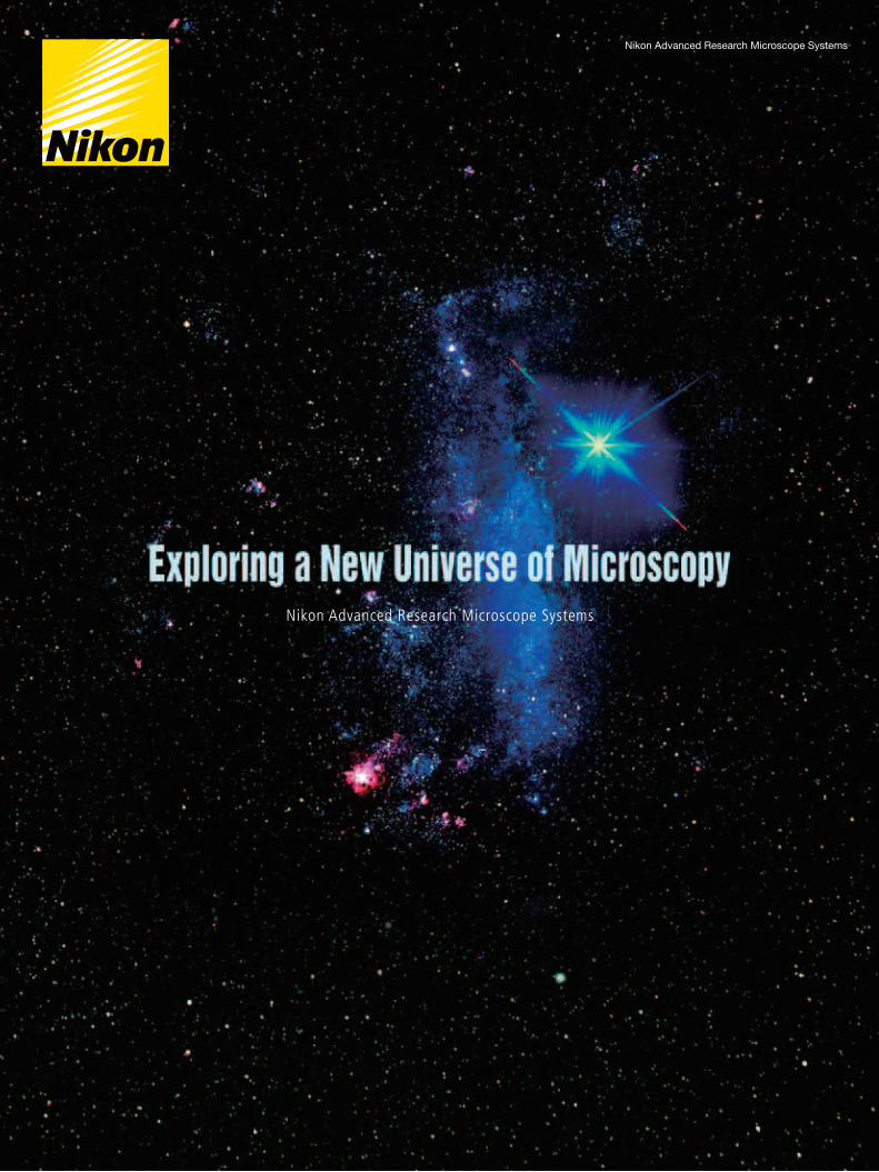
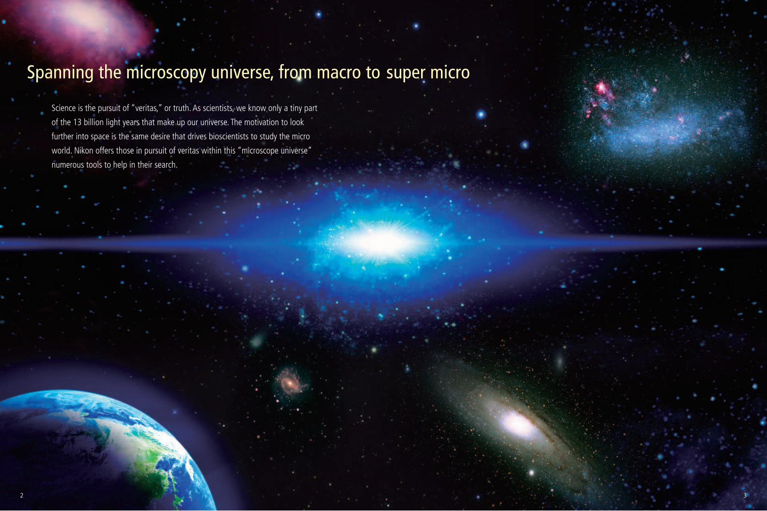
Spanning the microscopy universe, from macro to super micro
Science is the pursuit of “veritas,” or truth. As scientists, we know only a tiny part
of the 13 billion light years that make up our universe. The motivation to look
further into space is the same desire that drives bioscientists to study the micro
world. Nikon offers those in pursuit of veritas within this “microscope universe”
numerous tools to help in their search.
2 3
4 5
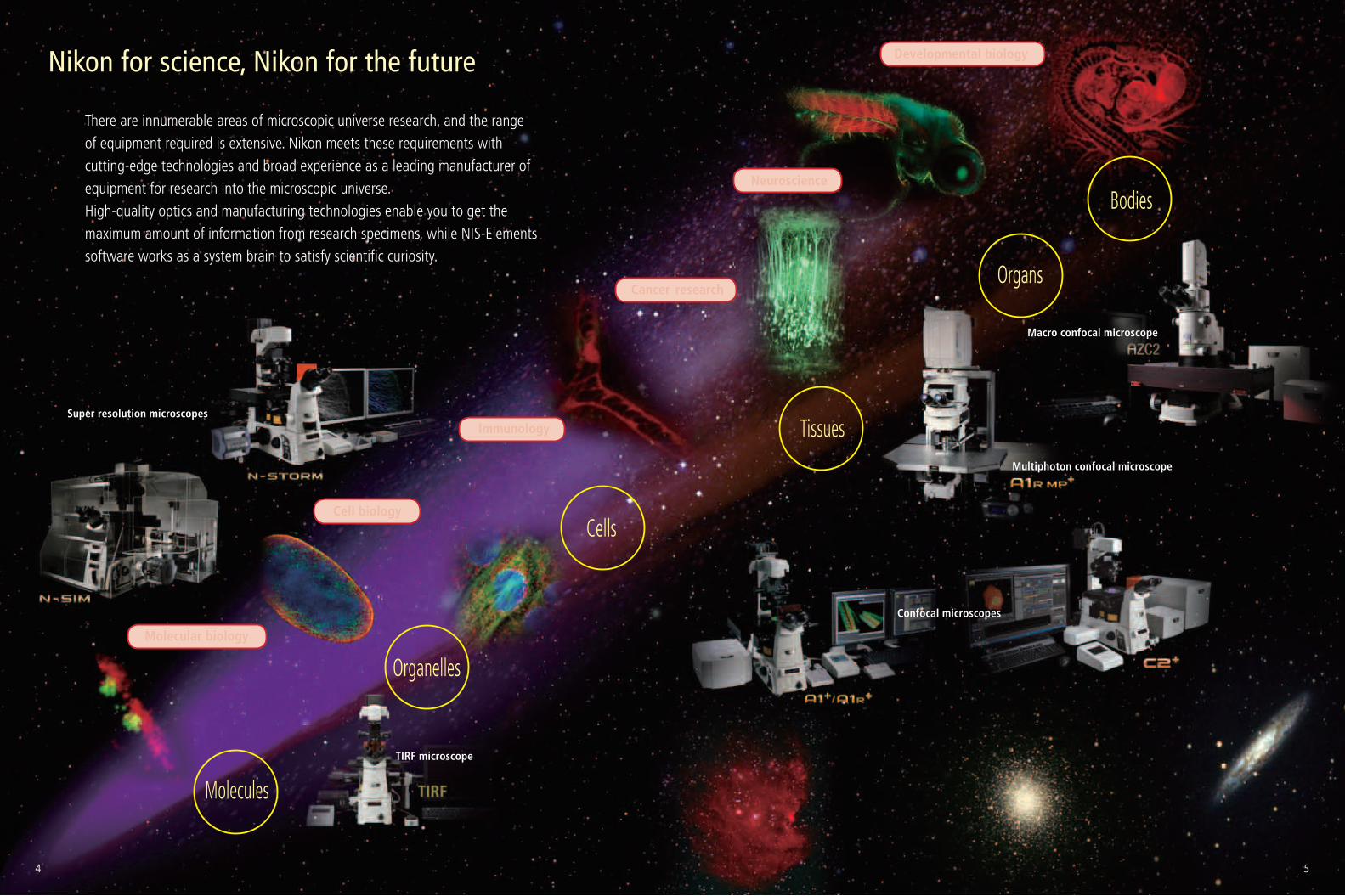
Nikon for science, Nikon for the future
There are innumerable areas of microscopic universe research, and the range of equipment required is extensive. Nikon meets these requirements with cutting-edge technologies and broad experience as a leading manufacturer of equipment for research into the microscopic universe.High-quality optics and manufacturing technologies enable you to get the maximum amount of information from research specimens, while NIS-Elements software works as a system brain to satisfy scientific curiosity.
Super resolution microscopes
TIRF microscope
Multiphoton confocal microscope
Confocal microscopes
Molecules
Organelles
Cells
Tissues
Organs
Bodies
Cell biology
Neuroscience
Developmental biology
Molecular biology
Cancer research
Immunology
Macro confocal microscope
4 5
6 7
Researcher’s testimonials
Maddy Parsons, Ph.D. Group Leader (Royal Society University Research Fellow), Randall Division of Cell and Molecular Biophysics, King’s College London
1. Please tell us about your research?We cover two main research fields; the first field focuses on adhesion receptors, such as integrins, and their roles in adhesion to the extracellular matrix, cell signaling and migration. We work with fibroblasts as a normal cell model, and also with different lines of breast carcinoma cells as a model for cancer cells. These different cell lines are known to show different levels of metastatic behavior. In cancer cells, we believe that the amount of time the receptors are in contact with the matrix, and the molecular composition of these adhesions is related to how likely the cell is to invade. We hypothesize that increased stability of adhesions results in a decreased tendency for cells to invade into the matrix. We are researching the molecules that regulate this process with an overall aim of finding novel ways to inhibit cell metastasis.
The second research field uses skin and lung epithelial cells as a model. These cells are generally non-migratory during normal homeostasis on humans, but undergo migration upon insult such as inflammation or wounding. With these cells our focus is studying how they adhere to other cells and to the extracellular matrix, and how this is controlled by external factors. This research helps us to understand wound healing, blistering skin diseases and inflammation in lung disease.
In all of our experiments we try to mimic the in vivo environment using in vitro model systems. The cells are put into a 3D matrix, and we use different models to study different aspects of adhesion. For example, if we were looking at cancer cell invasion out of the vasculature into the extracellular matrix, we would plate cancer cells in a ‘co-culture’ on top of human endothelial cells on a 3D collagen gel to analyze invasion through this ‘barrier’. We can also change the concentration of collagen in the matrix to simulate the different mechanical stresses on the cells that are seen in vivo. In the case of epithelial cells, we can instead plate them on top of a collagen gel containing fibroblasts to mimic the matrix environment within the dermis. These kinds of experiments allow us to study overall effects such as migration, but also smaller details of live individual adhesions all in the same model system.
Historically, experiments in this field were focused around plating cells on 2D surfaces, and there was a strong emphasis on extracting these cells and analyzing them using various biochemical methods. When these cells are in a 3D matrix and in co-culture, it makes it very difficult to extract them, so imaging becomes the method of choice as it gives a better idea of the in situ behavior of the cells. It also allows the capture of dynamics—something not possible with the traditional biochemical approach.
2D microscopy was also used historically to image bulk populations. The ability to image in 3D and 4D allows us to capture information about the z-dimension, as well as capture dynamic events. As these occur over second timescales it is necessary to be able to image at high-speed.
2. What applications do you use the Nikon A1Rsi and A1R-TIRF systems for?The resonant scanner of the A1R allows imaging of short-term dynamic events in live cells over long periods, and allows fast z-stack acquisition. We use the TIRF system for studying adhesion at the coverslip, and the turnover of the adhesion receptor components. We also combine this with XYZ imaging on the A1R in a single experiment so we can follow the fate of the components as they enter the cell.
We use the spectral detector on the A1Rsi for colocalization studies. We could potentially express three fluorophores in the same cell and want to know how these colocalize, especially during dynamic processes. The ability to spectrally image and unmix these fluorophores and remove crosstalk allows us to be confident of our colocalization results.
An example of an experiment unique to the A1R would be the experiments studying dynamic membrane protrusion events. Cells expressing fluorescent probes to label the F-actin cytoskeleton are put in a 3D collagen matrix. These cells form sub-micron protrusions that form and collapse in less than 5-10 seconds, and we believe these dynamic events contribute to cell invasion. Imaging these protrusions requires the acquisition of rapid z-stacks with the resonant scanner and piezo stage. The protrusion dynamics (length, lifetime, etc.) of cells with and without certain proteins are studied, as well as gathering information about invasion of the cell into the matrix over a longer time period to allow us to make correlations between protrusion dynamics and the ability of the cell to invade.
To study proteins that localize to cell contacts and study the recruitment and dynamics of the cell junction we also routinely carry out FRAP analysis of adhesion receptor turnover in epithelial cells. These cell junctions are often destabilized in inflammatory lung disease and the subsequent ‘leakiness’ is associated with an increased inflammatory response. Again, these are highly dynamic, localized processes and therefore require rapid, sensitive imaging approaches to permit accurate quantification.
We also use FRET biosensors to measure activation of proteins in protrusions, for example RhoA GTPase, which controls cytoskeletal dynamics. We look at this activity in various knockdown cells. This is another type of experiment that was historically measured via biochemical methods, but using imaging allows us to measure the localized activation dynamics in much more detail.
6. How has this system helped your research?With the A1R systems we have been able to obtain new data that could
not have been acquired or analyzed before, and this has led to new and surprising findings. Lots of assumptions have been made in the past from fixed cell studies that this work is now challenging. This has helped re-enforce our position as one of the foremost labs in the world in this area of study.
4. What was the performance specification that you valued most in selecting this system?The speed of the resonant scanner was a key consideration for us, as it allows us to capture dynamic events. The ability to flip between the resonant and non-resonant galvano scanners was one of the strongest points, as there is nothing else like it on the market.
5. What do you value about this Nikon system after having used it for three years?The Nikon Ti Perfect Focus System is excellent and when it comes to timelapse experiments it has changed our lives. The instrument does exactly what we wanted and has significantly increased the research possibilities available to us. We also value very highly the communication with Nikon. Their feedback and enthusiasm for science is something we have not experienced with other companies.
3. What was the deciding factor for choosing this Nikon system?One of the major factors that lead us to choose the A1R was the potential for flexibility in the system; the ability to cover all the types of experiments we wanted in a single microscope, and the possibility to add other components in the future as our research advances. Although there are other microscopes that could do parts of what we required, the A1R allows us to do everything in one system.
Systems:A1Rsi spectral confocal system and Ti-E inverted research microscope with OKOLab environmental chamber and 405, 440, 488, Multiline Argon, 561 and 640 lasers
A1R confocal system with TIRF attachment, Andor Ixon DU897 EMCCD camera, Ti-E inverted research microscope with OKOLab environmental chamber and 405, 440, 488, Multiline Argon, 561 and 640 lasers
Human lung epithelial cells grown as a confluent monolayer, fixed and stained for e-cadherin (green), CAR (red) and actin (blue). Scale bar 30 µm.
Human breast carcinoma cell expressing GFP-lifeact to label F-actin, plated on a thin layer of 2D collagen I labeled with Cy3 dye immobilized on a glass coverslip.
Analysis of RhoA GTPase activation in human epithelial cells using FRET biosensor. Image shows pseudocolor lookup table for RhoA FRET biosensor measured by acceptor photobleaching. Red shows high RhoA activity (20% FRET efficiency) and blue areas are low activity. Note high activity at cell-cell adhesion areas.
Human breast carcinoma cell expressing GFP-lifeact to label F-actin cytoskeleton, embedded in a Cy3-labeled collagen I gel. Images shown are top down projection (main image), or X/Z or Y/Z axis projections through the cell. Note the dramatic difference in morphology of this cell compared to the same cell plated on equivalent 2D surface in the image above.
6 7
8 9
Researcher’s testimonials
Professor Alberto Diaspro Professor of Applied Physics, Department of Physics, University of Genoa Director of the Department of Nanophysics, Istituto Italiano di Tecnologia (www.iit.it/en)
1. Please tell us about your research?My experience is in the design, realization and utilization of optical and biophysical instrumentation, as carried out with my research team. Our main aim is to improve on existing techniques to develop new, unique technologies. Our research centers on several main themes; one is the development of higher resolution imaging solutions, especially involving fluorescence. We are interested in techniques that give XY resolution below 30 nm and Z resolution below 100 nm. In order to do this we need to work with systems that have an excellent signal-to-noise ratio to preserve optical quality when imaging at nanometer resolution. We recently published a paper on this reporting about our IML-SPIM setup allowing 3D super-resolution on thick biological objects (http://www.nature.com/nmeth/journal/v8/n12/full/nmeth.1744.html).
Something else we’re interested in is developing multimodal imaging systems that allow the gathering of information from multiple techniques from a single specimen. An example of this is the recent combination of an optical nanoscope and atomic force microscope built on a customized Nikon platform.
Another goal is to implement faster techniques for super-resolution imaging through both software and hardware approaches. This might be through novel combinations of existing hardware, or by improved computational analysis of data during acquisition. An example of this would be being using on-the-fly data analysis during single molecule localization techniques to be able to stop acquisition once sufficient images have been acquired.
One of our biggest challenges is to be able to bring these high resolution imaging techniques to thick, highly scattering samples, tissues, organs, and ultimately in vivo. Some of the best candidates for this are methods based on multi-photon fluorescence excitation. As this is less efficient than single-photon excitation it means we require systems with very high optical performance.
2. What applications do you use the Nikon systems for?We work closely with several teams regarding applications for the newly developed systems. We have a particular focus on molecular oncology (chromatin and adhesion mechanisms), the production of insulin in beta cells, and neuroscience (nerve degeneration, Alzheimer’s disease, brain mapping, neuronal network signaling).
6. How has this system helped your research?The systems have allowed us to make affordable measurements with high reproducibility. The systems are really high-value work horses for nanobiotechnology and neuroscience labs.4. What was the performance specification that you
valued most in selecting these systems?The modularity and flexibility of the systems was very attractive as the basis for a platform for development. Nikon software is flexible and user friendly—Nikon has created some of the best software I’ve ever used. The Perfect Focus System is also very robust and useful for our time-lapse systems.
We are excited to explore the growing possibilities of intelligent microscope systems, such as CLEM (Controlled Light Exposure Microscopy).
5. What do you value about this Nikon system after having worked with it? We have at our disposal a new-generation confocal system, which has been overbooked throughout the last year, and it has been up and running 100 percent of the time.
3. What was the deciding factor for choosing Nikon systems?One of the main factors was Nikon’s reputation and experience in confocal laser scanning microscopy, which started with the PCM2000. Nikon systems are very efficient in terms of signal-to-noise ratio. When working at the nanoscale this becomes a critical factor.
Most importantly, the assistance given by Nikon is superb, with well qualified personnel and fast responses.
Systems:A1RMP multiphoton system and Ti-E inverted research microscopeN-STORM super-resolution system and Ti-E inverted research microscope
Photo by Massimo Brega–The Lighthouse
Photo by Massimo Brega–The Lighthouse
8 9
Microtubules (Cy3-Alexa 647)
Conventional image 2D N-STORM image
Microtubules (Cy3-Alexa 647)
Conventional image 3D N-STORM image
Three-dimensional N-STORM imaging of microtubules in Hela cells. Labeling has been performed by direct immunofluorescence protocol using primary antibodies against ß-tubulin labeled with Cy3 and Alexa 647. The z-position information is represented by a color-coded map according to the color scale bar.Images courtesy of Francesca Cella Zanacchi, Istituto Italiano di Tecnologia
Z (nm)+300
−300
0
Volume rendering image
Microtubules (Cy3-Alexa 647)
10 11
Researcher’s testimonials
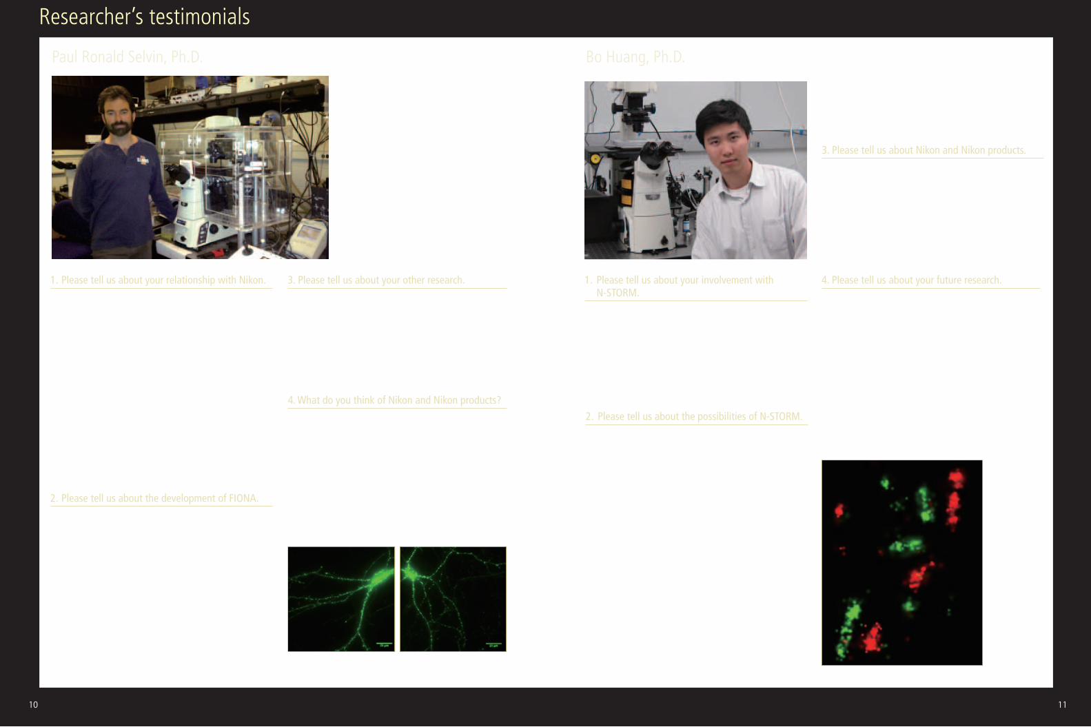
Paul Ronald Selvin, Ph.D. Professor, Department of Physics, University of Illinois at Urbana-Champaign Bo Huang, Ph.D. Assistant Professor, Department of Pharmaceutical Chemistry, Department of Biochemistry and Biophysics, University of California, San Francisco
1. Please tell us about your relationship with Nikon.Nikon understands the uncertainty and sensitivity of my field. I’ve worked hand in glove with the company to create a flexible system, perfectly tailored to my laboratory needs.
I have worked closely with Nikon Instruments to build an imaging system that fully supports my research goals. For example, side by side with Nikon engineers and product managers, I tweaked the stage of the company’s Ti microscope so it moves in incredible plus/minus 10 nanometer increments—improving upon the former range of plus/minus 30 nanometers and also reducing vibrations. I also collaborated with the company to better control the temperature of the system (to prevent drift), using different lasers to enable viewing of different fluorophores, and viewing work with a very high numerical aperture objective.
I have come to depend on Nikon to work with me through many iterations of change to my systems to get them just right. The support I received from the company was nothing short of fantastic. If I had known then what I know now, every single one of my microscopes would be Nikon.
1. Please tell us about your involvement with N-STORM.I was part of the team that developed the Stochastic Optical Reconstruction Microscopy (STORM) technology, and I have been working closely with Nikon engineers in building up the N-STORM hardware and software. With Nikon N-STORM, we can see things never before imagined. I am very happy to see our lab work turning into a commercial product, which allows many more research groups to have access to this tool in their pursuits to understand the fundamental mechanisms of the biological world.
2. Please tell us about the possibilities of N-STORM.They are countless. With a spatial resolution that is more than an order of magnitude better than conventional fluorescence microscopy, STORM enables the observation of subcellular and even sub-organelle structures with unprecedented detail. For many decades, scientists had to rely on indirect methods to investigate biological systems at these scales, leaving many questions unanswered simply because we could not "see." Now STORM makes it possible to address them by direct visualization. This is an opportunity for every one of us to see profound changes in how we understand the inner workings of life.
Besides the much improved resolution, N-STORM has two very important features: 3D imaging, which is often required to resolve intracellular structures, and multicolor imaging, which is essential for studying biomolecular interactions. In addition, it can utilize fluorescent probes identical to how biologists labeled specimens before. These features make N-STORM not only a powerful but also a versatile tool that will find applications in numerous fields of biological and biomedical science.
2. Please tell us about the development of FIONA.It’s a partnership that really blossomed when I became the 2010 Nikon Fellow and worked closely with the company to develop a specific system for my work at the Marine Biological Laboratory. Nikon allowed me to take my custom microscope back to the lab and shortly thereafter we received a grant that allowed us to purchase it. Our team continue to innovate new ways to produce clear images of the tiny specimens we study. We invented FIONA, or Fluorescence Imaging with One Nanometer Accuracy. As the name implies, FIONA allows researchers to determine the position of a fluorophore to about a nanometer in the x-y plane, anywhere from a millisecond or longer, depending on its brightness.
FIONA coupled with creative dye selection and manipulation allows researchers to study cytoplasmic molecular motors, both in vitro and in vivo. Our team has captured the movements of these motors and found that they move by “walking,” one “foot” in front of the other with a stride ranging from 16 to 74 nanometers.
3. Please tell us about your other research.Our team has also developed a series of super-resolution techniques by
separating the distance between dyes to achieve a better diffraction-
limit of about 250 nm. By relying on successive photobleaching of
the dyes—either permanently, or transiently—we have demonstrated
that it is possible to get down to about 8 nm between two dyes, and
approximately 50 nm measuring up to 20 dyes. We now use the system
to study neuronal synapses and measure distances on DNA and RNA.
3. Please tell us about Nikon and Nikon products.As a lab focused on both the development of new microscopy techniques
and the application of these techniques, we found Nikon instruments a
good balance between user-friendliness and customizability. N-STORM
offers the robustness and image quality for biologists in our group to
perform their everyday experiments. The same microscope also provides
the flexibility in design for our physicists to add in or modify components,
enabling it to perform new functionalities that have not been previously
conceived.
4. What do you think of Nikon and Nikon products?When you’re in the business of single molecules, good accuracy and
resolution is key. I count on our Nikon instrumentation to allow us to
see cells in a consistent, reliable fashion. The customization, in which
I collaborated with Nikon, has fine-tuned our equipment to create the
ideal conditions for viewing these tiny samples. However, what I value
most is the support I receive from Nikon. I have called up or e-mailed
them so many times with various questions or challenges, and I always
get a totally professional response in a rapid time-scale. That makes all
the difference.
4. Please tell us about your future research.In addition to pushing the super-resolution microscopy technology further into live cell observation and molecular-scale resolution, we have had our eye on the application of super-resolution microscopy in cell biology and neurobiology. We are using STORM in combination with Structured Illumination Microscopy (SIM), benefiting from the higher resolution of STORM as well as the fact that SIM puts fewer constraints on the sample. We have set out to study the architecture of subcellular structures such as the centrosome, as well as molecular mechanisms controlling the development of neurons in the central nervous system. In the end, we hope our work will set an example of how super-resolution microscopy can bridge our structural knowledge of macromolecules and their functionalities in the cellular context. There are an infinite number of biological questions that need to be answered. We are pleased that technology like STORM will help scientists begin to answer them.
A cultured neuron expressing homer-GFP, a postsynaptic marker. Bright fluorescent puncta along the dendrites show localization of synapses. Super-resolution image of synapses in mouse brain
10 11
12 13
Researcher’s testimonials
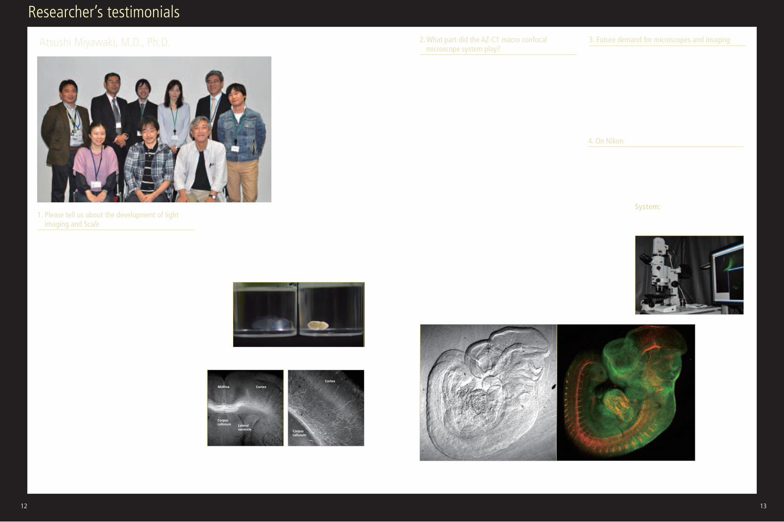
Atsushi Miyawaki, M.D., Ph.D. Senior Team Leader, Laboratory for Cell Function Dynamics, RIKEN Brain Science Institute
1. Please tell us about the development of light imaging and ScaleI have long believed that there is much information to be obtained
from the intermediate stage between a biochemical approach such as
cell componential analysis using electrophoresis and optical imaging
of living specimens—for example, from research into fixed specimens.
In particular, fluorescent protein technology and gene induction
technologies have enabled the selective labeling of nerve cells regardless
of depth; however, there is as yet little technology for visualizing the
neural networks running from the surface to the depths of the brain
together. The Scale reagent we have developed can make biological
tissue transparent like jelly. This has enabled deep imaging without
damage to the specimen.
Dr. Hiroshi Hama focused attention on the fact that the PVDF membrane
used for transferring proteins in western blotting becomes transparent
in high-concentration urea solution. In addition, we were already
aware that most fluorescent proteins were urea-resistant, though high-
concentration urea solution is generally used for protein denaturation.
The impetus for the Scale project was the belief that tissue clearing
technology could probably be developed based on urea. Initially, it took
quite a long time for the entire brain of a mouse to become transparent
when soaked in our early reagents. However, our capacity for patience
seems to have paid off. The Scale technology was improved and finally
the ScaleA2 reagent was completed. We now know that making a cut in
the mouse brain makes it become transparent more rapidly. Two or three
hours may be enough for a brain tissue block.
We have also focused attention on the biological debate over whether
the neural stem cells in the dentate gyrus of the hippocampus lie close
to the blood vessels. Through the observation of BrdU-labeled neural
stem cells and immunostained blood vessels, numerous researchers,
2. What part did the AZ-C1 macro confocal microscope system play?A wide range of applications are possible when used in combination with
Scale technology.
For example, if neurons fire, the transcription of immediate-early genes
such as c-fos and Arc is enhanced. Therefore, neuronal firing can be
monitored with the fluorescence signal of a reporter gene of fluorescent
protein such as Venus located downstream of a promoter that controls
c-fos and Arc expression. Quantifying this signal by capturing a number
of slices of the brain and reconstructing three-dimensional images
entailed tremendous effort. Using the AZ-C1, however, we proved that it
is possible to simplify the quantification by projecting three-dimensional
information of the brain, made transparent with Scale, to construct a
two-dimensional image.
The imaging depth limit with single-photon excitation is generally only
around 0.2 mm due to the influence of scattered light, while it is
0.8 mm with two-photon excitation. As Scale technology minimizes light
scattering in the sample, it is more effective when used with a single-
photon excitation microscope. It may also be beneficial for the AZ-C1.
For example, if the YFP gene is administered to the cerebral cortex of the
right hemisphere of the brain of an embryonic mouse, it is observed that
axons project into the left side of the cerebral cortex across the corpus
callosum during nervous system development (see the image below). The
AZ-C1 is effective in this kind of research, in which wide-area images
must be obtained.
in their papers, have concluded that they are close. However, the data
obtained from the observation of sliced tissue is not very persuasive, and
they could be criticized for choosing only favorable data. It is necessary
to measure the distances between the neural stem cells and the blood
vessels in three-dimensional space over the wide area of the dentate
gyrus of the hippocampus. We solved this problem with Scale technology.
Furthermore, for identification of neural stem cells, our Fucci technology
was used.
3. Future demand for microscopes and imagingIt would be ideal if there were a microscopic imaging system that could
freely zoom in and out.
I want to see our fluorescent-imaging technology and Scale technology
make a major contribution to human pathological diagnosis. I am
interested in the establishment of the technology for use in finding
lesions in large tissue mass. I believe that Scale technology will
contribute to medical science, as well as to academic disciplines such as
developmental biology. I also expect AZ-C1 to make a contribution.
4. On NikonProfessor Roger Tsien (2008 Nobel laureate for chemistry), who has
conducted numerous joint research and development projects with
Nikon, was my adviser. Therefore, I have a long-standing relationship
with Nikon. I hope that the company will develop products that
challenge researchers.
Callosal connections of postnatal day 10 mouse cerebrum.Neurons (a population of layer II/III pyramidal neurons) were labeled by in utero electroporation of EYFP into the dorsal ventricular zone on the right side of the mouse forebrain at E15.5. The mouse was fixed by PFA at P10 and subjected to treatment with ScaleA2 solution for tissue clearing.
Two mouse embryos, one (left) incubated in Sca/eA2 solution
System:AZ-C1 macro confocal microscope system1x objective lens (AZ-Plan Apo, NA 0.1, W.D. 35 mm)2x objective lens (AZ-Plan Fluor, NA 0.2, W.D. 45 mm)
1x 8x
Fucci transgenic mouse embryo (E10.5)Objective lens: 1x, AZ-C1 zoom: 3.9x, Image size: 512 x 512 pixelsLeft: Best focus for DIC image, Right: MIP for fluorescence image
Dr. Miyawaki (front row, center). On the right is Dr. Hama; on the left is Dr. Sawano
12 13
Midline Cortex
Cortex
Corpus callosum
Corpus callosum
Lateral ventricle
14 15
Researcher’s testimonials
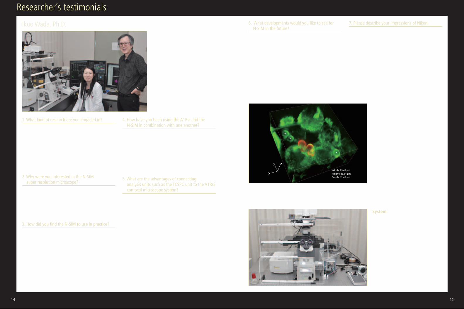
Ikuo Wada, Ph.D. Department of Cell Science, Institute of Biomedical Science, Fukushima Medical University
1. What kind of research are you engaged in?I have long been interested in the mechanism by which secreted proteins are efficiently produced. To study this, I have conducted a series of studies into the dynamics of inner membrane proteins and the dynamics of cargo protein molecules themselves. After my biochemistry research,I started fluorescence imaging, and then went on to use single molecule techniques, such as FCS (fluorescence correlation spectroscopy) and single molecule tracking. Because I want to understand what makes "living" cells, I have a boundless interest in changes in cell structure and in the dynamic behavior of multiple molecules.
2. Why were you interested in the N-SIM super resolution microscope?Initially, I anticipated that I would be able to observe the microstructures of endomembranes that cannot be recorded with a confocal microscope. When I used it in practice, I discovered that this was not the only thing I could now do—and when I used it in combination with an A1Rsi confocal microscope system, it felt as if a whole new world had been opened up.
3. How did you find the N-SIM to use in practice?It was extremely user-friendly and easier to use than I had imagined. It is simple to modify the analysis parameters, and the fact that it allows an intuitive understanding of the correlation between these parameters and the results of data acquisition makes it easy to use. Just modifying the parameters a little could eliminate the known artifacts of SIM. As all the original acquisition data are preserved, it is so easy to go back to the original and confirm that the reconstructed images are correct.
I was also particularly struck by the superb resolution in the Z-axis direction. Also, I was surprised to find that scanning with the N-SIM unit produces much less photobleaching than with a confocal LSM unit at the same magnification. The N-SIM unit is now our first choice.
4. How have you been using the A1Rsi and the N-SIM in combination with one another?I mainly use the N-SIM for observing cell structures as still images. On the other hand, the A1Rsi can perform ultra-high-speed image acquisition and allows FRAP and photo stimulation. Unless both devices are used, the information we can obtain is limited. The combination of N-SIM and A1Rsi allows us to acquire high-resolution images of specific structures. This is because A1Rsi highlights and activates only regions labeled with photoactivatable proteins such as PA-GFP. The ability to highlight only specific structures represents an extremely powerful capability.
5. What are the advantages of connecting analysis units such as the TCSPC unit to the A1Rsi confocal microscope system?First, the A1Rsi is highly extensible and operations, including external inputs, are extremely simple. This makes it easy to use—even for an amateur of optics like me. We use FLIM information obtained with the TCSPC unit for analyzing the FRET efficiency. Although there are sometimes difficulties with the acceptor-bleaching method, with FLIM, results are obtained quickly with few artifacts appearing, making it useful for FRET measurements when, for example, studying structural changes in molecules. Photon static information, including FLIM and FCS/PCH information, from living cells obtained with the TCSPC unit supplements detailed structural information obtained with the N-SIM. Nikon’s 60x water-immersion objective lens is the default lens for the TCSPC unit, while 40x is the default objective for other manufacturers. Nikon optical systems are surprisingly high-performance and give much higher photon counts per molecule than those acquired with other manufacturers’ optics. This is probably due to the extremely high brightness of Nikon objective lenses.
6. What developments would you like to see for N-SIM in the future?Firstly, faster image acquisition and simultaneous acquisition of two colors. Even without two cameras, if it could offer dual-view display with just a single camera, this would be quite effective—even with only 256 pixels. Users can probably be divided into those who want to view a larger area, and those who want to see faster change.
Secondly, improvements in illumination pattern contrast and a better CCD camera with a lower dark current may help improve reconstruction of dark (noisy) samples and should lead to wider use of the N-SIM in cell biology.
In addition, I feel that successful application of the N-SIM depends on proper imaging techniques and knowledge. I hope that Nikon is involved in promoting optical imaging education.
7. Please describe your impressions of Nikon.Nikon’s engineers have been giving thorough consideration to my requests. Instead of responding to my wishes by saying “it can’t be done,” they are properly considering how they can bring them to fruition. Nikon products are also outstanding in terms of system extensibility. It seems to be difficult to attach third-party peripheral devices to other manufacturer’s products, particularly in Japan.
Three-dimensional image of phagosomes taken with a super resolution microscope
J774/mVenus-SNAP-23 cells observed using a super resolution microscope (Nikon’s N-Structured Illumination Microscopy System). Phagosomes (highlighted with green fluorescence), which incorporate beads wrapped in mVenus-SNAP-23 (which are marked with a *), are visible in the cells. After Phagocytosis, the addition of an Alxa594-labeled antibody that recognizes the IgG used in opsonization allows confirmation that there are beads outside the cells (highlighted with red fluorescence).
System:N-SIM+A1Rsi+TCSPC (time-correlated single photon counting) unit (third-party peripheral device attached to the A1)
14 15
**
*
x
y
z
Width: 29.48 μm
Height: 28.54 μm
Depth: 12.40 μm
Dr. Wada (right)
16 1716 17
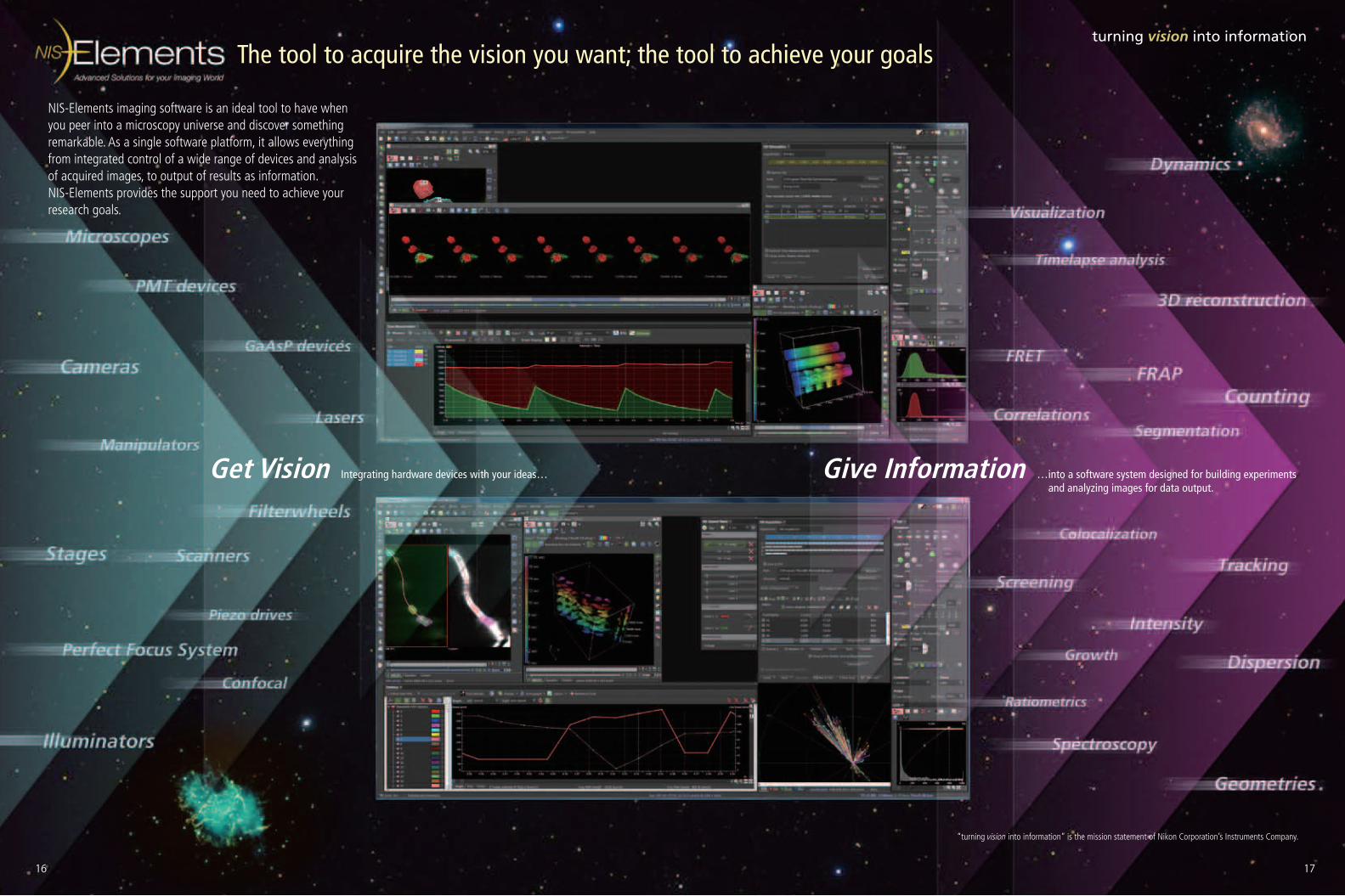
turning vision into information
Get Vision Integrating hardware devices with your ideas… Give Information …into a software system designed for building experiments and analyzing images for data output.
The tool to acquire the vision you want; the tool to achieve your goals
NIS-Elements imaging software is an ideal tool to have when you peer into a microscopy universe and discover something remarkable. As a single software platform, it allows everything from integrated control of a wide range of devices and analysis of acquired images, to output of results as information. NIS-Elements provides the support you need to achieve your research goals.
“turning vision into information” is the mission statement of Nikon Corporation’s Instruments Company.
18 19
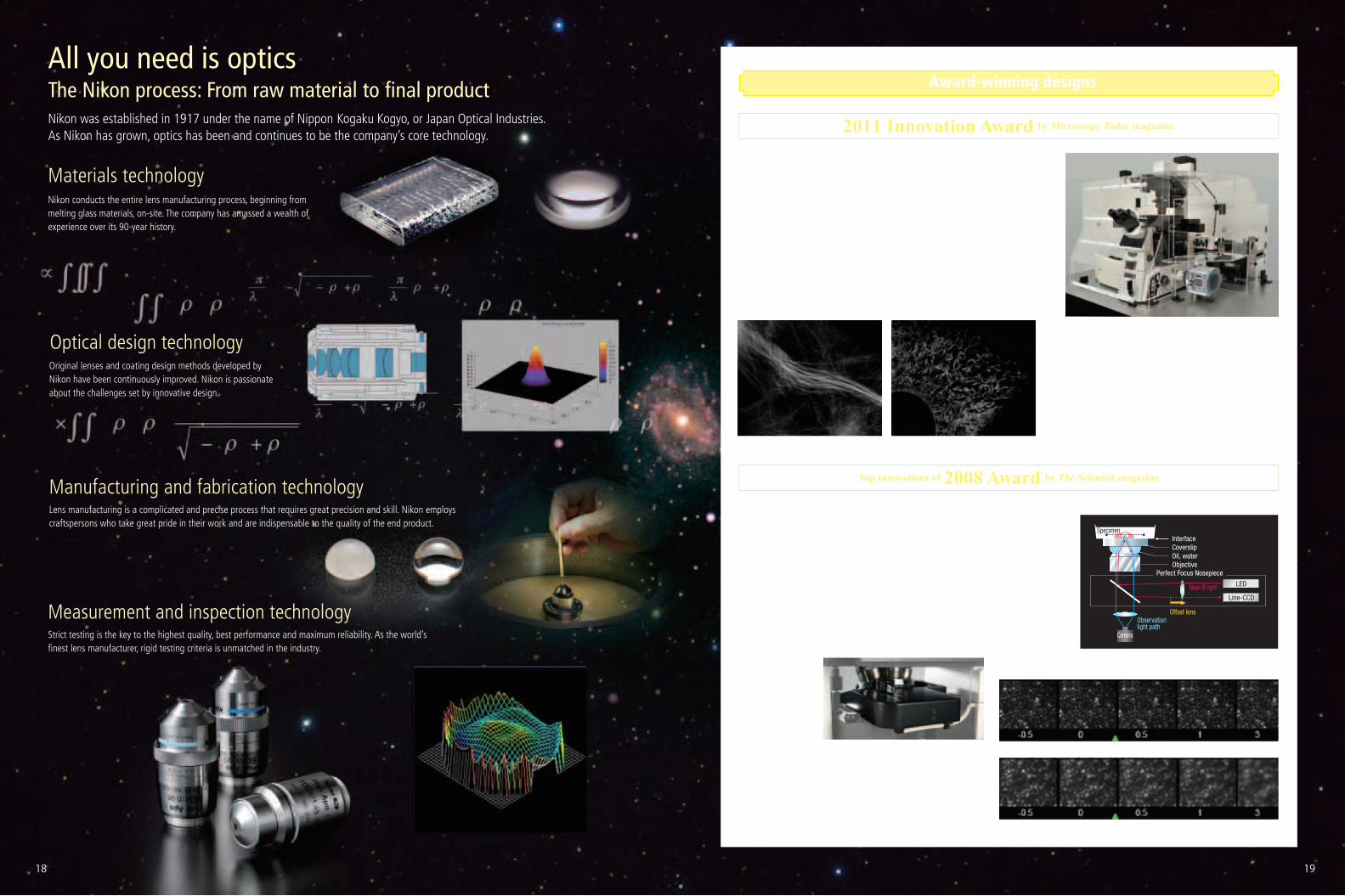
All you need is opticsThe Nikon process: From raw material to final product
Nikon conducts the entire lens manufacturing process, beginning from melting glass materials, on-site. The company has amassed a wealth of experience over its 90-year history.
Strict testing is the key to the highest quality, best performance and maximum reliability. As the world’s finest lens manufacturer, rigid testing criteria is unmatched in the industry.
Nikon’s Perfect Focus System was chosen as one of The Scientist magazine’s Top Innovations of 2008.The PFS is a real-time focus correction mechanism that keeps a specimen in focus by correcting focus drift caused by mechanical effects and thermal change over extended periods. It tracks optical offset and corrects it by sampling every five milliseconds with infrared light, enabling acquisition of more reliable data during time-lapse imaging of live cells.The panel of judges, including David Piston, Simon Watkins, Klaus Hahn and Steven Wiley, has collectively published more than 700 academic articles. They chose Nikon’s PFS as a product to most likely impact the life science market.
Nikon N-SIM super resolution microscope was chosen as one of the best 10 microscopy innovations in the 2011 awards sponsored by Microscopy Today (owner, Microscopy Society of America). The awards recognize new microscopy-related products and methods launched in the previous year. Areas judged include light microscopy, scanning probe microscopy, electron microscopy and microanalysis, as well as specimen preparation.The N-SIM was developed with “Structured Illumination Microscopy” technology licensed from the University of California, San Francisco (UCSF). It offers nearly double the resolution of conventional optical microscopes and achieves super resolution of up to 85 nm in multiple colors. It can also capture super resolution images at a temporal resolution of 0.6 sec/frame, allowing the study of dynamic interactions in living cells.
Microtubules in B16 melanoma cell labeled with YFP (left)Photographed with the cooperation of: Yasushi Okada, Ph.D., Department of Cell Biology and Anatomy, Graduate School of Medicine, University of Tokyo
Endoplasmic reticulum (ER) in living HeLa cell labeled with GFP (right)Photographed with the cooperation of: Ikuo Wada, Ph.D., Institute of Biomedical Sciences, Fukushima Medical University School of Medicine
Original lenses and coating design methods developed by Nikon have been continuously improved. Nikon is passionate about the challenges set by innovative design.
Lens manufacturing is a complicated and precise process that requires great precision and skill. Nikon employs craftspersons who take great pride in their work and are indispensable to the quality of the end product.
CoverslipInterface
Perfect Focus Nosepiece
Specimen
LED
Line-CCD
Camera
Observation light path
Offset lens
Oil, waterObjective
Near-IR light
Award-winning designs
Perfect Focus System:With PFS
Adding reagent
Adding reagent
Without PFS
Nikon was established in 1917 under the name of Nippon Kogaku Kogyo, or Japan Optical Industries. As Nikon has grown, optics has been and continues to be the company’s core technology.
Materials technology
Measurement and inspection technology
Optical design technology
Manufacturing and fabrication technologyTop Innovations of 2008 Award by The Scientist magazine
2011 Innovation Award by Microscopy Today magazine
Perfect Focus System
N-SIM Super Resolution Microscope
18 19
EnPrinted in Japan (1203-07)T Code No. 2CE-SCKH-2
Specifications and equipment are subject to change without any notice or obligation on the part of the manufacturer. March 2012 ©2012 NIKON CORPORATION
Company names and product names appearing in this brochure are their registered trademarks or trademarks.N.B. Export of the products* in this brochure is controlled under the Japanese Foreign Exchange and Foreign Trade Law. Appropriate export procedure shall be required in case of export from Japan.*Products: Hardware and its technical information (including software)Monitor images are simulated.
WARNINGTO ENSURE CORRECT USAGE, READ THE CORRESPONDING MANUALS CAREFULLY BEFORE USING YOUR EQUIPMENT.
This brochure is printed on recycled paper made from 40% used material.
NIKON CORPORATIONShin-Yurakucho Bldg., 12-1, Yurakucho 1-chome, Chiyoda-ku, Tokyo 100-8331, Japanphone: +81-3-3216-2375 fax: +81-3-3216-2385 http://www.nikon.com/instruments/
NIKON INSTRUMENTS INC.1300 Walt Whitman Road, Melville, N.Y. 11747-3064, U.S.A.phone: +1-631-547-8500; +1-800-52-NIKON (within the U.S.A. only)fax: +1-631-547-0306http://www.nikoninstruments.com/
NIKON INSTRUMENTS EUROPE B.V.Laan van Kronenburg 2, 1183 AS Amstelveen, The Netherlandsphone: +31-20-44-96-300 fax: +31-20-44-96-298http://www.nikoninstruments.eu/
NIKON INSTRUMENTS (SHANGHAI) CO., LTD.CHINA phone: +86-21-6841-2050 fax: +86-21-6841-2060(Beijing branch) phone: +86-10-5831-2028 fax: +86-10-5831-2026(Guangzhou branch) phone: +86-20-3882-0552 fax: +86-20-3882-0580
NIKON SINGAPORE PTE LTDSINGAPORE phone: +65-6559-3618 fax: +65-6559-3668
NIKON MALAYSIA SDN. BHD.MALAYSIA phone: +60-3-7809-3688 fax: +60-3-7809-3633
NIKON INSTRUMENTS KOREA CO., LTD.KOREA phone: +82-2-2186-8400 fax: +82-2-555-4415
NIKON CANADA INC.CANADA phone: +1-905-602-9676 fax: +1-905-602-9953
NIKON FRANCE S.A.S.FRANCE phone: +33-1-4516-45-16 fax: +33-1-4516-45-55
NIKON GMBHGERMANY phone: +49-211-941-42-20 fax: +49-211-941-43-22
NIKON INSTRUMENTS S.p.A.ITALY phone: +39-055-300-96-01 fax: +39-055-30-09-93
NIKON AGSWITZERLAND phone: +41-43-277-28-67 fax: +41-43-277-28-61
NIKON UK LTD.UNITED KINGDOM phone: +44-208-247-1717 fax: +44-208-541-4584
NIKON GMBH AUSTRIAAUSTRIA phone: +43-1-972-6111-00 fax: +43-1-972-6111-40
NIKON BELUXBELGIUM phone: +32-2-705-56-65 fax: +32-2-726-66-45
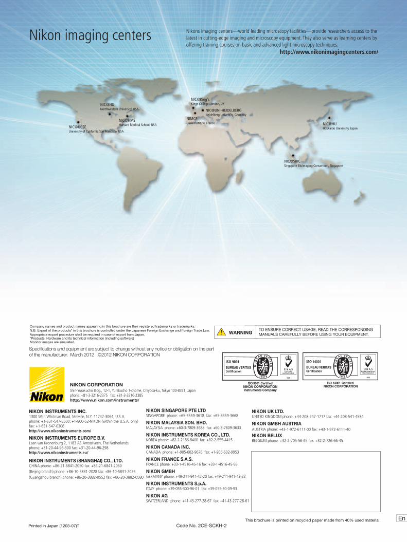
Nikon imaging centers Nikons imaging centers—world leading microscopy facilities—provide researchers access to the latest in cutting-edge imaging and microscopy equipment. They also serve as learning centers by offering training courses on basic and advanced light microscopy techniques.
http://www.nikonimagingcenters.com/
NIC@UCSF University of California San Francisco, USA
NIMCE Curie Institute, France
NIC@SBIC Singapore Bioimaging Consortium, Singapore
NIC@HU Hokkaido University, Japan
NIC@UNI-HEIDELBERG Heidelberg University, Germany
NIC@King's Kings College London, UKNIC@NU
Northwestern University, USA
NIC@HMS Harvard Medical School, USA